Embed Size (px)
Citation preview

Biocompatibility and Biodegradation Studies ofSubconjunctival Implants in Rabbit EyesYan Peng1., Marcus Ang2,3., Selin Foo3, Wing Sum Lee2, Zhen Ma3, Subbu S. Venkatraman1*, Tina T.
Wong1,2,3,4*
1 School of Materials Science and Engineering, Nanyang Technological University, Singapore, Singapore, 2 Singapore National Eye Centre, Singapore, Singapore,
3 Singapore Eye Research Institute, Singapore, Singapore, 4 Department of Ophthalmology, Yong Yoo Lin School of Medicine, National University of Singapore, Singapore,
Singapore
Abstract
Sustained ocular drug delivery is difficult to achieve. Most drugs have poor penetration due to the multiple physiologicalbarriers of the eye and are rapidly cleared if applied topically. Biodegradable subconjunctival implants with controlled drugrelease may circumvent these two problems. In our study, two microfilms (poly [d,l-lactide-co-glycolide] PLGA and poly[d,l-lactide-co-caprolactone] PLC were developed and evaluated for their degradation behavior in vitro and in vivo. We alsoevaluated the biocompatibility of both microfilms. Eighteen eyes (9 rabbits) were surgically implanted with one type ofmicrofilm in each eye. Serial anterior-segment optical coherence tomography (AS-OCT) scans together with serial slit-lampmicroscopy allowed us to measure thickness and cross-sectional area of the microfilms. In vitro studies revealed bulkdegradation kinetics for both microfilms, while in vivo studies demonstrated surface erosion kinetics. Serial slit-lampmicroscopy revealed no significant inflammation or vascularization in both types of implants (mean increase in vascularitygrade PLGA50/50 1260.5% vs. PLC70/30 1560.6%; P = 0.91) over a period of 6 months. Histology, immunohistochemistryand immuno-fluorescence also revealed no significant inflammatory reaction from either of the microfilms, which confirmedthat both microfilms are biocompatible. The duration of the drug delivery can be tailored by selecting the materials, whichhave different degradation kinetics, to suit the desired clinical therapeutic application.
Citation: Peng Y, Ang M, Foo S, Lee WS, Ma Z, et al. (2011) Biocompatibility and Biodegradation Studies of Subconjunctival Implants in Rabbit Eyes. PLoSONE 6(7): e22507. doi:10.1371/journal.pone.0022507
Editor: Maria Gasset, Consejo Superior de Investigaciones Cientificas, Spain
Received February 15, 2011; Accepted June 28, 2011; Published July 22, 2011
Copyright: � 2011 Peng et al. This is an open-access article distributed under the terms of the Creative Commons Attribution License, which permitsunrestricted use, distribution, and reproduction in any medium, provided the original author and source are credited.
Funding: This work was supported by National Research Foundation-Funded Translational & Clinical Research (TCR) Programme Grant [NMRC/TCR/002 - SERI/2008 - TCR 621/41/2008]. The funders had no role in study design, data collection and analysis, decision to publish, or preparation of the manuscript.
Competing Interests: The authors have declared that no competing interests exist.
* E-mail: [email protected] (SSV); [email protected] (TTW)
. These authors contributed equally to this work.
Introduction
The eye is the vital organ for sight and unique because it is both
anatomically and immunologically privileged. While the eye is
protected physiologically, it is also resistant to penetration by
drugs. Topical application is the mainstay of most ocular therapy,
but ocular bioavailability is poor due to the efficient protective
barriers [1,2,3]. The recognition of this limitation in efficient ocular
drug delivery has led to a range of systems that vary in mode of
administration, implantation site, composition and vehicles [4,5,6,
7,8,9,10,11,12], which all aim to circumvent the problems of drug
bioavailability, sustainability and feasibility of administration [13].
Biodegradable polymers are proven vehicles for sustained drug
delivery [5]. Polyhydroxyesters are easily fabricated with predict-
able biodegradation kinetics and biocompatible degradation [14].
These polymers, such as poly [d, l-lactide-co-glycolide] (PLGA) or
poly [d, l-lactide-co-caprolactone] (PLC), degrade through hydro-
lysis of their ester bonds into lactic acids, glycolic acids and caproic
acid – and eventually into water and carbon dioxide [15,16,17].
Since the body effectively deals with these degradation monomers,
there is very minimal systemic toxicity associated with its use in
human tissues. As such, these polymers have been used in US
FDA-approved implantable devices or injectable pharmaceutical
products.
PLGA has been used extensively in studies to deliver a wide
variety of drugs using various forms such as microparticles,
emulsions, implants and hydrogels [11,12,18,19,20]. The ability to
tailor the polymer degradation time by altering the ratio of the
monomers used during synthesis has made PLGA a common
choice in the production of a variety of biomedical devices such as
grafts, sutures, implants, root-canal fillings and prosthetic devices
[5]. In comparison, the copolymer PLC is relatively new, and is
currently finding application in implantable systems such as
occluders for atrial septal defects [21,22,23]. Several studies have
reported the development of biodegradable polymer microfilms
specifically for ocular drug delivery [11,21,22]. Apart from the
known advantages of using these biodegradable polymers, the
ability to cast the microfilms with varied thickness ranging from
microns to millimeters, is particularly useful for ease of insertion
into various layers of the eye [24]. Moreover, these biodegradable
microfilms may enable the release of multiple drugs with direc-
tionality and different release rates. Our authors have published
significant results from preliminary in vitro studies using commonly
used ophthalmic drugs, latanoprost and 5-fluorouracil [25].
In this study, we aim to develop and compare two different
biodegradable microfilms for their potential application as vehicles
for intraocular drug delivery, specifically on surgical insertion into
the subconjunctival space. We evaluated the degradation behavior
PLoS ONE | www.plosone.org 1 July 2011 | Volume 6 | Issue 7 | e22507

of both microfilms in vitro and in vivo, surgical feasibility and
biocompatibility of both types of microfilms using three comple-
mentary techniques: slit-lamp microscopy, anterior segment opti-
cal coherence tomography (AS-OCT) and histology with various
staining methods.
Materials and Methods
MaterialsPolymers used in this study are poly (d,l-lactide-co-glycolide),
PLGA50/50 (intrinsic viscosity 1.02 dl/g, Mw = 160 kDa) and
poly (d,l-lactide-co-e-caprolactone) PLC70/30 (intrinsic viscosity
1.66 dl/g, Mw = 210 kDa), which were purchased from Purac Far
East Pte. Ltd., Singapore. High-performance liquid chromatogra-
phy (HPLC)-grade dichloromethane and chloroform were from
Tedia Company. Phosphate buffer saline (PBS) tablets were
obtained from CalBioChem, England.
Sample preparationSamples of PLGA50/50 and PLC70/30 were weighed before
dissolving the appropriate amount in dichloromethane. Following
dissolution, the samples were dried in petri-dishes under a fume
hood for a day, followed by drying in a vacuum oven at 37uC until
the solvent level was less than 1% of the total weight, as measured
using a thermo-gravimetric analyzer (TGA, TA instruments
Q500). After drying, all samples were cut manually into standard
sized microfilms (6.063.060.5 mm) by using a sharp knife.
In vitro degradation studySamples were immersed in a closed vial containing 5 ml
Phosphate Buffered Saline (PBS, pH 7.4). PBS was prepared by
dissolving PBS tablets into 1 liter deionized water. All vials were
incubated at 37uC throughout the study. The buffer was refreshed
every week, and at every predetermined time point, samples were
taken out, rinsed with deionized water and dried in 37uC vacuum
oven for 7 days, before testing. Degradation of PLGA50/50 and
PLC 70/30 was monitored by film thickness (measured by
Elcometer 456), water absorption, weight loss and weight average
molecular mass (Mw) and poly dispersity index(PDI). Dried
samples were dissolved in chloroform (1–5 mg/ml) and filtered
through 0.22 mm regenerated cellulose syringe driven filters before
test. Weight average molar mass and poly dispersity of the sample
were determined by gel permeation chromatography (GPC, Agi-
lent 1100) at 35uC, using Agilent PLgel 5 mm mixed-C column,
under a flow rate of 1 ml chloroform per minute, using a Re-
fractive Index Detector (RID).
SterilizationAll the samples were sterilized by ethylene oxide (ETO) at 37uC
(used for normal medical device) in Tan Tock Seng Hospital
(Singapore) prior insertion into animals.
Surgical insertion of microfilmsWe obtained approval from the SingHealth Institute Animal
Care and Use Committee (IACUC Singhealth Approval Number
2009/SHS/478) and all procedures were performed in accor-
dance with the ARVO Statement for the Use of Animals in
Ophthalmic and Vision Research. Nine New Zealand white
rabbits (18 eyes) were used aged 4–6 months old with a weight
range of 2–2.5 kg each. Each rabbit was anesthetized with
intraperitoneal injection of ketamine hydrochloride (35–50 mg/
kg) and Xylazil (5–10 mg/kg). After the animal had been
adequately anaesthetized, the eye was cleaned with povidone–
iodine (10%) and draped with sterile cloth. A subconjunctival
pocket was created via blunt dissection just at the limbus with a 5–
6 mm incision in the superior-temporal aspect of the rabbit’s eye.
Microfilms were sterilized in ethyl alcohol and chlorhexidine
before soaking in sterile normal saline. The microfilm was then
inserted into the subconjunctival pocket 1 mm from the limbus
using a conjunctival forceps. Closure with 10-0 nylon sutures was
done to ensure secure implantation of each microfilm. In each
rabbit, PLC70/30 (n = 9) microfilms were inserted into the right
eye, whilst PLGA50/50 (n = 9) microfilms were inserted into the
left eye. Topical Tobradex (Tobramycin & Dexamethasone) was
administered each eye 4 times a day for 5 days.
Clinical monitoringVisual inspection of the operated eyes was conducted daily
following surgery. The animals’ eyes were also inspected for
changes at the insertion site, gross appearance of the implant and
for any evidence of infection. Slit-lamp examination of the exterior
and anterior chamber of the eyes was done prior to surgery and
weekly thereafter. All clinical and ocular observations were
recorded on a chart. The test animals were also monitored for
any gross changes such as eye discharge, squinting and, ocular
discomfort. A modified Hackett McDonald ocular score was used
to record the presence of conjunctival injection, swelling, discharge
and corneal clarity [26]. Two masked independent investigators
(MA, TTW) objectively graded each eye based on slit lamp
photography.
Anterior Segment Optical Coherence TomographyAnterior segment photographs and anterior segment optical
coherence tomography (AS-OCT, Visante OCT, Carl Zeiss
Meditec Inc., Germany) of the implanted eyes was performed
at monthly intervals. The Visante OCT is a high-resolution
biomicroscopic device for anterior segment imaging (axial resolu-
tion = 18 mm), based the principle of low coherence interferom-
etry using a 1310 nm light emitting diode [27]. Due to the optical
properties of different tissues, the AS-OCT image can help us
identify internal structures of the eye, such as fluid, scarring or
thinning of the sclera or conjunctiva [28]. We used a modified
technique previously described to obtain standardized images of
the implanted microfilm in each rabbit eye by a single, masked
operator (WSL) [29]. A radial anterior segment line scan was
chosen to include both the implanted microfilm and the surgical
insertion site. The site of conjunctival elevation from the micro-
film was determined by the location of a light reflex over the
conjunctiva during image acquisition. In cases where the light
reflex was absent, the observer manually assessed the surface of the
microfilm to select a radius that contained elevation. This AS-
OCT technique allowed us to image the layers of the eye, location
of implant as well as obtain standardized measurements of the
implant, which was included microfilm thickness and length. The
Anterior Segment OCT (AS-OCT) is calibrated internally to
detect internal structures of the eye using high-resolution corneal
and angle scans and pachymetry maps at a rate of up to 2048 A-
scans per second, with an optical axial resolution of up to 18 mm
and optical transverse resolution of up to 60 mm (Carl Zeiss
Meditec Inc, www.meditec.zeiss.com).
Enucleation and preparation of tissue and microfilmsThree rabbits were randomly selected for euthanasia at 1, 3 and
6 months post-implantation. Euthanasia was carried out with
intraperitoneal pentobarbitone (60–150 mg/kg) followed by
enucleation of both eyes. The eyes were immediately immersed
in a mixture of 4% glutaraldehyde and 2.5% neutral buffered
formalin for 24 hours. The globes were dehydrated and embedded
Subconjunctival Biodegradable Ocular Microfilms
PLoS ONE | www.plosone.org 2 July 2011 | Volume 6 | Issue 7 | e22507

in paraffin, then sent to the histopathological laboratory for
sectioning with a microtome, appropriate staining (Haematoxylin
and Eosin and Masson Trichrome stain) and reading – for signs of
inflammation, tissue damage, scarring and fibrosis. Immunohisto-
chemistry stains were used to quantify the amount of encapsula-
tion that developed in each eye.
ImmunohistochemistryThe sectioned, paraffin-embedded slides were heated to 60uC
for 1 hour, deparaffinized, and rehydrated. Endogenous peroxi-
dase activity was quenched by a 30-minute incubation in 3%
H2O2/PBS solution, washed, and blocked with 20% Aquablock
(East Coast Biologics, Inc., North Berwick, ME) in PBS/0.2%
Tween-20 for 30 minutes. Sections were incubated with a rabbit
anti-mouse CD45 monoclonal antibody (mAb) at 10 mg/ml (F4/
80, Serotec, Raleigh, NC; CD45, BD Biosciences Pharmingen,
San Diego, CA). Detection of the primary antibodies was
performed with biotinylated rabbit anti-rabbit IgG secondary
antibodies (2 mg/ml, Vector Laboratories, Orton Southgate, UK)
followed by incubation with 3,39-diaminobenzidine (DAB sub-
strate kit; Vector Laboratories, Burlingame, CA) and counter-
stained with Hematoxylin QS (Vector Laboratories).
ImmunofluoresenceThe sections were de-paraffinized with xylene and rehydrated.
Antigen retrieval was performed before heating the sections to
100uC for 25 minutes. The sections were cooled and washed with
PBS and 1%BSA for 30 minutes before incubating in a dark
incubation chamber for 90 mins with 50 ul of primary antibody at
room temperature. Images of the sections were then viewed and
digitally captured with a fluorescent microscope.
Histological analysisThe amount of fibrosis and scarring was evaluated by measuring
the thickness of the collagen capsule formed around each implant.
The sections were stained with Masson Trichrome, and the average
thickness of the collagen capsule was measured by integration of the
collagen-stained area throughout the entire length of the implant.
Next, the total inflammatory cell response was estimated by deter-
mining the percentage of the implant surface lined by inflammatory
cells. We calculated the ratio L: C [L = total length of the implant
surface and C = length of implant surface lined by inflammatory
cells (CD45 stained) immediately adjacent to the implant surface].
In sections where the implant was not clearly visible, the implant
site was sectioned and stained for inflammatory cells.
Statistical analysisStatistical analysis included descriptive statistics, where the
mean and standard deviation (SD) was calculated for the con-
tinuous variables; while frequency distribution and percentages
were used for categorical variables. The Student’s t-test was used
to analyze immunohistochemistry data and independent samples
t-test was used to study the rate of degradation of the microfilms.
P-values of less than 0.05 were defined as statistically significant.
All data was expressed as mean 6 SD unless otherwise stated. All
analyses were performed using STATA version 11 (StataCorp LP,
College Station, Texas, USA).
Results
In vitro degradation studyWe studied various factors in vitro, such as water absorption,
weight loss, change in thickness and change in molar mass, to
analyze the degradation of both types of microfilms.
Water absorption. As these polymers degrade in the body by
simple hydrolysis, water absorption rates are indicative of
hydrolysis rates. The amount of water absorbed by the sample
was calculated as: Water absorption = (Wwet2Wdry)/Wo %, where
Wwet represents the weight of the wet sample after wiping by
tissue, Wdry represent the final weight of the dried samples, and
Wo represent the sample’s initial weight. The amount of water
absorbed in the microfilm increased with immersion time for both
polymers (Figure 1). PLGA started to absorb significant amounts
of water after a week, whereas PLC 70/30 absorbed only about
1% of water over 6 weeks. This was primarily due to two factors:
first, the PLGA 50/50 is completely amorphous with a Tg close to
40uC, and this allows quicker water penetration. The PLC 70/30
is semi-crystalline, and water absorption is limited to the amor-
phous phase. Second, the glycolide segment is more hydrophilic
than the lactide moiety, which results in a more hydrophilic PLGA
50/50 matrix. Moreover, once significant water absorption occurs,
the hydrolysis is accelerated, resulting in oligomers that are
increasingly more water-soluble.
Mass loss. Mass loss was calculated as: (Wo2Wdry)/Wo %.
In conjunction with water absorption, mass loss started earlier for
PLGA 50/50. At 3 weeks, PLGA 50/50 started to lose significant
mass, whereas PLC 70/30 did not demonstrate any notable mass
loss until after day 56 (Figure 2). As water absorption increased,
PLGA 50/50 started to degrade, with oligomers being produced
with carboxylic end groups (-COOH). Such oligomers become
increasingly water-soluble as molar mass decreases. In contrast,
PLC had absorbed very little water and hence the observed
hydrolysis rate was low, with water-soluble oligomers not forming
to any measurable extent until day 56.
Change of molecular weight (Mw) and PDI. There was a
notable decrease in weight molecular mass for both PLGA50/50
and PLC70/30 over 56 days (Figure 3). The Mw/Mo versus time
graph showed that both polymers demonstrated bulk degradation
with a thickness of 0.5 mm. PLGA50/50 degraded faster than
PLC70/30, and the drop in Mw agreed with the corresponding
water absorption and mass loss in these two materials.
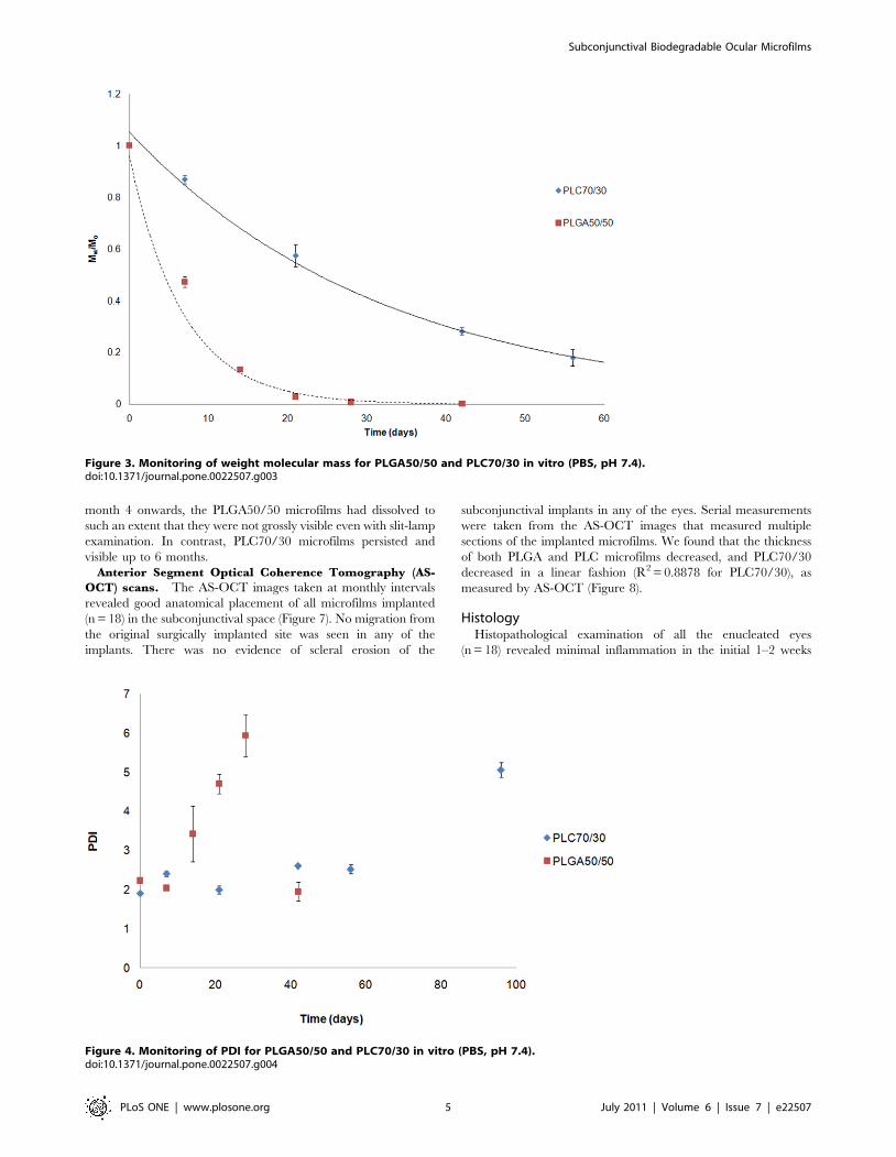
As shown in Figure 4, PDI of both polymer samples increased
with time. PDI for PLGA50/50 increased from week 2, but
dropped to a lower value (lower than its initial PDI) on day 42,
which shows it is fully degraded. PDI for PLC70/30 increased
slightly in the first 56 days of study, but increased suddenly to 5 or
more at the end of the study (not fully degraded at the end of the
study period).
Thickness change with degradation. The thickness of
PLGA50/50 was unchanged (within 10%) in the first 21 days, but
a sudden drop of 60% occurred on the day 28, and immeasurable
on day 42,. From the observed changes on mass loss, PLGA50/50
lost 90% of its initial weight on day 42, and corresponded to the
change in film thickness. In contrast, PLC70/30 maintained its
shape throughout the entire duration of the study period, with
minimal change in film thickness (Figure 5).
Results of in vivo study in rabbit eyesSlit-lamp examination of implanted microfilms. The
gross appearance and examination of the implanted microfilms
revealed minimal localized inflammation and vascularity using
serial vascularity grading scales [34] around the implanted
PLC70/30 (n = 9) and PLGA50/50 (n = 9). All eyes (n = 18) had
mild conjunctival hyperemia and chemosis, which resolved at one
week post-operatively (Figure 6). We compared conjunctival
vascularity of the insertion site before surgery and at the end of
the study in all eyes. We found no significant increase in ocular
score in all 18 eyes (mean percent increase in ocular score
Subconjunctival Biodegradable Ocular Microfilms
PLoS ONE | www.plosone.org 3 July 2011 | Volume 6 | Issue 7 | e22507

PLGA50/50 1260.5% vs. PLC70/30 1560.6%; P = 0.91, no
significant inter-observer variability). The cornea, anterior cham-
ber and lens remained clear with no evidence of inflammation or
scarring. We did notice on external ocular examination that the
PLGA50/50 microfilms appeared more pliable compared to the
PLC70/30 microfilms, which retained its original shape. However,
the animals did not display any signs of ocular discomfort, nor was
there any extrusion of any of the implants from either material. At
Figure 2. In vitro weight loss of PLGA50/50 and PLC70/30, immersed in PBS buffer (pH 7.4).doi:10.1371/journal.pone.0022507.g002
Figure 1. In vitro water absorption of PLGA50/50 and PLC70/30, immersed in PBS buffer (pH 7.4).doi:10.1371/journal.pone.0022507.g001
Subconjunctival Biodegradable Ocular Microfilms
PLoS ONE | www.plosone.org 4 July 2011 | Volume 6 | Issue 7 | e22507
month 4 onwards, the PLGA50/50 microfilms had dissolved to
such an extent that they were not grossly visible even with slit-lamp
examination. In contrast, PLC70/30 microfilms persisted and
visible up to 6 months.
Anterior Segment Optical Coherence Tomography (AS-
OCT) scans. The AS-OCT images taken at monthly intervals
revealed good anatomical placement of all microfilms implanted
(n = 18) in the subconjunctival space (Figure 7). No migration from
the original surgically implanted site was seen in any of the
implants. There was no evidence of scleral erosion of the
subconjunctival implants in any of the eyes. Serial measurements
were taken from the AS-OCT images that measured multiple
sections of the implanted microfilms. We found that the thickness
of both PLGA and PLC microfilms decreased, and PLC70/30
decreased in a linear fashion (R2 = 0.8878 for PLC70/30), as
measured by AS-OCT (Figure 8).
HistologyHistopathological examination of all the enucleated eyes
(n = 18) revealed minimal inflammation in the initial 1–2 weeks
Figure 3. Monitoring of weight molecular mass for PLGA50/50 and PLC70/30 in vitro (PBS, pH 7.4).doi:10.1371/journal.pone.0022507.g003
Figure 4. Monitoring of PDI for PLGA50/50 and PLC70/30 in vitro (PBS, pH 7.4).doi:10.1371/journal.pone.0022507.g004
Subconjunctival Biodegradable Ocular Microfilms
PLoS ONE | www.plosone.org 5 July 2011 | Volume 6 | Issue 7 | e22507

Figure 5. Change of film thickness of PLGA50/50 and PLC70/30 in vitro (PBS, pH 7.4).doi:10.1371/journal.pone.0022507.g005
Figure 6. Slit-lamp photographs of microfilms after surgical insertion into the subconjunctival space at 1, 3 and 6 months. A, B, C:1– Slit-lamp photographs of PLGA50/50 microfilm at 1, 3, 6 months respectively. A, B, C: 2 – Slit-lamp photograph of PLC70/30 microfilm at 1, 3, 6months respectively.doi:10.1371/journal.pone.0022507.g006
Subconjunctival Biodegradable Ocular Microfilms
PLoS ONE | www.plosone.org 6 July 2011 | Volume 6 | Issue 7 | e22507

post-operatively. This inflammation was seen to resolve by 1
month and there was minimal or no fibrosis or scarring detected.
By 4 months post-implantation, the PLGA50/50 microfilms were
not significantly visible on histology sections within the subcon-
junctival space. There was no significant fibrosis or collagen
capsule formation seen around the implant site (Figure 9).
However, capsule formation for PLC70/30 microfilms was noted
by 3 months on the histological sections. The mean collagen
capsule thickness surrounding the PLC70/30 microfilms increased
from 7.560.026 mm at 3 months to14.7560.11 mm at 6 months,
P,0.001. Histological examination did not show an obvious
foreign body encapsulation of the implanted films. This is im-
portant as excessive scarring and encapsulation often affects ocular
function or lead to surgical failure in surgeries such as glaucoma
filtration surgery [35].
ImmunohistochemistrySections stained with immunohistochemistry for CD45 T cells
were analyzed under polarized microscopy at 3 and 6 months
post-operatively. The PLC70/30 microfilms had significant
Figure 7. AS-OCT scans of microfilms after subconjunctival implantation. A, B, C: 1– AS-OCT of PLGA50/50 microfilms at 1, 3, 6 monthsrespectively. A, B, C: 2 – AS-OCT of PLC70/30 microfilms at 1, 3, 6 months respectively.doi:10.1371/journal.pone.0022507.g007
Figure 8. Serial AS-OCT thickness measurements of PLGA 50/50 and PLC70/30 microfilms in subconjunctival space.doi:10.1371/journal.pone.0022507.g008
Subconjunctival Biodegradable Ocular Microfilms
PLoS ONE | www.plosone.org 7 July 2011 | Volume 6 | Issue 7 | e22507

reduction in amount of inflammation surrounding the capsule
from 3 months post-implantation to the end of 6 months (inflam-
matory L/C ratio 8.560.6 vs. 5.560.8, P = 0.0018). Importantly,
there was minimal infiltration of T cells detected by immunohis-
tochemistry surrounding the implant site of the PLGA50/50
microfilms and the inflammatory L/C ratio could not be
calculated (Figure 10).
ImmunofluorescenceSections stained with immunofluorescence for CD45 T cells
were analyzed for both PLC70/30 and PLGA50/50 at 3 and 6
months. There was minimal inflammatory reaction, with scattered
T cells surrounding both implants. We also noted a reduction in T
cells surrounding the implant for both PLGA50/50 and PLC70/
30 when comparing implants at 3 and 6 months post-operatively
(Figure 11).
Discussion
The current mainstay of ocular therapy is via topical
administration. While it is easy to administer (for example, using
eye drops), there are many drawbacks - which include poor
bioavailability and penetration of the drugs, frequent instillation
leading to poor compliance and blurring of vision from viscous
vehicles [30]. Typically less than 5% of the topically applied drug
penetrates the cornea and reaches intraocular tissues, while a
major fraction of the instilled dose is often absorbed systemically
via the conjunctiva and nasolacrimal duct [31]. Thus, frequent
instillation of a relatively concentrated solution is required for a
sustained, therapeutic effect [32]. This need for frequent instil-
lation also leads to poor patient compliance, with often disastrous
consequences for vision.
We have used biodegradable polymers to create implants that
may be capable of sustained ocular drug delivery, to overcome the
disadvantages of topical medications and the issues with com-
pliance – a common problem faced by ophthalmologists in dealing
with diseases such as glaucoma, the second leading cause of
irreversible blindness in the world. This biopolymer microfilm
placed in the subconjunctival space may significantly improve
drug availability and reduce local ocular side effects, while over-
coming poor patient compliance.
Our study demonstrates that both PLGA50/50 and PLC70/30
microfilms are biocompatible and safe to be inserted subconjunc-
tivally in the rabbit eye. We also report for the first time, the use of
a novel AS-OCT imaging technique to serially measure the
degradation of the microfilms and describe the internal structures
of the eye for encapsulation in vivo over a prolonged period of 6
months. We used several techniques to demonstrate the biocom-
patibility, as well as behavior of these microfilms when inserted
into the subconjunctival space of the eye. Slit-lamp microscopy
Figure 9. Histological sections of PLGA50/50 and PLC70/30 microfilms in subconjunctival space. A, B, C: 1– Histology of eye implanted ofPLGA50/50 microfilm at 1, 3, 6 months respectively. A, B, C: 2 – Histology of eye implanted with PLC70/30 microfilm at 1, 3, 6 months respectively.doi:10.1371/journal.pone.0022507.g009
Subconjunctival Biodegradable Ocular Microfilms
PLoS ONE | www.plosone.org 8 July 2011 | Volume 6 | Issue 7 | e22507

allowed us to examine the location of the microfilms, while
monitoring inflammation and scarring of the overlying conjunc-
tiva. We did not find any significant increase in inflammation or
vascularity using serial vascularity grading scales [34]. We also
used an imaging technique, AS-OCT, to produce cross-sectional
scans of the implanted microfilms and eyes. The AS-OCT device
that is commonly used in the clinic on patients rapidly captures
reflections of light using low-coherence interferometry to create a
cross-sectional image in 8 meridians (128 sectional maps) to
produce high-resolution profile imaging of the anterior segment
Figure 10. Sections of PLGA50/50 and PLC70/30 with immunohistochemistry stains for CD45 T cells. Arrows point at the implant site. A,B: 1– Section of eye implanted with PLGA50/50 microfilm at 3 and 6 months respectively. A, B: 2 – Section of eye implanted with PLC70/30 microfilmat 3 and 6 months respectively.doi:10.1371/journal.pone.0022507.g010
Figure 11. Photographs with fluorescence microscopy of sections of PLGA50/50 and PLC70/30 with immunofluorescence stains forT cells. A, B: 1– Section of eye implanted with PLGA50/50 microfilm at 3 and 6 months respectively. A, B: 2 – Section of eye implanted with PLC70/30microfilm at 3 and 6 months respectively.doi:10.1371/journal.pone.0022507.g011
Subconjunctival Biodegradable Ocular Microfilms
PLoS ONE | www.plosone.org 9 July 2011 | Volume 6 | Issue 7 | e22507

[28]. This enabled us to not only examine the internal structure of
the eyes, but also the microfilm within the subconjunctival space in
situ during the entire study period. We used the AS-OCT to
serially measure the thickness of our microfilm implants with a
high resolution of up to microns and monitored each implant’s
position. We recognize that there are potential limitations due to
the resolution of the AS-OCT. However, we used a standard
technique to capture the cross-sectional image of each microfilm
and these cross-sectional images are taken in 8 meridians (128
sectional maps) to produce high-resolution profile. There was no
obvious fibrosis, effusion and encapsulation in the neighboring
ocular structures. Histological examination did not show an
obvious foreign body encapsulation of the implanted films. This is
important as excessive scarring and encapsulation often affects
ocular function or lead to surgical failure in surgeries such as
glaucoma filtration surgery [35]. All these studies revealed mini-
mal scarring and inflammation induced by the implanted
microfilm in the subconjunctiva over the 6-month study period.
It is generally accepted that, in this class of polymers used in our
study (poly a-hydroxy esters), there may be two different modes of
degradation. In the first mechanism, which is often referred to as
homogeneous or bulk degradation, the polymers degrade slowly
with no appreciable mass or volume loss until the degradation
products become water-soluble and leach out of the matrix, when
mass loss is then detectable. In the second mechanism, the
polymer degrades first at the surface, and the surface molecules
decrease in molecular weight to the point where the surface
molecules leach out, without affecting the interior of the material.
In this mode of degradation, which is sometimes referred to as
heterogeneous degradation or surface erosion, there is continuous
decrease in mass and in the material dimensions.
From the results of the study, the PLGA 50/50 films clearly
exhibited bulk degradation in vitro and in vivo. Although not as
evident (since no significant mass loss has been detected up to day
40 – Figure 2), in vitro, PLC70/30 also exhibited molecular weight
decrease (Figure 3) without any mass loss, which is a characteristic
of bulk degradation. However, PLC70/30 clearly behaves dif-
ferently when implanted into the rabbit eyes. PLC70/30 micro-
films underwent surface erosion in the subconjunctival space, as
evidenced by our serial measurements using slit-lamp microscopy
and AS-OCT techniques, since the width and length of the
microfilms did not change visually over 6 months (Figure 6), but
thickness of the films (Figure 7) decreased continuously. This is
typically observed in surface erosion or heterogeneous degrada-
tion. Usually, the polymer changes from a bulk degradation mode
to a surface erosion mode when the intrinsic hydrolysis rate (Rh)
becomes higher than the water ingress rate into the polymer (Rw).
We hypothesize that in the in vivo situation, Rh is being increased
relative to Rw, most likely due to the influence of enzymes
(esterases) or proteins present in the eye. A surface erosion mode is
the preferred mode in such applications, as bulk degradation may
lead to ‘‘catastrophic’’ breakdown into small fragments causing
localized irritation. Surface erosion also results in a constant
release of incorporated drug. PLGA and PLC are anionic poly-
mers that undergo bulk degradation in vitro. Embedded drugs are
released from the matrix via diffusion initially, followed by
degradation of the polymer matrix itself [33]. Thus the first
observation of surface erosion of this grade of polymers in the sub-
conjunctival space is exciting and opens the door for a more
efficient therapeutic route.
The subconjunctival space is a potential area in the eye that is
useful for delivering ocular drugs in a sustained manner. Cur-
rently, peribulbar or subtenon injections are used to deliver short
to intermediate duration of drugs to the eye [36]. Implanting the
microfilm in this space may bypass ocular blood and lymphatic
barriers, to achieve therapeutic levels in the eye with lower loading
concentrations of drug [37]. In this study, we have shown that the
PLC70/30 and PLGA50/50 microfilms can be placed into the
subconjunctival space using a simple surgical technique, and that
both microfilms remain stable in-situ for up to 6 months. Further-
more, we have demonstrated that surgical implantation of these
films in the subconjunctival space does not cause any associated
significant scarring, encapsulation or inflammation.
In conclusion, we report that biodegradable microfilms
prepared from PLGA50/50 and PLC70/30, are non-toxic and
well tolerated when implanted in the subconjunctival space and
therefore has the potential use as an ocular drug delivery platform.
PLGA50/50 always degraded at a faster rate than PLC70/30.
Both PLGA50/50 and PLC70/30 demonstrated bulk degradation
in vitro, whereas PLC70/30 exhibited surface erosion in vivo. The
observation of surface erosion in the sub-conjunctival space is
significant for controlling the release of drugs locally, and opens
the door for more efficient and sustained therapy.
Author Contributions
Conceived and designed the experiments: YP SSV TTW MA. Performed
the experiments: YP MA SF WSL ZM TTW. Analyzed the data: YP MA
SSV TTW. Wrote the paper: YP MA.
References
1. Ghate D, Edelhauser HF (2006) Ocular drug delivery. Expert Opin Drug Deliv
3: 275–287.2. Souto EB, Doktorovova S, Gonzalez-Mira E, Egea MA, Garcia ML (2010)
Feasibility of lipid nanoparticles for ocular delivery of anti-inflammatory drugs.
Curr Eye Res 35: 537–552.3. Barar J, Javadzadeh AR, Omidi Y (2008) Ocular novel drug delivery: impacts of
membranes and barriers. Expert Opin Drug Deliv 5: 567–581.4. Kato A, Kimura H, Okabe K, Okabe J, Kunou N, et al. (2004) Feasibility of
drug delivery to the posterior pole of the rabbit eye with an episcleral implant.
Invest Ophthalmol Vis Sci 45: 238–244.5. Gaudana R, Jwala J, Boddu SH, Mitra AK (2009) Recent perspectives in ocular
drug delivery. Pharm Res 26: 1197–1216.6. Jaffe GJ, Yang CH, Guo H, Denny JP, Lima C, et al. (2000) Safety and
pharmacokinetics of an intraocular fluocinolone acetonide sustained deliverydevice. Invest Ophthalmol Vis Sci 41: 3569–3575.
7. Kim H, Robinson MR, Lizak MJ, Tansey G, Lutz RJ, et al. (2004) Controlled
drug release from an ocular implant: an evaluation using dynamic three-dimensional magnetic resonance imaging. Invest Ophthalmol Vis Sci 45:
2722–2731.8. Okabe J, Kimura H, Kunou N, Okabe K, Kato A, et al. (2003) Biodegradable
intrascleral implant for sustained intraocular delivery of betamethasone
phosphate. Invest Ophthalmol Vis Sci 44: 740–744.
9. Okabe K, Kimura H, Okabe J, Kato A, Kunou N, et al. (2003) Intraocular tissue
distribution of betamethasone after intrascleral administration using a non-biodegradable sustained drug delivery device. Invest Ophthalmol Vis Sci 44:
2702–2707.
10. Kunou N, Ogura Y, Honda Y, Hyon SH, Ikada Y (2000) Biodegradable scleralimplant for controlled intraocular delivery of betamethasone phosphate.
J Biomed Mater Res 51: 635–641.11. Beeley NR, Rossi JV, Mello-Filho PA, Mahmoud MI, Fujii GY, et al. (2005)
Fabrication, implantation, elution, and retrieval of a steroid-loaded polycapro-
lactone subretinal implant. J Biomed Mater Res A 73: 437–444.12. Beeley NR, Stewart JM, Tano R, Lawin LR, Chappa RA, et al. (2006)
Development, implantation, in vivo elution, and retrieval of a biocompatible,sustained release subretinal drug delivery system. J Biomed Mater Res A 76:
690–698.13. Ali Y, Lehmussaari K (2006) Industrial perspective in ocular drug delivery. Adv
Drug Deliv Rev 58: 1258–1268.
14. Barbu E, Verestiuc L, Iancu M, Jatariu A, Lungu A, et al. (2009) Hybridpolymeric hydrogels for ocular drug delivery: nanoparticulate systems from
copolymers of acrylic acid-functionalized chitosan and N-isopropylacrylamide or2-hydroxyethyl methacrylate. Nanotechnology 20: 225108.
15. Lewis KJ, Irwin WJ, Akhtar S (1998) Development of a sustained-release
biodegradable polymer delivery system for site-specific delivery of oligonucle-
Subconjunctival Biodegradable Ocular Microfilms
PLoS ONE | www.plosone.org 10 July 2011 | Volume 6 | Issue 7 | e22507

otides: characterization of P(LA-GA) copolymer microspheres in vitro. J Drug
Target 5: 291–302.16. Kreuter J (1996) Nanoparticles and microparticles for drug and vaccine delivery.
J Anat 189(Pt 3): 503–505.
17. Kreuter J (1995) Nanoparticulate systems in drug delivery and targeting. J DrugTarget 3: 171–173.
18. Vega E, Gamisans F, Garcia ML, Chauvet A, Lacoulonche F, et al. (2008)PLGA nanospheres for the ocular delivery of flurbiprofen: drug release and
interactions. J Pharm Sci 97: 5306–5317.
19. Araujo J, Vega E, Lopes C, Egea MA, Garcia ML, et al. (2009) Effect of polymerviscosity on physicochemical properties and ocular tolerance of FB-loaded
PLGA nanospheres. Colloids Surf B Biointerfaces 72: 48–56.20. Agnihotri SM, Vavia PR (2009) Diclofenac-loaded biopolymeric nanosuspen-
sions for ophthalmic application. Nanomedicine 5: 90–95.21. Giordano GG, Refojo MF, Arroyo MH (1993) Sustained delivery of retinoic
acid from microspheres of biodegradable polymer in PVR. Invest Ophthalmol
Vis Sci 34: 2743–2751.22. Duong-Hong D, Tang YD, Wu W, Venkatraman SS, Boey F, et al. (2010) Fully
biodegradable septal defect occluder-a double umbrella design. CatheterCardiovasc Interv 76: 711–718.
23. Wang X, Venkatraman SS, Boey FY, Loo JS, Tan LP (2006) Controlled release
of sirolimus from a multilayered PLGA stent matrix. Biomaterials 27:5588–5595.
24. Joachim Loo SC, Jason Tan WL, Khoa SM, Chia NK, Venkatraman S, et al.(2008) Hydrolytic degradation characteristics of irradiated multi-layered PLGA
films. Int J Pharm 360: 228–230.25. Frank A, Rath SK, Venkatraman SS (2005) Controlled release from bioerodible
polymers: effect of drug type and polymer composition. J Control Release 102:
333–344.26. Munger RJ (2002) Veterinary ophthalmology in laboratory animal studies. Vet
Ophthalmol 5: 167–175.
27. Singh M, Chew PT, Friedman DS, Nolan WP, See JL, et al. (2007) Imaging of
trabeculectomy blebs using anterior segment optical coherence tomography.
Ophthalmology 114: 47–53.
28. Ramos JL, Li Y, Huang D (2009) Clinical and research applications of anterior
segment optical coherence tomography - a review. Clin Experiment Ophthalmol
37: 81–89.
29. Singh M, See JL, Aquino MC, Thean LS, Chew PT (2009) High-definition
imaging of trabeculectomy blebs using spectral domain optical coherence
tomography adapted for the anterior segment. Clin Experiment Ophthalmol 37:
345–351.
30. Koevary SB (2003) Pharmacokinetics of topical ocular drug delivery: potential
uses for the treatment of diseases of the posterior segment and beyond. Curr
Drug Metab 4: 213–222.
31. Zhang W, Prausnitz MR, Edwards A (2004) Model of transient drug diffusion
across cornea. J Control Release 99: 241–258.
32. Nomoto H, Shiraga F, Kuno N, Kimura E, Fujii S, et al. (2009)
Pharmacokinetics of bevacizumab after topical, subconjunctival, and intravitreal
administration in rabbits. Invest Ophthalmol Vis Sci 50: 4807–4813.
33. Houchin ML, Topp EM (2008) Chemical degradation of peptides and proteins
in PLGA: a review of reactions and mechanisms. J Pharm Sci 97: 2395–2404.
34. Wells AP, Crowston JG, Marks J, Kirwan JF, Smith G, et al. (2004) A pilot study
of a system for grading of drainage blebs after glaucoma surgery. J Glaucoma 13:
454–460.
35. Hitchings RA, Grierson I (1983) Clinico pathological correlation in eyes with
failed fistulizing surgery. Trans Ophthalmol Soc U K 103(Pt 1): 84–88.
36. Weijtens O, Feron EJ, Schoemaker RC, Cohen AF, Lentjes EG, et al. (1999)
High concentration of dexamethasone in aqueous and vitreous after subcon-
junctival injection. Am J Ophthalmol 128: 192–197.
37. Hughes PM, Olejnik O, Chang-Lin JE, Wilson CG (2005) Topical and systemic
drug delivery to the posterior segments. Adv Drug Deliv Rev 57: 2010–2032.
Subconjunctival Biodegradable Ocular Microfilms
PLoS ONE | www.plosone.org 11 July 2011 | Volume 6 | Issue 7 | e22507


![Biodegradation of Styrene-Butadiene-Styrene Coploymer via ... · A functional group when incorporated into a polymer can deliver enhanced properties, such as biocompatibility [2],](https://img.pdfslide.us/doc/110x75/5f01c2397e708231d400e5a8/biodegradation-of-styrene-butadiene-styrene-coploymer-via-a-functional-group.jpg)