Embed Size (px)
Citation preview
The Endoplasmic Reticulum Retention Signal of the E3/19K Protein of Adenovirus Type 2 Consists of Three Separate Amino Acid Segments at the Carboxy Terminus Reinhard Gaba thu le r and Sune Kvist
Ludwig Institute for Cancer Research, Stockholm Branch, S-104 01 Stockholm, Sweden
Abstract. The E3/19K protein of adenovirus type 2 is a resident of the ER. Immediately after synthesis it binds to human major histocompatibility complex class I antigens and prevents their departure from the ER compartment. The ER retention signal of the E3/19K protein is contained within the 15 amino acids that protrude on the cytoplasmic side at the carboxy terminus of the protein. To define the ER retention se- quence in more detail, we have generated 10 mutants of the E3/19K protein that differ only within this seg- ment. Analysis of the rate of intracellular transport and cell surface expression of HLA antigens as-
sociated to these mutants, show that the sequences Ser-Phe-Ile, located in the middle of the 15-residue segment and Met-Pro, at the extreme carboxy termi- nus, are crucial for retention. Four charged residues, Asp-Glu-Lys-Lys, are located between these two reten- tion elements but are of little or no importance. The basic cluster of amino acids close to the membrane also has some effect on retention. Thus, the retention signal of the E3/19K protein is not a contiguous se- quence of amino acids but has a complex spatial ar- rangement.
T HE human adenoviruses comprise a group of DNA viruses with more than 40 subtypes. Normally they cause a mild respiratory infection but can also induce
a latent infection that persists for extended periods of time (Evans, 1958; Flint, 1980). The two best characterized sub- types are the adenovirus type 2 (Ad-2) and 5 (for reviews see Shenk and Williams, 1984; Ginsberg, 1984; Doerfler, 1986).
The infection cycle can be divided into an early and a late phase. One of the early regions of Ad-2, the E3 region, en- codes an abundant protein called E3/19K (Ross and Levine, 1979; Persson et al., 1980b). The function of this protein is not known and the E3 region is dispensible for virus growth in cultured cells (Shenk and Williams, 1984). The E3/19K protein is synthesized on membrane-bound ribosomes and inserted into the membrane of the ER during or immediately after synthesis. It consists of 159 amino acids of which the 17 most amino-terminal ones constitute a cleavable signal se- quence, leaving a mature protein of 142 residues (Persson et al., 1980a; Ahmed et al., 1982; Wold et al., 1985). About 104 residues are present on the luminal side of the ER mem- brane, 23 amino acids span the membrane, and the remain- ing 15 are protruding into the cytoplasm. The protein is glycosylated at amino acids 12 and 61 (Kornfeld and Wold, 1981).
At least two different properties of the E9/19K protein make it interesting. First, several investigators have shown that the E3/19K protein associates with class I antigens of the
major histocompatibility complex (MHC) t (Kvist et al., 1978; Sign~is et al., 1982; K/impe et al., 1983; P~bo et al., 1986). These antigens (termed HLA in man) are cell surface glycoproteins composed of a heavy chain (45,000 tool wt) noncovalenfly bound to B2-microglobulin (12,000 tool w0 (for a review see Ploegh et al., 1981). The function of the HLA antigens is to present foreign viral peptides to cytolytic T lymphocyte precursors. These T cells develop into cytolyt- ic T cells with the ability to recognize and eliminate specif- ically the infected cells via the class I antigens (Klein, 1979; Zinkernagel and Doherty, 1979; Townsend et al., 1986; Braciale et al., 1987). We and others have shown that the as- sociation of the E3/19K protein to the HLA antigens prevents their cell surface expression (Burgert and Kvist, 1985; An- dersson et al., 1985). The association involves the ER lumi- nal part of the E3/19K protein and the od and or2 domains of the MHC antigens (Burgert and Kvist, 1987). Due to the decrease in density of cell surface MHC class I antigens, cytolytic T cells recognize cells expressing the E3/19K pro- tein considerably less efficiently (Burgert et al., 1987).
The second interesting property of the E3/19K protein is its cellular location. We and others have demonstrated it to be a resident of the ER (Burgert and Kvist, 1985; P~bo et al., 1987). Truncation of the eight most carboxy-terminal amino acids allows the cell surface expression of the mutated
1. Abbreviations used in this paper: endo H, endoglycosidase H; MHC, ma- jor histocompatibility complex.
© The Rockefeller University Press, 0021-9525/90/11/1803/8 $2.00 The Journal of Cell Biology, Volume 111, November 1990 1803-1810 1803
on November 18, 2018jcb.rupress.org Downloaded from http://doi.org/10.1083/jcb.111.5.1803Published Online: 1 November, 1990 | Supp Info:
E3/19K (P~i~ibo et al., 1987). Recently, Nilsson et al. (1989) showed by transplanting the E3/19K cytoplasmic tail onto the CD8 molecule that a short linear sequence at the car- boxy-terminus (DEKKMP) is responsible for ER retention of such a hybrid molecule.
Several proteins have been classified as resident ER pro- teins and can be divided into two groups: (a) soluble proteins present in the lumen of the ER and (b) integral ER membrane proteins. The common feature of all soluble ER proteins is a short amino acid sequence Lys-Asp-Glu-Leu (KDEL) pres- ent at the extreme carboxy terminus (Munro and Pelham, 1986; Lee et al., 1984; Edman et al., 1985). It has been shown that the KDEL sequence is crucial for the ER reten- tion of the immunoglobulin heavy chain binding protein (BiP) and, furthermore, after transfer to the chicken lyso- zyme, is sufficient to cause ER retention (Munro and Pel- ham, 1987).
Members of the integral ER membrane protein-group in- clude NS28 and VP7 of rotavirus (Petrie et al., 1984; Whit- feld et al., 1987), the docking protein (Hortsch et al., 1988), the 3-hydroxy-3-methylglutaryl coenzyme A reductase (Chin et al., 1984), the microsomal cytochrome P-450 (Sakaguchi et al., 1987), and the ribophorins I and II (Crimaudo et al., 1987). Only for the VP7 protein has the ER retention signal been localized to the luminal domain of the protein (Poru- chynsky and Atkinson, 1988; Stirzaker and Both, 1989). The structure(s) of the other proteins that localize them to the ER remains obscure. The E3/19K protein of Ad-2, which also belongs to this group, is unique with respect to its reten- tion signal being present within the 15 amino acids at the car- boxy terminus on the cytoplasmic side of the ER membrane.
We have taken a different approach than that of Nilsson et al. (1989) and characterized the ER retention signal on E3/19K protein by using the property of E3/19K protein to bind to HLA. We have, by site-directed mutagenesis, gener- ated 10 mutants of the E3/19K protein which differ from each other only within the short cytoplasmic tail containing the retention signal. Our results show that a complex noncontig- uous structure, consisting of three blocks of amino acids, constitutes the retention signal of the E3/19K protein.
Materials and Methods
Oligonucleotide-directed Mutagenesis of the E3/19K Protein The coding sequence of the E3/19K gene is contained within a 1,259-bp Sau I fragment of the Eco RID fragment of Ad-2 (Left et al., 1984). This frag- ment was inserted into the Sma I site of the vector WB 2311 (Barnes, 1980). Before insertion of this fragment the Sau I ends were made blunt by poly- merization with the Klenow DNA polymerase. In this way the Sau I sites at both ends of the fragment were reconstituted and could be used for rein- sertion into the Eco RID fragment after mutagenesis. For oligonucleotide- directed mntagenesis we used the method described by Zoller and Smith (1983). Oligonucleotides were synthesized in a DNA synthesizer (model 380 B; Applied Biosystems, Inc., Foster City, CA).
Cell Culture and DNA Transfection of Cells We have used the cell line 293 for expression of the wild-type E3/19K and its mutants (Graham et al., 1977). The cells were grown in DME containing 10% fetal calf serum, 20 mM Hepes, 2 mM glutamine, and antibiotics. Transfection of 293 cells with E3/19K mutant DNA was carried out as de- scribed previously (Arnold et al., 1984; Burgert and Kvist, 1985) with the neophosphotransferase gene as a selectable marker (Southern and Berg,
1982). Selection of cell clones was done in 800 #g/ml of G418 (Sigma Chemical Co., St. Louis).
Monoclonal Antibodies and Antisera The antibody W6/32 reacts with a framework determinant of HLA-A, B, and C antigens (Barnstable et al., 1978). The antiserum against E3/19K has been described previously (Persson et al., 1979).
Cell Labeling, Pulse-Chase Experiments, Endoglycosidase H Treatment, Immunoprecipitation, and SDS-PAGE Cells were washed in MEM medium without methionine 1 h before labeling and labeled with 150 #Ci/ml of [35S]methionine for 15 min. For the pulse- chase experiments, cells were labeled 15 min and then chased with normal DME medium containing 10% FCS, 20 mM Hepes, 2 mM glutamine, and antibiotics. A separate petri dish was used for each time point. Immunopre- cipitation and SDS-PAGE were carded out as described (Kvist et al., 1982). In Fig. 4, after immunoprecipitation, the material was digested with 5 mU of endoglycosidase H (endo H) (Boehringer Mannheim Diagnostics, Mann- heim, Federal Republic of Germany) for 20 h at 37°C, before SDS-PAGE analysis.
Immunoprecipitation of CeU Surface HLA Antigens Ceils were pulse labeled for 15 min with [35S]methionine and chased in medium containing an excess of cold methionine. At each time point, cells were split into two equal parts and washed twice with ice-cold medium. The cells were kept on ice. One aliquot was treated with mAb W6/32 for 30 min and was then washed three times with medium. The cells were lysed with solubilization buffer containing five times excess of cold cell lysate. Unla- beled HLA antigen in this lysate can then bind to any residual free antibody which might otherwise bind to radioactive intracellular HLA antigens ex- posed after lysis. After centrifugation, the immune complexes were recov- ered by using protein A-Sepharose and analyzed by SDS-PAGE. The second half of the ceils were lysed in normal solubilization buffer and total HLA antigens were immunoprecipitated and analyzed by SDS-PAGE.
Flow Cytometry Analysis To determine the cell surface expression of HLA antigens, we used fluores- cence-activated cell sorter analysis. After washing in DME containing 20 mM Hepes, 20 mM azide, and 5 % BSA, the cells were reacted with the W6/32 anti-HLA monoclonal antibody at4°C for 1 h and then washed three times with medium. The cells were stained with FITC-conjugated goat anti-mouse immunoglobulin serum (Sigma Chemical Co.). After 1 h of in- cubation, the cells were washed twice in medium and twice in PBS, the cells were then fixed in 1.5% paraformaidehyde for 20 min at 4°C. After two washes in PBS, the ceils were kept in PBS at 4°C and in darkness until ana- lyzed. The fluorescence profiles were obtained by analyzing •104 cells on a semilogarithmic plot in a fluorescence-activated cell sorter IV. In Fig. 6, we report the mean fluorescence obtained in a representative experiment out of a total of five separate experiments done with all 12 cell lines. Nega- tive control was without first antibody.
Other Reagents Protein A-Sepharose was from Pharmacia (Uppsaia, Sweden). [35S]Methi- onine (>800 Ci/mmol) was from Amersham International, Amersham, England).
Results
Experimental Strategy The ER retention signal of the E3/19K protein has been claimed to be contained within the eight most carboxy- terminal amino acids on the cytoplasmic side of the ER membrane (P~i~ibo et al., 1987). Our preliminary results, however, showed that also the basic cluster of amino acids close to the membrane influence retention (Gabathuler, R.,
The Journal of Cell Biology, Volume I I I, 1990 1804
E3/19K - / - ,~
H621
M129 ~ L
1,4121 ~ H123
N125
H131
M 133
M135
M137 ~ N139
/ / / / / ,
ER MEMBRANE
CYTOPLASM
S S F I D E K K P-EOOH
~(Z:~db:l~qg- [ooH
-EOOH
S S F I D E -EOOH
( ~ ( ~ 9 ( ~ ) ~ ) - -- - @ ( ~ ( ~ ( ~ - [ 0 0 H
- - - ( ~ - c o o H
~ - [ O O H
. . . . . ~ - C o o H
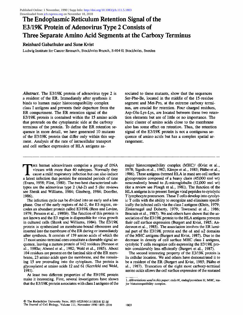
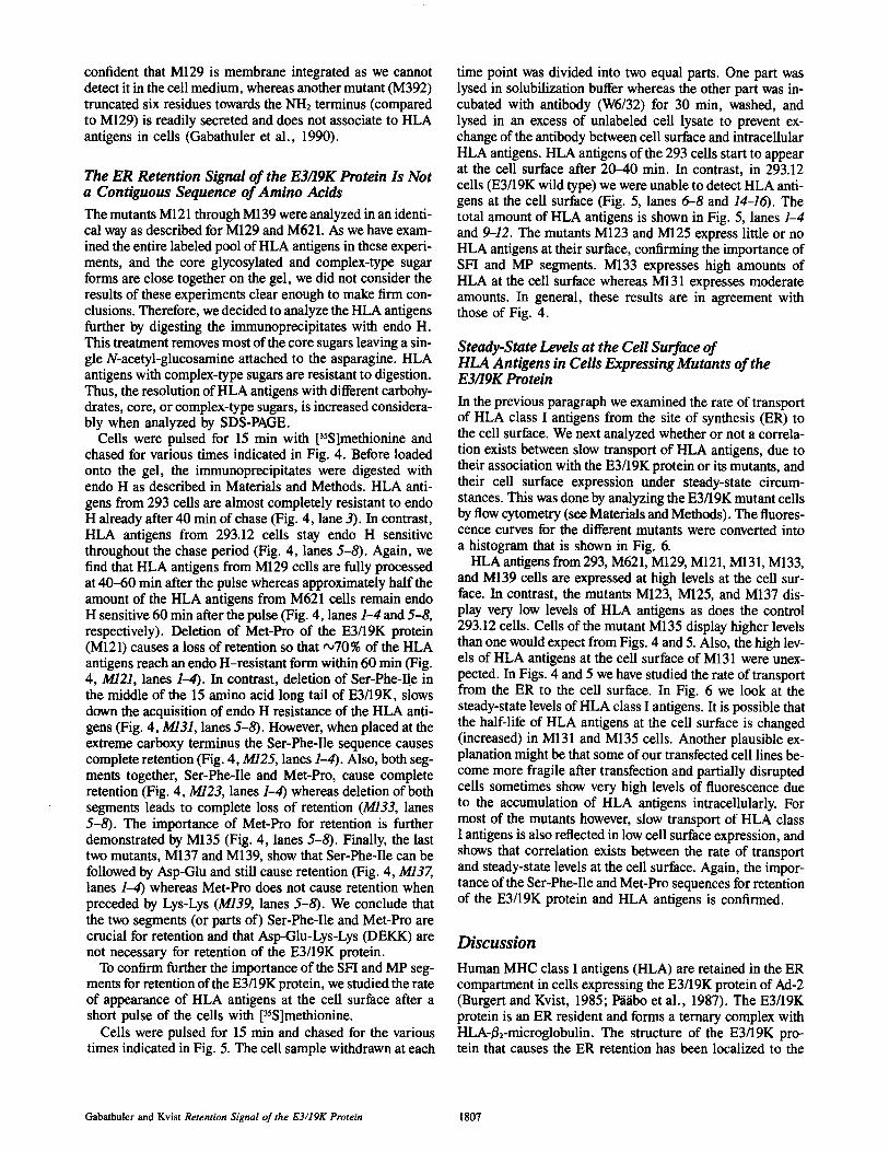
Figure 1. Schematic representation of the carboxy-terminal portion of the E3/19K protein and its mutants. The amino acid sequence of the wild-type E3/19K is shown on the top. The names of the different mutants are indicated to the left in the figure. The hatched area denotes the ER membrane and only two amino acids (YL) of the trans-membrane segment are shown. Dashes in the amino acid sequence indicate deleted residues. The one-letter code for amino acids is used.
and S. Kvist, unpublished results). Thus, all 15 residues of the cytoplasmic tail of the E3/19K protein might be impor- tant for retention. Our approach was to change these 15 amino acids in such a way that the E3/19K protein would be released from the ER compartment and transported towards the cell surface. However, not only did we want to disturb the retention signal but also to identify the amino acids cru- cial for ER retention. Even the limited number of 15 gives a very high number of different combinations of amino acids. We divided the cytoplasmic tail into two parts: (a) the basic cluster of residues closest to the membrane (six residues) and (b) the remaining carboxy-terminal nine amino acids. This was done by replacing the codon for the serine residue at position 7 with a termination codon (Fig. 1). The nine last carboxy-terminal amino acids were then further changed. We changed these amino acids in block deletions (at least two residues).
Immediately after synthesis, the E3/19K protein, or maybe the nascent chain, is found bound to HLA antigens (Burgert and Kvist, 1985). All our cell lines transfected with the E3/19K gene express an excess of the viral protein compared to HLA antigens. We have taken advantage of its firm inter- action with HLA antigens, and rather than analyzing the cel- lular location of the E3/19K protein itself, we have followed the rate of transport of HLA class I antigens. In favor of our approach are the following facts. (a) Little or no carbohy- drate processing can be observed for the E3/19K protein and its mutants. In pulse-chase experiments it is therefore difficult to follow transport by this method. In contrast, HLA antigens are well-characterized molecules that undergo car- bohydrate processing, which can readily be detected and fol- lowed during intracellular transport. (b) There is no good an- tiserum or antibody available against the E3/19K protein. Our rabbit anti-E3/19K serum also detects two other cellular
proteins that interfere with similar molecular weights when analyzed by SDS-PAGE. Excellent antibodies exist for HLA class I antigens that precipitate all three subunits: HLA, B2- microglobulin, and the E3/19K protein. (c) By analyzing the HLA antigens, associated with the E3/19K protein, we have an internal control that our modifications of the E3/19K pro- tein do not grossly alter its conformation on the luminal side of the ER membrane. Such changes might otherwise by themselves contribute to loss of retention.
Taken together, we preferred to analyze HLA class I anti- gens associated with the E3/19K protein rather than the E3/19K protein itself or the 15 amino acids containing the retention signal transferred to another protein of unknown behavior. However, our approach has two requirements that must be fulfilled. First, the E3/19K protein and its mutants must be expressed in excess compared to HLA antigens, i.e., all HLA class I molecules must be associated with E3/19K. Secondly, no mutant of the E3/19K protein should have suffered conformational alterations in such a way that associ- ation to HLA antigens would be disturbed, i.e., the ratio be- tween HLA antigens and the E3/19K protein should be con- stant. Both these requirements were fulfilled for all our mutants.
Site-directed Mutagenesis and Expression of the E3/19K Mutants in 293 Cells
By using oligonucleotide site-directed mutagenesis we have generated 10 mutants of the E3/19K protein that all differ in the 15 most carboxy-terminal amino acids (Fig. 1; see Mate- rials and Methods for details). Plasmid DNAs for the mu- tants were used to transfect 293 cells, an epithelium em- bryonic kidney cell line (Graham et al., 1977; Burgert and Kvist, 1985). Stable transformants were selected as clones,
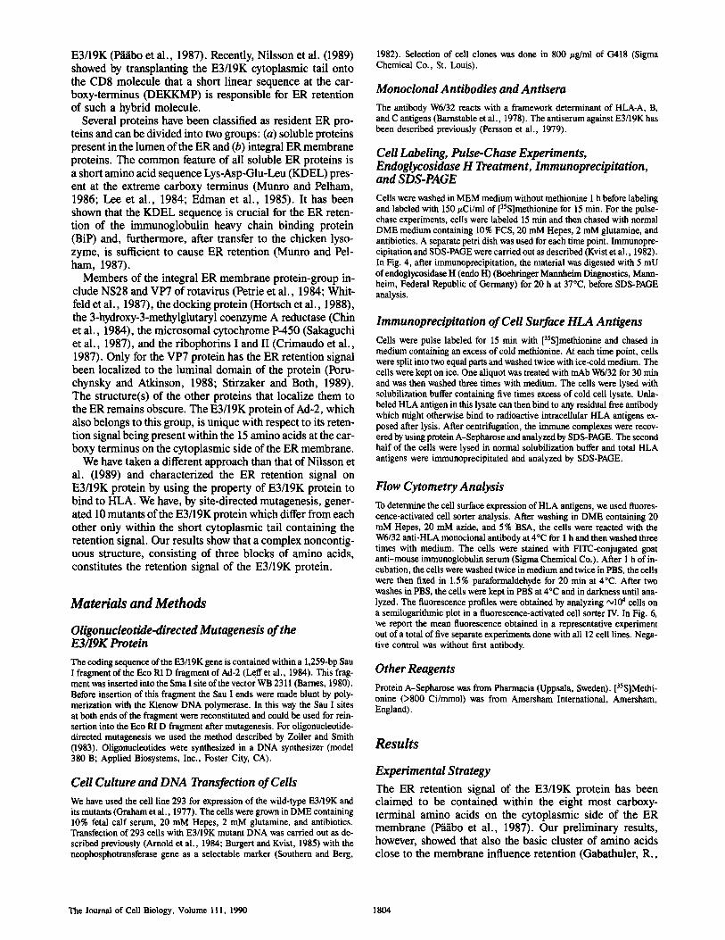
Figure 2. Expression of E3/19K and its mutants in transfected 293 cells. Cells were labeled for 15 min with [35S]methionine, lysed, immunoprecipitated with a rabbit anti-E3/19K serum, and ana- lyzed by SDS-PAGE. The parental cell line 293 was used as a con- trol. The cell lines expressing the wild-type E3/19K (293.12) and the E3/19K mutants are indicated at the top of the figure. The shift in apparent molecular weight for the E3fI9K mutants correlate ap- proximately to the number of amino acids deleted. All mutants are expressed at a higher level (2-10-fold) than the wild type. The faint band below the major band corresponds to a partially glycosylated form of the protein. See text for further details.
Gabathuler and Kvist Retention Signal of the E3/19K Protein 1805
Table L Association of Wild-Type and Mutant E3/19K Proteins to HLA Molecules
Mutant Ratio E3/19K:HLA proteins (mean values)
Wild type 1.00 M621 1.33 + 0.30 M121 1.07 + 0.47 M123 1.69 + 0.75 M125 1.32 + 0.58 M129 1.00 + 0.39 M131 1.49 + 0.44 M133 1.19 + 0.52 M135 1.72 + 0.51 M137 1.59 + 0.83 M139 1.36 + 0.86
Cells were labeled for 15 rain with [35S]methionine, lysed, and immunopre- cipitated by W6/32. This material was analyzed by SDS-PAGE. The exposed x-ray films were scanned by a laser densitometer (LKB Instruments Inc., Bromma, Sweden). The densities between HLA and E3/19K related bands were compared for the same immunoprecipitation. The ratio between E3/19K wild-typo protein and HLA bands was fixed arbitrarily to one for each individu- al experiment. The mean ratios for five independent experiments are reported in the table with the standard deviations.
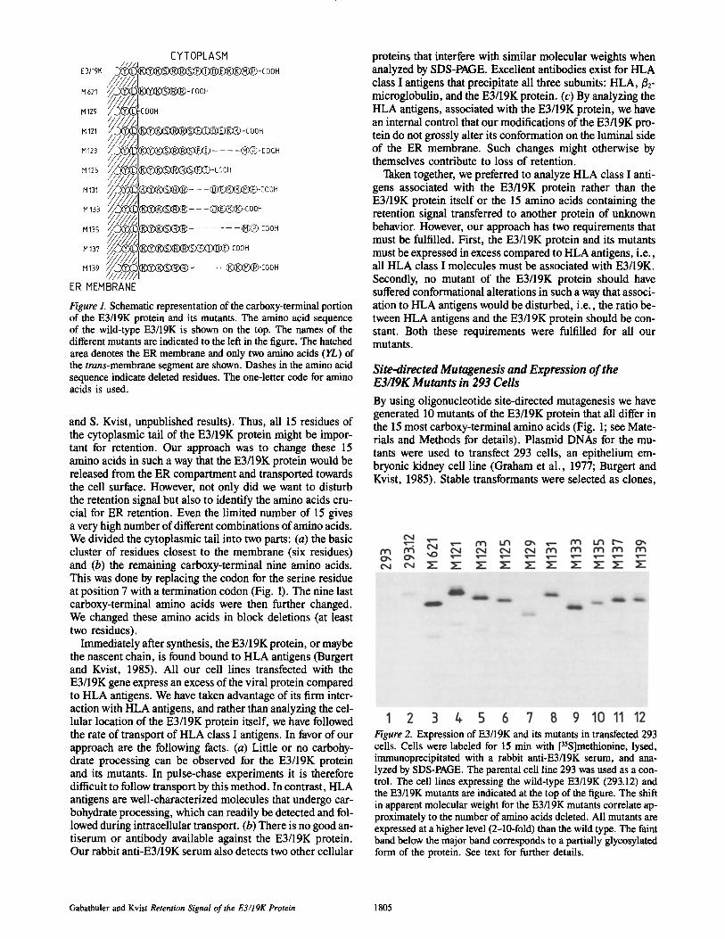
class I antigens of these two mutant cell lines were compared with those of 293 cells (absence of E3/19K) and 293.12 cells (wild-type E3/19K). Cells were labeled with [35S]methio- nine for 15 min and then chased in an excess of cold methio- nine. Analysis was as described above at the times indicated in Fig. 3, HLA antigens from 293 cells and M129 cells have undergone complete carbohydrate processing already after 40 min of chase (Fig. 3, lanes 1-3). In contrast, class I anti- gens from 293.12 cells are not processed at all during the 2-h chase (Fig. 3, lanes 1-5). HLA antigens from M621 cells show partial processing at 2 h of chase. The majority of the radioactivity is found in the lower band at early time points (Fig. 3, lanes 1 and 2), equal amounts are found at 40 and 60 min of chase (lanes 3 and 4) whereas at 2 h the great majority is found in the two upper bands (lane 5). As HLA antigens from 293 and M129 cells are fully processed at 40 rain of chase, we conclude that a certain influence is exerted by the first six amino acids (the basic cluster). We are
labeled with [35S]methionine, immunoprecipitated with a rabbit anti-E3/19K serum, and finally analyzed by SDS- PAGE. Clones expressing high amounts of the E3/19K pro- tein were chosen for further experiments and were compared with the cell line 293.12, which expresses the wild-type E3/19K protein (Fig. 2). All mutants selected expressed at least a twofold excess of the E3/19K protein compared to the wild-type (Fig. 2, compare lanes 3-12 with lane 2). We have previously shown that the wild-type E3/19K protein ex- pressed in 293.12 cells causes an almost complete inhibition of cell surface expression of HLA class I antigens (Burgert and Kvist, 1985). As the cell lines analyzed in Fig. 2 express considerably more of the E3/19K mutant proteins (2-10 fold) than the 293.12 cells, one would expect also these to cause an almost complete block in HLA expression, provided the retention signal is intact.
We next analyzed whether or not all the mutants of the E3/19K protein associate to HLA class I antigens to a similar degree. Cells were labeled with [35S]methionine, lysed, and reacted with the W6/32 (monoclonal anti-HLA), and ana- lyzed by SDS-PAGE. In addition to HLA heavy chains and ~2-microglobulin a protein band of ~ 24,000 mol wt, rep- resenting the E3/19K mutant, was seen for all the mutant cell lines (data not shown). Table I is a compilation of five in- dividual experiments and shows that all mutant E3/19K bind to HLA antigens approximately with the same efficiency as the wild-type E3/19K protein. We conclude that changes in the cytoplasmic tail do not disturb binding of the mutant pro- teins to the HLA antigens (Table I). As mentioned above this was a requirement that had to be fulfilled in order to study carbohydrate processing of HLA antigens as a measurement for loss of retention.
Role of the Basic Cluster in ER Retention of the E3/19K Protein
We started to analyze mutants M129 and M621 as these two mutants have lost the entire and about half, respectively, of the 15 amino acids in the cytoplasmic tail (see Fig. 1). HLA
Figure 3. Carbohydrate processing of HLA antigens in cells ex- pressing E3/19K and two of its mutants. Cells were labeled for 15 min with [35S]methionine and chased in an excess of cold methio- nine for the times (minutes) indicated at the top of the figure. Solu- bilized antigens were immunoprecipitated with the W6/32 antibody and analyzed by SDS-PAGE. The arrows on the left hand side indi- cate the positions for core glycosylation of HLA antigens immedi- ately after the pulse. The starred arrows on the right hand side indi- cate the positions for complex type sugars attached to the antigens. Note that the upper band of core glycosylation (293, lane 1 ) coin- cide with the lower band for complex type sugars (293, lanes 3-5). No carbohydrate processing is observed for 293.12 cells (express- ing wild-type E3/19K). See text for further details.
The Journal of Cell Biology, Volume 111, 1990 1806
confident that M129 is membrane integrated as we cannot detect it in the cell medium, whereas another mutant (M392) truncated six residues towards the NH2 terminus (compared to M129) is readily secreted and does not associate to HLA antigens in cells (Gabathuler et al., 1990).
The ER Retention Signal of the E3/19K Protein Is Not a Contiguous Sequence of Amino Acids
The mutants M121 through M139 were analyzed in an identi- cal way as described for M129 and M621. As we have exam- ined the entire labeled pool of HLA antigens in these experi- ments, and the core glycosylated and complex-type sugar forms are close together on the gel, we did not consider the results of these experiments clear enough to make firm con- clusions. Therefore, we decided to analyze the HLA antigens further by digesting the immunoprecipitates with endo H. This treatment removes most of the core sugars leaving a sin- gle N-acetyl-glucosamine attached to the asparagine. HLA antigens with complex-type sugars are resistant to digestion. Thus, the resolution of HLA antigens with different carbohy- drates, core, or complex-type sugars, is increased considera- bly when analyzed by SDS-PAGE.
Cells were pulsed for 15 min with [35S]methionine and chased for various times indicated in Fig. 4. Before loaded onto the gel, the immunoprecipitates were digested with endo H as described in Materials and Methods. HLA anti- gens from 293 cells are almost completely resistant to endo H already after 40 min of chase (Fig. 4, lane 3). In contrast, HLA antigens from 293.12 cells stay endo H sensitive throughout the chase period (Fig. 4, lanes 5-8). Again, we find that HLA antigens from M129 cells are fully processed at 40-60 min after the pulse whereas approximately half the amount of the HLA antigens from M621 cells remain endo H sensitive 60 min after the pulse (Fig. 4, lanes 1-4 and 5-8, respectively). Deletion of Met-Pro of the E3/19K protein (M121) causes a loss of retention so that ~70% of the HLA antigens reach an endo H-resistant form within 60 min (Fig. 4, M121, lanes 1-4). In contrast, deletion of Ser-Phe-II.e in the middle of the 15 amino acid long tail of E3/19K, slows down the acquisition of endo H resistance of the HLA anti- gens (Fig. 4, M131, lanes 5-8). However, when placed at the extreme carboxy terminus the Ser-Phe-Ile sequence causes complete retention (Fig. 4, M125, lanes 1-4). Also, both seg- ments together, Ser-Phe-Ile and Met-Pro, cause complete retention (Fig. 4, M123, lanes 1-4) whereas deletion of both segments leads to complete loss of retention (M133, lanes 5-8). The importance of Met-Pro for retention is further demonstrated by M135 (Fig. 4, lanes 5-8). Finally, the last two mutants, M137 and M139, show that Ser-Phe-Ile can be followed by Asp-GIu and still cause retention (Fig. 4, M/37, lanes 1-4) whereas Met-Pro does not cause retention when preceded by Lys-Lys (M139, lanes 5-8). We conclude that the two segments (or parts of) Ser-Phe-Ile and Met-Pro are crucial for retention and that Asp-Glu-Lys-Lys (DEKK) are not necessary for retention of the E3/19K protein.
To confirm further the importance of the SFI and MP seg- ments for retention of the E3/19K protein, we studied the rate of appearance of HLA antigens at the cell surface after a short pulse of the cells with [35S]methionine.
Cells were pulsed for 15 min and chased for the various times indicated in Fig. 5. The cell sample withdrawn at each
time point was divided into two equal parts. One part was lysed in solubilization buffer whereas the other part was in- cubated with antibody (W6/32) for 30 min, washed, and lysed in an excess of unlabeled cell lysate to prevent ex- change of the antibody between cell surface and intracellular HLA antigens. HLA antigens of the 293 cells start to appear at the cell surface after 20--40 min. In contrast, in 293.12 cells (E3/19K wild type) we were unable to detect HLA anti- gens at the cell surface (Fig. 5, lanes 6--8 and 14-16). The total amount of HLA antigens is shown in Fig. 5, lanes 1-4 and 9-12. The mutants M123 and M125 express little or no HLA antigens at their surface, confirming the importance of SFI and MP segments. M133 expresses high amounts of HLA at the cell surface whereas M131 expresses moderate amounts. In general, these results are in agreement with those of Fig. 4.
Steady-State Levels at the Cell Surface of HLA Antigens in Cells Expressing Mutants of the E3/19K Protein
In the previous paragraph we examined the rate of transport of HLA class I antigens from the site of synthesis (ER) to the cell surface. We next analyzed whether or not a correla- tion exists between slow transport of HLA antigens, due to their association with the E3/19K protein or its mutants, and their cell surface expression under steady-state circum- stances. This was done by analyzing the E3/19K mutant cells by flow cytometry (see Materials and Methods). The fluores- cence curves for the different mutants were converted into a histogram that is shown in Fig. 6.
HLA antigens from 293, M621, M129, M121, M131, M133, and M139 cells are expressed at high levels at the cell sur- face. In contrast, the mutants M123, M125, and M137 dis- play very low levels of HLA antigens as does the control 293.12 cells. Cells of the mutant M135 display higher levels than one would expect from Figs. 4 and 5. Also, the high lev- els of HLA antigens at the cell surface of M131 were unex- pected. In Figs. 4 and 5 we have studied the rate of transport from the ER to the cell surface. In Fig. 6 we look at the steady-state levels of HLA class I antigens. It is possible that the half-life of HLA antigens at the cell surface is changed (increased) in M131 and M135 cells. Another plausible ex- planation might be that some of our transfected cell lines be- come more fragile after transfection and partially disrupted cells sometimes show very high levels of fluorescence due to the accumulation of HLA antigens intracellularly. For most of the mutants however, slow transport of HLA class I antigens is also reflected in low cell surface expression, and shows that correlation exists between the rate of transport and steady-state levels at the cell surface. Again, the impor- tance of the Ser-Phe-Ile and Met-Pro sequences for retention of the E3/19K protein and HLA antigens is confirmed.
Discussion
Human MHC class I antigens (HLA) are retained in the ER compartment in cells expressing the E3/19K protein of Ad-2 (Burgert and Kvist, 1985; P~/ibo et al., 1987). The E3/19K protein is an ER resident and forms a ternary complex with HLA-/~2-microglobulin. The structure of the E3/19K pro- tein that causes the ER retention has been localized to the
Gabathuler and Kvist Retention Signal of the E3/19K Protein 1807
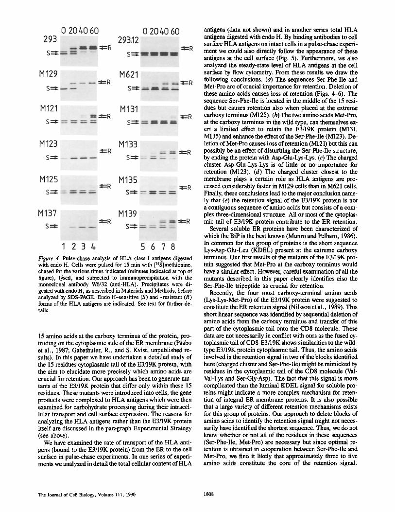
Figure 4. Pulse-chase analysis of HLA class I antigens digested with endo H. Cells were pulsed for 15 min with [35S]methionine, chased for the various times indicated (minutes indicated at top of figure), lysed, and subjected to immunoprecipitation with the monoclonal antibody W6/32 (anti-HLA). Precipitates were di- gested with endo H, as described in Materials and Methods, before analyzed by SDS-PAGE. Endo H-sensitive (S) and -resistant (R) forms of the HLA antigens are indicated. See text for further de- tails.
15 amino acids at the carboxy terminus of the protein, pro- truding on the cytoplasmic side of the ER membrane (P~i~ibo et al., 1987; Gabathuler, R., and S. Kvist, unpublished re- suits). In this paper we have undertaken a detailed study of the 15 residues cytoplasmic tail of the E3/19K protein, with the aim to elucidate more precisely which amino acids are crucial for retention. Our approach has been to generate mu- tants of the E3/19K protein that differ only within these 15 residues. These mutants were introduced into cells, the gene products were complexed to HLA antigens which were then examined for carbohydrate processing during their intracel- lular transport and cell surface expression. The reasons for analyzing the HLA antigens rather than the E3/19K protein itself are discussed in the paragraph Experimental Strategy (see above).
We have examined the rate of transport of the HLA anti- gens (bound to the E3/19K protein) from the ER to the cell surface in pulse-chase experiments. In one series of experi- ments we analyzed in detail the total cellular content of HLA
antigens (data not shown) and in another series total HLA antigens digested with endo H. By binding antibodies to cell surface HLA antigens on intact cells in a pulse-chase experi- ment we could also directly follow the appearance of these antigens at the cell surface (Fig. 5). Furthermore, we also analyzed the steady-state level of HLA antigens at the cell surface by flow cytometry. From these results we draw the following conclusions. (a) The sequences Ser-Phe-Ile and Met-Pro are of crucial importance for retention. Deletion of these amino acids causes loss of retention (Figs. 4-6). The sequence Ser-Phe-Ile is located in the middle of the 15 resi- dues but causes retention also when placed at the extreme carboxy terminus (M125). (b) The two amino acids Met-Pro, at the carboxy terminus in the wild type, can themselves ex- ert a limited effect to retain the E3/19K protein (M131, M135) and enhance the effect of the Ser-Phe-Ile (M123). De- letion of Met-Pro causes loss of retention (M121) but this can possibly be an effect of disturbing the Ser-Phe-Ile structure, by ending the protein with Asp-Glu-Lys-Lys. (c) The charged cluster Asp-Glu-Lys-Lys is of little or no importance for retention (M123). (d) The charged cluster closest to the membrane plays a certain role as HLA antigens are pro- cessed considerably faster in M129 cells than in M621 cells. Finally, these conclusions lead to the major conclusion name- ly that (e) the retention signal of the E3/19K protein is not a contiguous sequence of amino acids but consists of a com- plex three-dimensional structure. All or most of the cytoplas- mic tail of E3/19K protein contribute to the ER retention.
Several soluble ER proteins have been characterized of which the BiP is the best known (Munro and Pelham, 1986). In common for this group of proteins is the short sequence Lys-Asp-Glu-Leu (KDEL) present at the extreme carboxy terminus. Our first results of the mutants of the E3/19K pro- tein suggested that Met-Pro at the carboxy terminus would have a similar effect. However, careful examination of all the mutants described in this paper clearly identifies also the Ser-Phe-Ile tripeptide as crucial for retention.
Recently, the four most carboxy-terminal amino acids (Lys-Lys-Met-Pro) of the E3/19K protein were suggested to constitute the ER retention signal (Nilsson et al., 1989). This short linear sequence was identified by sequential deletion of amino acids from the carboxy terminus and transfer of this part of the cytoplasmic tail onto the CD8 molecule. These data are not necessarily in conflict with ours as the fused cy- toplasmic tail of CD8-E3/19K shows similarities to the wild- type E3/19K protein cytoplasmic tail. Thus, the amino acids involved in the retention signal in two of the blocks identified here (charged cluster and Ser-Phe-Ile) might be mimicked by residues in the cytoplasmic tail of the CD8 molecule (Val- Val-Lys and Ser-Gly-Asp). The fact that this signal is more complicated than the luminal KDEL signal for soluble pro- teins might indicate a more complex mechanism for reten- tion of integral ER membrane proteins. It is also possible that a large variety of different retention mechanisms exists for this group of proteins. Our approach to delete blocks of amino acids to identify the retention signal might not neces- sarily have identified the shortest sequence. Thus, we do not know whether or not all of the residues in these sequences (Ser-Phe-Ile, Met-Pro) are necessary but since optimal re- tention is obtained in cooperation between Ser-Phe-Ile and Met-Pro, we find it likely that approximately three to five amino acids constitute the core of the retention signal.
The Journal of Cell Biology, Volume 111, 1990 1808
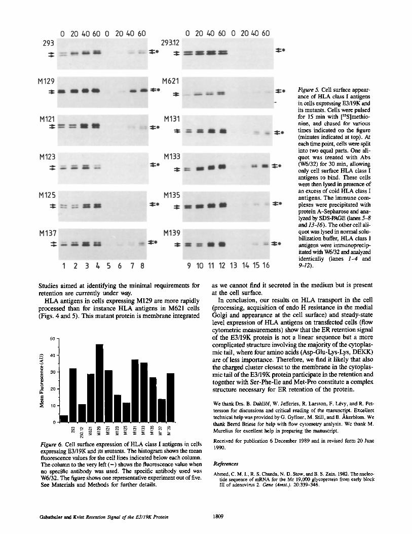
Figure 5. Cell surface appear- ance of HLA class I antigens in ceils expressing E3/19K and its mutants. Cells were pulsed for 15 min with [35Slmethio- nine, and chased for various times indicated on the figure (minutes indicated at top). At each time point, cells were split into two equal parts. One ali- quot was treated with Abs (W6/32) for 30 min, allowing only cell surface HLA class I antigens to bind. These cells were then lysed in presence of an excess of cold HLA class I antigens. The immune com- plexes were precipitated with protein A-Sepharose and ana- lyzed by SDS-PAGE (lanes 5-8 and 13-16). The other cell ali- quot was lysed in normal solu- bilization buffer, HLA class I antigens were immunoprecip- itated with W6/32 and analyzed identically (lanes 1-4 and 9-12).
Studies aimed at identifying the minimal requirements for retention are currently under way.
HLA antigens in cells expressing M129 are more rapidly processed than for instance HLA antigens in M621 cells (Figs. 4 and 5). This mutant protein is membrane integrated
50 ¸
4O
" 30
o 20 t~
o
Figure 6. Cell surface expression of HLA class I antigens in cells expressing E3/19K and its mutants. The histogram shows the mean fluorescence values for the cell lines indicated below each column. The column to the very left ( - ) shows the fluorescence value when no specific antibody was used. The specific antibody used was W6/32. The figure shows one representative experiment out of five. See Materials and Methods for further details.
as we cannot find it secreted in the medium but is present at the cell surface.
In conclusion, our results on H L A transport in the cell (processing, acquisition of endo H resistance in the medial Golgi and appearance at the cell surface) and steady-state level expression of HLA antigens on transfected cells (flow cytometric measurements) show that the ER retention signal of the E3/19K protein is not a linear sequence but a more complicated structure involving the majority of the cytoplas- mic tail, where four amino acids (Asp-Glu-Lys-Lys, DEKK) are of less importance. Therefore, we find it likely that also the charged cluster closest to the membrane in the cytoplas- mic tail of the E3/19K protein participate in the retention and together with Ser-Phe-Ile and Met-Pro constitute a complex structure necessary for ER retention of the protein.
We thank Drs. B. Dahll6f, W. Jefferies, R. Larsson, F. l~vy, and R. Pet- tersson for discussions and critical reading of the manuscript. Excellent technical help was provided by G. Gyllner, M. Still, and B./~kerblom. We thank Bernd Briese for help with flow cytometry analysis. We thank M. Murelius for excellent help in preparing the manuscript.
Received for publication 6 December 1989 and in revised form 20 June 1990.
References
Ahmed, C. M. I., R. S. Chanda, N. D. Stow, andB. S. Zain. 1982. The nucleo- tide sequence of rnRNA for the Mr 19,000 glycoprotein from early block III of adenovirus 2. Gene (Amst.). 20:339-346.
Gabathuler and Kvist Retention Signal of the E3/19K Protein 1809
Andersson, M., S. P~bo, T. Nilsson, and P. A. Peterson. 1985. Impaired in- tracellular transport of class I MHC antigens as a possible means for adenoviruses to evade immune surveillance. Cell. 43:215-222.
Arnold, B., H.-G. Burgert, U. Hamann, G. Hgnunerling, U. Kees, and S. Kvist. 1984. Cytolytic T cells recognize the two amino-terminal domains of H-2 antigens in tandem in influenza A infected cells. Cell. 38:79-87.
Barnes, W. M. 1980. DNA cloning with single stranded phage vectors. In Genetic Engineering. Vol. 2. J. K. Setlow and A. Hollaender, editors. Ple- num Publishing Corp., New York. 185-200.
Barnstable, C. J., W. F. Bodmer, G. Brown, G. Galfre, C. Milstein, A. F. Wil- hams, and A. Ziegler. 1978. Production of monoclonal antibodies to group A erythrocytes, HLA and other human cell surface antigens: new tools for genetic analysis. Cell. 14:9-20.
Braciale, T. J., L. A. Morrison, M. T. Sweetser, J. Sambrook, M. J. Gething, and V. L. Braciale. 1987. Antigen presentation pathways to class I and class II MHC-restricted T lymphocytes, lmmunol. Rev. 98:95-114.
Burgert, H.-G., and S. Kvist. 1985. An adenovirus type 2 glycoprotein blocks cell surface expression of human histocompatibility class I antigens. Cell. 41:987-997.
Burgert, H.-G., and S. Kvist. 1987. The E3/19K protein of adenovirus type 2 binds to the domains of histocompatibility antigens required for CTL recog- nition. EMBO (Fur. Mol. Biol. Organ.) J. 6:2019-2026.
Burgert, H.-G., J. L. Maryanski, and S. Kvist. 1987. ~E3/19K" protein of adenovirns type 2 inhibits lysis of cytolytic T lymphocytes by blocking cell surface expression of histocompatibility class I antigens. Proc. Natl. Acad. Sci. USA. 84:1356-1360.
Chin, D. J., G. Gil, D. W. Russell, L. Liscum, K. L. Luskey, S. K. Basu, H. Okayama, P. Berg, J. L. Golstein, and M. S. Brown. 1984. Nucleotide se- quence of 3-hydroxy-3-methyl-glutaryl coenzyme A reductase, a glycopro- tein of endoplasmic reticulum. Nature (Lond.). 308:613-617.
Crimaudo, C., M. Hortsch, H. Gansepohl, and D. I. Meyer. 1987. Human ribophorins I and II: the primary structure and membrane topology of two highly conserved rough endoptasmic reticulum specific glycoproteins. EMBO (Fur. Mol. Biol. Organ.)J. 6:75-82.
Doerfler, W. 1986. Adenovirus DNA. Dev. Mol. Virol. 8:223-246. Edman, J. C., L. Ellis, R. W. Blucher, R. A. Roth, and W. J. Rutter. 1985.
Sequence of protein disulfide isomerase and implications of its relationship to thioredoxin. Nature (Lond.). 317:267-270.
Evans, A. S. 1958. Latent adenovirus infections of the human respiratory tract. Am. J. Hyg. 67:256-266.
Flint, S. J. 1980. Structure and genomic organization of adenoviruses. In DNA Tumor Viruses..l. Tooze, editor. Cold Spring Harbor Laboratory, Cold Spring Harbor, NY. 383-575.
Gabathuler, R., F. L6vy, and S. Kvist. 1990. Requirements for the association of adenovirus type 2 E3/19K wild-type and mutant proteins with HLA anti- gens. J. Virol. 64:3679-3685.
Ginsberg, H. S. 1984. The Adenovirnses. H. S'. Ginsberg, editor. Plenum Pub- lishlng Corp., New York. 205-258.
Graham, F. L., J. Smiley, W. C. Russell, and R. Nairn. 1977. Characterization of a human cell line transformed by DNA from human adenovirus type 5. J. Gen. Virol. 36:59-72.
Hortsch, M., S. Labeit, and D. I. Meyer. 1988. Complete cDNA sequence cod- ing for the human docking protein. Nucleic Acids Res. 16:361-362.
K~znpe, O., D. Bellgrau, U. Hammerling, P. Lind, S. P~ibo, L. Severinsson, and P. A. Peterson. 1983. Complex formation of class I transplantation anti- gens and a viral glycoprotein. J. Biol. Chem. 258:10594-10598.
Klein, J. 1979. The major histocompatibility complex of the mouse. Science (Wash. DC). 203:516-521.
Kornfeld, R., and W. S. M. Wold. 1981. Structures of the oligosaccharides of the glycoprotein coded by early region E3 of adenovirus 2. J. Virol. 40:440-449.
Kvist, S., L. Ostberg, H. Persson, L. Philipson, and P. A. Peterson. 1978. Mo- lecular association between transplantation antigens and a cell surface anti- gen in an adenovirus-transformed cell line. Proc. Natl. Acad. Sci. USA. 75:5674-5678.
Kvist, S., K. Wiman, L. Claesson, P. A. Peterson, and B. Dobberstein. 1982. Membrane insertion and oligomeric assembly of HLA-DR histocompatibil- ity antigens. Cell. 29:61-69.
Lee, A. S., J. Bell, and J. Ting. 1984. Biochemical characterization of the 94-
and 78-kilodalton glucose-regulated proteins in hamster fibroblasts. J. Biol. Chem. 259:4616--4621.
Left, T., R. Elkaim, C. R. Goding, P. Jalinot, P. Sassone-Corsi, M. Per- ricaudet, C. K6dinger, and P. Chambon. 1984. Individual products of the adenovirus 12S and 13S EIa mRNAs stimulate viral EIIa and EIII expression at the transcriptional level. Proc. Natl. Acad. Sci. USA. 81:4381--4385.
Munro, S., and H. R. B. Pelham. 1986. An Hsp70-protein in the ER: identity with the 78 kd glucose-regulated protein and immunoglobulin heavy chain binding protein. Cell. 46:291-300.
Munro, S., and H. R. B. Pelham. 1987. A C-terminal signal prevents secretion of luminal ER proteins. Cell. 48:899-907.
Nilsson, T., M. Jackson, and P. A. Peterson. 1989. Short cytoplasmic se- quences serve as retention signals for transmembrane proteins in the en- doplasmic reticulum. Cell. 58:707-718.
P~i~bo, S., F. Weber, T. Nilsson, W. Schaffner, and P. A. Peterson. 1986. Structural and functional dissection of an MHC class I antigen-binding adenovirus glycoprotein. EMBO (Fur. Mol. Biol. Organ.)J. 5:1921-1927.
P[iibo, S., B. M. Bhat, W. S. M. Wold, and P. A. Peterson. 1987. A short sequence in the COOH-terminus makes an adenovirus membrane glycopro- tein a resident of the endoplasmic reticulum. Cell. 50:311-317.
Persson, H., C. Sign~is, and L. Philipson. 1979. Purification and characteriza- tion of an early glycoprotein from Ad-2 infected cells. J. Virol. 29:939-948.
Persson, H., M. Jansson, and L. Philipson. 19g0a. Synthesis and genomic site for an adenovirus type 2 early glycoprntein. J. Mol. Biol. 136:375-394.
Persson, H., H. J6rnvall, and J. Zabielski. 1980b. Multiple mRNA species for the precursor to an adenovirus-encoded glycoprotein: identification and structure of the signal sequence. Proc. Natl. Acad. Sci. USA. 77:6349-6353.
Petrie, B. L., H. B. Greenberg, D. Y. Graham, and M. K. Estes. 1984. Ultra- structural localization of rotavirus antigens using colloidal gold. Virus Res. 1:133-152.
Ploegh, H. L., H. T. Orr, and J. L. Strominger. 1981. Major histocompatibility antigens: the human (HLA-A,-B,-C) and mudne (H-2K, H-2D) class I mole- cules. Cell. 24:287-~299.
Poruchynsky, M. S., and P. H. Atkinson. 1988. Primary sequence domains re- quired for the retention of rotavirus VP7 in the endoplasmic reticulum. J. Cell Biol. 107:1697-1706.
Ross, S., and A. J. Levine. 1979. The genomic map position of the adenovirus type 2 glycoprotein. Virology. 99:427-430.
Sakagushi, M., K. Mihara, and R. Sato. 1987. A short amino-terminal segment of microsomal cytochrome P-450 functions both as an insertion signal and as a stop-transfer sequence. EMBO (Eur. Mol. Biol. Organ.) J. 6:2425- 2431.
Sbenk, T., and J. Williams. 1984. Genetic analysis of adenoviruses. Curt. Top. Microbiol. lmmunol. 111 : 1-39.
Sign/is, C., M. G. Katze, H. Persson, and L. Philipson. 1982. An adenovirus glycoprotein binds heavy chains of class I transplantation antigens from man and mouse. Nature (Lond.). 299:175-178.
Southern, P. J., and P. Berg. 1982. Transformation of mammalian cells to an- tibiotic resistance with a bacterial gene under control of the SV40 early re- gion promoter. J. Mol. Appl. Genet. 1:327-341.
Stirzaker, S. C., and G. W. Both. 1989. The signal peptide of the rotavirus gly- coprotein VP7 is essential for its retention in the ER as an integral membrane protein. Cell. 56:741-747.
Townsend, A. R. M., J. Rothbard, F. M. Gotch, G. Bahadur, D. Wraith, and A. J. McMichael. 1986. The epitopes of influenza nucleoprotein recognized by cytolytic T lymphocytes can be defined with short synthetic peptides. Cell. 44:959-968.
Whitfeld, P. L., C. Tyndall, S. C. Stirzaker, A. R. Bellamy, and G. Both. 1987. Location of sequences within rotavirus SA 11 glycoprotein VF'/which direct it to the endoplasmic reticulum. Mol. Cell. Biol. 7:2491-2497.
Wold, W. S. M., C. Cladaras, S. L. Deutscher, and Q. S. Kapoor. 1985. The 19 KDa glycoprotein coded by region E3 of adenovirus. J. Biol. Chem. 260:2424-2431.
Zinkernagel, R. M., and P. C. Doherty. 1979. MHC-restricted cytotoxic T cells: studies on the biological role of polymorphic major transplantation an- tigens determining T-cell restriction: specificity, function, and responsive- ness. Adv. lmmunol. 27:51-177.
Zoller, M. J., and M. Smith. 1983. Oligonucleotide-directed mutagenesis of DNA fragments cloned into MI3 vectors. Methods Enzymol. 100:468-500.
The Journal of Cell Biology, Volume 111, 1990 1810


![Endoplasmic reticulum[1]](https://img.pdfslide.us/doc/110x75/58ed5fc71a28aba1678b4611/endoplasmic-reticulum1.jpg)