Embed Size (px)
Citation preview
The Elbow
Anatomy of the Elbow
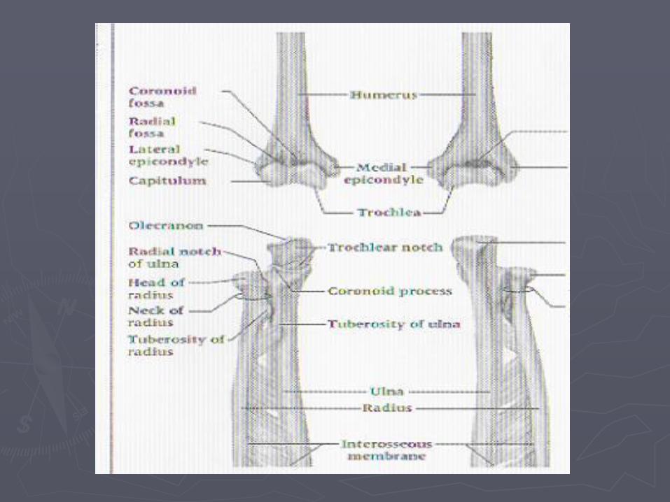
Bones
►Humerus
►Radius
►Ulna
What Motions Does the Elbow Perform?
► Flexion
► Extension
► Pronation
► Supination
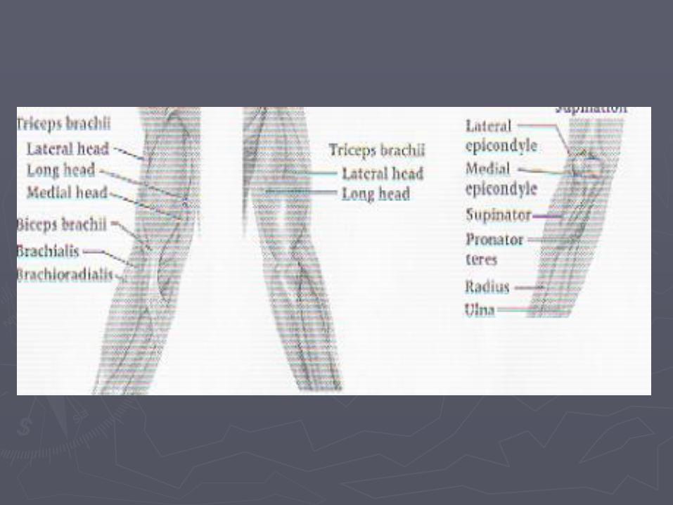
Muscles in Motion
►FLEXION
Biceps brachii (supinated)
Brachialis (pronated)
Brachioradialis (neutral)
Prime movers depends on position of the forearm
►EXTENSION
Triceps brachii
►Primary mover
Anconeus
►Secondary mover
►SUPINATION
Supinator
►Primary mover
Biceps brachii
►Secondary mover
Brachioradialis
►Secondary mover
►Also pronates forearm from a supinated position
►PRONATION
Pronatror teres
Pronator quadratus
►Primary movers
Tendons
►Biceps
►Triceps
►Flexor tendon group
►Extensor tendon group
Joints
► Humeroulnar joint
Humerus and ulna
Allows for flexion and extension
► Humeroradial joint
Humerus and radius
Flexion and extension
Pronation and supination
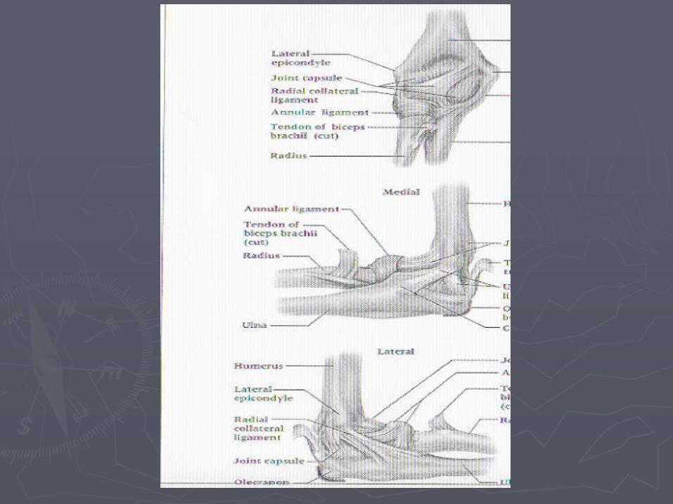
Ligaments
Medial – resists valgus stress
► ulnar collateral ligament (UCL)
three bands
anterior oblique band
► medial epicondyle to coronoid process
► resists against valgus stress
transverse band
► medial epicondyle to coronoid process
posterior oblique band
► medial epicondyle to olecranon process
Lateral – resists varus stress►Lateral Collateral Ligament (LCL)
Main lateral stabilizer Middle of the lateral epicondyle to ulnar
tuberacle►Radial collateral ligament
Thickened capsule Lateral epicondyle to annular ligament Maintain close relationship between
humeral head and radial head
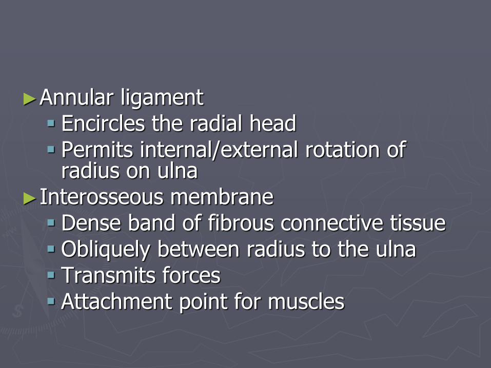
►Annular ligament Encircles the radial head Permits internal/external rotation of
radius on ulna►Interosseous membrane
Dense band of fibrous connective tissue Obliquely between radius to the ulna Transmits forces Attachment point for muscles
Assessment of the Elbow
►History
Past history
Mechanism of injury
When and where does it hurt?
Motions that increase or decrease pain
Type of, quality of, duration of, pain?
Sounds or feelings?
How long were you disabled?
Swelling?
Previous treatments?
►Observations
Deformities and swelling?
Carrying angle►Cubitus valgus versus cubitus varus
Flexion and extension►Cubitus recurvatum
Elbow hyperextension?
►Palpation
Be sure to check sites of pain and deformity
Assess epicondyles, olecranon, distal aspect of humerus and proximal aspect of ulna
Soft tissue – muscles, tendons, joint capsules and ligaments surrounding joint
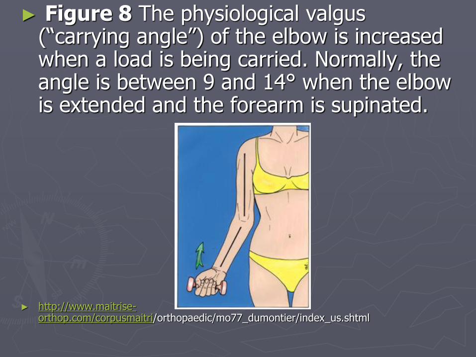
► Figure 8 The physiological valgus (“carrying angle”) of the elbow is increased when a load is being carried. Normally, the angle is between 9 and 14° when the elbow is extended and the forearm is supinated.
► http://www.maitrise-orthop.com/corpusmaitri/orthopaedic/mo77_dumontier/index_us.shtml
Special tests
►Methods to assess elbow and forearm injuries include:
ROM test for elbow flexion
ROM test for elbow extension
ROM test for elbow supination
ROM test for elbow pronation
Manual Muscle Tests for the Elbow
►Flexion strength test
►Extension strength test
►Supination strength test
►Pronation strength test
Prevention of Elbow, Forearm, and Wrist Injuries
►Vulnerable to a variety of acute and chronic injuries
►Protective gear is always recommended to reduce severity of injury
►Chronic injury reduction
Limit repetitions (baseball, tennis)
Utilize proper mechanics
Use equipment that is appropriate for skill level
Maintain appropriate levels of strength, flexibility, and endurance for activity
Injuries to the Elbow and Arm
►Fractures
►Dislocations and subluxations
►Contusions
►Sprains
Valgus stress test for the elbow
Varus stress test for the elbow
Injuries to the Elbow and Arm
►Impingement
Tinel’s sign
►Synovitis and bursitis
►Biceps brachii rupture
►Epicondylitis
►Volkmann’s contracture
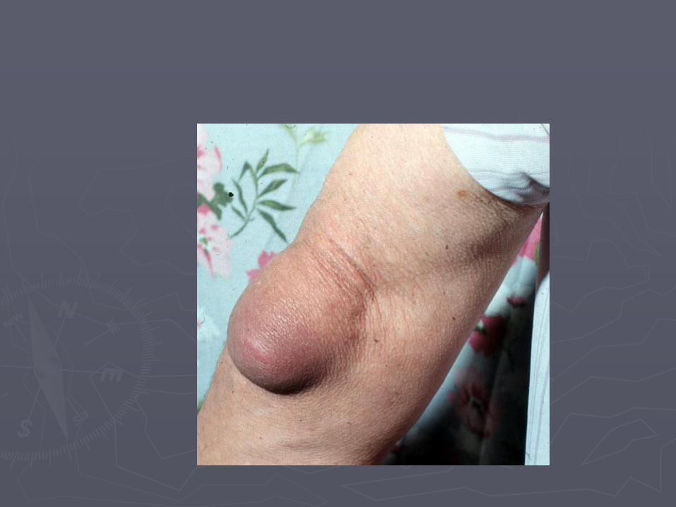
Common Injuries►Olecranon Bursitis
Cause of Injury
►Superficial location makes it extremely susceptible to injury (acute or chronic) --direct blow
Signs of Injury
►Pain, swelling, and point tenderness
►Swelling will appear and w/out usual pain and heat
►Contusion
Cause of Injury
►Vulnerable area due to lack of padding
►Result of direct blow or repetitive blows
Signs of Injury
►Swelling (rapidly after irritation of bursa or synovial membrane)
Care
►Treat w/ RICE immediately for at least 24 hours
►If severe, refer for X-ray to determine presence of fracture
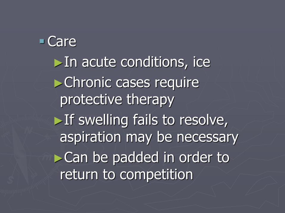
Care
►In acute conditions, ice
►Chronic cases require protective therapy
►If swelling fails to resolve, aspiration may be necessary
►Can be padded in order to return to competition
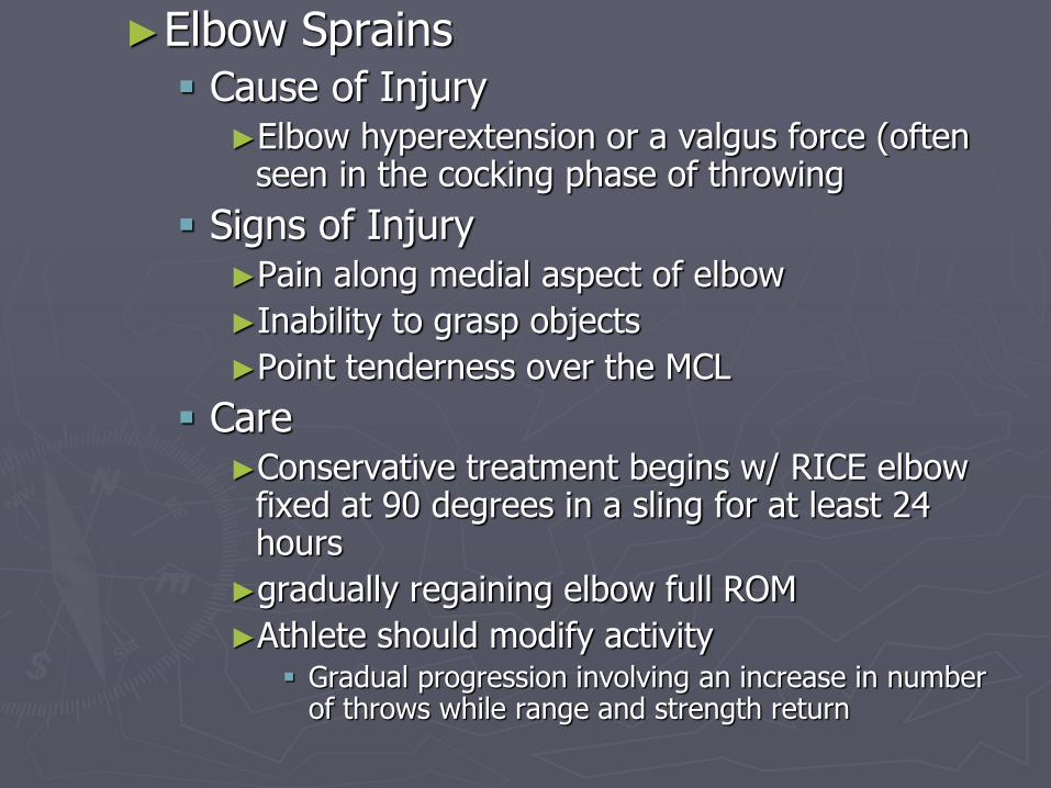
►Elbow Sprains Cause of Injury
►Elbow hyperextension or a valgus force (often seen in the cocking phase of throwing
Signs of Injury►Pain along medial aspect of elbow
►Inability to grasp objects
►Point tenderness over the MCL
Care►Conservative treatment begins w/ RICE elbow
fixed at 90 degrees in a sling for at least 24 hours
►gradually regaining elbow full ROM
►Athlete should modify activity Gradual progression involving an increase in number
of throws while range and strength return
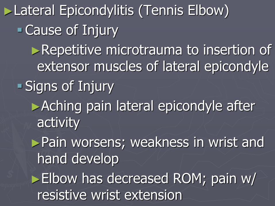
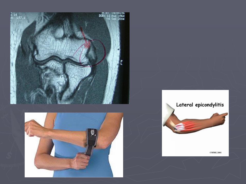
►Lateral Epicondylitis (Tennis Elbow)
Cause of Injury
►Repetitive microtrauma to insertion of extensor muscles of lateral epicondyle
Signs of Injury
►Aching pain lateral epicondyle after activity
►Pain worsens; weakness in wrist and hand develop
►Elbow has decreased ROM; pain w/ resistive wrist extension
►Lateral Epicondylitis (continued)
Care
►RICE, NSAID’s and analgesics
►ROM exercises and PRE, deep friction mass., avoidance of pronation motions
►Mobilization and stretching in pain free ranges
►Use of a counter force or neoprene sleeve
►Proper mechanics and equipment instruction is critically important
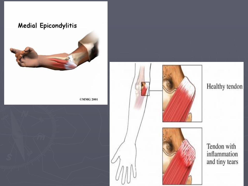
►Medial Epicondylitis Cause of Injury
►Repeated forceful flexion of wrist and extreme valgus torque of elbow
Signs of Injury►Pain produced w/ forceful flexion or extension
►Point tenderness and mild swelling
►Passive movement of wrist seldom elicits pain, but active movement does
Care►Sling, rest, cryotherapy or heat through
ultrasound
►Analgesic and NSAID's
►Curvilinear brace below elbow to reduce elbow stressing
►Severe cases may require splinting and complete rest for 7-10 days
►Elbow Osteochondritis Dissecans
Cause of Injury
►Impairment of blood supply to anterior surface resulting in degeneration of articular cartilage, and bone creating loose bodies within the joint
Signs of Injury
►Sudden pain, locking; range usually returns in a few days
►Swelling, pain and crepitation may also occur
Care
►If repeated locking occurs, loose bodies may be removed surgically
►Without removal, arthritis may develop
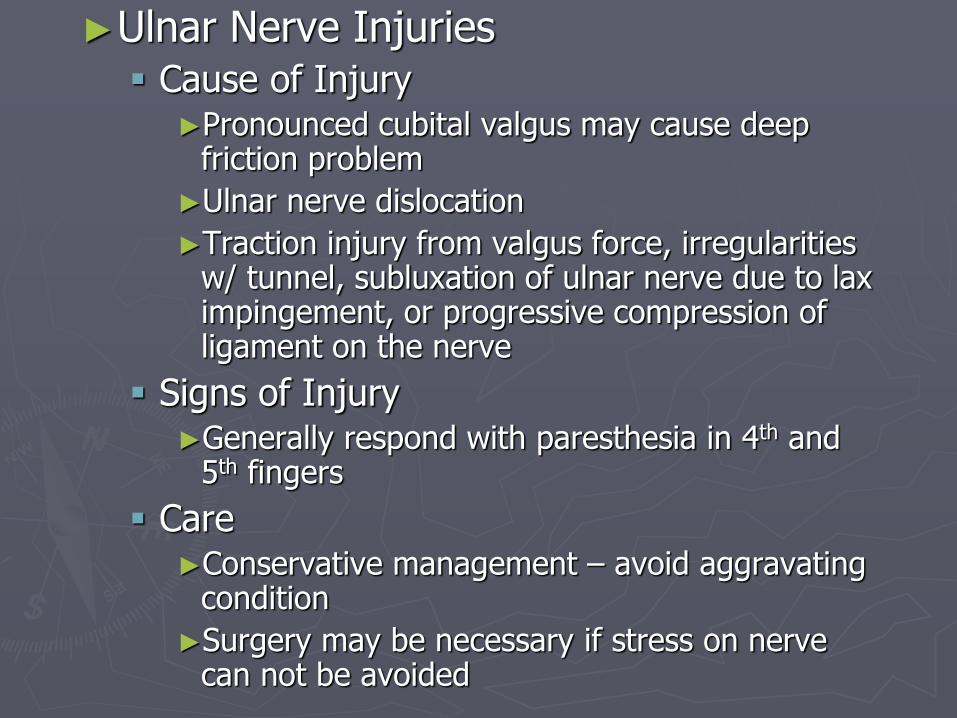
►Ulnar Nerve Injuries Cause of Injury
►Pronounced cubital valgus may cause deep friction problem
►Ulnar nerve dislocation
►Traction injury from valgus force, irregularities w/ tunnel, subluxation of ulnar nerve due to lax impingement, or progressive compression of ligament on the nerve
Signs of Injury ►Generally respond with paresthesia in 4th and
5th fingers
Care►Conservative management – avoid aggravating
condition
►Surgery may be necessary if stress on nerve can not be avoided
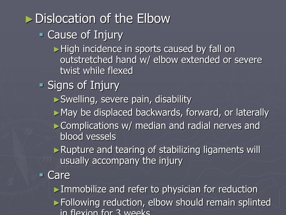
►Dislocation of the Elbow
Cause of Injury ►High incidence in sports caused by fall on
outstretched hand w/ elbow extended or severe twist while flexed
Signs of Injury►Swelling, severe pain, disability
►May be displaced backwards, forward, or laterally
►Complications w/ median and radial nerves and blood vessels
►Rupture and tearing of stabilizing ligaments will usually accompany the injury
Care►Immobilize and refer to physician for reduction
►Following reduction, elbow should remain splinted in flexion for 3 weeks
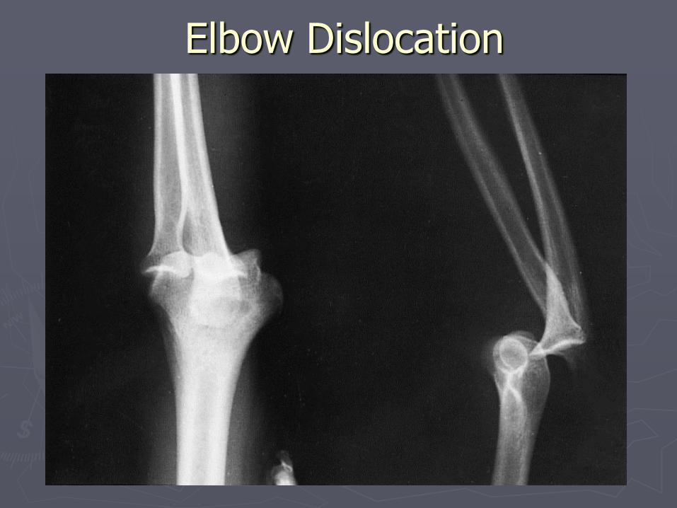
Elbow Dislocation
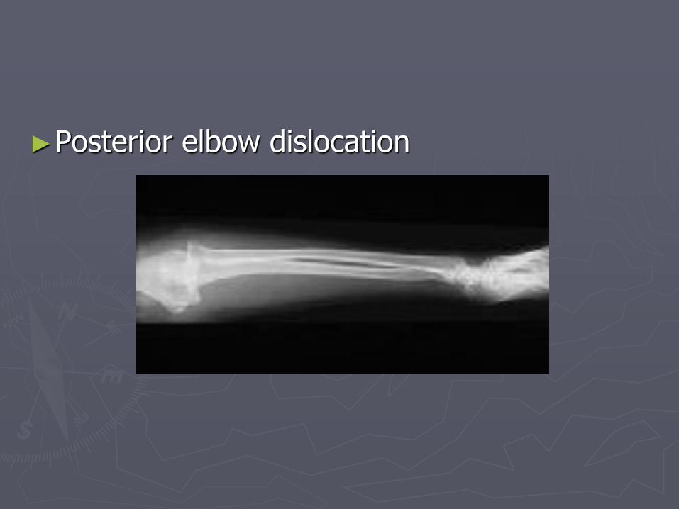
►Posterior elbow dislocation
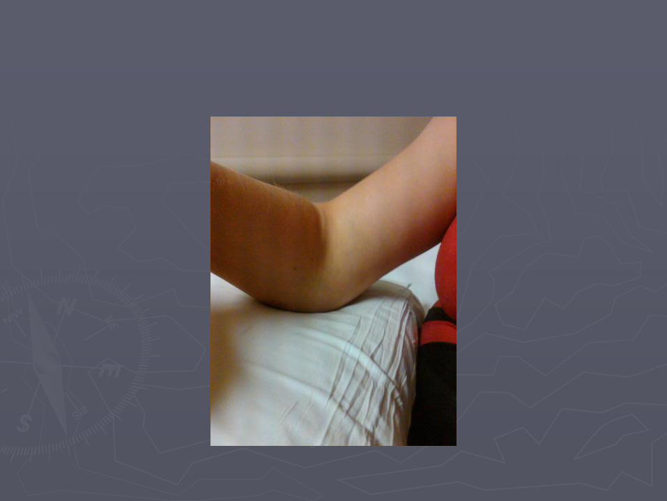
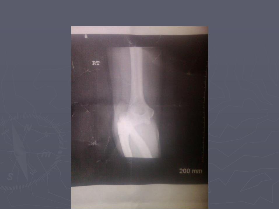
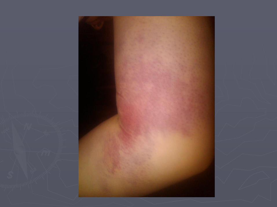
►Fractures of the Elbow
Cause of Injury ►Fall on flexed elbow or from a direct blow
►Fracture can occur in any one or more of the bones
►Fall on outstretched hand often fractures humerus above condyles or between condyles
Signs of Injury ►May or may not result in visual deformity
►Hemorrhaging, swelling, muscle spasm
Care►Ice and sling for support – refer to physician
►Video
►Hungarian wt lifter
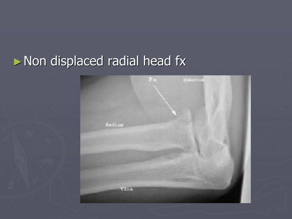
►Non displaced radial head fx
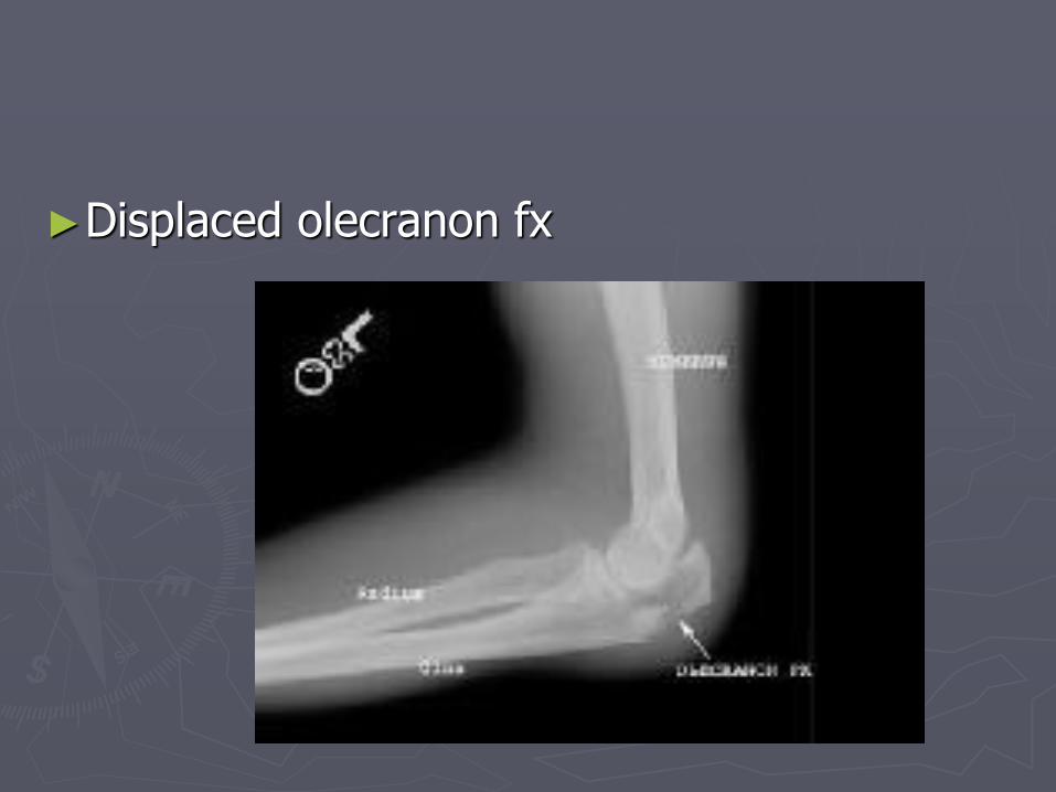
►Displaced olecranon fx
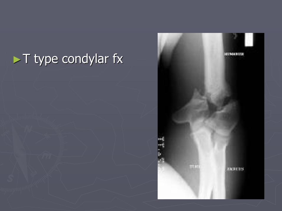
►T type condylar fx
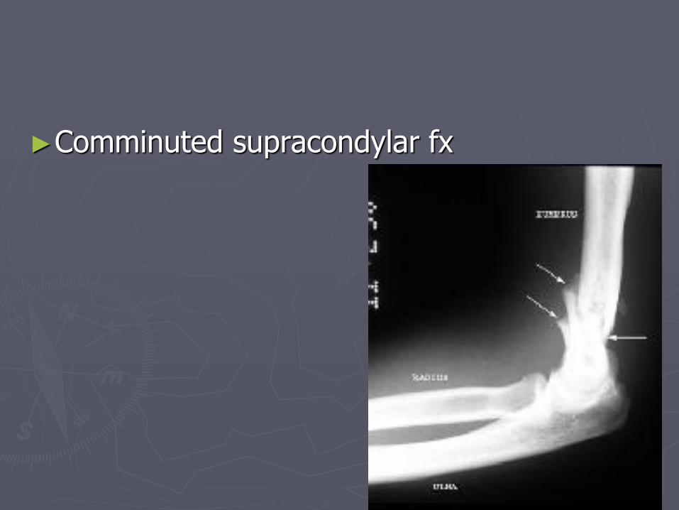
►Comminuted supracondylar fx
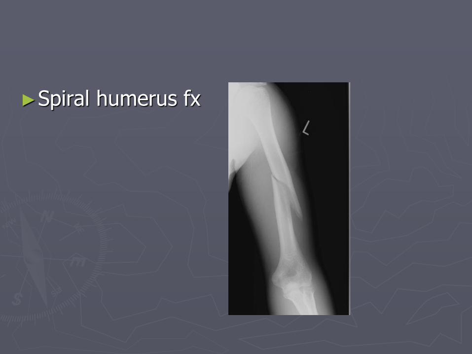
►Spiral humerus fx