Embed Size (px)
Citation preview

The effects of norepinephrine microinjections into the central
nucleus of the amygdala on serotonergic activity in the
dorsal raphe nucleus and fear-related behavior
Erika Kolisch
Department of Integrative Physiology
University of Colorado at Boulder
Defended April 8, 2013
Thesis Advisor
Dr. Christopher Lowry, Department of Integrative Physiology and the Center for Neuroscience
Committee Members
Dr. David Sherwood, Department of Integrative Physiology
Dr. Paul Strom, Honors Program Faculty

2
ABSTRACT The central nucleus of the amygdala (CE) projects to target areas that elicit autonomic and behavioral symptoms of fear. It has been shown that in stressful or fearful paradigms, endogenous norepinephrine (NE) is released into the CE. Microinjections of NE into the CE could activate fear circuits in the brain and lead to fear-like behavior. The CE projects to an area of the brain called the dorsal raphe nucleus (DR), specifically the dorsal raphe nucleus, ventrolateral part (DRVL) and the adjacent ventrolateral periaqueductal gray (VLPAG). We hypothesize that NE binds to adrenergic receptors in the CE to stimulate corticotropin-releasing factor (CRF) projections to the DR, where CRF binds to CRF type 2 receptors (CRFR2), which can elicit freezing behavior through serotonergic projections to a multisynaptic circuit controlling somatomotor neurons in the spinal cord. To test this hypothesis, we gave bilateral microinjections of NE (250 nmoles in 0.2 µL per side) into the CE and then immediately measured freezing behavior using the open-field test paradigm. Norepinephrine microinjections into the CE increased both the frequency and duration of freezing behavior. As the project is currently in the process of completion, we hope to also show that serotonergic neurons in the DR were activated in projection areas associated with previously established fear circuitry. This study was conducted to gain insight into control of serotonergic pathways that could lead to advancement of our understanding of stress-related mental illnesses.

3
Background
Stress-related neuropsychiatric disorders affect millions of people each year and represent a significant public health problem in the United States (NIMH, 2005). Mechanisms through which stress activates serotonergic systems are thought to be central to both the etiology and pathophysiology of stress-related mental disease (Hale, Shekhar, Lowry, 2012). It is paramount that we continue to conduct this research to attempt to mitigate the personal and societal detriment of mental disease.
Dysregulation of serotonergic pathways is thought to be a major contributor to the development of stress-related psychiatric disorders (Owens & Nemeroff, 1994). Evidence in humans suggests that those with stress-related neuropsychiatric disorders have abnormal serotonin (5-HT) systems as indicated by a four-fold increase in brain serotonin turnover when compared to healthy controls (Esler et al., 2007). Anxiety is a complex emotional state associated with heightened physiological and behavioral arousal (Lowry et al., 2005). Although the mechanisms underlying the regulation of anxiety-states are not well understood, the physiological and behavioral arousal appears to be controlled by a distributed and interconnected system of forebrain and hindbrain structures (Singewald, Salchner, Sharp, 2003; Singewald and Sharp, 2000). The central amygdaloid nucleus (CE) and the dorsal raphe nucleus (DR) are two structures critically involved in the control of anxiety states and anxiety-related behaviors (Lowry et al., 2005, 2008). The emotional state of anxiety is characterized by concern about the uncertainty of positive or negative outcomes of future events and has many of the same signs and symptoms of fear, which involves a feeling of impending doom (Davis, Walker, Lee, 1997).
It is also well known that the CE is critically involved in fear-like behaviors (Campeau & Davis, 1995). Lesions of the CE will block fear-like behaviors such as freezing and fear-potentiated startle in rats (Davis, Walker, Lee, 1997; Kim & Davis 1986). More recently this has also been seen in humans. Patients suffering from Urbach-Wiethe disease show bilateral amygdala lesions and subsequently fail to recognize fearful faces (Adolphs & Tranel, 2000) and fail to display fear during a fear-provoking event, such as a life-threatening traumatic event (Feinstein et al., 2011).
In a fearful or a fight-or-flight situation, stress hormones, neurohormones, neuromodulators, and neurotransmitters are released and have a large effect on behavioral and physiological responses to these types of stimuli. Norepinephrine (NE) is a catecholamine that is released in stressful situations and can act as both hormone (i.e., into the blood) and a neurotransmitter. This dual role allows NE to come in contact with a number of different tissues and NE has complex neuronal networks in the brain. Norepinephrine is endogenously released in the CE during a fearful event (Singewald, Kaehler, Philippu, 1999). Norepinephrine in the CE increases expression of c-Fos, the protein product of the immediate-early gene, c-fos, and a marker of neuronal activity (Singewald, Kaehler, Philippu, 1999). Previous research has shown that an increase in amygdala NE is mediated by influences involving the nucleus of the solitary tract (nTS) (Williams et al, 1998), which contains large numbers of norepinephrine synthesizing neurons (specifically, the A2 noradrenergic cell group). Retrograde tracing has confirmed that norepinephrine is the major neurotransmitter in projection neurons from the nTS to the amygdala (William et al., 1998). The nTS is associated with neuroendocrine changes in response to emotionally arousing experiences.
In addition, injections of corticotropin-releasing factor (CRF), an anxiety- and stress-related neuropeptide, into the DR induce a freezing response by the activation of corticotropin-releasing factor type 2 receptors (CRFR2) (Forster et al., 2006). Activation of CRFR2 in the DR enhances firing rates of 5-HT neurons (Pernar et al., 2004). CRF appears to be an important modulator of serotonergic neuronal activity (Lowry et al., 2000; Kirby et al., 2000; Price et al., 1998). Further, activation of the DR by CRFR2 can be blocked with a CRFR2 antagonist injected into the DR (Forster et al., 2008), suggesting that the CE is activating the DR through CRF projection neurons. We hypothesize that the CRF-induced freezing response is mediated by serotonergic projections from the DR to a multi-synaptic pathway terminating on somatomotor neurons in the spinal cord. These serotonergic projections control vestibulospinal reflexes that could ultimately control the paralysis of movement seen in a freezing response (Yates et al., 1993). Figure 1. details a proposed circuit of fear.

4
Figure 1. Hypothetical model illustrating proposed fear circuitry that we hypothesize is initiated by microinjections of norepinephrine (NE) into the central nucleus of the amygdala (CE). Through this circuit, NE acts to elevate corticotropin-releasing factor (CRF) release (represented by orange projections) in the dorsal raphe nucleus, ventrolateral part (DRVL) binding to corticotropin-releasing factor type 2 receptors (CRFR2) in the dorsal raphe nucleus (DR). This cascade leads to an activation of fear circuitry and fear-like behavior such as freezing. Abbreviations: CE, central nucleus of the amygdala; CRF, corticotropin-releasing factor; CRFR2, corticotropin-releasing factor type 2 receptors; DPAG, dorsal periaqueductal gray; DR, dorsal raphe nucleus; DRVL, dorsal raphe nucleus, ventrolateral part; NE, norepinephrine. (Paxinos and Watson, 1998)
To test this hypothesis it is important to look at the effects of intra-CE microinjections of NE on 5-HT activity in the DR and associated brain areas in the fear circuit, which will aide in developing a better understanding of stress-related mental disease.
The measurement of freezing, a fear-like behavior in rodents (Blanchard, Flannelly, Blanchard, 1986), provides an indication of fear induced by intra-CE NE injections. Freezing is defined as the absence of all visible movement, except for movements associated with respiration for at least 3 seconds (Pugh et al., 1998; Martinez, De Oliveira, Brandão, 2006). Freezing behavior is tested using the open-field paradigm where an animal is placed in a novel environment and desired behaviors are recorded and scored (Denenberg, 1969; Walsh & Cummins, 1976). This paradigm is designed to assess conflict anxiety and is based on a conflict between the animal’s internal drive to explore a novel environment (based on the potential for rewarding outcomes) versus the internal drive to avoid a novel environment (based on the potential for aversive outcomes) (Bouwknecht et al., 2007). Since rodents are nocturnal they tend to avoid brightly-lit places. They also dislike wide-open spaces and prefer to stay close to vertical references such as walls, an innate behavioral response referred to as thigmotaxis (Treit & Fundytus, 1988). Freezing behavior can occur at any location in the open-field and is considered an indication of fear.
We hypothesize that rats receiving intra-CE NE microinjections will display increased freezing behavior. This is proposed to be dependent upon the activation of CRF projections from the CE to the DR
5
and serotonergic projections from the DR, via multisynaptic pathways, to the spinal cord in response to an NE microinjection. We propose that this mechanism will increase freezing behavior and increase the amount of 5-HT metabolism in serotonergic projection areas.
Methods Animals
Adult male Sprague Dawley rats (275-300 g; Charles River, Kingston, NY, USA) were maintained under standard environmental conditions on a 12 hour light-dark cycle. Upon arrival, rats were pair-housed in standard polycarbonate cages (38 cm W x 48 cm L x 21 cm H; Techniplast cages, Techniplast, Kettering, UK). Food (Cat. No. 8640; Teklad 22/5 Rodent diet, Harlan Laboratories) and tap water were provided ad libitum. Following surgery for intra-CE cannulation, rats were singly housed in the same room using the same conditions listed above. All animal experiments were conducted in accordance with the National Institute of Health Guide for the Care and Use of Laboratory Animals, Eighth Edition (Institute for Laboratory Animal Research, The National Academies Press, Washington, D.C., 2011) and were approved by the University of Colorado Institutional Animal Care and Use Committee. All possible efforts were made to minimize the number of animals used and their suffering.
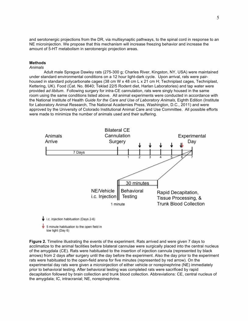
Figure 2. Timeline illustrating the events of the experiment. Rats arrived and were given 7 days to acclimatize to the animal facilities before bilateral cannulae were surgically placed into the central nucleus of the amygdala (CE). Rats were habituated to the insertion of injection cannula (represented by black arrows) from 2 days after surgery until the day before the experiment. Also the day prior to the experiment rats were habituated to the open-field arena for five minutes (represented by red arrow). On the experimental day rats were given a microinjection of either vehicle or norepinephrine (NE) immediately prior to behavioral testing. After behavioral testing was completed rats were sacrificed by rapid decapitation followed by brain collection and trunk blood collection. Abbreviations: CE, central nucleus of the amygdala; IC, intracranial; NE, norepinephrine.

6
Surgery
Seven days prior to the NE microinjections and behavior tests bilateral intra-CE cannula were stereotaxically placed in each animal. Rats were anesthetized with isoflurane (2-5%, Cat. No. 502017; VET ONE distributed by MWI Veterinary Supply; Meridian, ID, USA). The scalp was shaved and a small incision (approximately 7.5 mm; caudal to rostral) was made using a scalpel. The tissue was separated using a scalpel and, using a Dremel 300, small burr holes were drilled into the skull and the guide cannulae (Cat No. C315G/Spc, cut 8 mm below pedestal, Plastics One, Roanoke, VA, USA) were positioned at the following coordinates AP: –2.12 mm, DV: –7.1 mm, ML: ± 3.8 mm, relative to bregma (Figure 3). Cannulae were secured to the skull using a skull screw and dental cement. Dummy cannulae (Cat No. C315DC/Spc, cut 8 mm below pedestal with no projection outside the guide cannula, Plastics One) were placed in the guide cannulae to maintain patency.
Following surgery, rats were placed into a warm environment and monitored until they were fully awake. Postsurgical analgesics (Loxicom, 5 mg/kg, s.c., Cat No. NDC 55529-040-10, Norbrook, Newry, Northern Ireland, UK) were administered. Once awake the rats were allowed to recover in their home cages for 7 days prior to behavioral testing. The rats had one full day for recovery with no handling and were habituated for less than 10 minutes each day for the following 6 days before behavioral testing. Habituation to intracranial injections consisted of removing the rats from their home cage and placing them onto the lab bench. Rats were placed under a blue towel and the dummy cannula were briefly removed and replaced. Rats were then placed back into their home cages. Habituation to the open-field arena consisted of removing the rats from their home cages and then placing them into the open-field arena for 5 minutes at the same time and in the same conditions as the when the behavioral testing was executed. Rats were then placed back into their home cages.
Figure 3. Figure illustrating the desired cannula placement for stereotaxic surgery. The central nucleus of the amygdala, medial part, was the desired target. This area of the amygdala contains corticotropin-releasing factor (CRF) projections to the dorsal raphe nucleus (DR). The black dot represents the end of the guide cannula, which is 1 mm from the red dot, which represents the end of the injection cannula. Abbreviations: CRF, corticotropin-releasing factor; DR, dorsal raphe nucleus. Behavioral Procedures
Rats were randomly assigned to two treatment groups, 250 nmol NE (Stone et al., 1997) (n=5) or vehicle (n=5), and tested in an open-field arena (36”L x 36”W x 12” H) under low light, familiar conditions

7
(8-10 Lux) (Bouwknecht et al., 2007). Treatment groups were run in a random order starting at 0900 h to mitigate any potential effects of circadian levels of endogenous corticosterone (CORT).
Rats received intra-CE microinjections of either vehicle (aCSF) (0.402 g KCL, 17.18 g NaCl, 0.248 g NaH2PO4, 0.398 g Na2HPO4, 0.203 g MgCl2, and 0.176 g CaCl2 in 1 liter of distilled water, pH 7.2-7.4) or NE (250 nmol; 0.2 µl/ min per side; total volume of 0.2 µl/ min per side) (L-Noradrenaline L-hydrogentartrate, Sigma-Aldrich, St. Louis, MO, USA) immediately before rats were placed singly in an open-field arena. Behavior was videotaped for 30 minutes to be analyzed for freezing behavior. Rats were then placed in DecapiCones (Cat No. DC200, Braintree Scientific, Braintree, MA, USA) in preparation for rapid decapitation. After rapid decapitation, trunk blood was collected and brains were removed and flash frozen on dry ice.
Cannula Placement Verification Brain tissue was sectioned at 30 µm using a cryostat (Leica CM1900, North Central Instruments, Denver, CO, USA). The 30 µm sections were mounted on slides (VistaVision HistoBond slides, Cat No. 16004-406, VWR, Radnor, PA, USA) then stained using cresyl-violet and viewed under a microscope in order to verify that bilateral cannulae were placed in the CE. Behavior Analysis Behavior videos were coded and the analysis was performed blind to treatment groups. The first 5 minutes of the videos were scored for instances and duration of freezing behavior using Observer XT 10 (Noldus, Wageningen, Netherlands). Freezing behavior was defined as the absence of all movement for at least 3 seconds except those required for respiration (Pugh et al., 1998; Martinez, De Oliveira, Brandão, 2006). Statistics A Student’s t-test (SPSS 21, IBM, Armonk, NY, USA) was used to compare the freezing instances and duration between the treatment groups. Significance was determined at an α level of p < 0.05. Results Microinjections of NE into the CE produced significant differences in the number of freezing events and duration of freezing behavior between treatment groups. Rats were only included in the analysis if bilateral cannulae were placed in the CE. Figure 4. shows all of the cannula placements for the experiment and Figure 5 shows an example of an actual cannula placement.

8
Figure 4. Figure illustrating the cannula placements for all rats that underwent stereotaxic surgery. Open and closed circles represent vehicle and norepinephrine (NE) treated rats with placements found inside the central nucleus of the amygdala (CE) respectively. Open and closed squares represent vehicle and NE treated rats with placements found outside the CE respectively. Abbreviations: BLA, basolateral amygadaloid nucleus; CE, central nucleus of the amygdala; CeC, central amygadaloid nucleus, capsular part; CeL, central amygdaloid nucleus, lateral division; CeM, central amygdaloid nucleus, medial division; cst, commissural stria terminalis; NE, norepinephrine (Paxinos and Watson, 1998).

9
Figure 5. Photomicrograph of a cannula placement found in the target area, CeM. Both guide cannula and injection cannula placements are visible, with the injection cannula hitting the target area. The photomicrograph is of a section at approximately −2.0 mm bregma. Abbreviations: BLA, basolateral amygdaloid nucleus; CeC, central amygdaloid nucleus, capsular part; CeL, central amygdaloid nucleus, lateral division; CeM, central amygdaloid nucleus, medial division; cst, commissural stria terminalis (Paxinos and Watson, 1998). There was a significant effect of bilateral intra-CE microinjections of NE (250 nmol) on fear-like behavior measured by the amount of time spent frozen (p < 0.05) and of the number of freezing events (p < 0.001; Figure 6 & 7) in the 5-minute observation period.

10
Figure 6. Graph illustrating the average time (s) that individual rats spent frozen and the standard error of the means during the first 5 minutes of behavioral testing. The asterisk represents a significant difference of p < 0.05 between the vehicle (n=5) and norepinephrine (NE) (n=5) treated rats. Abbreviations: NE, norepinephrine; s, seconds.
Figure 7. Graph illustrating the mean number of freezing events and standard error of the means observed during the first 5 minutes of behavioral testing. The three asterisks represent a significant difference of p < 0.001 between the vehicle (n=5) and norepinephrine (NE) (n=5) treated rats. Abbreviations: NE, norepinephrine.

11
Discussion
Through the obtained results it can be hypothesized that intra-CE NE microinjections activate serotonergic projections from the DR to multisynaptic circuits converging on the spinal cord to induce freezing. The mechanism by which we believe NE may be operating is by binding to adrenergic receptors in the CE. We hypothesize that the norepinephrine/adrenergic receptor interaction acts to stimulate CRF projections to the DRVL, where CRF binds to CRFR2 and promotes serotonin release in target areas that converge, via multi-synaptic pathways, on somatomotor neurons in the spinal cord controlling somatomotor function, ultimately resulting in the paralysis of movement, or the behavioral phenotype known as freezing. Without stimulation of the CRF receptors in the DRVL it has been shown that CRF-mediated serotonergic output is abolished. The serotonergic output relies on CRF receptor activation in the DRVL in order for the freezing behavior to occur.
Freezing is thought to be a behavior associated with this circuit in the brain although more data will be collected in order to confirm that serotonergic projection areas displayed an increase in 5-HT metabolism. The implications of the currently obtained data align with the fear model adding to a better understanding of this idea. The microinjection of NE into the CE chemically induced a stress response in the rat. Animals that received an NE microinjection displayed freezing behavior more often and for a longer amount of time when compared to animals in the vehicle group. Using findings from previous studies we were able to create a new approach to interpreting the DR-CE fear pathway.
The selected supra-physiological dose of 250 nmol (0.2 µl/ min per side) for the NE microinjection was chosen because previous research has shown that when using 25 nmol (0.5 µl) NE there is an intense induction of c-Fos expression in the CE (Stone et al., 1997). In the future a dose response study would most likely show that a dose smaller than 25 nmol would not have a strong behavioral response or c-Fos activation associated with it. The behavioral effects seen in these results only partially answer the question of serotonergic activation by intra-CE NE microinjections. Future analysis of the serotonergic projection areas will allow us to combine the behavioral data and relate it to the brain neurochemistry. Nonetheless the obtained behavioral data lead us closer to a better understanding of the pathophysiology behind stress-related mental disease.
Future Directions
This project will continue in the hopes of reinforcing the results already obtained. There are many other factors to consider besides the behavioral effects of the study. This work will be furthered with analysis of brain tissue and blood plasma. These additional steps will allow us to determine if there was an activation of serotonergic systems during those behavioral events. Brain tissue will be used to measure the amount of 5-HT and its metabolite 5-HIAA that were present in the serotonergic projections areas during the freezing response. Subsequent research will use these results as a platform for new approaches and understanding behind the role of serotonergic systems in neuropsychiatric disorders.

12
Sources
1. National Institute of Mental Health. (2005). Anxiety Disorder Among Adults Statistics. Retrieved from http://www.nimh.nih.gov/statistics/1ANYANX_ADULT.shtml
2. Hale, M. W., Shekhar, A., & Lowry, C. A. (2012). Stress-related serotonergic systems: implications for symptomatology of anxiety and affective disorders. Cellular and molecular neurobiology, 32(5), 695–708. doi:10.1007/s10571-012-9827-1
3. Owens, M. J., & Nemeroff, C. B. (1994). Role of serotonin in the pathophysiology of depression: focus on the serotonin transporter. Clinical chemistry, 40(2), 288–95. Retrieved from http://www.ncbi.nlm.nih.gov/pubmed/7508830
4. Esler M, Lambert E, Alvarenga M, Socratous F, Richards J, Barton D, Pier C, Brenchley C, Dawood T, Hastings J, Guo L, Haikerwal D, Kaye D, Jennings G, Kalff V, Kelly M, Wiesner G, Lambert G (2007). Increased brain serotonin turnover in panic disorder patients in the absence of a panic attack: reduction by a selective serotonin reuptake inhibitor. Stress, 10(3), 295–304. doi: 10.1080/10253890701300904
5. Lowry, C. A., Johnson, P. L., Hay-Schmidt, A., Mikkelsen, J., & Shekhar, A. (2005). Modulation of anxiety circuits by serotonergic systems. Stress, 8(4), 233–46. doi:10.1080/10253890500492787
6. Singewald N, Salchner P, Sharp T. (2003). Induction of c-Fos expression in specific areas of the fear circuitry in rat forebrain by anxiogenic drugs. Biological Psychiatry 15(53), 275–283.
7. Singewald, N. & T. Sharp. (2000). Neuroanatomical targets of anxiogenic drugs in the hindbrain as revealed by Fos immunocytochemistry. Neuroscience 98(4), 759–770.
8. Lowry, C. A., Hale, M. W., Evans, A. K., Heerkens, J., Staub, D. R., Gasser, P. J., & Shekhar, A. (2008). Serotonergic systems, anxiety, and affective disorder: focus on the dorsomedial part of the dorsal raphe nucleus. Annals of the New York Academy of Sciences, Dec(1148), 86–94. doi:10.1196/annals.1410.004
9. Davis, M., Walker, D. L., & Lee, Y. (1997). Roles of the amygdala and bed nucleus of the stria terminalis in fear and anxiety measured with the acoustic startle reflex. Possible relevance to PTSD. Annals of the New York Academy of Sciences, 821(203), 305–31. Retrieved from http://www.ncbi.nlm.nih.gov/pubmed/9238214
10. Campeau, S., & Davis, M. (1995). Involvement of the central nucleus and basolateral complex of the amygdala in fear conditioning measured with fear-potentiated startle in rats trained concurrently with auditory and visual conditioned stimuli. The Journal of Neuroscience : the official journal of the Society for Neuroscience, 15(3 Pt 2), 2301–11. Retrieved from http://www.ncbi.nlm.nih.gov/pubmed/7891168
11. Kim, M & Davis, M. (1986). Lack of temporal gradient of retrograde amnesia in rats with amygdala lesions assessed with the fear-potentiated startle paradigm. Behavioral Neuroscience. 107, 1088- 1092.
12. R. Adolphs and D. Tranell. (2000). Emotion Recognition and the Human Amygdala. The Amygdala. 2, 587-630.
13. Feinstein JS, Adolphs R, Damasio A, Tranel D. (2011). The human amygdala and the induction and experience of fear. Current Biology. 21(1), 34.
14. Singewald, N., Kaehler, S. T., & Philippu, A. (1999). Noradrenaline release in the locus coeruleus of conscious rats is triggered by drugs, stress and blood pressure changes. Neuroreport, 10(7), 1583–7. Retrieved from http://www.ncbi.nlm.nih.gov/pubmed/10380985

13
15. Williams, C. L., Men, D., Clayton, E. C., & Gold, P. E. (1998). Norepinephrine release in the amygdala
after systemic injection of epinephrine or escapable footshock: contribution of the nucleus of the solitary tract. Behavioral neuroscience, 112(6), 1414–22. Retrieved from http://www.ncbi.nlm.nih.gov/pubmed/9926823
16. Forster, G. L., Feng, N., Watt, M. J., Korzan, W. J., Mouw, N. J., Summers, C. H., & Renner, K. J. (2006). Corticotropin-releasing factor in the dorsal raphe elicits temporally distinct serotonergic responses in the limbic system in relation to fear behavior. Neuroscience, 141(2), 1047–55. doi:10.1016/j.neuroscience.2006.04.006
17. Pernar, L., Curtis, A.L., Vale, W.W., Rivier, J.E. & Valentino, R.J. (2004) Selective activation of corticotrophin-releasing factor-2 receptors on neuro- chemically identified neurons in the rat dorsal raphe nucleus reveals dual actions. J. Neurosci., 24, 1305–1311.
18. Lowry, C.A., J.E. Rodda, S.L. Lightman & C.D. Ingram. (2000). Corticotropin-releasing factor in- creases in vitro firing rates of serotonergic neurons in the rat dorsal raphe nucleus: evidence for activation of a topographically organized mesolimbocortical serotonergic system. J. Neurosci. 20: 7728–7736.
19. Kirby, L.G., K.C. Rice & R.J. Valentino. (2000). Effects of corticotropin-releasing factor on neuronal activity in the serotonergic dorsal raphe nucleus. Neuropsychopharmacology 22: 148–162.
20. Price, M.L., A.L. Curtis, L.G. Kirby, et al. (1998). Effects of corticotropin-releasing factor on brain serotonergic activity. Neuropsychopharmacology 18: 492– 502.
21. Forster, G. L., Pringle, R. B., Mouw, N. J., Vuong, S. M., Watt, M. J., Burke, A. R., Lowry, C. a, et al. (2008). Corticotropin-releasing factor in the dorsal raphe nucleus increases medial prefrontal cortical serotonin via type 2 receptors and median raphe nucleus activity. The European journal of neuroscience, 28(2), 299–310. doi:10.1111/j.1460-9568.2008.06333.x
22. Yates, B. J., Goto, T., Kerman, I., & Bolton, P. S. (1993). Responses of caudal medullary raphe neurons to natural vestibular stimulation Responses of Caudal Medullary Raphe Neurons to Natural Vestibular Stimulation. Journal of Neurophysiology, (70), 938–946.
23. Paxinos, G. & Watson, C. (1998) The Rat Brain in Stereotaxic Coordinates, Fourth Edition. Academic Press, San Diego.
24. Blanchard RJ, Flannelly KJ, Blanchard DC. (1986). Defensive behavior of laboratory and wild Rattus norvegicus. J Comp Psychol, 100(2):101-7.
25. Pugh, C. R., Kumagawa, K., Fleshner, M., Watkins, L. R., Maier, S. F., & Rudy, J. W. (1998). Selective effects of peripheral lipopolysaccharide administration on contextual and auditory-cue fear conditioning. Brain, behavior, and immunity, 12(3), 212–229. doi:10.1006/brbi.1998.0524
26. Martinez, R. C. R., De Oliveira, A. R., & Brandão, M. L. (2006). Conditioned and unconditioned fear organized in the periaqueductal gray are differentially sensitive to injections of muscimol into amygdaloid nuclei. Neurobiology of learning and memory, 85(1), 58–65. doi:10.1016/j.nlm.2005.08.007
27. Denenberg, V. H. (1969). Denenberg : Open-Field Behavior. Annals of the New York Academy of Sciences, 159, 852–859.
28. Walsh, R. N., & Cummins, R. A. (1976). The Open-Field Test: a critical review. Psychological bulletin, 83(3), 482–504. Retrieved from http://www.ncbi.nlm.nih.gov/pubmed/17582919

14
29. Bouwknecht, J. A., Spiga, F., Staub, D. R., Hale, M. W., & Lowry, C. A. (2007). Differential effects of
exposure to low-light or high-light open- field on anxiety-related behaviors ; relationship to c-Fos expression in serotonergic and non-serotonergic neurons in the dorsal raphe nucleus. Brain Research Bulletin 72(1), 32–43. doi:10.1016/j.brainresbull.2006.12.009.
30. Treit D, Fundytus M. (1998). Thigmotaxis as a test for anxiolytic activity in rats. Pharmacol. Biochem. Behav. 31,959–962. [PubMed: 3252289]
31. Stone, E., Zhang, Y., Hiller, J. M., Simon, E. J., & Hillman, D. E. (1997). Activation of fos in mouse amygdala by local infusion of norepinephrine or atipamezole. Brain research, 778(1), 1–5. Retrieved from http://www.ncbi.nlm.nih.gov/pubmed/9462871

15
Acknowledgements This thesis project could not have been completed without the assistance and skill of Christopher Stamper and the guidance of my thesis advisor, Dr. Christopher Lowry. I would like to thank them both for their time and encouragement. I would also like to thank Dr. James Fox for his initial training and aid throughout my participation in the lab. And of course I would like to thank the Behavioral Neuroendocrinology Laboratory as whole for supporting me along the way. Finally, my thesis committee for their time and attention: Dr. Lowry, Dr. Sherwood, and Dr. Strom. The Undergraduate Research Opportunities Program, Howard Hughes Medical Institute grant at the University of Colorado at Boulder supported me with funding for this project.


![Plasma l-[3H]Norepinephrine, d-['4C]Norepinephrine, › ... › JCI83111134.pdf · 2014-01-30 · Plasma l-[3H]Norepinephrine, d-['4C]Norepinephrine, and d,l-[3H]Isoproterenol Kinetics](https://img.pdfslide.us/doc/110x75/5f0f14b47e708231d44264fd/plasma-l-3hnorepinephrine-d-4cnorepinephrine-a-a-jci83111134pdf.jpg)