Embed Size (px)
Citation preview
The Effects of Energy Drinks on the Structure and Function of Epithelial
Cells and Fibroblasts
Eric Shide, Dr. Vidya Chandrasekaran
I. Abstract Energy drinks and their ingredients were studied at the cellular level. This project
continued previous research that was conducted at Saint Mary’s College of California during the
summer of 2010.8
Cellular effects of energy drinks were assessed on two cell types: Madin-
Darby Canine Kidney (MDCK) cells and rat embryonic fibroblasts. To examine effects of energy
drinks on cellular structure and function, cells were treated with Monster Energy and 5-hour
Energy. It was discovered that both types of energy drinks negatively impacted cell structure
and function in epithelial cells and fibroblasts.
II. Introduction Energy drink consumption has grown exponentially since the introduction of Red Bull in
the United States in 1997. Despite the widespread marketing of energy drinks, the United States
does not regulate the ingredients found in energy drinks.
The most common ingredients
manufacturers include in energy drinks are caffeine, taurine, guaranà, and B vitamins.1 The FDA
defines these ingredients to be “dietary ingredients” so energy drinks are defined to be “dietary
supplements” under the Dietary Supplement Health and Education Act of 1994.2 This allows
manufactures to include supplements in unspecified amounts even if some of the supplements
are insufficiently researched.2,3
The lack of regulation of energy drinks has raised concerns. Consumption of energy
drinks has been associated with high-risk behaviors such as drug use, alcohol abuse, smoking,
fighting, sexual risk taking, and accepting risky dares.1 Serious adverse effects of energy drinks

have especially been linked to children, adolescents, and young adults diagnosed with conditions
like seizures, diabetes, cardiac abnormalities, or behavioral disorders.
4 Clinical studies show that
energy drinks have been associated with cerebral vasculopathy, acute mania, and epilepsy.5
The goal of this project was to examine the effects of energy drinks at the cellular level in
order to understand the cellular changes underlying the use of energy drink consumption. This
study focused on the effects of energy drinks on two types of cells: epithelial cells and
fibroblasts. Kidney epithelial cells were chosen for this study because many energy drink
ingredients are excreted through urine, which suggests potential deleterious effects on kidney
cells. Fibroblasts were chosen because they are present in the connective tissue and are necessary
for secreting the extracellular matrix, which helps maintain the structure of tissues. These cells
have a dynamic actin cytoskeleton and are known to be important for wound healing.
The results of the project indicate that in both kidney cells and in fibroblasts, the function
of the actin cytoskeleton is disrupted without affecting the microtubule cytoskeleton. However,
the underlying reason for the disrupted function is different in the two cell types. In the kidney
epithelial cells, the actin cytoskeleton is disrupted, whereas the fibroblast showed a normal actin
cytoskeleton but aberrant filopodia/lamellopodia formation.
III. Methods
Culturing Cells/ Treatment
MDCK cells and fibroblast cells obtained from ATCC biological resource center were
grown in media consisting of MEM containing 10% Fetal Bovine Serum, and 1% penicillin-
streptomycin. Cells were maintained at 37oC in an environment of 95% air and 5% CO2. For
imaging, cells were grown on coverslips coated with poly-l-lysine (100 μg/ml). Cells were

treated with Monster Energy and 5-hour Energy. The energy drinks were filtered through a 0.2
micron filter and added to media. An energy drink concentration of 10% was used for the
treatment experiments. Since energy drinks are acidic, the energy drink solutions were
neutralized by adding drops of 1M NaOH with a transfer pipet. Cells were also treated with
caffeine, taurine, and a combination of caffeine and taurine on twelve well plates at
concentrations of 40μM.
Immunstaining
Cells were treated with energy drinks or their ingredients as described above for a three
day period. The cells were fixed with 4% paraformaldehyde, permeabilized with 0.1% triton and
stained with Rhodamine Phalloidine antibody (2.5%) for 2 hours. They were mounted on slides
using Fluoromount. The slides were observed using the confocal microscope at UC Berkley.
Wound Healing
Cells were grown on 6 well plates as described above until they reached 100%
confluence. The wells were pre-treated with 10% energy drink a day prior to experimentation. A
cell scraper was used to make a vertical wound from top to bottom of each well. The wounds
were observed in a 1 cm2 area near the center of the well. Throughout the duration of the wound
healing process, the wells were photographed. A calibration slide was photographed and Image J
(NIH) software was used to measure its wound distance in micrometers. Using the calibrated
software, four wound distance measurements were taken and averaged for each photograph.
Western Blot

Cells were grown in large flasks until they reached 100% confluence. Control flasks
were fed with media and experimental flasks were treated with 10% 5-hour Energy. After a
three day treatment, the proteins of the cells were extracted by lysing the cells with buffer
containing water, 1% SDS, 5% TRIS-HCL, 2% PMSF, 2% 2-mercaptoethanol. The buffer
solution was scraped throughout the flask and then transferred with a syringe to a centrifuge
tubes. The tubes were boiled for three minutes and then centrifuged for 15 minutes. To
determine concentrations, a protein assay was performed. 65μl of sample was mixed with sample
buffer and run on a 4-12% Bis-Tris gel at 200 V for 35 min. The proteins were transferred to a
nitrocellulose membrane. The proteins were detected by standard immunoblotting procedure
(NuPAGE Technical Guide provided by Invitrogen).
The membrane was rinsed, blocked with 5% non fat milk for 1 hour, followed by
incubation with an anti-actin antibody (.5%). The staining was detected by incubating with a
streptavidin conjugated secondary antibody (1%) and visualized by staining with
diaminobenzidine.
IV. Results
MDCK Cells Show a disruption in the Actin Cytoskeleton Structure
In cells, actin cytoskeleton networks appear as cables running across cells. In this experiment,
the actin of the MDCK cells was stained with rhodamine phalloidin in order to see how energy drinks
impact the actin cables within cells. Cells treated with control media showed a clearly defined actin
cytoskeleton. When treated with Monster Energy and 5-hour Energy, cells displayed actin cytoskeleton
problems. Figure 1 compares the number of “normal” and “disrupted” cells among control, Monster
Energy, and 5-hour Energy.

The control MDCK cells in Figure 2A show distinguished cells containing clear actin
cables. Figure 2B shows MDCK cells treated with 10% Monster Energy. Compared to control,
Monster Energy treated cells do not have visible actin cytoskeleton networks. It was observed
that larger MDCK cells in Monster Energy treated fields displayed a visibly normal actin
cytoskeleton while the majority of cells, the smaller MDCK cells, experienced a significant
structural disruption. Cells treated with 5-hour Energy displayed less actin cables compared to
control (Figure 2C). Compared to control, the actin cables of the 5-hour Energy treated cells are
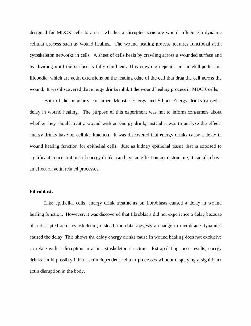
elongated and have a more web-like structure. Stack images of the 5-hour Energy MDCK cells
reveal that the actin cables extend past the plasmamembranes of the cells (Appendix A). A stack
photograph displays images of the same viewing field at different depth layers.
Figure 1
0
100
200
300
400
500
600
700
800
900
Control 1 Monster Energy 1 5 Hour 1
Nu
mb
er
of
Ce
lls C
ou
nte
d
Treatment
MDCK 3-Day Energy Drink Treatment
Normal
Disrupted
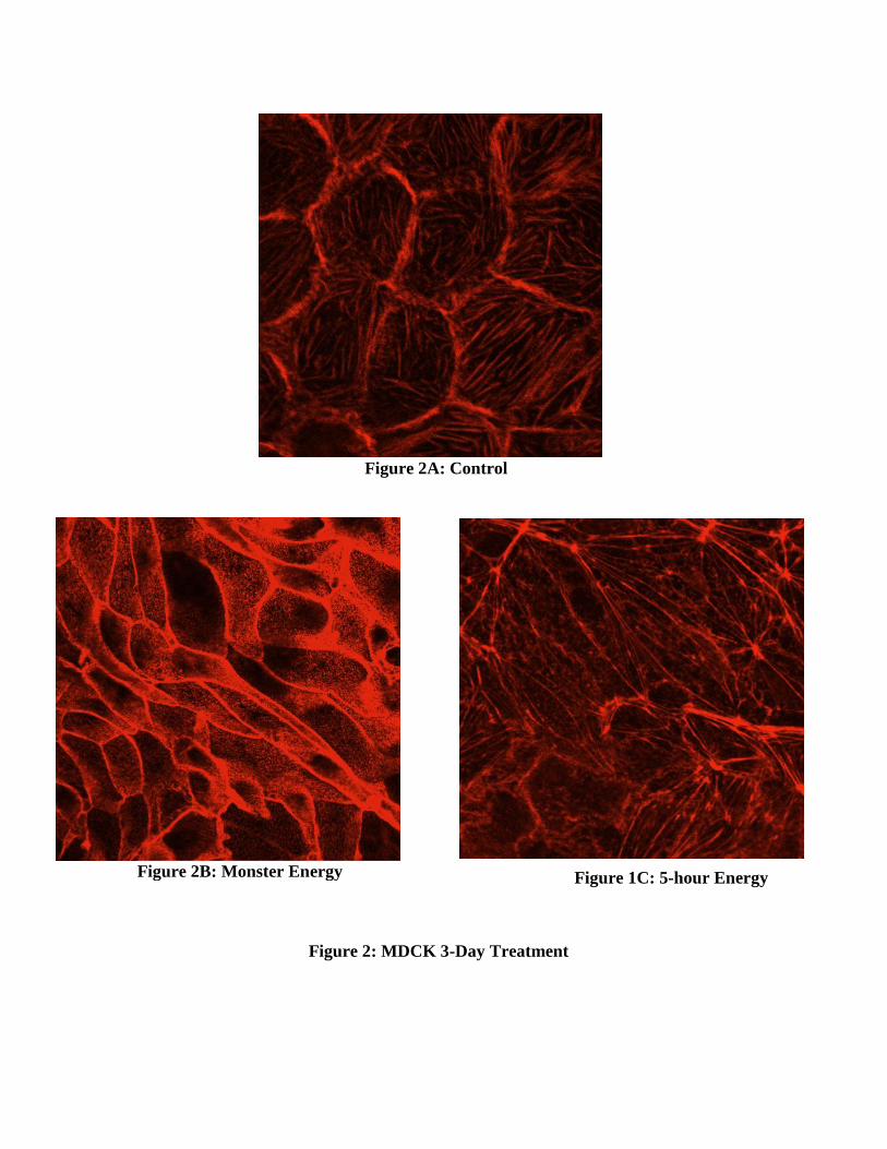
Figure 2A: Control
Figure 2B: Monster Energy Figure 1C: 5-hour Energy
Figure 2: MDCK 3-Day Treatment
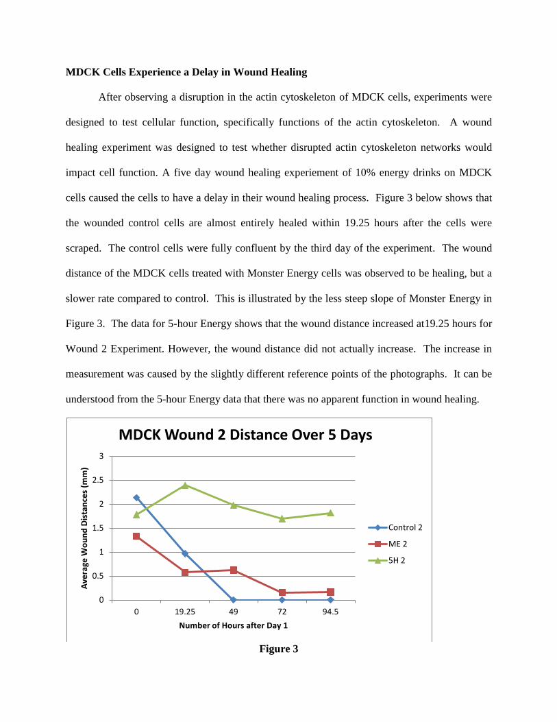
MDCK Cells Experience a Delay in Wound Healing
After observing a disruption in the actin cytoskeleton of MDCK cells, experiments were
designed to test cellular function, specifically functions of the actin cytoskeleton. A wound
healing experiment was designed to test whether disrupted actin cytoskeleton networks would
impact cell function. A five day wound healing experiement of 10% energy drinks on MDCK
cells caused the cells to have a delay in their wound healing process. Figure 3 below shows that
the wounded control cells are almost entirely healed within 19.25 hours after the cells were
scraped. The control cells were fully confluent by the third day of the experiment. The wound
distance of the MDCK cells treated with Monster Energy cells was observed to be healing, but a
slower rate compared to control. This is illustrated by the less steep slope of Monster Energy in
Figure 3. The data for 5-hour Energy shows that the wound distance increased at19.25 hours for
Wound 2 Experiment. However, the wound distance did not actually increase. The increase in
measurement was caused by the slightly different reference points of the photographs. It can be
understood from the 5-hour Energy data that there was no apparent function in wound healing.
Figure 3
0
0.5
1
1.5
2
2.5
3
0 19.25 49 72 94.5
Ave
rage
Wo
un
d D
ista
nce
s (m
m)
Number of Hours after Day 1
MDCK Wound 2 Distance Over 5 Days
Control 2
ME 2
5H 2

Fibroblasts Experience a Delay in Wound Healing
Fibroblasts treated with 10% energy drink concentrations did not experience disruptions
in the actin cytoskeleton network. A wound healing experiment was carried out for fibroblasts in
order to assess if cell function would correlate with the observations of normal actin
cytoskeletons in treated fibroblasts.
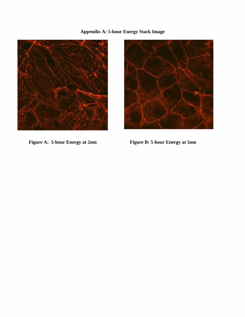
Fibroblasts that were treated with 10% energy drink experienced a delay in their wound
healing process. Over the 4 day period of the experiment, only the control cells fully healed.
Figure 4 shows that the control cells are almost entirely confluent 19 hours after the scrape,
while Monster Energy and 5-hour Energy treated cells did not fully heal after 67 hours. The
slopes displayed in the graphed data of Figure 4 show that 5-hour energy delayed wound healing
function more than Monster Energy delayed wound healing function. The pictures in Appendix
B of the fibroblasts show qualitatively that Monster Energy and 5-Hour healed slower than
control.
Figure 4

Fibroblasts Display a Change in Membrane Dynamics
In an attempt to find a mechanism for the delayed wound healing process, the wound
edges of fibroblasts were more closely observed. Upon closer examination at higher
magnification (40x), the fibroblasts showed significant differences in membrane dynamics.
Figure 5A shows the edge of the control cells, which display a normal ruffling of edges. These
ruffled edges (darker areas of edge) are caused by filopodia/lamellopodia extensions that the
cells use to crawl. Figure 5B shows the smooth edges of Monster Energy treated cells. These
cells responded to the Monster Energy treatment by not putting out filopodia/lamellopodia
extensions at the edge of the wound. Figure 5C shows aberrant formation of
filopodia/lamellopodia. The 5-hour Energy treated cells show thin, over extensions of the
membrane along the edge of the wound.
Figure 5A: Control (40x)
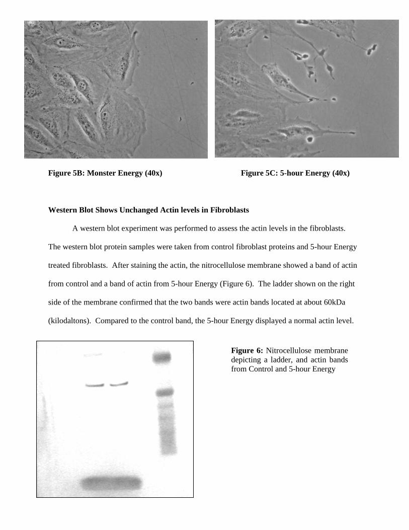
Figure 5B: Monster Energy (40x) Figure 5C: 5-hour Energy (40x)
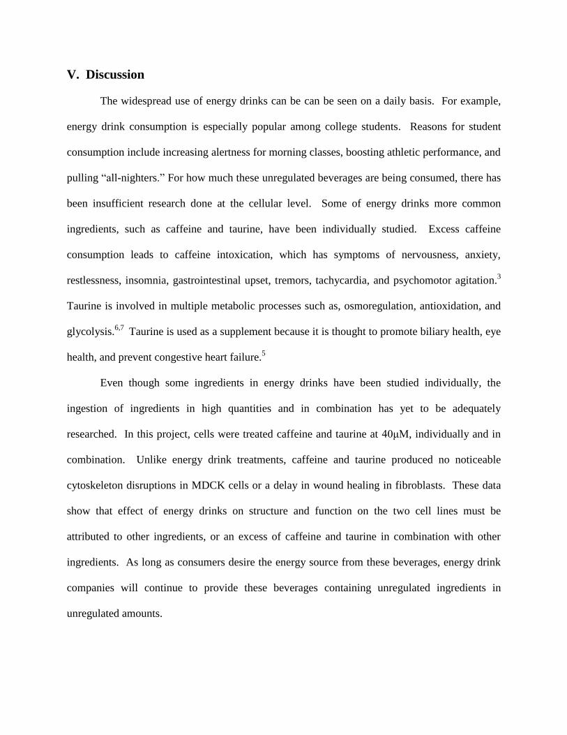
Western Blot Shows Unchanged Actin levels in Fibroblasts
A western blot experiment was performed to assess the actin levels in the fibroblasts.
The western blot protein samples were taken from control fibroblast proteins and 5-hour Energy
treated fibroblasts. After staining the actin, the nitrocellulose membrane showed a band of actin
from control and a band of actin from 5-hour Energy (Figure 6). The ladder shown on the right
side of the membrane confirmed that the two bands were actin bands located at about 60kDa
(kilodaltons). Compared to the control band, the 5-hour Energy displayed a normal actin level.
Figure 6: Nitrocellulose membrane
depicting a ladder, and actin bands
from Control and 5-hour Energy
V. Discussion
The widespread use of energy drinks can be can be seen on a daily basis. For example,
energy drink consumption is especially popular among college students. Reasons for student
consumption include increasing alertness for morning classes, boosting athletic performance, and
pulling “all-nighters.” For how much these unregulated beverages are being consumed, there has
been insufficient research done at the cellular level. Some of energy drinks more common
ingredients, such as caffeine and taurine, have been individually studied. Excess caffeine
consumption leads to caffeine intoxication, which has symptoms of nervousness, anxiety,
restlessness, insomnia, gastrointestinal upset, tremors, tachycardia, and psychomotor agitation.3
Taurine is involved in multiple metabolic processes such as, osmoregulation, antioxidation, and
glycolysis.6,7
Taurine is used as a supplement because it is thought to promote biliary health, eye
health, and prevent congestive heart failure.5
Even though some ingredients in energy drinks have been studied individually, the
ingestion of ingredients in high quantities and in combination has yet to be adequately
researched. In this project, cells were treated caffeine and taurine at 40μM, individually and in
combination. Unlike energy drink treatments, caffeine and taurine produced no noticeable
cytoskeleton disruptions in MDCK cells or a delay in wound healing in fibroblasts. These data
show that effect of energy drinks on structure and function on the two cell lines must be
attributed to other ingredients, or an excess of caffeine and taurine in combination with other
ingredients. As long as consumers desire the energy source from these beverages, energy drink
companies will continue to provide these beverages containing unregulated ingredients in
unregulated amounts.

Epithelial Cells
Treating MDCK cells with energy drinks showed disruptions of the actin cytoskeleton
networks in cells. While energy drinks may provide a consumer with desired energy, they could
possibly be disrupting kidney epithelial tissue. If epithelial tissue is exposed to a high enough
concentration of energy drink ingredients, the data from this project suggests that the actin
cytoskeleton structure of the cells would be disrupted. The data above shows that 5-hour Energy
more significantly disrupted MDCK actin cytoskeleton structure. Upon further examination with
the confocal microscope, it was discovered that the cells that Monster Energy treated cells that
were disrupted were much smaller than the Monster Energy treated cells with normal actin
cytoskeletons. After referring to the ATCC website, it was understood that the MDCK cell line
had two types of cells: tube and dome.
The data show that Monster Energy affected the tube cells more severely than the larger
dome cells. Extrapolating this data, Monster Energy might affect a certain part of the kidney
epithelia more severely than another part. Compared to Monster Energy, 5-hour Energy
disrupted the cells more uniformly and had a more severe affect on the wound healing process.
The difference in severity in cell disruption and cell function could be possibly due to a dose
response between the two drinks. A 1.93 ounce bottle of 5-hour Energy relatively produces the
same desired energetic effects for a consumer as a 16 ounce can of Monster Energy. The
difference in severity between the two energy drinks on cell sheets may point to the results of
more concentrated energy drink ingredients in the human body from excessive consumption or
long term consumption.
In addition to cell structure, actin filaments are essential for dynamic cellular processes
such as cell movement, phagocytosis, and cell division. Wound healing experiments were
designed for MDCK cells to assess whether a disrupted structure would influence a dynamic
cellular process such as wound healing. The wound healing process requires functional actin
cytoskeleton networks in cells. A sheet of cells heals by crawling across a wounded surface and
by dividing until the surface is fully confluent. This crawling depends on lamelellipodia and
filopodia, which are actin extensions on the leading edge of the cell that drag the cell across the
wound. It was discovered that energy drinks inhibit the wound healing process in MDCK cells.
Both of the popularly consumed Monster Energy and 5-hour Energy drinks caused a
delay in wound healing. The purpose of this experiment was not to inform consumers about
whether they should treat a wound with an energy drink; instead it was to analyze the effects
energy drinks have on cellular function. It was discovered that energy drinks cause a delay in
wound healing function for epithelial cells. Just as kidney epithelial tissue that is exposed to
significant concentrations of energy drinks can have an effect on actin structure, it can also have
an effect on actin related processes.
Fibroblasts
Like epithelial cells, energy drink treatments on fibroblasts caused a delay in wound
healing function. However, it was discovered that fibroblasts did not experience a delay because
of a disrupted actin cytoskeleton; instead, the data suggests a change in membrane dynamics
caused the delay. This shows the delay energy drinks cause in wound healing does not exclusive
correlate with a disruption in actin cytoskeleton structure. Extrapolating these results, energy
drinks could possibly inhibit actin dependent cellular processes without displaying a significant
actin disruption in the body.

Studies have shown that proteins such as CDC-42, Rac, and Rho have an active role in
actin polymerization and depolymerization.10
The difference in membrane dynamics between
control, Monster Energy, and 5-hour Energy could be possibly caused by the energy drinks
disrupting actin-binding proteins. The data from the Western Blot experiment show that the
actin levels of the treated fibroblast cells were unchanged. The experiment showed that a lack of
actin was not causing a change in membrane dynamics, which further suggests that the
disruption of actin-binding proteins are the cause of the change in membrane dynamics.
Consuming energy drinks may not reduce actin levels in fibroblasts, but could have an affect on
actin-binding proteins necessary for actin related processes in the human body.
VI. Conclusion
Monster Energy and 5-hour Energy cause a disruption in epithelial actin cytoskeletons
and a change in fibroblast membrane dynamics. Both epithelial cells and fibroblasts experience
a delay in wound healing when treated with the two energy drinks. 5-hour Energy treatments had
a more negative effect on cell structure and function than Monster Energy for both cell types.
While energy drinks negatively impacted structure and function of cells, the ingredients caffeine
and taurine did not.
VII. Acknowledgements
Dr. Vidya Chandrasekaran for all her guidance and support throughout the summer and
school year. Charlotte Lea for taking care of the MDCK cells for two weeks and for repeating a
fibroblast wound healing experiment. Amy Bockman for ordering the antibodies and kits
necessary for the project. The Science Department of Saint Mary’s college of California for this
opportunity to do undergraduate research.

References
1. Higgins, John et al. Energy Drinks: Content and Safety. Rochester (2010) vol. 85 (11) pp.
1033-1042.
2. US Food and Drug Administration. Overview of Dietary Supplements. FDA (2009)
http://www.fda.gov/Food/DietarySupplements/ConsumerInformation/ucm110417.htm
3. Reissig et al. Caffeinated energy drinks—A growing problem. Drug Alcohol Depend (2008)
vol. 99 (1-3) pp. 1-10
4. Seifert, Sara M. et al. Health Effects of Energy Drinks on Children, Adolescents, and Young
Adults. PEDIATRICS (2011) vol. 127 (3) pp. 511-528
5. Iyadurai, Stanley, and Steve S. Chung. New-onset seizures in adults: Possible association with
consumption of popular energy drinks. Epilepsy Behav (2007) vol. 10 (3) pp. 504-508
6. Babu et al. Energy Drinks: The New Eye-Opener For Adolescents. Clinical Pediatric
Emergency Medicine (2008) vol. 9 (1) pp. 35-42
7. Timbrell et al. The In Vivo and In Vitro Protective Properties of Taurine. Gen Pharmacol
(1995) vol. 26 (3) pp. 453-62
8. Doyle, Wayne. The Cellular Response Of Neural and Kidney Cells After Exposure to
Commonly Consumed Energy Drinks. Saint Mary’s College of CA (2010) pp. 1-16
9. Clauson et al. Safety issues associated with commercially available energy drinks. Journal of
the American Pharmacists Association : JAPhA (2008) vol. 48 (3) pp. 55-63
10. Eisen et al.: Regulation of epithelial tubule formation by Rho family GTPases. Am J Physiol
Cell Physiol: (2005) C1297-C1309
Appendix A: 5-hour Energy Stack Image
Figure A: 5-hour Energy at 2nm Figure B: 5-hour Energy at 5nm

Appendix B: Fibroblast Wound Healing (4x)
Control
Figure A: Initial Scrape
19 Hours After Scrape 28 Hours After Scrape
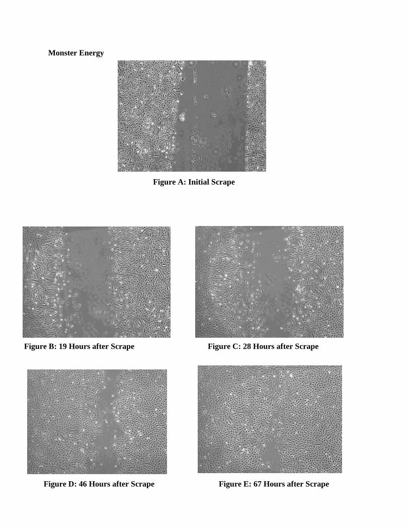
Monster Energy
Figure A: Initial Scrape
Figure B: 19 Hours after Scrape Figure C: 28 Hours after Scrape
Figure D: 46 Hours after Scrape Figure E: 67 Hours after Scrape

5-hour Energy
Figure A: Initial Scrape
Figure B: 19 Hours after Scrape Figure C: 28 Hours after Scrape
Figure D: 46 Hours after Scrape Figure E: 67 Hours after Scrape