Embed Size (px)
Citation preview
i
THE EFFECT OF THE ISOLITETM SUCTION ON AEROSOLS AND SPLATTER DURING ULTRASONIC SCALING
Jessica Laurin Holloman
A thesis submitted to the faculty at the University of North Carolina at Chapel Hill in partial fulfillment of the requirements for the degree of Master of Science in Dental Hygiene Education
in the Dental Hygiene Department in the School of Dentistry.
Chapel Hill 2014
Approved by:
Sally Mauriello
Roland Arnold
Luiz Pimenta

ii
© 2014 Jessica Laurin Holloman
ALL RIGHTS RESERVED
iii
ABSTRACT
Jessica Laurin Holloman: The Effect of the IsoliteTM Suction on Aerosols and Splatter During
Ultrasonic Scaling (Under the direction of Sally Mauriello)
This study compared the IsoliteTM and saliva ejector on aerosol reduction during
ultrasonic scaling. Fifty participants were randomly assigned to control (n=25, saliva ejector) or
test groups (n=25, IsoliteTM). Plaque extent scores were recorded and aerosols were collected
both during (timed period) and post (35 minute period) ultrasonic scaling in buffer in Petri dishes
placed six inches from the participant’s mouth. Participants were surveyed regarding device
acceptance. The during and post suspensions were plated to blood agar plates and recoverable
colonies (CFU) were counted following anaerobic incubation. Significant contamination
occurred during ultrasonic scaling in both device groups, as indicated by high CFU and the
identification of strict oral anaerobes on all plates. A Student t-test revealed that the test device
did not reduce aerosol contamination compared to the control device (p=0.25). Additional
caution should be exercised with these devices to reduce the risk of exposure to potential
pathogens.
iv
ACKNOWLEDGEMENTS
I would like to thank IsoliteTM Systems for funding the lab supplies for this study,
especially Mark McIntyre and Maureen Sullivan for helping to secure the loan of the test device
during the duration of the study. I’d also like to thank the GO Health Clinic and their wonderful
staff for the use of their facilities and patient population. I’d like to extend a special thanks to
Wendy Lamm who helped schedule and confirm every single patient. I greatly appreciate the
expertise of Eric Simmons, who helped immensely during the microbiological process. I would
also like to acknowledge Bob Link and John Marley for helping to install and disassemble the
test device. The capture of my study design and lab methods was made possible by Ramona
Hutton-Howe’s extraordinary photography and videography skills.
v
TABLE OF CONTENTS
LIST OF TABLES................................................................................................................................................... vii
LIST OF FIGURES ............................................................................................................................................... viii
ABBREVIATIONS .................................................................................................................................................. ix
CHAPTER 1: INTRODUCTION........................................................................................................................ 10
CHAPTER 2: A REVIEW OF THE LITERATURE ....................................................................................... 12 Introduction .......................................................................................................................................................................... 12 Defining Aerosols and Splatter ...................................................................................................................................... 13 Aerosols and Healthcare .................................................................................................................................................. 14 Aerosols and Dentistry ..................................................................................................................................................... 15 The Ultrasonic Scaler ........................................................................................................................................................ 16 Reduction of Aerosols and Splatter .............................................................................................................................. 17 The IsoliteTM dryfield illuminator ................................................................................................................................. 19 Previous Research Methods to Collect and Quantify Aerosols and Splatter .................................................. 23
CHAPTER 3: MATERIALS AND METHODS ............................................................................................... 28 Test Device........................................................................................................................................................................... 28 Control Device .................................................................................................................................................................... 28 Ultrasonic Scaling Equipment........................................................................................................................................ 28 Lab Equipment .................................................................................................................................................................... 28 Methods ................................................................................................................................................................................. 29
vi
CHAPTER 4: RESULTS ...................................................................................................................................... 36 Primary Objective .............................................................................................................................................................. 36 Secondary Objective ......................................................................................................................................................... 38
CHAPTER 5: DISCUSSION ............................................................................................................................... 40 Key Findings........................................................................................................................................................................ 40 Findings related to microbiological methodology ................................................................................................... 42 Secondary Objective ......................................................................................................................................................... 43
CHAPTER 6: CONCLUSIONS ........................................................................................................................... 44 Limitations ........................................................................................................................................................................... 45 Future Studies ...................................................................................................................................................................... 45
APPENDIX 1: TEST DEVICE ............................................................................................................................ 54
APPENDIX 2: IRB APPROVED TELEPHONE SCRIPT ............................................................................ 55
APPENDIX 3: STUDY DESIGN PHASE ......................................................................................................... 56
APPENDIX 4: PLAQUE EXTENT DATA FORM ........................................................................................ 57
APPENDIX 5: PLACEMENT OF PETRI DISH ............................................................................................. 58
APPENDIX 6: SURVEY INSTRUMENT ......................................................................................................... 59
APPENDIX 7: LAB CFU RECORDING FORM ............................................................................................. 60
REFERENCES ....................................................................................................................................................... 61
vii
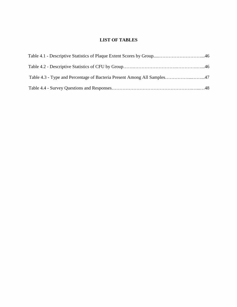
LIST OF TABLES
Table 4.1 - Descriptive Statistics of Plaque Extent Scores by Group.....………………………...46
Table 4.2 - Descriptive Statistics of CFU by Group…….……………………….………….…...46
Table 4.3 - Type and Percentage of Bacteria Present Among All Samples……………...……...47
Table 4.4 - Survey Questions and Responses…………………………………………….…...…48
viii
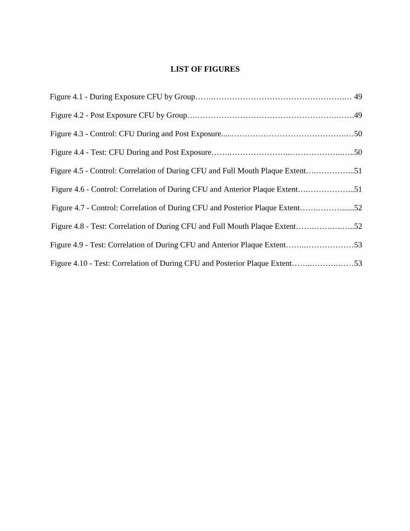
LIST OF FIGURES
Figure 4.1 - During Exposure CFU by Group…….………………………………………….… 49
Figure 4.2 - Post Exposure CFU by Group….…………………………………………….…….49
Figure 4.3 - Control: CFU During and Post Exposure.....…………………………………….…50
Figure 4.4 - Test: CFU During and Post Exposure…….…………………..………………..…..50
Figure 4.5 - Control: Correlation of During CFU and Full Mouth Plaque Extent….…………...51
Figure 4.6 - Control: Correlation of During CFU and Anterior Plaque Extent….……………...51
Figure 4.7 - Control: Correlation of During CFU and Posterior Plaque Extent…….………......52
Figure 4.8 - Test: Correlation of During CFU and Full Mouth Plaque Extent…….…….….…..52
Figure 4.9 - Test: Correlation of During CFU and Anterior Plaque Extent……..………………53
Figure 4.10 - Test: Correlation of During CFU and Posterior Plaque Extent……..……….……53
ix
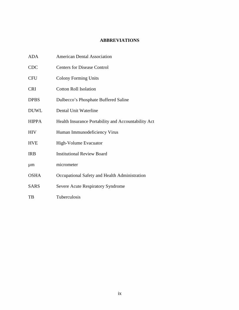
ABBREVIATIONS ADA American Dental Association
CDC Centers for Disease Control
CFU Colony Forming Units
CRI Cotton Roll Isolation
DPBS Dulbecco’s Phosphate Buffered Saline
DUWL Dental Unit Waterline
HIPPA Health Insurance Portability and Accountability Act
HIV Human Immunodeficiency Virus
HVE High-Volume Evacuator
IRB Institutional Review Board
µm micrometer
OSHA Occupational Safety and Health Administration
SARS Severe Acute Respiratory Syndrome
TB Tuberculosis
10
CHAPTER 1: INTRODUCTION According to the Centers for Disease Control and Prevention (CDC), almost all new
infections in the United States are contracted through the aerosol route from infected patients
who are coughing and dispersing infective droplet nuclei into the air.1 Therefore, the presence
and dispersal of aerosols and splatter are a concern in healthcare due to their potential adverse
health effects on patients and healthcare workers. The effect of these aerosols on
immunocompromised patients is concerning due to their increased susceptibility to the
potentially infectious bacteria that are present in these aerosols. Many routine dental procedures
produce aerosols and splatter, which may contain infectious material such as blood, saliva, and
other organic matter.2 Published data showed that ultrasonic scalers, as well as other types of
dental hand pieces, had a significant effect on the number of colony-forming units (CFU)
cultivable from the air when compared to preprocedural levels.3, 4 The ultrasonic scaler, in
particular, has been shown to produce three times the bacterial aerosol contamination as that
produced by dental operative equipment.1,5 Based on supportive data, the American Dental
Association (ADA) has recommended that the high-volume evacuator (HVE) be employed
during the use of ultrasonic scalers to minimize the spread of airborne bacterial contamination
during ultrasonic scaling.6,7 This recommendation creates a unique challenge for dental health
care workers, specifically dental hygienists, that traditionally provide clinical care without a
dental assistant. Due to the nature of most suction devices, dental hygienists must sacrifice their
non-dominant hand, as well as light and indirect vision, while using the ultrasonic scaler.
11
Further, the HVE can be bulky and difficult to maneuver when an assistant is not available,
making the saliva ejector the device of choice to remove excess fluids from the oral cavity. The
IsoliteTM dryfield illuminator, a product created with “hands-free” dentistry in mind, was
introduced to the commercial market in 2005.8 The primary purpose of this system is to provide
isolation and illumination to the oral cavity, but has been purported to have an added benefit of
aerosol reduction by as much as 65% when compared to the saliva ejector in a simulated clinical
environment.8,9 There is currently no published data on how the IsoliteTM compares to the saliva
ejector when used with an ultrasonic scaler in an actual clinical environment. Further, there is no
published literature regarding patient acceptance of the IsoliteTM in an adult United States
population.
After reviewing the literature and developing the protocol for the current study it seemed
prudent to attempt to identify an alternative methodology for the collection and quantification of
aerosols and splatter. The methods available often included the use of equipment that was either
expensive or not readily available and these methods typically did not include both the
quantification of aerosols and splatter and identification of specific bacteria collected.
Therefore, the purpose of this study was two-fold:
(1) Compare the effect of the IsoliteTM suction and saliva ejector on aerosols and splatter during
ultrasonic scaling, and
(2) Determine patient acceptance of the IsoliteTM in an adult U.S. population.
Additional specific objectives included:
(3) Develop an alternative methodology to collect and quantify aerosols and splatter, and
(4) Confirm that recovered bacteria originated from an oral source.
12
CHAPTER 2: A REVIEW OF THE LITERATURE
Introduction Since the 1960’s, the detrimental effects of aerosols and splatter and their role in the
spread of infectious diseases have been extensively researched and documented. The role of
aerosols and splatter in the spread of infectious diseases have been linked to outbreaks of
influenza, chickenpox, tuberculosis (TB), legionnaire’s disease, severe acute respiratory
syndrome (SARS), etc.1,5,6 It is known that the transmission of these diseases occurs via the
airborne route from human sources through the inhalation of droplet nuclei that are aerosolized
by respiratory secretions such as coughing, sneezing, or talking.10 Aspiration of pathogenic
bacteria from the oral cavity has also been linked to certain systemic infections.11,12 Pneumonia,
an infection of the lungs, has been extensively studied and is most common in an
immunocompromised population, such as the elderly.13
In order to address the control of potentially infectious aerosols it was necessary to first
examine their characteristics and capabilities. Assessment of aerosols and splatter included
factors such as generation, particle size and concentration, infectivity and virulence, viability, air
flow, environmental sampling, and analysis.10 These factors have been studied and reported by a
range of experts, including those who specialize in aerobiology to dentists who were interested in
reducing the presence of aerosols and splatter in the dental office. The spread of aerosols and
splatter were first appraised by exploring their history in healthcare and dentistry and their
impact on healthcare workers.
13
This was followed by identifying those procedures known to produce the greatest amounts of
aerosols and splatter, which has proven key in the process of sampling and studying the
microorganisms that are linked to the spread of infectious diseases. Finally, the mechanisms to
control the spread of aerosols and splatter are the final piece to preventing their dissemination in
the dental office.
Defining Aerosols and Splatter Although the Occupational Safety and Health Administration (OSHA) does not
explicitly define aerosols according to size, the consensus among the literature states that
aerosols can be liquid or solid particles that are approximately 50µm or less in diameter, are
suspended in the air, and are capable of penetrating deep into the respiratory system.3,14,15,17
Previous aerobiology studies have shown that aerosols tend to stay airborne for an extended
period of time before they settle on surfaces or enter the respiratory tract. 3,10,17,18 It is the smaller
particles of an aerosol (.5µm -10µm in diameter) that are of particular concern because they are
thought to carry the greatest potential for transmitting infections due to their ability to penetrate
and lodge into the smaller passages of the lungs.5
Splatter particles differ from aerosols because they are visible to the naked eye and are
considered too large to be inhaled and imposed deep within the lung.3,16 Splatter particles are
known to behave in a ballistic manner, in that they follow an arc trajectory from the oral cavity
until they contact a surface.15 It is because of this trajectory that splatter particles do not remain
suspended in the air for long periods of time, making them less likely to transmit disease via the
airborne route.
Another important consideration when discussing aerosols and splatter is the viability of
the microorganisms within the environment once they have left the host. The viability of the
14
microorganism is essentially the ability of the microorganism to reproduce. When
microorganisms leave their host and are aerosolized, they are potentially injured during the
generation process.10 Previous data have shown that microorganisms could remain viable in the
airborne state for long enough to permit their wide dissemination.19 Factors such as temperature,
relative humidity, air flow, and oxygen sensitivity will impact whether or not microorganisms
are able to survive outside of the host and replicate.19 Engineering controls should be in place
within healthcare settings to eliminate these microorganisms and limit the exposure of the staff
and public to their presence. It is necessary to understand the factors related to aerosol and
splatter generation and survival when considering disease transmission. This knowledge is
crucial to the task of implementing protocol for the elimination of aerosols and splatter, therefore
preventing disease transmission.
Aerosols and Healthcare
With regard to healthcare workers, OSHA stated that the primary routes of infectious
disease transmission in U.S. healthcare settings are through contact, droplet, and airborne
particles.1,6 Airborne diseases such as tuberculosis, influenza, measles, chickenpox, and
Legionnaires’ disease have been well documented as capable of remaining viable and airborne
within the indoor environment.20,21-24 The ability of aerosols to remain suspended in the air and
disperse over considerable distances increases the likelihood of cross-contamination.25 Because
of these characteristics, it is not surprising that healthcare workers and patients are at an
increased risk of infection.
In cases where measles and tuberculosis have been spread in an indoor environment,
airflow studies revealed that droplet nuclei were generated throughout the entire office.24, 26
Legionnaire’s disease, a severe form of pneumonia, is also spread via the aerosol route. This
15
disease is estimated to infect 10,000 to 15,000 persons per year in the United States, where 9% of
cases occur at least six months before or after a hospital outbreak.23 This disease has become
especially concerning in dentistry due to its link to dental unit waterlines (DUWL).4,19,23 Another
systemic infection that has been extensively studied is pneumonia. This infection is especially
common in the elderly and can be acquired from cross-contamination or self ingestion.13
Aspiration pneumonia, a type of pneumonia caused by the inhalation of a substance into the
lung, is often associated with anaerobic bacteria.11 This type of pneumonia is especially likely
during dental procedures, which can yield high amounts of bacteria laden aerosols and splatter.
Based on this knowledge, the presence and dispersal of aerosols in indoor environments
are a growing concern in epidemiology.25, 27 Further, special patient populations, such as the
immunocompromised patient, have been shown to be at a greater risk of infection than the
average healthy patient.20
Aerosols and Dentistry In the dental clinic, aerosols are of particular concern due to the inherent nature of most
dental procedures. Studies have demonstrated that many routine dental procedures that
incorporate the use of water sprays or rotary instruments artificially generate aerosols that
produce significantly greater numbers of bacteria than those activities produced by non-dental
related oral activities, such as coughing and sneezing.17, 28 In a review of the literature by Harrell
et al., it was emphasized that saliva and nasopharyngeal secretions may contain pathogenic
organisms such as herpes viruses, streptococci, staphylocci, and the SARS virus.85,29 Further,
bloodborne diseases such as Hepatitis and Human Immunodeficiency Virus (HIV) can be
transmitted into the air via blood droplets.5,29
16
In addition to the transmission of infectious diseases, aerosols can be a source of irritants,
allergens, and other toxic substances, which are a potential source of acute or chronic respiratory
disease.17 A survey of aerosol-related symptoms in dental hygienists who frequently use
ultrasonic scalers revealed that symptoms such as nasal irritation, persistent cough, runny eyes,
itchy and dry skin, were more common in dental hygienists than in nurses and hospital staff.30
Dental staff must consider protecting themselves and their patients from common airborne
diseases and infectious diseases that are not characteristically airborne and are being transmitted
into the air during dental procedures. The inhalation of these substances may not be of concern to
the average healthy patient, but special patient populations, such as the immunocompromised
patient, can be especially susceptible to the adverse effects of these aerosols. Both the CDC and
the ADA have recommended reducing the risk of infection posed by aerosols by the use of
rubber dams, high-velocity air evacuation, and proper patient positioning, along with standard
precautions.1 Based on the evidence surrounding the generation of water spray by the ultrasonic
scaler, one may infer a relationship between the production of infectious aerosols and treatment
techniques. Thus, mechanisms to reduce aerosol spray should be considered during ultrasonic
instrumentation.
The Ultrasonic Scaler Those dental procedures shown to create high amounts of aerosols and splatter are of
particular concern in oral epidemiology. Hand scaling, for example, has been shown to create
negligible amounts of aerosol and splatter.2 Ultrasonic scalers and high-speed hand pieces have
been studied extensively and were shown to produce measurable amounts of aerosols and
splatter.2, 6,16,17,28,31,32,42 Since the water spray emitted from the working tip of an ultrasonic
scaling hand piece bears a strong physical resemblance to the spray of high-speed dental hand
17
piece, many studies have advised the same type of aerosol reduction device.33 A dental hygienist
commonly uses ultrasonic scalers during periodontal instrumentation and routine prophylaxis.
These devices utilize high-frequency vibrations and water as a medium of ultrasonic energy to
remove calculus deposits and have been labeled as one of the major sources of potential aerosol
contamination in the dental setting due to the large amount of aerosols expelled into the air
during their use.9, 33
Reduction of Aerosols and Splatter
Abundant research is available regarding the presence and dispersal of aerosols and
various devices have been evaluated regarding their effectiveness.2-5, 16,20,31,33-36 Traditional
methods that reduce potentially infectious aerosols during dental procedures include the low and
high-volume evacuator, dental dam, pre-procedural rinses, and various air quality devices.2,
8,16,31,35-37 A landmark study conducted by Micik et al. in 1969 was published regarding the
reduction of aerosols during routine dental procedures and found that the HVE demonstrated the
highest efficiency.15 Since then, various studies have shown that when compared to no suction
or the saliva ejector the HVE has proven to be the most effective at reducing aerosols created
during dental procedures by as much as 90%.14,15,34-36 The use of a rubber dam has also been
shown to eliminate almost all contamination that arises from saliva or blood, but this type of
device is not feasible for most periodontal and dental hygiene procedures. 32,37,38 During most
dental procedures, it is the assistant who manipulates the HVE due to the manner in which it
must be used to properly control aerosol and splatter. The HVE can be cumbersome and
uncomfortable to the patient and clinician if not used correctly. These actualities make the HVE
difficult to use as a single clinician, which is often the case during procedures rendered by a
dental hygienist.
18
In 1996, Harrel et al. published an in vitro study, which primarily investigated the
reduction of aerosols and splatter with the use of an HVE attachment compared to no suction
during use of the ultrasonic scaler.34 Recognizing the limitations of the HVE, a sheath was
engineered to connect the HVE to the ultrasonic scaler. To assess aerosol reduction, a plastic
enclosure with one centimeter square gridlines was assembled to enclose around a dentoform
model that was mock scaled for one minute with an ultrasonic scaler. Instead of water, red
erythrosine solution was used to represent contamination. Each square containing at least one
erythrosine spot was considered contaminated and squares were counted twice following the
exposure by an evaluator. The scaling procedure was repeated ten times by two operators,
resulting in a total of twenty trials. Mean numbers of contaminated squares were calculated and
results indicated gross differences based on the operator, making the findings variable based on
practices of the clinician. Overall, the study found that the HVE attachment device greatly
reduced detectable aerosol and splatter contamination by as much as 100% in a single trial. These
results represent a greater than 93% reduction in the average amount of contamination produced
by the ultrasonic scaler with the HVE attachment when compared to no suction device.34 A
potential limitation of this study is that it was a small sample size and was completed in vitro,
making the results difficult to generalize and apply to a clinical environment.
As an extension of the Harrel et al. study, King et al. published in 1997 an in vivo study
regarding the reduction of aerosols with an ultrasonic scaler utilizing the same type of engineered
HVE attachment to the ultrasonic scaler.33 Twelve subjects were enrolled and each subject served
as his/her own control. Each subject was scaled with an identical ultrasonic unit, insert, power,
and water setting. Three blood agar plates were placed at a 50° angle and 6-inches from the
subject’s mouth to collect aerosols. In separate closed-door rooms, ultrasonic instrumentation
19
was performed for five minutes on each side of the patient’s mouth, one side with the HVE
attachment and one side without. After being exposed for 25 minutes, the blood agar plates were
covered and incubated at 37°C for three days prior to counting CFU.33 Results were in
concurrence with the study completed in 1996 by Harrel et al. and revealed that the use of a HVE
attachment significantly reduced aerosols and splatter.33, 34
The IsoliteTM dryfield illuminator
The IsoliteTM is a device designed to provide isolation, suction, illumination, and
retraction simultaneously when used by a single operator. The bite-block component of the
mouthpiece allows for isolation of the maxillary and mandibular quadrants simultaneously so
that the patient can rest open during the entire dental procedure.8 Due to the relatively new status
of this product, little research has evaluated the company’s reported benefits of decreased
procedure time, increased retention rates for restorations and sealants, and reduction of aerosols
in the operatory. IsoliteTM Systems (Santa Barbara, CA) specifically purports to reduce airborne
aerosols by up to 65% compared to the saliva ejector, which is of particular interest due to the
role that aerosols play in the spread of infectious diseases.8, 35 Because the IsoliteTM is designed to
attach to the high-volume suction hose and previous research has shown that the ultrasonic scaler
produces the greatest amount of aerosols, it would be prudent to determine how effectively the
IsoliteTM reduces aerosols and splatter while performing ultrasonic scaling in an actual clinical
environment.
An unpublished study by Jacks and Pollard in 2007, compared the IsoliteTM to the HVE
alone and saliva ejector in an independent laboratory trial and measured the amount of aerosol
particles that reached the breathing space of the clinician.35 A total of 21 trials were conducted
and involved mock scaling for two minutes with an ultrasonic scaler. Scaling was performed on
20
all surfaces of all teeth on a DENTOFORM model. The HVE, saliva ejector, and IsoliteTM were
compared and each trial was divided into two minute sampling periods: pre-exposure, exposure,
and post exposure. Aerosols were measured with the DataRAM Real-Time Aerosol Monitor
every 10 seconds during all phases. Each group was statistically different when compared. The
IsoliteTM was shown to reduce aerosols by as much as 66% and the HVE by as much as 76%
when both were compared to the saliva ejector. The study’s overall recommendation was to alter
the design of the IsoliteTM mouthpiece to increase airflow, which may deliver a closer reduction
amount to that of the HVE.35 A limitation of this study is that is was performed in a laboratory
environment so all of the variables present in a clinical environment were not taken into account.
In 2009, Noro et al. published the first study evaluating the IsoliteTM and examined its
clinical usefulness in a Japanese population.37 Volunteer resident dentists in the Department of
General Dentistry at Tokyo Dental College Chiba Hospital were utilized as study subjects.
Subjects were randomly divided into two groups of 15 and paired with an individual from the
opposite group. In each pair, the subject playing the role of the clinician placed the IsoliteTM into
the oral cavity of the subject playing the role of the patient and used an air turbine hand piece
equipped with a dummy bur to simulate tooth preparation for a crown. Following the simulation,
all subjects completed a survey composed of nine questions regarding their experience with the
device. Based on the mean overall ratings, the subjects playing the role of the surgeon rated the
device higher in satisfaction than the patients. The lowest patient ratings were in response to the
question regarding how the IsoliteTM fits in the mouth, leading to the conclusion that the IsoliteTM
needed to be altered for a better fit with the Japanese people.37 A drawback of the study was their
exclusive focus on the Japanese population, making these conclusions difficult to apply to other
populations.
21
In 2010, a study by Colette et al. assessed patient acceptance of the IsoliteTM during
sealant placement compared to cotton roll isolation (CRI) while evaluating sealant application
times.38 A total of 48 children were seen at the pediatric dental clinic at Children’s Hospital in
Cincinnati, Ohio. Data on patient acceptance were collected via a verbal survey, consisting of
nine questions, which asked the patient about his or her experience with both the IsoliteTM and
CRI. Subjects were randomly assigned to one of two groups, both utilizing the IsoliteTM and CRI
but alternating which method was used first. The survey was administered following sealant
placement. The results of the study revealed discomfort was reported with the IsoliteTM for the
following reasons: stretching of the cheeks, inducing their gag reflex, and high amounts of noise.
The author concluded that because the participants of the study were all under the age of 11 it
would be beneficial to utilize older children or adults in future research to further evaluate
patient acceptance.38
A study published in November 2012, by Dahlke et al. compared the effectiveness of a
splatter reduction at an operative site by three groups: the IsoliteTM alone, the HVE with dental
dam, and the HVE alone in a patient simulated environment.36 During the study, a total of 72
trials were completed in a closed door operatory and tooth preparation was simulated on a
typodont manikin during a benchtop exercise. A high-speed hand piece with a carbide bur was
used to create splatter with a fluorescein dye solution, which was added to the water supply. A
bulletin board was mounted to surround the typodont head and was used to collect splatter
emitted from the hand piece and contaminated squares were counted following the conclusion of
each trial. Each type of dry-field technique was used while tooth prep was simulated on tooth
numbers 18-20. The control consisted of the HVE alone during simulated tooth preparation. The
first experimental group consisted of the IsoliteTM set at maximum strength during tooth-
22
simulated preparation. The second experimental group consisted of the standard 6-inch dental
dam with only three holes to isolate the three teeth being prepped. The HVE for the control and
experimental groups was oriented in an identical position and the IsoliteTM was kept in the same
position throughout the entire experiment. The study found no significant difference in the
reduction of splatter between the two experimental devices, but splatter was decreased
significantly when compared to the HVE alone. The only statistical difference found was that
when tooth prep was simulated on a more anterior tooth, the HVE with dental dam reduced
splatter slightly more than the IsoliteTM, likely due to the design of the mouthpiece and the focus
of evacuation being in the posterior. The conclusions stated that because the IsoliteTM reduced
aerosols just as well as the HVE with dental dam during tooth preparation, it may be the
preferred device because of the other benefits it offers, specifically illumination, isolation,
protection of adjacent soft tissues, assistance in opening the mouth and protecting from
accidental aspiration.36 The observation that the IsoliteTM was not as effective in the anterior was
beneficial when identifying possible cofounding variables for the current study.
Bacterial contamination from ultrasonic scaler aerosol has been well documented in the
past.3-5, 8,16,31,32,34,35 Developing protocol for the reduction of aerosols during the treatment of
immunocompromised patients can help protect these patients during dental procedures, such as
ultrasonic scaling, that create high amounts of potentially infectious aerosols. Presently, there is
no known research regarding the IsoliteTM’s ability to reduce aerosols or its performance
compared to other suction devices in an actual clinical environment during use of the ultrasonic
scaler. Based on the findings of the study, if the IsoliteTM reduces aerosols more effectively
during ultrasonic scaling than the saliva ejector alone then it may be recommended as a standard
of care, especially during the treatment of immunocompromised patients.
23
Previous Research Methods to Collect and Quantify Aerosols and Splatter
A variety of methods have been employed in research to collect and quantify dental
aerosols and splatter. Historically, these methods have depended on the type of particle being
studied (aerosol vs. splatter) and/or the type of microbe in questions (anaerobic vs. aerobic).
Techniques for aerosol and/or splatter collection have ranged from benchtop exercises with
fluorescein dye to elaborate air sampler devices.
The most basic approach to evaluate aerosols and splatter has been the use of benchtop
studies where dental procedures were simulated and no actual microbes were created or
measured.5, 21,29,34 In these cases, the primary objective was the testing of aerosol and splatter
reduction devices instead of collecting and quantifying actual microbes. These studies have
helped to identify devices that produce as well as reduce the greatest amounts of aerosols and
splatter.5, 21,29,34 In two separate studies by Harrel et al., aerosol production and reduction were
evaluated.29, 34 The first of the two studies examined aerosol reduction with an HVE attachment
to an ultrasonic scaler29 while the subsequent study identified differences in aerosol production
between hand scaling and various ultrasonic inserts.34 In both studies the coolant water for the
ultrasonic scaler was replaced with a fluorescein solution and a grid containing one centimeter
squares surrounded a dentoform model. Squares containing a drop of the fluorescein solution
were considered contaminated and were counted and recorded.29, 34 A more recent study, which
used this method while evaluating the IsoliteTM was conducted by Dahlke et al., and compared
the splatter reduction of the IsoliteTM to the HVE with the rubber dam and HVE alone.36 The
methods used were similar in that an overlay grid was used to show contaminated squares of
fluorescein dye. Similar to previous studies, this method proved appropriate for their aims but
was limited in its inability to collect, identify, and quantify actual microbes.

24
Some of the earlier studies in the 1940’s and 1950’s were critiqued because of their
inability to differentiate between viable and non-viable aerosols.39 The earliest published study to
identify an instrument that was able to collect and count viable airborne particles was in 1958, by
Ariel Andersen.39 This device is known as the Andersen sampler, consisting of six-stages
through which the air or aerosol is drawn in by air flow, at a rate of one cubic foot per minute,
through perforated (400) holes into a Petri dish filled with agar medium to collect the microbes.
Petri dishes were removed and incubated for an undisclosed amount of time. The way in which
colonies were counted were not specified but stated as being quantified “in a usual manner” as
defined in microbiology and in the case of heavily loaded plates, by a dissecting type microscope
before the colonies were able to merge.39 Although groundbreaking at the time, this method
proved problematic due to its inability to include splatter as well as its tendency to also include
dust, molds, yeast, and other particles present in the environment at the time of sampling. This
study was followed by a very similar study by Larato et al., in 1967, which utilized a similar
device; the Reyner air sampler.27 An advantage of this study was the specific identification of
multiple types of bacteria sampled from the air. It was determined that most of the bacteria
collected were mold or yeast that were either often found in the air or water. The exception to
this was the identification of alpha streptococcus, which is present in large numbers in the oral
cavity. This device faced the same drawbacks as the Andersen air sampler with its inability to
account for splatter and its tendency to include airborne particles such as mold and yeast because
of the focused airflow that draws particles into the machine.27
As an attempt to address these problems, Micik and Miller et al. in 1969 implemented a
two-part dental aerobiology study to examine characteristics of bacterial aerosols generated from
a patient’s mouth during dental procedures.15 These studies utilized a human aerosol test
25
chamber which enclosed the patient’s head and used sealed slots allowing for entry of hands and
equipment needed to conduct various dental procedures such as hand scaling and cavity
preparation. Four Anderson six-stage sieve samplers used to collect aerosols and Petri dishes
containing heart infused agar were incubated for 48 hours at 37oC. Only aerobic bacteria capable
of growing on heart infusion agar were counted according to the Andersen method and were
expressed as CFU/min. A noted limitation of the study was the inability to include splatter and
anaerobic bacteria. Therefore, in 1971 a subsequent study by Micik and Miller et al. utilized a
different technique to collect and quantify aerosols.2 This time, the aim was to target splatter and
a system was engineered to rapidly open and close strategically placed Petri dishes. An apparatus
was built and installed out of wood battens three feet above the floor radiating one foot below the
patient’s mouth with the sides extending to the end of the operatory (8 x 10 x 7.5 feet). Petri
dishes containing heart infused agar were fixed with suction cups to each wooden batten and
were rotated 360o, opened for exposure, and then closed immediately by rotating the battens in
the opposite direction. Test dental procedures were performed for 30 seconds to create splatter.
Once plates had been exposed and closed they were incubated at 48 hours at 37oC and colonies
were counted. CFU were computed and expressed as CFU/foot.2 A benefit of this study was the
ability to differentiate from aerosols and splatter. But because of elaborate study design, it would
be difficult to replicate and reproduce these results.
A more common method to collect and quantify aerosols and splatter has been the
placement of blood agar plates in the vicinity of where aerosols and splatter are being produced.
This method has shown success in multiple studies in the collection, quantification, and
identification of specific microbes sampled from the air.13,31-33,30 This approach was used by
King et al. in 1997 during an attempt to evaluate the effectiveness of an HVE attachment to the
26
ultrasonic scaler at reducing aerosols.33 Prior to initiation of the study, a pilot study was
conducted and results showed that it took approximately 30 minutes for aerosolized bacteria to
return to baseline levels. Three blood agar plates were mounted at a 50o angle six-inches from
the subject’s oral cavity and exposed to the ultrasonic for five minutes and then left often for 25
minutes following scaling. Plates were then incubated at 37oC for 72 hours and recorded. There
was no noted attempt to culture or identify types of bacteria in this study. Instead, the primary
outcome was assessed by the quantification of CFU in order to verify the findings of the in-vivo
Harrel et al. study.33
A study conducted in 2006 by Rautemaa et al. took this process a step further and used
Gram stain to classify aerosolized bacteria.40 In this particular study, aerosol samples were
collected using horse blood chocolate agar plates. Plates were strategically placed, a set of two in
six different areas of the operatory, ranging from 0.5 to 2m from the patient’s oral cavity. Each
plate was opened when treatment was initiated and one plate from each group was closed after
1.5 hours and the other closed after three hours. The plates were incubated at 37oC for 48 hours
followed by counting and classifying with Gram stain using a light microscope with 1000x
magnification. The most common types of bacteria identified were Gram-positive cocci, namely
viridians streptococci and staphylococci.40 Authors did not state whether plates were incubated in
an aerobic or anaerobic environment.
In 2001, Klyn et al. sought to identify methods to reduce bacteria-containing spray during
ultrasonic scaling.32 To collect aerosols during ultrasonic scaling, three blood agar plates were
placed six inches from the oral cavity and one plate was placed two feet from the oral cavity.
Plates were kept covered until testing and were left open for five additional minutes following
exposure to the ultrasonic scaler. Plates were immediately incubated at 37oC for 72 hours before
27
being recorded. The organisms collected were aerobic and were recognized as mostly
staphylococci, which are not considered pathogenic due to their tendency to be found in the
saliva of healthy adults. The results of the study were generalized to include pathogenic
organisms because literature “supports the potential presence of these organisms in aerosols and
splatter.” 32 It would have been beneficial to culture and identify anaerobic bacteria to confirm
their presence, which would be constructive when discussing aerosols and the spread of
infectious disease.
As outlined in the above-mentioned studies, there are various methods to collect and
quantify aerosols and splatter. Those methods that were able to collect bacteria-laden aerosols
and identify specific microbes were most telling when considering the potential to spread
infectious diseases in a dental setting. A major downfall of studies that utilized blood agar to
directly collect splatter and aerosols without the use of an air sampling device was the inability to
differentiate CFU when there were high counts of aerosols and splatter present. On the other
hand, studies that did use air-sampling devices were not able to take splatter into account, which
can be a major source of contamination in the dental office. It seemed prudent to consider an
alternative method to include both aerosols and splatter while uniformly dispersing the microbes
so that they could be counted and quantified in a manner that would be as comprehensive as
possible.
28
CHAPTER 3: MATERIALS AND METHODS
Test Device The test device utilized for the study was the Isolite™ dryfield illuminator (IsoliteTM
Systems, Santa Barbara, CA) (Appendix 1). This system was designed to retro-fit onto the high
volume suction hose and consists of an autoclavable control head with built in LED light and
disposable mouthpiece. The control head portion of the system can be removed between patients.
The mouthpiece portion has an integrated bite-block that is continuous with a piece of malleable
plastic (tongue and cheek shield) that fits in the vestibule and oropharynx area. The tongue/cheek
shield assists with suction, retraction, and blockage of the throat to help prevent aspiration.
Control Device The positive control for the study was the saliva ejector, a disposable attachment to the
low-volume suction hose. This device consists of a straight tube of plastic with a standard 4mm
slot attachment and assists with suction and retraction.
Ultrasonic Scaling Equipment The ultrasonic unit for the study was a 30KHz Cavitron SPS ultrasonic scaler and a
Dentsply 30K slimline scaling tip (Dentsply Preventive Care, York, PA).
Lab Equipment The liquid medium used to collected aerosols and splatter was Dulbecco’s phosphate
buffered saline (DPBS) (GIBCO® DPBS, pH 7.4 from invitrogenTM Grand Island, NY 14072).
29
Pre-gassed Brucella agar with 5% sheep Brucella enriched with hemin and vitamin K (BRU by
Anaerobe Systems, Morgan Hill, CA 95037) was used for the growth of anaerobes. To assess
DUWL contamination prior to initiation of the study, R2A Agar (BBL® R2A Agar, from Becton
Dickinson and Company, Cockeysville, MD 21030) was utilized. A Model D Spiral Plater
(Spiral SystemTM by Microbiology International, Frederick, MD 21701) was utilized to plate
replicate aliquots of the dispersed suspension onto Brucella agar for the quantitation of CFU. The
inoculated Brucella agar was incubated at 37oC in a Coy anaerobic chamber with an atmosphere
of 5% CO2 / 10% H2 / 85% N2 for up to seven days. A ProtoCOL automated CFU counter
(ProtoCOL RGB, Model No. 9000, Synoptics Ltd, UK) was utilized to calibrate the hand
counted CFU by the principal investigator.
Methods All dental cleanings were conducted in the same enclosed dental operatory, fully
equipped with high and low volume suction hoses, air/water syringe, an IsoliteTM dryfield
illuminator, and a DentsplyTM Cavitron Jet. The air in the operatory was set to change over at a
rate of six to eight times per hour. Each patient was seated in a supine position during their
cleaning and was treated by the same clinician. The clinician was a licensed dental hygienist with
five years of clinical experience and three years of experience with the test device. Each subject
was asked to refrain from oral hygiene care, such as brushing, flossing or rinsing for 12 hours
prior to his or her appointment.
Participants were English-speaking adults, 18 years of age or older, receiving treatment in
the General and Oral (GO) Systemic Health clinic located within the School of Dentistry at the
University of North Carolina Chapel Hill. Participants were recruited by contacting individuals
of previous Institutional Review Board (IRB) approved studies who still attend the GO Health
30
clinic for a dental hygiene services. Subjects’ rights have been protected by the IRB and written
informed consent was granted from all subjects.
To be included in the study, subjects met the following criteria: (1) had not received
dental scaling, root planing, or prophylaxis in the last three months (2) absence of tooth
sensitivity that would prevent use of the ultrasonic scaler, and (3) willing to refrain from oral
hygiene practices for 12 hours prior to the appointment. Subjects were excluded from the study if
they presented with the following: (1) presence of a respiratory infection (2) presence of a
cardiac pacemaker (3) chronic disease with oral manifestations (4) exhibited gross oral pathology
(5) currently taking antibiotics or steroids, and (6) presence of active infectious diseases such as
HIV, tuberculosis or Hepatitis B.
A telephone script was provided to the scheduling coordinator at the GO Health clinic
(Appendix 2). As an incentive, participants were offered an oral prophylaxis at no charge.
Participants who met the inclusion/exclusion criteria were scheduled with the principal
investigator for a dental cleaning.
Prior to initiation of the study methods were tested on a volunteer to establish a protocol
for the following: placement of Petri dishes, type of medium for microbiological collection, and
lab procedures for the sampling and quantification of CFU. Two different types of media were
tested, Brucella agar (solid) and 20 ml of sterile DPBS (liquid). Three separate plate groups, one
to the left, right, and center, were placed six inches from the subject’s oral cavity (Appendix 3).
Each plate group contained one Brucella agar and one plate containing 20ml of DPBS.
Following the manufacturer’s directions, water lines were flushed prior to the initiation of
scaling. Each plate was opened to the operatory atmosphere during the entire ultrasonic scaling
procedure and then closed immediately once scaling was completed. Time spent scaling was
31
recorded as the during exposure period. Each plate was then replaced with a fresh plate for the
post exposure time period of 35 minutes. Hand scaling was initiated following ultrasonic scaling
and no other procedures that would create aerosols or splatter were completed (e.g. polishing
with a prophy cup or air powder polishing). Following the final collection period, Brucella agar
plates were immediately incubated while the plates containing DPBS were spiral plated onto
Brucella agar, as detailed in the study procedures. Both types of plates were incubated and CFU
were counted after 7 days. After quantifying the CFU on each type of plate, those containing the
DPBS and plates centered in front of the subject’s oral cavity were found to produce consistently
higher CFU, providing a more complete representation of the actual infectious load.
To ensure that CFU collected originated from an oral source an atmospheric baseline of
aerosols was obtained as well as a baseline of the DUWL bacteria. To obtain an atmospheric
baseline, a single Petri dish containing Brucella agar was placed in the center of the closed door
operatory and uncovered for thirty minutes. The Brucella agar plate was not inoculated and
instead kept at 37oC and checked after 48 hours, and did not show any growth, indicating a
negative atmospheric baseline for aerosols. Further, DUWL were tested for the presence of
bacterial contamination by plating water samples onto R2A agar, which was incubated at room
temperature and was not enriched. Oral bacteria are unable to grow on this medium, but the
bacteria characteristically associated with DUWL contamination can. This test was also negative
for the presence of bacteria. Therefore, it was inferred that any CFU collected originated from
the treatment subject.
Each patient was instructed to refrain from any oral hygiene care for 12 hours prior to the
study. Upon arrival to their appointment, subjects were given an IRB and Health Insurance
Portability and Accountability Act (HIPAA) consent form. Once consented, subjects were
32
randomized with the flip of a coin into one of two treatment groups (test device or positive
control) and the appointment for the oral prophylaxis was initiated. The medical history was
updated, an oral cancer screening was performed, and a plaque index recorded (Appendix 4). To
determine the extent of plaque a Modified Greene and Vermilion plaque index was performed.
Each tooth was given a single score for the facial surface only based on the following criteria:
Score Amount of plaque
0 No plaque present
1 Plaque covering not more than one third of the tooth surface
2 Plaque covering more than one third but less than two thirds of the tooth surface
3 Plaque covering more than two thirds of the tooth surface
Once all teeth were scored a sum was calculated and divided by the total number of teeth present
to give a number ranging from 0-3, with zero being the lowest possible score and three being the
highest possible score.
Prior to ultrasonic instrumentation, a single Petri dish containing 20ml of DPBS was
centrally placed six inches from the oral cavity (Appendix 5). At the onset of ultrasonic
instrumentation the lid to the Petri dish was removed for the duration of ultrasonic scaling and
exposure time was recorded. On completion of the ultrasonic scaling procedure, the Petri dish
was re-capped and replaced with a new Petri dish containing fresh 20ml of sterile DPBS. The
second Petri dish remained open to operatory air for 35 minutes to collect aerosols for the post-
exposure period and was then re-capped. The purpose of keeping the plate open for 35 minutes
following use of the ultrasonic scaler is based on previous studies which have shown that
33
aerosols settle back to baseline an average of 35 minutes after use of a high speed drill or
ultrasonic scaler.27, 31,32,41 The remainder of the subjects’ cleaning proceeded without the use of
any devices that would create aerosols or splatter such as coronal polishing or use of an air
powder polisher. To prevent cross-contamination of aerosols, only one subject was scheduled per
day. The operator position was set at 11 o’clock during the entire procedure. In addition, the
amount of water dispensed and the power settings on the ultrasonic unit were identical for each
subject, at 50% power and lavage. The dual vacuum levers, which control the suction strength on
the test device, were kept at 75% for the maxillary and mandibular arches while in use. At the
completion of their appointment, subjects were asked to fill out a nine item survey with questions
regarding the suction device used during their appointment to assess patient acceptance
(Appendix 6). Each subject received the same survey regardless of the device used. Each survey
question was related to the patient’s comfort and experience with the device used during their
cleaning. A section for comments was provided for feedback regarding the device used.
At the end of each collection period, the exposed DPBS samples were aseptically
transferred to a sterile disposable 50ml centrifuge tube and then transferred to the lab within
fifteen minutes following the final collection period. Once in the lab the CFU in the solution
were dispersed by vortexing and then spiral plated to fresh anaerobic Brucella blood agar. The
spiral plater delivered a spiral gradient at a total volume of 0.049 ml sampled from the 20 ml
volume of the dispersed inoculum. Recovered CFU/ml of liquid were quantified after incubation.
All Brucella agar plates were pre-gassed from the manufacturer and kept packaged until ready
for use. The inoculated Brucella agar was incubated at 37oC in a Coy anaerobic chamber for
seven days. These conditions permit bacterial species to grow that could only have come from
the oral cavity such as α-hemolytic streptococci, actinomycetes and strict oral anaerobes. Most
34
environmental bacteria and mold will not grow in this atmosphere or at this temperature.
Following incubation, Brucella agar plates were counted by the principal investigator and
recorded onto a data collection form (Appendix 7). Each sample was marked with a number so
that the principal investigator was blinded as to the device used for each sample when recording
CFU.
Extent of aerosol contamination was determined by counting CFU, which were recorded
by the principal investigator after plates had incubated for the requisite times. CFU were
recorded by plate number only and were not linked with participants. The total CFU per surface
area were determined by multiplying by 20 (volume of liquid) and the inverse of the counted
dilution. This number would be comparable to the total number of CFU landing on the surface of
the agar plate without consideration for aggregates. The CFU were counted by the principal
investigator using a counting grid designed to center over the spiral plate and were compared to
counts determined by the ProtoCOL automated counter. The grid used by the principal
investigator consisted of five concentric circles and eight radial lines, which create annular
segments. These segments are further divided creating a number of marked areas. Each and
every area marked on the grid corresponds to a known, constant volume of sample deposited on
the spiral plate. The number of CFU was then divided by the corresponding volume for that
marked area. Following incubation, plates were inspected by the naked eye and by microscope
for the following: alpha and beta hemolysis, pigmentation, and morphology.
All sample counts were expressed as colonies of bacteria per milliliter (CFU/ml) and
were then transformed to log10 for normalization. All statistical tests were given an alpha level
of significance of 0.05. To determine if a difference existed in aerosols and splatter reduction
between the two device groups a Student t-test was utilized to compare the average log10 CFU
35
collected during ultrasonic scaling in each device group. A Student t-test was also performed to
assess aerosol and splatter reduction within each device group (from during to post exposure)
and to determine if there was a significant difference between the two device groups in terms of
average time spent ultrasonic scaling. Spearman’s correlation coefficient was used to measure
the relationship between full mouth plaque extent and CFU, as well as location of plaque
(anterior vs. posterior) and CFU. Survey responses were entered into an Excel spreadsheet and
analyzed for frequencies following completion of the survey.
36
CHAPTER 4: RESULTS
A total of fifty-two subjects were enrolled in the study. Data on two subjects were
excluded from the data analysis due to an incorrect dilution of the DPBS in their samples.
Twenty-six subjects were randomized into each treatment group. A majority of subjects in each
group were female, with 77% in the test group and 76% in the control group. The average age of
subjects in the test group was 40 compared to 45 in the control group. Maximum, median, and
minimum tooth counts were also calculated for each group, revealing a maximum of 32 teeth and
a median of 28 teeth in each group, and a minimum of 24 teeth in the test group and 21 teeth in
the control group. Subjects within each group represented a range of plaque extent scores, which
were not statistically different between groups (Table 4.1).
To assess for bias, time spent ultrasonic scaling and plaque extent scores were assessed
between the two groups. A Wilcoxon Rank Sums test revealed that there was not a statistically
significant difference between the two groups in the average time spent ultrasonic scaling during
the procedure (P= 0.68). Similarly, a Student t-test revealed there was not a significant difference
in the average full-mouth plaque extent scores between the two groups (p=0.56).
Primary Objective There was not a statistically significant difference in aerosol and splatter reduction during
ultrasonic scaling between the test and control group (p=0.25). Descriptive statistics of aerosols
37
and splatter collected in both exposure periods are displayed in Table 4.2. The range of aerosols
and splatter collected during ultrasonic scaling in each group can be compared in Figure 4.1. As
shown in this figure, the average number of aerosols and splatter collected in the control group
was approximately log10 3.6 CFU/ml (4,000 CFU/ml) compared to log10 3.3 CFU/ml (2,000
CFU/ml) in the test group. The range of aerosols and splatter collected in the 35-minute post
exposure period in each group is displayed in Figure 4.2. As shown in this figure, the average
number of aerosols and splatter collected in the control group and test groups in the post
exposure period was approximately log10 2.0 CFU/ml (100 CFU/ml) and log10 1.6 CFU/ml (45
CFU/ml) respectively.
When looking within each device group, there was a significantly sharp decline in
aerosols and splatter following ultrasonic scaling (p<0.0001), as displayed in Figures 4.3 and 4.4.
Overall, each group exhibited a maximum amount of aerosol and splatter contamination of
approximately log10 5.0 CFU/ml (100,000 CFU/ml) during ultrasonic scaling with plates
exhibiting no growth in the post exposure period. Within the control group (Figure 4.3) the
average number of aerosols and splatter declined from log10 3.6 CFU/ml (4,000 CFU/ml) to log10
2.0 CFU/ml (100 CFU/ml) representing an almost 98% reduction. A significant decline was also
found in the test group (p<0.0001) where the average number of aerosols and splatter declined
from log10 3.3 CFU/ml (2,000 CFU/ml) to log10 1.6 CFU/ml (45 CFU/ml) representing an almost
98% reduction, displayed in Figure 4.4.
To assess for potential bias, Spearman correlations were performed to assess the
relationship between plaque extent and aerosols and splatter collected in each device group.
Within the control group there was a significant positive correlation (p=0.003) between aerosols
and splatter collected during ultrasonic scaling and full-mouth plaque extent with an R2 value of
38
0.27, displayed in Figure 4.5. When this relationship was assessed in the anterior region a
significant positive correlation (p<0.0001) was also found with an R2 value of 0.43, displayed in
Figure 4.6. However, no significant relationship (p=0.201) was found in the posterior region,
displayed in Figure 4.7. These same relationships were also examined in the test group. When
assessing full-mouth extent a significant relationship was not found (p=0.087) displayed in
Figure 4.8. In contrast to the control group, a significant positive correlation was not found in the
anterior region (p=0.105) with an R2 value of 0.18, displayed in Figure 4.9. In the posterior
region no correlation was found (p=0.290) with an R2 value of 0.04, displayed in Figure 4.10.
All samples were assessed for the presence of oral bacteria. The types and frequencies of
identified bacteria types are shown in Table 4.3. The most prominent type of bacteria present
was alpha hemolytic streptococcus, present in 100% of the samples, followed by: Fusiform
(64%), black pigmented (26%), beta hemolytic bacteria (20%), Eikenella corrodens (12%),
Prevotella intermedia (10%), Tannerella forsythia (4%), and Porphyromonas gingivalis (2%).
Secondary Objective Survey responses indicated that the test device was not well liked. When subjects were
asked, “Would you like to have this device used during your next cleaning?” 92% of those in the
control group said “yes” where only 64% of those in the test group said “yes.” When asked if the
device made them feel as if they were going to gag, or stretched their cheek and lips, there were
no subjects that answered “yes” in the control group where a range of 12-20% said “yes” in the
test group. Table 4.4 displays the percentage of “yes”, “no”, and “don’t know” responses of each
question, bolding those questions with considerable differences in responses.
When examining comments related to their experience 24% in the test group and 28% in
the control group reported a “good” experience. A more common theme among comments from
39
subjects was related to the size and design of the test device mouthpiece. Specific emic
expressions regarding the test device related to this theme include:
• “It does hurt being inserted but once there it’s comfortable.”
• “It felt only a little bit uncomfortable initially and only made me feel like I was going to
gag at first.”
• “Kept the back of my mouth dry, but didn’t really help with the fluids in front. Had more
dribble down my face and neck than previous traditional suction devices and the same
sonic cleaner.”
• “My only negative issue was slight buildup of water at the back of my throat.”
• “I have a small mouth so it was a lot in my mouth….”
• “…..I felt a little stretch to the cheeks and lips but this may be normal for inserting for
comfort/fit….”
• “Device made me feel as if not getting enough air (intermittent), [I] needed to take a
deep breath occasionally.”
40
CHAPTER 5: DISCUSSION
Introduction
Due to the amount of contamination that takes place during most dental procedures, it is
of great importance to minimize the presence of potentially infectious aerosols and splatter as
much as possible. Minimizing aerosols and splatter during treatment may be one mechanism for
achieving this goal.
Key Findings The purpose of this study was to compare the effect of the IsoliteTM suction and saliva
ejector on aerosols and splatter during ultrasonic scaling. Results indicated that there was not a
significant difference in the average number of aerosols and splatter collected during ultrasonic
scaling between the two groups tested (p=0.25). However, the amount of contamination taking
place during ultrasonic scaling, as indicated by high counts (approximately log10 5.0 CFU/ml or
100,000 log10 CFU/ml) in both groups, is concerning. When considering the saliva ejector only,
these findings were in agreement with previous studies and confirmed that the saliva ejector was
not effective at removing aerosols and splatter created during ultrasonic scaling.27, 31,33 It was
unexpected to find similar amounts of aerosols and splatter collected during ultrasonic scaling in
the test group due its design to attach to the high-volume suction hose. These findings conflicted
with the findings of the 2007 Jacks and Pollard study and the 2012 Dahlke et al. study.35,36 Each
of these studies detected a significant reduction of aerosols when the test device was compared to
the saliva ejector35 and HVE alone.36 Further, the Dahlke et al. study found the test device to be
41
comparable to the HVE with rubber dam at aerosol reduction, except when in the anterior
region.36 Because the current study was the first of its kind to evaluate the IsoliteTM in regards to
aerosol/splatter reduction in an actual clinical environment, it was presumed that the addition of
other variables (e.g. plaque, saliva, patient/operator positioning) contributed to dissimilar
findings. Therefore, it can be concluded from these studies that although attempts can be made to
create an environment identical to a clinical environment, it is difficult through bench-top studies
to capture all of the variables involved in actual clinical studies.
When aerosol and splatter reduction within each group was assessed, the reduction was
significant (p<0.0001). A reduction of approximately 98% was seen in both device groups, with
some samples in the during exposure period containing as much as log10 5.0 CFU/ml (100,000
CFU/ml) and some samples in the post exposure period containing no growth at all. However,
this sharp decline of aerosols and splatter cannot be attributed to any properties of the suction
devices because both suction devices were turned off immediately following ultrasonic scaling.
This significant decline was thought to be directly related to the air clearance by the air handling
system of the operatory. Due to the use of a closed-door operatory for the current study, it may
be beneficial to examine this effect in an open-bay clinical environment.
The significant relationship between anterior location of plaque and higher counts of
aerosols and splatter in the control group can likely be explained by the inherent nature of the
saliva ejector (i.e. to remove saliva not aerosols). Because the saliva ejector sits in the floor of
the mouth and suctions out pooled saliva, the aerosols were allowed to escape into the
environment. In contrast, the IsoliteTM did not show a significant relationship between anterior
location of plaque and higher counts of aerosols and splatter. This finding can be explained by
the design of the IsoliteTM mouthpiece, which is able to provide suction in multiple areas of the
42
mouth, allowing it to target aerosols and splatter instead of just saliva alone. This finding is not
in agreement with the Dahlke et al. study, which found a significant positive correlation between
increased aerosol contaminations in the anterior region during use of the IsoliteTM.36
Findings related to microbiological methodology The presence of strict oral bacteria in all samples (e.g. alpha hemolytic streptococci)
confirmed that the bacteria collected had originated from the oral cavity (saliva and/or plaque) of
the host and not from the skin or the environment. As detailed in the methods, each sample was
collected in a liquid medium (DPBS) and spiral plated onto Brucella agar. Utilizing a liquid
medium instead of a solid medium and vortexing all samples before spiral plating allowed for a
more complete representation of the actual infectious load. Further, the use of Brucella agar
enriched with hemin and Vitamin K incubated in an anaerobic chamber excluded the growth of
bacteria not typically associated with the oral cavity.
Previously, the most basic approach to evaluate aerosol and splatter reduction devices has
been in a simulated clinical environment where no actual bacteria were involved.27, 29,34,38 These
methods eventually progressed to the collection of aerosols by use of an air sampling machine
9,15,39 or placement of blood agar plates in the vicinity of the patient’s oral cavity during dental
treatment.14,31-33,40 A downfall of air sampling devices were their inclusion of environmental
contaminants and their inability to evaluate splatter, which eventually led to the use of blood agar
plates. Blood agar plates, although useful for collecting aerosols and platter, can make it difficult
to differentiate individual microbial colonies when high amounts of aerosols and splatter are
collected during a single collection period. Further, a majority of studies that utilized blood agar
plates for collection only allowed incubation to occur for 48-72 hours, which did not give some
periodontal pathogens long enough to grow. A seven-day incubation period in the current study
allowed for the growth of black-pigmented bacteria, which were inspected and identified as
43
having morphology consistent with periodontal pathogens (i.e. P.gingivalis and P.intermedia).
The presence of these anaerobic pathogens is disconcerting due their link to acute bacterial
endocarditis12 and more commonly, aspiration pneumonia.11 The process of ultrasonic scaling
almost always involves working sub-gingivally, which disrupts the microbes present in the
periodontal pocket and aerosolizes it. A benefit not offered by the saliva ejector but reported by
the IsoliteTM is the protection against aspiration. The IsoliteTM’s mouthpiece provides this benefit
by wrapping around the back of the mouth and blocking the oropharynx.8 This advantage should
be considered, especially when working with an immunocompromised population who are more
susceptible to these infections.
Secondary Objective When assessing patient acceptance of the test device it was determined that the
mouthpiece should be modified so that it is less likely to stretch the cheeks and lips during
insertion and cause gagging. Leading factors to this conclusion were based on the frequency of
“yes” responses to survey question #’s 2-4 which asked subjects if the suction device used during
their cleaning made them feel as if they were going to gag and stretched their cheeks and/or lips.
Moreover, when subjects were asked if they wanted to have that suction device used during their
cleaning, 92% said “yes” in the control group where only 64% said “yes” in the test group. These
findings are in direct agreement with two previous studies which evaluated patient acceptance of
the test device.37,38 It was recommended by Colette et al. to modify and trim the flange portion of
the mouthpiece to decrease gagging.38 This recommendation along with decreasing the width of
the bite-block portion may better increase patient acceptance of the test device.
44
CHAPTER 6: CONCLUSIONS
It remains, as recommended by the ADA and CDC, that the HVE, along with proper
patient positioning, should continue to be used during dental procedures that yield high amounts
of aerosols and splatter.6, 7 However, due to its design, the test device has been reported to help
prevent aspiration8 which should be taken into account, especially when treating
immunocompromised patients. The test device also has various benefits not related to aerosol
reduction (i.e. isolation, illumination, etc.) and if being used for those reasons it is recommended
that additional measures be taken to further reduce aerosols and splatter. For example, it would
be beneficial to follow the ultrasonic scaler with an additional suction device in the anterior
region while the test device is being used. Further, preprocedural mouth rinses have been shown
to reduce bacterial laden aerosols and splatter prior to use of aerosol creating devices42 and
should therefore be used in instances where aerosols creating devices, such as the ultrasonic
scaler, will be utilized. In addition to this, removal of gross biofilm would be helpful in reducing
the patient’s plaque load by having the patient brush before initiating ultrasonic scaling. It is also
recommended that dental offices and institutional settings consider air clearance as a way to
further reduce cross-contamination, especially in open-bay clinic environments where aerosols
have the opportunity to spread much further. Patient positioning should also be considered when
optimizing aerosol reduction. A supine patient position was utilized in the current study and
proper patient positioning should be used whenever possible during aerosol producing
procedures, as outlined by the ADA and CDC.6, 7
45
Limitations A limitation of the current study is the small number of participants in each group when
analyzing by device. An increased sample size would be beneficial in future studies.
Future Studies Future research conducted in this area should be designed to allow comparison of the IsoliteTM
and HVE in a randomized clinical trial utilizing the aerosol and splatter collection methods of the
current study.
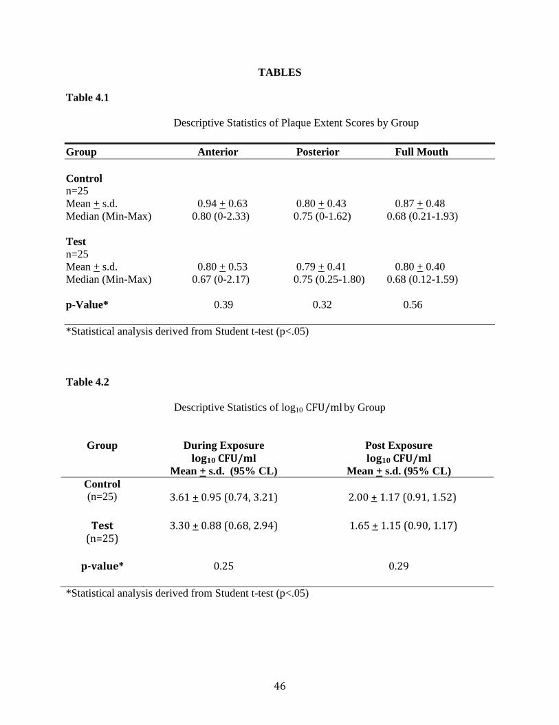
46
TABLES
Table 4.1
Descriptive Statistics of Plaque Extent Scores by Group
Group Anterior Posterior Full Mouth Control n=25 Mean + s.d. 0.94 + 0.63 0.80 + 0.43 0.87 + 0.48 Median (Min-Max) 0.80 (0-2.33) 0.75 (0-1.62) 0.68 (0.21-1.93) Test n=25 Mean + s.d. 0.80 + 0.53 0.79 + 0.41 0.80 + 0.40 Median (Min-Max) 0.67 (0-2.17) 0.75 (0.25-1.80) 0.68 (0.12-1.59) p-Value* 0.39 0.32 0.56 *Statistical analysis derived from Student t-test (p<.05)
Table 4.2
Descriptive Statistics of log10 CFU/ml by Group
Group During Exposure log10 CFU/ml
Mean + s.d. (95% CL)
Post Exposure log10 CFU/ml
Mean + s.d. (95% CL) Control (n=25)
3.61 + 0.95 (0.74, 3.21)
2.00 + 1.17 (0.91, 1.52)
Test
(n=25)
3.30 + 0.88 (0.68, 2.94) 1.65 + 1.15 (0.90, 1.17)
p-value*
0.25 0.29
*Statistical analysis derived from Student t-test (p<.05)
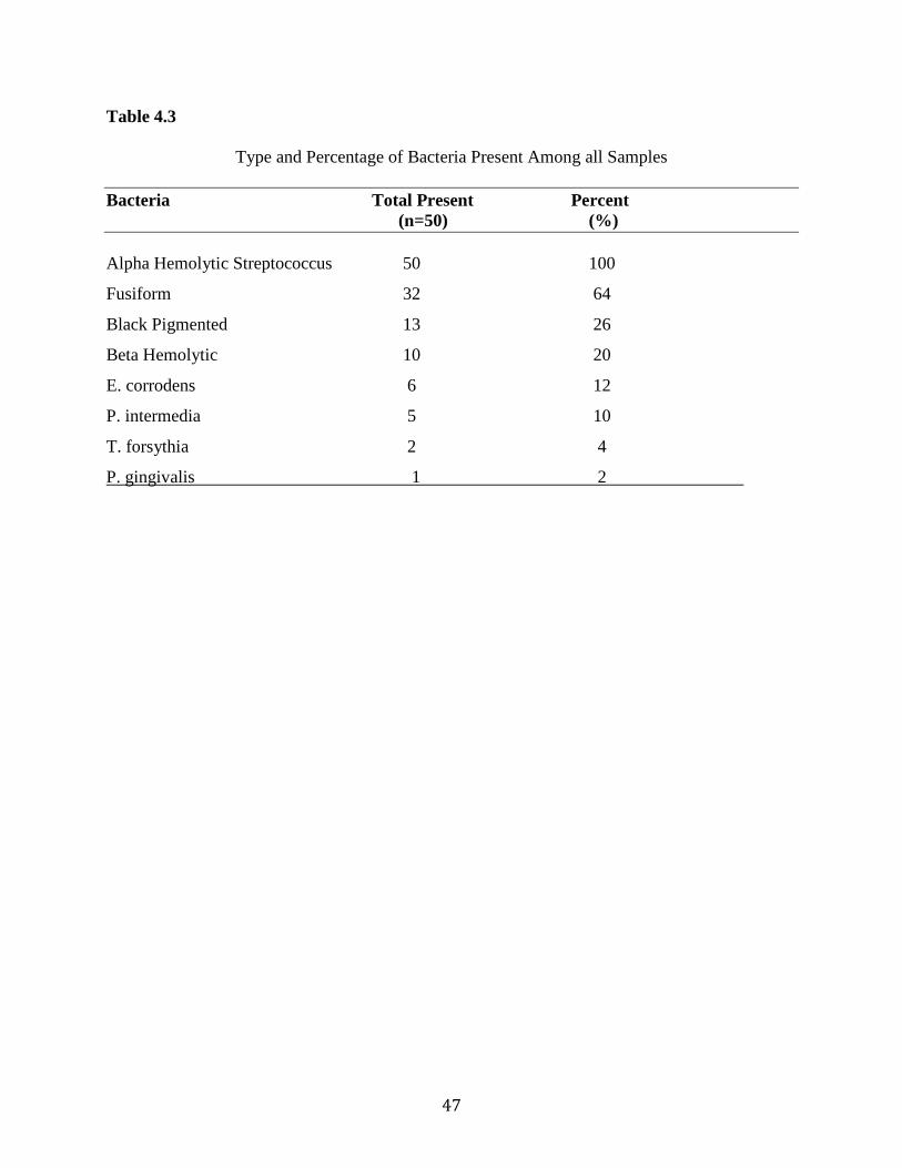
47
Table 4.3
Type and Percentage of Bacteria Present Among all Samples
Bacteria Total Present Percent (n=50) (%) Alpha Hemolytic Streptococcus 50 100
Fusiform 32 64
Black Pigmented 13 26
Beta Hemolytic 10 20
E. corrodens 6 12
P. intermedia 5 10
T. forsythia 2 4
P. gingivalis 1 2
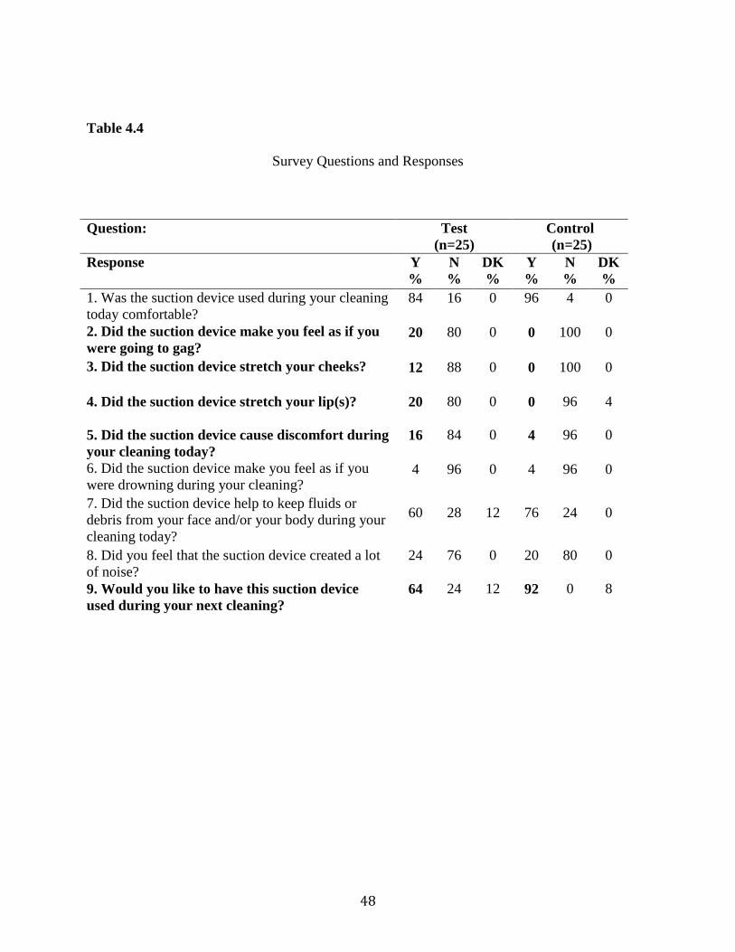
48
Table 4.4
Survey Questions and Responses
Question: Test
(n=25) Control (n=25)
Response Y %
N %
DK %
Y %
N %
DK %
1. Was the suction device used during your cleaning today comfortable?
84 16 0 96 4 0
2. Did the suction device make you feel as if you were going to gag?
20 80 0 0 100 0
3. Did the suction device stretch your cheeks? 12 88 0 0 100 0
4. Did the suction device stretch your lip(s)? 20 80 0 0 96 4
5. Did the suction device cause discomfort during your cleaning today?
16 84 0 4 96 0
6. Did the suction device make you feel as if you were drowning during your cleaning?
4 96 0 4 96 0
7. Did the suction device help to keep fluids or debris from your face and/or your body during your cleaning today?
60 28 12 76 24 0
8. Did you feel that the suction device created a lot of noise?
24 76 0 20 80 0
9. Would you like to have this suction device used during your next cleaning?
64 24 12 92 0 8
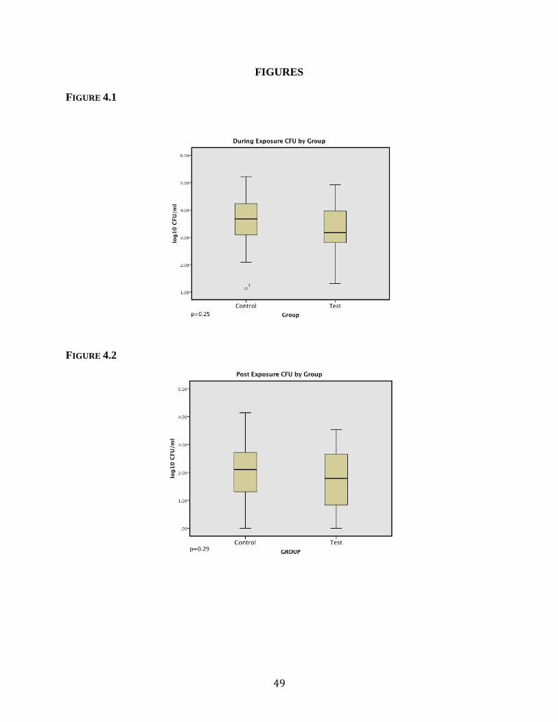
49
FIGURES FIGURE 4.1
FIGURE 4.2
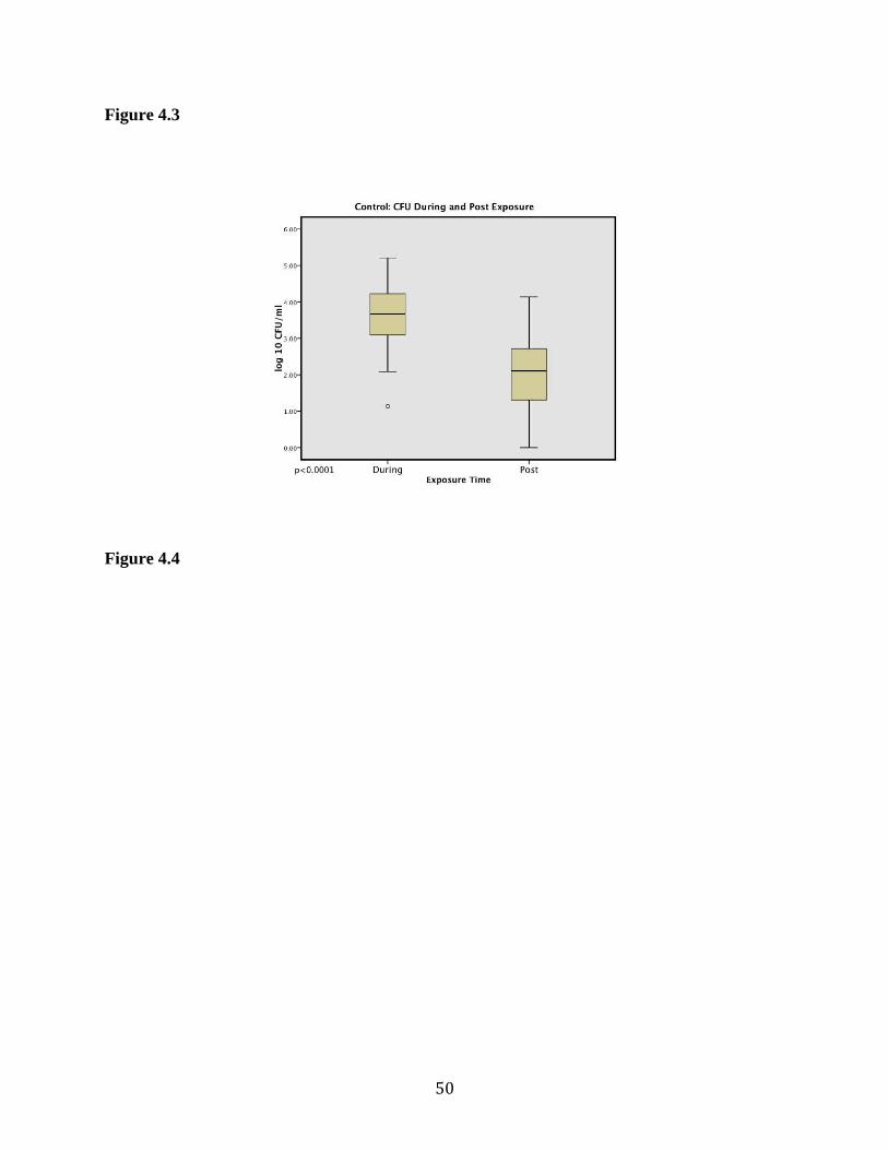
Figure 4.3
Figure 4.4
50
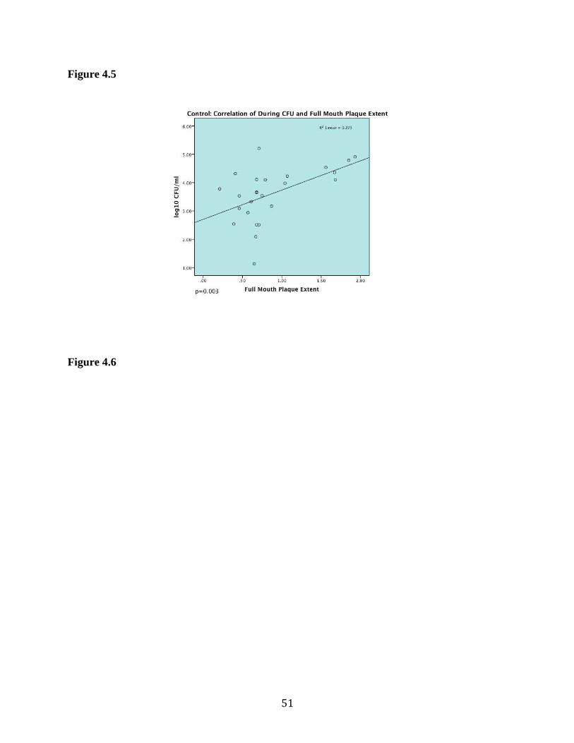
Figure 4.5
Figure 4.6
51
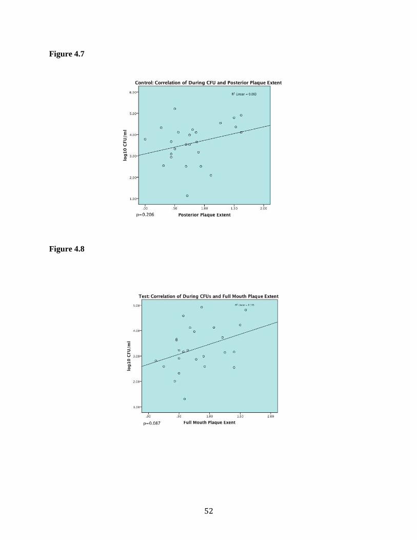
52
Figure 4.7
Figure 4.8
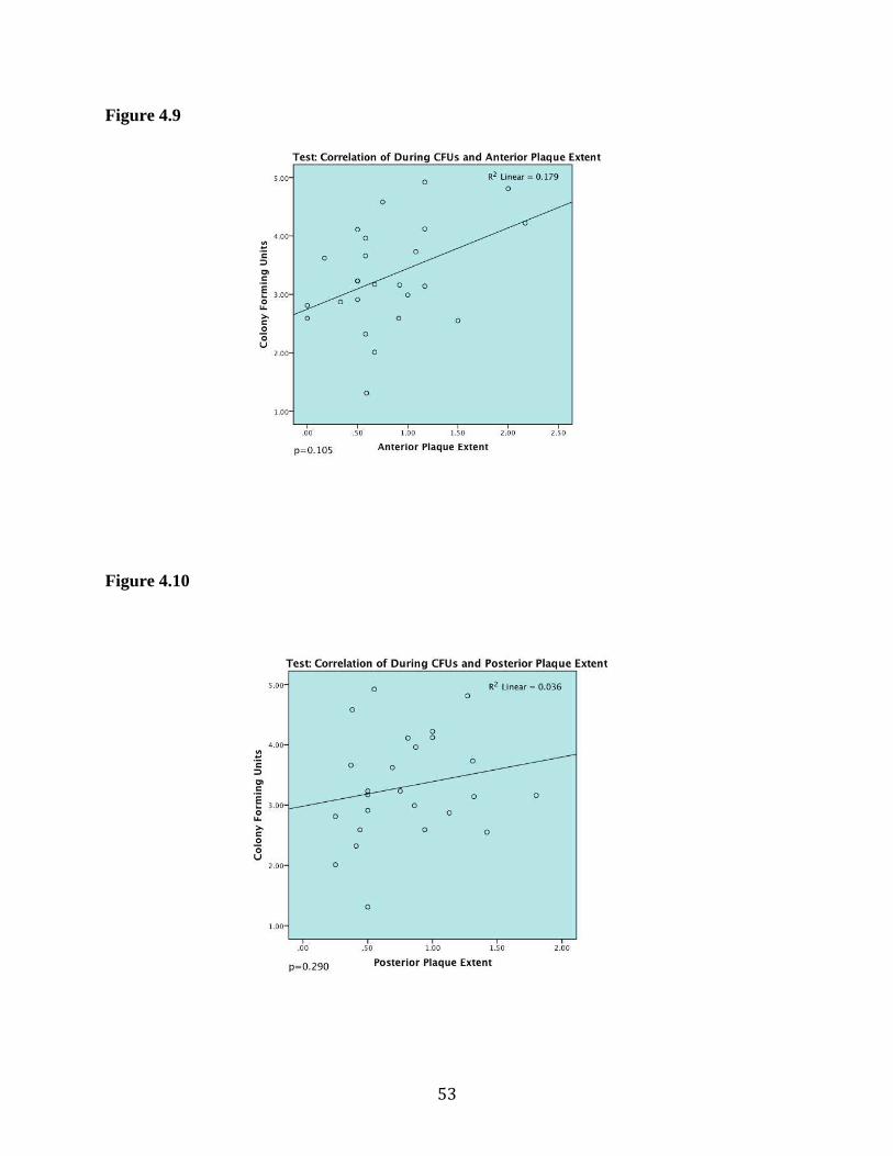
53
Figure 4.9
Figure 4.10
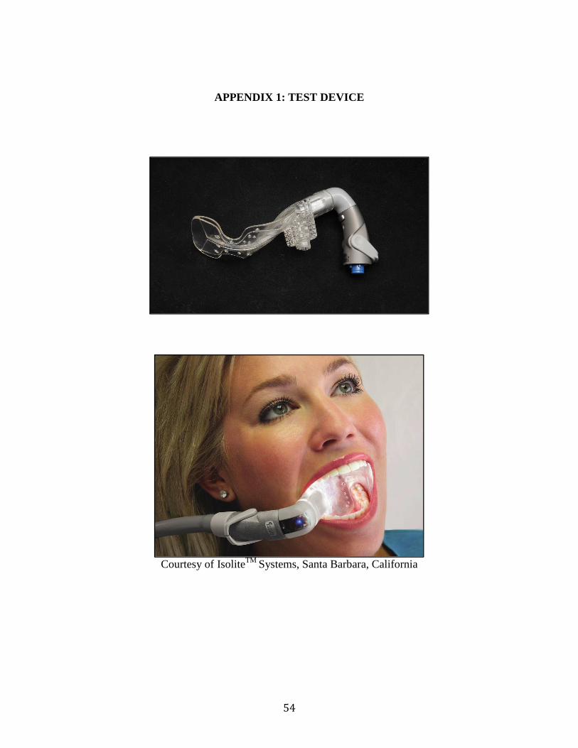
54
APPENDIX 1: TEST DEVICE
Courtesy of IsoliteTM Systems, Santa Barbara, California
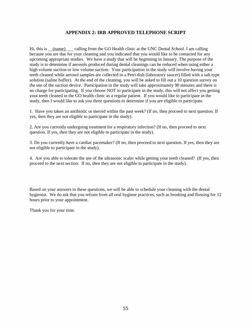
55
APPENDIX 2: IRB APPROVED TELEPHONE SCRIPT Hi, this is (name) calling from the GO Health clinic at the UNC Dental School. I am calling because you are due for your cleaning and you indicated that you would like to be contacted for any upcoming appropriate studies. We have a study that will be beginning in January. The purpose of the study is to determine if aerosols produced during dental cleanings can be reduced when using either a high volume suction or low volume suction. Your participation in the study will involve having your teeth cleaned while aerosol samples are collected in a Petri dish (laboratory saucer) filled with a salt-type solution (saline buffer). At the end of the cleaning, you will be asked to fill out a 10 question survey on the use of the suction device. Participation in the study will take approximately 90 minutes and there is no charge for participating. If you choose NOT to participate in the study, this will not affect you getting your teeth cleaned in the GO health clinic as a regular patient. If you would like to participate in the study, then I would like to ask you three questions to determine if you are eligible to participate. 1. Have you taken an antibiotic or steroid within the past week? (If no, then proceed to next question. If yes, then they are not eligible to participate in the study). 2. Are you currently undergoing treatment for a respiratory infection? (If no, then proceed to next question. If yes, then they are not eligible to participate in the study). 3. Do you currently have a cardiac pacemaker? (If no, then proceed to next question. If yes, then they are not eligible to participate in the study). 4. Are you able to tolerate the use of the ultrasonic scaler while getting your teeth cleaned? (If yes, then proceed to the next section. If no, then they are not eligible to participate in the study). Based on your answers to these questions, we will be able to schedule your cleaning with the dental hygienist. We do ask that you refrain from all oral hygiene practices, such as brushing and flossing for 12 hours prior to your appointment. Thank you for your time.
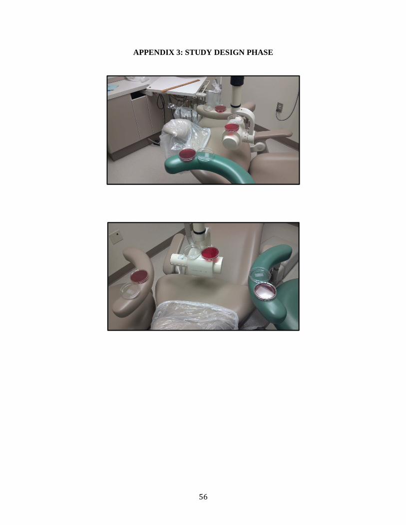
56
APPENDIX 3: STUDY DESIGN PHASE
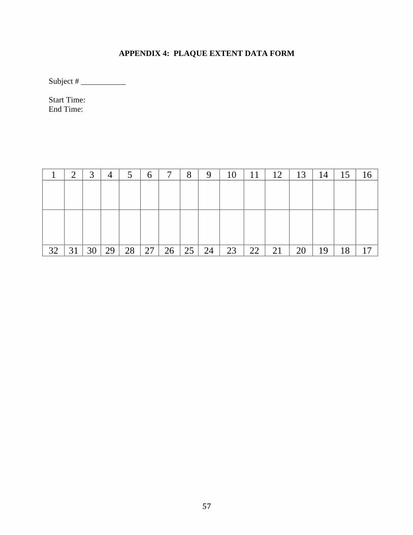
57
APPENDIX 4: PLAQUE EXTENT DATA FORM
Subject # ___________ Start Time: End Time:
1 2 3 4 5 6 7 8 9 10 11 12 13 14 15 16
32 31 30 29 28 27 26 25 24 23 22 21 20 19 18 17
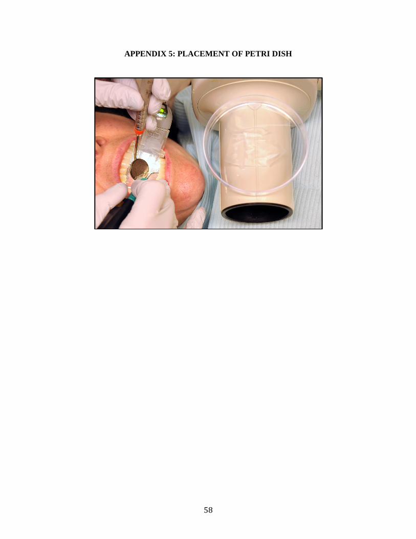
58
APPENDIX 5: PLACEMENT OF PETRI DISH
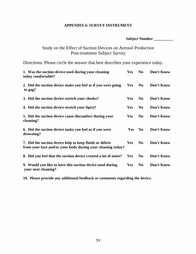
59
APPENDIX 6: SURVEY INSTRUMENT
Subject Number __________
Study on the Effect of Suction Devices on Aerosol Production Post-treatment Subject Survey
Directions: Please circle the answer that best describes your experience today. 1. Was the suction device used during your cleaning Yes No Don’t Know today comfortable? 2. Did the suction device make you feel as if you were going Yes No Don’t Know to gag? 3. Did the suction device stretch your cheeks? Yes No Don’t Know 4. Did the suction device stretch your lip(s)? Yes No Don’t Know 5. Did the suction device cause discomfort during your Yes No Don’t Know cleaning? 6. Did the suction device make you feel as if you were Yes No Don’t Know drowning? 7. Did the suction device help to keep fluids or debris Yes No Don’t Know from your face and/or your body during your cleaning today? 8. Did you feel that the suction device created a lot of noise? Yes No Don’t Know 9. Would you like to have this suction device used during Yes No Don’t Know your next cleaning? 10. Please provide any additional feedback or comments regarding the device.
60
APPENDIX 7: LAB CFU RECORDING FORM
SUBJECT # ________ Bacteria Present:
CFU COUNTS – During use of ultrasonic Plate #1 Plate #2 Plate #3 Average:
@ 24-48 hrs method:
@ 72-96 hrs method:
@ 5-7 days method:
CFU COUNTS – Post use of Ultrasonic Plate #1 Plate #2 Plate #3 Average:
@ 24-48 hrs method:
@ 72-96 hrs method:
@ 5-7 days method:
61
REFERENCES
1. Snider D (1992). Occupational Safety Health Report- Centers for Disease Control and Prevention, 1125.
2. Miller RL, Micik RE, Able C, Ryge G (1971). Studies of dental aerobiology: II. microbial splatter discharge from the oral cavity of dental patients. Journal of Dental Research 50:621-625.
3. Miller RL, Micik RE (1978). Air pollution and its control in the dental office. Dental Clinics of North America Journal 22:453-476.
4. Kohn WG, Collins AS, Clebeland JL, Harte JA, Eklund KJ, Malvitz DM (2003). Centers for Disease Control and Prevention (CDC) Guidelines for infection control in dental health-care setting. Morbidity and Mortality Weekly Report, 52(17):16-70.
5. Harrell SK (2004). Airborne spread of disease- implication for dentistry. Journal of the California Dental Association 32:901-906.
6. Centers for Disease Control and Prevention. Recommended infection-control practices for dentistry. MMWR (1993) (accessed2012 Sept 12]; 42(No. RR-8). Available from http://www.cdc.gov/mmwr/preview/mmwrhtml/00021095.htm
7. American Dental Association 2012 Current Policies. [Internet] 2012 [cited 2012 Sept]; Available from: http://www.ada.org/currentpolicies.aspx
8. IsoliteTM Systems, Santa Barbara, California
9. Jacks, ME (2002). A laboratory comparison of evacuation devices on aerosol reduction. Journal of Dental Hygiene 76(3):202-206.
10. Cole EG, Cook CE (1998). Characterization of infectious aerosols in health care facilities: An aid to effective engineering controls and preventive strategies. American Journal of Infection Control 26(4):453-464.
11. Finegold SM (1991). Aspiration pneumonia. Review of Infectious Disease. 13:S737-S742.
12. Siegman-Igra Y, Schwartz D, Ophir O, Konforti N. (1984). Endocarditis caused by Actinobacillus actinomycetemcomittans. European Journal of Clinical Microbiology 3:556-559. 13. Limeback, H (1998). Implications of oral infections on systemic disease in the institutionalized elderly with a special focus on pneumonia. Annals of Periodontology. 3(1): 262-275.
62
14. Muir KF, Ross PW, Macphee IT, Holbrook WP, Kowolik MJ (1978). Reduction of microbial contamination from ultrasonic scalers. British Dental Journal. 145:76-78. 15. Micik RE, Miller RL, Mazzarella MA, Ryge G (1969). Studies on dental aerobiology: I. bacterial aerosols generated during procedures. Journal of Dental Research, 48:49-56. 16. Micik RE, Miller RL, Leong AC (1971). Studies on dental aerobiology, III: Efficacy of surgical masks in protecting dental personnel from airborne bacterial particles. Journal of Dental Research. 50:626-630 17. Abel LC, Miller RL, Micik RE, Ryge G (1971). Studies on dental aerobiology, IV: Bacterial contamination of water delivered by dental units. Journal of Dental Research. 50: 1567-1590 18. Sze To GN, Wan MP, Chao C, Wei F, Yu SCT, Kwan JKC. (2008). A methodology for estimating airborne virus exposures in indoor environments using the spatial distribution of expiratory aerosols and virus viability characteristics. Journal of Indoor Air. 18:425-438. 19. Wells WF, Stone WR (1934). On air-borne infection. Study III. Viability of droplet nuclei infection. American Journal of Dental Hygiene. 20:619-26 20. Wells WF (1955). Airborne contagion and air hygiene. Cambridge University Press,117-122. 21. Eickhoff TC (1994). Airborne nosocomial infection: a contemporary perspective. Infection Control Hospital Epidemiology.15: 663-72. 22. Beck-Sague C, Dooley SW, Hutton MD, Crawford JT, Williams J, Sordillo EM et al. (1992). An outbreak of multidrug resistant tuberculosis among hospitalized patients with the acquired immunodeficiency syndrome. New England Journal of Medicine. 326: 1514-21. 23. Joseph CA, Watson JM, Harrison TG, Bartlett CL (1994). Nosomocial legionnaires’ diseases in England and Wales, 1980-1992. Epidemiol Infect 112: 329-45. 24. Bloch AB, Orenstein WA, Ewing WM, Spain WH, Mallison GF, Herrmann KL, et al. (1985) Measles outbreak in a pediatric practice: airborne transmission in an office setting. Pediatrics 75: 676-83. 25. Leggat PA, Kedjaurine U (2001). Bacterial aerosols in the dental clinic: A review. International Dental Journal 51(1): 39-44. 26. Centers for Disease Control and Prevention (1992). Transmission of multidrug resistant tuberculosis among immunocompromised persons in a correctional system. Massachusetts Medical Society, Waltham. MMWR Morbidity and Mortality Weekly Report July 1992 41(28):507-590.
63
27. Larato DC, Ruskin P, Martin A (1967). Effect of an ultrasonic scaler on bacterial counts in air. Journal of Periodontology 38:550-554. 28. Williams GH, Pollok NL, Shay De Barr CE (1970). Laminar air purge of microorganisms in dental aerosols: Prophylactic procedures with the ultrasonic scaler. Journal of Dental Research 49: 1498-1504 29. Harrel S, Barnes J, Rivera-Hidalgo F (1998). Aerosol and splatter contamination from the operative site during ultrasonic scaling. Journal of the American Dental Association 129: 1241-1249. 30. Basu MK, Browne RM, Potts AJC. (1988). A survey of aerosol related symptoms in dental hygienists. J Soc Occup Med 38: 23-35. 31. Bentley CD, Burkhart NW, Crawford JJ (1994). Evaluating splatter and aerosol contamination during dental procedures. Journal of the American Dental Association 125: 579-584 32. Klyn SL, Cummings DE, Richardon BW, Davis RD (2001). Reduction of bacteria-containing spray produced during ultrasonic scaling. Journal of General Dentistry 49(6): 648-652 33. King TB, Muzin KB, Berry CW, Ander LM (1997) The effectiveness of an aerosol reduction device for ultrasonic scalers. Journal of Periodontology 68:45-49 34. Harrel SK, Barnes JB, Rivera-Hidalgo F (1996). Reduction of aerosols produced by ultrasonic scalers. Journal of Periodontology 67(1): 28-32 35. Jacks M, Pollard C (2007). IsoliteTM v. High Volume Evacuation: Effects on dental aerosol reduction. Unpublished. 36. Dahlke WO, Cottam MR, Herring MC, Leavitt JM, Ditmyer MM, Walker RS (2012). Evaluation of the spatter-reduction effectiveness of two dry-field isolation techniques. Journal of the American Dental Association 143(11):1199-1204. 37. Noro A, Kameyama A, Asami M, Sugiyama T, Morinaga K, Kondou Y, Tsunoda M (2009). Clinical usefulness of "IsoliteTM plus: For oral environment of Japanese people. The Bulletin of Tokyo Dental College 50(3):149-155. 38. Colette J, Wilson S, Sullivan D (2010). A study of the IsoliteTM system during sealant placement: Efficacy and patient acceptance. Journal of American Pediatric Dentistr 32(2): 146-150. 39. Andersen A (1958) A new sampler for the collection, sizing, and enumeration of viable airborne particles. J Bact 76:471-484.
64
40. Rautemaa R, Nordberg A, Wuolijoki-Saaristo K, Meruman JH (2006). Bacterial aerosols in dental practice – a potential hospital infection problem? Journal of Hospital Infection 64:76-81. 41. Grenier D (1995). Quantitative analysis of bacterial aerosols in two different dental clinic environments. Applied Environmental Microbiology 61:3165-3168. 42. Feres M, Figueiredo LC, Faveri M (2010). The effectiveness of a preprocedural mouthrinse containing cetylpyridinium chloride in reducing the bacteria in the dental office. Journal of the American Dental Association. 141(4):415-422.