Embed Size (px)
Citation preview
Campbell University Campbell University
CU FIND CU FIND
Osteopathic Medicine, Jerry M. Wallace School of Faculty Research and Publications
7-1990
The effect of oxygen on corneal neovascularization The effect of oxygen on corneal neovascularization
M. Culton
D. B. Chandler
A. D. Proia
D. Hickingbotham
G. K. Klintworth
Follow this and additional works at: https://cufind.campbell.edu/medicine_school
Part of the Medical Sciences Commons, and the Other Pharmacy and Pharmaceutical Sciences
Commons
Recommended Citation Recommended Citation Culton, M.; Chandler, D. B.; Proia, A. D.; Hickingbotham, D.; and Klintworth, G. K., "The effect of oxygen on corneal neovascularization" (1990). Osteopathic Medicine, Jerry M. Wallace School of. 502. https://cufind.campbell.edu/medicine_school/502
This Article is brought to you for free and open access by the Faculty Research and Publications at CU FIND. It has been accepted for inclusion in Osteopathic Medicine, Jerry M. Wallace School of by an authorized administrator of CU FIND. For more information, please contact [email protected].
Investigative Ophthalmology & Visual Science, Vol. 31, No. 7, July 1990Copyright © Association for Research in Vision and Ophthalmology
The Effect of Oxygen on Corneol NeovascularizationMark Culton, David B. Chandler, Alan D. Proia, Dyson Hickingborham, and Gordon K. Klinrworrh
Since tissue oxygen levels are believed to play a pivotal role in new vessel growth in several situations,we studied the effect of several oxygen concentrations (0,10, 21,50, 75, or 100%) on corneal vascular-ization induced in the rat by chemical cautery. We achieved this by perfusing known concentrations ofoxygen through goggles fitted over both eyes of the rat after corneal cauterization. Neovascularizationwas measured in flat corneal preparations with India ink-filled vessels 4 days postcautery usingcomputerized image analysis. The angiogenic response of rats whose eyes were continuously exposedto 0-75% oxygen were not significantly different from each other. The mean response in corneasexposed to 100% oxygen was 10-21% lower than all of the other groups, and this difference wasstatistically significant when compared to oxygen concentrations of 0, 21 and 75%. The reason for theinhibitory effect of 100% oxygen remains to be determined, but it may represent a toxic effect ofoxygen free radicals on the vascular endothelium. Invest Ophthalmol Vis Sci 31:1277-1281,1990
Tissue hypoxia is considered to play an essentialrole in the induction of neovascularization in certaindisorders, such as the retinopathies of prematurityand diabetes mellitus1'2 and in wound-healing.3 Hyp-oxia also leads to an increased capillary density inskeletal and cardiac muscle.4"8 Conversely, direct va-soobliterative and constructive effects of oxygen havebeen observed in the mammalian retina,9 and in-creased oxygen tension is believed to underlie therelative absence of capillaries around retinal arteri-oles as compared to venules.101' In a clinical study ona few patients using a subjective rating scale based onslit-lamp biomicroscopic examination and minimalfollow-up, subconjunctival oxygen injections havebeen reported to have a vasoobliterative and vasoin-hibitory effect on small superficial corneal vessels inhumans.1213
Several investigators have studied the effect of hy-peroxia and hyperbaric oxygen on neovascularizationinduced by corneal injuries in rabbits and guineapigs.9>14"17 All showed no effect of oxygen on the cor-neal neovascular response, but all of these studieslacked objective quantitative methods for evaluatingthe angiogenic response. We investigated the effect of
From the Departments of Ophthalmology and Pathology, DukeUniversity Medical Center, Durham, North Carolina.
Supported by grants R01 EY-00146, R01 EY-05883, P30EY-05722, and IS 10 RR02339 from the National Eye Institute.
Presented in part on May 2, 1988 at the annual meeting of theAssociation for Research in Vision and Ophthalmology, Sarasota,Florida.
Submitted for publication: July 28, 1989; accepted September20, 1989.
Reprint requests: Gordon K. Klintworth, MD, PhD, Duke Uni-versity Eye Center, Durham, NC 27710.
various oxygen concentrations on corneal neovascu-larization induced in the rat by chemical cautery. Forthese studies we constructed special goggles thatcould be perfused with known concentrations of oxy-gen without influencing the breathing environmentof the animal. In this way, extreme hyperoxia couldbe studied for longer periods of exposure without thelimiting constraint of animal morbidity and mortal-ity due to pulmonary oxygen toxicity. Likewise, theeffect of extreme local hypoxia could be evaluated.The degree of corneal neovascularization in the dif-ferent experimental groups was compared using com-puterized image analysis of corneal flat preparationswith India ink-filled vessels.
Materials and Methods
Corneal Cauterization
Both eyes of Sprague Dawley male rats weighing250-375 g (Harlan Sprague Dawley, Indianapolis,IN) were cauterized by applying the tip of a silver/potassium nitrate applicator (75% silver nitrate / 25%potassium nitrate, no. 2867-1590; Graham-FieldSurgical, New Hyde Park, NY) to the center of thecornea of ether anesthetized rats for 4.5 sec. A freshapplicator stick was used for each cornea. Immedi-ately after cauterization, the cautery site was gentlyblotted with tissue paper. The care and maintenanceof the rats used in these experiments conformed tothe ARVO Resolution on the Use of Animals in Re-search.
Goggles
To permit the maintenance of local nonatmo-spheric oxygen concentrations to the eyes, goggles
1977
Downloaded from iovs.arvojournals.org on 08/20/2019
1278 INVESTIGATIVE OPHTHALMOLOGY & VISUAL SCIENCE / July 1990 Vol. 31
were constructed from soft rubber microscope eyepieces (Carl Zeiss, Oberkochen, West Germany),which were contoured to fit a rat's head. Plexiglass®(Rohm and Haas, Philadelphia, PA) covers weremolded and cut to the curvature of the eyepieces andattached circumferentially with epoxy resin. Holeswere then drilled in the right and left sides of thePlexiglass® plates for the insertion of stainless steelinlet and outlet ports to which flexible plastic tubingwas attached. The soft rubber rim of the goggles wassecured to the head of anesthetized animals with tape(Durapore; 3M, St. Paul, MN), a thin circumferentialstrip of foam underpadding (Depend underpads;Kimberly-Clark Health Care Products Group, Ros-well, GA), and cyanoacrylate glue (Duro Quick-Gel,No-Run Superglue; Loctite, Cleveland, OH) (Fig. 1).The tape and foam padding were necessary to ensurean airtight seal. When in place, the eyes of each ani-mal were enclosed, with a margin left between theeyes and the edge of the mask to allow free movementof the eyelids.
Animal Preparation and Maintenance
To prevent the animals from removing their gog-gles, each rat was restrained in a harness (HarvardBiosciences, South Natick, MA) that allowed freemovement of all four limbs, the tail, head, and neck.Prior to placement in the harness, the upper halves ofthe forelegs and shoulders of each rat were wrappedwith surgical tape (Durapore; 3M). Each animal,within its harness/frame apparatus, was providedconstant access to food and water.
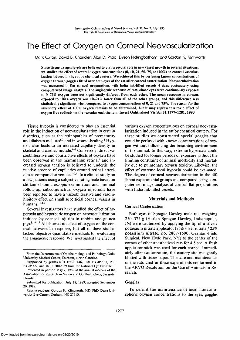
0 2 PROJECTGas Regulators
Humidifier Inlet Tube
Fig. 1. Method of administration of different percentages ofoxygen to the eyes.
Administration of Oxygen
Different mixtures of oxygen and nitrogen (HighPurity Nitrogen/Extra Dry Grade Oxygen; NationalWelders, Raleigh, NC) were used to achieve oxygenconcentrations of 0, 10, 21 (simulating room air), 50,75, and 100. The eyes of 20 rats were continuouslyexposed to each of the different oxygen concentra-tions for 4 days via the goggles. The gas was distrib-uted through plastic tubing and circulated through ahumidifier before reaching the inlet port of the gog-gles. The regulation of gas flow from the gas tanks wasaccomplished through the use of low pressure, two-stage regulators (Mathison Instruments, Newark,NJ). When 100% oxygen or nitrogen was adminis-tered, the gas was channeled directly from the tanksto the humidifier. The gas finally reached the gogglesthrough plastic tubing attached directly to the metal-lic inlet port positioned over the left eye. A section ofthe tubing was attached to the outlet port over theright eye and was vented over the edge of the rodentcage (Fig. 1). For mixing of oxygen and nitrogen, agas proportioner (Mathison Instruments), was used.The tubing was connected to each gas proportioner sothat the gas was channeled through the same systemof humidification and Y-tubing. The flow rate wasapproximately 0.5 liters/min.
Gas Sampling
On days 1 and 3 of each experiment, the oxygenconcentration reaching the corneas was determinedby slowly withdrawing 20 ml of the gas from the out-let tubing of the goggles into a plastic syringe, whichwas quickly sealed with an airtight rubber cap. Con-firmation of the oxygen concentration in the speci-mens with 0 and 100% concentrations was performedusing a pH/blood gas analyzer (Instrument Labora-tory Systems; Lexington, MA) that gave oxygen read-ings in mmHg. Determination of the oxygen concen-tration in the oxygen/nitrogen mixtures was per-formed with a pH/blood gas analyzer (InstrumentLaboratory Systems) that provided readings as per-centage oxygen concentration and was accurate towithin 0.1%.
Visualization of Corneal Blood Vessels
Four days after corneal cauterization, an intervalthat consistently provides prominent corneal vascu-larization in this experimental model,18 each rat wasdeeply anesthetized with intraperitoneal 1.0% so-dium pentobarbital (50 mg/kg) and the upper torsowas perfused with a mixture of 11% gelatin-10%India ink-lactated Ringer's solution. The degree ofvascularization of each cornea was quantitated in
Downloaded from iovs.arvojournals.org on 08/20/2019
No. 7 THE EFFECT OF OXYGEN ON CORNEAL NEOVASCULARIZATION / Culron er ol 1279
OH,
corneal flat preparations by computerized imageanalysis as described by Proia et al.19
Results
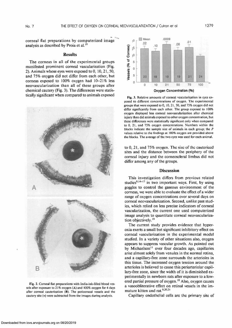
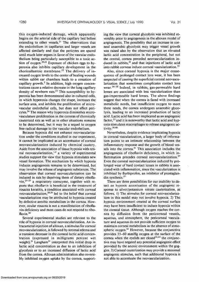
The corneas in all of the experimental groupsmanifested prominent corneal vascularization (Fig.2). Animals whose eyes were exposed to 0, 10, 21, 50,and 75% oxygen did not differ from each other, butcorneas exposed to 100% oxygen had 10-21% lessneovascularization than all of these groups afterchemical cautery (Fig. 3). The differences were statis-tically significant when compared to animals exposed
.0009
Fig. 2. Corneal flat preparations with India-ink-hTled blood ves-sels after exposure to 21% oxygen (A) and 100% oxygen for 4 daysafter corneal cauterization (B). The pericorneal vessels and thecautery site (*) were subtracted from the images during analysis.
10 21 50
Oxygen Concentration (%)
24
100
Fig. 3. Relative amounts of corneal vascularization in eyes ex-posed to different concentrations of oxygen. The experimentalgroups that were exposed to 0, 10, 21, 50, and 75% oxygen did notdiffer significantly from each other. The group exposed to 100%oxygen displayed less corneal neovascularization after chemicalinjury than did animals exposed to other oxygen concentration, butthese differences were statistically significant only when comparedto 0, 21, and 75% oxygen concentrations. Numbers within theblocks indicate the sample size of animals in each group; the Pvalues relative to the findings at 100% oxygen are provided abovethe blocks. The average of the two eyes was used for each animal.
to 0, 21, and 75% oxygen. The size of the cauterizedsites and the distance between the periphery of thecorneal injury and the corneoscleral Hmbus did notdiffer among any of the groups.
Discussion
This investigation differs from previous relatedstudies9'14"17 in two important ways. First, by usinggoggles to control the gaseous environment of thecorneas, we were able to evaluate the effect of a widerrange of oxygen concentrations over several days oncorneal neovascularization. Second, unlike past stud-ies, which relied on less precise indicators of cornealvascularization, the current one used computerizedimage analysis to quantitate corneal neovasculariza-tion objectively.19
The current study provides evidence that hyper-oxia exerts a small but significant inhibitory effect oncorneal vascularization in the experimental modelstudied. In a variety of other situations also, oxygenappears to suppress vascular growth. As pointed outby Michaelson" over four decades ago, capillariesarise almost solely from venules in the normal retina,and a capillary-free zone surrounds the arterioles inthis tissue. The increased oxygen tension around thearterioles is believed to cause this periarteriolar capil-lary-free zone, since the width of it is diminished ex-perimentally in newborn rats after exposure to a low-ered partial pressure of oxygen.10 Also, oxygen causesa vasoobliterative effect on retinal vessels in the im-mature kitten and rat.9'20'21
Capillary endothelial cells are the primary site of
Downloaded from iovs.arvojournals.org on 08/20/2019
1280 INVESTIGATIVE OPHTHALMOLOGY & VISUAL SCIENCE / July 1990 Vol. 31
this oxygen-induced damage, which apparentlybegins on the arterial side of the capillary bed beforeextending to other vessels.21 The observations thatthe endothelium in capillaries and larger vessels areaffected similarly and that the pericytes are spareduntil much later argues in favor of the vascular endo-thelium being particularly susceptible to a toxic ac-tion of oxygen.20'21 Exposure of chicken eggs to hy-peroxia also inhibits capillary development in thechorioallantoic membrane.22 The delivery of in-creased oxygen levels to the center of healing woundswithin rabbit ear chambers leads to a cessation ofcapillary growth.3 In addition, high oxygen concen-trations cause a relative decrease in the lung capillarydensity of newborn rats.23 This susceptibility to hy-peroxia has been demonstrated also in cultured cells,in which hyperoxia changes the shape, increases thesurface area, and inhibits the proliferation of micro-vascular endothelial cells, but does not affect peri-cytes.24 The reason why hyperoxia diminishes micro-vasculature proliferation in the corneas of chemicallycauterized rats as well as in other situations remainsto be determined, but it may be a sequel to oxygenfree-radical damage to the vascular endothelium.
Because hypoxia did not enhance neovasculariza-tion under the conditions studied in our experiment,it cannot be implicated as an accentuator of cornealneovascularization induced by chemical cautery.Aside from the association of tissue hypoxia with reti-nal neovascularization,12 a variety of experimentalstudies support the view that hypoxia stimulates newvessel formation. The mechanism by which hypoxiainduces angiogenesis remains to be determined, butmay involve the release of angiogenic substances. Theobservation that corneal neovascularization can beinduced in rats by depriving them of dietary ribofla-vin,25"27 a respiratory coenzyme, together with re-ports that riboflavin is beneficial in the treatment ofrosacea keratitis, a condition associated with cornealneovascularization,2829 led to the belief that cornealvascularization may be attributed to hypoxia createdby defective aerobic metabolism in the cornea. How-ever, ocular rosacea is not a manifestation of ribofla-vin deficiency and most cases do not respond to ribo-flavin.30
Several experimental studies are relevant to therole of hypoxia in corneal neovascularization. An in-tracameral injection of alloxan, a stimulus for cornealneovascularization, is followed by stromal edema anda transient decrease in the corneal lactic acid concen-tration (expressed in milligrams percent wetweight).31 Langham31 interpreted this initial drop inlactic acid concentration as due to an inhibition ofglycolysis or to an increased diffusion of lactic acidfrom the cornea. Alloxan administration also reversi-bly inhibited oxygen uptake by the cornea, support-
ing the view that corneal glycolysis was inhibited re-versibly prior to angiogenesis in the alloxan model ofangiogenesis. The possibility that an increase in cor-neal anaerobic glycolysis may trigger vessel growthwas raised also by the observation that an elevatedlactic acid concentration in the peripheral, but notthe central, cornea preceded neovascularization in-duced in rabbits,32 and that injections of lactic acidinto rabbit corneas induce corneal vascularization.33
Also, since corneal hypoxia is the major conse-quence of prolonged contact lens wear, it has beensuspected of causing the superficial corneal neovascu-larization that sometimes complicates contact lenswear.34"38 Indeed, in rabbits, gas-permeable hardlenses are associated with less vascularization thangas-impermeable hard lenses. The above findingssuggest that when the cornea is faced with increasedmetabolic needs, but insufficient oxygen to meetthese needs, the cornea undergoes anaerobic glyco-lysis, leading to an increased production of lacticacid. Lactic acid has been implicated as an angiogenicfactor,33 and it is noteworthy that lactic acid and hyp-oxia stimulates macrophages to release angiogenic ac-tivity.39'40
Nevertheless, despite evidence implicating hypoxiain corneal vascularization, a larger body of informa-tion points to an intimate relationship between theinflammatory response and the growth of blood ves-sels into the cornea.41 This association includes theangiogenesis of riboflavin deficiency, in which in-flammation precedes corneal neovascularization.27
Even the corneal neovascularization induced by pro-longed wear of hard contact lenses in rabbits is asso-ciated with inflammation, since the vascularization isinhibited by flurbiprofen, an inhibitor of prostaglan-din synthesis.37
There are three possibilities for our inability to de-tect an hypoxic accentuation of the angiogenic re-sponse to silver/potassium nitrate cauterization, asfollows. 1) The stimulus for corneal neovasculariza-tion in this model may not involve hypoxia. 2) Thehypoxic environment created at the corneal surfacemay have been insufficient to induce hypoxia withinthe corneal tissue. Although oxygen reaches the cor-nea by diffusion from the pericorneal vessels,aqueous, and atmosphere, the pericorneal vascula-ture and aqueous do not provide sufficient oxygen tomaintain corneal metabolism in the absence of atmo-spheric oxygen.42 However, because the conjunctivaprovides 55-60 mmHg oxygen at the surface of thecornea when the eyelids are closed35'43 the conjunc-tiva may have negated any potential angiogenic effectprovided by the anoxic environment within the gog-gles. 3) Corneal cauterization may provide a maximalangiogenic stimulus, such that additional hypoxia isnot able to accentuate the neovascularization.
Downloaded from iovs.arvojournals.org on 08/20/2019
No. 7 THE EFFECT OF OXYGEN ON CORNEAL NEOVASCULARIZATION / Culron er ol 1281
In summary, we have shown that perfusing thecorneal surface with 100% oxygen causes a small butsignificant reduction in corneal neovascularizationafter silver nitrate/potassium nitrate cauterization.Hypoxia had no affect on the neovascular response inthis model. These results suggest that tissue oxygenlevels are not a major influence on corneal neovascu-larization in our experimental model.
Key words: hypoxia, hyperoxia, angiogenesis, rat, cornea
References
1. Garner A: Ocular angiogenesis. Int Rev Exp Pathol 28:249,1986.
2. Patz A: Current concepts of the effect of oxygen on the devel-oping retina. Curr Eye Res 3:159, 1984.
3. Knighton DR, Silver IA, and Hunt TK: Regulation of wound-healing angiogenesis: Effect of oxygen gradients and inspiredoxygen concentration. Surgery 90:262, 1981.
4. Banchero N: Capillary density of skeletal muscle in dogs ex-posed to simulated altitude. Proc Soc Exp Biol Med 148:435,1975.
5. Hudlicka O: Growth of capillaries in skeletal and cardiac mus-cle. Circ Res 50:451, 1982.
6. Mai JV, Edgerton VP, and Barnard RJ: Capillary of red, whiteand intermediate muscle fibers in trained and untrained guineapigs. Experimentia 26:1222, 1970.
7. Adolfsson J, Ljunquist A, Tornling G, and Unge G: Capillaryincrease in the skeletal muscle of trained young and adult rats.J Physiol (Lond) 310:529, 1981.
8. Gerdes AM, Callas G, and Kasten TH: Differences in regionalcapillary distribution and myocyte sizes in normal and hyper-trophic rat hearts. Am J Anat 156:523, 1979.
9. Ashton N, and Cook C: Direct observation of the effect ofoxygen on developing vessels. Br J Ophthalmol 38:433, 1954.
10. Campbell FW: The influence of a low atmospheric pressure onthe development of the retinal vessels in the rat. Trans Oph-thalmol Soc UK 71:287, 1951.
11. Michaelson IC: The model of development of the vascularsystem of the retina with some observations on its significancefor certain retinal diseases. Trans Ophthalmol Soc UK 68:137,1948.
12. Chhabra HN and Consul BN: Oxygen in corneal vasculariza-tion. J All India Ophthalmol Soc 18:41, 1970.
13. Chhabra HN and Sharma DP: Subconjunctival oxygen in cor-neal vascularization. Eye Ear Nose Throat Monthly 50:17,1971.
14. Michaelson IC, Herz N, and Kertesz D: Effect of increasedoxygen concentration on new vessel growth in the adult cor-nea. Br J Ophthalmol 38:588, 1954.
15. Henkind P: Hyperbaric oxygen and corneal neovasculariza-tion. Lancet 2:836, 1964.
16. Lazar M, Lieberman TW, and Leopold IH: Hyperbaric oxygenand corneal neovascularization in the rabbit. Am J Ophthal-mol 66:107, 1968.
17. Kaiser RJ and Klopp DW: Hyperbaric air and corneal vascu-larization. Ann Ophthalmol 5:44, 1973.
18. Klintworth GK and Burger PC: Neovascularization of the cor-nea: Current concepts of its pathogenesis. Int Ophthalmol Clin23:27, 1983.
19. Proia AD, Chandler DB, Haynes WL, Smith CF, SuvarnamaniC, Erkel FH, and Klintworth GK: Quantitation of corneal
neovascularization using computerized image analysis. LabInvest 58:473, 1988.
20. Ashton N and Pedler C: Studies on developing retinal vessels:Reaction of endothelial cells to oxygen. Br J Ophthalmol46:257, 1962.
21. Agrawal PK, Agarwal LP, and Tandon HD: Oxygen and reti-nal blood vessels. Orient Arch Ophthalmol 4:77, 1966.
22. Temple GF and Metcalf J: The effects of increased incubatoroxygen tension on capillary development in the chick chorio-allantois. Respir Physiol 9:216, 1970.
23. Roberts RJ, Weesner KM, and Bucher JR: Oxygen-inducedalterations in lung vascular development in the newborn rat.PediatrRes 17:368, 1983.
24. D'Amore P and Sweet E: Effects of hyperoxia on microvascu-lar cells in vitro. In Vitro Cell Dev Biol 23:123, 1987.
25. Bessey OA and Wolbach SB: Vascularization of the cornea ofthe rat in riboflavin deficiency, with a note on corneal vascular-ization in vitamin A deficiency. J Exp Med 69:1, 1939.
26. Bowles LL, Allen L, Sydenstricker VP, Hock CW, and HallWK: The development and demonstration of corneal vascular-ization in rats deficient in vitamin A and in riboflavin. J Nutr32:19, 1946.
27. Fromer CH and Klintworth GK: An evaluation of the role ofleukocytes in the pathogenesis of experimentally induced cor-neal vascularization: I. Comparison of experimental models ofcorneal vascularization. Am J Pathol 79:537, 1975.
28. Johnson L and Eckardt RE: Rosacea keratitis and conditionswith vascularization of the cornea treated with riboflavin. ArchOphthalmol 23:899, 1940.
29. Johnson LV and Eckhard RE: Ocular conditions associatedwith clinical riboflavin deficiency. Arch Ophthalmol 24:1001,1940.
30. Wise G: Ocular rosacea. Am J Ophthalmol 26:591, 1943.31. Langham M: Observations on the growth of blood vessels into
the cornea: Application of a new experimental technique. Br JOphthalmol 37:210, 1953.
32. Levene R, Shapiro A, and Baum J: Experimental corneal vas-cularization. Arch Ophthalmol 70:242, 1963.
33. Imre G: The mechanism of corneal vascularization. ActaMorphol Acad Sci Hung Tomus 14:99, 1966.
34. Binder PS: Complications associated with extended wear ofsoft contact lenses. Ophthalmology 86:1093, 1979.
35. Binder PS: The physiologic effects of extended wear soft con-tact lenses. Ophthalmology 87:745, 1980.
36. Nesburn AB: Complications associated with therapeutic softcontact lenses. Ophthalmology 86:1130, 1979.
37. Duffin RM, Weissman BA, Glasser DB, and Pettit TH: Flur-biprofen in the treatment of corneal neovascularization in-duced by contact lenses. Am J Ophthalmol 93:607, 1982.
38. Duffin RM, Weissman BA, and Ueda J: Complications ofextended wear hard contact lenses on rabbits. Int Contact LensClin 9:101, 1982.
39. Jensen DR, Hunt TK, Scheuenstuhl H, and Banda MJ: Effectof lactate, pyruvate, and pH on secretion of angiogenesis andmitogenesis factors by macrophages. Lab Invest 54:574, 1986.
40. Knighton DR, Hunt TK, Scheuenstuhl H, Halliday BJ, WerbZ, and Banda MJ: Oxygen tension regulates the expression ofangiogenesis factor by macrophages. Science 221:1283, 1983.
41. Klintworth GK: Neovascularization of the cornea: An over-view. In Ocular Circulation and Neovascularization, BenEzraD, Ryan SJ, Glaser B, and Murphy RP, editors. Dordrecht,Martinus Nijhoff/Dr. W. Junk Publishing, 1987.
42. Langham M: Utilization of oxygen by the component layers ofthe living cornea. J Physiol 117:461, 1952.
43. Poise KA and Mandell RB: Critical oxygen tension at thecorneal surface. Arch Ophthalmol 84:505, 1970.
Downloaded from iovs.arvojournals.org on 08/20/2019


![l Journal of Clinical & Experimental Ophthalmology · 2020. 2. 7. · management of corneal neovascularization [7,8]. However, both therapies have several limitations. Verteporfin](https://img.pdfslide.us/doc/110x75/612f019e1ecc515869432b67/l-journal-of-clinical-experimental-ophthalmology-2020-2-7-management.jpg)