Embed Size (px)
Citation preview

Ghoz, Noha (2020) Ocular neovascularization. PhD thesis, University of Nottingham.
Access from the University of Nottingham repository: http://eprints.nottingham.ac.uk/60563/1/Thesis.pdf
Copyright and reuse:
The Nottingham ePrints service makes this work by researchers of the University of Nottingham available open access under the following conditions.
This article is made available under the University of Nottingham End User licence and may be reused according to the conditions of the licence. For more details see: http://eprints.nottingham.ac.uk/end_user_agreement.pdf
For more information, please contact [email protected]

Ocular neovascularization:
Pathological changes in cornea, conjunctiva and retina
Noha Mohamed Ghoz M.B,Ch.B, MSc
Thesis submitted to the University of Nottingham for the degree of doctor of Philosophy (Phd)
August 2019
Ophthalmology, Division of Clinical Neuroscience, School of Medicine
Supervisors
Professor Harminder S Dua
Mr. Winfried MK Amoaku

‘’Allah, advance me in knowledge’’
The Holy Quran
i
ACKNOWLEDGMENT
Undertaking this PhD has been a truly life changing experience for me and it is my pleasure
to acknowledge the roles of all those people who helped and guided me for completion of
my journey.
First of all, I would like to acknowledge my supervisors, Professor Harminder S Dua and Mr.
Winfried M Amoaku. I was very privileged to work under their supervision. I greatly
appreciate their skillful guidance, innovative ideas and patience. Professor Dua created a
research environment that stimulated original thinking and initiative. His clinical and
scientific knowledge were invaluable to my PhD work. Mr. Amoaku always motivated me
and was very helpful with his extensive knowledge. His continuous support and guidance
were essential to complete my work. I must also thank Emily Hodgins for her great guidance
with the imaging process and Mr. Tahseen for his invaluable statistical input in my studies. I
would also like to acknowledge the valuable input of my dear friend and colleague Mrs Dalia
Said for her contribution with her knowledge, immense support and precious advice. I
would like to express my gratitude to my colleagues: Andrew Ross, Mouhamed Al-Aqaba
and Imran Mohammed. It was a great pleasure working with them and I appreciate their
help, advice, ideas and good humor.
My deepest appreciation goes to my husband Mohamed for his love and continuous
support through this long journey and my two beautiful children; Alia and Ali whose
presence always motivated me to continue this journey.
Finally, but by no means least, thanks go to my two lifelines; mum and dad for their
unbelievable love, moral support and encouragement. They are the most important people
in my world and I dedicate this thesis to them.

ii
Abstract
This thesis is about neovascularization in the eye including neovascularization on the ocular
surface and the retina. The pathogenesis of the neovascularization is similar in different tissues
of the eye. Ischaemia of tissues results in production of vascular endothelial growth factor
(VEGF) which in turn leads to growth of abnormal neovessels which can lead to complications
by leakage or bleeding.
Chapter one discusses in details the anatomy of the ocular surface and retina and the
pathogenesis of neovascularization as well as the mediators involved in the complex process of
neovascularization.
Chapter two discusses a clinical trial that studies the effect of combined Anti-VEGF injection
(Avastin) and Anti-fibrosis injection (5 Fluorouracil) on pterygium. Pterygium is a condition that
affects the conjunctiva due to degenerative changes and can affect vision by causing
astigmatism and blocking the visual axis. Pterygium has both vascular and fibrous elements and
hence the idea of injecting it with 2 agents. It was found via previous studies that the release of
several mediators is responsible for the formation and growth of pterygium. Of these
mediators, VEGF is the main inducer of the vascular component of the pterygium and its
receptors are blocked by Avastin while the fibrotic component is blocked by 5 fluorouracil (5
FU) injections. Pterygium is mainly treated by surgical excision with the draw back of
recurrence. Several studies were conducted and investigated the use of Avastin and 5FU
separately but never combined. This clinical trial was designed to evaluate the effect of
combined injections of 5FU and bevaciazumab in patients presenting with pterygium. This
approach resulted in reduction in the clinical grade, thickness and vascularity of the pterygium.
This two pronged approach addressing both the main pathological processes may work
synergistically affecting thickness and vascularity of the pterygium. This medical approach will
also reduce the need for surgery in many patients in whom injections alone might be an option
to stop the progression of pterygium.
Chapter three is an observational study to observe the healing of grafts in patients who
underwent surgical excision of pterygium and had autologous conjunctival grafts to cover the
defect. The healing of the donor site was also observed. Such an observational study
monitoring the graft and donor site was never carried out before. The results were very

iii
interesting as it was found that the graft showed signs of reperfusion injury during the early
stages of healing, a phenomenon which also occurs in organs like kidneys during renal
transplantation surgery. Reperfusion injury happens due to sudden ischaemia caused by
severed blood supply but is essential as it induces healing to start in the tissue which in this
study was the autoconjunctival graft. These findings are specifically useful for clinical follow up
and differentiation from early signs of recurrence of pterygium which may clinically look very
similar to the normal healing signs of the graft.
Chapter four studies a different component of the ocular surface which is the cornea. The
cornea is responsible for the major proportion of refraction. A retrospective study was
conducted to evaluate the effect of different techniques to treat active and established corneal
neovessels which can cause inflammation and severe visual impairment. The techniques used
were fine needle diathermy, Anti-VEGF injections or both. All patients who received both fine
needle diathermy and injections had complete regression of the corneal neovessels for the
whole study period.
Chapter five was about the HOLOCORE study, a multi-center prospective study across several
centers in Europe for exvivo limbal stem cell expansion. It involved evaluating the results of
Holoclar product- a small limbal biopsy specimen from the unaffected eye or from a normal
limbal zone in case of bilateral burns, followed by in vitro expansion to generate a sheet of
corneal epithelial cells including both differentiated and stem cells resting on a supportive
fibrin layer and kept in a nutrient transport medium- on the corneal neovessels regression and
epithelial defect healing in patients with unilateral or bilateral partial chemical or physical burn.
Two patients were recruited after meeting the eligibility criteria of the clinical trial. According
to the outcome measures of the trial, one patient was considered a success as there was
complete regression of the corneal neovessels and healing of the epithelial defect. The second
patient failed as the vessels have failed to regress and the epithelial defect failed to heal. The
patient underwent other different procedures for treating the neovessels and the defect.
Finally, chapter six was about retinal neovascularization. It was a single centre prospective cross-
sectional study to investigate corneal neuropathy in the different stages of diabetes with and without
retinopathy and the effect of anti-VEGF and PASCAL laser photocoagulation on corneal nerves in eyes
with diabetic macular oedema in non-proliferative diabetic retinopathy and proliferative diabetic
retinopathy. This was the first study that has evaluated corneal innervation following anti-VEGF
iv
therapy in a large number of diabetic eyes at different stages of diabetic retinopathy. The results
showed that PASCAL is a safe modality of treatment for patients who need focal, macular grid or PRP
laser treatment. It also showed that anti-VEGF injections might be implicated in the damage of corneal
nerves seen in diabetic eyes especially at the non-proliferative diabetic retinopathy stage. In eyes with
proliferative diabetic retinopathy, it is most probable that the long-standing diabetes rather than the
laser treatment or anti-VEGF injections are responsible for reduced corneal nerve parameters.
However, higher numbers of anti-VEGF injections might play role in the extent of corneal nerve
damage.

v
CONTENTS
ACKNOWLEDGMENT ............................................................................................................................... ii
LIST OF PUBLICATIONS ............................................................................................................................ v
LIST OF FIGURES ..................................................................................................................................... vi
LIST OF TABLES ....................................................................................................................................... ix
CHAPTER 1: GENERAL INTRODUCTION ................................................................................................... 1
1.1 Ocular surface anatomy ................................................................................................................ 1
1.1.1 Corneal layers......................................................................................................................... 1
1.1.2 Limbal structure ..................................................................................................................... 2
1.1.3 Conjunctiva ............................................................................................................................ 4
1.1.4 Blood supply and lymphatics ................................................................................................. 4
1.1.5 Corneal innervation ............................................................................................................... 5
1.2 Corneal angiogenesis .................................................................................................................... 5
1.2.1 Epidemiology and risk factors ................................................................................................ 6
1.2.2 Causes .................................................................................................................................... 6
1.2.3 Mechanism of angiogenesis ................................................................................................... 6
1.2.4 Suppressors of corneal angiogenesis ..................................................................................... 7
1.2.5 Stimulators of corneal angiogenesis ...................................................................................... 9
1.3 Management of corneal vascularisation ..................................................................................... 12
1.3.1 Medical therapy ................................................................................................................... 12
1.3.2 Surgical treatment ............................................................................................................... 16
1.4 Consequences of corneal vascularization ................................................................................... 18
1.5 Pterygium: demographics, role of vessels and vascular factors ................................................. 19
1.6 Pterygium recurrence ................................................................................................................. 20
1.7 Management of primary pterygium ........................................................................................... 21
1.7.1 Medical ................................................................................................................................. 21
1.7.2 Surgical excision ................................................................................................................... 22
1.8 Management of recurrent pterygium ......................................................................................... 24
1.8.1 Medical ................................................................................................................................. 24
1.8.2 Surgical excision ................................................................................................................... 25
1.8.3 Anti-VEGF as Adjuvant to surgery ........................................................................................ 26
1.9 Limbal stem cell deficiency ...................................................................................................... 26
1.10 Retinal anatomy ........................................................................................................................ 28

vi
1.10.1 Retinal layers .......................................................................................................................... 28
1.11 Retinal vascular structure and blood supply ............................................................................. 31
1.12 Retinal neovascularisation ........................................................................................................ 31
1.13 Diabetic retinopathy (DR) ......................................................................................................... 32
1.13.1 Epidemiology and risk factors ............................................................................................ 32
1.13.2 DR forms ............................................................................................................................. 32
1.13.3 Pathophysiology of DR ....................................................................................................... 32
1.13.4 Consequences of PDR ........................................................................................................ 34
1.14 Aims and objectives
CHAPTER 2: MANAGEMENT OF PRIMARY PTERYGIUM WITH INTRA-LESIONAL INJECTION OF 5
FLUROURACIL AND BEVACIZUMAB (AVASTIN), A CLINICAL TRIAL (REPEAT) ........................................ 35
2.1 Introduction ................................................................................................................................ 35
2.2 Methods ...................................................................................................................................... 36
2.2.1 Ethical issues ........................................................................................................................ 36
2.2.2 Patient recruitment .............................................................................................................. 37
2.2.3 Clinical intervention and follow up ...................................................................................... 38
2.2.4 Laboratory study method .................................................................................................... 40
2.2.5 Outcome measurements ..................................................................................................... 41
2.2.6 Statistics and Data analysis .................................................................................................. 42
2.3 Results ......................................................................................................................................... 42
2.3.1 Recruitment ......................................................................................................................... 42
2.3.2 Cases .................................................................................................................................... 43
2.3.3 Laboratory study results ...................................................................................................... 47
2.4 Discussion .................................................................................................................................... 51
CHAPTER 3: HEALING OF AUTOLOGOUS CONJUNCTIVAL GARFTS IN PTERYGIUM SURGERY ............... 54
3.1 Introduction ................................................................................................................................ 54
3.2 Methods ...................................................................................................................................... 55
3.2.1 Ethical issues ........................................................................................................................ 55
3.2.2 Patient recruitment .............................................................................................................. 55
3.2.3 Clinical intervention and follow up ...................................................................................... 55
3.3 Results ......................................................................................................................................... 58
3.3.1 Cases .................................................................................................................................... 58
3.3.2 Conjunctival autograft angiography ..................................................................................... 70
3.4 Discussion .................................................................................................................................... 74
CHAPTER 4: MANAGEMENT OF ACTIVE AND ESTABLISHED CORNEAL NEOVASCULARISATION TO
PREVENT VISUAL IMPAIRMENT ............................................................................................................ 77
vii
4.1 Introduction ................................................................................................................................ 77
4.2 Methods ...................................................................................................................................... 78
4.2.1 Ethical issues ........................................................................................................................ 78
4.2.2 Patients ................................................................................................................................ 78
4.2.3 Clinical intervention and follow up ...................................................................................... 79
4.2.4 Outcome measurements ..................................................................................................... 83
4.2.5 Statistics and data analysis................................................................................................... 83
4.3 Results ......................................................................................................................................... 83
4.4 Discussion .................................................................................................................................... 90
CHAPTER 5: AUTOLOGOUS CULTIVATED LIMBAL STEM CELLS TRANSPLANTATION FOR RESTORATION
OF CORNEAL EPITHELIUM IN PATIENTS WITH LIMBAL STEM CELL DEFICIENCY DUE TO OCULAR
BURNS (HOLOCORE Study).................................................................................................................... 93
5.1 Introduction ................................................................................................................................ 93
5.2 Methods ...................................................................................................................................... 95
5.2.1 Ethical issues ........................................................................................................................ 95
5.2.2 Patient recruitment .............................................................................................................. 95
5.2.3 Clinical Intervention and follow up ...................................................................................... 98
5.2.4 Outcome measurements ................................................................................................... 112
5.3 Results ....................................................................................................................................... 112
5.3.1 Recruitment ....................................................................................................................... 112
5.3.2 Cases .................................................................................................................................. 113
5.4 Discussion .................................................................................................................................. 124
CHAPTER 6: CORRELATION BETWEEN DIABETIC CORNEAL NEUROPATHY AND DIFFERENT STAGES OF
DIABETIC RETINOPATHY ...................................................................................................................... 126
6.1 Introduction .............................................................................................................................. 126
6.2 Methods .................................................................................................................................... 128
6.2.1 Ethical issues ...................................................................................................................... 128
6.2.2 Patient recruitment ............................................................................................................ 128
6.2.3 Clinical intervention and follow up .................................................................................... 130
6.2.4 Outcome measurements ................................................................................................... 131
6.2.5 Statistics and data analysis................................................................................................. 131
6.3 Results ....................................................................................................................................... 132
6.3.1 Recruitment and Cases ...................................................................................................... 132
6.4 Discussion .................................................................................................................................. 145
CHAPTER 7: SUMMARY AND CONCLUSIONS ...................................................................................... 149
BIBLIOGRAPHY .................................................................................................................................... 154

viii
LIST OF PUBLICATIONS
List of publication related to work conducted in this thesis: 1. Ghoz N, Elalfy M, Said D, Dua H. Healing of autologous conjunctival grafts in pterygium
surgery. Acta Ophthalmol. 2018 Dec;96(8):e979-e988. doi: 10.1111/aos.13794. Epub
2018 Aug 29.PMID: 30156059
2. Ghoz N, Britton J, Ross AR, Mohammed I, Hogan E, Said DG, Dua HS. Management of
primary pterygium with intra-lesional injection of 5 flurouracil and bevacizumab (Avastin).
Eye (Lond). 2019 Nov;33(11):1776-1783. doi: 10.1038/s41433-019-0493-0. Epub 2019
Jun 19.

ix
LIST OF ABBREVIATIONS
ACER Amnion assisted conjunctival epithelial redirection
ACLSC Autologous Cultivated Limbal Stem Cell
ACLSCT Autologous Cultivated Limbal Stem Cell Transplantation
AL Autolimbal transplantation
AMT Aminiotic membrane graft transplantation
Ang 1,2. Angiopoietins 1,2.
APCs Antigen presenting cells
bFGF Basic fibroblast growth Factor
CD Cadaver donor
CHMP Committee for Medicinal Products for Human use
CNV Choroidal neovascularization
COMET Cultivated Oral Mucosal Epithelial Transplantation.
DM Diabetes Mellitus
DME Diabetic macular edema
DR Diabetic retinopathy
EGF Epidermal growth factor
EMA European medicines agency
ERG Electroretinography
FGFs Fibroblast growth factors
FND Fine needle diathermy
HRA Health research authority
IPL Inner plexiform layer
IVCM Invivo confocal microscopy
LEC Limbal epithelial crypts
LRD Living donor
LSCD Limbal stem cell deficiency
MHRA Medicines and healthcare products regulatory agency
MMC Mitomycin C
MMPs Matrix metalloproteinases

x
NV Corneal neovascularization
NVAMD Neovascular age related macular degeneration.
OCT Optical coherence tomography
OPL Outer plexiform layer
PASCAL Pattern scanning laser
PEDF Pigment epithelium derived factor
PDGFs Platelet derived growth factors
R&I Research and innovation
RPE Retinal pigment epithelium
SC Stem cells
SSCE Sequential sectoral conjunctival epitheliectomy
VEGF Vascular endothelial growth factor
5FU 5 fluorouracil

xi
LIST OF FIGURES
Figure 1.1: A composite image illustrating different corneal layers adapted from Dua
et al 2013. .............................................................................................................................. .2
Figure 1.2: A cross-sectional diagram of the human corneal limbus……………………4
Figure 1.3: A three-dimensional section of the human cornea showing the corneal
nerves distribution and their density and width at different layers adapted from
Mansoor et al ……………………………………………………………………………….7
Figure 1.4: Slit-lamp anterior segment photograph of a primary pterygium…………..23
Figure 1.5: Slit-lamp anterior segment photograph of a recurrent pterugium…………24
Figure 1.6: Illustration of retinal layers …………………………………………………..36
Figure 1.7: Anatomy of ocular circulation…………………………………………………38
Figure 2.1: Slit lamp anterior segment photograph of a pterygium post injection ………52
Figure 2.2: Slit-lamp anterior segment photograph of REPEAT09 before and after 5 injections..53
Figure 2.3: Slit lamp anterior segment photographs of REPEAT14 before and after 3 injections..54
Figure 2.4: Slit lamp anterior segment photographs images of REPEAT16 before and
after 5 injections .................................................................................................................. 54
Figure 2.5: Immunofluorescence analysis of control and injected pterygium samples …56
Figure 2.6: Representative sections of sample from control (n=3) and injected (n=4)
groups (Double labeling of bFGF and SPARC) ............................................................... 57
Figure 2.7: Representative micrographs of control (n=3) and injected (n=4) samples.
(Co- localization of vWF (vascular) and LVYE-1 (lymphatic) channels) ..................... 58
Figure 3.1: Slit lamp anterior segment photographs of the conjunctival autograft
within 1 week of transplant ................................................................................................ 69
Figure 3.2: Slit lamp anterior segment photographs of the conjunctival autograft
within 1 week of transplant ................................................................................................ 70
Figure 3.3: Slit lamp anterior segment photographs of the conjunctival autograft
within 1 week of transplant (without and with fluorescein staining (2% eye drops) ..... 71
Figure 3.4: Slit lamp anterior segment photographs of the conjunctival autograft
within 1 week of transplant (without and with fluorescein 2% eye drops) ....................71
xii
Figure 3.5: Slit lamp anterior segment photographs images of the conjunctival
autograft of a patient from 1 week of surgery to 6 months (without and with
fluorescein 2% eye drops) ..................................................................................................... 72
Figure 3.6: Slit lamp anterior segment images of the conjunctival autograft of a patient from
1 week of transplant to 6 months (without and with fluorescein 2% eye drops) ......................... 73
Figure 3.7: Representative image of the conjunctival autograft of a patient at 1 month
postoperative (slit lamp image, diffuse illumination) ......................................................... 75
Figure 3.8: Slit lamp anterior segment photograph of the donor site of a patient at 1
week postoperative .............................................................................................................. 76
Figure 3.9: Slit lamp anterior segment photograph of the donor site of a patient at 3 months
postoperative ....................................................................................................................... 77
Figure 3.10: Slit lamp anterior segment photograph of the donor site of a patient at 2
weeks postoperative ............................................................................................................ 78
Figure 3.11: Image of the conjunctival autograft of a patient at 1 day postoperative (A)
Slit lamp diffuse anterior segment photograph of conjunctival autograft (B) Anterior
segment fluorescein angiogram at 1 minute and 45 seconds ............................................. 78
Figure 3.12: Anterior segment fluorescein angiogram at 4 min and 10 seconds of the
conjunctival autograft of a patient at 3 months postoperative
................................................................................................................................................. 79
Figure 3.13: Image of the conjunctival autograft of a patient at 3 days postoperative
(A) Slit lamp diffuse anterior segment photograph of conjunctival autograft (B)
Anterior segment fluorescein angiogram at 56 seconds ………………………………..80
Figure 3.14: Anterior segment fluorescein angiogram at 10 seconds of the conjunctival
autograft of a patient at 2 days postoperative ................................................................ 81
Figure 4.1: Classification of corneal vessels ..................................................................... 88
Figure 4.2: Slit lamp anterior segment photographs of a patient before treatment (top
image) and after treatment with FND, Avastin and PK (bottom image) ...................... 95
Figure 4.3: Slit lamp anterior segment photographs of a patient before treatment (top
image) and after treatment with FND, Avastin (bottom image) ...................................... 96
xiii
Figure 4.4: Slit lamp anterior segment photographs of a patient with lipid keratopathy
before treatment (top image) and after treatment with FND (bottom image) .............. 97
Figure 5.1: Slit lamp anterior segment photographs of Case 1 on V1 ............................ 115
Figure 5.2: Cobalt blue filter image with fluorescein sodium 2% eye drops of Case 1 on V10…118
Figure 5.3: Cobalt blue filter image with fluorescein sodium 2% eye drops of Case 1 2
weeks after V10 .................................................................................................................... 119
Figure 5.4: Slit lamp anterior segment photographs of Case 1 9 months on V1 ........... 120
Figure 5.5: Slit-lamp anterior segment photograph of Case 2 at V1 .............................. 122
Figure 5.6: Slit-lamp anterior segment photograph of Case 2 at V10 ............................ 123
Figure 5.7: Slit-lamp anterior segment photograph of Case 2 at V11 ............................. 124
Figure 5.8: Slit-lamp anterior segment photograph of Case 2 two weeks after V11 .... 125
Figure 5.9: Slit-lamp anterior segment photograph of Case 2 at V12 ............................. 126
Figure 6.1: IVCM image of a left eye of a patient showing sub-basal corneal nerves (depth 167
microns) of normal length and density…………………………………………………….133
Figure 6.2: IVCM image of a left eye of a patient showing sub-basal corneal nerves (depth 62 microns)
of reduced length and density………………………………………………………………134
Figure 6.3: Box plot showing insignificant difference in nerve length between NPDR eyes that did not
receive laser treatment and NPDR eyes that received laser treatment ........................... 139
Figure 6.4: Box plot showing insignificant difference in nerve density between NPDR
eyes that did not receive laser treatment and NPDR eyes have received laser
treatment………………………………………………………………………………140
Figure 6.5: Box plot showing significant difference in nerve length between NPDR eyes
that did not receive injections and NPDR eyes that received injection………………140
Figure 6.6: Box plot showing significant difference in nerve density between NPDR eyes
that did not receive injections and NPDR eyes that received injection………141

xiv
Figure 6.7: Box plot showing insignificant difference in nerve length between NDR eyes
that did not receive laser treatment and NPDR eyes that received laser treatment ...... 142
Figure 6.8: Box plot showing insignificant difference in nerve density between PDR
eyes that did not receive injections and PDR eyes that received injections ................... 142
Figure 6.9: Box plot showing insignificant difference in nerve length between PDR
eyes that did not receive injections and PDR eyes that received injections ............. 143
Figure 6.10: Box plot showing insignificant difference in nerve density between PDR eyes
that did not receive injections and PDR eyes that received injections .................... 144
xv
LIST OF TABLES
Table 2.1: Follow up scheme of REPEAT .......................................................................... 48
Table 2.2. Primary antibodies used for immunofluorescence .......................................... 49
Table 2.3: Demographic data of REPEAT cases …………………………………………51
Table 2.4: Marked reduction in average number of vWF and LYVE-1 positive vessels ..58
Table 3.1: Age and sex of patients and laterality of the pterygium ................................. 66
Table 4.1 Demographic data and procedures done for each case .................................... 91
Table 4.2: End stage of NV in each of the 3 treatment regimens ..................................... 93
Table 5.1: Details of patient’s visits during the study course ........................................... 106
Table 5.2: Details of procedures carried out in each visit during the study course…….111
Table 5.3: Demographic data of HOLOCORE cases ....................................................... 115
Table 6.1: Demographics of study participants ................................................................ 136
Table 6.2: p-value of statistical test for combining nerve length and nerve density for both eyes in in
Control, NPDR, PDR and diabetics with no retinopathy groups………………………..137
Table 6.3: Normality of distribution testing of each group. Non-parametric Kolmogorov Smirnov test
was used……………………………………………………………………………………..137
Table 6.4: p-value of statistical test for combining nerve length and nerve density for both eyes in in
Control, NPDR, PDR and diabetics with no retinopathy groups………………………..138
1
CHAPTER 1: GENERAL INTRODUCTION
1.1 Ocular surface anatomy
The ocular surface consists of the corneal epithelium, the limbal epithelium and the
epithelial lining of the conjunctiva and lid margins. The transparent cornea is the part of the
ocular surface that is responsible for 65% of refraction in the eye allowing the light to pass
and focus on its way to the retina.
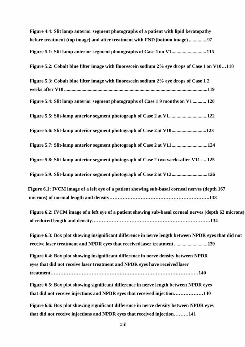
1.1.1 Corneal layers
The cornea consists of 6 layers: epithelium which is the outer most layer with its basement
membrane. Posterior to the epithelial basement membrane lies the Bowman’s membrane
which is a condensation of types 1 and 4 and proteoglycans. The stroma which forms 90%
of the corneal thickness is formed of stromal fibers, extracellular matrix and keratocytes.
The stromal fibers are mainly made of collagen type 1 but also contain types 4 and 12
collagen. Pre- Descemet’s layer (Dua’s layer) lies posterior to the stroma and is formed of
collagen fibers(1). Descemet’s membrane lies posterior to Dua’s layer and contains type 4
collagen and laminin and is acellular. The endothelium which is the inner most layer is a
single layer of hexagonal cells (Figure 1.1). The corneal epithelium is a uniform non-
keratinised stratified squamous epithelium extending from limbus to limbus and is
continuous with the limbal epithelium. It is made of 4 to 6 layers and has 3 types of cells
which are the superficial flat cells, wing cells and basal cells. The transparency of the cornea
is attributed to the extremely well organised epithelium and organisation of the collagen
fibres which lie parallel to each other and to the plane of the cornea within the stroma as
well as corneal lack of vascularity (2). The basement membrane is secreted by the basal
epithelial cells and is made of different proteins mainly type IV collagen, fibrin, fibronectin
and laminin. It acts as a foundation for the epithelial cells (3).
2
Figure 1.1: A composite image illustrating different corneal layers (adapted from Dua et al 2013, 1).
1.1.2 Limbal structure
The palisades of Vogt with their interpalisade rete ridges, the specialized connective tissue
architecture, the various epithelial immunophenotypes and the limbal epithelial crypts; as
well as a highly organized vascular and nervous supply provide the physiological
microenvironment and the anatomical support for the limbus (corneoscleral junction) to
maintain and sustain the limbal epithelial stem cells. Stem cells (SC) are progenitor cells with
the role of tissue regeneration and cells replacement. SC are slow cycling and live as long as
the organ they serve and constitute a very small part of the total cell mass of the organ.
They are the source from which almost all other cells arise that constitute a given organ
served by the SC. Corneal epithelial stem cells are committed progenitors and their
physiological role is to only differentiate to corneal epithelial phenotype, though in
experimental conditions can be made to transdifferentiate to hair follicles (4, 5). SC play a
major role in healing and regeneration of epithelium after injury but have a minimal role in
physiological homeostasis of the corneal epithelium (6, 7). Limbal stem cells reside in a well-

3
protected microenvironment called the niche. The niche surrounds stem cells and
modulates their function and fate through internal and external factors. They are protected
from Ultraviolet radiation by melanocytes that reside in the basal layers of the limbal
epithelium and by the upper and lower eyelid that act as a protective cover to the superior
and inferior limbus (8-10). It acquires its oxygen supply, cytokines, growth factors and other
several nutrients from the limbal stromal blood vessels and mesenchymal cells (2, 11-13).
The niche also regulates the limbal stem cell cycle to keep them in an undifferentiated
resting state (11, 14). The inter palisade rete ridges of the palisades of Vogt constitute the
limbal SC niche (4, 5, 15, 16).
The palisades of Vogt are radially oriented fibrovascular ridges and are very prominent along
the upper and lower limbus. They are visible in pigmented individuals due to the high
melanin content of the epithelial cells. The ‘valley’ between the adjacent palisades is packed
with epithelial cells that form the interpalisade (epithelial) rete ridge. Impression cytology
studies show that the limbal epithelial cells are smaller, with a higher cell density and a
greater nucleus to cytoplasm ratio compared to central corneal epithelial cells (17, 18). The
connective tissue of the palisade is rich in arteries, veins, nerves and lymphatics. The arterial
supply is from the anterior ciliary arteries derived from arteries of the rectus muscles. The
epithelium covering the palisade is 2 to 3 cells thick. The epithelial cells in inter palisade rete
ridges are 10 to 15 cells thick (19).
Limbal epithelial crypts (LEC) are solid cords of epithelial cells that arise from the posterior
end of the inter palisade rete ridge and extend into the surrounding substantia propria
peripherally, centrally towards the cornea or circumferentially in a clockwise or
anticlockwise direction (18, 20). LEC could be minor and major, with the major ones ranging
from 40 to over 200 microns. The average number of LEC is 9 per eye. LEC are packed with
epithelial phenotype of cells, basal, suprabasal and central. The cell membrane of the basal
cells in contact with the basement membrane have complex finger-like processes with
special hemidesmosomes, allowing for a strong attachment between these putative SC and
the substrate. The peripheral corneal basal cell attachments are also firmer than the central
epithelial attachments. Hence following injury, a peripheral rim of corneal epithelium (and
the limbal epithelium) remain attached even when a large area of the central cornea is lost.
Cells from this rim migrate centripetally as convex sheets that close the defect and restore

4
epithelial integrity. Lines of contact between these sheets stain with fluorescein and appear
as the pseudo dendrites seen during healing of corneal epithelial defects (21).
Figure 1.2: A cross-sectional diagram of the human corneal limbus. (corneal epithelial stem cells, deficiency and regulations (adapted from G.Secker,et al,21)
1.1.3 Conjunctiva
The conjunctiva is the highly specialised mucosal lining of the ocular surface and it extends
from the lid margin, posterior to the grey line and lines the posterior aspect of the lids
forming the palpebral or tarsal conjunctiva. It then folds over to cover the eye ball as the
bulbar conjunctiva. The folds between the bulbar and palpebral conjunctiva are called
superior, inferior, lateral and medial fornices. The conjunctiva is made of epithelial cells and
substantia propria which is loose connective tissue underlying the epithelium. The
conjunctival epithelium is squamous, non-keratinized, with goblet cells which are found all
over the conjunctiva especially in the nasal palpebral and forniceal areas. The conjunctival
cells are rich in cytoplasmic organelles. The substantia propria is very rich in blood vessels
and contains lymphoid tissue made of plasma cells, lymphocytes, neutrophils and mast cells
(22-24).
1.1.4 Blood supply and lymphatics

5
The cornea is an avascular structure and acquires nutrients from the peri-corneal limbal
vasculature, the aqueous humour and the tear film (25-27).
Limbal vessels consist of basement membrane and endothelial cell lining. The basement
membrane is made of fine filaments with embedded collagen fibrils. The filaments are
arranged in alternating layers, each layer is less than 0.1 microns thick. The endothelial cells
form the vessel wall and are organised in a single layer attached to their basement
membrane (28).
Two types of capillaries have been described in the limbus; muscle type capillaries with thick
endothelium and fenestrated visceral type capillaries. There is no gap between the
endothelial cells in most of the capillaries. However, a thin membrane covering this gap was
described in some localised areas where the vessel appears to be fenestrated. Pericytes
were seen on the outer surface of the arterial capillaries and in proximity to the endothelial
cells (29). Pericytes have similar structure to the endothelial cells but are functionally

6
different (30). It is commonly believed that pericytes are one major source of mesenchymal
stem cells (31-34).
Pericytes also lead to specific induction of fibronectin and nidogen-1 (i.e., matrix-bridging
proteins that link together basement membrane components) as well as perlecan and
laminin isoforms (35).
The cornea is normally devoid of lymphatic vessels thus allowing for its unique immune
privileged status. Corneal neovascularisation is usually associated with lymphatics-
subsequent studies of vascularised corneas supported the presence of lymphatic vessels in
the cornea using lymphatic specific markers which augment the afferent arc of the immune
response and is related to the abrogation of the immune privileged status of the cornea (36,
37).
1.1.5 Corneal innervation
The cornea is a highly innervated structure and has both sensory and autonomic nerves and
is mainly vasomotor in function (38). Corneal innervation is mainly sensory form the
ophthalmic division of the trigeminal nerve via the long posterior ciliary nerves. These
nerves branch forming the peri-limbal plexus(39).
The autonomic nerve fibers have sympathetic fibers that originate from the superior
cervical ganglion as well as parasympathetic fibers that come from the ciliary ganglion.
Demylinated radial nerves emerge from the peri-limbal plexus and enter the cornea at mid
stromal level. There are about 11 radial nerves in each quadrant. They run towards the
centre and superficially to form the Bowmans plexus. Smaller branches penetrate Bowmans
membrane ending in the sub-basal plane of the epithelium, in terminal bulbs which are 20
to 40 micrins in diameter. The penetration sites are mainly situated in the mid-peripheral
cornea. Finer neurites arise from the bulbs as naked endings (without Schwan cells) forming
the sub-basal plexus (40). Terminal nerves from the sub-basal nerve plexus enter the
epithelium and end inter and intra-cellulary (41).

7
Figure 1.3: A three-dimensional section of the human cornea showing the corneal nerves distribution and their density and
width at different layers. Diabetic corneal neuropathy (adapted from Hassan Mansoor et al, 39)
1.2 Corneal angiogenesis
1.2.1 Epidemiology and risk factors
Corneal neovascularization (NV) is a condition that can result in severe visual impairment
and represents a major public health problem (42). It is estimated that for a given year, 1.4
million patients in the US may develop corneal NV; and 20% of corneal samples taken during
corneal transplantation revealed histopathologic evidence of vascularization (43). Recently,
it was found to affect up to 4.14% of patients coming for eye care (44). The rate of corneal
NV may be as high as 60% in patients with inflammatory disorders such as atopic
8
keratoconjunctivitis (45). A study was performed to assess corneal NV after penetrating
keratoplasty in patients without active inflammation, previous corneal NV or persistent
epithelial defects. The study reported a remarkably higher risk of corneal NV when suture
knots were buried in the host stroma, when a large recipient was used or when active
blepharitis was present. They also reported that 41% of eyes receiving corneal grafts
developed corneal NV 6 to 9 months after the corneal transplant (46). The most common
cause for corneal neovascularisation was found to be herpes simplex keratitis and it remains
to be the most common cause of recurrence of NV after corneal grafting (47, 48).
1.2.2 Causes
Corneal neovascularisation could occur due to the following disorders (44, 49, 50):
1. Hypoxia due to contact lens wear
2. Infectious keratitis due to viral, bacterial, fungal and parasitic infections
3. Limbal stem cell deficiency
4. Trauma
5. Ocular surface neoplasia such as conjunctival, corneal intraepithelial neoplasia and
papilloma.
6. Inflammatory ocular surface disorders such as Steven-Johnson syndrome, atopic
conjunctivitis, Rosacea and Mucous membrane pemphigoid.
1.2.3 Mechanism of angiogenesis
9
In corneal angiogenesis, corneal NV sprout mainly from the venules and capillaries of the
limbal plexus. Depending on the underlying pathology, corneal NV are present clinically in
three different forms (1) stromal NV mainly correlated with stromal keratitis, (2) deep NV
overlying Descemet’s membrane in herpetic keratitis and (3) vascular pannus which is
associated with connective tissue growing in the superficial periphery of the cornea and is
seen mainly in ocular surface disorders (46, 51).
Insults to the cornea activate inflammatory and immune-mediated pathways leading to an
imbalance between angiogenic and antiangiogenic factors and this balance may be tilted in
favour of NV due to the upregulation of angiogenic factors and/or the downregulation of
antiangiogenic factors (52-55). The angiogenic factors include vascular endothelial growth
factor (VEGF), fibroblast growth factor, platelet derived growth factor, inflammatory
mediators and angiopoietins with VEGF being the most prominent molecule involved in the
promotion of NV. Antiangiogenic factors are pigment epithelium–derived factor, VEGF
Receptor-2, endostatin and angiostatin (56). MMPs can act as a stimulator or inhibitor of
angiogenesis in different circumstances (57, 58).
The antiangiogenic molecules were found to work together to maintain the cornea in an
avascular state. Alternatively, it was mentioned that molecules can act individually to
suppress corneal vascularisation.
Corneal NV can be superficial or deep. Superficial NV is either localized or diffuse (pannus).
Pannus is fibrovascular tissue that separates the epithelium from Bowman’s membrane,
occasionally extending than Bowman’s membrane. With trachoma it tends to involve the upper
part of the cornea while in in exposure or bullous keratopathy it involves the lower part of the
cornea. In chemical burns, it is massive and covers the whole cornea. Each vessel of a pannus
consists of a small arteriole forming a loop with one large venule or more via a small capillary
bed. The vessels move by simple migration of the loop. On the other hand, localized superficial
NV consists of a layer of vessels that includes interstitial and superficial vessels. These vessels
are directed towards a specific lesion and is most commonly seen in recurrent herpetic keratitis.
Histologically, superficial NV are located under the epithelium and are associated with
inflammatory cells during their active progression. Pannus wll be associated with plenty of
connective tissue. Deep NV tends to occur between the corneal stroma and Descemet’s
membrane. They tend to occur following interstitial keratitis (Corneal vascularization, David G.

10
Cogan, IOVS, 1962)
1.2.4 Suppressors of corneal angiogenesis
A) VEGF Receptors
Multiple VEGEF receptors are expressed by the cornea and these act as “decoy” receptors
for the proangiogenic VEGF molecules. These receptors are soluble VEGF-1 and soluble
VEGF-2. Soluble VEGF-1 suppresses angiogenesis via sequestration of VEGF-A molecules (56,
59). The corneal epithelium is responsible for expressing membrane bound VEGF-3 which in
turn sequesters VEGF-C and VEGF-D (60).This leads to indirect suppression of angiogenesis
by inhibiting the recruitment of VEGF secreting macrophages (61).
B) Pigment epithelium–derived factor (PEDF)
PEDF is thought to act individually to maintain the cornea in an avascular state (62). PEDF is
found in large amounts in the corneal stroma, iris and retinal pigment epithelium (63, 64).
When an antibody that suppresses the action of PEDF was delivered to the cornea, it was
found that the cornea became invaded with blood vessels. On delivery of large amounts
PEDF to the corneal stroma the blood vessels showed reduction (65).
C) Angiostatin
Angiostatin is present in the tear fluid and the corneal epithelium (66). Angiostatin
suppresses corneal NV via binding to integrin αvβ3 (67), vascular endothelial cell surface-
expressed F1-F0 ATP synthase R (68, 69)and hepatocyte growth factor receptor (c-met) (70-
72) resulting in suppression of cell migration and proliferation(68, 73).
D) Angiogenin
Angiogenin is present in tears and reduces the inflammation caused by tumour necrosis
factor-alpha or lipopolysaccharide in human corneal fibroblasts by suppressing IkappaB
kinase-epsilon mediated activation of nuclear factor-kappaB (74),
E) Matrix metalloproteinases (MMPs)

11
MMPs play a query role in corneal vascularization as the same molecule is capable of being
a stimulator or inhibitor of angiogenesis in different circumstances (57, 58). MMP-7
(matrilysin) promotes vascularization by stimulating the production of VEGF and promoting
proliferation of vascular endothelial cells (75, 76). MMP-7 produced by the corneal basal
epithelial layer is believed to play a role in suppressing angiogenesis and this is evidenced by
the massive increase of the angiogenic response to corneal trauma when MMP-7 becomes
deficient (77, 78). This may be a function of the MMP-7-mediated cleavage of type XVIII
collagen, the precursor of antiangiogenic endostatin (79) (80).
F) Endostatin

12
Endostatin is a proteolytic portion of collagen XVIII and suppresses angiogenesis by
inhibiting the VEGF and fibroblast growth factor (81, 82). Collagen XVIII is present in lens
capsule, lens and cornea (50).
1.2.5 Stimulators of corneal angiogenesis
A) Vascular Endothelial Growth Factors (VEGF)
VEGF are secreted growth factor peptides generated by alternative splicing in five isoforms
(VEGF 115, VEGF 121, VEGF 165, VEGF 189 and VEGF 206). The members of the VEGF family
are VEGF-A, VEGF-B, VEGF-C, VEGF-D and placental growth factor (PlGF) (83). VEGFs bind to
tyrosine kinase receptor VEGF-1, VEGF-2 and VEGF-3 (83, 84).
VEGF is produced by T cells, pericytes, smooth muscle cells, astrocytes, retinal pigment
epithelial cells and macrophages secondary to inflammation and hypoxia. Hypoxia is the
main regulator of VEGF gene expression. VEGF expression is also regulated by several
cytokines such as epidermal growth factor, prostaglandin E2, interleukin 1a, interleukin 6
and transforming growth factor-B (85, 86).
VEGF is a peptide present massively in the epithelium of vascularized corneas secondary to
inflammation(87)and is a strong promoter of corneal inflammation induced by hypoxia and
angiogenesis(88). VEGF is also produced in the stromal keratocytes and endothelium (89).
VEGF-A is the most important member of VEGF family regarding angiogenesis (90, 91).VEGF-
A binds to VEGF-2(92) resulting in vascular endothelial cell proliferation, migration and
increased vascular permeability and dilation (86, 93, 94). Moreover, VEGF attracts
inflammatory cells (e.g. macrophages) chemically leading to production of more
proangiogenic molecules(95).When VEGF-C or VEGF-D bind to VEGF-2 or -3
lymphangiogenesis is stimulated in a similar way (96, 97).
B) Inflammatory Mediators
All causes of corneal angiogenesis result from inflammation. Inflammatory cells are
abundant in VEGF molecules and thus encourage angiogenesis (98).
13
Inflammatory mediators such as chemokines can promote angiogenesis either directly or by
recruiting inflammatory cells (99). Integrins help with the migration of vascular endothelial
cells and inflammatory cells (100). Interleukin 6, transforming growth factor and tumour
necrosis factor organize the process of VEGF production and activate inflammatory cells
(101-104).
C) Fibroblast Growth Factors (FGFs)
There are 18 types of FGs that bind to FGF receptors (105, 106). FGF1 and FGF 2 are strong
stimulators of angiogenesis (107, 108). FGF2 is produced by corneal epithelial cells and
released as a result to injury to the epithelium (50, 109) and binds to receptors on the
epithelial basement membrane, Bowman membrane and Descemet membrane (110, 111)
thus promoting vascular endothelial cell proliferation and migration (112, 113).
D) Platelet-derived growth factors (PDGFs)
PDGF and VEGF are related both in structure and function (114). Humans have four PDGF
chains (PDGF-A, -B, -C, and –D) that bind to tyrosine kinase receptor complexes of PDGFR-α
or PDGFR–β(115). PDGF-A and –B are present in corneal endothelial cells, epithelial cells and
stromal fibroblasts (116). PDGFR-α and PDGFR–β are expressed by corneal epithelial cells,
stromal fibroblasts, and endothelial cells (116) (117).
E) Angiopoietins (Ang)
Once vessels are formed, Ang family including Ang1, 2, and 4 ((118) come into place in the
process of angiogenesis. Ang-1 induces maturation of blood vessels and this is evidenced by
an increase in vessels perfusion and density. The role of Ang-2 has remained controversial,
however, with recent reports suggest that in some circumstances, it may be pro-angiogenic
via promoting a rapid increase in capillary diameter, remodelling of the basal lamina,
proliferation and migration of endothelial cells and stimulating the sprouting of new blood
vessels (119).
1.2.3 Quantification of Corneal NV

14
Faraj et al. (47) have recently carried out a study with the aim of clinically characterizing the
corneal NV in 165 patients with various causes of corneal pathologies. They assessed the
vessels location, depth, length, branching pattern, colour, lipid leakage, blood flow and
presence or absence of haemorrhage and developed a clinical grading system for corneal
NV. They staged the vessels into five categories based on their morphological characteristics
and created a system for the quantification of NV as follows (Please see Chapter 4, figure
4.1):
1. Active young vessels: Newly formed vessels that contain a lot of blood, bright red
with minimal fibrous tissue layering . They are seen to be actively progressing in the
cornea and a well- defined network of fine capillary vessels. The corneal stroma
surrounding the vessels shows signs of leakage
2. Active old vessels: These are vessels that reach and surround or cover the lesion in
the cornea. Their progression stops but the fibrosis will keep progressing. They are
less bright than the young vessels and still have an active circulation
3. Partially regressed: These vessels are present when corneal pathology has stopped
due to therapy or the arrival of corneal vessels or following fine needle diathermy of
corneal vessels. The blood circulation is slow, the vessels are less dilated and some
parts of the complex become less visible or reduced. Both active old vessels and
partly regressed vessels show arborizing pattern from the limbus towards the centre,
with the vessels becoming narrower towards the centre
4. Mature: These are larger vessels, with less branching and regressed or absent
capillary networks, seen to persist in scar tissue or in the corneal stroma after the
corneal pathology has abated. These vessels maintain a blood circulation
5. Regressed (ghost vessels): These can be seen as fine white lines mirroring the
morphology of the original vessels and are seen as ‘ghost vessels’. These do not
contain a circulation and the surrounding cornea is not swollen. Ghost vessels are
situated in the stroma

15
1.3 Management of corneal vascularisation
Choosing the appropriate method for managing the corneal neovascularisation is dependent
on the state of maturity of these vessels. Mature vessels are not dependent on angiogenic
mediators and thus surgical interventions such as fine needle diathermy are the most
effective method for treating them, while actively growing vessels indicate an underlying
ongoing pathology which is best treated by anti-VEGF in the form of drops or
subconjunctival injections (120).
1.3.1 Medical therapy
A) Steroids
The impact of corticosteroids in inhibiting the corneal vessels both in clinical and
experimental conditions is well documented especially topical and subconjunctival
Triamcinolone (121, 122) . Their anti-angiogenic effect is a result of their anti-inflammatory
properties, which includes the suppression of inflammatory cellular chemotaxis, the
inhibition of synthesis of pro-inflammatory cytokines as well as direct suppression of
vascular endothelial cell migration and proliferation (122-124) . Ashton N et al reported
that moderate doses of systemic (IM) and subconjunctival steroids have remarkably
decreased the corneal NV in rabbits and that subconjunctival injection was found to be
more superior than systemic treatment, They also found that subconjunctival steroid
injection has caused a remarkable reduction in the corneal opacity(125). Another study has
shown that adding heparin to steroid can greatly improve its efficacy in reduction of corneal
NV (126). However, the long term use of steroids can result in several complications such as
posterior subcapsular cataracts, glaucoma and enhancement of infection especially herpes
simplex and its inhibitory effect is incomplete (127).
B) Anti VEGFs
VEGF is the most fundamental molecule that promotes and controls major steps in the
process of corneal angiogenesis. When hypoxia or inflammation occur, endothelial cells
lining the limbal vessels together with corneal epithelial and endothelial cells release VEGF
16
(60). Anti-VEGFs will suppress the process of angiogenesis thus preventing and reducing the
corneal NV formation.
Ranibizumab (Lucentis®) is a recombinant humanized monoclonal antibody directed against
human VEGF-A. It is produced in Escherichia coli using recombinant DNA technology and has
a molecular mass of 48 kDa and a single antigen binding site. Ranibizumab neutralizes all
active forms of VEGF-A. It has been genetically engineered via a process of selective
mutation to increase its affinity for binding and suppressing the growth factor (128).
Ranibizumab was developed specifically for intraocular use and was approved by the FDA in
2006 for the treatment of neovascular age related macular degeneration (NVAMD) (129).
The indications for ranibizumab have expanded to include macular oedema following retinal
vein occlusion and diabetic macular oedema. Ranibizumab has greater pharmacokinetic
properties than bevacizumab as it is a smaller molecule and hence has better corneal
bioavailability (89).
A combined in vivo and in vitro study showed that ranibizumab had dual antiangiogenic
mechanisms via simultaneous inhibition of blood and lymphatic vessels, highlighting its
therapeutic role in corneal NV. A study in rabbits was conducted and showed a reduction of
VEGF concentration in the bulbar conjunctiva, cornea, aqueous humor and iris following
subconjunctival ranibizumab injections indicating high bioavailability (130).
Several studies were conducted for the clinical evaluation of ranibizumab. One study used
topical ranibizumab as 1% solution, 4 times a day in eyes with stable NV. The results showed
that ranibizuamb is effective in controlling stable NV by narrowing of vessels but not by
reduction of the corneal area invaded by NV (131).
Another randomized clinical trial compared subconjunctival and intralesional injections of
bevacizumab and ranibizumab. It was suggested that corneal NV management was better in
the bevacizumab group than in those treated with ranibizumab (132). This was contradicted
by another study which showed that subconjunctival and intrastromal ranibizumab
injections were successful in treating a case with NV due to herpetic keratoconjunctivitis
after failure to treat her with bevacizumab injections by the same route of delivery (133).

17
Bevacizumab (Avastin®) is a full-length humanized monoclonal anti-VEGF antibody that
binds all VEGF-A isoforms thus blocking the VEGF-/VEGF receptor interaction thereby
suppressing the role of VEGF in angiogenesis. It is a recombinant IgG1 antibody that is
produced in Chinese Hamster Ovary mammalian cell expression system and its molecular
weight is 149kD. It was initially designed as an anti-angiogenic therapy to treat solid
tumours (134). In 1997, phase 1 trials of bevacizumab for the treatment of cancer was
started and it was established that it had minimal toxicity (135). Phase 2 and 3 trials were
carried out later and their results led to the approval of the U.S. Food and Drug
Administration (FDA) of bevacizumab as therapy of colon cancer along with chemotherapy
(136, 137).
Although bevacizumab was initially approved for treating colorectal cancer, it has been used
off-label to treat several ophthalmic conditions (138, 139). It has been first introduced as an
intravitreal injection for treating ocular diseases in July 2005 (140).
Several clinical cases have been published showing dramatic resolution of macular fluid in
central retinal vein occlusion and NVAMD (140). Other studies showed the ability of
bevacizumab to penetrate the retina after being injected intravitreally (141, 142).
Bevacizumab has also been used for the treatment of other ophthalmological conditions
such as juxtafoveal telengectasia (142-144), myopic choroidal neovascularization (CNV)
(145, 146) and CNV secondary to angioid streaks (147, 148)and has shown promising results.
Many retina specialists have also noted impressive results in the setting of CNV, proliferative
diabetic retinopathy, neovascular glaucoma, diabetic macular oedema, retinopathy of
prematurity and macular oedema secondary to retinal vein occlusions (149).
The comparison of Age related Macular Degeneration Treatments Trials is one of the largest
studies that has been conducted with the aim of evaluating the relative efficacy and safety
of treatment of NVAMD. It was concluded that Avastin and Lucentis had the same effect on
visual acuity over a 5 year period. There were no differences between the 2 drugs in rates of
death or arteriothrombotic events (150).

18
A systematic review and meta-analysis was done with the objective of evaluating the
efficacy and safety in treatment of ocular neovascularization by bevacizumab versus
ranibizumab. The results showed that bevacizumab has equal efficacy and safety to
ranibizumab in treatment of ocular neovascularization (151).
Anti-VEGFs have been first used to treat corneal neovascularization in a rat model. Corneal
injury was induced to in order to stimulate VEGF release, and the resulting NV were
subsequently blocked by anti-VEGF antibodies (85). Another animal trial conducted later
demonstrated that corneal vessels secondary to herpes simplex virus were reduced by
suppressing VEGF via the delivery of a murine soluble VEGF receptor protein, mFlt (1–3)-
immunoglobulin G (152). These studies led to the hypothesis that anti-VEGFS might be an
effective treatment option for active corneal NV (153).
Several studies have been conducted to evaluate the efficacy and safety of bevacizumab
when used as subconjunctival injections to treat corneal NV. They showed that the
treatment of active corneal NV with subconjunctival injections of bevacizumab could be of
great benefit as they noted either a regression or stabilization of the corneal NV and this
effect was also associated with improved lipid deposition in cases with lipid keratopathy.
The intervention was well tolerated without adverse ocular or systemic effects (154-158).
However, there has been reports that it could delay epithelial wound healing (159).This was
contradicted by another group who used similar doses of bevacizumab (1%, twice daily for 2
months) but instilled the drops in the reservoir of a Boston Ocular Surface Prosthesis. This
indirect drug application may have resulted in quite low drug concentrations at the corneal
surface (160).
Other studies also reported improved mean visual acuity at the final follow up and also
reported that the injections were beneficial and effective in high-risk keratoplasty, with
regards to preventing corneal NV after keratoplasty with improvement of graft survival
(161-164).
Bevacizumab 1% eye drops used 4 times/day showed remarkable decrease in superficial and
deep stromal neovascularization in a number of pilot studies (165, 166) (167).

19
C) Topical therapeutics
New topical therapeutics are being developed and might be used clinically in the future for
treatment of corneal NV (168). These are protein phosphatase magnesium dependent-1,
melatonin, omentum, 0.5% ketorolac tromethamine, Rho-associated protein kinase
inhibitors (AMA0526), prospero homeobox 1 and aloe vera (169-176).
1.3.2 Surgical treatment
A) Laser photocoagulation
Argon laser has been first described by Cherry PM in treating corneal NV on 4 human
patients. There was 1 success, one partial regression of NV and two failures (177).In 1976
Cherry and Garner used 11 rabbits to test the impact of argon laser in obliterating corneal
vessels. They concluded that the obliteration of corneal vessels by argon laser was
successful as long as there was no stimulus for vascularisation. There was slight iris damage
in their experiment but they did not think that this could necessarily contradict the laser
usage on humans(178).The argon laser was used further in treatment of lipid keratopathy
by Marsh RJ. The lipid deposition was reduced in more than half of the cases, however there
were complications such as iris damage, peaking of pupil and corneal thinning(179). Yellow
dye laser 577 nm was used by Baer and Foster to treat corneal NV in 25 eyes of 23 patients
(180). They reported a significant decrease in the vascularised region from 46.4% to 27.3%
in 9 patients who had lipid keratopathy. They also reported reversal of graft rejection and a
marked reduction in the vascularisation area (68% decrease in the vascularised area).
However, patients with severe corneal neovascularisation did not show improvement.
B) Photodynamic Therapy (PDT)
PDT is a potential treatment for the management of corneal NV. The idea of PDT is to
administer systemic porphyrin derivatives which will build up in the replicating endothelial
cells. This is followed by laser energy application which will activate those derivatives
resulting in the release of cytotoxic oxygen free radicals leading to corneal vessels
obliteration (181-184). Several porphyrin derivatives were used such as verteporfin(185)
and diheamtoporphyrin ether(182). A clinical trial was carried out using PDT with

20
verteporfin to treat corneal neovascularisation and showed that it is a safe and effective
method of reducing corneal neovascularization (186) . Other trials used a combination of
PDT and subconjunctival bevacizumab and showed that this combination can effectively
inhibit corneal neovascularization (187, 188).
C) Fine needle diathermy (FND)
Fine needle diathermy(FND) is a method for occluding corneal vessels that has been
developed by Professor Harminder Dua (189). FND was found to be superior to the methods
mentioned earlier in terms of being an effective, simple and inexpensive method of
occluding mature vessels. FND can be used as a preconditioning and/or post-conditioning
treatment of high-risk corneal grafts (190). It can be one element of an overall plan to
reduce the risk of graft rejection or deal with refractory episodes of rejection (120).
The procedure involves the insertion of stainless steel 3/8 needle attached to a 10–0
monofilament black nylon suture close to the limbus, parallel to and at approximately the
same depth as the blood vessel(s) to be occluded than the needle is touched with a
monopolar cautery at low power (Covidien Force X) to produce the coagulation (120).
Several studies were carried out to evaluate the safety and efficacy FND for occlusion of
corneal vessels (189, 191). They concluded that FND is a safe, inexpensive and effective
treatment option for the occlusion of corneal blood vessels both before and after
penetrating keratoplasty. It can be done repeatedly and is also beneficial in stopping the
progress of lipid keratopathy. However, it was noted that the deposited lipids did not
regress any faster by the occlusion of the corneal vessels (189).
In 2014, a study was carried out by Faraj et al in the same centre where FND technique was
first described more than a decade ago(120).
The study has shown that FND was the method of choice in occlusion of active old and
mature vessels. It was also found that FND was very effective in treating lipid keratopathy.
Another important conclusion that was drawn out from the study, was that active young
vessels were usually a sign of underlying continuous pathology which keeps on inducing
more vascularisation. It was pointed out that such eyes will require treatment with anti-
21
VEGF subconjunctival injections or eye drops and several studies have been conducted to
assess the efficacy and safety of combined FND with adjuvant anti-VEGF bevacizumab eye
drops, subconjunctival or intrastromal injections in both adults and children (192, 193).
The studies concluded that applying combined FND and bevacizumab is both successful and
safe and that it improves the corneal transparency as well as prepares the cornea for future
keratoplasty.
1.4 Consequences of corneal vascularization
In the presence of corneal pathology, NV’s are necessary to mount a host defence reaction,
promote healing of the cornea, get rid of toxic material and transfer medications to the
inflammation site(194, 195).However, when the pathology is no longer active, the blood
vessels formed may remain in the cornea and the vessels are tortuous, leaky and wide
resulting in infiltration of corneal stroma with inflammatory cells and cytokines due to loss
of pericytes and separation of endothelial cells. Keratocytes proliferate and transform into
fibroblasts which secrete fibrin leading to scarring. All these events will eventually lead to
corneal opacity and haze which will consequently reduce vision. This opacification is a result
of circulation of blood cells in the blood vessels, changes in the spaces between stromal
collagen, chronic or recurrent inflammation, scarring, higher order aberrations caused by
irregular vessel wall and lipid leakage and deposition in the cornea (50),(49, 196). Lipid
keratopathy was related to mature and partially regressed vessels. The most common
aetiology of lipid keratopathy was found to be secondary to viral keratitis (47).
Corneal vessels promote easy lipid access to the cornea(197). Lipids become deposited by
lipid overloaded fibroblasts in the corneal stroma in the form of droplets of cholesterol
crystals(198). Corneal NV may as well cause bleeding intraoperatively and hyphema. Corneal
NV affect the corneal immunological privilege which may eventually lead to graft rejection
(199, 200)as it provides an efferent arm in the immune reflex arc and accelerates immune
reaction by facilitating access for immune effector cells to the graft and thereby resulting in
corneal rejection(49, 50). Lymphatic vessels that are microscopically undetectable have
been detected with blood vessels in high-risk vascularized corneas (201). These lymphatic
22
vessels facilitate the access of host and donor antigen-presenting cells (APCs) to regional
lymph nodes thereby accelerating sensitization to the graft antigens and causing further
disruption of the immune privilege status of the cornea. Lymphatics represent the afferent
arm of the immune reflex arc (201-203). A meta-analysis of 19 studies reporting on a total of
24,944 grafts undergoing keratoplasty was done. It was found that the chances of graft
failure due to rejection becomes higher with increasing number of corneal quadrants that
contain corneal NV before keratoplasty (44).
1.5 Pterygium: demographics, role of vessels and vascular factors
Pterygium is a potentially sight threatening condition that originates from the bulbar
conjunctiva as fleshy fibrovascular tissue and grows centripetally most often from the nasal
conjunctiva although it can occur temporally. It can be bilateral and asymmetric with one
eye affected by a larger pterygium, encroaching on the cornea which could result in corneal
scarring, chronic ocular surface inflammation, irregular astigmatism as well as restriction of
ocular motility (204-206). Although the etiology of pterygium remains an enigma and is so
complex, environmental factors such as chronic exposure to ultraviolet light, dryness,
genetic and immunological factors play a big role in its formation (207-209). It is more
prevalent within the peri-equatorial latitudes” 37 degrees north and south”, where the
ultraviolet radiation intensity is strongest. Recently, other important factors such as
fibrovascular proliferation, inflammation and vascularisation and localised stem cell
deficiency have been implicated in the pterygium pathogenesis and progression (210, 211).
Pterygium can be classified as quiescent or progressing and active (212, 213).
Many studies have also demonstrated that the pathogenesis and recurrence of pterygia is
highly attributed to the increased expression of molecules such as vascular endothelial
growth factor (VEGF), transforming growth factor, platelet derived growth factor and basic
fibroblast growth factor(214-216). The most prominent of these was found to be VEGF
molecule and it was found to be highly increased in pterygia as well as being involved
directly or indirectly in formation of pterygia by inducing the formation of blood vessels. It
has also been postulated that the development of pterygia depends on a disturbed

23
angiogenic stimulator to inhibitor ratio (216-219). Moreover, further immunochemistry
studies have shown that immunostaining of VEGF in pterygial sections was much stronger
than normal conjunctival sections(216). Based on these findings, several clinical studies
aimed to reduce the vascular density and prevent its recurrence using anti-VEGFs (207, 210).
In many of these cases, there was a brisk reduction of inflammation and vascularization
when pterygia where injected subconjunctivaly with anti-VEGF which gives more evidence
of the role of VEGF in pterygium angiogenesis(208, 210); (216);(219). Studies have also
shown that in anti-VEGFs suppress fibroblast proliferation and induce fibroblast apoptosis in
addition to its effect on angiogenesis. In one study, injection of bevacizumab with a
concentration greater than 7.5 mg/ml had a remarkable effect on the anti-fibrotic effect,
changes in morphology, and reduction in the collagenase activity of human Tenons’
fibroblasts cultured from both primary and recurrent pterygia (220, 221)(Figures 1.4, 1.5).
Figure 1.4: Slit-lamp anterior segment photograph of a primary pterygium
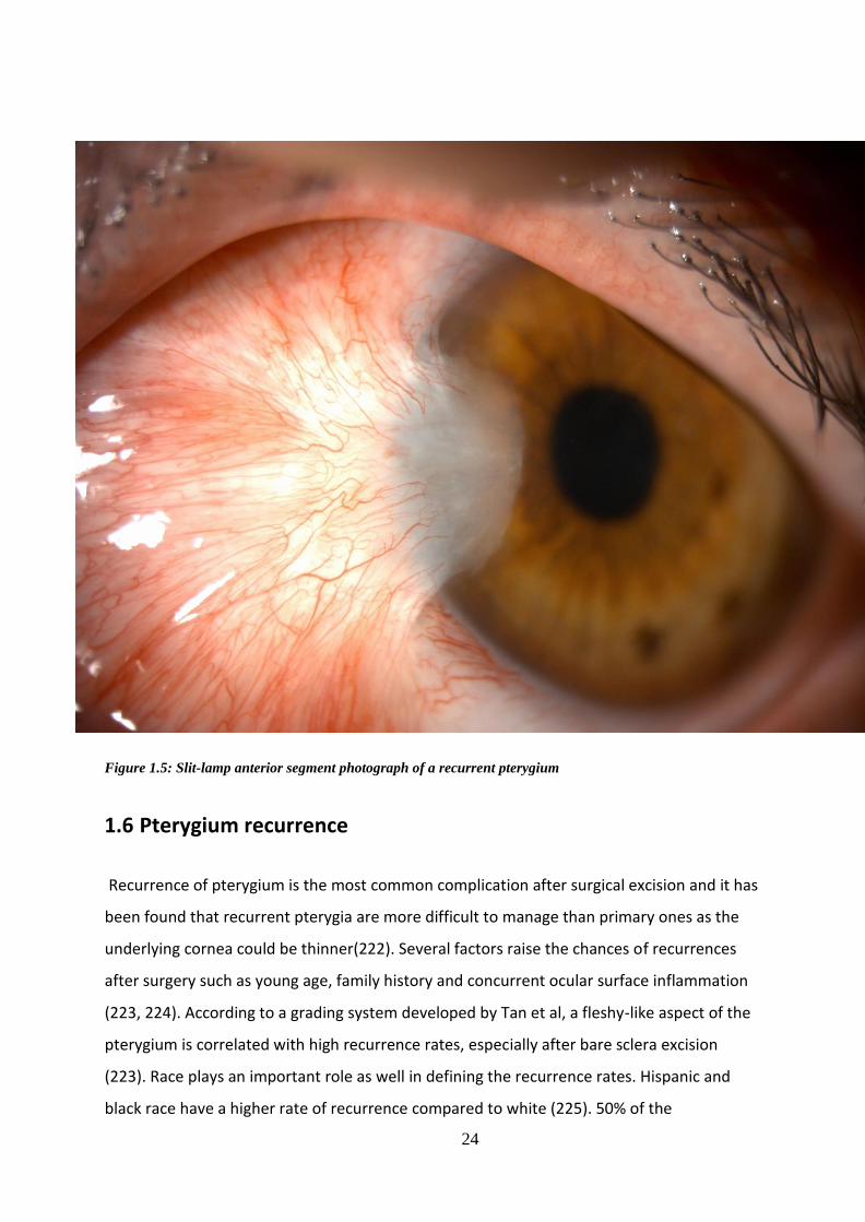
24
Figure 1.5: Slit-lamp anterior segment photograph of a recurrent pterygium
1.6 Pterygium recurrence
Recurrence of pterygium is the most common complication after surgical excision and it has
been found that recurrent pterygia are more difficult to manage than primary ones as the
underlying cornea could be thinner(222). Several factors raise the chances of recurrences
after surgery such as young age, family history and concurrent ocular surface inflammation
(223, 224). According to a grading system developed by Tan et al, a fleshy-like aspect of the
pterygium is correlated with high recurrence rates, especially after bare sclera excision
(223). Race plays an important role as well in defining the recurrence rates. Hispanic and
black race have a higher rate of recurrence compared to white (225). 50% of the
25
recurrences appear in the first 4 months while 97% appear within the first 12 months (226).
Surgical technique of pterygium excision also attributes to the rate of recurrence. High
recurrences rates were reported in patients who had pterygium excision by bare sclera
technique with no adjuvants or radiotherapy alone and this was attributed to the fact that
small tissue residues may be left in the scleral bed leading to higher recurrence rates which
ranged from 38 and 88 % in many publications (227-229). Adjuvant amniotic membrane
transplantation has also high rate of recurrence which can be up to 28.1 % but no
postulations were given regarding the high recurrence rate (230).
The recurrence of pterygium has been also attributed to fibroblast migration and
proliferation, therefore 5 fluorouracil is being injected subconjunctivally in the recurrent
pterygium body to reduce or inhibit the activity of fibroblasts (231).
1.7 Management of primary pterygium
1.7.1 Medical
A) 5 Flouorouracil (5 FU)
5 fluorouracil (5FU) is a pyrimidine analogue that inhibits thymidylate synthetase and hence
DNA and RNA synthesis, suppresses proliferating fibroblasts and promotes apoptosis of
Tenon’s fibroblasts. It is widely used in ophthalmology especially in glaucoma surgery with
favourable outcomes due to its anti-fibrotic characteristics.(232, 233). A number of pilot
studies were conducted to study the efficacy of intra-lesional injection of 5FU in primary
pterygium. They reported cosmetic and clinical improvement. One study reported no
statistically significant changes of corneal astigmatism after the injections (234, 235).
B) Anti-VEGF alone (no surgery)
Injections
Several studies demonstrated that vascularization and inflammation were rapidly controlled
by subconjunctival bevacizumab injections in many patients, and this provided more

26
evidence for the role of increased VEGF in pterygium (153, 208, 215, 216, 219, 236-238).
However, some reported that the changes were clinically insignificant as they were clinically
not obvious with no beneficial effect on the regression of the vessels (239, 240) and one
study reported transient regression and irritation for only one week (209).
Topical
One study used 2 different doses of topical bevacizumab and compared them to a control
group and reported good results in the group with higher bevacizumab dose. They had
recurrences in both the lower dose group and the control group (241).
1.7.2 Surgical excision
There are several techniques for surgical management of primary pterygium.
Bare sclera technique: This technique involves complete excision of the pterygium head,
removal of some of the surrounding nasal bulbar conjunctiva as well as excision of Tenon’s
capsule resulting in a bare sclera. The recurrence rate for this method is quite high 30% to
80%). That is why some of the surgeons will combine this technique with other adjunctive
therapies to reduce the recurrence rate (229, 242, 243).
Aminiotic membrane graft Transplantation (AMT): Human aminiotic membrane inhibits
inflammation and formation of fibrovascular tissues, provides numerous growth factors and
promotes proliferation of epithelial cells without the risk of immunological reactions (244,
245). Due to these properties, it has been used as a graft to cover the bare sclera after
pterygium excision, typically with the basement membrane facing up and the stroma facing.
It has the advantage of short surgical time, fast recovery, less post-operative pain and better
cosmetic outcome following surgery (as no donor site is injured)(246). However, it has
higher recurrence rates when compared to the conjunctival autograft technique (247, 248).
AMT effectiveness is improved by using fibrin glue to promote aminiotic membrane graft
fixation to the underlying sclera. It is useful in situations such as covering large conjunctival

27
defects after pterygium excision and to preserve conjunctiva in glaucomatous patients.
Combining AMT with intra-operative Mitomycin-C improves the outcome of the surgery and
reduces the recurrence rate (249) . AMT has been used in combination with conjunctival
autograft to reduce the recurrence rate especially in cases with large or severely inflamed
pterygia or persistent recurrence (250).
Conjunctival autograft: This technique involves covering the bare sclera by using autologous
conjunctival tissue. The free graft is obtained from the superior bulbar conjunctiva and
sutured or more preferably, glued to the bare scleral defect after pterygium excision.
Additional use of intra-operative Mitomycin-C reduces the recurrence rate (251).
Recurrence rates range from 3.3% to 13.5% (252-255). Higher recurrence rates have been
reported in cases of recurrent pterygium (31.3% to 33.3%) (253). It is considered to be one
of the most effective method for management of pterygium (256). Conjunctival autografts
can be fixed to the scleral bed using sutures or fibrin glue. Sutures are associated with
inflammation, discomfort and granuloma formation (257). Fibrin glue is used as an
alternative and remains the most effective method for conjunctival autograft fixation in
pterygium surgery due to reduced operative time, reduced postoperative discomfort and
lower recurrence rate (258).
Conjunctival transpositional flap
Rotational conjunctival flaps have been employed since the 40’s to cover the pterygium
excision site with different recurrence rates ranging from 3.2% as reported by McCoombes
et al. (259) to 33.33% as reported by Alpay (227). Bilge compared this technique to
conjunctival autografting and reported that both procedures had low recurrence rates
without severe complications (253). This procedure is useful in patients with insufficient
conjunctiva (260).
Mitomycin C (MMC) is an antineoplastic drug that has been suggested as an adjuvant for
pterygium surgery in the 60’s (261). Intraoperative application of 0.02% MMC had a positive
outcome as it has been reported that recurrence in primary pterygium was as low as 7%
after use of MMC while rates after recurrent pterygia were reduced to 9%. When applied
for 5 minutes with bare sclera technique, the recurrence rate was as low as 5% (262). When

28
combined with rotational conjunctival flap approach, no differences have been noted
between postoperative 0.02% MMC drops for 5 days or intraoperative MMC at different
dosages (263-268). However, it has been shown that combining intraoperative MMC with
conjunctival autografting is associated with remarkably lower recurrence rates regardless of
the application technique or dosage (269-271). The most common used concentration of
MMC is 0.02% (0.2 mg/ml) and the most common application time length is three minutes
(229).
1.7.1.3 Anti-VEGF as Adjuvant to surgery
Numerous clinical trials studied the effect of bevacizumab as an adjunct to surgery, however
the trials employed different surgical techniques.
Two studies were conducted using bevacizumab as an adjunct to pterygium excision using
bare sclera technique. One reported that subconjunctival bevacizumab injections before and
after surgical excision, were a safe and valid option in preventing recurrence after bare scleral
procedures (272), however the other study showed no changes and that was attributed to
the low dose of bevacizumab injected.
Trials have demonstrated reduced vascularity of pterygium as well as reduced recurrence rate
after using subconjunctival bevacizumab injections with conjunctival autografts for primary
pterygium (273-275).
Karlezli et al. (276) used topical bevacizumab after pterygium excision with conjunctival
autografting for 1 month after surgery and compared to them to a control group who were
only given steroids and antibiotics and reported no significant difference in the recurrence
rate between the two groups after a follow-up of at least 2 years. Kasetuwan et al. (277) had
similar results.
1.8 Management of recurrent pterygium
1.8.1 Medical

29
A) Steroids

30
Local subconjunctival steroid injections have shown efficacy in about 50% of the cases of
recurrence but no large randomized controlled clinical trials have been carried out to
confirm their efficacy (278) .
B) 5 fluorouracil
The effective role of 5FU in halting pterygium recurrence and further progression has been
studied by many (279, 280).
Pherwani at al. (281) reported a single case of multiple post-surgical recurrences of
pterygium in an Afro-Caribbean lady who was successfully treated with intra-lesional 5FU
injections following the fourth operation. Other studies have also reported the safety and
efficacy of 5FU in limiting the progression and inducing the regression of recurrent
pterygium and that the number of injections can be tailored according to clinical need. They
have also shown that by avoiding repeated surgery for recurrent pterygium by injecting 5FU,
less fibrosis occurred and the risk of intra and post-operative complications has decreased
(231, 282, 283).
C) Anti-VEGF (no surgery)
Subconjunctival injections of bevacizumab were used safely and effectively for treatment of
recurrent pterygia (207, 236, 284, 285).
1.8.2 Surgical excision
Recurrent pterygia are more difficult and challenging to excise due to the scarring already
present which increases the risk of damage to the extraocular muscles and obliterates the
tissue planes. In cases of extensive scarring, primary closure techniques are unreliable.
Pedicle or sliding flaps can be used (278).
Surgical management is the first line management for aggressive pterygium recurrences or
recurrent pterygia that extend more centrally than the initial pterygia, with 4 to 6 months

31
form the primary surgery. Conjunctival flaps, conjunctival or limbal autograft
transplantation have been used for management of recurrent pterygium (286).
1.8.3 Anti-VEGF as Adjuvant to surgery
In 2013, Ozgurhan et al. used topical application of bevacizumab 4 times daily for 2 months
after excision of recurrent pterygium. This technique had less recurrence rate compared to
the control group (287). Similarly, Nava Castaneda at al. (275) reported reduced vascularized
area as well as reduced corneal opacification area in all recurrent pterygia and the effect
remained until the end of the study.
1.9 Limbal stem cell deficiency
Limbal stem cell deficiency (LSCD) could be congenital as in cases of aniridia or acquired
secondary to ocular burns (chemical and thermal), chronic immune-mediated inflammatory
diseases (Ocular cicatricial pemphigoid and Stevens Johnson syndrome), contact lens related
pathology and ocular surface malignancy. Ocular burns have the most devastating impact in
relation to visual impairment, quality of life and ability to work. LSCD has several sequeal
which include superficial and deep corneal neovascularisation, recurrent and /or persistent
epithelial defects, development of fibrovascular pannus, stromal scarring,
conjunctivalization of the cornea, symblepharon formation and ultimately corneal melting
(288, 289).
There are several management options for LSCD and it all begins with a thorough
assessment of the diagnosis, patient factors and the extent of deficiency. The diagnosis
depends mainly on the history and the clinical signs of which conjunctivalization of the
cornea is the hallmark. Impression cytology and/or biopsy and invivo confocal microscopy
are done to demonstrate the conjunctival epithelial cells and goblet cells on the cornea and
these procedures can be performed to complement the clinical diagnosis (6, 288-291). LSCD
can be partial or total, unilateral or bilateral (290).
Ideally all abnormalities related to the lds and fornices should be addressed and intraocular
pressure should be controlled before considering limbal transplantation.

32
Partial SC deficiency with conjunctivalized epithelium on the cornea; where the visual axis is
not involved, requires no surgical intervention. When the visual axis is involved and vision is
affected, the first line of management should be Sequential Sector Conjunctival
Epitheliectomy (SSCE). SSCE involves brushing off the conjunctival epithelium away from the
corneal surface so that the healthy corneal epithelium from the surviving limbus can
repopulate the denuded area. As the cells derived from the corneal epithelium cover the
denuded area, the conjunctival epithelial cells will be also invading the corneal surface,
usually faster than the corneal epithelial cells. That is why brushing should be repeated
every 24 to 48 hours as often as required to keep the advancing conjunctival sheet in check
and to allow the corneal surface to be covered by cells migrating from the remaining intact
limbal epithelium.
Partial SC deficiency with fibrovascular pannus over the cornea may require sector limbal
transplant, although SSCE could help (292, 293).
For cases with total unilateral SC deficiency, the procedure of choice is auto limbal
transplantation (AL) (17, 294). In total bilateral SC deficiency, the only treatment option
would be the use of allografts from a living donor (LRD) or a cadaver donor (CD). Living non-
related donor tissue can be potentially used as well. The advantage of using autologous and
LRD tissue is that they are very fresh and of younger age, on the other hand CD is available
in abundance but is usually between 24 to 48 hours old (post death). Screening tests for HIV,
HTLV, syphilis and Hepatitis B and C must be done for all donors (294-296). Both AL and LRD
should not be considered in the acute stage of the disease as there is ongoing
inflammation.CD is associated with very high failure rate, systemic immunosuppression is
required in LRD and CD transplantation for at least 18 months after the transplantation or
from the last rejection episode (297-299).
SSCE can also be applied in association with AL transplants and LRD transplants as often the
conjunctival epithelium from the peritomised conjunctiva migrates at a rapid pace and
crosses the limbus to cover the corneal surface resulting in conjunctivalisation of the
cornea. SSCE is used successfully to prevent this from occurring (293, 294, 300). However
there are limitations in these circumstances which are bleeding form surface vessels, pain
and multiple visits required for treatment and observation. Based on that Dua et al. (301)
have devised an approach, using amniotic membrane, to redirect conjunctival epithelium on

33
to the membrane allowing the corneal surface to be covered by explant-derived epithelium,
thus completely avoiding admixture of limbal/corneal and conjunctival epithelium and
avoiding the need for SSCE. This procedure has been termed amnion-assisted conjunctival
epithelial redirection (ACER) and is a useful adjuvant in the management of patients
undergoing limbal transplantation for LSCD.
Exvivo expansion of limbus derived cells methods are an essential option in the surgical
management of total LSCD whether unilateral or bilateral. These methods will be discussed
in details in Chapter 5.
1.10 Retinal anatomy
1.10.1 Retinal layers
The retina is the neurosensory part of the eye. It is formed of multiple layers of
photoreceptors and neuronal cells with a complex structure. The adult posterior pole, also
known as the anatomical macula, is about 4.5 to 6 mm in diameter and lies between the
superior and inferior temporal arcades and the fovea lies in its center . The macula, also
known as the anatomical fovea centralis, is located approximately 3 mm temporal to the
optic disc and is about 1.5 mm in diameter. The macula has a disproportionately big
density of cones which are responsible for hyperacuity and colour differentiation. The
fovea represents the retinal region of greatest visual acuity. It is thinner than the rest of
the retina and is seen as a dip (foveal pit) in cross sectional view. The central 500 µm of the
fovea contains no retinal capillaries (the foveal avascular zone) and the fovea receives its
blood supply from the choriocapillaris. The foveola has the highest density of cone
photoreceptors (199,000/mm2). The foveola consists of cone photoreceptors and some
Muller cells (302, 303).
The peripheral retina is the part of the retina that lies outside the temporal retinal arteries.
The peripheral retina is made of one layer of ganglion cells. The long posterior ciliary
arteries and nerves mark the horizontal meridian, while the ampullae of the vortex are
located posterior to the equator (Figure 1.6).

34
Retinal Pigment Epithelium (RPE)
There are about 3.5 million RPE cells and they interdigitate with the outer segments of the
photoreceptors (304). The density of RPE cells is greater in the fovea (5000 cells/ mm2) than
in the periphery (2000 cells/ mm2) RPE cells contain melanin and that is the reason they are
pigmented. RPE cells are essential for recycling of the visual pigments necessary for the
photoreceptor to function normally as well as removing the debris from the degenerating
photoreceptors, pumping fluid out of the retina, forming the outer blood retinal barrier due
to the tight junctions between adjacent RPE cells (preventing flow of fluid out of choroidal
blood vessels into the retina) and secretion of anti-angiogenic factors. RPE cells lie on a thick
basement membrane called Bruchs membrane. Bruchs membrane contains collagen 5 and
probably originates from the overlying RPE cells and the underlying choriocapillaris. It acts
as a water impermeable membrane between the overlying RPE and retina and the
underlying vascular choroid (303).
Photoreceptor layer
Photoreceptors act as the sensors of the visual system and are responsible for converting
photons to a nerve signal in a process called phototransduction (305). It is the only light
sensitive part of the neuroretina. The nerve signals generated will be transmitted by all the
other layers of the neuroretina. There are approximately 4 to 5 million cones and 77 to 107
million rods in the human retina (305-307). The foveola contains only cones , whereas rods
are dominant in the rest of the fovea and the peripheral retina. Each photoreceptor is made
of an outer segment, inner segment, a nucleus, an inner fibre and a synaptic terminal (308).
The outer segment contains photopigment that captures the photon. The outer and inner
segments are connected by non-mobile cilia. The inner segment is full of mitochondria that
produces ATP and rough endoplasmic reticulum for synthetic activity as well as
microtubules for transport within the cell. The nucleus contains nonmitochondrial DNA. The
inner fibre acts as the axon of the photoreceptor cell and is responsible for the transmission
of photoreceptors signals to the outer plexiform layer via its synaptic terminals.
External Limiting Membrane
35
The external limiting membrane is not a real membrane, it is made of junctional complexes
between adjacent Muller cells as well as between Muller cells and photoreceptor cells. The
subretinal space is a potential space between the outer blood retina barrier and the
external limiting membrane (309, 310).
Outer Nuclear Layer
The outer nuclear layer is made of the nuclei of the photoreceptor cells and is thickest in the
foveolar area (305-307).
Outer Plexiform Layer (OPL)
In the OPL, photoreceptor cells of the outer nuclear layer form connections with the
horizontal and bipolar cells of the inner nuclear layers. The OPL consists of 2 parts: the
axons of photoreceptor, bipolar and horizontal cells and their synaptic connections
(307, 311).
Inner Nuclear Layer
This layer contains the nuclei of five different types of cells: the bipolar, the amacrine, the
horizontal, the interplexiform and Muller cells. The amacrine cells face the inner plexiform
layer while the horizontal cells are situated along the outer limit of the inner nuclear layer
facing the OPL (307, 311, 312).
Inner Plexiform Layer (IPL)
The IPL contains the connections between amacrine, bipolar and ganglion cells. The IPL is
further divided into six layers which is imperative for the processing of the photoreceptor
input through highly specialized interactions between the bipolar, amacrine, and ganglion
cells in each of the six layers of the IPL (313).
Ganglion cell Layer
There are 1.2 million ganglion cells in in the ganglion cell layer in addition to other cells
including astrocytes, pericytes and endothelial cells. The ganglion cell layer is thickest in the
perifoveal macula where it is made of 8 to 10 rows of nuclei and becomes only one raw

36
outside the macular area. It is not present in the foveola. The small midget and larger
parasol cells make up 80% of the ganglion cell layer (306, 307, 313).
Nerve Fibre Layer
The nerve fibre layer has the ganglion cell axons travelling within it towards the optic nerve
head. The nerve fibre layer is thick towards the disc due to the convergence of all retinal
ganglion axon fibres on the optic disc and becomes thinner towards the periphery. The
axons are associated with astrocytes and are separated into small bundles by the cellular
processes of Müller cells and the internal limiting membrane (314, 315).
Figure 1.6: Illustration of retinal layers(adapted from retinal imaging and image analysis, biomedical engineering, Michael
Abramoff et al, 315)
1.11 Retinal vascular structure and blood supply

37
The inner two thirds of the retina receives its oxygen supply and nutrients form the retinal
vascular circulation via a network of blood vessels that emanates from the central retinal
artery at the optic nerve head. The central retinal artery is a branch from the ophthalmic
artery which is ultimately a branch of the internal carotid artery. The central retinal artery
branches into four arterioles at the optic nerve head which further subdivide to supply each
retinal quadrant via a network of capillaries. Each arteriole is associated with a
corresponding venule which carries deoxygenated blood back to the optic nerve head and
into the central retinal vein. The choroidal circulation supplies the outer one third of the
retina (photoreceptor cell bodies, photoreceptor inner segments, and photoreceptor outer
segments). Each person’s retinal vasculature is said to be unique and may provide a form of
finger print because of the multiple subdivisions of each retinal vessel (316)(Figure 1.7).

38

39
Figure 1.7: Anatomy of ocular circulation (a-artery, b-vein, n-nerve). A, Cut away drawing along the superioreinferior axis of
the human eye through the optic nerve, showing the vascular supply to the retina and choroid. B, Drawing showing
vasculature of the retina and choroid. (adapted from Dave Schmick et al, 317 )
1.12 Retinal neovascularisation
Retinal neovascularization occurs due to ischemic retinopathy changes in which damage to
retinal vessels leads to retinal ischemia. In diabetic retinas, leukocytes form adhesions and
infiltrations causing physical occlusion of capillaries and hence damage to the vascular
endothelial cells and neuroglial cells. There is also release of inflammatory mediators and
superoxide in diabetic retinas hence leading to more retinal ischemia. Severe retinal
ischemia results in retinal neovascularization which might bleed leading to pre-retinal or
vitreous hemorrhage and /or grow into the vitreous cavity causing tractional retinal
detachment. There are several ischemic retinopathies that lead to retinal
neovascularization and the most prevalent of these are diabetic retinopathy and retinal vein
occlusions (317).
1.13 Diabetic retinopathy (DR)
1.13.1 Epidemiology and risk factors
Diabetic retinopathy (DR) is the most common microvascular complication in diabetic
patients, with a higher incidence in type 1 compared with type 2 diabetes mellitus (318). DR
is the fundamental cause of loss of vision globally in working middle aged adults which is
consistent with the increasing prevalence of diabetes in developing and developed
countries. Diabetic macular edema (DME) affects central vision at any stage of DR. Among
the diabetic populations, the estimated prevalence of any type of DR is 34.6% and those of
PDR and DME are 6.96% and 6.81%, respectively (318). A major risk factor for DR is
sustained hyperglycemia but dyslipidemia, hypertension and pregnancy have also been
implicated (318-320).
1.13.2 DR forms
Diabetic retinopathy is divided clinically into non-proliferative (NPDR) and proliferative
40
(PDR) forms (319, 320). NPDR is classified into mild, moderate or severe based on the
severity of the retinal vascular lesions. Severe NPDR is associated with more distinct
features of retinal ischaemia such as intra-retinal microvascular abnormalities and venous
beading that are adjacent to non-perfusion areas. PDR has the defining features of
neovascularization of optic disc or elsewhere, pre-retinal hemorrhage or vitreous
hemorrhage (319, 320).
1.13.3 Pathophysiology of DR
Diabetic mouse modules were frequently employed to gain insights into the molecular and
cellular mechanisms that underlie the pathophysiology of DR. Optical coherence
tomography (OCT), Optical coherence tomography angiography (OCTA) and
electroretinography (ERG) have been used to monitor the structure and function of mouse
retinae (321-323). In vivo imaging of retinal cell dynamics of anesthetized diabetic mice has
been made possible using 2-photon and confocal laser scanning fluorescence microscopy,
combined with a cataract-preventing contact lens (324). The metabolic changes caused by
diabetes induce excessive production of mitochondrial superoxide in vascular endothelial
cells which results in activation of protein kinase C pathway and over activity of the
hexosamine pathway. These pathways increase the levels of intracellular reactive oxygen
species leading to irreversible cell destruction through epigenetic changes such as DNA
methylation, non-coding RNAs and histone changes (325, 326).
Continuous hyperglycemia and oxidative stress induce inflammation (325-327). The levels of
pro-inflammatory cytokines and chemokines, such as monocyte chemoattractant protein 1,
interleukin1B, interleukin 6 and tumor necrosis factor are increased in eyes with DR (328). In
diabetic retinas, the infiltration and adhesion of leukocytes might destruct the vascular
endothelial cells and neuroglial cells via obstruction of capillaries and release of
inflammatory mediators and superoxide (329). Although it is still not known why retinal
microvascular abnormalities occur over years of hyperglycemic periods (more than five
years in type 2 diabetes mellitus), experimental and clinical evidence has shown irreversible
loss of neurons preceding the vascular damage in diabetic retinas (330-333).
After the discovery of VEGF in the 1980s (334, 335), elevated VEGF levels were reported in
eyes with PDR (336). Following that, VEGF injections were used in monkeys’ eyes and led to

41
the formation of retinal vascular abnormalities that were seen in NPDR and PDR (337).
During ischemic conditions, several VEGFA isoforms are generated such as VEGFA121,
VEGFA165, VEGFA189 in humans (338-340). VEGFA isoforms have binding affinities to
extracellular matrices, VEGFR2 co-receptor and neuropilin-1. VEGFA in vascular endothelial
cells bind to VEGFR2, activating several signal transduction cascades, as well as the
phosphatidyl inositol 3-kinase (PI3K)/Akt pathway and the mitogen-activated protein kinase
pathway, thereby inducing cell proliferation and migration and the subsequent formation of
new blood vessels (338, 339). Moreover, VEGFA-VEGFR2 signal damages the endothelial
cells’ tight junctions resulting in fluid extravasation and vascular hyperpermeability (338,
339). Thus, VEGF-VEGFR2 signal is essential in vascular leakage and retinal angiogenesis in
DR (340, 341). Angiopoietin 1 (Ang1) stabilizes vessels integrity by promoting endothelial
cells survival, inhibiting inflammatory pathways and preventing vascular permeability (342).
Angiopoietin 2 (Ang2) renders the vascular endothelial cells more sensitive to pro-
inflammatory, pro-angiogenic and pro-permeable stimuli such as VEGFA. Further, Ang2 is
upregulated by ischemia, hyperglycemia and VEGFA. These findings signify that Ang2
facilitates vascular permeability, inflammation and retinal angiogenesis under specific
disease conditions. Ang2 is upregulated in eyes with ischemic conditions such as DR, retinal
vein occlusion and age related macular degeneration (343).
1.13.4 Consequences of PDR
New blood vessels that protrude from the ischemic retina cause vitreous hemorrhages
(344). 8% of PDR patients will have a fibrovascular membrane growth accompanying the
aberrant neoangiogenesis, inducing retinal detachment (345). Retinal ischemia persistence
results in neovascularization of the iris and refractory glaucoma (344).
1.14 Aims and Objectives
This thesis contains several studies to evaluate the effect of different modalities of treatment on ocular
neovessels in the ocular surface which includes the cornea and conjunctiva as well as observational studies
that observe the effect of treatment modalities such as surgery, laser and injections on ocular surface

42
neovascularization and retinal neovascularization secondary to proliferative diabetic retinopathy.
Chapter’s 2 aim was to evaluate the effect of combined injections of 5FU and
bevaciazumab in patients presenting with pterygium which is a condition that is mainly
treated surgically. This would reduce the need to perform surgery for excision of the
pterugium.
Chapter’s 3 aim was to study of the healing of conjunctival autograft and the donor site
after pterygium excision surgery. This would have implications for clinical follow‐up and
differentiation from signs of early recurrence of pterygium.
Chapter’s 4 aim was to compare the effects of FND alone, subconjunctival
bevacizumab alone and the combined FND and subconjunctival bevacizumab
injections on the corneal NV.
Chapter’s 5 aim was to evaluate the effect of LSCT using an exvivo method for patients
who had extensive LSCD and NV. This was done as as part of a multi-center study
(HOLOCORE). The success was measured by reduction of corneal NV and healing of
corneal epithelial defect.
Chapter’s 6 aim was to investigate corneal neuropathy in the different stages of diabetes with
and without retinopathy and the effect of anti-VEGF and Pattern Scanning Laser
photocoagulation on corneal nerves in eyes with different stages of diabetic retinopathy.

43
CHAPTER 2: MANAGEMENT OF PRIMARY PTERYGIUM WITH
INTRA-LESIONAL INJECTION OF 5 FLUROURACIL AND
BEVACIZUMAB (AVASTIN), A CLINICAL TRIAL (REPEAT)
2.1 Introduction
Pterygium is an elastotic degeneration of the subconjunctival tissue that affects the
exposed bulbar conjunctiva, nasally more than temporally. It affects sight when the fleshy
fibrovascular tissue advances across the limbus and encroaches on to the visual axis of the
cornea. It may result in corneal scarring, chronic ocular surface inflammation and irregular
astigmatism and can restrict ocular movement (204). Although the aetiology of the
pterygium remains an enigma, environmental factors such as ultraviolet light, dryness,
genetic and immunological factors play a big role in its occurrence (209). Other important
factors such as fibrovascular proliferation, inflammation and angiogenesis have been
implicated in the pathogenesis and progression of pterygium (211). Increased expression of
several growth factors, such as epidermal growth factor (EGF), vascular endothelial growth
factor (VEGF), basic fibroblast growth factor (bFGF), transforming growth factor-beta,
connective tissue growth factor, insulin-like growth factor and nerve growth factor has
been reported in primary and recurrent pterygia (346). VEGF, VEGF receptors and other
molecules that play a role in angiogenesis are particularly expressed in pterygium (347-
349).
The gold standard for treatment of primary pterygium is surgical excision with an
autoconjunctival graft (350). Recurrence rates of 0 to 14.29 % have been reported (351).
Recently Said et al (282) emphasised the importance of recognising conjunctival recurrence
of pterygium and demonstrated that intralesional 5 fluorouracil (5FU) could arrest
progression of pterygium avoiding need for surgery. This has also been the experience of
others (235). Studies have also shown that intralesional injection of bevacizumab can treat

44
recurrent pterygium (275, 352).
Taking a cue from the above reports, investigators looked at treating primary pterygium in a
similar manner. Two studies were carried out in 2016 to study the effect of intralesional 5FU
injection in primary pterygium and reported good results (234, 235). Further studies were
carried out on the use of bevacizumab in primary pterygium and also reported good results
(273, 352).
We conducted a clinical trial to evaluate the effect of combined injections of 5FU and
bevaciazumab in patients presenting with primary pterygium. Immunohistology for different
growth factors and markers for blood vessels and lymphatics was carried out in tissue
samples obtained after excision of injected pterygia. Our results have implications for
clinical application of these injections to arrest progression and reduce vascularity of
pterygium as the only treatment or prior to surgical excision. This chapter has been
published by Dr Ghoz et al and is her own work (Please refer to list of publications).
2.2 Methods
2.2.1 Ethical issues
The study adhered to the tenets of the Declaration of Helsinki. The study proposal with the
relevant forms and documents were approved by the Nottingham research ethics service
committee II, Medicines & Healthcare products Regulatory Agency MHRA, the Health
Research Authority HRA and local NHS Research and Innovation authorities (Eudract
Number: 2015-003217-20), (REC reference Number: 16/EM/0002). It was registered online
and adopted by National Institute for Health Research.
The ethical approval application process involved the submission of the project proposal
and completed IRAS form to all parties (Research ethics committee, MHRA, NUH NHS trust
R&I and HRA), the ethics committee reviewed the application on 22nd February 2016 and
was approved on 14th March 2016. MHRA approved the study on 23rd March 2016. Four
substantial amendments were applied for and they were all approved. The first patient was
recruited on 10th January 2017.

45
2.2.2 Patient recruitment
Whilst the recruitment target was extended to 40 participants, the decision was taken to
complete recruitment at 20 as we have collected sufficient data to achieve the protocol
objectives. Participants were recruited from specialist cornea and ocular surface clinics at
the Queen’s Medical Centre, Nottingham University Hospitals NHS Trust under the
supervision of Professor Harminder Dua (Consultant Ophthalmologist). 11 patients were
retrospectively recruited into the study.
Inclusion criteria
- Patients over the age of 18.
- Patients able to give informed consent.
- Patients with progressive primary pterygium.
- Patients with early recurrent pterygium within 6 months of original excision (conjunctival
recurrence or recurrence extending just across the limbus).
Recurrence will be defined as follows:
1. Increased vascularity of the conjunctiva at the site of original excision and
autologous conjunctival grafting, showing tortuous and dilated vessels associated with
thickening/oedema of the conjunctiva appearing after the initial redness and swelling
related to surgery had settled.
2. Clinically documented progression of fibrovascular tissue over two or more follow-up
visits with or without frank encroachment on to the cornea, which did not settle with
increased doses of topical steroids
-Use of effective contraception in females of childbearing age.
Exclusion criteria
- Patients under 18 years of age.
- Patients unable or refusing to provide informed consent.
- Patients who are needle phobic.
- Pregnant women, women aiming for conception and breastfeeding women.

46
- Patients with hypersensitivity to the active substance or to any of the excipients.
- Patients with active or suspected ocular or periocular infections.
- Patients with active severe intraocular inflammation.
- Patients with raised intraocular pressure or on glaucoma medication.
2.2.3 Clinical intervention and follow up
A complete eye examination at base line and after injections was carried. Visual acuity was
assessed using LogMAR charts followed by pupil reaction, anterior segment biomicroscopic
examination, corneal topography (Oculus Pentacam HR, Oculus Optikgerate GmbH, Wetzlar,
Germany) to assess corneal astigmatism, Optical coherence tomography (OCT) (Spectralis
Heidelberg engineering, Heidelberg, Germany) to assess thickness, intraocular pressure
measurement and fundal examination. Demographic data were documented. A
standardised protocol was followed for anterior segment photography. The patient was
asked to look straight and ×10 and ×16 images were taken. Brightness, colour and
background illumination were always set at the standard chosen for the study for all
patients at all time-points. The ×10 magnification image was used for length measurement.
Images were imported to a PC and measurements were taken with the Neuron J software
(ImageJ V.1.31, Maryland, USA) that was calibrated in accordance with the pixel resolution
of the images as detailed in instructions. The pterygium length, from limbus to apex was
measured before and after injections. A standardised protocol was followed for optical
coherence tomography. The Spectralis Heidelberg OCT was used in all patients at all time-
points. The patient was asked to look at a predefined fixation target (looking straight) and a
vertical scan was performed at two pre-determined points corresponding to anatomical
landmarks in the body of the pterygium. The landmark corresponded to the bifurcation of a
major vessel. From previous validation studies it was noted that this landmark did not shift
in position with treatment though the ‘redness’ and thickness of the vessels changed. The
thickness was measured from the epithelial surface to the surface of the underlying scleral
reflection on OCT. The Spectralis Heidelberg software was used to measure the thickness in
microns. Any complications and adverse events were noted and recorded. Anterior segment
photographs were taken at every follow up visit to assess response to treatment in terms of
47
redness and progression. The pterygium was graded as severe (red areas more than white
(heavily vascularized with dilated and full vessels and fleshy); moderate (more white areas
then red, (intermediate vascularization with thin and narrow vessels and not fleshy), and
mild (atrophic and thin with minimal vasculature). A score of 3, 2 and 1 was ascribed to the
three grades respectively. Astigmatic changes were analysed using Vector Analyzer software
(VectorAnalyzer 1.2, Dr.Peyman) and the magnitude of difference vector was measured.
Injections were administered every 2 weeks for a maximum of 5 injections or until the
desired endpoint was achieved (atrophic pterygium, mild grade). Further follow up was at
monthly intervals for three months (Table 2.1).
Injection method
All injections were given in the outpatient clinic using a slit lamp. Topical anaesthesia
(tetracaine minims 1% w/v, Bausch & Lomb, UK) and Povidone iodine (5% w/v, Bausch &
Lomb, UK) eye drops were instilled in the eye 10 minutes prior to the injection. With a pre-
loaded 1 ml syringe of 5FU (0.3 ml of 2.5mg 5FU per 0.1ml) 0.1 to 0.2 ml of 5FU (2.5 to 5 mg)
was injected into the body of the pterygium using a 30 gauge needle (Sterican, B. Braun,
Hessen, Germany). Formation of a bleb indicated intra-lesional injection of drug (Fig. 1). This
was followed by injection of bevacizumab, which too was delivered in a pre-loaded 1 ml
syringe containing 0.2 ml of 5 mg bevacizumab. 0.1 to 0.2 ml of bevacizumab (2.5mg to
5mg) was injected in the body of the pterygium. Post-injection, 1–2 drops of
chloramphenicol 0.5% (preservative free minims, Bausch and Lomb, UK) were instilled
topically and continued four times a day for 3 days after each injection. All patients had the
ability to access the 24 hour on call emergency eye service and contact the investigators for
any queries during the study period.

48
Table 2.1: Follow up scheme of REPEAT.
Table giving details of the follow up scheme of recruited patients.
Name and code of visit Interpretation
Visit 01 Date of recruitment
Visit 02 Date of first treatment
Visit 03 Two weeks from first treatment and
possibly date of second treatment
Visit 04 Two weeks from second treatment and
possibly date of third treatment
Visit 05 Two weeks from third treatment and
possibly date of fourth treatment
Visit 06 Two weeks from fourth treatment and
possibly date of fifth treatment
Post-injection visit 01 1 month from last treatment
Post-injection visit 02 2 months from last treatment
Post-injection visit 03 3 months form last treatment
2.2.4 Laboratory study method
The laboratory part complements the clinical study. This clinical study was published and
I contributed for three-quarters of the work. The cases in the study were my patients and
I was the first author on the publication. The laboratory team carried out the laboratory
part of the study.
Four patients returned for excision of pterygium for cosmetic reasons, 5 months after last
injection. Pterygium tissue from these patients and from three non-injected controls
matched for severity (controls) were subjected to immunohistological examination.
Pterygium tissue was fixed in 4% paraformaldehyde for 2 hours and embedded in paraffin.
Thin sections (4 um) were cut, deparaffinized and antigen retrieval carried out by incubating
slides in 10mM sodium citrate solution, pH 6.0 (IHC antigen retrieval solution – low pH;
Thermofisher Scientific, UK) for 15 minutes at 60oC. The slides were then incubated with 5%
normal donkey serum in 0.03% Triton-X100 for 1 hour to block any non-specific binding

49
sites. Sections were stained with specific primary antibodies (Table 1) or negative control
(no primary or normal mouse IgG1) overnight at 4oC followed by secondary antibody
staining (alexa fluor 488 or 594 conjugated donkey anti rabbit IgG or donkey anti mouse IgG,
Thermofisher scientific) at room temperature for 1 hour. The stained sections were
mounted in DAKO fluorescent mounting media and examined using fluorescent microscope
(B51X Olympus, Japan) and processed with Adobe Photoshop CS4 (Adobe Systems, CA).
Histological sections in duplicate, from each of the three control samples and four injected
pterygia were used for each of the seven antibodies indicated in table 2.2.
Table 2.2. Primary antibodies used for immunofluorescence.
Target Antibody
type Host Clone Supplier
Catalog no.
Dilution
bFGF IgG Goat Polyclonal R&D Systems AF-233-
NA 1 in 200
VEGF- 189
IgG1 Mouse VG1 Thermofisher
Scientific MA5- 12184
1 in 100
EGF IgG1 Mouse 10825 R&D Systems MAB236 1 in 100
Collagen- I
IgG Rabbit Polyclonal Abcam AB34710 1 in 200
vWF IgG Rabbit Polyclonal Dako A0082 1 in 400
LYVE-1 IgG Goat Polyclonal R&D Systems AF2089 1 in 200
Isotype IgG1 Mouse 11711 R&D Systems MAB002 1 in 100
2.2.5 Outcome measurements
The primary efficacy measures were arrest of progression and reduction of vascularity
(redness) of lesion. The secondary efficacy measures were cosmetic improvement as
perceived by the patient, improvement in symptoms of irritation, reduction in thickness and
effect on astigmatism. All patients were clearly informed that the treatment would not
cause the lesion to disappear and that for cosmetic or other reasons surgery to remove the
lesion might still be required.
Safety measurements were specifically investigated with reporting any adverse events
related to the injections. Patients had the ability to access the 24 hour on call emergency
eye service, and at the same time were provided with contact numbers for investigators for
any queries during the study period.
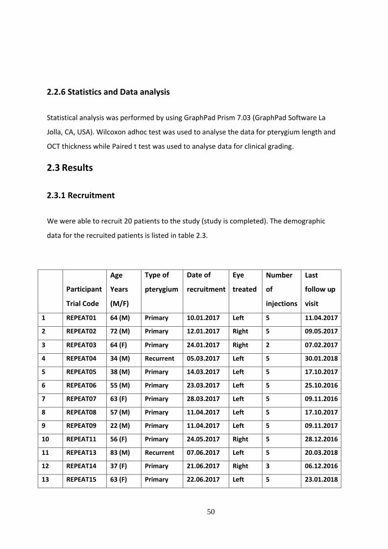
50
2.2.6 Statistics and Data analysis
Statistical analysis was performed by using GraphPad Prism 7.03 (GraphPad Software La
Jolla, CA, USA). Wilcoxon adhoc test was used to analyse the data for pterygium length and
OCT thickness while Paired t test was used to analyse data for clinical grading.
2.3 Results
2.3.1 Recruitment
We were able to recruit 20 patients to the study (study is completed). The demographic
data for the recruited patients is listed in table 2.3.
Participant
Trial Code
Age
Years
(M/F)
Type of
pterygium
Date of
recruitment
Eye
treated
Number
of
injections
Last
follow up
visit
1 REPEAT01 64 (M) Primary 10.01.2017 Left 5 11.04.2017
2 REPEAT02 72 (M) Primary 12.01.2017 Right 5 09.05.2017
3 REPEAT03 64 (F) Primary 24.01.2017 Right 2 07.02.2017
4 REPEAT04 34 (M) Recurrent 05.03.2017 Left 5 30.01.2018
5 REPEAT05 38 (M) Primary 14.03.2017 Left 5 17.10.2017
6 REPEAT06 55 (M) Primary 23.03.2017 Left 5 25.10.2016
7 REPEAT07 63 (F) Primary 28.03.2017 Left 5 09.11.2016
8 REPEAT08 57 (M) Primary 11.04.2017 Left 5 17.10.2017
9 REPEAT09 22 (M) Primary 11.04.2017 Left 5 09.11.2017
10 REPEAT11 56 (F) Primary 24.05.2017 Right 5 28.12.2016
11 REPEAT13 83 (M) Recurrent 07.06.2017 Left 5 20.03.2018
12 REPEAT14 37 (F) Primary 21.06.2017 Right 3 06.12.2016
13 REPEAT15 63 (F) Primary 22.06.2017 Left 5 23.01.2018

51
14 REPEAT16 49 (M) Primary 28.06.2017 Left 5 17.01.2017
15 REPEAT17 54 (M) Primary 05.07.2017 Left 5 21.02.2017
16 REPEAT18 53 (F) Recurrent 15.08.2017 Left 2 15.08.2017
17 REPEAT19 70 (M) Primary 29.08.2017 Right 5 16.01.2018
18 REPEAT20 42 (F) Primary 11.10.2017 Left 4 27.02.2018
19 REPEAT22 37 (F) Primary 03.11.2017 Right 5 10.04.2018
20 REPEAT23 69 (M) Recurrent 09.11.2017 Left 5 09.11.2017
Table 2.3 Demographic data of REPEAT cases
2.3.2 Cases
16 patients had primary pterygium and 4 patients had recurrent pterygium. A total of 20
eyes of 20 patients received 5 FU and bevacizumab injections. Of the twenty patients, 8
were females (40%), and 12 were males (60%). Mean age was 54 years (range was from 22-
83 years). Sixteen patients received 5 injections, 1 received 4 injections, 1 received 3
injections and 2 received 2 injections. All patients experienced stinging during injection of
bevacizumab but not with 5FU. All patients showed subconjunctival and/or intra-lesional
haemorrhage after injection (Figure 2.1), which resolved in two weeks. All patients showed
arrest of progression over the follow up period.

52
Figure 2.1: Slit lamp anterior segment photograph of a pterygium post injection.
The bleb created by the injection is visible with some conjunctival haemorrhage at the site of needle entry.
There was a mean reduction of 0.10 mm+/-0.1 in pterygium length in 56% of the patients
while the rest did not show any change ( p=0.0039, Wilcoxon test, median= 0.041, 95%
confidence interval (CI) = 0.024-0.177). Twenty two percent of the patients showed a
reduction in astigmatism (mean magnitude of difference vector was 10.1), 11% showed no
difference (mean magnitude of difference vector was 1) while 67% had increased
astigmatism (mean magnitude of difference vector was 5) following the injections. The
mean magnitude of difference vector was 5.7 and the range of astigmatic change was from
0.8-19.4. Eighty nine percent of patients had reduced thickness of pterygium as evidenced
by difference in OCT measurements while the rest had no change. The mean reduction in
thickness was 35.8 µm+/- 31.29, (p=0.0078, Wilcoxon test, median= 29 µm, 95% CI= 11.7-
59.9).
The clinical grade of the pterygium had significantly reduced in 62.5 % while it remained the
same in 37.5%. No patient showed an increase in clinical grade of pterygium. Ninety percent
of the patients who showed reduction of clinical grade had one clinical grade improvement
while 10% had a 2 clinical grades improvement (Figures 2.2-2.4) (Mean pre-injection grade
was 2.313, mean post-injection grade was1.625, p=0.0004, paired-test, 95% CI= 0.367 to

53
1.008). Two patients developed superficial punctate keratitis after the 4th and 5th injection.
This resolved on cessation of injection.
Figure 2.2: Slit-lamp anterior segment photograph of REPEAT09 before and after 5 injections.
A, C: Pre-injection image of pterygium with patient looking straight (A) and temporally (C). The lesion
appears fleshy with dilated and full vessels. B, D: Post-injection images with patient looking straight (B)
and temporally (D) show reduction in vascularity.

54
Figure 2.3: Slit lamp anterior segment photographs of REPEAT14 before and after 3 injections.
A, C: Pre-injection images of pterygium with patient looking straight (A) and temporally (C). The lesion
appears fleshy with dilated and full vessels (arrow). B, D: Post-injection images show reduction in
vascularity with patient looking straight (B) and temporally (D). The arrow in (D) points to a vessel that
has attenuated with barely visible branches.
Figure 2.4: Slit lamp anterior segment photographs images of REPEAT16 before and after 5 injections.
A: The pterygium before injections is fleshy and vascular obscuring the underlying limbus (arrow). B:
The pterygium after injections is atrophic as evidenced by increased visibility of the outline of the
underlying limbus especially inferiorly (arrow).

55
2.3.3 Laboratory study results
Atypical fibroblast cells and vascular endothelium showed positive staining for bFGF in
control pterygium samples (Figure 2.5A), whereas in injected pterygium (Figure 2.5B) weak
bFGF immunoreactivity was noted only at the basal aspect of vascular endothelium.
Monoclonal antibody against VEGF showed positive immunoreactivity in control pterygium
samples (Figure 2.5C) and a significantly reduced staining in injected samples (Figure 2.5D).
EGF (epidermal growth factor) was abundantly present in pterygium epithelium and around
basal aspects of vascular endothelium of control samples (Figure 2.5E), but in injected
samples it was only detected at low levels around basal aspect of vascular endothelium
(Figure 2.5F). Reactivity of collagen-I was moderately reduced in injected samples (Fig. 5H)
compared to control samples (Figure 2.5G). SPARC (secreted protein acidic and cysteine
rich) was shown to be positively expressed in all control samples but unlike bFGF, it
remained unchanged in sections of injected samples (Figure 2.6). Antibodies against von-
Willebrand factor (vWF) were used to detect blood vessels, and against lymphatic vessel
endothelial hyaluronan receptor (LYVE-1) to detect lymphatic vessels. Some vascular
structures showed dual staining for both vWF and LYVE-1 in sections of control samples
(Figure 2.7) whereas reduced expression was noted in injected samples. This amounted to
64.3% reduction in vWF staining and 61.9% in LYVE-1 stained vasculature after injection
(Table 2.4).

56
Figure 2.5: Immunofluorescence analysis of control and injected pterygium samples.

57
Representative sections of sample from control (A, C, E, G; n=3) and injected (B, D, F, H; n=4) groups.
Positive immunoreactivity for bFGF in fibroblast cells (A); VEGF in vascular endothelium and
extracellular matrix (C), EGF in epithelial cells (E) and Collagen-I in vascular basement membrane (G)
are demonstrated in the control sections. Reduced or weakened staining for these proteins is seen in
corresponding images post injection (B, D, F, H). Scale bar = 50 microns (x400).
Figure 2.6: Representative sections of sample from control (n=3) and injected (n=4) groups (Double
labeling of bFGF and SPARC).
Labeling for bFGF is demonstrated by red fluorescence (A and D) and SPARC by green fluorescence (B
and E) in multiple sections of the same pterygium vessel in control and injected samples. Overlay
micrographs indicate double labeling for bFGF and SPARC (orange/yellow fluorescence; C and F). The
staining for bFGF is markedly reduced in the injected sample but that of SPARC is unchanged. Scale bar
= 50 microns (x400).

58
Figure 2.7: Representative micrographs of control (n=3) and injected (n=4) samples. (Co-localization of
vWF (vascular) and LVYE-1 (lymphatic) channels).
vWF is indicated in green (A and D) and LYVE-1 in red (B and E). Overlay micrograph of control
section (C) indicates co-localisation of vWF and LYVE-1. In injected sample (F), LYVE-1 stained cells
surround vWF stained endothelium.
Table 2.4 Marked reduction in average number of vWF and LYVE-1 positive vessels.
vWF LYVE-1 Control (n=3) 28 21 Injected (n=4) 10 8
% reduction 64.3 61.9
59
2.4 Discussion
Recurrence of pterygium is the major complication after surgical excision of the lesion. The
two main pathological processes in pterygium are fibroblast proliferation and angiogenesis.
Many studies have demonstrated that the pathogenesis and recurrence of pterygia can be
attributed to the increased expression of molecules such as VEGF, transforming growth
factor, platelet derived growth factor and bFGF (236). VEGF was found to be highly
increased in pterygia and is believed to be involved directly or indirectly in formation of
pterygia (215).
Intervention to reduce incidence of recurrence following surgery for primary or recurrent
pterygia include the use of autologous conjunctival graft and application of antimetabolites
either 5FU or MMC intraoperatively. Intra lesion injection of antimetabolites and anti-VEGF
drugs have also been used to treat recurrent pterygium (235, 275, 282, 352). Encouraged by
the beneficial effects of treating recurrence, both agents have also been used individually in
treatment of primary pterygium with considerable success (349). 5FU inhibits DNA synthesis
and proliferation of fibroblast (353). Anti-VEGF antibodies bind to isoforms of VEGF and
inhibit the VEGF-receptor interaction preventing angiogenesis (352).
In our study we injected both agents together, for the treatment of primary pterygium. We
found that this approach resulted in reduction in the clinical grade, thickness and vascularity
of the pterygium. This two pronged approach addressing both the main pathological
processes may work synergistically affecting thickness and vascularity of the pterygium.
Some patients also showed a slight reduction in the length of the pterygium. This is most
likely due to the reduction in thickness at the apex, but in any case was less than 0.2mm and
clinically insignificant. Significantly, the progression of pterygium was arrested in all cases
and the cosmesis too had improved in 62.5% with 56% of the concerned patients noticing
the difference subjectively. Some of these individuals are unlikely to proceed to surgery
making the study clinically more relevant. Interestingly however, the astigmatism increased
in 67% of patients post-injection. This could be related to the retraction of the fibrotic tissue
post-injection exerting a greater force on the cornea inducing further deformation. Malik et

60
al (235) compared the mean change in corneal astigmatism and clinical appearance after
intralesional injection of 5FU in primary and recurrent pterygia. They treated all patients
with 0.1 ml of 5FU (5mg) weekly injections for four weeks and demonstrated a reduction of
the astigmatism. However, Singh et al (273) use only bevacizumab injection and showed not
difference in astigmatism. Unlike these studies, we injected both 5FU and bevacizumab,
which could account for the increased astigmatism we noted, probably as a consequence of
fibrous tissue contraction. Most studies that have used either 5FU or bevacizumab (234,
236, 273) concluded that improvements in appearance, colour and surface area of
pterygium could be safely achieved.
Despite anaesthesia, all patients complained of a stinging sensation during injection of
bevacizumab but not with 5FU. Hence we injected the latter before the former.
Bevacizumab solution is acidic (pH 6.13) and 5FU is alkaline (pH 8.89), which could explain
the difference in patient experience. Apart from mild punctate keratitis, no adverse events
were recorded. Post-injection, four patients requested surgery to remove the residual
pterygium. Intraoperative bleeding was much less compared to previous experience with
un-injected lesions. This is an anecdotal observation as the study was not designed to pick
up this difference. These four samples were examined by immunohistology.
Immunohistology:
Immunohistochemical studies have shown increased expression of VEGF in pterygium
compared to normal conjunctiva (215, 349). VEGF receptors 2 and 3 are also increased in
pterygium (349). Mohamed et al (353) conducted a randomised control study with one
group receiving a single bevacizumab injection while the other did not receive any. Similar
to our results, they found a reduced mean vessel count and a significant reduction in VEGF
expression in injected pterygium samples. vWF is one of the proteins involved in blood
vessel function. It has been demonstrated in the pterygial blood vessel walls and could be
involved in pterygial angiogenesis (354). Lymphatic vessel endothelial cells express specific
proteins including LYVE-1 (201). Our study demonstrated that there was a reduction in both
vWF and LYVE-1 in the injected samples indicating decreased angiogenesis and
lymphangiogenesis, which is a novel finding. Interestingly, in control pterygium samples
61
there was a close localisation of both vWF and LYVE-1 in relation some vessels, which could
be related to immature vessels or proximity of mononuclear cells (355).
Detorakis et al (356) found that levels of bFGF were higher in pterygium than normal
conjunctiva. Collagen types I, III and IV were found in normal conjunctiva and pterygium
(357). In our injected samples both bFGF and Collagen I were reduced, which is more likely
to be due to the effect of 5FU as it primarily targets the proliferation of fibroblasts. SPARC,
is a matrix protein which has several roles in wound remodelling and modification of
collagen production as well as in angiogenesis and high levels have been reported in
pterygium (349). We were not able to demonstrate any effect of our injections on the
SPARC expression despite reduction in vascularity and collagen suggesting that bFGF plays a
greater role in the pathophysiology of pterygium. Pterygium epithelium and fibroblasts
derived from pterygia also express epidermal growth factor receptors (EGFR) (358). We
detected EGF at the basal aspect of pterygium vascular endothelium, which was reduced
after injections suggesting that EGF also contributed to angiogenesis in pterygium.
This study demonstrates that proliferative activity in pterygium and its progression can be
modified by drugs targeting the various growth factors and cells involved in the
pathogenesis of pterygium. Such a medical approach in the management of pterygium, the
treatment of which is essentially surgical, is a viable option to consider for both recurrent
and primary pterygia.
The strong point in this clinical study is using 2 different types of combined injections to determine their effect
on the arrest of pterygium progression as all previous studies investigated one type of injection only. The
limitation in this study was the sample size. In the future, a bigger sample size would give more data and also
another clinical study could be carried out to follow up the patients who required surgery despite of injections to
look into the rate of recurrence to determine whether the multiple combined injections reduced the recurrences
rates or not .
62
CHAPTER 3: HEALING OF AUTOLOGOUS CONJUNCTIVAL
GARFTS IN PTERYGIUM SURGERY
3.1 Introduction
Pterygium is a common ocular disorder which presents as a progressive triangular growth
encroaching on the cornea from the bulbar conjunctiva. It consists of fibrovascular
subconjunctival tissue with elastotic degeneration and conjunctival epithelial
hypertrophy(359). It typically affects inter‐palpebral conjunctiva, nasal more than temporal.
It can affect vision by direct obstruction of the passage of light or induce corneal distortion
and irregular astigmatism. In advanced cases, it can affect ocular movements with
symptoms of diplopia. For many patients, it is a cosmetic blemish (204, 360-363). There is
no medical therapy for pterygium. When indicated for the above reasons, surgical excision
with autologous conjunctival graft is the intervention of choice with intraoperative use of an
anti‐metabolite such as Mitomycin‐C (MMC) as an adjunct, especially in recurrent cases.
Traditionally the autologous graft was secured in place with sutures but in many parts of the
world sutures have been replaced with tissue glue (fibrin glue) or its variations such as
autologous blood (364-366).
Both occurrence and recurrence of pterygium after surgery are related to race, skin colour
and geographic location (207-209). The surgical success rate (lack of recurrence) following
excision and conjunctival autograft is reported to be between 0 and 14.29% (228, 351, 367-
370). Use of fibrin glue instead of sutures is reported to favourably influence the recurrence
rate. Although much has been studied and reported in relation to use of autologous grafts
and fibrin glue (254, 368, 371-379), there is no information on the fate or healing of the
grafted conjunctival tissue and the changes associated with the donor site. We undertook a
temporal observational study of the healing of conjunctival autograft and the donor site in
28 consecutive patients and report our results herein that have implications for clinical
follow‐up and differentiation from signs of early recurrence.
This chapter has been published by Dr Ghoz et al and is her own work (Please refer to list of
publications).
63
3.2 Methods
3.2.1 Ethical issues
The study was conducted in accordance to the declaration of Helsinki. The study was classed
as a ‘healthcare evaluation project’ and approved by the Clinical Quality, Risk and Safety
directorate, Nottingham University Hospitals, NHS Trust (project number: 17-206c).
3.2.2 Patient recruitment
Thirty‐two eyes of 28 consecutive patients attending the cornea and ocular surface clinic of
the Queens Medical Centre, Nottingham were included. All patients had visually or
cosmetically significant pterygium requiring surgery.
3.2.3 Clinical intervention and follow up
All thirty two eyes of the 28 patients underwent surgical excision of pterygium using the
conjunctival autograft technique.
Surgical procedure
After due consent, surgery was carried out under regional anaesthesia (sub‐Tenon's block)
or general anaesthesia. A blunt scissors was inserted through the conjunctiva at the neck of
the pterygium (at limbus) and the head peeled off the corneal surface. The head and neck
were excised and the corneal surface polished. The conjunctiva covering the body of the
pterygium was carefully dissected off the underlying fibrovascular tissue and the latter off
the sclera, up to the medial fornix but anterior to the caruncle. The fibrovascular tissue was
excised and bleeding points cauterized with a bipolar cautery (Force EZ, Valleylab, USA).
Donor site: the superior bulbar conjunctiva of the same eye was exposed with a superior
rectus suture (4/0 silk). The donor conjunctiva, corresponding to the size of the defect

64
created by pterygium excision, was marked with ink (4–6 mm vertically and 5–15 mm
horizontally) preserving 2 mm of peri‐limbal conjunctiva of the donor site. The donor
conjunctiva, sparing Tenon's capsule, was undermined with a blunt scissors and excised such
that the ink marks were included on the epithelial surface. The donor conjunctiva was
spread epithelial side down, on the cornea and covered with a layer of the thick component
(fibrinogen, aprotinin, coagulation factor 13) of (Tisseel®, Baxter Healthcare Corporation,
Berkshire, UK) in a double barrel syringe. The thawed components of each barrel were
withdrawn separately into 2 ml syringes and glue applied via a Rycroft cannula (Sterimedix,
Redditch, Worcestershire, UK). The recipient bed was covered with a layer of the thin
component (thrombin and calcium chloride) of Tisseel®. The donor conjunctiva was flipped
across to cover the recipient bed and smoothened such that the edges were not rolled.
Gentle pressure was applied with a dry surgical sponge to approximate the conjunctival
autograft with the underlying episclera/sclera (scleral bed). Further glue, each component
sequentially, was applied under the edge of the recipient site conjunctival edges
(conjunctival bed), and it was approximated to the donor conjunctiva edge as the glue set.
The posterior part of the conjunctiva at the donor site was undermined, both components
of glue applied to the surface of exposed Tenon's, and the conjunctiva pulled down to cover
the defect and approximated to the rim of peri‐limbal conjunctiva.
Mitomycin‐C was applied as an adjunct in 25 eyes using sponge pieces soaked in 0.04%
solution and placed between sclera and overlying conjunctiva of the pterygium for 5 min,
and on the exposed sclera for 2 min. Mitomycin‐C (MMC) was used intraoperatively in all
patients with recurrent pterygium and patients with Middle‐Eastern and Afro‐Caribbean
origin undergoing excision of primary pterygium. The eye was then washed with 20 ml of
balanced salt solution before affixing the donor conjunctiva.
Postoperative management
All patients were prescribed chloramphenicol eye drops 0.5% (Chloramphenicol minims,
Bausch & Lomb/Chauvain), instilled four times a day for 2 weeks and prednisolone acetate
eye drops 0.5% (Predsol minims, Bausch & Lomb/Chauvain), instilled four times a day for
1 week then tapered to three times a day for 1 week, twice a day for 1 week and then once
a day for another week.

65
Patients were followed up within the first week and then at weeks 2, 4, and 8. Sixteen
patients were followed up to 6 months postoperatively. Slit lamp images were taken with a
Topcon camera SLD701 preoperatively and at each visit. Patients were examined, without
and with fluorescein staining, for presence of graft, for graft haemorrhage, swelling,
vascularity/ischaemia and retraction of edges. The graft beds (scleral and conjunctival) and
the conjunctiva at the donor site were also similarly examined. Fluorescein angiography (FA)
was performed in four patients within the first week and in one patient 3 months after the
surgery.
Anterior segment fluorescein angiography
Anterior segment fluorescein angiography (FA) was performed on five eyes of five patients
who had pterygium excision and conjunctival autografts. The terms of the study approval
required that only one angiogram be performed per patient and for five patients only as
part of an angiography pilot study. Hence, FA was performed at 1 day postoperative for two
patients, 2 days postoperative for one patient, 3 days postoperative for one patient and at
3 months postoperative for one patient.
Procedure of angiography
The angiograms were obtained using the Heidelberg Retina Angiograph, and the degree of
field of view was chosen according to the size of the conjunctival autograft/vessels. The
focus was set on +32.00. An initial infrared image of the selected area was taken as baseline.
Five millilitres of 10% sodium fluorescein were then injected intravenously by cannulating
the antecubital vein at a rate of 1 millilitre per second. Two single images of the selected
area were taken at the start and end of the injection. A video recording of 35 seconds
duration was initiated with the first appearance of the dye, which was usually 12 seconds
after the start of the injection. After 35 seconds, no images were taken till 1 min elapsed
from the start of the injection and then a single image was captured every second till 2 min
elapsed from the start of the injection. At minute 2 from the start of the injection, two
single images of the selected eye were taken and another two single images were captured
between minutes 4 and 5 from the start of the injection.

66
3.3 Results
3.3.1 Cases
A total of thirty‐two eyes of 28 patients underwent pterygium excision with autologous
conjunctival graft affixed with fibrin glue. Patient demographics are shown in Table 3.1. The
age range was 33–78 years with a mean of 50.8 years and there were 17 males and 11
females. Twenty‐four cases were unilateral and four were bilateral. Mitomycin‐C (MMC)
was applied to 25 eyes of 22 patients. Twenty‐seven eyes of 24 patients had primary
pterygium while five eyes of four patients had recurrent pterygium. None of the primary
pterygium cases that underwent surgery developed recurrence over the study period.
Age Sex Eye
1 54 M L
2
41
M
L
3
72
F
R
4
39
M
R
5
51
M
L
6
45
M
R
7
33
M
L
8
78
F
L
9
61
M
BE

67
Age Sex Eye
10 70 F R
11
39
M
L
12
46
M
R
13
45
M
R
14
54
M
L
15
46
M
BE
16
50
F
BE
17
50
M
R
18
45
F
R
19
64
F
BE
20
36
F
R
21
64
F
L
22
56
M
L
23
51
F
L
24
39
F
L
25
34
M
L
26
57
F
R

68
Table 3.1: Age and sex of patients and laterality of the pterygium.
BE = both eyes; F = female; L = left; M = male; R = right.
Within 1 week
The conjunctival autograft was not lost in any eye. About 94% of conjunctival autografts
showed diffuse or patchy intra-graft haemorrhage (Figure 3.1). Haemorrhage started as
early as 1 day postoperative. Blood vessels were obscured by the diffuse haemorrhage
(Figure 3.1). In grafts with patchy haemorrhage, the visible vessels were narrow and/or
dilated (Figure 3.1). Vascular connections between graft and host bed were not clearly
visible. All grafts showed variable amount of swelling. The swelling was either due to
oedema and/or transudation, which appeared as a straw‐coloured accumulation of fluid
(Figure 3.2).The corneal edge of the conjunctival autograft was overlapping the limbus in all
patients. About 11% conjunctival autografts showed areas of localized under‐perfusion
(areas devoid of blood vessels compared to the surrounding perfused conjunctiva in the
autograft) (Figure 3.3). The episclera under the conjunctival bed of 17% conjunctival
autografts showed engorged vessels. The conjunctival beds of 89% of conjunctival
autografts showed congestion and 33% showed haemorrhage (Fig. 3A). The edge of the
conjunctival beds were swollen with oedema causing the edge to form a narrow hump
giving it a ‘rolled’ appearance in some sectors as illustrated (Figures 3.1A and 3.1D).
Fluorescein staining showed no loss of epithelium in any conjunctival autograft. Residual
fibrin glue at the junction of conjunctival autograft and conjunctival bed, stained brightly
with fluorescein (Figure 3.4). A separation or gap between the edges of the conjunctival
autograft and conjunctival bed was seen in 33% of patients. This gap was fully epithelialized
as evidenced by fluorescein staining. About 12.5% of the patients with a gap showed under‐
perfusion of the scleral bed (Figures 3.5 and 3.6).
R M 66 28
L M 37 27
Eye
Sex
Age
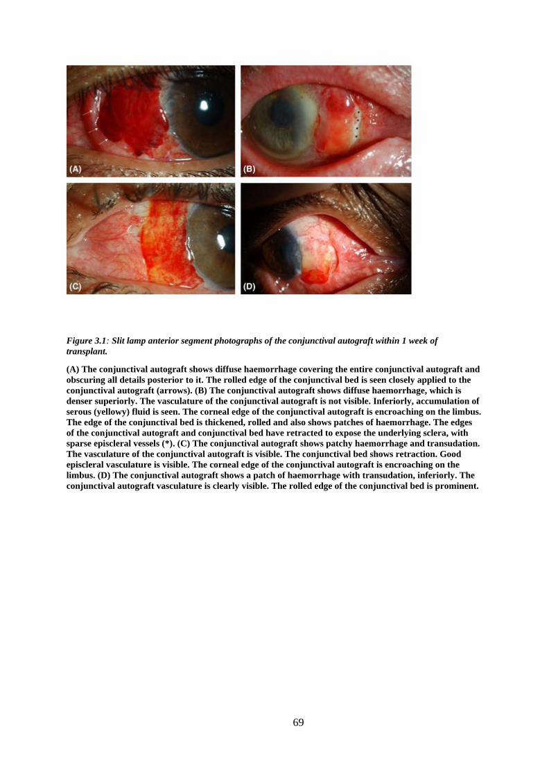
69
Figure 3.1: Slit lamp anterior segment photographs of the conjunctival autograft within 1 week of
transplant.
(A) The conjunctival autograft shows diffuse haemorrhage covering the entire conjunctival autograft and
obscuring all details posterior to it. The rolled edge of the conjunctival bed is seen closely applied to the
conjunctival autograft (arrows). (B) The conjunctival autograft shows diffuse haemorrhage, which is
denser superiorly. The vasculature of the conjunctival autograft is not visible. Inferiorly, accumulation of
serous (yellowy) fluid is seen. The corneal edge of the conjunctival autograft is encroaching on the limbus.
The edge of the conjunctival bed is thickened, rolled and also shows patches of haemorrhage. The edges
of the conjunctival autograft and conjunctival bed have retracted to expose the underlying sclera, with
sparse episcleral vessels (*). (C) The conjunctival autograft shows patchy haemorrhage and transudation.
The vasculature of the conjunctival autograft is visible. The conjunctival bed shows retraction. Good
episcleral vasculature is visible. The corneal edge of the conjunctival autograft is encroaching on the
limbus. (D) The conjunctival autograft shows a patch of haemorrhage with transudation, inferiorly. The
conjunctival autograft vasculature is clearly visible. The rolled edge of the conjunctival bed is prominent.

70
Figure 3.2: Slit lamp anterior segment photographs of the conjunctival autograft within 1 week of
transplant.
The vasculature of the conjunctival autograft is visible in all photographs. The vessels are of variable
thickness and some show stagnant columns of blood (arrow). The nasal edge of the conjunctival autograft
is in close apposition with the conjunctival bed, with no gap in all four cases. (A) The conjunctival
autograft shows swelling due to oedema. (B) The conjunctival autograft shows swelling due to oedema.
The conjunctival bed is congested and shows an elevated fold where it apposes the edge of the
conjunctival
autograft. (C) The conjunctival autograft shows patchy haemorrhage and diffuse transudation of straw‐
coloured fluid which contrasts with the appearance of oedema seen in (A) and (B) above. The corneal
edge of the conjunctival autograft is encroaching on the limbus. The conjunctival bed is not congested
but is
thickened. (D) The conjunctival autograft shows haemorrhage inferiorly with diffuse transudation of
straw‐ coloured fluid.

71
Figure 3.3: Slit lamp anterior segment photographs of the conjunctival autograft within 1 week of transplant
(without and with fluorescein staining (2% eye drops).
(A) The conjunctival autograft shows localized under‐perfused area (outlined), patchy haemorrhage
with minimal transudation. The conjunctival bed shows areas of congestion. (B) Cobalt blue filter image
with fluorescein sodium 2% eye drops of (A) showing the outline of conjunctival autograft where
pooling of dye occurs. The cornea shows an epithelial defect (arrow). (C) The conjunctival autograft
shows patchy
haemorrhage and a localized area of under‐perfusion (outlined). (D) Cobalt blue filter image with
fluorescein sodium 2% eye drops of (C) showing the outline of conjunctival autograft.
Figure 3.4: Slit lamp anterior segment photographs of the conjunctival autograft within 1 week of transplant
(without and with fluorescein 2% eye drops).

72
(A) The conjunctival autograft is swollen and shows patchy haemorrhage and transudation. There is
residual fibrin glue at the junction of the conjunctival autograft and the conjunctival bed (arrow). The
corneal edge of the conjunctival autograft is encroaching on the limbus. (B) Cobalt blue filter image with
fluorescein sodium 2% eye drops of (A) showing fibrin glue staining brightly with fluorescein.
Figure 3.5: Slit lamp anterior segment photographs images of the conjunctival autograft of a patient from
1 week of surgery to 6 months (without and with fluorescein 2% eye drops).
(A) The conjunctival autograft is swollen, haemorrhagic and its nasal edge has retracted from the
conjunctival bed. The underlying sclera has sparse vessels. (B) Cobalt blue filter image with fluorescein
sodium 2% eye drops of (A) showing the outline of conjunctival autograft. The exposed underlying sclera
is epithelialized. A small defect in the healing corneal epithelium is seen. (C) The conjunctival autograft
showed less swelling, haemorrhage and transudation at 2 weeks postoperative. The corneal edge of the
conjunctival autograft has retracted from the limbus. (D) The conjunctival autograft swelling has
resolved, and there is minimal haemorrhage at 1 month postoperative. There is a well‐established
network of vessels in the conjunctival autograft. Some under‐perfused areas persisted (outlined) but
small vessels (arrows) are seen to migrate into this area. (E) The conjunctival autograft and conjunctival
bed have settled completely at 6 months postoperative. The under‐perfusion of the scleral bed has
resolved. The conjunctival autograft shows multiple reconnections with the bed. (F) Cobalt blue filter
image with fluorescein sodium 2% eye drops of (E) showing the outline of conjunctival autograft
although it seems to have imperceptibly merged with the conjunctival bed in (E).
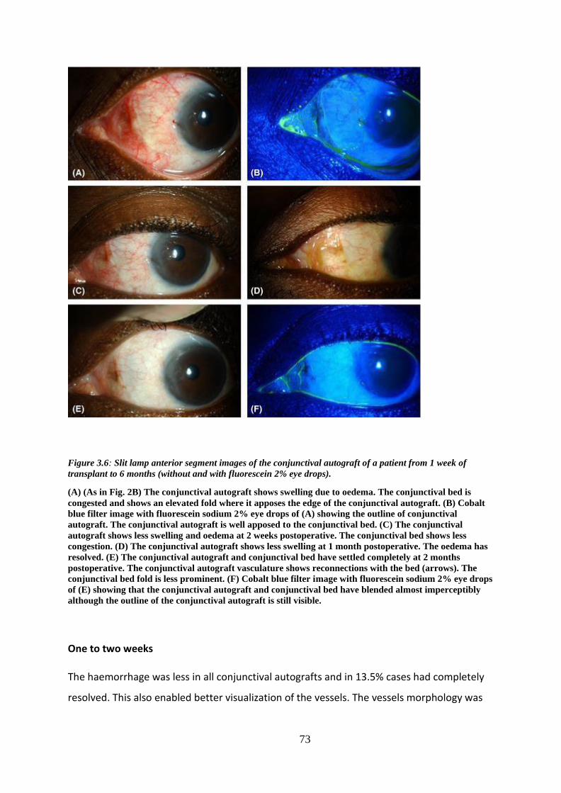
73
Figure 3.6: Slit lamp anterior segment images of the conjunctival autograft of a patient from 1 week of
transplant to 6 months (without and with fluorescein 2% eye drops).
(A) (As in Fig. 2B) The conjunctival autograft shows swelling due to oedema. The conjunctival bed is
congested and shows an elevated fold where it apposes the edge of the conjunctival autograft. (B) Cobalt
blue filter image with fluorescein sodium 2% eye drops of (A) showing the outline of conjunctival
autograft. The conjunctival autograft is well apposed to the conjunctival bed. (C) The conjunctival
autograft shows less swelling and oedema at 2 weeks postoperative. The conjunctival bed shows less
congestion. (D) The conjunctival autograft shows less swelling at 1 month postoperative. The oedema has
resolved. (E) The conjunctival autograft and conjunctival bed have settled completely at 2 months
postoperative. The conjunctival autograft vasculature shows reconnections with the bed (arrows). The
conjunctival bed fold is less prominent. (F) Cobalt blue filter image with fluorescein sodium 2% eye drops
of (E) showing that the conjunctival autograft and conjunctival bed have blended almost imperceptibly
although the outline of the conjunctival autograft is still visible.
One to two weeks
The haemorrhage was less in all conjunctival autografts and in 13.5% cases had completely
resolved. This also enabled better visualization of the vessels. The vessels morphology was

74
more varied, being tortuous or straight, narrow or dilated. Vascular connections were
clearly seen between the conjunctival autograft and conjunctival bed in all patients. About
60% conjunctival autografts were less swollen while 40% showed no improvement in
swelling. The corneal edge of 93% had retracted from the limbus, which was now clearly
visible. The under‐perfused area had not resolved in any of the cases. The episcleral vessels
(scleral bed) were less engorged. About 80% of conjunctival beds still showed congestion
but the degree of congestion was less than week one in all congested eyes while the rest of
the congested eyes has resolved and residual minimal haemorrhage was seen in 27% of
cases. All beds had several tortuous, straight, narrow or dilated blood vessels
(Figures 3.5 and 3.6).
Two to four weeks
About 70.5% of conjunctival autografts continued to show minimal haemorrhage, which had
completely resolved in the rest. All conjunctival autografts demonstrated a well‐formed
network of blood vessels with well‐established connections with the conjunctival bed.
Approximately half (47%) showed complete resolution of swelling. Under‐perfusion of
conjunctival autograft was persistent. The congestion in all conjunctival beds had resolved
and only 10% showed minimal haemorrhage. At this time point, ‘shunt vessels’ were clearly
seen in the conjunctival bed (Figure 3.7).The scleral bed in the gap between conjunctival
autograft and conjunctival bed continued to show under‐perfusion although small vessels
were seen to migrate into the under‐perfused area (Figure 3.5D).

75
Figure 3.7: Representative image of the conjunctival autograft of a patient at 1 month postoperative (slit
lamp image, diffuse illumination).
The conjunctival autograft shows swelling due to oedema and patchy haemorrhages. There is a
localized area of under‐perfusion (outlined). The conjunctival bed shows a shunt vessel, the afferent
starting at the
black arrow and forming a loop to the efferent (white arrow). No terminal branches are seen from the
distal part of the loop. The conjunctival bed is congested.
Four to eight weeks
About 16% conjunctival autografts had some remaining haemorrhage. Multiple
reconnections with the bed were seen in all patients. About 21% conjunctival autografts
continued to show residual swelling. The under‐perfused areas in the conjunctival
autografts persisted. Clinically, the junction of the edge of the conjunctival autografts and
the conjunctival bed was difficult to discern except in those who had developed a gap in one
or more sector. Under‐perfusion of the scleral bed in the gap persisted. With fluorescein
staining, the conjunctival autograft‐conjunctival bed junction was clearly outlined with
pooling of fluorescein in 80% and in cases where there was a gap, the pooling was more
obvious as a broader outline. About 10% of the conjunctival beds continued to show
minimal haemorrhage (Figures 3.5 and 3.6).
Eight weeks up to 6 months

76
All patients had completely settled grafts and beds (Figures 3.5 and 3.6). The conjunctival
autografts showed no haemorrhage or swelling and all of them had a well‐formed network
of vessels connected with the beds. Some shunt vessels persisted. The under‐perfusion of
scleral beds had completely resolved.
The findings from our temporal observation of ischaemia/under‐perfusion, haemorrhage,
swelling or gaping at the edge between the conjunctival autograft and conjunctival bed
were consistent, with no variation in eyes which received adjunct MMC during pterygium
surgery from those which did not have MMC application.
Donor site
About 27% of donor sites showed haemorrhage and congestion in the first week, which had
completely resolved within 8 weeks (Figure 3.8). One patient had telangiectasia, which
developed at 1 week and persisted thereafter (Figure 3.9). One patient had retraction of the
conjunctival edge from the limbus with exposed Tenon's capsule, which developed a
pyogenic granuloma (Figure 3.10). This had settled and epithelialized by the week eight
follow‐up visit. No other complications were noted.
Figure 3.8: Slit lamp anterior segment photograph of the donor site of a patient at 1 week postoperative.

77
The donor site is congested and shows multiple areas of haemorrhages. The superior conjunctiva that
was pulled towards the limbus and glued to cover the defect has remained in situ without any
retraction.
Figure 3.9: Slit lamp anterior segment photograph of the donor site of a patient at 3 months postoperative.
The donor site shows an area of aneurysmal/telangiectatic dilation of the vasculature. The lesion was
noted at 2 weeks postoperative and changed minimally over the follow‐up period. The margins remained
well‐ defined and the colour did not fade. The probability of this lesion being an intraconjunctival
haemorrhage could not be ruled out.
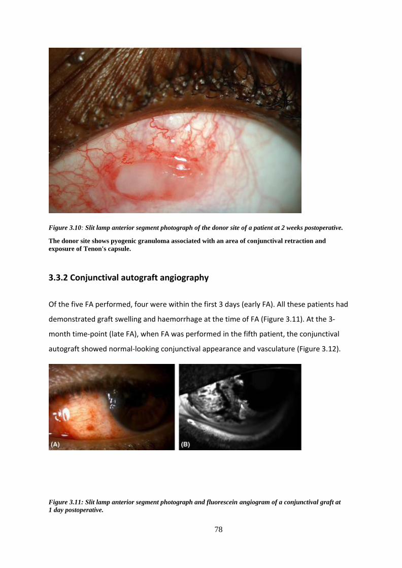
78
Figure 3.10: Slit lamp anterior segment photograph of the donor site of a patient at 2 weeks postoperative.
The donor site shows pyogenic granuloma associated with an area of conjunctival retraction and
exposure of Tenon's capsule.
3.3.2 Conjunctival autograft angiography
Of the five FA performed, four were within the first 3 days (early FA). All these patients had
demonstrated graft swelling and haemorrhage at the time of FA (Figure 3.11). At the 3‐
month time‐point (late FA), when FA was performed in the fifth patient, the conjunctival
autograft showed normal‐looking conjunctival appearance and vasculature (Figure 3.12).
Figure 3.11: Slit lamp anterior segment photograph and fluorescein angiogram of a conjunctival graft at
1 day postoperative.

79
(A) Slit lamp anterior segment photograph of the conjunctival autograft of a patient at 1 day
postoperative. Swelling due to oedema and patchy haemorrhages is seen. (B) Anterior segment
fluorescein angiogram at
1 min and 45 seconds. There is hyperfluorescence along the conjunctival bed. Areas of hyperfluorescence
are also seen on the conjunctival autograft. The dark band does not correspond to the haemorrhage seen
in
(A) and is likely to represent an area of non‐perfusion with some masking.
Figure 3.12: Anterior segment fluorescein angiogram at 4 min and 10 seconds of the conjunctival
autograft of a patient at 3 months postoperative.
A normal vasculature network is clearly visible in the conjunctival autograft. There is no leakage in the
conjunctival autograft but areas of hyperfluorescence persist along the edge of conjunctival bed which is
accentuated by light reflection. Long loops of shunt vessels, running parallel to the limbus (arrows) are
seen at the junction of the conjunctival autograft and the conjunctival bed.
All early FA showed intense fluorescence along the conjunctival bed early in the passage of
dye (Figure 3.11).The conjunctival autograft appeared largely non‐perfused or under‐
perfused in three of the four grafts. In conjunctival autograft where there was
haemorrhage, the area of haemorrhage obscured the fluorescence (Figures 3.11 and 3.13)
but in these conjunctival autografts even areas where there was no haemorrhage showed
the absence of fluorescence indicating poor or absent circulation (Figure 3.11). In all these

80
early FAs, 0–2 small vascular twigs were seen at some point along the edge of the
conjunctival bed and multiple fluorescent spots were seen in the underlying scleral bed. One
of the four early FAs (one‐day postop) showed an extensive network of vessels in the
conjunctival autograft, unlike the other three (Figure 3.11). Tissue leakage of dye was
evident as late hyperfluorescence in all conjunctival autografts with early FA indicating a
persistent breach in the blood–tissue barrier (Figure 3.11).
Figure 3.13: Image of the conjunctival autograft of a patient at 3 days postoperative (A) Slit lamp diffuse
anterior segment photograph of conjunctival autograft (B) Anterior segment fluorescein angiogram at
56 seconds.
(A) There is an area of diffuse haemorrhage involving almost the entire conjunctival autograft. (B) The
conjunctival autograft shows a large area of masking corresponding to the diffuse haemorrhage seen in
(A) obscuring the underlying fluorescence. The conjunctival bed shows intense fluorescence (accentuated
by the light reflection) due congestion of the conjunctival bed.
In the only late FA, the conjunctival bed showed three sites from which feeder vessels
connected to a well‐established network of vessels in the conjunctival autograft. Some of
these vessels filled early in the sequence (afferents) and others late (efferents). Two areas in
the conjunctival autograft remained hypo‐fluorescent for 7 seconds, compared to other
areas, before the vascular network became visible, suggesting a degree of residual ‘relative
under‐perfusion’. This conjunctival autograft did not demonstrate any leakage/persistent
fluorescence, indicating re‐establishment of the blood–tissue barrier.
Disappearance of dye (drainage) from conjunctival bed was earlier than from the
conjunctival autograft in all cases. One conjunctival autograft showed increased
fluorescence of the underlying scleral bed rather than of the conjunctival autograft itself.
Here stagnant columns of blood were seen in the conjunctival autograft at this time‐point

81
(Figure 3.14).
Figure 3.14: Anterior segment fluorescein angiogram at 10 seconds of the conjunctival autograft of a patient
at 2 days postoperative.
The conjunctival autograft shows increased fluorescence of the underlying scleral bed rather than of the
conjunctival autograft itself. It also shows stagnant columns of blood.
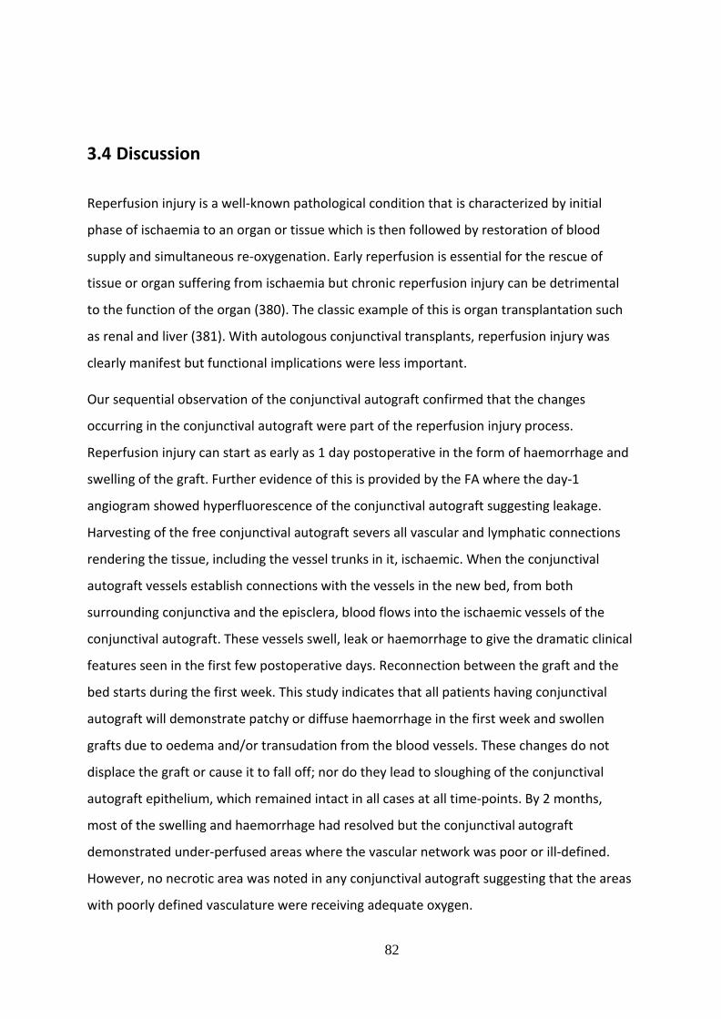
82
3.4 Discussion
Reperfusion injury is a well‐known pathological condition that is characterized by initial
phase of ischaemia to an organ or tissue which is then followed by restoration of blood
supply and simultaneous re‐oxygenation. Early reperfusion is essential for the rescue of
tissue or organ suffering from ischaemia but chronic reperfusion injury can be detrimental
to the function of the organ (380). The classic example of this is organ transplantation such
as renal and liver (381). With autologous conjunctival transplants, reperfusion injury was
clearly manifest but functional implications were less important.
Our sequential observation of the conjunctival autograft confirmed that the changes
occurring in the conjunctival autograft were part of the reperfusion injury process.
Reperfusion injury can start as early as 1 day postoperative in the form of haemorrhage and
swelling of the graft. Further evidence of this is provided by the FA where the day‐1
angiogram showed hyperfluorescence of the conjunctival autograft suggesting leakage.
Harvesting of the free conjunctival autograft severs all vascular and lymphatic connections
rendering the tissue, including the vessel trunks in it, ischaemic. When the conjunctival
autograft vessels establish connections with the vessels in the new bed, from both
surrounding conjunctiva and the episclera, blood flows into the ischaemic vessels of the
conjunctival autograft. These vessels swell, leak or haemorrhage to give the dramatic clinical
features seen in the first few postoperative days. Reconnection between the graft and the
bed starts during the first week. This study indicates that all patients having conjunctival
autograft will demonstrate patchy or diffuse haemorrhage in the first week and swollen
grafts due to oedema and/or transudation from the blood vessels. These changes do not
displace the graft or cause it to fall off; nor do they lead to sloughing of the conjunctival
autograft epithelium, which remained intact in all cases at all time‐points. By 2 months,
most of the swelling and haemorrhage had resolved but the conjunctival autograft
demonstrated under‐perfused areas where the vascular network was poor or ill‐defined.
However, no necrotic area was noted in any conjunctival autograft suggesting that the areas
with poorly defined vasculature were receiving adequate oxygen.
83
Changes were also seen in the conjunctival bed surrounding the conjunctival autograft. The
edge of the conjunctiva was hyperaemic and swollen and haemorrhage was also seen in
some places. Fibrin glue is known to ‘seal’ vessels and is used for haemostasis (382).
As fibrin glue was also used to attach the conjunctival bed, it is likely that the clinical picture
seen, both in the conjunctival bed and the conjunctival autograft was influenced by its
presence. Fibrin glue has been shown to mechanically interfere with epithelial cell migration
which proceeds normally once the glue dissolves in 10–14 days (383). The physical presence
of the glue at the site between the conjunctival autograft and the conjunctival bed could
also have influenced the re‐establishment of vascular connections. Resolution of the
oedema of the conjunctival bed corresponded to the appearance of ‘shunt’ vessels that
were noted to connect severed vessels in the bed. These shunt vessels were seen clinically
in the conjunctival bed of two eyes. A continuous remodelling of the vasculature occurs in
the postoperative period as new connections appear and some disappear. Eventually a
mature vasculature is established which remains permanent. The scleral bed also
contributes to the re‐vascularization of the conjunctival autograft. These vessels were seen
as large (relatively thicker) trunks but were sparse and not seen in every case.
Although the edges of the conjunctival autograft were closely approximated to the
conjunctival bed intraoperatively, gaps were noted between the edges at places, in some
cases, in the postoperative period. This represented a retraction of the tissue, exposing the
sclera/episclera. This exposed area initially stained with fluorescein but was rapidly covered
by migration of epithelial cells from the conjunctival bed. It is possible that the epithelium
on the ‘ischaemic’ graft is less likely to respond by proliferation and migration compared to
the epithelium on the conjunctival bed. These epithelized gaps persisted as thin furrows
that showed pooling of fluorescein dye. The epithelial migration too could have been
influenced by the fibrin glue applied (383).
At 6 months postoperative, all grafts and beds had settled completely and all haemorrhage
and swelling had resolved indicating complete re‐establishment of vascular and lymphatic
connections.
The donor site changes all resolved satisfactorily although complications of telangiectasia
and pyogenic granuloma were see in one patient each. In the majority, the donor site

84
conjunctiva settled satisfactorily indicating that covering the donor site with conjunctiva and
use of fibrin glue, restores conjunctival integrity. Exposed tenons can cause pyogenic
granuloma formation and adequate cover should be ensured to avoid this complication. The
changes described in the conjunctival autograft are not be confused with conjunctival
recurrence of pterygium (282). Most recurrence occurs in the first 6 months, which is the
maximum time taken for all changes in conjunctival autograft to completely resolve.
Conjunctival recurrence of pterygium precedes corneal involvement and appears as an area
of focal vascularity and swelling that increases over time as it progresses towards the
limbus. Re‐perfusion injury on the other hand, although also associated with vascularity and
swelling, is more diffuse, usually is associated with conjunctival haemorrhage and the
changes regress with time.
Fluorescein angiography data were limited on account of the single time‐point and small
number of patients studied thus no definitive conclusions could be drawn from these
findings. However, some observations were made that helped to corroborate the clinical
inferences made above. Reperfusion from the conjunctival and scleral beds was evident.
Early clearance of dye from the conjunctival bed compared to the conjunctival autograft
suggests that the lymphatic and efferent vasculature was more efficient in the bed than in
the conjunctival autograft, which took longer to re‐establish.
The small number of patients available to do FFA was a limitation in this study. The unique thing about this
study is that it offers explanations to several clinical findings during the healing process of conjunctival
autografts and the donor bed after pterygium surgical excision which is something that was not fully understood
before. In the future, more FFAs can be done for a bigger number of patients at more time points after the
surgery.

85
CHAPTER 4: MANAGEMENT OF ACTIVE AND ESTABLISHED
CORNEAL NEOVASCULARISATION TO PREVENT VISUAL
IMPAIRMENT
4.1 Introduction
Corneal neovascularization (NV) is a condition that can result in severe visual impairment
(42). The normally avascular cornea may vascularize in situations in which a disequilibrium
between angiogenic and antiangiogenic stimuli leads to a surplus of pro-angiogenic factors
(such as vascular endothelial growth factor [VEGF], basic fibroblast growth factor [bFGF],
and matrix metalloproteinases) and a deficiency in antiangiogenic angiostatin, and
endostatin (56). Corneal NV could occur due to a wide range of infectious, inflammatory,
degenerative, or traumatic disorders as well as chemical burns, limbal stem cell deficiency
and prolonged contact lens wear (50, 384). Corneal vascularity is the single most important
risk factor for graft rejection (200). Grafting onto vascularized corneal beds, which by
definition are corneas that have NV in 2 or more quadrants or history of previous graft
rejection, is known as high-risk transplantation because of the frequent occurrence of
immune rejection (385). Deep vascularisation of the recipient may more than double the
risk of graft rejection (44) (53).
Fine needle diathermy (FND) is a surgical procedure that is used in the treatment of corneal
NV. It is a safe and effective method for corneal vessels occlusion (191). It was described by
Harminder S Dua in Nottingham (120, 189). Faraj et al, 2014 (120), evaluated the efficacy of
FND in occlusion of corneal vessels and they concluded that FND was most suitable for active
old and mature vessels. This has also been confirmed by others (386). In 2015, Vita et al and
Spiteri N (195) et al did FND successfully under angiographic guidance as they believe
selective targeting of the afferent vessels is effective in reducing the area of NV while
applying minimal thermal energy to the limbus.
It has been shown that vascular endothelial growth factor (VEGF) plays a crucial role in
corneal NV in different pathological conditions (44). Anti-VEGF agents such as bevacizumab-
a recombinant humanised monoclonal antibody that binds human VEGF-A isoforms and

86
prevents activation of VEGF receptors-were found to be beneficial in treating corneal
vessels when injected (167). Anti-VEGF injections are indicated for active young vessels
(189). Several studies have been conducted to evaluate the efficacy and safety of Avastin
when used as subconjunctival injections to treat corneal NV (154, 161, 164, 193, 207, 387-
392). Some of these studies have used Avastin injections as preconditioning treatment
before keratoplasty to promote corneal graft survival and improve their prognosis.
It became later known that applying FND can lead to release of endothelial growth factors
and those will be inhibited from causing further NV by the anti-VEGF agents that will be
injected subconjunctivally (393). Based on that, several trials were conducted using
combined FND and bevacizumab (192, 193, 394) and Koenig et al, Elbaz et al and Hussein et
al showed that this combination is a well-tolerated and effective option to regress corneal NV as well as
giving good results in patients with lipid deposition, children and preconditions the cornea for future
keratoplasty.
We retrospectively reviewed 15 patients to compare the effects of FND alone,
subconjunctival bevacizumab alone and the combined FND and subconjunctival
bevacizumab injections on the NV and rejection rates in patients who had keratoplasty
following treatment with any of the 3 treatment regimens.
4.2 Methods
4.2.1 Ethical issues
The work was conducted in accordance to the declaration of Helsinki. The work was classed
as a ‘healthcare evaluation project’ and approved by the Clinical Quality, Risk and Safety
directorate, Nottingham University Hospitals, NHS Trust (project number: 17-205c).
4.2.2 Patients
Patients were seen in routine specialist corneal clinics under the supervision of Professor H
Dua (consultant ophthalmologist) at a single centre based within the outpatients’ clinic in
the Ophthalmology department at Queen’s Medical Centre, Nottingham University
87
Hospitals NHS Trust. Patients were consented in theatre prior to the FND and
subconjunctival procedures.
4.2.3 Clinical intervention and follow up
The medical notes of 15 patients from Feb 2014 till July 2018 were reviewed and clinical and
surgical information were extracted from the notes. As part of the standard protocol for
managing patients with corneal NV in the department; visual acuity, pupillary reaction,
intraocular pressure measurement, anterior segment bio-microscopic examination and
fundus examination were performed at the first visit. High quality diffuse digital
standardised imaging of the cornea and vessels were taken with a standard anterior
segment camera at the first three visits and at the final visit. Visual acuity and intraocular
pressure were measured at each visit.
Corneal vessels were examined with respect to stage; number in each quadrant, depth,
superficial or deep and extent in to the cornea from periphery to centre. Grading of corneal
vascularisation was performed using the corneal NV scoring method that was published by Faraj
et al (please look page 11 chapter 1). Stages of NV were classified into five categories based
on their clinical grades (Figure 4.1).

88
Figure 4.1: Classification of corneal vessels (47).
(A) Active young: bright red, brush-like vessels invading in to the cornea with interstitial keratitis;(B)
Active young: bright red vessels emerging from previously mature corneal vessels and invading a
recurrent herpetic keratitis lesion;(C) Active old: dull red vessels with an active circulation of blood and
surrounding oedema of the cornea.;(D) Partially regressed: The vessels are dull red with a slow
circulation and some parts of the complex are less visible or have undergone attrition;(E) Mature,
relatively large vessels with blood circulation but no established fine network (capillary) of vessels;(F)
Regressed: Vessels appearing as fine white lines (ghost vessels) displaying the outline of the original
vessels. The above figures were used as a guideline for the study.
Severity of vascularisation was ascertained according to their scoring system (Faraj et al, 47)
which included number of quadrants involved (1–4), depth of involvement
(superficial=anterior one-third of corneal thickness or deep=posterior two-thirds) and
location of vessels (peripheral=outer one-third, mid-peripheral=middle third and
central=central third). A score of 1 was given for each quadrant affected; a score of 1 for

89
superficial vessels and 2 for deep vessels; and a score of 1 for peripheral vessels, 2 for mid-
peripheral vessels and 3 for central vessels. When one quadrant had more than one vessel,
the score attributed to the worst vessel(s) was counted for that quadrant. Thus, the
maximum score for any quadrant would be 6 and for the whole cornea 24 (47).
Patients had 3 different treatment regimens. Group 1 had FND (n=3), group 2 had
subconjunctival bevacizumab injections (n=4) and group 3 had combined FND and
subconjunctival bevacizumab (n=8).
Six patients had corneal transplant surgery. As part of the standard protocol for
management of patients with corneal NV in the department; all patients were given
chloramphenicol 0.5% eye drops QID for one month, dexamethasone 0.1% eye drops QID
for 2 months, then TID for two months and then OD for two months. At 6 months the drops
were reduced to OD and continued for another six months.
As part of the standard protocol for management of patients with corneal NV in the
department; patients were followed up at two weeks post-treatment and then at 1 month,
two months and every 3 months till the end of one year.
FND technique and injection method
Patients received subconjunctival injections of Avastin® around the limbus in the
quadrant(s) affected (total dose of between 0.2 ml-0.3mlof the 5 mg/0.2 ml solution). The
subconjunctival injections were administered in a clean room under topical anaesthesia. The
subconjunctival injections were administered after FND in the same treated quadrants,
close to the limbus. For fine needle diathermy, the eye was prepared as for extraocular
surgery and draped. The ground electrode of a monopolar cautery (Covidien Force X) was
attached to the patient’s leg or abdomen. With topical anaesthesia under an operating
microscope, a spatulated needle attached to a 10/0 monofilament nylon suture was
inserted radially at the limbus in the vicinity of the vessel(s) to be cauterised. At a very low
setting of 1 on the dial, the diathermy probe was brought in contact with the corneal needle
and switched on until mild blanching of the stroma occurred. Further cautery central to the
initial application was carried out to occlude the length of the vessel. When several vessels

90
were present close to each other, the needle was inserted into the stroma tangential to the
limbus and the vessels cauterised in a single pass of the needle.
Adverse events, epithelial defects and haemorrhage, were recorded if present. For patients
who underwent keratoplasty, graft clarity (oedema and lipid), evidence of graft rejection
and recurrence of vascularisation were noted. All patients had the ability to access the 24
hours on call emergency eye service and contact the investigators for any queries during the
study period.
4.2.4 Outcome measurements
Primary outcome measures were decreased corneal neovascularisation. Secondary outcome
measures were improved/maintained Snellen’s visual acuity, decreased corneal opacity
related to lipid and reduced episodes of graft rejection (in patients with corneal grafts).
4.2.5 Statistics and data analysis
Statistical analysis was performed by using GraphPad Prism 8.02 (GraphPad Software La
Jolla, CA, USA). Paired t test was used to analyse the data for the clinical scoring of NV.
4.3 Results
We reviewed the notes of 15 patients who fit the criteria. The demographic data and
procedures for the recruited patients is listed in table 4.1.
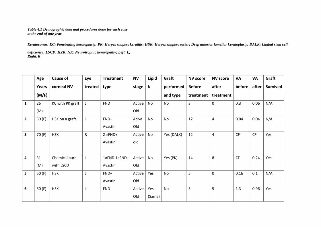
Table 4.1 Demographic data and procedures done for each case
at the end of one year.
Keratoconus: KC; Penetrating keratoplasty: PK; Herpes simplex keratitis: HSK; Herpes simplex zoster; Deep anterior lamellar keratoplasty: DALK; Limbal stem cell
deficiency: LSCD; HZK; NK: Neurotrophic keratopathy; Left: L, Right: R
Age
Years
(M/F)
Cause of
corneal NV
Eye
treated
Treatment
type
NV
stage
Lipid
k
Graft
performed
and type
NV score
Before
treatment
NV score
after
treatment
VA
before
VA
after
Graft
Survived
1 26
(M)
KC with PK graft L FND Active
Old
No No 3 0 0.3 0.06 N/A
2 50 (F) HSK on a graft L FND+
Avastin
Acive
Old
No No 12 4 0.04 0.04 N/A
3 70 (F) HZK R 2 ×FND+
Avastin
Active
old
No Yes (DALK) 12 4 CF CF Yes
4 31
(M)
Chemical burn
with LSCD
L 1×FND 1×FND+
Avastin
Active
Old
No Yes (PK) 14 8 CF 0.24 Yes
5 50 (F) HSK L FND+
Avastin
Active
Old
Yes No 5 0 0.16 0.1 N/A
6 50 (F) HSK L FND Active
Old
Yes
(Same)
No 5 5 1.3 0.96 Yes
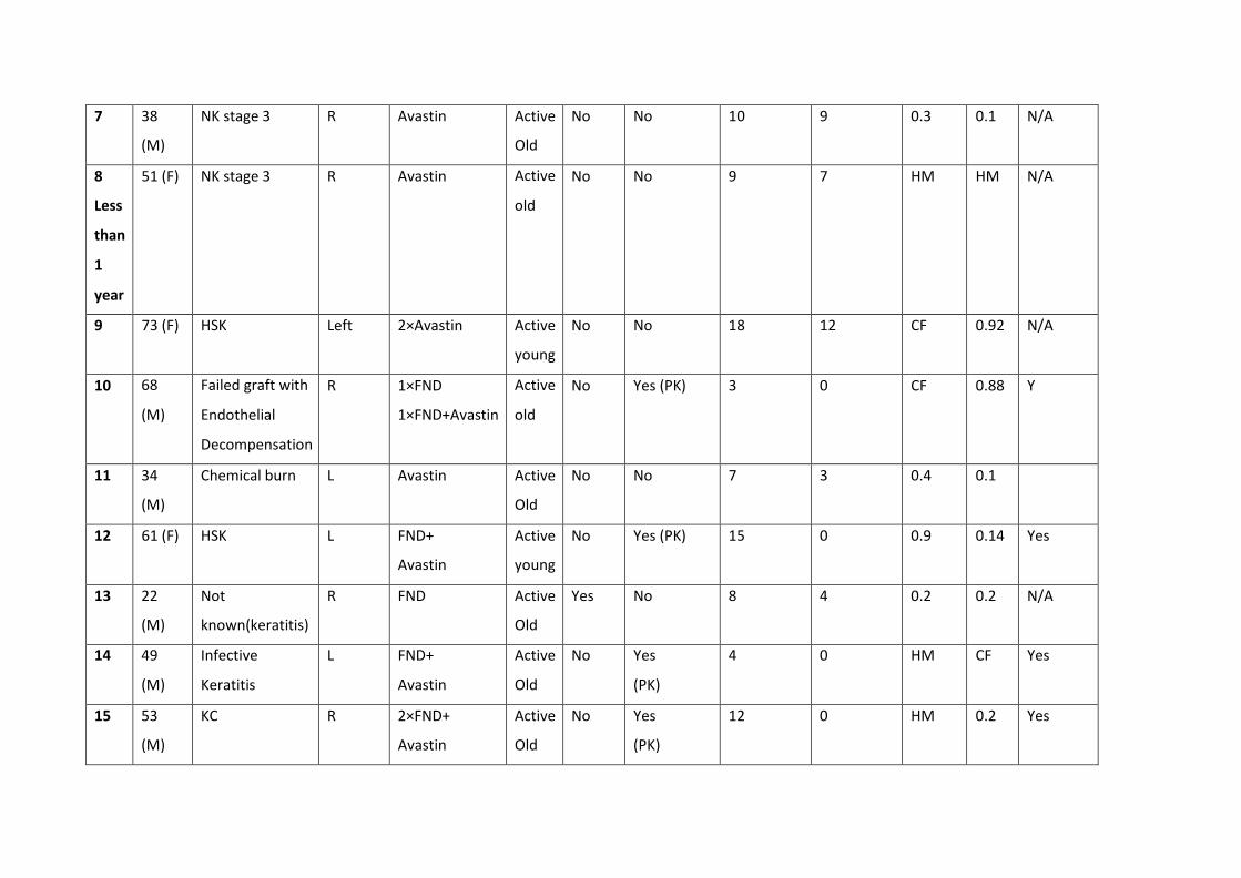
7 38
(M)
NK stage 3 R Avastin Active
Old
No No 10 9 0.3 0.1 N/A
8
Less
than
1
year
51 (F) NK stage 3 R Avastin Active
old
No No 9 7 HM HM N/A
9 73 (F) HSK Left 2×Avastin Active
young
No No 18 12 CF 0.92 N/A
10 68
(M)
Failed graft with
Endothelial
Decompensation
R 1×FND
1×FND+Avastin
Active
old
No Yes (PK) 3 0 CF 0.88 Y
11 34
(M)
Chemical burn L Avastin Active
Old
No No 7 3 0.4 0.1
12 61 (F) HSK L FND+
Avastin
Active
young
No Yes (PK) 15 0 0.9 0.14 Yes
13 22
(M)
Not
known(keratitis)
R FND Active
Old
Yes No 8 4 0.2 0.2 N/A
14 49
(M)
Infective
Keratitis
L FND+
Avastin
Active
Old
No Yes
(PK)
4 0 HM CF Yes
15 53
(M)
KC R 2×FND+
Avastin
Active
Old
No Yes
(PK)
12 0 HM 0.2 Yes

93
Fourteen patients had active old NV while 1 patient had active young NV. Of the 15 patients,
8 were females (50%), and 8 were males (50%). Mean age was 48 years (range was from 22-
73 years). Ten patients (67%) had 1 treatment session while 5 (33%) had 2 sessions of
treatment. Three patients had FND, 4 patients had bevacizumab injections and 8 patients
had combined FND and bevacizumab injections. Six patients had a graft with or after one of
the 3 treatment options. Five of these grafts were a penetrating keratoplasty (PK) while one
was a deep anterior lamellar keratoplasty (DALK). Five of the patients with a graft had no
rejection episodes or failure. Only one patient who had a penetrating graft had an
endothelial rejection episode 2 months after combined FND and bevacizumab injection and
was aborted after appropriate treatment. Seventy-three% of the patients had improved
visual acuity while 27% had stable vision. After treatment, 6 patients had mature NV, 5
patients had regressed NV and 4 patients had partially regressed NV. Three patients had
lipid keratopathy. Two of them had remarkable reduction of lipid deposition following
treatment while one remained the same. Table 4.2 shows the end stage of the NV in each of
the 3 treatment regimens. The data reported is after one year from the start of treatment.
Temporal decrease of NV was not analyzed.
Table 4.2: End stage of NV in each of the 3 treatment regimens.
End stage FND Avastin FND and Avastin
Regressed 1 0 4
Partially regressed
0 1 3
Matured 2 3 0
There was a mean reduction of 2.33 +/- 2.08 in the clinical scoring of NV in the FND group
(p=0.1917, paired test, 95% confidence interval (CI) = 2.838-7.504), which was not
statistically significant. For the Avastin injection group, the mean reduction was 3.25+/-
2.217 (p= 0.0609, paired test., 95% CI= 0.2783-6.778), which was not statistically significant.
The mean reduction in the FND and Avastin group was 8.143+/4.14 (p= 0.0020, paired test,
95% CI= 4.134-11.97), which was statistically significant. None of the patients had adverse
events related to the FND or the subconjunctival injections.

94
That is already mentioned in table 4.1 Patients 2,5,6 gender, age and laterality is just a co-incidence.

95
Figures 4.2- 4.4 are for 3 patients before and after treatment with different procedures.
Figure 4.2: Slit lamp anterior segment photographs of a patient before treatment (top image) and after
treatment with FND, Avastin and PK (bottom image).
The bottom image shows no NV and a clear cornea.
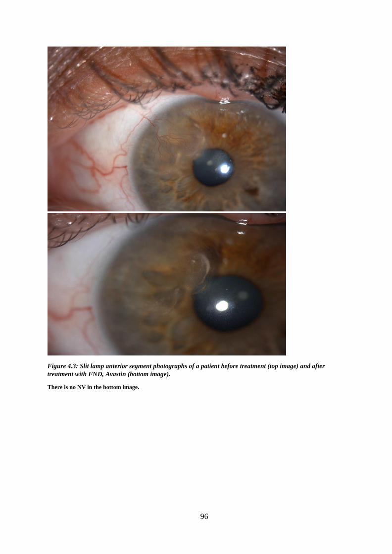
96
Figure 4.3: Slit lamp anterior segment photographs of a patient before treatment (top image) and after
treatment with FND, Avastin (bottom image).
There is no NV in the bottom image.

97
Figure 4.4: Slit lamp anterior segment photographs of a patient with lipid keratopathy before treatment (top
image) and after treatment with FND (bottom image).
Top image shows superficial superior NV and dense lipid keratopathy. The bottom image shows less lipid
keratopathy after FND.
4.4 Discussion
It is well established that corneal stromal vascularisation is both a risk factor and harbinger
of rejection in penetrating keratoplasty. Topical steroids have been used to suppress corneal

98
vascularisation and to decrease corneal scarring. However, long term topical use may lead
to cataract, glaucoma, superinfection and reactivation of herpes simplex virus and they are
unlikely to cause complete regression of the vessels. There are no current NICE guidelines
for established corneal neovascularisation.
The only definitive treatment that is available now is fine needle diathermy which is used as
a routine for treatment of corneal NV. However, FND itself induces trauma to the vascular
endothelium that might stimulate further vessel formation. It is hypothesised that if an anti-
VEGF is combined with FND it will address any surgically induced factor for further
vascularisation thus reduce the risk of rejection even further (393).
Several studies were performed where FND was used for corneal NV of different aetiologies
(120, 189, 191, 386, 395). They reported that FND was a safe and effective method for both
active and established vessels, can be used before PK to reduce rejection episodes and is
associated with improvement of vision. However, Thatte et al (191) noted that the
deposited lipids did not regress any faster by the occlusion of the corneal vessels. This was
contradicted by Faraj et al (120), who reported that NV regression resulted in gradual
reduction of lipid keratopathy if present. They also reported that active young vessels were
usually a sign of underlying continuous pathology which keeps on inducing more
vascularisation and such eyes will require treatment with anti-VEGF subconjunctival
injections.
Combined FND and bevacizumab was also used. Koenig et al (192), used FND combined with
bevacizumab eye drops as well as bevacizumab injections in some of the eyes. They
reported good outcomes in terms of regression of the vascularised area and visual acuity
improvement which was however statistically insignificant with some patients needing a
second treatment session. They also reported better results in eyes that received injections
and drops rather than drops therapy alone reported similar results using FND and

99
subconjunctival bevacizumab injections with reduction of lipid deposition (394). However,
they noted that the management of cases with ongoing inflammatory was much more
challenging. Elbaz et al (193), reported similar results in children with NV using combined
FND and bevacizumab subconjunctival injections.
Many studies used subconjunctival bevacizumab and eye drops to treat NV. They reported
statistically significant reduction of the calibre of vessels, area of NV (166, 167) (207, 390)
(387, 396, 397).
Subconjunctival bevacizumab was also used successfully as an adjunctive treatment in
vascularized corneas at the time of lamellar and penetrating keratoplasty to reduce the risk
of graft rejection (388, 391). Vassileva et al and Symes et al reported that bevacizumab is an efficient
and safe additional treatment option for improvement of prognosis in high-risk corneal transplantation
with pre- and postoperative corneal NV.
In this retrospective study, there were 3 different treatment regimens. The limitations were
the small number of patients in each group. The results show that there was a reduction in
the clinical score of NV in all groups but the FND and Avastin injection group is the only
group that showed a statistically significant reduction. This is most probably because FND
occludes all stages of vessels and the VEGF produced by FND trauma to the vascular
endothelium is blocked by the bevacizumab injections leading to effective and prolonged
reduction of NV as our patients had a maintained effect for a year after the last treatment.
Similar to the previous studies, some of our patients required a second session of
treatment. We had no side effects related to the injections and the FND. Gradual reduction
of lipid keratopathy was seen in 2 of the 3 patients following treatment. The improvement
of visual acuity in 73% of the patients was related to the reduction of NV, reduction of lipid
keratopathy and the reduction of corneal opacity in patients who had grafts. None of the
patients had worse vision. We noted that patients who did not achieve partial regression or
regression of vessels had stable, inactive vessels after the treatment which is considered a
success as these patients had no further progression of NV or lipid deposition. Only one
patient had an episode of endothelial rejection following treatment with FND and Avastin
which was controlled by steroid treatment. Based on the results, a combination of anti
VEGF with FND produces the best outcome of eliminating corneal neovascularisation with
no reported side effects. Different treatments for HSK patients in this study were given

100
based on the clinical picture with regards to the NV stage and clinical scoring as treatment
option is based on the NV status and not the aetiology of the NV. This is because the
mechanism of corneal angiogenesis is the same for any etiology. The main inducer of NV in
all etiologies is inflammation. All patients who had HSK in this study were females which
was a coincidence as HSK almost affects both genders equally. Avastin was used for patients
with NV secondary to NK as the NV stage was active which is usually a sign of underlying
continuous pathology which keeps on inducing more vascularisation and such eyes will
require treatment with anti-VEGF subconjunctival injections.
The main limitations in this study was the sample and the fact that data collection was
retrospective. Future studies with larger sample sizes need to be carried out with longer
follow-up to ascertain the effect of combined treatment for corneal NV and rejection rates
following corneal grafting.

101
CHAPTER 5: AUTOLOGOUS CULTIVATED LIMBAL STEM CELLS TRANSPLANTATION FOR RESTORATION OF CORNEAL EPITHELIUM IN PATIENTS WITH LIMBAL STEM CELL DEFICIENCY DUE TO OCULAR BURNS (HOLOCORE Study)
5.1 Introduction
Ex-vivo expansion of limbus derived cells with or without a substrate (collagen, fibrin or
amnion) is an essential option in the surgical management of total limbal stem cell deficiency whether
unilateral or bilateral. Biopsies are obtained from the unaffected eye or from living related donors and
are used to extract limbal epithelial cells or can be directly applied as explants on culture plates to
produce stratified sheets to be used for transplantation (398-400). Excellent results were reported with
long term follow-up following use of ex-vivo expansion of these stratified sheets
(401).
Oral mucosal cells have been used as potential cells in the treatment of LSCD and it was
reported that oral mucosal cells can successfully improve vision in patients with LSCD
obviating the need for immunosuppression (402). Cultivated oral mucosal epithelial
transplantation (COMET) utilizes a transplanted epithelial sheet which contains stem cells
that help to reconstruct and sustain the integrity of the corneal surface and is a surgical
option for bilateral LSCD (403).
Autologous Cultivated Limbal Stem Cell Transplantation (ACLSCT) is an advanced treatment
for LSCD which utilizes a small limbal-biopsy specimen from the unaffected eye or from a
normal limbal zone in case of bilateral burns, followed by in vitro expansion to generate a
sheet of corneal epithelial cells including both differentiated and stem cells. The final
product is named Holoclar and is a transparent round sheet that consists of 300,000 to
1,200,000 viable autologous human corneal epithelial cells, with an average of 3.5% limbal

102
stem cells as well as differentiated cells and stem cell derived transient amplifying cells. This
epithelial sheet rests on a supportive fibrin layer and is kept in a nutrient transport medium
(401,404,405).
This technique has the advantage of being autologous which means that no donor tissue is
used and no immunosuppression is required.
Autologous cultivated limbal stem cells (ACLSC) have been obtained using standardized and
optimized methods since 1998. Since then, ACLSCT was performed on more than 200
patients with moderate or severe LSCD in several clinical centres in Italy. The acquired
experience has been reported in independent published studies (401, 404, 405) and in three
Chiesi-sponsored, retrospective, case-series, non-randomised, non-controlled studies
(HLSTM01, HLSTM02 and HLSTM04), that gathered all clinical information from all treated
patients with available source documentation with a follow-up of up to 10 years.
Specific amendments and improvements in quality and manufacturing have been
introduced after 2007 to comply with the current legislation and regulations regarding
Advanced Therapy Medicinal Products (ATMPs), in particular in respect of the quality of the
reagents (recombinant growth factors, pharmacopoeia-grade reagents) and some excipients
( the supporting layer of human fibrin for cell attachment). The results of HLSTM04 study
showed that after the introduction of improvements in quality and manufacturing of ACLSCT
in 2007, most of the patients with moderate to severe LSCD due to ocular burns, who
received cultured limbal stem-cell grafts for corneal transplantation, achieved a positive
clinical outcome with a favourable safety profile. Improved visual acuity was achieved
despite the majority of study population exhibiting stromal scarring. In addition, clinically-
and functionally-beneficial effects from Holoclar were observed among patients who failed
a full restoration of corneal epithelium and who are candidates for Holoclar re-treatment
and keratoplasty. The data from these retrospective studies demonstrated the efficacy and
safety of Holoclar for human use and were included as clinical data into the marketing
authorization application for Holoclar which was filed by Chiesi Farmaceutici S.p.A. in March
2013 (application EMEA/H/C/2450).
Holoclar has been approved by the European Medicines Agency EMA only recently (Committee for
103
Medicinal Products for Human Use (CHMP) meeting 15-18 December 2014). During that meeting, the
CHMP recommended Holoclar, the first advanced therapy medicinal product containing stem cells for
usage in the European Union. The CHMP considered that Holoclar provided a first treatment option for
this eye condition and recommended a conditional marketing authorisation because, although the
data supplied by the applicant show that the medicine's benefits outweigh its risks, the data was based
on retrospective studies and were not yet comprehensive. This data has been reported in independent
published studies (401, 404, 405) and in three Chiesi-sponsored, retrospective, case-series, non-
randomised, non-controlled studies (HLSTM01, HLSTM02 and HLSTM04). Therefore, an additional study
(Holocore) on the use of Holoclar was conducted and this clinical trial CCD- GPLSCD01-03 has been
agreed with the regulatory body in order to satisfy the need of additional and more comprehensive data
obtained in a controlled setting.
5.2.1 Methods
5.2.2 Ethical issues The study was conducted in accordance to the declaration of Helsinki. All relevant study
approvals were present before the actual started trial. Chiesi Farmaceutici S.p.A. (the sponsor
of Holocore study) supplied us with all the relevant documents from the regulatory
authorities before we started recruiting the patients. (REC reference number: 15/LO/1480).
5.2.3 Patient recruitment
Holocore is a multinational, multicentre, prospective, open label, uncontrolled clinical trial.
Queens Medical Centre (Centre number 82602) was one of the 12 sites in 8 countries across
Europe in which the study was carried out. It was conducted in routine specialist cornea
clinics under the supervision of Professor Harminder Dua (Consultant Ophthalmologist and
principal investigator of Holocore study).
Inclusion Criteria
Subjects had to meet all of the following inclusion criteria to be eligible for enrolment into
the study:

104
1. Written informed consent prior to any study-related procedures
2. Adult male and female patients (≥18 years old).
3. LSCD secondary to unilateral or bilateral physical or chemical ocular burns, with at least 1-
2 mm2 of undamaged limbus to harvest stem cells for expansion in culture. LSCD was
considered for inclusion in the presence of superficial neo-vascularization invading at least
two corneal quadrants with evidence of central corneal (6mm) involvement (including
corneal neovascularisation, corneal opacity or corneal dyschromia).
4. Stability of LSCD, defined by a duration of disease of at least 24 months at the time of the
screening visit and the presence of continuous epithelial cover by fluorescein staining
scored as none or trace.
5. Presence of severe impairment in visual acuity defined by a score of 1/10th or below
20/200 at the Snellen chart (legal blindness) after best correction (i.e. Best Corrected Visual
Acuity).
6. Absence of other clinical contraindications to ACLSC transplantation based upon
investigators’ judgment.
7. A cooperative attitude to follow up the study procedures (Caregivers in case of minors).
Exclusion Criteria
The presence of any of the following excluded a subject from study enrolment:
1. LSCD of mild degree (i.e. below 2 quadrants of neo-vessel invasion without central
corneal), due to a recent burn (less than 24 months before screening), or secondary to
medical conditions other than burns (i.e. radiotherapy).
2. Severe ocular inflammation according to the Efron Grading Scale for Contact Lens
complications.
3. Presence of eyelids malposition.
4. Conjunctival scarring with fornix shortening.
5. Severe tear secretion deficiency, determined by Schirmer’s test type I (<5 mm/ 5 min).
6. Corneal anaesthesia and conjunctival anaesthesia.
105
7. Active local or systemic infections at the time of screening.
8. Diagnosis of local or systemic neoplastic disease.
9. Congenital diseases (i.e., Aniridia).
10. Bilateral inflammatory diseases (i.e. Stevens-Johnson syndrome, pemphigoid).
11. A pre-existing blindness precluding a functional recovery.
12. Female subjects: pregnant or lactating women and all women physiologically capable of
becoming pregnant (i.e. women of childbearing potential) unless they were willing to use
one or more reliable methods of contraception (e.g. oral contraceptives, IUD, tubal ligature).
Reliable contraception should be maintained throughout the study.
A urine pregnancy test was performed at the screening visit for all women of childbearing
potential and repeated before biopsy and treatment. Any postmenopausal women
(physiologic menopause defined as “12 consecutive months of amenorrhea”) or women
permanently sterilized (e.g. tubal occlusion, hysterectomy or bilateral salpingectomy) was a
potential candidate for enrolment into the study
13. Allergy, sensitivity or intolerance to concomitant drugs or excipients (Hypersensitivity to
any of the excipients listed in section 6.1 or to bovine serum and murine 3T3-J2 cells).
14. Contraindications to the local or systemic antibiotics and/ or corticosteroids foreseen by
the protocol.
15. Contraindications to the surgical procedure.
16. Clinically significant or unstable concurrent disease or other clinical contraindications to
stem cell transplantation based upon investigators’ judgment or other concomitant medical
conditions affecting grafting procedure.
17. Patients unlikely to comply with the study protocol or unable to understand the nature
and scope of the study or the possible benefits or unwanted effects of the study procedures
and treatments.
18. Participation in another clinical trial where investigational drug was received less than 4
weeks prior to the screening visit.

106
5.2.4 Clinical Intervention and follow up
Fourteen clinic visits were performed during the study. The study duration period was 19
months +/- 15 days:
Five to 6 weeks- screening visit up to biopsy.
Six months roll-in period starting from the biopsy and up to autologous limbal stem cells
transplant ACLSCT.
Twelve months from the autologous limbal stem cells transplant ACLSCT up to endpoint
visit.
Table 5.1 shows the follow up scheme of HOLOCORE.
Table 5.1: Details of patient’s visits during the study course
Name and code of visit Interpretation
Visit 0 (V0) Date of recruitment
Visit 1 (V1) Two weeks from V0
Visit 2 (V2) Biopsy day (Four weeks from V1)
Visit 3 (V3) Two months after the Biopsy
Visit 4 (V4) Four months after the Biopsy
Visit 5 (V5) Day -13 +/- 2
Visit 6 (V6) Transplantation day
Visit 7 (V7) One day after V6 (done over the phone)
Visit 8 (V8) Four +/- 1 day after V6
Visit 9 (V9) Two weeks +/- 3 days after V6
Visit 10 (V10) Twenty nine days+/- 5 days after V6
Visit 11 (V11) Three months +/- 14 days after V6
Visit 12 (V12) Six months +/- 14 days after V6

107
Visit 13 (V13) Nine months +/- days after V6
Visit 14 (V14) End of the study, 1 year +/- 14 days after V6
A) Standard haematology, blood chemistry and infectious assessment
Standard haematology and biochemistry and infectious assessments were performed at the
prescribed visits as in at the phlebotomy clinic at Queen Medical Centre and transferred to a
central laboratory in Germany for analysis. The blood samples for standard haematology
and blood chemistry were collected in the morning after an overnight fasting (10 hours) at
screening visit, visit 5 and visit 10. The blood samples for infectious assessments were
collected at visit 1 and visit 2. The collection was always done after vital signs
measurements and before any further study assessment.
The following parameters were assessed:
Haematology: Red blood cells count, white blood cells count and differential, total
haemoglobin, hematocrit , platelets count and prothrombin time.
Blood Chemistry: Creatinine, Blood urea nitrogen, fasting serum glucose, Aspartate
aminotransferase, Alanine aminotransferase, Gamma-glutamyl transpeptidase, total
bilirubin, albumin, Alkaline phosphatase, sodium, potassium, calcium, and chloride
electrolytes.
Infectious Assessment: HAV-Ag, HAV-IgG, HAV-IgM, HBsAg, HBsAb,
HBcAb, HCVAg, HCVAb, HIV1&HIV2 Ab and Ag, Treponema Pallidum Ab, HTLV – Human T
Lymphotropic Virus-Ag and Ab, HWN -Human West Nile virus- Ab measurement.
B) Surgical procedures
Holocore study includes obtaining a biopsy from the unaffected eye (or from a normal zone
in bilateral LSCD cases) then a single administration of Holoclar implant through a dedicated
surgical procedure of corneal surface scraping and product application. Both the biopsy and
Holoclar implantation were performed at the Ophthalmology theatre at Queens Medical
Centre by the same surgeon (Professor Harminder Dua).

108
Biopsy (Visit 2)
The patients had corneal biopsy taken from the unaffected limbus of the contralateral eye
according to the following multi-steps procedure:
· Topical anaesthesia was procured with parabulbar anaesthesia with marcaine without and
adrenaline.
· Ocular surface lavage was performed with balanced salt solution (BSS).
• A horizontal clear-cornea incision, 2 mm long, was made with a disposable scalpel of
15 - 30°, just anterior to the limbal vessels. Lamellar dissection in a "pocket" fashion
using a disk lamellar knife or a crescent blade (the depth was about 50 to 100 μm to
enable the lifting of the lamella from the corneal limbus) was made from the incision
extending posterior to the limbus. The conjunctiva at the limbus was then incised to
expose the posterior limit of the lamellar dissection. The sample was obtained from
the upper area.
•Two vertical incisions from each end of the horizontal incision were then made in the
limbus and adjacent sclera with a disposable scalpel of 15 - 30°. A posterior horizontal
incision was then made to join the posterior end of the vertical incisions to complete
the excision of a 2mm piece of limbus. Cautery was not used to coagulate bleeding
vessels before the tissue sample was removed, in order to avoid damaging the stem
cells.
• Biopsy was inserted in the sterile test tube containing the transport medium.
• The conjunctiva was sutured (tacked down) with 2 nylon 10/0 sutures.
Treatment after biopsy
Patients received topical antibiotic prophylaxis with single-dose preservative-free
levofloxacin, 3-4 drops three times per day for 7 days.
Holocalr implantation procedure (Visit 6)
Surgery was performed according to the following procedure:

109
• General anaesthesia was done.
• Limbal peritomy was done a few millimetres outside the limbus, with proper coagulation.
The bulbar conjunctiva was freed for about 4-5 mm in order to create a pocket into which
Holoclar implant was inserted.
• Pannusectomy for removal of corneal fibrovascular layer of conjunctival origin was done,
trying to find the cleavage level between the pannus and the cornea for avoiding, when
possible, keratectomy.
• Lavage with BSS was done, whilst checking for an absence of consistent blood loss that
could form blood collections (‘sacks’) under the epithelial graft.
• Holoclar implant was transferred from the transport container to a suitable dish. Under
the microscope it was possible to recognize the fibrin "nude" side (smooth and
translucent) from the cells-seeded side (rough). It is absolutely crucial to place the fibrin
sheet with the cultivated cells outside and not upside down.
• Holoclar was allowed to slide onto the recipient’s prepared graft area assisting with BSS
and slight traction with tweezers at the edge of the graft as necessary.
• The excess fibrin sheet (Holoclar) was trimmed and the edge covered with the
conjunctiva applying 2 or 3 stitches (sutures) of Vicryl 8/0.
• Eyelids were kept closed with Steri-strips.
Treatment after administration of Holoclar
• Systemic antibiotic treatment: Doxycyclin 100 mg tablets twice per day for 2 weeks.
• Systemic corticosteroid treatment: Prednisone P.O. at a daily dose of 0.5 mg/kg for 2
weeks after the implant at a daily dose to a maximum of 25 mg for 2 weeks, then tapered
to 0.25 mg/kg (maximum 12.5 mg) per day for 1 week, to 0.125 mg/kg (maximum 5.0 mg)
per day for the following week and then stopped. In case of persistent inflammation, the
corticosteroid treatment was allowed to be maintained or reintroduced.

110
• Topical corticosteroid treatment: 2 weeks after surgery, preservative-free dexamethasone
(dexafree 1 mg/ml PF B&L) eye drops for a total of 4 weeks: 1 drop TID for 2 weeks
followed by 1 drop bid for 1 week and then 1 drop QID for 1 week. Topical corticosteroid
can be maintained for longer in case of persistent inflammation according to the judgment
of the doctor.
C) Details of the visits:
All of the visits and medications received by the patient were recorded in the patient’s
medical notes as well as in electronic CRF. I was the coordinator of this study. I was
responsible for performing all the clinical examination during all the visits and
recording the data on the medical notes as well as doing all correspondence with the
sponsor of the study. The slit lamp images were taken by the imaging technicians in
the department.

111
Table 5.2: Details of procedures carried out in each visit during the study course
VISITs
Pre- Screen.
Screen Roll-in start
Limbal Biopsy
Pre- Transp
Pre- Transp
Confir mation
ACLSCT Post-
Transp Post-
Transp Post-
Transp Post-
Transp Post-
Transp Post-
Transp Post-
Transp End of Study
V0 V1 V2(1) V3 V4(2) V5 V6 V7 (4) V8 V9 V10 V11 V12 V13 V14
Date from 1st ACLSCT
Day-225 ± 30
Day-210 ± 30
Day-180 ± 30
Day-120 ± 30
Day -60 ± 30
Day -13 ± 2
Day 1 Day 2 Day 4
± 1 Day 15
± 3 Day 29
± 5 Day 90
± 14 Day 180
± 14 Day 270
± 14 Day 360
± 14
ACTIVITY/ ASSESSMENT
Informed Consent ✓
Demographic Data ✓
Inclusion/ Exclusion Criteria
✓ ✓ (3)
Medical History and concomitant diseases ✓
Previous and concomitant medications
✓ ✓ ✓ ✓ ✓ ✓ ✓ ✓ ✓ ✓ ✓ ✓ ✓ ✓ ✓
Adverse Events (AE) ✓ ✓ ✓ ✓ ✓ ✓ ✓ ✓ ✓ ✓ ✓ ✓ ✓ ✓ ✓ Adverse Event of
Special Interest (AESI) ✓ ✓ ✓ ✓ ✓ ✓ ✓ ✓ ✓ ✓ ✓ ✓ ✓ ✓ ✓
Physical examination(5)
✓ ✓ ✓ ✓ ✓ ✓ ✓ ✓ ✓ ✓ ✓ ✓ ✓ ✓ Vital signs ✓ ✓ ✓ ✓ ✓ ✓ ✓ ✓ ✓ ✓ ✓ ✓ ✓
Haematology and blood chemistry
✓ ✓ ✓
Infectious Assessment ✓ ✓
ECG ✓ ✓
Urine Pregnancy Test ✓ ✓ ✓ ✓

112
VISITs
Pre- Screen.
Screen Roll-in start
Limbal Biopsy
Pre- Transp
Pre- Transp
Confir mation
ACLSCT Post-
Transp Post-
Transp Post-
Transp Post-
Transp Post-
Transp Post-
Transp Post-
Transp End of Study
V0 V1 V2(1) V3 V4(2) V5 V6 V7 (4) V8 V9 V10 V11 V12 V13 V14
Date from 1st ACLSCT
Day-225 ± 30
Day-210 ± 30
Day-180 ± 30
Day-120 ± 30
Day -60 ± 30
Day -13 ± 2
Day 1 Day 2 Day 4
± 1 Day 15
± 3 Day 29
± 5 Day 90
± 14 Day 180
± 14 Day 270
± 14 Day 360
± 14
ACTIVITY/ ASSESSMENT Corneal epithelial
defects by fluorescein staining
✓ ✓ ✓ ✓ ✓
✓ ✓ ✓ ✓ ✓
Superficial corneal neo- vascularization
✓ ✓ ✓ ✓ ✓ ✓ ✓ ✓ ✓ ✓
Digital photography ✓ ✓ ✓ ✓ ✓ ✓ ✓ ✓ ✓ Best-Corrected Visual
Acuity ✓
✓ ✓ ✓
✓ ✓ ✓ ✓ ✓
Ocular tonometry ✓ ✓ ✓ ✓ ✓ ✓ ✓ ✓ ✓ ✓ Slit lamp examination: • Corneal endothelium • Anterior chamber
✓ ✓ ✓ ✓ ✓
✓ ✓ ✓ ✓ ✓ ✓ ✓
Conjunctival inflammation (limbal
and bulbar)
✓ ✓ ✓ ✓ ✓
✓ ✓ ✓ ✓ ✓ ✓ ✓
Corneal sensitivity ✓ ✓ ✓ ✓ ✓ ✓ ✓ ✓ ✓ ✓
Corneal involvement ✓ ✓ ✓ ✓ ✓ ✓ ✓ ✓ ✓ ✓
Schirmer’s test type I ✓ ✓ Symptoms: • Pain • Burning • Photophobia
✓
✓
✓
✓
✓
✓ (6)
✓
✓
✓
✓
✓
✓
✓
NEI-VFQ-25 and EQ- 5D-3L(Y) QoL
questionnaires
✓
✓
✓ ✓
✓
Limbal biopsy and shipment
✓
ACLSC grafting procedure
✓
✓
(7)
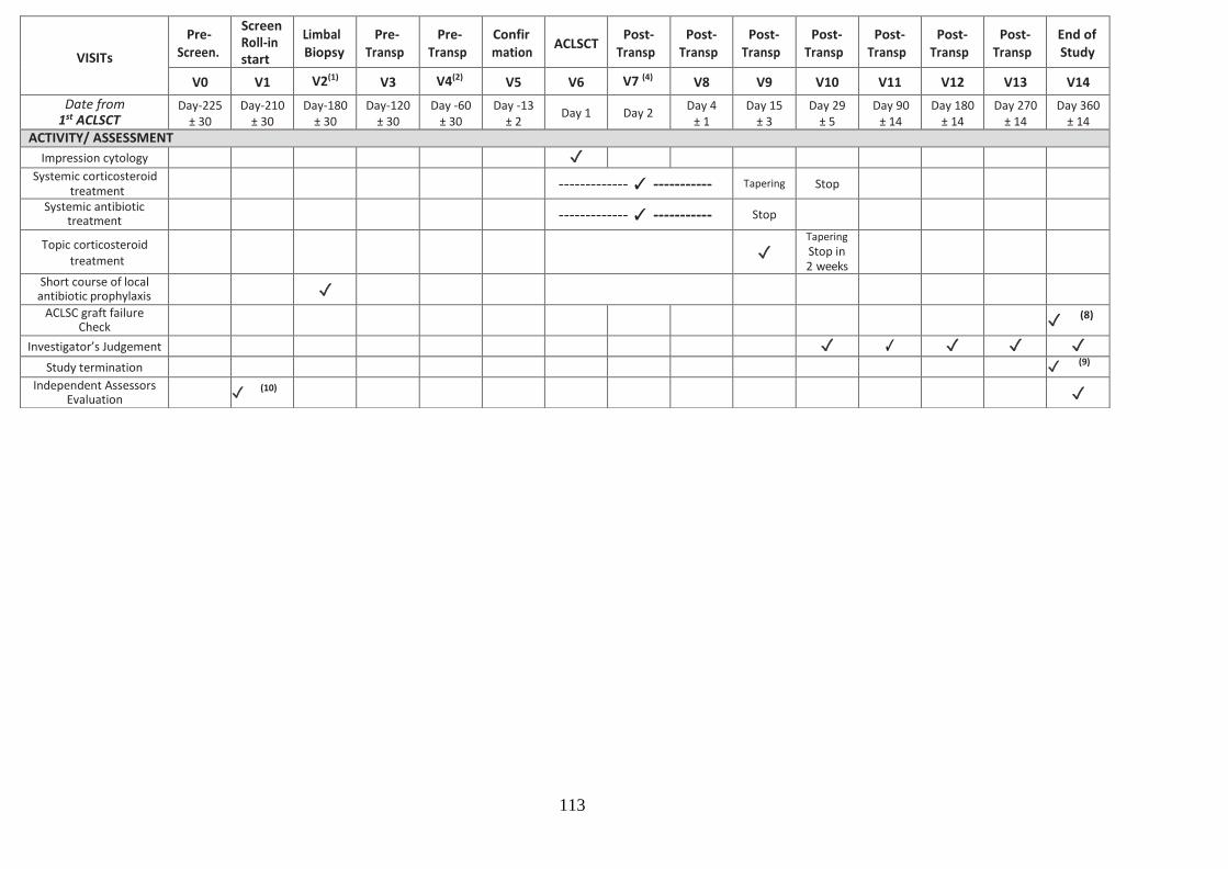
113
VISITs
Pre- Screen.
Screen Roll-in start
Limbal Biopsy
Pre- Transp
Pre- Transp
Confir mation
ACLSCT Post-
Transp Post-
Transp Post-
Transp Post-
Transp Post-
Transp Post-
Transp Post-
Transp End of Study
V0 V1 V2(1) V3 V4(2) V5 V6 V7 (4) V8 V9 V10 V11 V12 V13 V14
Date from 1st ACLSCT
Day-225 ± 30
Day-210 ± 30
Day-180 ± 30
Day-120 ± 30
Day -60 ± 30
Day -13 ± 2
Day 1 Day 2 Day 4
± 1 Day 15
± 3 Day 29
± 5 Day 90
± 14 Day 180
± 14 Day 270
± 14 Day 360
± 14
ACTIVITY/ ASSESSMENT
Impression cytology ✓
Systemic corticosteroid treatment
------------- ✓ ----------- Tapering Stop
Systemic antibiotic treatment
------------- ✓ ----------- Stop
Topic corticosteroid treatment
✓
Tapering
Stop in 2 weeks
Short course of local antibiotic prophylaxis
✓
ACLSC graft failure Check
✓ (8)
Investigator’s Judgement ✓ ✓ ✓ ✓ ✓
Study termination ✓ (9)
Independent Assessors Evaluation
✓ (10)
✓

114
The study was terminated at this visit 14 for all patients who are judged as a success.
5.2.5 Outcome measurements
Efficacy measurements were made in terms of the degree of superficial corneal
neovascularisation and epithelial defects. Safety measurements were specifically
investigated with reporting any adverse effects related to the biopsy and Holoclar
transplantation. Patients had the ability to access the 24 hour on call emergency eye service,
and at the same time were provided with contact numbers for investigators for any queries
during the study period.
5.3 Results
5.3.1 Recruitment
We were able to recruit 2 patients to this study as these were the only patients that
met all the eligibility criteria at the time of recruitment. The demographic data for the
recruited patients is listed in table 5.2. The two recruited patients in this study are
reported as individual case reports.

115
Table 5.3: Demographic data of HOLOCORE cases.
LSCD: Limbal stem cell deficiency.
5.3.2 Cases
Case one (82602-01)
This patient was a 57 year old gentleman who was recruited on the 7th of February 2017 for
treatment of the right eye. He had a dense fibrovascular membrane involving the central
cornea due to an acid chemical burn in 1993. He had severe superficial corneal NV in all 4
quadrants and his vision was hand movement. The 4 quadrants of the cornea were invaded
with superficial corneal NV and they extended to the central cornea (Figure 5.1). The visual
acuity was hand movement. He had normal intraocular pressure in both eyes throughout
the study (range from 8-12). Schirmer test type I was 10 mm on V1. He had normal corneal
sensitivity which was assessed by a cotton tip. On V1, the patient had moderate
photophobia but no pain or burning sensation. His physical examination and vital signs were
always normal. He was started on Citalopram tablets 20 mg once daily (selective serotonin
re-uptake inhibitor) because of anxiety related to holocore trial and continued for the whole
period of the trial. Otherwise, he had no history of any medical conditions. His infectious
assessment was always normal. The biopsy was done on 30th of May 2017. The biopsy was
taken from the superior limbus and placed in a sterile tube containing the transport
Participant
Trial Code
Age
Years
(M/F)
Cause of
LSCD
Date of
recruitment
Eye
treated
Last follow
up visit
1 82602-01 57 (M) Acid
chemical
burn
07/02/2017 Right 19/12/2018
2 82602-02 27 (F) Chemical
burn
(Unknown)
08/02/2017 Left 19/12/2018
19

116
medium. The standard operating procedures were followed carefully and the biopsy was
shipped on the same day to Holostem (The site where the biopsy will be processed into
Holoclar). The patient was prescribed preservative-free levofloxacin eye drops, three times
per day for 1 week. Conjunctiva was closed using 2 sutures. The patient presented to the
eye casualty 2 days after the biopsy procedure with pain which was due to a broken suture
and was removed. The other suture was removed 2 weeks after the biopsy procedure. The
wound was healing well underneath. There were no adverse events. The holoclar implant
was performed on the 6th of December 2017. Holoclar was delivered on the same day to the
hospital inside a sterile, plastic Petri dish which was placed in a secondary container that
was only removed in the operating theatre. The surgery was done under general
anaesthesia. The surgery was successful and Holocolar was in place. The patient was started
on the treatment mentioned before. On V8, the ocular bandage was removed and replaced
with new bandage, Steri-strips and shield. He continued on Doxycycline tablets, 25 mgs of
prednisolone tablets. On V9, the ocular bandage was removed. The vision was not recorded
on that visit. The patient had mild burning sensation and photophobia but no pain. He was
asked to stop doxycycline, taper Prednisolone to 12.5 mgs and to start dexafree eye drops
TID. There were no adverse events. On V10, the vision was counting fingers. On
examination, he had marked limbal and conjunctival hyperaemia with an epithelial defect (>
2mm) (Figure 5.2). He had a deep central corneal opacity. The anterior chamber was deep
and quite. He was prescribed levofloxacin 5mg/ml eye drops QID. Dexafree eye drops were
tapered by 1 drop every week. He was seen two weeks later. His vision was still counting
fingers and he had moderate pain and photophobia. The epithelial defect was much larger
and was involving the centre of the cornea (Figure 5.3). A bandage contact lens (BCL) was
inserted and Celluvisc 0.5% eye drops QID were prescribed in addition to the levofloxacin
eye drops that he was using. It was decided that the patient should be withdrawn from the
trial and the epithelial defect was reported to Chiesi as a serious advert event. He was listed
for penetrating keratoplasty with autolimbal stem cell transplant. The patient had a
persistent epithelial defect for 5 months despite eyelid taping with Steri-strips so a decision
was took to list him for Omnigen transplant. During these 5 months he was on levofloxacin
preservative free eye drops QID, predsol preservative free eye drops 0.5% and Visuxl 6 times
per day. The Omnigen was transplanted on 20th June 2018 and sutured. A BCL was inserted.
Gradually over 3 months, the eye became more comfortable. During that time the patient

117
was seen frequently and the BCL was replaced until the epithelial defect healed completely
(Figure 5.4). The final vision was hand movement and there were superficial NV in all
quadrants involving the corneal centre. The patient is currently on the waiting list for
penetrating keratoplasty with autolimbal stem cell transplant.
Figure 5.1: Slit lamp anterior segment photographs of Case 1 on V1.
Dense fibrovascular pannus with central corneal opacity.

118
Figure 5.2: Cobalt blue filter image with fluorescein sodium 2% eye drops of Case 1 on V10.
Inferior epithelial defect larger than 2mm.

119
Figure 5.3: Cobalt blue filter image with fluorescein sodium 2% eye drops of Case 1 2 weeks after V10.
Enlargement of epithelial defect to involve the central cornea.

120
Figure 5.4: Slit lamp anterior segment photographs of Case 1 9 months on V1.
Dense superficial NV in all quadrants involving the centre of the cornea.
Case two (82602-02)
This patient was a 27 year old lady who was recruited on the 8th of March 2017 for
treatment of the left eye. She had dense fibrovascular pannus involving the central cornea
due to an assault chemical injury in 2011. The 4 quadrants of the cornea were invaded with
superficial corneal NV and they extended to the central cornea (Figure 5.5). The visual acuity
was hand movement. She had normal ocular pressure in both eyes throughout the study
(range from 12-15 mmHg). The anterior chamber was always visible and it was always deep
with no cells or abnormal contents. Schirmer test type I was 10 mm on V1 and dropped to
6mm on V14. She had corneal anaesthesia which was assessed using a cotton tip. On V1, the
patient had moderate photophobia but no pain or burning sensation. Her physical
examination and vital signs were always normal and she was not on any concomitant
medications and had no history of any medical conditions. Her infectious assessment was
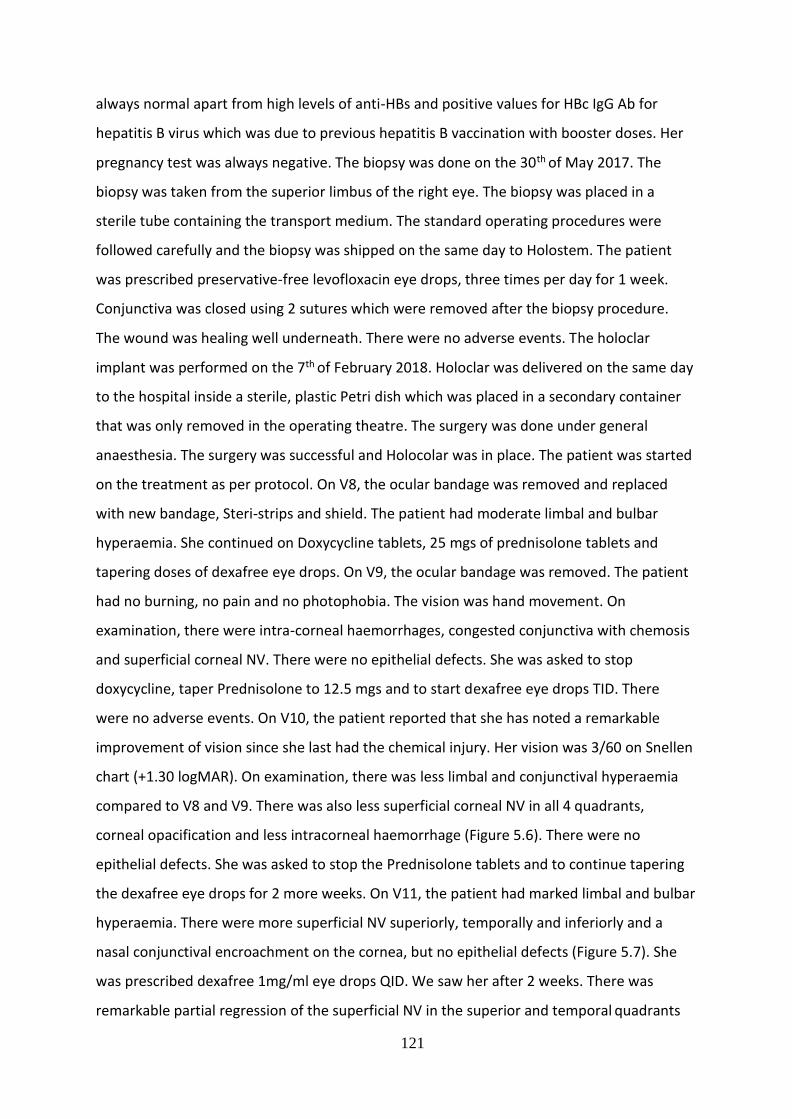
121
always normal apart from high levels of anti-HBs and positive values for HBc IgG Ab for
hepatitis B virus which was due to previous hepatitis B vaccination with booster doses. Her
pregnancy test was always negative. The biopsy was done on the 30th of May 2017. The
biopsy was taken from the superior limbus of the right eye. The biopsy was placed in a
sterile tube containing the transport medium. The standard operating procedures were
followed carefully and the biopsy was shipped on the same day to Holostem. The patient
was prescribed preservative-free levofloxacin eye drops, three times per day for 1 week.
Conjunctiva was closed using 2 sutures which were removed after the biopsy procedure.
The wound was healing well underneath. There were no adverse events. The holoclar
implant was performed on the 7th of February 2018. Holoclar was delivered on the same day
to the hospital inside a sterile, plastic Petri dish which was placed in a secondary container
that was only removed in the operating theatre. The surgery was done under general
anaesthesia. The surgery was successful and Holocolar was in place. The patient was started
on the treatment as per protocol. On V8, the ocular bandage was removed and replaced
with new bandage, Steri-strips and shield. The patient had moderate limbal and bulbar
hyperaemia. She continued on Doxycycline tablets, 25 mgs of prednisolone tablets and
tapering doses of dexafree eye drops. On V9, the ocular bandage was removed. The patient
had no burning, no pain and no photophobia. The vision was hand movement. On
examination, there were intra-corneal haemorrhages, congested conjunctiva with chemosis
and superficial corneal NV. There were no epithelial defects. She was asked to stop
doxycycline, taper Prednisolone to 12.5 mgs and to start dexafree eye drops TID. There
were no adverse events. On V10, the patient reported that she has noted a remarkable
improvement of vision since she last had the chemical injury. Her vision was 3/60 on Snellen
chart (+1.30 logMAR). On examination, there was less limbal and conjunctival hyperaemia
compared to V8 and V9. There was also less superficial corneal NV in all 4 quadrants,
corneal opacification and less intracorneal haemorrhage (Figure 5.6). There were no
epithelial defects. She was asked to stop the Prednisolone tablets and to continue tapering
the dexafree eye drops for 2 more weeks. On V11, the patient had marked limbal and bulbar
hyperaemia. There were more superficial NV superiorly, temporally and inferiorly and a
nasal conjunctival encroachment on the cornea, but no epithelial defects (Figure 5.7). She
was prescribed dexafree 1mg/ml eye drops QID. We saw her after 2 weeks. There was
remarkable partial regression of the superficial NV in the superior and temporal quadrants
122
as well as regression of the nasal conjunctival growth. The inferior vessels regressed
completely. There was also less bulbar and limbal hyperaemia (Figure 5.8). The dexafree
drops were tapered to bid. On V12, there were very fine vessels superiorly and temporally
and the nasal vessels were inactive and stable (Figure 5.9). On V13 and V14, the patient had
stable inactive NV in 3 quadrants with no epithelial defects. The case was considered a
success as she had no epithelial defects at any visits after the implant (V6) and mild degree
of superficial NV at the end of the study (V14). She has been added to the waiting list for a
left eye penetrating keratoplasty.
Figure 5.5: Slit-lamp anterior segment photograph of Case 2 at V1.
Fibrovascular pannus with superficial NV in all four quadrants and central corneal opacity.

123
Figure 5.6: Slit-lamp anterior segment photograph of Case 2 at V10.
Intracorneal haemorrhages and superficial NV.
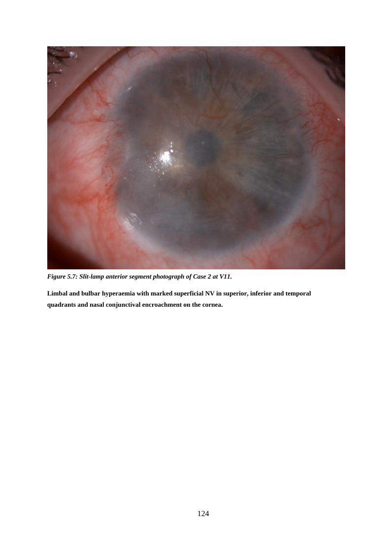
124
Figure 5.7: Slit-lamp anterior segment photograph of Case 2 at V11.
Limbal and bulbar hyperaemia with marked superficial NV in superior, inferior and temporal
quadrants and nasal conjunctival encroachment on the cornea.

125
Figure 5.8: Slit-lamp anterior segment photograph of Case 2 two weeks after V11.
Less limbal and conjunctival hyperaemia compared to Figure 5.3 (V11). Less active vessels nasally.
Inferior vessels regressed.

126
Figure 5.9: Slit-lamp anterior segment photograph of Case 2 at V12.
Very fine vessels superiorly and temporally and the nasal vessels were inactive and stable.
5.4 Discussion
We recruited 2 patients as part of the Holocore study. Case 2 was a success and case 1 was
considered a failure as it did not achieve the outcomes of the study which were the
reduction of the degree of superficial corneal neovascularisation and absence of epithelial
defects. Case 1 is currently on the waiting list for penetrating keratoplasty and autolimbal
stem cell transplant. We could not ascertain the reason for the failure as the patient had no
underlying systemic or local causes such as blepharitis, lid malposition, infections, blinking
or exposure problems that could explain the failure of the implant. Another study centre
reported a similar case where the patient suffered from persistent epithelial defect in
addition to corneal melting and the development of a descemetocele and they attributed
this to the Holoclar implant. Case 2 is on the waiting list for penetrating keratoplasty as she

127
had residual central deep corneal opacity. Recently, NICE has approved Holoclar as an
option in people with moderate to severe limbal stem cell deficiency after eye burns, only if
it is used to treat 1 eye and people have already had a conjunctival limbal autograft or if
there is not enough tissue for a conjunctival limbal autograft.
Scholz et al (406), evaluated the visual acuity and graft success in 34 eyes with LSCD due to
chemical and thermal burns using autologous transplantation of ex vivo on human aminiotic
membrane -cultured and expanded limbus epithelium. They reported stable corneal surface
in 75.4% of the eyes. Visual acuity increased significantly in 40 (65.6%) eyes, was stable in
19.7% of the eyes and decreased in 14.8% of the eyes. They considered this method to be
safe and successful on the long term. Shimazaki et al (407) transplanted cultivated human
limbal epithelium on aminiotic membrane for 13 eyes with severe LSCD and achieved
corneal epithelialisation in 46.2% of the eyes.
Two long-term studies have been published about simple limbal epithelial transplantation
(SLET). A study by Basu et al (408) reported long-term clinical outcomes of autologous SLET
on a population of 125 patients suffering from unilateral LSCD. Seventy-six percent of eyes
maintained a successful outcome with survival probability of 80% after 1 year. Progressive
corneal conjunctivalization was recorded in 18.4% of the eyes in this study.
Another retrospective, multicentre study with 6-month follow-up on 68 eyes showed the
success of SLET in 83.8% of the cases with survival probability greater than 80% after 1 year
(409).
The main limitation of HOLOCORE is the method of assessment of the severity of LSCD
which was based on only the extent of corneal neovascularization and corneal opacity.
There was no impression cytology or IVCM performed to assess the quality of epithelium
on the corneal surface.
Further trials need to be conducted to compare the results of Holoclar and other exvivo
methods for expansion of limbus derived cells with regards to efficacy and safety.

128
CHAPTER 6: CORRELATION BETWEEN DIABETIC CORNEAL
NEUROPATHY AND DIFFERENT STAGES OF DIABETIC
RETINOPATHY
6.1 Introduction
The prevalence of diabetes mellitus (DM) has continued to increase across the world. As at
2013, 382 million people have diabetes worldwide (410). Type 2 DM makes up about 90%
of the cases. This equated to 8.3% of the adult population with equal rates in women and
men (411). Diabetes can affect any ocular tissue. Diabetic retinopathy (DR) is the
commonest complication of diabetes (412) and meets the World Health Organization
(WHO) criteria of suitability for screening (413). It is the most significant ocular
complication of diabetes and is a major cause of vision loss worldwide (318). Approximately
one third of people with diabetes have DR, and a third of the DR patients may have sight-
threatening DR, defined as proliferative diabetic retinopathy (PDR) or clinically significant
macular oedema (DMO) (318). The natural history of DR is relatively well understood, with
recognizable stages. Major risk factors for developing DR include duration of diabetes (414,
415), severity of hyperglycaemia (416, 417), hypertension (418) and dyslipidaemia (419).
Once sight-threatening DR is present, progression is the norm and complications are
unpredictable. Twenty years after diagnosis, almost all people with Type 1 DM and 60% of
people with Type 2 DM will have some degree of diabetic DR (414, 415). Recently, diabetic
retinal neurodegeneration (DRN) has been described, where progressive neuronal changes
occur in the inner retina before or in the absence of clinically visible microvascular
abnormalities (318, 420-422) and may contribute to DR (421, 423-425). OCT studies of
diabetic retinal neuropathy (in eyes without DR) have been reported by several
investigators (426-430).
Other ocular complications including keratopathy, glaucoma and cataracts may be
associated with diabetes (431). The diabetic cornea suffers from cellular dysfunction and
dysfunctional repair mechanisms, which manifests as recurrent erosions, delayed wound

129
healing ulcers, and oedema. In addition and undoubtedly related to epithelial dysfunction,
alterations in epithelial basement membrane occur.
More recently, diabetic corneal neuropathy, has been reported as resulting in loss of
corneal sensation and innervations (432-435) which may be related to corneal epithelial
defects. Progressive reduction in corneal sensitivity and increasing corneal nerve
degeneration has been linked with, and may occur before diabetic neuropathy elsewhere
in the body (433). In-vivo confocal microscopy (IVCM) (a rapid and non-invasive new tool
to examine sub-epithelial and stromal corneal nerves) has demonstrated a decrease in
nerve fibre bundles (436), nerve beadings, and nerve branching patterns as well as an
increase in nerve fibre tortuosity (437), and abnormal stromal nerves (438) in diabetic
patients.
Some studies have shown the involvement of corneal sub-basal nerve plexus in diabetes
and their marked reduction with increasing severity of DR and neuropathy using IVCM (436,
439). These studies also showed alterations in basal epithelial cell density in different
retinopathy stages, and demonstrated that reduced density in basal epithelial cell is
correlated with changes in innervations (436).
A few studies have reported on the effect of laser photocoagulation on sub-basal nerve
plexus in patients with diabetic retinopathy (440, 441). Anti-vascular endothelial growth
factor (anti-VEGF) drugs have been introduced as treatments for diabetic macular oedema
(DMO) (442-444). It is known that VEGF is neuroprotective (445, 446). It is, therefore,
theoretically possible that intraocular innervation may be affected by multiple intravitreal
injections of anti-VEGFs. In addition, a few studies have reported on the effect of anti-VEGF
on corneal nerves in humans and animals (447-450).
However, to date, the author is unaware of any studies that have evaluated corneal
innervation following anti-VEGF therapy in a large number of diabetic eyes at different
stages of DR. There is a further paucity of data on how the corneal nerve parameters in
diabetes correlate with corneal sensitivity.

130
6.2 Methods
6.2.1 Ethical issues
The study was conducted in accordance to the Declaration of Helsinki (Version 5, 2000).
The study proposal with the relevant forms and documents were approved by the
Nottingham Research Ethics Service Committee II, the Health Research Authority HRA and
local NHS (Nottingham University Hospitals [NUH] NHS Trust) Research and Innovation
authorities (REC reference Number: 15/SW/0186).
The ethical approval application process involved the submission of the project proposal
and completed IRAS form to all parties (Research Ethics Committee, NUH NHS trust R&I
and HRA), the ethics committee reviewed the application on 6th July 2015 and was
approved on 11th August 2015. One substantial amendment was applied for and was
approved. The first patient was recruited on 23rd June 2016.
6.2.2 Patient recruitment
This single centre prospective longitudinal study included the identification of 86 patients.
The non-diabetic and diabetic patients with type 1 and 2 were recruited from eye clinics at
Queen’s Medical Centre, NUH Trust, Nottingham.
Participants were recruited from specialist macula clinics, diabetic clinics and diabetic
screening clinics at the Queen’s Medical Centre, NUH Trust, Nottingham under the
supervision of Mr Winfried Amoaku (Consultant Ophthalmologist). Based on the diagnostic
criteria and stage classification of DR, the patients were divided into the following 5 groups:
Non-diabetic patients (control), diabetic patients with no diabetic retinopathy (DR), Non-
proliferative DR (NPDR) and Proliferative DR (PDR) patients including those who have and
have not received Anti-VEGF injections and/or retinal laser photocoagulation. Twenty-

131
three patients were recruited into the control group, 24 patients in the group of diabetic
patients with no DR, 27 patients in the NPDR group, and 12 patients in the PDR group.
Control group inclusion criteria:
- Patients over the age of 18
-Patients with no diabetes
-Able to consent
Control group exclusion criteria:
-Patients under age of 18
-Patients unable or unwilling to provide informed consent
-Any other known cause of neuropathy e.g. herpes keratitis
-History of corneal trauma or surgery
-Contact lens wearers
Diabetic groups inclusion criteria:
-Patients over the age of 18
-Patients with type 1 or type 2 diabetes
-Patients who have received retinal laser photocoagulation or intravitreal injections
- Able to consent
Diabetic group exclusion criteria:
-Patients under age of 18
-Patients unable or unwilling to provide informed consent
-Any other known cause of neuropathy e.g. herpes keratitis
-History of corneal trauma or surgery
-Contact lens wearers

132
6.2.3 Clinical intervention and follow up
Each participant attended one research session. All participants and groups had identical
processes and procedures. A full ophthalmological examination was undertaken for all
patients.
Relevant ophthalmic history was obtained. Information was collected on diabetes duration,
hypertension state, renal problems and smoking. Visual acuity with glasses and pinhole (best
habitual) was measured using ETDRS LogMAR charts (for both literate and illiterate) followed
by assessment of pupil reaction. Corneal sensitivity was assessed with Cochet Bonnet
esthesiometer (Luneau, Paris, France) was used to assess the sensation of the corneal nerves.
A 0.08-mm monofilament was used to gently touch the inferior portion of the cornea in the
vertical direction. The filament length was sequentially reduced in 5-mm steps, starting from
60 mm, until a positive response (verbal or a blink) occurred. The length of the monofilament
at the time of response was recorded as the corneal sensitivity threshold and graded on a
scale from 10 to 60. Anterior segment biomicroscopic examination, intraocular pressure (IOP)
measurement with the Goldmann applanation tonometer, and fundal examination after pupil
dilatation were performed.
Retinal stereo photographs were obtained in 4 ETDRS fields using the Topcon TRC 50IA
camera. DR severity was based on clinical examination and the colour photographic grading
by trained ophthalmologists according to the International DR Classification (modified from
ETDRS classification). Corneal nerves were examined and quantified by in IVCM in all patients
(with the Heidelberg Retina Tomogram II-Rostock Cornea Module; HRTII-RCM; Heidelberg
Engineering GmbH, Heidelberg, Germany). This is a laser scanning confocal microscope that
uses a class I diode laser (670-nm wavelength) with a 63× water-immersion lens. IVCM was
performed under topical anaesthesia with Guttae Proxymetacaine 0.5% (B&L, UK). A digital
camera mounted on a side arm furnished a lateral view of the eye and objective lens to
monitor the position of the objective lens on the surface of the eye. A drop of polyacrylic gel
(Carbomer 0.2% eye gel, Blumont, UK) was used as coupling medium between the contact
cap and objective lens of the microscope.

133
The IVCM images were then analyzed on a special software for analyzing corneal nerves (Image J, LOCI,
University of Wisconsin). This software measures 4 main parameters for corneal nerves which are nerve
fibre density and that is defined as the number of main nerves per frame, the nerve branch density which
is the number of main branches per frame, the nerve fibre length which is the total length of all nerves
per frame and the nerve fibre tortuosity which is the main nerves per frame.
Three IVCM frames (the clearest images) were selected from among hundreds of scans for
each eye. To analyze the image, the parameter that needs to be measure was selected from the
metric box on the software. The density and length of the sub-basal nerves in each of the three
frames was measured the using the tracing in analysis tool. The average was calculated and
so for each eye two values were calculated: the density and length of sub-basal nerves in that
eye.
Patients were provided with contact numbers for investigators for any queries, and access
the 24 hour on call emergency eye service, during the study period.
Figure 6.1: IVCM image of a left eye of a patient showing sub-basal corneal nerves (depth 167 microns) of
normal length and density.

134
Figure 6.2: IVCM image of a left eye of a patient showing sub-basal corneal nerves (depth 62 microns) of
reduced length and density.
6.2.4 Outcome measurements
The outcome measure was to detect and grade the changes in corneal nerves in correlation
with different stages of diabetic retinopathy.
Data will be captured using Microsoft Office Access and cleaned using Excel. Analysis will be
done using statistical package for social sciences (SPSS Version 24.0.). Continuous numerical
data will be presented as Mean and Standard deviation (SD) and categorical data as
percentages (%).
6.2.5 Statistics and data analysis
Statistical analysis was performed with SPSS software version 24.0 (SPSS Inc, Chicago,
Illinois, USA). The statistical analysis was carried out on corneal nerve parameter (length and
density) between the control group, NPDR, PDR and diabetics with no retinopathy groups.

135
Normality of the data was tested between the different groups using the non-parametric
test Kolmogorov-Smirnov as the groups were not age and co-morbidity matched. The non-
parametric Mann-Whitney test and Kruskal-Wallis test were subsequently used to
determine differences in corneal nerve parameters between the different groups.
Significance was set at α=0.05.

136
6.3 Results
6.3.1 Recruitment and Cases
A total of 86 patients were recruited into this study. Of the 86 patients, 35 were females and
51 males. Table 6.1 shows demographics of the study participants.
Table 6.1 Demographics of study participants
Controls Diabetics with no retinopathy
NPDR PDR
Age (years)
Mean: 37 Range: 23-64
Mean: 70 Range: 48-95
Mean: 58 Range: 26-83
Mean: 52 Range: 26-80
Sex (F, M) 7, 16 13, 11 10, 17 5, 7
Diabetes Type
N/A 23 type 2 1 type 1
25 type 1 2 type 2
8 type1 4 type 2
Duration of diabetes (years)
N/A Mean: 14 Range: 1-40
Mean: 17 Range: 4-45
Mean: 25 Range: 12-45
Smoker 0 2 1 0
HTN 0 10 7 4
Renal problems
0 1 2 2
A) Combining both eyes in each group:
Data from both eyes for each patient were combined for analysis of nerve length and
density parameters for the control, NPDR, PDR and diabetics with no diabetic retinopathy as
there was no statistically significant difference between nerve length and density between 2
in all of the above mentioned groups. Tables 6.2-6.9 show the significance level of each of
the above mentioned groups. Mann- Whitney test was used for all groups.

137
Table 6.2: p-value of statistical test for combining nerve length and nerve density for both eyes in in Control, NPDR, PDR and diabetics with no retinopathy groups
Null Hypothesis Significance Decision
Combining both eyes in nerve length of control group (same across all categories of eyes)
.783 Retain the null hypothesis
Combining both eyes in nerve density of control group
.767 Retain the null hypothesis
Combining both eyes in nerve length of NPDR group
.406 Retain the null hypothesis
Combining both eyes in nerve density of NPDR group
.356 Retain the null hypothesis
Combining both eyes in nerve length for PDR group
.321 Retain the null hypothesis
Combining both eyes in nerve density of PDR group
.314 Retain the null hypothesis
Combining both eyes in nerve length of diabetics with no retinopathy group
.918 Retain the null hypothesis
Combining both eyes in nerve density of diabetics with no retinopathy group
.869 Retain the null hypothesis
Table 6.3: Normality of distribution testing of each group. Non-parametric Kolmogorov Smirnov test was used
Group Normal distribution
NPDR group nerve length Not normally distributed
Control group nerve length Normally distributed
PDR group nerve length Normally distributed
Diabetics with no retinopathy group nerve length Not normally distributed
NPDR group nerve density Normally distributed
Control group nerve density Normally distributed
PDR group nerve density Normally distributed
Diabetics with no retinopathy group nerve density Not normally distributed

138
B) Comparing different groups:
Table 6.4: p-value of statistical test for comparing nerve length and nerve density in Control, NPDR, PDR and diabetics with no retinopathy groups
C) The effect of laser and intravitreal injection on nerve length and density in NPDR and PDR
groups:
1. Focal and macular grid laser has effect on NPDR-length (mm/mm2) (No) both eyes
considered combined
Non-parametric Mann- Whitney test was applied to compare the nerve length of laser/ non-
laser NPDR groups. The p-value = 0.41. Therefore, laser treatment had no significant effect
on the nerve length in the NPDR group (Figure 6.1).
Group Test used p-value Significance
Control vs NPDR nerve length
Mann-Whitney 0.007 Significantly different
Control vs NPDR nerve density
Mann-Whitney <0.001 Significantly different
Control vs PDR nerve length
Mann-Whitney <0.0001 Significantly different
Control vs PDR nerve density
Mann-Whitney <0.0001 Significantly different
NPDR vs diabetics with no retinopathy nerve length
Mann-Whitney 0.84 No significant difference
NPDR vs diabetics with no retinopathy nerve length
Mann-Whitney 0.84 No significant difference
PDR vs diabetics with no retinopathy nerve length
Kruskal-Wallis 0.020 No significant difference
PDR vs diabetics with no retinopathy nerve density
Kruskal-Wallis 0.019 No significant difference
NPDR vs PDR nerve length Kruskal-Wallis 0.048 No significant difference
NPDR vs PDR nerve density Kruskal-Wallis 0.018 No significant difference

139
Figure 6.3: Box plot showing insignificant difference in nerve length between NPDR eyes that did not
receive laser treatment and NPDR eyes that received laser treatment. 0:group that did not receive laser, 1:
group that received laser.
2. Focal and macular grid laser has effect on NPDR-density (no/mm2) (No) both eyes considered combined
Non-parametric Mann-Whitney test was applied to compare the nerve density of laser/ non-
laser NPDR groups. The p-value = 0.36. Therefore, laser treatment had no significant effect on
the nerve density in the NPDR group (Figure 6.2).

140
Figure 6.4: Box plot showing insignificant difference in nerve density between NPDR eyes that did not
receive laser treatment and NPDR eyes have received laser treatment. 0: group that did not receive laser, 1:
group that received laser.
3. Anti-VEGF Injection has effect on NPDR-length (mm/mm2) (yes) both eyes considered combined
Non-parametric Mann-Whitney test was applied to compare the nerve length of injection/
non-injection NPDR groups. The p-value = 0.046. Therefore, anti-VEGF injections had a
significant effect on the nerve length in the NPDR group (Figure 6.3).
Figure 6.5: Box plot showing significant difference in nerve length between NPDR eyes that did not receive
injections and NPDR eyes that received injections. 0:group that did not receive laser, 1: group that received
laser.

141
4. Anti-VEGF Injection has effect on NPDR-density (no/mm2) (yes) both eyes
considered combined
Non-parametric Mann-Whitney test was applied to compare the nerve density of injection/
non-injection NPDR groups. The p-value = 0.016. Therefore, anti-VEGF injections had a
significant effect on the nerve density in the NPDR group (Figure 6.4).
Figure 6.6: Box plot showing significant difference in nerve density between NPDR eyes that did not receive
injections and NPDR eyes that received injections. 0:group that did not receive injections, 1: group that
received injections.
5. PASCAL laser has effect on PDR-length (mm/mm2) (No) both eyes considered combined
Non-parametric Mann-Whitney test was applied to compare the nerve density of laser/ non-
laser PDR groups. The p-value = 0.32. Therefore, laser treatment had no significant effect on
the nerve length in the PDR group (Figure 6.5).

142
Figure 6.7: Box plot showing insignificant difference in nerve length between NDR eyes that did not receive
laser treatment and NPDR eyes that received laser treatment. 0:group that did not receive laser, 1: group
that received laser.
6. PASCAL laser has effect on PDR-density (no/mm2) (No) both eyes considered combined
Non-parametric Mann-Whitney test was applied to compare the nerve density of laser/ non-
laser PDR groups. The p-value = 0.22. Therefore, laser treatment had no significant effect on
the nerve density in the PDR group (Figure 6.6).
Figure 6.8: Box plot showing insignificant difference in nerve density between PDR eyes that did not receive
injections and PDR eyes that received injections. 0:group that did not receive laser, 1: group that received
laser.

143
7. Anti-VEGF Injection has effect on PDR-length (mm/mm2) (No) both eyes considered combined
Non-parametric test Mann-Whitney test was applied to compare the nerve length of
injection/ non-injection PDR groups. The p-value = 0.26. Therefore, anti-VEGF injections had
no significant effect on the nerve length in the PDR group (Figure 6.7).
Figure 6.9: Box plot showing insignificant difference in nerve length between PDR eyes that did not receive
injections and PDR eyes that received injections. 0:group that did not receive injections, 1: group that
received injections.
8. Anti-VEGF Injection has effect on PDR-density (no/mm2) (No) both eyes considered combined
Non-parametric Mann-Whitney test was applied to compare the nerve length of injection/
non-injection PDR groups. The p-value = 0.22. Therefore, anti-VEGF injections had no
significant effect on the nerve density in the PDR group (Figure 6.8).

144
Figure 6.10: Box plot showing insignificant difference in nerve density between PDR eyes that did not
receive injections and PDR eyes that received injections. 0:group that did not receive injections, 1: group
that received injections.
The results of the study are summarised in Table 6.10. The length and density of fibres of
the sub-basal nerve plexus are significantly lower in the NPDR and PDR groups compared to
the control group. There was no significant difference between the nerve length and density
in the NPDR and diabetic patients with no retinopathy groups. The nerve length and density
of the diabetic patients with no retinopathy group are significantly lower than those of the
PDR group. The nerve length and density of the NPDR group were significantly lower than
those in the PDR group.
The PASCAL laser treatment had no significant effect on the corneal nerve length and
density in both the NPDR and PDR groups. Anti-VEGF treatment had a significant reduction
of corneal nerve length and density of the NPDR group but not the PDR group.
Aflibercept and ranibizumab were the anti-VEGF drugs used in the current study. The
average number of intravitreal injections of anti-VEGF in NPDR group was 4.3, and 2.8 in the
PDR eyes group.

145
Table 6.4: Features of the sub-basal plexus nerves in the study participants and corneal nerve sensitivity
Parameters Length (mm) Density (mm/mm2) Corneal sensitivity
Control Mean +/- SD: 2.278+/- 0.89 Range: 0.8- 4.2
Mean: 13.943+/- 5.15 Range: 5-25
Mean: 57 Range: 55-60
NPDR Mean +/- SD: 1.5802+/-0.71 Range: 0.2- 3.4
Mean: 10.245+/- 4.38 Range: 1.2- 21.5
Mean: 55.4 Range: 25-60
PDR Mean: 1.241+/-0.65 Range: 0.5- 2.7
Mean: 7.745+/- 3.40 Range: 2.9- 16.1
Mean: 50 Range: 25-60
Diabetics with no retinopathy
Mean: 1.635+/- 0.70 Range: 0.6- 3.1
Mean: 10.185+/- 4.24 Range: 3.8- 19.5
Mean: 55.7 Range: 40-60
6.4 Discussion
Confocal microscopy is a non- invasive method for quantifying corneal nerve length and
density. Studies have shown that there is a marked reduction of the sub-basal plexus nerve
density in patients with DR which increased with the increasing severity of DR as well as
decreased corneal sensitivity (435, 436, 439, 451). The results from this study showed a
significant difference between the sub-basal plexus nerve length and density in diabetic
eyes with different stages of retinopathy compared to control eyes, and support findings
from previous studies. Similarly, there were differences in corneal nerve parameters
between NPDR eyes when compared to PDR eyes, and PDR eyes when compared to diabetic
eyes with no retinopathy. However, there was no significant difference in corneal nerve
parameters between the NPDR eyes and eyes of diabetics with no retinopathy. These
findings suggest that neuronal changes in the cornea precede the development of clinical
DR, and may coincide with development of retinal neuropathy as previously described (318,
420-422).
Nerve branching is one of the parameters measured in some previous studies, but was found
to be unpredictable as nerve fibre branches were sometimes difficult to grade, as for
example, a single nerve branch could be easily interpreted as two fibres without branches
(441). Tortuosity was another measurement used in some studies but the subjective (438,

146
440) and objective grading methods (452, 453) that have been used for quantification were
not repeatable or comparable. For these reasons, the sub-basal nerve length and density
were thought to be the most accurate parameters to compare between the different groups
in this study.
The results from this study have shown that PASCAL laser treatment, whether focal, macular
grid or PRP did not lead to significant changes in the length and density of the sub-basal
corneal nerve plexus in NPDR and PDR eyes. Misra et al (441), reported the same as they
found no significant difference in nerve density between 19 PDR eyes that had no PRP
compared to 19 PDR eyes that had PRP. They did not specify the type of laser equipment
that was used. They suggested that the decreased nerve density found in their study was
due to diabetes rather than the laser treatment.
De cilla et al (440) recruited 50 patients with DR and 50 normal control subjects. They studied
the effect of argon laser PRP on the corneal sub-basal nerve plexus of 14 patients with PDR
who had argon laser and compared them to 13 patients who had PDR but did not receive any
laser treatment. They used IVCM to quantify the number of nerves, tortuosity and fibre
reflectivity. Contrary to results from this study, they reported significant reduction of sub-
basal nerve plexus number in the laser treated group compared to the non-treated group.
Similar to results from the present study, they reported significant differences between the
PDR and NPDR eyes and diabetic eyes (NPDR and PDR) when compared to normal control
eyes. The results from the present study might be different from those from De Cilla et. al. as
the PASCAL laser was used in the present study (Synthesis 577 nm, Topcon, Essebaan,
Netherlands) which is a frequency-doubled (Nd:YAG) solid-state laser rather than argon. The
number of laser burns might be another factor of difference. The other possible reason might
be that De Cilla et. al. study included patients who had previous cataract surgery which is known to
have damaging effect on corneal nerves. Schiodte et al (454) suggested that the argon laser
might be causing damage to the short ciliary nerves as they cross the suprachoroidal space
having left the ciliary ganglion on their way to the ciliary body and iris. However, corneal
innervation is generally derived from the long ciliary nerves, rather than the short ciliaries.
In this study, the corneal sensitivity worsened with the increasing severity of the diabetic
retinopathy.

147
VEGF was reported to be an essential player in nerve growth. VEGF and its receptor are
expressed by neurons and they are responsible for promoting neurotrophic and
neuroprotective activities (445, 455, 456). These activities include protection of neurons
from hypoxia-induced death, expansion of cortical neurones and stimulation of outgrowth
of axons in peripheral neurons (457). One study reported that systemic inhibition of VEGF
with bevacizumab may cause apoptosis in retinal neurons (458).
Studies were carried out to investigate the influence of VEGF on promoting corneal nerve
regeneration and repair. VEGF was found to have established trophic effects on nerve
growth including studies of neurite outgrowth of trigeminal ganglion cultures (455, 459-
463). Li et al, investigated the contribution of acute inflammation to sensory nerve
regeneration in the murine cornea using a model of corneal abrasion that removes the
stratified epithelium and sub-basal nerve plexus (464). Their results showed that VEGF is
necessary for efficient corneal nerve repair following epithelial abrasion.
Results from this study showed that anti-VEGF injections have an effect on the sub-basal
nerve plexus length and density in eyes with NPDR. This effect may be due to the role that
VEGF plays in nerve growth. The non-significant corneal nerve changes in eyes with PDR
maybe that the decreased nerve density and length are due to the effect of the long-
standing diabetes rather than the injections. Another possibility is that the average number
of injections in the NPDR was greater than in PDR eyes. Goldhardt et al (450) carried out a
retrospective study and compared between eyes in 39 patients who had anti-VEGF in one
eye only and the fellow eye. They also compared corneal nerve parameters between 50
eyes of 50 individuals who had anti-VEGF and 80 individuals with no history of injections.
That study only included 8 diabetic patients among the 89 patients; the other cases included
retinal vein occlusion, wet age-related macular degeneration and other mixed aetiologies and
did not specify the degree of retinopathy in the 8 diabetic patients. They reported
reduced corneal nerve length and density in all eyes that received injections regardless of
the indication for the injection.
Bock et al (447) used bevacizumab eye drops to treat corneal neovascularisation in murine
corneas. They measured the corneal nerve density following treatment with the drops.The findings
showed that the nerves were not affected by the treatment and this may imply Anti-VEGf drops might
be a safer option than injections .
148
However, a direct comparison in a randomised study will be required for confirmation of the
hypothesis.
The current study was prospective, and showed that PASCAL is a safe modality of treatment
for patients who need focal, macular grid or PRP laser treatment. It also shows that anti-VEGF
injections might be implicated in the damage of corneal nerves seen in diabetic eyes especially
at the NPDR stage. In eyes with PDR, it is most probable that the long-standing diabetes rather
than the laser treatment or anti-VEGF injections are responsible for reduced corneal nerve
parameters. However, higher numbers of anti-VEGF injections might play role in the extent of
corneal nerve damage.
149
CHAPTER 7: SUMMARY AND CONCLUSIONS
Ocular neovascularisation is an important feature in the pathophysiology of many vision-
threatening conditions including corneal vascularisation and diabetic retinopathy.
The cornea is the transparent part of the eye and is responsible for the majority of its
refractive power. The cornea is normally an avascular structure, but may vascularize in
situations in which a disequilibrium between angiogenic and antiangiogenic stimuli occurs.
Such vascularisation leads to oedema, further inflammation and eventually scar formation
resulting in severe visual impairment if uncontrolled. Vascularisation is the single most
important risk for corneal graft rejection. Treatments for corneal vascularisation have been
ineffective to date.
Treatments for corneal vascularisation (NV) have been ineffective to date. As part of this
project, fifteen patients with corneal vascularisation resulting from different pathologies who
had fine needle diathermy (FND) only, subconjunctival injection of bevacizumab (Avastin,
Roche) only, or a combination of both modalities were studied. The corneal vessels were
graded using the scoring system published by Faraj et al at Queens Medical Centre,
Nottingham University hospitals. The study results showed that the only statistically
significant method of reduction of corneal NV was the combined FND and bevacizumab
injections. This is most probably due to the fact that FND induces trauma which stimulates
the release of vascular endothelial growth factor (VEGF). Anti-VEGF injections are thus
required to block these factors and prevent further NV formation following FND. Both FND
and bevacizumab injections had no significant complications. A randomised control trial with
a larger number of patients is required to establish this combined method of treatment as the
number one option for managing corneal NV especially in patients with NV undergoing
keratoplasty.
In another study (HOLOCORE), Holoclar - a sheet of autologous cultivated limbal stem cells
was used to treat patients with moderate to severe limbal stem cell deficiency complicated
by deep and superficial corneal vascularisation. This work was part of a global multi-centre
study. Two patients with unilateral chemical burns with moderate corneal vascularisation

150
were recruited from Nottingham. One patient failed with treatment in HOLOCORE, and had
persistent epithelial defect which was managed with aminiotic membrane transplant. The
reason of the failure was not clear but could have been related to the age of the patient, the
condition of the cornea and conjunctiva or the corneal nerves status after the chemical burn.
The other chemical burn case successfully treated and was listed for a penetrating
keratoplasty due to residual deep central corneal scar. There are other studies which have
used autologous transplantation of ex vivo cultivated cells on human aminiotic membrane
and simple limbal epithelial transplantation (SLET), which reported safe and successful
outcomes in the majority of the cases on the long term (406-409). NICE has approved Holoclar
as an option but only if it is used to treat 1 eye in a patient who have already had a conjunctival
limbal autograft or if there is not enough tissue in the healthy eye for a conjunctival limbal
autograft. Further trials need to be conducted to compare the results of Holoclar and other
ex vivo methods for expansion of limbus derived cells with regards to efficacy and safety.
Pterygium is a potentially sight threatening condition and is composed of wing shaped fleshy
fibrous and vascular tissue that grows towards the cornea. Two studies were carried to
investigate the medical management and healing of primary and recurrent pterygium. In the
first study (REPEAT), 20 patients with primary and recurrent were recruited and treated with
combined subconjunctival injections of 5 fluorouracil (5FU) and Avastin. Tissue was obtained
by surgical excision of primary pterygium from 4 eyes who received injections and 3 eyes with
primary pterygium who did not receive injections (control) and subjected to
immunohistological examination for beta fibroblast growth factor (bFGF), vascular
endothelial growth factor (VEGF), epidermal growth factor (EGF), von-Willebrand factor
(vWF), lymphatic vessel endothelial hyaluronan receptor (LYVE-1) and collagen-I. All of the
patients had arrest of progression of their pterygia, as well as reduction in the clinical grade,
thickness and vascularity of the pterygium. This is related to the effect of agents on
fibroblasts, collagen, and vascular tissues. There was also a reduction of all the above
mentioned molecules on immunohistochemical study in the injected samples compared to
the control samples. Fifty six percent of the patients were happy with their eyes being less
red and irritated and refrained from further surgery. Four patients subsequently decided to
proceed with surgical excision for cosmetic reasons. Astigmatism was found to be increased
in 67% of the treated patients which could be related to the retraction of the fibrotic tissue
151
post-injection exerting a greater force on the cornea inducing further deformation. This is the
first time both 5FU and Avastin were combined to treat pterygia. Other studies that used
either 5FU or bevacizumab alone (234, 236, 273) concluded that improvements in
appearance, colour and surface area of pterygium could be safely achieved with their
regimen. However, Avastin on its own was not very effective to halt progression. The results
of injecting 5 FU on its own in primary and recurrent pterygia are very similar to the results of
REPEAT study (234, 282). As such, 5FU on its own might be used in primary or recurrent
pterygium. However, this study found that intraoperative bleeding was much less in eyes
treated with the combination regime compared to un-injected eyes. This is most probably
related to the use of Avastin which reduced vascularity of the membranes. Combined
intralesional injection of Avastin and 5FU was found to be safe and viable option in the
management of pterygium on its own or as an adjuvant with surgical excision. Longer follow
up is needed to ascertain whether this will reduce the recurrence rate following excision.
In the second study, 32 eyes of 28 patients underwent pterygium surgery for primary and
recurrent pterygia. The healing of the conjunctival autografts was temporally studied
following the surgery. The changes that occurred in the autoconjunctival grafts were part of
the reperfusion injury process which can start as early as 1 day postoperative in the form of
haemorrhage and swelling of the graft. These changes were evidenced by fluorescein
angiography which was performed in 5 patients in the study. Harvesting of the free
conjunctival autograft severs all vascular and lymphatic connections rendering the tissue
ischaemic. When the conjunctival autograft vessels establish connections with the episcleral
vessels in the new host bed, blood flows into the ischaemic vessels of the conjunctival
autograft so they recanalize, swell and leak, giving dramatic clinical features seen in the first
few postoperative days. Reconnection between the graft and the bed starts during the first
week. All patients having conjunctival autograft will demonstrate patchy or diffuse
haemorrhage and swollen grafts due to oedema and/or transudation from the blood vessels
in the first week. All the grafts remained in place and the conjunctival autograft epithelium
remained intact in all cases at all time‐points. At 2 months, most of the swelling and
haemorrhage had resolved but the conjunctival autograft demonstrated under‐perfused
areas where the vascular network was poor or ill‐defined. Changes were also seen in the
conjunctival bed surrounding the conjunctival autograft in the form of hyperaemia and

152
scattered haemorrhages. Fibrin glue used to fix the graft, was shown to have an influence on
the clinical picture seen both in the conjunctival bed and the conjunctival autograft as it
mechanically interfered with epithelial cell migration which proceeds normally once the glue
dissolves in 10-14 days. Donor site complications were only seen in 2 eyes in the form of
telangectasia and pyogenic granuloma. This study showed that reperfusion injury occurs in
the autoconjunctival graft and is an essential feature of early healing. The changes described
in the conjunctival autograft should not be confused with conjunctival recurrence of
pterygium. Patients should be assured that the dramatic clinical picture seen in the early post-
operative is actually a sign of healing and will steadily decline over time.
Ocular neovascularisation also includes retinal neovascularisation which can occur as a result
of diabetic retinopathy (DR). DR is the leading cause of loss of vision in the working age
population in the developed world,and is fast gaining importance in the low and middle
income countries. Diabetes affects all ocular tissues, although most significant visual loss
results from the retinopathy. Clinically, DR is characterized by the formation of
microaneurysms, exudates in the outer plexiform layer, retinal haemorrhages, cotton wool
spots and retinal vascular calibre changes in the NPDR stage. Recent evidence indicates that
diabetic retinal neuropathy is an important consequence of diabetes, which precedes clinical
DR. Neovascularisation of the retina (PDR) and iris (iris neovascularisation/rubeosis iridis)
occur at the later stages of DR,and are attributed to retinal ischaemia. Retinal
neovascularisation may result in bleeding from the fragile vessels leading to pre-retinal and
vitreous haemorrhage. Further changes may result in fibrous tissue formation and traction
retinal detachment. The effect of anti-VEGF and PASCAL laser treatment whether in the form
of panretinal photocoagulation (PRP), focal or macular has not been widely investigated.
This study on diabetics aimed to show the correlation between DR and retinal neuropathy,
corneal neuropathy and corneal sensitivity. The study was able to show that the corneal and
retinal neuropathy precedes the DR (vascular) changes as there was no significant difference
in the corneal nerve parameters between the non-proliferative diabetic retinopathy eyes
(NPDR) and eyes of diabetics with no retinopathy, and confirms findings from previous studies
(318, 420-422). The study, addition, showed that both corneal sensitivity and corneal
neuropathy worsen as the DR becomes more advanced. The study, further, demonstrated
that PASCAL laser is a safe option for treating different stages of retinopathy. Anti-VEGF
153
injections were found to be associated some potentially damaging effect especially in NPDR
eyes rather than PDR eyes, as corneal sensitivity was significantly reduced. On the contrary,
in eyes with PDR, the corneal neuropathy is most probably due to the long-standing diabetes.
The number of injections might be another factor.
The main limitations of this study on diabetic eyes is the inclusion of a small number of eyes
within the PDR group and the short follow up visits. Furthermore, information from patient
records on the details of the laser sessions for patients in NPDR and PDR groups were
incomplete. Further longitudinal studies are needed, with a larger number of PDR eyes and
with longer follow up on the same patients before and after PRP and anti-VEGF injections.
This thesis has investigated vascularisation and its modulation in the cornea and retina in
different eye diseases. Further studies on the different mechanisms and therapies will be
required to optimise treatments.

154
BIBLIOGRAPHY
1. Dua HS, Faraj LA, Said DG, Gray T, Lowe J. Human corneal anatomy redefined: a novel pre- Descemet's layer (Dua's layer). Ophthalmology. 2013;120(9):1778-85. 2. Gipson IK. The epithelial basement membrane zone of the limbus. Eye (Lond). 1989;3 ( Pt 2):132-40. 3. Krachmer JH, Mannis MJ, Holland EJ. Cornea. St. Louis: Mosby; 1997. 4. Alison MR, Poulsom R, Forbes S, Wright NA. An introduction to stem cells. J Pathol. 2002;197(4):419-23. 5. Sehic A, Utheim OA, Ommundsen K, Utheim TP. Pre-Clinical Cell-Based Therapy for Limbal Stem Cell Deficiency. J Funct Biomater. 2015;6(3):863-88. 6. Dua HS, Miri A, Alomar T, Yeung AM, Said DG. The role of limbal stem cells in corneal epithelial maintenance: testing the dogma. Ophthalmology. 2009;116(5):856-63. 7. Majo F, Rochat A, Nicolas M, Jaoude GA, Barrandon Y. Oligopotent stem cells are distributed throughout the mammalian ocular surface. Nature. 2008;456(7219):250-4. 8. Higa K, Shimmura S, Miyashita H, Shimazaki J, Tsubota K. Melanocytes in the corneal limbus interact with K19-positive basal epithelial cells. Experimental eye research. 2005;81(2):218-23. 9. Chee KY, Kicic A, Wiffen SJ. Limbal stem cells: the search for a marker. Clin Exp Ophthalmol. 2006;34(1):64-73. 10. Dziasko MA, Armer HE, Levis HJ, Shortt AJ, Tuft S, Daniels JT. Localisation of epithelial cells capable of holoclone formation in vitro and direct interaction with stromal cells in the native human limbal crypt. PLoS One. 2014;9(4):e94283. 11. Shortt AJ, Secker GA, Munro PM, Khaw PT, Tuft SJ, Daniels JT. Characterization of the limbal epithelial stem cell niche: novel imaging techniques permit in vivo observation and targeted biopsy of limbal epithelial stem cells. Stem Cells. 2007;25(6):1402-9. 12. Daniels JT, Dart JK, Tuft SJ, Khaw PT. Corneal stem cells in review. Wound Repair Regen. 2001;9(6):483-94. 13. Kruse FE, Tseng SC. Growth factors modulate clonal growth and differentiation of cultured rabbit limbal and corneal epithelium. Investigative ophthalmology & visual science. 1993;34(6):1963- 76. 14. Schofield R. The stem cell system. Biomed Pharmacother. 1983;37(8):375-80. 15. Fuchs E, Segre JA. Stem cells: a new lease on life. Cell. 2000;100(1):143-55. 16. Janes SM, Lowell S, Hutter C. Epidermal stem cells. J Pathol. 2002;197(4):479-91. 17. Dua HS, Azuara-Blanco A. Autologous limbal transplantation in patients with unilateral corneal stem cell deficiency. The British journal of ophthalmology. 2000;84(3):273-8. 18. Dua HS, Shanmuganathan VA, Powell-Richards AO, Tighe PJ, Joseph A. Limbal epithelial crypts: a novel anatomical structure and a putative limbal stem cell niche. The British journal of ophthalmology. 2005;89(5):529-32. 19. Goldberg MF, Bron AJ. Limbal palisades of Vogt. Transactions of the American Ophthalmological Society. 1982;80:155-71. 20. Shanmuganathan VA, Foster T, Kulkarni BB, Hopkinson A, Gray T, Powe DG, et al. Morphological characteristics of the limbal epithelial crypt. The British journal of ophthalmology. 2007;91(4):514-9. 21. Dua HS, Forrester JV. Clinical patterns of corneal epithelial wound healing. American journal of ophthalmology. 1987;104(5):481-9.

155
22. Haynes RJ, Tighe PJ, Scott RAH, Dua HS. Human conjunctiva contains high endothelial venules that express lymphocyte homing receptors. Experimental eye research. 1999;69(4):397-403. 23. Knop N, Knop E. Conjunctiva-associated lymphoid tissue in the human eye. Investigative ophthalmology & visual science. 2000;41(6):1270-9. 24. Dua HS, Gomes JA, Jindal VK, Appa SN, Schwarting R, Eagle RC, Jr., et al. Mucosa specific lymphocytes in the human conjunctiva, corneoscleral limbus and lacrimal gland. Curr Eye Res. 1994;13(1):87-93. 25. Maurice DM. The structure and transparency of the cornea. The Journal of physiology. 1957;136(2):263-86. 26. Benedek GB. Theory of transparency of the eye. Applied optics. 1971;10(3):459-73. 27. Farrell RA, McCally RL, Tatham PE. Wave-length dependencies of light scattering in normal and cold swollen rabbit corneas and their structural implications. The Journal of physiology. 1973;233(3):589-612. 28. Fenton BM, Zweifach BW, Worthen DM. Quantitative morphometry of conjunctival microcirculation in diabetes mellitus. Microvasc Res. 1979;18(2):153-66. 29. Iwamoto T, Smelser GK. Electron microscope studies on the mast cells and blood and lymphatic capillaries of the human corneal limbus. Invest Ophthalmol. 1965;4(5):815-34. 30. Papas EB. The limbal vasculature. Cont Lens Anterior Eye. 2003;26(2):71-6. 31. Caplan AI. All MSCs are pericytes? Cell Stem Cell. 2008;3(3):229-30. 32. Crisan M, Corselli M, Chen CW, Peault B. Multilineage stem cells in the adult: a perivascular legacy? Organogenesis. 2011;7(2):101-4. 33. Bernardo ME, Cometa AM, Pagliara D, Vinti L, Rossi F, Cristantielli R, et al. Ex vivo expansion of mesenchymal stromal cells. Best Pract Res Clin Haematol. 2011;24(1):73-81. 34. Corselli M, Chen CW, Crisan M, Lazzari L, Peault B. Perivascular ancestors of adult multipotent stem cells. Arterioscler Thromb Vasc Biol. 2010;30(6):1104-9. 35. Stratman AN, Malotte KM, Mahan RD, Davis MJ, Davis GE. Pericyte recruitment during vasculogenic tube assembly stimulates endothelial basement membrane matrix formation. Blood. 2009;114(24):5091-101. 36. Dua HS, Azuara-Blanco A. Corneal allograft rejection: risk factors, diagnosis, prevention, and treatment. Indian J Ophthalmol. 1999;47(1):3-9. 37. Patel SP, Dana R. Corneal lymphangiogenesis: implications in immunity. Semin Ophthalmol. 2009;24(3):135-8. 38. Bonini S, Rama P, Olzi D, Lambiase A. Neurotrophic keratitis. Eye (Lond). 2003;17(8):989-95. 39. Marfurt CF, Cox J, Deek S, Dvorscak L. Anatomy of the human corneal innervation. Experimental eye research. 2010;90(4):478-92. 40. Al-Aqaba MA, Fares U, Suleman H, Lowe J, Dua HS. Architecture and distribution of human corneal nerves. The British journal of ophthalmology. 2010;94(6):784-9. 41. Stepp MA, Tadvalkar G, Hakh R, Pal-Ghosh S. Corneal Epithelial Cells Function as Surrogate Schwann Cells for Their Sensory Nerves. Glia. 2017;65(6):851-63. 42. Beebe DC. Maintaining transparency: a review of the developmental physiology and pathophysiology of two avascular tissues. Seminars in cell & developmental biology. 2008;19(2):125- 33. 43. Lee P, Wang CC, Adamis AP. Ocular neovascularization: an epidemiologic review. Surv Ophthalmol. 1998;43(3):245-69. 44. Bachmann B, Taylor RS, Cursiefen C. Corneal Neovascularization as a Risk Factor for Graft Failure and Rejection after Keratoplasty An Evidence-Based Meta-analysis. Ophthalmology. 2010;117(7):1300-U42. 45. Power WJ, Tugal-Tutkun I, Foster CS. Long-term follow-up of patients with atopic keratoconjunctivitis. Ophthalmology. 1998;105(4):637-42. 46. Dana MR, Schaumberg DA, Kowal VO, Goren MB, Rapuano CJ, Laibson PR, et al. Corneal neovascularization after penetrating keratoplasty. Cornea. 1995;14(6):604-9.
156
47. Faraj LA, Said DG, Al-Aqaba M, Otri AM, Dua HS. Clinical evaluation and characterisation of corneal vascularisation. Brit J Ophthalmol. 2016;100(3):315-22. 48. Centifantofitzgerald YM, Yamaguchi T, Kaufman HE, Tognon M, Roizman B. Ocular Disease Pattern Induced by Herpes-Simplex Virus Is Genetically-Determined by a Specific Region of Viral- DNA. J Exp Med. 1982;155(2):475-89. 49. Dana MR, Streilein JW. Loss and restoration of immune privilege in eyes with corneal neovascularization. Investigative ophthalmology & visual science. 1996;37(12):2485-94. 50. Azar DT. Corneal angiogenic privilege: angiogenic and antiangiogenic factors in corneal avascularity, vasculogenesis, and wound healing (an American Ophthalmological Society thesis). Transactions of the American Ophthalmological Society. 2006;104:264-302. 51. Cursiefen C, Kuchle M, Naumann GO. Angiogenesis in corneal diseases: histopathologic evaluation of 254 human corneal buttons with neovascularization. Cornea. 1998;17(6):611-3. 52. Hanahan D, Folkman J. Patterns and emerging mechanisms of the angiogenic switch during tumorigenesis. Cell. 1996;86(3):353-64. 53. Folkman J, Shing Y. Angiogenesis. The Journal of biological chemistry. 1992;267(16):10931-4. 54. Kato T, Kure T, Chang JH, Gabison EE, Itoh T, Itohara S, et al. Diminished corneal angiogenesis in gelatinase A-deficient mice. FEBS letters. 2001;508(2):187-90. 55. Beck L, Jr., D'Amore PA. Vascular development: cellular and molecular regulation. FASEB journal : official publication of the Federation of American Societies for Experimental Biology. 1997;11(5):365-73. 56. Ambati BK, Nozaki M, Singh N, Takeda A, Jani PD, Suthar T, et al. Corneal avascularity is due to soluble VEGF receptor-1. Nature. 2006;443(7114):993-7. 57. Itoh T, Tanioka M, Yoshida H, Yoshioka T, Nishimoto H, Itohara S. Reduced angiogenesis and tumor progression in gelatinase A-deficient mice. Cancer research. 1998;58(5):1048-51. 58. O'Reilly MS, Wiederschain D, Stetler-Stevenson WG, Folkman J, Moses MA. Regulation of angiostatin production by matrix metalloproteinase-2 in a model of concomitant resistance. The Journal of biological chemistry. 1999;274(41):29568-71. 59. Ambati BK, Patterson E, Jani P, Jenkins C, Higgins E, Singh N, et al. Soluble vascular endothelial growth factor receptor-1 contributes to the corneal antiangiogenic barrier. The British journal of ophthalmology. 2007;91(4):505-8. 60. Cursiefen C, Chen L, Saint-Geniez M, Hamrah P, Jin Y, Rashid S, et al. Nonvascular VEGF receptor 3 expression by corneal epithelium maintains avascularity and vision. Proceedings of the National Academy of Sciences of the United States of America. 2006;103(30):11405-10. 61. Chung ES, Chauhan SK, Jin Y, Nakao S, Hafezi-Moghadam A, van Rooijen N, et al. Contribution of macrophages to angiogenesis induced by vascular endothelial growth factor receptor-3-specific ligands. The American journal of pathology. 2009;175(5):1984-92. 62. Tombran-Tink J, Chader GG, Johnson LV. PEDF: a pigment epithelium-derived factor with potent neuronal differentiative activity. Experimental eye research. 1991;53(3):411-4. 63. Ortego J, Escribano J, Becerra SP, Coca-Prados M. Gene expression of the neurotrophic pigment epithelium-derived factor in the human ciliary epithelium. Synthesis and secretion into the aqueous humor. Investigative ophthalmology & visual science. 1996;37(13):2759-67. 64. Karakousis PC, John SK, Behling KC, Surace EM, Smith JE, Hendrickson A, et al. Localization of pigment epithelium derived factor (PEDF) in developing and adult human ocular tissues. Molecular vision. 2001;7:154-63. 65. Dawson DW, Volpert OV, Gillis P, Crawford SE, Xu H, Benedict W, et al. Pigment epithelium- derived factor: a potent inhibitor of angiogenesis. Science. 1999;285(5425):245-8. 66. Hernandez-Quintela E, Azar DT, O'Reilly MS, Ye HQ, Lu PC, Rachid MD. The role of angiostatin in the cornea after wounding. Investigative ophthalmology & visual science. 1999;40(4):S97-S. 67. Tarui T, Miles LA, Takada Y. Specific interaction of angiostatin with integrin alpha(v)beta(3) in endothelial cells. The Journal of biological chemistry. 2001;276(43):39562-8.

157
68. Moser TL, Stack MS, Asplin I, Enghild JJ, Hojrup P, Everitt L, et al. Angiostatin binds ATP synthase on the surface of human endothelial cells. Proceedings of the National Academy of Sciences of the United States of America. 1999;96(6):2811-6. 69. Ma G, Molla-Abbassi A, Kritikos M, Ilyukhin A, Jalilehvand F, Kessler V, et al. Structure of the dimethyl sulfoxide solvated thallium(III) ion in solution and in the solid state. Inorganic chemistry. 2001;40(25):6432-8. 70. Wajih N, Sane DC. Angiostatin selectively inhibits signaling by hepatocyte growth factor in endothelial and smooth muscle cells. Blood. 2003;101(5):1857-63. 71. Coffer PJ, Jin J, Woodgett JR. Protein kinase B (c-Akt): a multifunctional mediator of phosphatidylinositol 3-kinase activation. The Biochemical journal. 1998;335 ( Pt 1):1-13. 72. Mirza AM, Kohn AD, Roth RA, McMahon M. Oncogenic transformation of cells by a conditionally active form of the protein kinase Akt/PKB. Cell Growth Differ. 2000;11(6):279-92. 73. Patterson BC, Sang QA. Angiostatin-converting enzyme activities of human matrilysin (MMP-
7) and gelatinase B/type IV collagenase (MMP-9). The Journal of biological
chemistry. 1997;272(46):28823-5. 74. Lee SH, Kim KW, Joo K, Kim JC. Angiogenin ameliorates corneal opacity and neovascularization via regulating immune response in corneal fibroblasts. BMC Ophthalmol. 2016;16:57. 75. Huo N, Ichikawa Y, Kamiyama M, Ishikawa T, Hamaguchi Y, Hasegawa S, et al. MMP-7 (matrilysin) accelerated growth of human umbilical vein endothelial cells. Cancer Lett. 2002;177(1):95-100. 76. Ito TK, Ishii G, Chiba H, Ochiai A. The VEGF angiogenic switch of fibroblasts is regulated by MMP-7 from cancer cells. Oncogene. 2007;26(51):7194-203. 77. Lu PC, Ye H, Maeda M, Azar DT. Immunolocalization and gene expression of matrilysin during corneal wound healing. Investigative ophthalmology & visual science. 1999;40(1):20-7. 78. Kure T, Chang JH, Kato T, Hernandez-Quintela E, Ye H, Lu PC, et al. Corneal neovascularization after excimer keratectomy wounds in matrilysin-deficient mice. Investigative ophthalmology & visual science. 2003;44(1):137-44. 79. Fukai N, Eklund L, Marneros AG, Oh SP, Keene DR, Tamarkin L, et al. Lack of collagen XVIII/endostatin results in eye abnormalities. EMBO J. 2002;21(7):1535-44. 80. Yeh SI, Han KY, Sabri A, Rosenblatt MI, Azar DT, Jain S, et al. MMP-7 knock-in corneal fibroblast cell lines secrete MMP-7 with proteolytic activity towards collagen XVIII. Curr Eye Res. 2010;35(9):799-805. 81. Ohlmann AV, Ohlmann A, Welge-Lussen U, May CA. Localization of collagen XVIII and endostatin in the human eye. Curr Eye Res. 2005;30(1):27-34. 82. O'Reilly MS, Boehm T, Shing Y, Fukai N, Vasios G, Lane WS, et al. Endostatin: an endogenous inhibitor of angiogenesis and tumor growth. Cell. 1997;88(2):277-85. 83. Neufeld G, Cohen T, Gengrinovitch S, Poltorak Z. Vascular endothelial growth factor (VEGF) and its receptors. FASEB journal : official publication of the Federation of American Societies for Experimental Biology. 1999;13(1):9-22. 84. Stuttfeld E, Ballmer-Hofer K. Structure and function of VEGF receptors. IUBMB Life. 2009;61(9):915-22. 85. Amano S, Rohan R, Kuroki M, Tolentino M, Adamis AP. Requirement for vascular endothelial growth factor in wound- and inflammation-related corneal neovascularization. Investigative ophthalmology & visual science. 1998;39(1):18-22. 86. Ferrara N, Gerber HP, LeCouter J. The biology of VEGF and its receptors. Nat Med. 2003;9(6):669-76. 87. Kvanta A. Ocular angiogenesis: the role of growth factors. Acta Ophthalmol Scand. 2006;84(3):282-8.
158
88. Singh N, Higgins E, Amin S, Jani P, Richter E, Patel A, et al. Unique homologous siRNA blocks hypoxia-induced VEGF upregulation in human corneal cells and inhibits and regresses murine corneal neovascularization. Cornea. 2007;26(1):65-72. 89. Stevenson W, Cheng SF, Dastjerdi MH, Ferrari G, Dana R. Corneal neovascularization and the utility of topical VEGF inhibition: ranibizumab (Lucentis) vs bevacizumab (Avastin). Ocul Surf. 2012;10(2):67-83. 90. Hiratsuka S, Kataoka Y, Nakao K, Nakamura K, Morikawa S, Tanaka S, et al. Vascular endothelial growth factor A (VEGF-A) is involved in guidance of VEGF receptor-positive cells to the anterior portion of early embryos. Mol Cell Biol. 2005;25(1):355-63. 91. Takahashi H, Shibuya M. The vascular endothelial growth factor (VEGF)/VEGF receptor system and its role under physiological and pathological conditions. Clin Sci. 2005;109(3):227-41. 92. Campochiaro PA. Ocular neovascularisation and excessive vascular permeability. Expert Opin Biol Ther. 2004;4(9):1395-402. 93. Bates DO, Hillman NJ, Williams B, Neal CR, Pocock TM. Regulation of microvascular permeability by vascular endothelial growth factors. J Anat. 2002;200(6):581-97. 94. Hood JD, Meininger CJ, Ziche M, Granger HJ. VEGF upregulates ecNOS message, protein, and NO production in human endothelial cells. Am J Physiol-Heart C. 1998;274(3):H1054-H8. 95. Luttun A, Tjwa M, Moons L, Wu Y, Angelillo-Scherrer A, Liao F, et al. Revascularization of ischemic tissues by PlGF treatment, and inhibition of tumor angiogenesis, arthritis and atherosclerosis by anti-Flt1. Nat Med. 2002;8(8):831-40. 96. Joukov V, Pajusola K, Kaipainen A, Chilov D, Lahtinen I, Kukk E, et al. A novel vascular endothelial growth factor, VEGF-C, is a ligand for the Flt4 (VEGFR-3) and KDR (VEGFR-2) receptor tyrosine kinases. Embo Journal. 1996;15(2):290-8. 97. Achen MG, Jeltsch M, Kukk E, Makinen T, Vitali A, Wilks AF, et al. Vascular endothelial growth factor D (VEGF-D) is a ligand for the tyrosine kinases VEGF receptor 2 (Flk1) and VEGF receptor 3 (Flt4). Proceedings of the National Academy of Sciences of the United States of America. 1998;95(2):548-53. 98. Dana MR. Angiogenesis and lymphangiogenesis-implications for corneal immunity. Semin Ophthalmol. 2006;21(1):19-22. 99. Li A, Dubey S, Varney ML, Dave BJ, Singh RK. IL-8 directly enhanced endothelial cell survival, proliferation, and matrix metalloproteinases production and regulated angiogenesis. J Immunol. 2003;170(6):3369-76. 100. Chen L, Huq S, Gardner H, de Fougerolles AR, Barabino S, Dana MR. Very late antigen 1 blockade markedly promotes survival of corneal allografts. Arch Ophthalmol-Chic. 2007;125(6):783- 8. 101. Li J, Perrella MA, Tsai JC, Yet SF, Hsieh CM, Yoshizumi M, et al. Induction of vascular endothelial growth factor gene expression by interleukin-1 beta in rat aortic smooth muscle cells. The Journal of biological chemistry. 1995;270(1):308-12. 102. Cohen T, Nahari D, Cerem LW, Neufeld G, Levi BZ. Interleukin 6 induces the expression of vascular endothelial growth factor. The Journal of biological chemistry. 1996;271(2):736-41. 103. Ryuto M, Ono M, Izumi H, Yoshida S, Weich HA, Kohno K, et al. Induction of vascular endothelial growth factor by tumor necrosis factor alpha in human glioma cells. Possible roles of SP-
1. The Journal of biological chemistry. 1996;271(45):28220-8. 104. Pertovaara L, Kaipainen A, Mustonen T, Orpana A, Ferrara N, Saksela O, et al. Vascular Endothelial Growth-Factor Is Induced in Response to Transforming Growth-Factor-Beta in Fibroblastic and Epithelial-Cells. Journal of Biological Chemistry. 1994;269(9):6271-4. 105. Olsen SK, Garbi M, Zampieri N, Eliseenkova AV, Ornitz DM, Goldfarb M, et al. Fibroblast growth factor (FGF) homologous factors share structural but not functional homology with FGFs. Journal of Biological Chemistry. 2003;278(36):34226-36. 106. Itoh N, Ornitz DM. Evolution of the Fgf and Fgfr gene families. Trends Genet. 2004;20(11):563-9.

159
107. Beenken A, Mohammadi M. The FGF family: biology, pathophysiology and therapy. Nat Rev Drug Discov. 2009;8(3):235-53. 108. Miller DL, Ortega S, Bashayan O, Basch R, Basilico C. Compensation by fibroblast growth factor 1 (FGF1) does not account for the mild phenotypic defects observed in FGF2 null mice. Mol Cell Biol. 2000;20(6):2260-8. 109. Adamis AP, Meklir B, Joyce NC. In situ injury-induced release of basic-fibroblast growth factor from corneal epithelial cells. The American journal of pathology. 1991;139(5):961-7. 110. Folkman J, Klagsbrun M, Sasse J, Wadzinski M, Ingber D, Vlodavsky I. A Heparin-Binding Angiogenic Protein - Basic Fibroblast Growth-Factor - Is Stored within Basement-Membrane. American Journal of Pathology. 1988;130(2):393-400. 111. Soubrane G, Jerdan J, Karpouzas I, Fayein NA, Glaser B, Coscas G, et al. Binding of basic fibroblast growth factor to normal and neovascularized rabbit cornea. Investigative ophthalmology & visual science. 1990;31(2):323-33.
112. Ware JA, Simons M. Angiogenesis in ischemic heart disease. Nat Med. 1997;3(2):158-64. 113. Yanagisawa-Miwa A, Uchida Y, Nakamura F, Tomaru T, Kido H, Kamijo T, et al. Salvage of infarcted myocardium by angiogenic action of basic fibroblast growth factor. Science. 1992;257(5075):1401-3. 114. Munier AI, Doucet D, Perrodou E, Zachary D, Meister M, Hoffmann JA, et al. PVF2, a PDGF/VEGF-like growth factor, induces hemocyte proliferation in Drosophila larvae. Embo Rep. 2002;3(12):1195-200. 115. Fredriksson L, Li H, Eriksson U. The PDGF family: four gene products form five dimeric isoforms. Cytokine Growth F R. 2004;15(4):197-204. 116. Kim WJ, Mohan RR, Mohan RR, Wilson SE. Effect of PDGF, IL-1alpha, and BMP2/4 on corneal fibroblast chemotaxis: expression of the platelet-derived growth factor system in the cornea. Investigative ophthalmology & visual science. 1999;40(7):1364-72. 117. Hoppenreijs VP, Pels E, Vrensen GF, Felten PC, Treffers WF. Platelet-derived growth factor: receptor expression in corneas and effects on corneal cells. Investigative ophthalmology & visual science. 1993;34(3):637-49. 118. Valenzuela DM, Griffiths JA, Rojas J, Aldrich TH, Jones PF, Zhou H, et al. Angiopoietins 3 and 4: diverging gene counterparts in mice and humans. Proceedings of the National Academy of Sciences of the United States of America. 1999;96(5):1904-9. 119. Lobov IB, Brooks PC, Lang RA. Angiopoietin-2 displays VEGF-dependent modulation of capillary structure and endothelial cell survival in vivo. Proceedings of the National Academy of Sciences of the United States of America. 2002;99(17):11205-10. 120. Faraj LA, Elalfy MS, Said DG, Dua HS. Fine needle diathermy occlusion of corneal vessels. The British journal of ophthalmology. 2014;98(9):1287-90. 121. Murata M, Shimizu S, Horiuchi S, Taira M. Inhibitory effect of triamcinolone acetonide on corneal neovascularization. Graefe's archive for clinical and experimental ophthalmology = Albrecht von Graefes Archiv fur klinische und experimentelle Ophthalmologie. 2006;244(2):205-9. 122. BenEzra D, Griffin BW, Maftzir G, Sharif NA, Clark AF. Topical formulations of novel angiostatic steroids inhibit rabbit corneal neovascularization. Investigative ophthalmology & visual science. 1997;38(10):1954-62. 123. Boneham GC, Collin HB. Steroid inhibition of limbal blood and lymphatic vascular cell growth. Curr Eye Res. 1995;14(1):1-10. 124. McNatt LG, Weimer L, Yanni J, Clark AF. Angiostatic activity of steroids in the chick embryo CAM and rabbit cornea models of neovascularization. J Ocul Pharmacol Ther. 1999;15(5):413-23. 125. Ashton N, Cook C, Langham M. Effect of cortisone on vascularization and opacification of the cornea induced by alloxan. The British journal of ophthalmology. 1951;35(11):718-24. 126. Chang CT, Chen YL, Lee SH, Lue CM, Lin MT. The inhibition of prostaglandin E1-induced corneal neovascularization by steroid eye drops. Taiwan yi xue hui za zhi Journal of the Formosan Medical Association. 1989;88(7):707-11.
160
127. Dinning WJ. Steroids and the eye--indications and complications. Postgraduate medical journal. 1976;52(612):634-8. 128. Steinbrook R. The price of sight--ranibizumab, bevacizumab, and the treatment of macular degeneration. The New England journal of medicine. 2006;355(14):1409-12. 129. Kaiser PK. Antivascular endothelial growth factor agents and their development: therapeutic implications in ocular diseases. American journal of ophthalmology. 2006;142(4):660-8. 130. Liarakos VS, Papaconstantinou D, Vergados I, Douvali M, Theodossiadis PG. The effect of subconjunctival ranibizumab on corneal and anterior segment neovascularization: study on an animal model. Eur J Ophthalmol. 2014;24(3):299-308. 131. Ferrari G, Dastjerdi MH, Okanobo A, Cheng SF, Amparo F, Nallasamy N, et al. Topical Ranibizumab as a Treatment of Corneal Neovascularization. Cornea. 2013;32(7):992-7. 132. Kim JH, Seo HW, Han HC, Lee JH, Choi SK, Lee D. The effect of bevacizumab versus ranibizumab in the treatment of corneal neovascularization: a preliminary study. Korean J Ophthalmol. 2013;27(4):235-42. 133. Ahn YJ, Hwang HB, Chung SK. Ranibizumab injection for corneal neovascularization refractory to bevacizumab treatment. Korean J Ophthalmol. 2014;28(2):177-80. 134. Grisanti S, Ziemssen F. Bevacizumab: off-label use in ophthalmology. Indian J Ophthalmol. 2007;55(6):417-20. 135. Gordon MS, Margolin K, Talpaz M, Sledge GW, Jr., Holmgren E, Benjamin R, et al. Phase I safety and pharmacokinetic study of recombinant human anti-vascular endothelial growth factor in patients with advanced cancer. J Clin Oncol. 2001;19(3):843-50. 136. Kabbinavar F, Hurwitz HI, Fehrenbacher L, Meropol NJ, Novotny WF, Lieberman G, et al. Phase II, randomized trial comparing bevacizumab plus fluorouracil (FU)/leucovorin (LV) with FU/LV alone in patients with metastatic colorectal cancer. J Clin Oncol. 2003;21(1):60-5. 137. Hurwitz H. Integrating the anti-VEGF-A humanized monoclonal antibody bevacizumab with chemotherapy in advanced colorectal cancer. Clin Colorectal Cancer. 2004;4 Suppl 2:S62-8. 138. Avery RL, Pieramici DJ, Rabena MD, Castellarin AA, Nasir MA, Giust MJ. Intravitreal bevacizumab (Avastin) for neovascular age-related macular degeneration. Ophthalmology. 2006;113(3):363-72 e5. 139. Avery RL, Pearlman J, Pieramici DJ, Rabena MD, Castellarin AA, Nasir MA, et al. Intravitreal bevacizumab (Avastin) in the treatment of proliferative diabetic retinopathy. Ophthalmology. 2006;113(10):1695 e1-15. 140. Rosenfeld PJ, Fung AE, Puliafito CA. Optical coherence tomography findings after an intravitreal injection of bevacizumab (avastin) for macular edema from central retinal vein occlusion. Ophthalmic Surg Lasers Imaging. 2005;36(4):336-9. 141. Shahar J, Avery RL, Heilweil G, Barak A, Zemel E, Lewis GP, et al. Electrophysiologic and retinal penetration studies following intravitreal injection of bevacizumab (Avastin). Retina. 2006;26(3):262-9. 142. Heiduschka P, Fietz H, Hofmeister S, Schultheiss S, Mack AF, Peters S, et al. Penetration of bevacizumab through the retina after intravitreal injection in the monkey. Investigative ophthalmology & visual science. 2007;48(6):2814-23. 143. Jorge R, Costa RA, Calucci D, Scott IU. Intravitreal bevacizumab (Avastin) associated with the regression of subretinal neovascularization in idiopathic juxtafoveolar retinal telangiectasis. Graefe's archive for clinical and experimental ophthalmology = Albrecht von Graefes Archiv fur klinische und experimentelle Ophthalmologie. 2007;245(7):1045-8. 144. Mandal S, Venkatesh P, Abbas Z, Vohra R, Garg S. Intravitreal bevacizumab (Avastin) for subretinal neovascularization secondary to type 2A idiopathic juxtafoveal telangiectasia. Graefe's archive for clinical and experimental ophthalmology = Albrecht von Graefes Archiv fur klinische und experimentelle Ophthalmologie. 2007;245(12):1825-9. 145. Laud K, Spaide RF, Freund KB, Slakter J, Klancnik JM, Jr. Treatment of choroidal neovascularization in pathologic myopia with intravitreal bevacizumab. Retina. 2006;26(8):960-3.

161
146. Tewari A, Dhalla MS, Apte RS. Intravitreal bevacizumab for treatment of choroidal neovascularization in pathologic myopia. Retina. 2006;26(9):1093-4. 147. Lommatzsch A, Spital G, Trieschmann M, Pauleikhoff D. [Intraocular application of bevacizumab for the treatment of choroidal neovascularization secondary to angioid streaks]. Ophthalmologe. 2007;104(4):325-8. 148. Teixeira A, Moraes N, Farah ME, Bonomo PP. Choroidal neovascularization treated with intravitreal injection of bevacizumab (Avastin) in angioid streaks. Acta Ophthalmol Scand. 2006;84(6):835-6. 149. Azad R, Chandra P. Intravitreal bevacizumab in aggressive posterior retinopathy of prematurity. Indian J Ophthalmol. 2007;55(4):319; author reply 20. 150. Comparison of Age-related Macular Degeneration Treatments Trials Research G, Martin DF, Maguire MG, Fine SL, Ying GS, Jaffe GJ, et al. Ranibizumab and bevacizumab for treatment of neovascular age-related macular degeneration: two-year results. Ophthalmology. 2012;119(7):1388- 98. 151. Ma N, He XG. [Efficacy and safety in treatment of ocular neovascularization by Bevacizumab versus Ranibizumab: a meta-analysis]. Zhonghua Yan Ke Za Zhi. 2010;46(3):263-7. 152. Zheng M, Deshpande S, Lee S, Ferrara N, Rouse BT. Contribution of vascular endothelial growth factor in the neovascularization process during the pathogenesis of herpetic stromal keratitis. J Virol. 2001;75(20):9828-35. 153. Hosseini H, Nejabat M. A potential therapeutic strategy for inhibition of corneal neovascularization with new anti-VEGF agents. Med Hypotheses. 2007;68(4):799-801. 154. Petsoglou C, Dart J, Balaggan K, Bunce C, Xing W, Tuft S. Pilot Randomized Placebo- Controlled Double-Masked Clinical Trial of Subconjunctival Bevacizumab on Eyes with Recent Onset of Corneal Neovascularization. Clin Exp Ophthalmol. 2011;39:24-. 155. Chu HS, Hu FR, Yang CM, Yeh PT, Chen YM, Hou YC, et al. Subconjunctival injection of bevacizumab in the treatment of corneal neovascularization associated with lipid deposition. Cornea. 2011;30(1):60-6. 156. Doctor PP, Bhat PV, Foster CS. Subconjunctival bevacizumab for corneal neovascularization. Cornea. 2008;27(9):992-5. 157. You IC, Kang IS, Lee SH, Yoon KC. Therapeutic effect of subconjunctival injection of bevacizumab in the treatment of corneal neovascularization. Acta Ophthalmol. 2009;87(6):653-8. 158. Gueudry J, Richez F, Tougeron-Brousseau B, Genevois O, Muraine M. [Subconjunctival bevacizumab for corneal neovascularization]. J Fr Ophtalmol. 2010;33(9):630-6. 159. Kim TI, Chung JL, Hong JP, Min K, Seo KY, Kim EK. Bevacizumab Application Delays Epithelial Healing in Rabbit Cornea. Investigative ophthalmology & visual science. 2009;50(10):4653-9. 160. Jacobs DS, Lim M, Carrasquillo KG, Rosenthal P. Bevacizumab for corneal neovascularization. Ophthalmology. 2009;116(3):592-3; author reply 3-4. 161. Krizova D, Vokrojova M, Liehneova K, Studeny P. Treatment of Corneal Neovascularization Using Anti-VEGF Bevacizumab. J Ophthalmol. 2014;2014:178132. 162. Dastjerdi MH, Saban DR, Okanobo A, Nallasamy N, Sadrai Z, Chauhan SK, et al. Effects of topical and subconjunctival bevacizumab in high-risk corneal transplant survival. Investigative ophthalmology & visual science. 2010;51(5):2411-7. 163. Awadein A. Subconjunctival bevacizumab for vascularized rejected corneal grafts. J Cataract Refract Surg. 2007;33(11):1991-3. 164. Zaki AA, Farid SF. Subconjunctival bevacizumab for corneal neovascularization. Acta Ophthalmol. 2010;88(8):868-71. 165. DeStafeno JJ, Kim T. Topical bevacizumab therapy for corneal neovascularization. Arch Ophthalmol. 2007;125(6):834-6. 166. Koenig Y, Bock F, Horn F, Kruse F, Straub K, Cursiefen C. Short- and long-term safety profile and efficacy of topical bevacizumab (Avastin) eye drops against corneal neovascularization. Graefe's
162
archive for clinical and experimental ophthalmology = Albrecht von Graefes Archiv fur
klinische und experimentelle Ophthalmologie. 2009;247(10):1375-82. 167. Dastjerdi MH, Al-Arfaj KM, Nallasamy N, Hamrah P, Jurkunas UV, Pineda R, 2nd, et al. Topical bevacizumab in the treatment of corneal neovascularization: results of a prospective, open-label, noncomparative study. Arch Ophthalmol. 2009;127(4):381-9. 168. Bakunowicz-Lazarczyk A, Urban B. Assessment of therapeutic options for reducing alkali burn-induced corneal neovascularization and inflammation. Adv Med Sci. 2016;61(1):101-12. 169. Atiba A, Wasfy T, Abdo W, Ghoneim A, Kamal T, Shukry M. Aloe vera gel facilitates re- epithelialization of corneal alkali burn in normal and diabetic rats. Clin Ophthalmol. 2015;9:2019-26. 170. Rho CR, Choi JS, Seo M, Lee SK, Joo CK. Inhibition of Lymphangiogenesis and Hemangiogenesis in Corneal Inflammation by Subconjunctival Prox1 siRNA Injection in Rats. Investigative ophthalmology & visual science. 2015;56(10):5871-9. 171. Sijnave D, Van Bergen T, Castermans K, Kindt N, Vandewalle E, Stassen JM, et al. Inhibition of Rho-Associated Kinase Prevents Pathological Wound Healing and Neovascularization After Corneal Trauma. Cornea. 2015;34(9):1120-9. 172. Lima TB, Ribeiro AP, Conceicao LF, Bandarra M, Manrique WG, Laus JL. Ketorolac eye drops reduce inflammation and delay re-epithelization in response to corneal alkali burn in rabbits, without affecting iNOS or MMP-9. Arq Bras Oftalmol. 2015;78(2):67-72. 173. Cai J, Dou G, Zheng L, Yang T, Jia X, Tang L, et al. Pharmacokinetics of topically applied recombinant human keratinocyte growth factor-2 in alkali-burned and intact rabbit eye. Experimental eye research. 2015;136:93-9. 174. Shadmani A, Kazemi K, Khalili MR, Eghtedari M. Omental transposition in treatment of severe ocular surface alkaline burn: an experimental study. Med Hypothesis Discov Innov Ophthalmol. 2014;3(2):57-61. 175. Dvashi Z, Sar Shalom H, Shohat M, Ben-Meir D, Ferber S, Satchi-Fainaro R, et al. Protein phosphatase magnesium dependent 1A governs the wound healing-inflammation-angiogenesis cross talk on injury. The American journal of pathology. 2014;184(11):2936-50. 176. Crooke A, Guzman-Aranguez A, Mediero A, Alarma-Estrany P, Carracedo G, Pelaez T, et al. Effect of melatonin and analogues on corneal wound healing: involvement of Mt2 melatonin receptor. Curr Eye Res. 2015;40(1):56-65. 177. Cherry PM, Faulkner JD, Shaver RP, Wise JB, Witter SL. Argon laser treatment of corneal neovascularization. Annals of ophthalmology. 1973;5(8):911-20. 178. Cherry PM, Garner A. Corneal neovascularization treated with argon laser. The British journal of ophthalmology. 1976;60(6):464-72. 179. Marsh RJ, Marshall J. Treatment of lipid keratopathy with the argon laser. The British journal of ophthalmology. 1982;66(2):127-35. 180. Baer JC, Foster CS. Corneal laser photocoagulation for treatment of neovascularization. Efficacy of 577 nm yellow dye laser. Ophthalmology. 1992;99(2):173-9. 181. Epstein RJ, Stulting RD, Hendricks RL, Harris DM. Corneal neovascularization. Pathogenesis and inhibition. Cornea. 1987;6(4):250-7. 182. Epstein RJ, Hendricks RL, Harris DM. Photodynamic therapy for corneal neovascularization. Cornea. 1991;10(5):424-32. 183. Schmidt-Erfurth U, Hasan T, Schomacker K, Flotte T, Birngruber R. In vivo uptake of liposomal benzoporphyrin derivative and photothrombosis in experimental corneal neovascularization. Lasers Surg Med. 1995;17(2):178-88. 184. Gohto Y, Obana A, Kaneda K, Nakajima S, Takemura T, Miki T. Accumulation of photosensitizer ATX-S10 (Na) in experimental corneal neovascularization. Jpn J Ophthalmol. 2000;44(4):348-53. 185. Holzer MP, Solomon KD, Vroman DT, Sandoval HP, Margaron P, Kasper TJ, et al. Photodynamic therapy with verteporfin in a rabbit model of corneal neovascularization. Investigative ophthalmology & visual science. 2003;44(7):2954-8.
163
186. Diaz-Davalos CD, Carrasco-Quiroz A, Rivera-Diez D. [Neovascularization corneal regression in patients treated with photodynamic therapy with verteporfin]. Rev Med Inst Mex Seguro Soc. 2016;54(2):164-9. 187. Hamdan J, Boulze M, Aziz A, Alessi G, Hoffart L. [Corneal neovascularisation treatments compared: Subconjunctival bevacizumab injections and/or photodynamic therapy]. J Fr Ophtalmol. 2015;38(10):924-33. 188. Yoon HJ, Kim MK, Seo KY, Ueta M, Yoon KC. Effectiveness of photodynamic therapy with verteporfin combined with intrastromal bevacizumab for corneal neovascularization in Stevens- Johnson syndrome. Int Ophthalmol. 2019;39(1):55-62. 189. Pillai CT, Dua HS, Hossain P. Fine needle diathermy occlusion of corneal vessels. Investigative ophthalmology & visual science. 2000;41(8):2148-53. 190. Cursiefen C, Colin J, Dana R, Diaz-Llopis M, Faraj LA, Garcia-Delpech S, et al. Consensus statement on indications for anti-angiogenic therapy in the management of corneal diseases associated with neovascularisation: outcome of an expert roundtable. The British journal of ophthalmology. 2012;96(1):3-9. 191. Thatte S. Fine needle diathermy - a choice for managing corneal vascularization. Nepal J Ophthalmol. 2011;3(1):23-6. 192. Koenig Y, Bock F, Kruse FE, Stock K, Cursiefen C. Angioregressive Pretreatment of Mature Corneal Blood Vessels Before Keratoplasty: Fine-Needle Vessel Coagulation Combined With Anti- VEGFs. Cornea. 2012;31(8):887-92. 193. Elbaz U, Mireskandari K, Shen C, Ali A. Corneal Fine Needle Diathermy With Adjuvant Bevacizumab to Treat Corneal Neovascularization in Children. Cornea. 2015;34(7):773-7. 194. Conn H, Berman M, Kenyon K, Langer R, Gage J. Stromal vascularization prevents corneal ulceration. Investigative ophthalmology & visual science. 1980;19(4):362-70. 195. Spiteri N, Romano V, Zheng Y, Yadav S, Dwivedi R, Chen J, et al. Corneal angiography for guiding and evaluating fine-needle diathermy treatment of corneal neovascularization. Ophthalmology. 2015;122(6):1079-84. 196. Gupta D, Illingworth C. Treatments for corneal neovascularization: a review. Cornea. 2011;30(8):927-38. 197. Forsius H. Lipid keratopathy. A clinical and serum lipid chemical study of sixteen cases. Acta Ophthalmol (Copenh). 1961;39:273-83. 198. Gaynor PM, Zhang WY, Salehizadeh B, Pettiford B, Kruth HS. Cholesterol accumulation in human cornea: Evidence that extracellular cholesteryl ester-rich lipid particles deposit independently of foam cells. J Lipid Res. 1996;37(9):1849-61. 199. Koay PY, Lee WH, Figueiredo FC. Opinions on risk factors and management of corneal graft rejection in the United kingdom. Cornea. 2005;24(3):292-6. 200. Chong EM, Dana MR. Graft failure IV. Immunologic mechanisms of corneal transplant rejection. Int Ophthalmol. 2008;28(3):209-22. 201. Cursiefen C, Schlotzer-Schrehardt U, Kuchle M, Sorokin L, Breiteneder-Geleff S, Alitalo K, et al. Lymphatic vessels in vascularized human corneas: Immunohistochemical investigation using LYVE- 1 and podoplanin. Investigative ophthalmology & visual science. 2002;43(7):2127-35. 202. Cursiefen C. Immune privilege and angiogenic privilege of the cornea. Chem Immunol Allergy. 2007;92:50-7. 203. Chen L, Cursiefen C, Barabino S, Zhang Q, Dana MR. Novel expression and characterization of lymphatic vessel endothelial hyaluronate receptor 1 (LYVE-1) by conjunctival cells. Investigative ophthalmology & visual science. 2005;46(12):4536-40. 204. Detorakis ET, Spandidos DA. Pathogenetic mechanisms and treatment options for ophthalmic pterygium: trends and perspectives (Review). Int J Mol Med. 2009;23(4):439-47. 205. Tong L, Lan WW, Sim HS, Hou AH. Conjunctivochalasis is the precursor to pterygium. Medical Hypotheses. 2013;81(5):927-30.

164
206. Clearfield E, Muthappan V, Wang X, Kuo IC. Conjunctival autograft for pterygium. Cochrane Database Syst Rev. 2016;2:CD011349. 207. Bahar I, Kaiserman I, McAllum P, Rootman D, Slomovic A. Subconjunctival bevacizumab injection for corneal neovascularization in recurrent pterygium. Curr Eye Res. 2008;33(1):23-8. 208. Mansour AM. Treatment of inflamed pterygia or residual pterygial bed. The British journal of ophthalmology. 2009;93(7):864-5. 209. Teng CC, Patel NN, Jacobson L. Effect of subconjunctival bevacizumab on primary pterygium. Cornea. 2009;28(4):468-70. 210. Hosseini H, Nejabat M, Khaliti MR. Bevacizumab (Avastin) as a potential novel adjunct in the management of pterygia. Medical Hypotheses. 2007;69(4):925-7. 211. Mauro J, Foster CS. Pterygia: pathogenesis and the role of subconjunctival bevacizumab in treatment. Semin Ophthalmol. 2009;24(3):130-4. 212. Xu YX, Zhang LY, Zou DL, Liu ZS, Shang XM, Wu HP, et al. Differential expression and function of survivin during the progress of pterygium. Investigative ophthalmology & visual science. 2014;55(12):8480-7. 213. Coroneo MT, Di Girolamo N, Wakefield D. The pathogenesis of pterygia. Curr Opin Ophthalmol. 1999;10(4):282-8. 214. Kria L, Ohira A, Amemiya T. Growth factors in cultured pterygium fibroblasts: immunohistochemical and ELISA analysis. Graefe's archive for clinical and experimental ophthalmology = Albrecht von Graefes Archiv fur klinische und experimentelle Ophthalmologie. 1998;236(9):702-8. 215. Lee JK, Song YS, Ha HS, Park JH, Kim MK, Park AJ, et al. Endothelial progenitor cells in pterygium pathogenesis. Eye (Lond). 2007;21(9):1186-93. 216. Jin J, Guan M, Sima J, Gao G, Zhang M, Liu Z, et al. Decreased pigment epithelium-derived factor and increased vascular endothelial growth factor levels in pterygia. Cornea. 2003;22(5):473-7. 217. Marcovich AL, Morad Y, Sandbank J, Huszar M, Rosner M, Pollack A, et al. Angiogenesis in pterygium: morphometric and immunohistochemical study. Curr Eye Res. 2002;25(1):17-22. 218. Aspiotis M, Tsanou E, Gorezis S, Ioachim E, Skyrlas A, Stefaniotou M, et al. Angiogenesis in pterygium: study of microvessel density, vascular endothelial growth factor, and thrombospondin-1. Eye (Lond). 2007;21(8):1095-101. 219. Gebhardt M, Mentlein R, Schaudig U, Pufe T, Recker K, Nolle B, et al. Differential expression of vascular endothelial growth factor implies the limbal origin of pterygia. Ophthalmology. 2005;112(6):1023-30. 220. O'Neill EC, Qin Q, Van Bergen NJ, Connell PP, Vasudevan S, Coote MA, et al. Antifibrotic activity of bevacizumab on human Tenon's fibroblasts in vitro. Investigative ophthalmology & visual science. 2010;51(12):6524-32. 221. Park YM, Kim CD, Lee JS. Effect of Bevacizumab on Human Tenon's Fibroblasts Cultured from Primary and Recurrent Pterygium. Korean J Physiol Pharmacol. 2015;19(4):357-63. 222. Busin M, Halliday BL, Arffa RC, McDonald MB, Kaufman HE. Precarved lyophilized tissue for lamellar keratoplasty in recurrent pterygium. American journal of ophthalmology. 1986;102(2):222- 7. 223. Tan DT, Chee SP, Dear KB, Lim AS. Effect of pterygium morphology on pterygium recurrence in a controlled trial comparing conjunctival autografting with bare sclera excision. Arch Ophthalmol. 1997;115(10):1235-40. 224. Kim KW, Kim JC. Current approaches and future directions in the management of pterygium. Int J Ophthalmol. 2018;11(5):709-11. 225. Campagna G, Adams M, Wang L, Khandelwal S, Al-Mohtaseb Z. Comparison of Pterygium Recurrence Rates Among Different Races and Ethnicities After Primary Pterygium Excision by Surgeons in Training. Cornea. 2018;37(2):199-204. 226. Hirst LW, Sebban A, Chant D. Pterygium recurrence time. Ophthalmology. 1994;101(4):755- 8.
165
227. Alpay A, Ugurbas SH, Erdogan B. Comparing techniques for pterygium surgery. Clin Ophthalmol. 2009;3:69-74. 228. Ozer A, Yildirim N, Erol N, Yurdakul S. Long-term results of bare sclera, limbal-conjunctival autograft and amniotic membrane graft techniques in primary pterygium excisions. Ophthalmologica. 2009;223(4):269-73. 229. Kaufman SC, Jacobs DS, Lee WB, Deng SX, Rosenblatt MI, Shtein RM. Options and adjuvants in surgery for pterygium: a report by the American Academy of Ophthalmology. Ophthalmology. 2013;120(1):201-8. 230. Luanratanakorn P, Ratanapakorn T, Suwan-Apichon O, Chuck RS. Randomised controlled study of conjunctival autograft versus amniotic membrane graft in pterygium excision. The British journal of ophthalmology. 2006;90(12):1476-80. 231. Prabhasawat P, Tesavibul N, Leelapatranura K, Phonjan T. Efficacy of subconjunctival 5- fluorouracil and triamcinolone injection in impending recurrent pterygium. Ophthalmology. 2006;113(7):1102-9. 232. Tsai YY, Lin JM, Shy JD. Acute scleral thinning after pterygium excision with intraoperative mitomycin C: a case report of scleral dellen after bare sclera technique and review of the literature. Cornea. 2002;21(2):227-9. 233. Safianik B, Ben-Zion I, Garzozi HJ. Serious corneoscleral complications after pterygium excision with mitomycin C. The British journal of ophthalmology. 2002;86(3):357-8. 234. Khan MS, Malik S, Basit I. Effect of intralesional 5 fluorouracil injection in primary pterygium. Pak J Med Sci. 2016;32(1):130-3. 235. Malik S, Khan MS, Basit I. Comparison of primary versus recurrent pterygium after intralesional 5-Fluorouracil. J Pak Med Assoc. 2016;66(5):559-62. 236. Besharati MR, Manaviat MR, Souzani A. Subconjunctival bevacizumab injection in treatment of pterygium. Acta Med Iran. 2011;49(3):179-83. 237. Sarac O, Demirel S, Oltulu R. Efficacy of intralesional bevacizumab administration in primary pterygium: a quantitative analysis. Eye Contact Lens. 2014;40(1):46-50. 238. Nakasato H, Uemoto R, Mizuki N. Treatment of pterygium by ligation and bevacizumab injection. Cornea. 2012;31(11):1339-41. 239. Fallah MR, Khosravi K, Hashemian MN, Beheshtnezhad AH, Rajabi MT, Gohari M. Efficacy of topical bevacizumab for inhibiting growth of impending recurrent pterygium. Curr Eye Res. 2010;35(1):17-22. 240. Mandalos A, Tsakpinis D, Karayannopoulou G, Tsinopoulos I, Karkavelas G, Chalvatzis N, et al. The effect of subconjunctival ranibizumab on primary pterygium: a pilot study. Cornea. 2010;29(12):1373-9. 241. Motarjemizadeh Q, Aidenloo NS, Sepehri S. A comparative study of different concentrations of topical bevacizumab on the recurrence rate of excised primary pterygium: a short-term follow-up study. Int Ophthalmol. 2016;36(1):63-71. 242. Johnson RD, Pai VC, Hoft RH. Historical Approaches to Pterygium Surgery, Including Bare Sclera and Adjunctive Beta Radiation Techniques. Pterygium: Techniques and Technologies for Surgical Success. 2012:27-36. 243. Ang LP, Chua JL, Tan DT. Current concepts and techniques in pterygium treatment. Curr Opin Ophthalmol. 2007;18(4):308-13. 244. Segev F, Jaeger-Roshu S, Gefen-Carmi N, Assia EI. Combined mitomycin C application and free flap conjunctival autograft in pterygium surgery. Cornea. 2003;22(7):598-603. 245. Tseng SC, Li DQ, Ma X. Suppression of transforming growth factor-beta isoforms, TGF-beta receptor type II, and myofibroblast differentiation in cultured human corneal and limbal fibroblasts by amniotic membrane matrix. J Cell Physiol. 1999;179(3):325-35. 246. Katircioglu YA, Altiparmak U, Engur Goktas S, Cakir B, Singar E, Ornek F. Comparison of Two Techniques for the Treatment of Recurrent Pterygium: Amniotic Membrane vs Conjunctival Autograft Combined with Mitomycin C. Semin Ophthalmol. 2015;30(5-6):321-7.

166
247. Tananuvat N, Martin T. The results of amniotic membrane transplantation for primary pterygium compared with conjunctival autograft. Cornea. 2004;23(5):458-63. 248. Prabhasawat P, Barton K, Burkett G, Tseng SC. Comparison of conjunctival autografts, amniotic membrane grafts, and primary closure for pterygium excision. Ophthalmology. 1997;104(6):974-85. 249. Ma DH, See LC, Hwang YS, Wang SF. Comparison of amniotic membrane graft alone or combined with intraoperative mitomycin C to prevent recurrence after excision of recurrent pterygia. Cornea. 2005;24(2):141-50. 250. Cho H, Chuck RS. Pterygium Excision and Placement of Amniotic Membrane Grafts. Pterygium: Techniques and Technologies for Surgical Success. 2012:91-100. 251. Kenyon KR, Wagoner MD, Hettinger ME. Conjunctival autograft transplantation for advanced and recurrent pterygium. Ophthalmology. 1985;92(11):1461-70. 252. Fernandes M, Sangwan VS, Bansal AK, Gangopadhyay N, Sridhar MS, Garg P, et al. Outcome of pterygium surgery: analysis over 14 years. Eye (Lond). 2005;19(11):1182-90. 253. Bilge AD. Comparison of conjunctival autograft and conjunctival transposition flap techniques in primary pterygium surgery. Saudi J Ophthalmol. 2018;32(2):110-3. 254. Koranyi G, Seregard S, Kopp ED. The cut-and-paste method for primary pterygium surgery: long-term follow-up. Acta Ophthalmol Scand. 2005;83(3):298-301. 255. Al Fayez MF. Limbal versus conjunctival autograft transplantation for advanced and recurrent pterygium. Ophthalmology. 2002;109(9):1752-5. 256. Nuzzi R, Tridico F. How to minimize pterygium recurrence rates: clinical perspectives. Clinical Ophthalmology. 2018;12:2347-62. 257. Kumar S, Singh R. Pterygium excision and conjunctival autograft: A comparative study of techniques. Oman journal of ophthalmology. 2018;11(2):124-8. 258. Koranyi G, Seregard S, Kopp ED. Cut and paste: a no suture, small incision approach to pterygium surgery. Brit J Ophthalmol. 2004;88(7):911-4. 259. McCoombes JA, Hirst LW, Isbell GP. Sliding conjunctival flap for the treatment of primary pterygium. Ophthalmology. 1994;101(1):169-73. 260. Bamdad S, Kooshki AS, Yasemi M. Surgical outcome of conjunctival rotational autograft- mitomycin C (MMC) versus free conjunctival autograft-MMC for pterygium removal: A randomized clinical trial. Electron Physician. 2017;9(12):5877-84. 261. Bekibele CO, Ashaye A, Olusanya B, Baiyeroju A, Fasina O, Ibrahim AO, et al. 5-Fluorouracil versus mitomycin C as adjuncts to conjunctival autograft in preventing pterygium recurrence. Int Ophthalmol. 2012;32(1):3-8. 262. Frucht-Pery J, Ilsar M, Hemo I. Single dosage of mitomycin C for prevention of recurrent pterygium: preliminary report. Cornea. 1994;13(5):411-3. 263. Mahar PS. Conjunctival autograft versus topical mitomycin C in treatment of pterygium. Eye. 1997;11:790-2. 264. Manning CA, Kloess PM, Diaz MD, Yee RW. Intraoperative mitomycin in primary pterygium excision - A prospective, randomized trial. Ophthalmology. 1997;104(5):844-8. 265. Sharma A, Gupta A, Ram J, Gupta A. Low-dose intraoperative mitomycin-C versus conjunctival autograft in primary pterygium surgery: long term follow-up. Ophthalmic Surg Lasers. 2000;31(4):301-7. 266. Ari S, Caca I, Yildiz ZO, Sakalar YB, Dogan E. Comparison of mitomycin C and limbal- conjunctival autograft in the prevention of pterygial recurrence in Turkish patients: A one-year, randomized, assessor-masked, controlled trial. Curr Ther Res Clin Exp. 2009;70(4):274-81. 267. Biswas MC, Shaw C, Mandal R, Islam MN, Chakroborty M. Treatment of pterygium with conjunctival limbal autograft and mitomycin C--a comparative study. J Indian Med Assoc. 2007;105(4):200, 2, 4.

167
268. Keklikci U, Celik Y, Cakmak SS, Unlu MK, Bilek B. Conjunctival-limbal autograft, amniotic membrane transplantation, and intraoperative mitomycin C for primary pterygium. Ann Ophthalmol (Skokie). 2007;39(4):296-301. 269. Frucht-Pery J, Raiskup F, Ilsar M, Landau D, Orucov F, Solomon A. Conjunctival autografting combined with low-dose mitomycin C for prevention of primary pterygium recurrence. American journal of ophthalmology. 2006;141(6):1044-50. 270. Koranyi G, Artzen D, Seregard S, Kopp ED. Intraoperative mitomycin C versus autologous conjunctival autograft in surgery of primary pterygium with four-year follow-up. Acta Ophthalmol. 2012;90(3):266-70. 271. Cardillo JA, Alves MR, Ambrosio LE, Poterio MB, Jose NK. Single intraoperative application versus postoperative mitomycin C eye drops in pterygium surgery. Ophthalmology. 1995;102(12):1949-52. 272. Nuzzi R, Tridico F. Efficacy of Subconjunctival Bevacizumab Injections before and after Surgical Excision in Preventing Pterygium Recurrence. J Ophthalmol. 2017;2017:6824670. 273. Singh P, Sarkar L, Sethi HS, Gupta VS. A randomized controlled prospective study to assess the role of subconjunctival bevacizumab in primary pterygium surgery in Indian patients. Indian J Ophthalmol. 2015;63(10):779-84. 274. Bekibele CO, Sarimiye TF, Ogundipe A, Olaniyan S. 5-Fluorouracil vs avastin as adjunct to conjunctival autograft in the surgical treatment of pterygium. Eye (Lond). 2016;30(4):515-21. 275. Nava-Castaneda A, Ulloa-Orozco I, Garnica-Hayashi L, Hernandez-Orgaz J, Jimenez-Martinez MC, Garfias Y. Triple subconjunctival bevacizumab injection for early corneal recurrent pterygium: one-year follow-up. J Ocul Pharmacol Ther. 2015;31(2):106-13. 276. Karalezli A, Kucukerdonmez C, Akova YA, Koktekir BE. Does topical bevacizumab prevent postoperative recurrence after pterygium surgery with conjunctival autografting? Int J Ophthalmol. 2014;7(3):512-6. 277. Kasetsuwan N, Reinprayoon U, Satitpitakul V. Prevention of recurrent pterygium with topical bevacizumab 0.05% eye drops: a randomized controlled trial. Clin Ther. 2015;37(10):2347-51. 278. Bradley JC. Management of Recurrent Pterygium. Pterygium: Techniques and Technologies for Surgical Success. 2012:121-34. 279. Khaw PT, Ward S, Porter A, Grierson I, Hitchings RA, Rice NS. The long-term effects of 5- fluorouracil and sodium butyrate on human Tenon's fibroblasts. Investigative ophthalmology & visual science. 1992;33(6):2043-52. 280. Blumenkranz MS, Ophir A, Claflin AJ, Hajek A. Fluorouracil for the treatment of massive periretinal proliferation. American journal of ophthalmology. 1982;94(4):458-67. 281. Pherwani A, Vakil V, Eatamadi H, Singh R, Dua HS. Postoperative subconjunctival 5- fluorouracil in the management of recurring pterygium. The British journal of ophthalmology. 2007;91(3):398-9. 282. Said DG, Faraj LA, Elalfy MS, Yeung A, Miri A, Fares U, et al. Intra-lesional 5 fluorouracil for the management of recurrent pterygium. Eye (Lond). 2013;27(10):1123-9. 283. Pikkel J, Porges Y, Ophir A. Halting pterygium recurrence by postoperative 5-fluorouracil. Cornea. 2001;20(2):168-71. 284. Hosseini H, Nejabat M, Khalili MR. Bevacizumab (Avastin) as a potential novel adjunct in the management of pterygia. Med Hypotheses. 2007;69(4):925-7. 285. Stival LR, Lago AM, Figueiredo MN, Bittar RH, Machado ML, Nassaralla Junior JJ. Efficacy and safety of subconjunctival bevacizumab for recurrent pterygium. Arq Bras Oftalmol. 2014;77(1):4-7. 286. Mutlu FM, Sobaci G, Tatar T, Yildirim E. A comparative study of recurrent pterygium surgery: limbal conjunctival autograft transplantation versus mitomycin C with conjunctival flap. Ophthalmology. 1999;106(4):817-21. 287. Ozgurhan EB, Agca A, Kara N, Yuksel K, Demircan A, Demirok A. Topical Application of Bevacizumab as an Adjunct to Recurrent Pterygium Surgery. Cornea. 2013;32(6):835-8.
168
288. Dua HS, Saini JS, Azuara-Blanco A, Gupta P. Limbal stem cell deficiency: concept, aetiology, clinical presentation, diagnosis and management. Indian J Ophthalmol. 2000;48(2):83-92. 289. Dua HS, Joseph A, Shanmuganathan VA, Jones RE. Stem cell differentiation and the effects of deficiency. Eye (Lond). 2003;17(8):877-85. 290. Dua HS, Azuara-Blanco A. Limbal stem cells of the corneal epithelium. Surv Ophthalmol. 2000;44(5):415-25. 291. De Nicola R, Labbe A, Amar N, Dupas B, Baudouin C. In vivo confocal microscopy and ocular surface diseases: anatomical-clinical correlations. J Fr Ophtalmol. 2005;28(7):691-8. 292. Dua HS. The conjunctiva in corneal epithelial wound healing. The British journal of ophthalmology. 1998;82(12):1407-11. 293. Dua HS, Miri A, Said DG. Contemporary limbal stem cell transplantation - a review. Clin Exp Ophthalmol. 2010;38(2):104-17. 294. Holland EJ, Schwartz GS. The evolution of epithelial transplantation for severe ocular surface disease and a proposed classification system. Cornea. 1996;15(6):549-56. 295. Dua HS, Azuara-Blanco A. Allo-limbal transplantation in patients with limbal stem cell deficiency. Brit J Ophthalmol. 1999;83(4):414-9. 296. Tsubota K, Toda I, Saito H, Shinozaki N, Shimazaki J. Reconstruction of the corneal epithelium by limbal allograft transplantation for severe ocular surface disorders. Ophthalmology. 1995;102(10):1486-96. 297. Djalilian AR, Mahesh SP, Koch CA, Nussenblatt RB, Shen D, Zhuang Z, et al. Survival of donor epithelial cells after limbal stem cell transplantation. Investigative ophthalmology & visual science. 2005;46(3):803-7. 298. Tsubota K, Satake Y, Kaido M, Shinozaki N, Shimmura S, Bissen-Miyajima H, et al. Treatment of severe ocular-surface disorders with corneal epithelial stem-cell transplantation. The New England journal of medicine. 1999;340(22):1697-703. 299. Joseph A, Raj D, Shanmuganathan V, Powell RJ, Dua HS. Tacrolimus immunosuppression in high-risk corneal grafts. The British journal of ophthalmology. 2007;91(1):51-5. 300. Dua HS, Forrester JV. The corneoscleral limbus in human corneal epithelial wound healing. American journal of ophthalmology. 1990;110(6):646-56. 301. Dua HS, Miri A, Elalfy MS, Lencova A, Said DG. Amnion-assisted conjunctival epithelial redirection in limbal stem cell grafting. The British journal of ophthalmology. 2017;101(7):913-9. 302. Curcio CA, Sloan KR, Kalina RE, Hendrickson AE. Human photoreceptor topography. J Comp Neurol. 1990;292(4):497-523. 303. Sharma R, Whinger B. Development and structure of the retina. In: Kaufman BA, A., editor. Adler's physiology of the eye. 12 ed. St Louis: Mosby; 2003. 304. Panda-Jonas S, Jonas JB, Jakobczyk-Zmija M. Retinal pigment epithelial cell count, distribution, and correlations in normal human eyes. American journal of ophthalmology. 1996;121(2):181-9. 305. Levin LA. Optic nerve. In: Kaufman PLA, A., editor. Adler's Physiology of the Eye. 10 ed. St Louis: Mosby; 2003. p. 603-38. 306. Tessier-Lavigne M. Visual processing by the retina. In: Kandel ERS, J.H.; Jessell, T.M., editor. Principles of Neural Science. 4 ed. New York: McGraw-Hill; 2000. p. 507-22. 307. Wurtz RH, Kandel ERS, J.H.; Jessell, T.M. Central visual pathways. In: Kandel ERS, J.H.; Jessell, T.M., editor. Principles of Neural Science. 4 ed. New york: McGraw-Hill; 2000. p. 523-47. 308. Roof DJ, Makino CL. The structure and function of retinal photoreceptors. In: Albert DAJ, F.A., editor. Principles and Practice of Ophthalmology. 2 ed. Philadelphia: Saunders; 2000. p. 1624- 73. 309. Alm A, Bill A. Ocular and Optic-Nerve Blood-Flow at Normal and Increased Intraocular Pressures in Monkeys (Macaca-Irus) - Study with Radioactively Labeled Microspheres Including Flow Determinations in Brain and Some Some Other Tissues. Experimental eye research. 1973;15(1):15- 29.

169
310. Alm A, Bill A, Young FA. The effects of pilocarpine and neostigmine on the blood flow through the anterior uvea in monkeys. A study with radioactively labelled microspheres. Experimental eye research. 1973;15(1):31-6. 311. Ogden TE. Nerve fiber layer of the macaque retina: retinotopic organization. Investigative ophthalmology & visual science. 1983;24(1):85-98. 312. Newman EA. Muller cells and the retinal pigment epithelium In: Albert DAJ, F.A., editor. Principles and Practice of Ophthalmology. 2 ed. Philadelphia: Saunders; 2000. p. 1763-85. 313. Zhu MD, Madigan MC, Van Driel D, Maslim J, Billson FA, Provis JM, et al. The human hyaloid system: Cell death and vascular regression. Experimental eye research. 2000;70(6):767-76. 314. Radius RL, Anderson DR. The histology of retinal nerve fiber layer bundles and bundle defects. Arch Ophthalmol. 1979;97(5):948-50. 315. Hogan MJ, Alvarado JA, Weddell JE. Histology of the Human Eye. Retina. Philadelphia: Saunders; 1971. p. 393-521. 316. Cioffi G, Gransteam E, Alm A. Ocular circulation. In: Kaufman BA, A., editor. Adler's physiology of the eye. 12 ed. St Louis: Mosby; 2003. 317. Campochiaro PA. Molecular pathogenesis of retinal and choroidal vascular diseases. Prog Retin Eye Res. 2015;49:67-81. 318. Yau JW, Rogers SL, Kawasaki R, Lamoureux EL, Kowalski JW, Bek T, et al. Global prevalence and major risk factors of diabetic retinopathy. Diabetes Care. 2012;35(3):556-64. 319. Wong TY, Sun J, Kawasaki R, Ruamviboonsuk P, Gupta N, Lansingh VC, et al. Guidelines on Diabetic Eye Care: The International Council of Ophthalmology Recommendations for Screening, Follow-up, Referral, and Treatment Based on Resource Settings. Ophthalmology. 2018;125(10):1608- 22. 320. Solomon SD, Chew E, Duh EJ, Sobrin L, Sun JK, VanderBeek BL, et al. Diabetic Retinopathy: A Position Statement by the American Diabetes Association. Diabetes Care. 2017;40(3):412-8. 321. Robinson R, Barathi VA, Chaurasia SS, Wong TY, Kern TS. Update on animal models of diabetic retinopathy: from molecular approaches to mice and higher mammals. Dis Model Mech. 2012;5(4):444-56. 322. Nakao S, Arita R, Nakama T, Yoshikawa H, Yoshida S, Enaida H, et al. Wide-field laser ophthalmoscopy for mice: a novel evaluation system for retinal/choroidal angiogenesis in mice. Investigative ophthalmology & visual science. 2013;54(8):5288-93. 323. Park JR, Choi W, Hong HK, Kim Y, Park SJ, Hwang Y, et al. Imaging Laser-Induced Choroidal Neovascularization in the Rodent Retina Using Optical Coherence Tomography Angiography. Investigative ophthalmology & visual science. 2016;57(9):Oct331-Oct40. 324. Ikeda W, Nakatani T, Uemura A. Cataract-preventing contact lens for in vivo imaging of mouse retina. Biotechniques. 2018;65(2):101-4. 325. Giacco F, Brownlee M. Oxidative stress and diabetic complications. Circ Res. 2010;107(9):1058-70. 326. Reddy MA, Natarajan R. Epigenetic mechanisms in diabetic vascular complications. Cardiovasc Res. 2011;90(3):421-9. 327. El-Osta A, Brasacchio D, Yao D, Pocai A, Jones PL, Roeder RG, et al. Transient high glucose causes persistent epigenetic changes and altered gene expression during subsequent normoglycemia. J Exp Med. 2008;205(10):2409-17. 328. Rubsam A, Parikh S, Fort PE. Role of Inflammation in Diabetic Retinopathy. Int J Mol Sci. 2018;19(4). 329. Miyamoto K, Khosrof S, Bursell SE, Rohan R, Murata T, Clermont AC, et al. Prevention of leukostasis and vascular leakage in streptozotocin-induced diabetic retinopathy via intercellular adhesion molecule-1 inhibition. Proceedings of the National Academy of Sciences of the United States of America. 1999;96(19):10836-41. 330. Sohn EH, van Dijk HW, Jiao CH, Kok PHB, Jeong W, Demirkaya N, et al. Retinal neurodegeneration may precede microvascular changes characteristic of diabetic retinopathy in
170
diabetes mellitus. Proceedings of the National Academy of Sciences of the United States of
America. 2016;113(19):E2655-E64. 331. van Dijk HW, Kok PH, Garvin M, Sonka M, Devries JH, Michels RP, et al. Selective loss of inner retinal layer thickness in type 1 diabetic patients with minimal diabetic retinopathy. Investigative ophthalmology & visual science. 2009;50(7):3404-9. 332. Frydkjaer-Olsen U, Hansen RS, Simo R, Cunha-Vaz J, Peto T, Grauslund J, et al. Correlation between Retinal Vessel Calibre and Neurodegeneration in Patients with Type 2 Diabetes Mellitus in the European Consortium for the Early Treatment of Diabetic Retinopathy (EUROCONDOR). Ophthalmic Research. 2016;56(1):10-6. 333. Barber AJ, Lieth E, Khin SA, Antonetti DA, Buchanan AG, Gardner TW. Neural apoptosis in the retina during experimental and human diabetes. Early onset and effect of insulin. J Clin Invest. 1998;102(4):783-91. 334. Senger DR, Galli SJ, Dvorak AM, Perruzzi CA, Harvey VS, Dvorak HF. Tumor cells secrete a vascular permeability factor that promotes accumulation of ascites fluid. Science. 1983;219(4587):983-5. 335. Leung DW, Cachianes G, Kuang WJ, Goeddel DV, Ferrara N. Vascular Endothelial Growth- Factor Is a Secreted Angiogenic Mitogen. Science. 1989;246(4935):1306-9. 336. Aiello LP, Avery RL, Arrigg PG, Keyt BA, Jampel HD, Shah ST, et al. Vascular endothelial growth factor in ocular fluid of patients with diabetic retinopathy and other retinal disorders. The New England journal of medicine. 1994;331(22):1480-7. 337. Tolentino MJ, Miller JW, Gragoudas ES, Jakobiec FA, Flynn E, Chatzistefanou K, et al. Intravitreous injections of vascular endothelial growth factor produce retinal ischemia and microangiopathy in an adult primate. Ophthalmology. 1996;103(11):1820-8. 338. Simons M, Gordon E, Claesson-Welsh L. Mechanisms and regulation of endothelial VEGF receptor signalling. Nat Rev Mol Cell Biol. 2016;17(10):611-25. 339. Koch S, Claesson-Welsh L. Signal transduction by vascular endothelial growth factor receptors. Cold Spring Harb Perspect Med. 2012;2(7):a006502. 340. Miller JW, Le Couter J, Strauss EC, Ferrara N. Vascular Endothelial Growth Factor A in Intraocular Vascular Disease. Ophthalmology. 2013;120(1):106-14. 341. Campochiaro PA, Aiello LP, Rosenfeld PJ. Anti-Vascular Endothelial Growth Factor Agents in the Treatment of Retinal Disease: From Bench to Bedside. Ophthalmology. 2016;123(10S):S78-S88. 342. Augustin HG, Koh GY, Thurston G, Alitalo K. Control of vascular morphogenesis and homeostasis through the angiopoietin-Tie system. Nat Rev Mol Cell Biol. 2009;10(3):165-77. 343. Regula JT, Lundh von Leithner P, Foxton R, Barathi VA, Cheung CM, Bo Tun SB, et al. Targeting key angiogenic pathways with a bispecific CrossMAb optimized for neovascular eye diseases. EMBO Mol Med. 2016;8(11):1265-88. 344. Cheung N, Mitchell P, Wong TY. Diabetic retinopathy. Lancet. 2010;376(9735):124-36. 345. Shu DY, Lovicu FJ. Myofibroblast transdifferentiation: The dark force in ocular wound healing and fibrosis. Progress in Retinal and Eye Research. 2017;60:44-65. 346. Di Girolamo N, Chui J, Coroneo MT, Wakefield D. Pathogenesis of pterygia: role of cytokines, growth factors, and matrix metalloproteinases. Prog Retin Eye Res. 2004;23(2):195-228. 347. Gumus K, Karakucuk S, Mirza GE, Akgun H, Arda H, Oner AO. Overexpression of vascular endothelial growth factor receptor 2 in pterygia may have a predictive value for a higher postoperative recurrence rate. Brit J Ophthalmol. 2014;98(6):796-800. 348. Ling S, Li Q, Lin H, Li W, Wang T, Ye H, et al. Comparative evaluation of lymphatic vessels in primary versus recurrent pterygium. Eye (Lond). 2012;26(11):1451-8. 349. Bornstein P, Sage EH. Matricellular proteins: extracellular modulators of cell function. Curr Opin Cell Biol. 2002;14(5):608-16. 350. Riordan-Eva P, Kielhorn I, Ficker LA, Steele AD, Kirkness CM. Conjunctival autografting in the surgical management of pterygium. Eye (Lond). 1993;7 ( Pt 5):634-8.
171
351. Kheirkhah A, Hashemi H, Adelpour M, Nikdel M, Rajabi MB, Behrouz MJ. Randomized Trial of Pterygium Surgery with Mitomycin C Application Using Conjunctival Autograft versus Conjunctival- Limbal Autograft. Ophthalmology. 2012;119(2):227-32. 352. Bayar SA, Kucukerdonmez C, Oner O, Akova YA. Subconjunctival bevacizumab in the impending recurrent pterygia. Int Ophthalmol. 2014;34(3):541-7. 353. Mohamed TA, Soliman W, Fathalla AM, El Refaie A. Effect of single subconjunctival injection of bevacizumab on primary pterygium: clinical, histopathological and immunohistochemical study. Int J Ophthalmol. 2018;11(5):797-801. 354. Naib-Majani W, Eltohami I, Wernert N, Watts W, Tschesche H, Pleyer U, et al. Distribution of extracellular matrix proteins in pterygia: an immunohistochemical study. Graefe's archive for clinical and experimental ophthalmology = Albrecht von Graefes Archiv fur klinische und experimentelle Ophthalmologie. 2004;242(4):332-8. 355. Schlereth SL, Neuser B, Caramoy A, Grajewski RS, Koch KR, Schrodl F, et al. Enrichment of lymphatic vessel endothelial hyaluronan receptor 1 (LYVE1)-positive macrophages around blood vessels in the normal human sclera. Investigative ophthalmology & visual science. 2014;55(2):865- 72. 356. Detorakis ET, Zaravinos A, Spandidos DA. Growth factor expression in ophthalmic pterygia and normal conjunctiva. Int J Mol Med. 2010;25(4):513-6. 357. Dake Y, Mukae R, Soda Y, Kaneko M, Amemiya T. Immunohistochemical localization of collagen types I, II, III, and IV in pterygium tissues. Acta Histochem. 1989;87(1):71-4. 358. Kase S, Osaki M, Sato I, Takahashi S, Nakanishi K, Yoshida K, et al. Immunolocalisation of E- cadherin and beta-catenin in human pterygium. The British journal of ophthalmology. 2007;91(9):1209-12. 359. Kim KW, Park SH, Kim JC. Fibroblast biology in pterygia. Experimental eye research. 2016;142:32-9. 360. Lin A, Stern G. Correlation between pterygium size and induced corneal astigmatism. Cornea. 1998;17(1):28-30. 361. Gazzard G, Saw SM, Farook M, Koh D, Widjaja D, Chia SE, et al. Pterygium in Indonesia: prevalence, severity and risk factors. The British journal of ophthalmology. 2002;86(12):1341-6. 362. Fotouhi A, Hashemi H, Khabazkhoob M, Mohammad K. Prevalence and risk factors of pterygium and pinguecula: the Tehran Eye Study. Eye (Lond). 2009;23(5):1125-9. 363. Li Z, Cui H. Prevalence and associated factors for pterygium in a rural adult population (the Southern Harbin Eye Study). Cornea. 2013;32(6):806-9. 364. Pan HW, Zhong JX, Jing CX. Comparison of fibrin glue versus suture for conjunctival autografting in pterygium surgery: a meta-analysis. Ophthalmology. 2011;118(6):1049-54. 365. Boucher S, Conlon R, Teja S, Teichman JC, Yeung S, Ziai S, et al. Fibrin glue versus autologous blood for conjunctival autograft fixation in pterygium surgery. Can J Ophthalmol. 2015;50(4):269-72. 366. Kurian A, Reghunadhan I, Nair KG. Autologous blood versus fibrin glue for conjunctival autograft adherence in sutureless pterygium surgery: a randomised controlled trial. The British journal of ophthalmology. 2015;99(4):464-70. 367. Gris O, Guell JL, del Campo Z. Limbal-conjunctival autograft transplantation for the treatment of recurrent pterygium. Ophthalmology. 2000;107(2):270-3. 368. Yuksel B, Unsal SK, Onat S. Comparison of fibrin glue and suture technique in pterygium surgery performed with limbal autograft. Int J Ophthalmol. 2010;3(4):316-20. 369. Mak RK, Chan TCY, Marcet MM, Choy BNK, Shum JWH, Shih KC, et al. Use of anti-vascular endothelial growth factor in the management of pterygium. Acta Ophthalmologica. 2017;95(1):20-7. 370. Kampitak K, Leelawongtawun W, Leeamornsiri S, Suphachearaphan W. Role of artificial tears in reducing the recurrence of pterygium after surgery: a prospective randomized controlled trial. Acta Ophthalmol. 2017;95(3):e227-e9. 371. Koranyi G, Artzen D, Wijk T. Learning curve in the Cut and Paste method for surgery of primary pterygium. Acta Ophthalmol. 2013;91(5):463-8.
172
372. Ayala M. Results of pterygium surgery using a biologic adhesive. Cornea. 2008;27(6):663-7. 373. Karalezli A, Kucukerdonmez C, Akova YA, Altan-Yaycioglu R, Borazan M. Fibrin glue versus sutures for conjunctival autografting in pterygium surgery: a prospective comparative study. The British journal of ophthalmology. 2008;92(9):1206-10. 374. Por YM, Tan DT. Assessment of fibrin glue in pterygium surgery. Cornea. 2010;29(1):1-4. 375. Ratnalingam V, Eu ALK, Ng GL, Taharin R, John E. Fibrin Adhesive Is Better Than Sutures in Pterygium Surgery. Cornea. 2010;29(5):485-9. 376. Nieuwendaal CP, van der Meulen IJE, Mourits M, Lapid-Gortzak R. Long-Term Follow-Up of Pterygium Surgery Using a Conjunctival Autograft and Tissucol. Cornea. 2011;30(1):34-6. 377. Cha DM, Kim KH, Choi HJ, Kim MK, Wee WR. A comparative study of the effect of fibrin glue versus sutures on clinical outcome in patients undergoing pterygium excision and conjunctival autografts. Korean J Ophthalmol. 2012;26(6):407-13. 378. Welder JD, Pandya HK, Nassiri N, Djalilian AR. Conjunctival Limbal Autograft and Allograft Transplantation Using Fibrin Glue. Ophthal Surg Las Im. 2012;43(4):323-7. 379. Sandra S, Zeljka J, Zeljka VA, Kristian S, Ivana A. The influence of pterygium morphology on fibrin glue conjunctival autografting pterygium surgery. Int Ophthalmol. 2014;34(1):75-9. 380. Eltzschig HK, Eckle T. Ischemia and reperfusion--from mechanism to translation. Nat Med. 2011;17(11):1391-401. 381. Duehrkop C, Rieben R. Ischemia/reperfusion injury: effect of simultaneous inhibition of plasma cascade systems versus specific complement inhibition. Biochem Pharmacol. 2014;88(1):12- 22. 382. Rousou JA. Use of Fibrin Sealants in Cardiovascular Surgery: A Systematic Review. J Cardiac Surg. 2013;28(3):238-47. 383. Yeung AM, Faraj LA, McIntosh OD, Dhillon VK, Dua HS. Fibrin glue inhibits migration of ocular surface epithelial cells. Eye (Lond). 2016;30(10):1389-94. 384. Mazhdrakova I, Demerdjieva Z. [Neovascularisation of the cornea]. Klin Monbl Augenheilkd. 2005;222(8):623-9. 385. Maguire MG, Stark WJ, Gottsch JD, Stulting RD, Sugar A, Fink NE, et al. Risk factors for corneal graft failure and rejection in the collaborative corneal transplantation studies. Collaborative Corneal Transplantation Studies Research Group. Ophthalmology. 1994;101(9):1536-47. 386. Trikha S, Parikh S, Osmond C, Anderson DF, Hossain PN. Long-term outcomes of Fine Needle Diathermy for established corneal neovascularisation. The British journal of ophthalmology. 2014;98(4):454-8. 387. Benayoun Y, Petellat F, Leclerc O, Dost L, Dallaudiere B, Reddy C, et al. [Current treatments for corneal neovascularization]. J Fr Ophtalmol. 2015;38(10):996-1008. 388. Vassileva PI, Hergeldzhieva TG. Avastin use in high risk corneal transplantation. Graef Arch Clin Exp. 2009;247(12):1701-6. 389. Erdurmus M, Totan Y. Subconjunctival bevacizumab for corneal neovascularization. Graefe's archive for clinical and experimental ophthalmology = Albrecht von Graefes Archiv fur klinische und experimentelle Ophthalmologie. 2007;245(10):1577-9. 390. Carrasco MA. Subconjunctival bevacizumab for corneal neovascularization in herpetic stromal keratitis. Cornea. 2008;27(6):743-5. 391. Symes RJ, Poole TR. Corneal graft surgery combined with subconjunctival bevacizumab (avastin). Cornea. 2010;29(6):691-3. 392. Fasciani R, Mosca L, Giannico MI, Ambrogio SA, Balestrazzi E. Subconjunctival and/or intrastromal bevacizumab injections as preconditioning therapy to promote corneal graft survival. International Ophthalmology. 2015;35(2):221-7. 393. Romano V, Steger B, Kaye SB. Fine-Needle Diathermy Guided by Angiography. Cornea. 2015;34(9):e29-30. 394. Hussain R, Savant V. Fine-Needle Diathermy with Simultaneous Subconjunctival Bevacizumab. Semin Ophthalmol. 2017;32(5):550-2.

173
395. Romano V, Steger B, Brunner M, Ahmad S, Willoughby CE, Kaye SB. Method for Angiographically Guided Fine-Needle Diathermy in the Treatment of Corneal Neovascularization. Cornea. 2016;35(7):1029-32. 396. Cheng SF, Dastjerdi MH, Ferrari G, Okanobo A, Bower KS, Ryan DS, et al. Short-term topical bevacizumab in the treatment of stable corneal neovascularization. American journal of ophthalmology. 2012;154(6):940-8 e1. 397. Agarwal S, Angayarkanni N, Iyer G, Srinivasan B, Natarajan R, Charola S, et al. Clinico- biochemical correlation of the effect of subconjunctival bevacizumab for corneal neovascularization. Cornea. 2014;33(10):1016-21. 398. Daya SM, Watson A, Sharpe JR, Giledi O, Rowe A, Martin R, et al. Outcomes and DNA analysis of ex vivo expanded stem cell allograft for ocular surface reconstruction. Ophthalmology. 2005;112(3):470-7. 399. Meller D, Pires RTF, Tseng SCG. Ex vivo expansion of human limbal epithelial stem cells on amniotic membrane. Investigative ophthalmology & visual science. 2000;41(4):S454-S. 400. Shortt AJ, Secker GA, Lomas RJ, Wilshaw SP, Kearney JN, Tuft SJ, et al. The effect of amniotic membrane preparation method on its ability to serve as a substrate for the ex-vivo expansion of limbal epithelial cells. Biomaterials. 2009;30(6):1056-65. 401. Rama P, Matuska S, Paganoni G, Spinelli A, De Luca M, Pellegrini G. Limbal stem-cell therapy and long-term corneal regeneration. The New England journal of medicine. 2010;363(2):147-55. 402. Sotozono C, Inatomi T, Nakamura T, Koizumi N, Yokoi N, Ueta M, et al. Visual improvement after cultivated oral mucosal epithelial transplantation. Ophthalmology. 2013;120(1):193-200. 403. Takeda K, Nakamura T, Inatomi T, Sotozono C, Watanabe A, Kinoshita S. Ocular Surface Reconstruction Using the Combination of Autologous Cultivated Oral Mucosal Epithelial Transplantation and Eyelid Surgery for Severe Ocular Surface Disease. American journal of ophthalmology. 2011;152(2):195-201. 404. Pellegrini G, Traverso CE, Franzi AT, Zingirian M, Cancedda R, De Luca M. Long-term restoration of damaged corneal surfaces with autologous cultivated corneal epithelium. Lancet. 1997;349(9057):990-3. 405. Rama P, Bonini S, Lambiase A, Golisano O, Paterna P, De Luca M, et al. Autologous fibrin- cultured limbal stem cells permanently restore the corneal surface of patients with total limbal stem cell deficiency. Transplantation. 2001;72(9):1478-85. 406. Scholz SL, Thomasen H, Hestermann K, Dekowski D, Steuhl KP, Meller D. [Long-term results of autologous transplantation of limbal epithelium cultivated ex vivo for limbal stem cell deficiency]. Ophthalmologe. 2016;113(4):321-9. 407. Shimazaki J, Aiba M, Goto E, Kato N, Shimmura S, Tsubota K. Transplantation of human limbal epithelium cultivated on amniotic membrane for the treatment of severe ocular surface disorders. Ophthalmology. 2002;109(7):1285-90. 408. Basu S, Sureka SP, Shanbhag SS, Kethiri AR, Singh V, Sangwan VS. Simple Limbal Epithelial Transplantation: Long-Term Clinical Outcomes in 125 Cases of Unilateral Chronic Ocular Surface Burns. Ophthalmology. 2016;123(5):1000-10. 409. Vazirani J, Ali MH, Sharma N, Gupta N, Mittal V, Atallah M, et al. Autologous simple limbal epithelial transplantation for unilateral limbal stem cell deficiency: multicentre results. The British journal of ophthalmology. 2016;100(10):1416-20. 410. Shi Y, Hu FB. The global implications of diabetes and cancer. Lancet. 2014;383(9933):1947-8. 411. Vos T, Flaxman AD, Naghavi M, Lozano R, Michaud C, Ezzati M, et al. Years lived with disability (YLDs) for 1160 sequelae of 289 diseases and injuries 1990-2010: a systematic analysis for the Global Burden of Disease Study 2010. Lancet. 2012;380(9859):2163-96. 412. Yau JWY, Rogers SL, Kawasaki R, Lamoureux EL, Kowalski JW, Bek T, et al. Global Prevalence and Major Risk Factors of Diabetic Retinopathy. Diabetes Care. 2012;35(3):556-64. 413. Wilson JMG, Jungner G. Principles and practice of screening for disease. Geneva,: World Health Organization; 1968. 164 p. p.

174
414. Klein R, Klein BE, Moss SE, Davis MD, DeMets DL. The Wisconsin Epidemiologic Study of Diabetic Retinopathy. IX. Four-year incidence and progression of diabetic retinopathy when age at diagnosis is less than 30 years. Arch Ophthalmol. 1989;107(2):237-43. 415. Klein R, Klein BE, Moss SE, Davis MD, DeMets DL. The Wisconsin epidemiologic study of diabetic retinopathy. III. Prevalence and risk of diabetic retinopathy when age at diagnosis is 30 or more years. Arch Ophthalmol. 1984;102(4):527-32. 416. Stratton IM, Kohner EM, Aldington SJ, Turner RC, Holman RR, Manley SE, et al. UKPDS 50: risk factors for incidence and progression of retinopathy in Type II diabetes over 6 years from diagnosis. Diabetologia. 2001;44(2):156-63. 417. Intensive blood-glucose control with sulphonylureas or insulin compared with conventional treatment and risk of complications in patients with type 2 diabetes (UKPDS 33). UK Prospective Diabetes Study (UKPDS) Group. Lancet. 1998;352(9131):837-53. 418. Tight blood pressure control and risk of macrovascular and microvascular complications in type 2 diabetes: UKPDS 38. UK Prospective Diabetes Study Group. BMJ. 1998;317(7160):703-13. 419. Klein R, Sharrett AR, Klein BEK, Moss SE, Folsom AR, Wong TY, et al. The association of atherosclerosis, vascular risk factors, and retinopathy in adult's with diabetes - The atherosclerosis risk in communities study. Ophthalmology. 2002;109(7):1225-34. 420. Simo R, Hernandez C, European Consortium for the Early Treatment of Diabetic R. Neurodegeneration in the diabetic eye: new insights and therapeutic perspectives. Trends Endocrinol Metab. 2014;25(1):23-33. 421. Barber AJ. Diabetic retinopathy: recent advances towards understanding neurodegeneration and vision loss. Sci China Life Sci. 2015;58(6):541-9. 422. Stem MS, Gardner TW. Neurodegeneration in the pathogenesis of diabetic retinopathy: molecular mechanisms and therapeutic implications. Curr Med Chem. 2013;20(26):3241-50. 423. Abcouwer SF, Gardner TW. Diabetic retinopathy: loss of neuroretinal adaptation to the diabetic metabolic environment. Ann N Y Acad Sci. 2014;1311:174-90. 424. Jindal V. Neurodegeneration as a Primary Change and Role of Neuroprotection in Diabetic Retinopathy. Mol Neurobiol. 2015;51(3):878-84. 425. Simo R, Hernandez C. Novel approaches for treating diabetic retinopathy based on recent pathogenic evidence. Prog Retin Eye Res. 2015;48:160-80. 426. Santos AR, Ribeiro L, Bandello F, Lattanzio R, Egan C, Frydkjaer-Olsen U, et al. Functional and Structural Findings of Neurodegeneration in Early Stages of Diabetic Retinopathy: Cross-sectional Analyses of Baseline Data of the EUROCONDOR Project. Diabetes. 2017;66(9):2503-10. 427. Debuc DC, Somfai GM. Early detection of retinal thickness changes in diabetes using Optical Coherence Tomography. Med Sci Monitor. 2010;16(3):Mt15-Mt21. 428. Park HY, Kim IT, Park CK. Early diabetic changes in the nerve fibre layer at the macula detected by spectral domain optical coherence tomography. The British journal of ophthalmology. 2011;95(9):1223-8. 429. Vujosevic S, Midena E. Retinal layers changes in human preclinical and early clinical diabetic retinopathy support early retinal neuronal and Muller cells alterations. J Diabetes Res. 2013;2013:905058. 430. Chhablani J, Sharma A, Goud A, Peguda HK, Rao HL, Begum VU, et al. Neurodegeneration in Type 2 Diabetes: Evidence From Spectral-Domain Optical Coherence Tomography. Investigative ophthalmology & visual science. 2015;56(11):6333-8. 431. Fong DS, Aiello LP, Ferris FL, 3rd, Klein R. Diabetic retinopathy. Diabetes Care. 2004;27(10):2540-53. 432. Tavakoli M, Malik RA. Corneal confocal microscopy: a novel non-invasive technique to quantify small fibre pathology in peripheral neuropathies. J Vis Exp. 2011(47). 433. Tavakoli M, Petropoulos IN, Malik RA. Assessing corneal nerve structure and function in diabetic neuropathy. Clin Exp Optom. 2012;95(3):338-47.

175
434. Zhivov A, Winter K, Hovakimyan M, Peschel S, Harder V, Schober HC, et al. Imaging and Quantification of Subbasal Nerve Plexus in Healthy Volunteers and Diabetic Patients with or without Retinopathy. Plos One. 2013;8(1). 435. Malik RA, Kallinikos P, Abbott CA, van Schie CH, Morgan P, Efron N, et al. Corneal confocal microscopy: a non-invasive surrogate of nerve fibre damage and repair in diabetic patients. Diabetologia. 2003;46(5):683-8. 436. Chang PY, Carrel H, Huang JS, Wang IJ, Hou YC, Chen WL, et al. Decreased density of corneal basal epithelium and subbasal corneal nerve bundle changes in patients with diabetic retinopathy. American journal of ophthalmology. 2006;142(3):488-90. 437. Midena E, Brugin E, Ghirlando A, Sommavilla M, Avogaro A. Corneal diabetic neuropathy: a confocal microscopy study. J Refract Surg. 2006;22(9 Suppl):S1047-52. 438. Mocan MC, Durukan I, Irkec M, Orhan M. Morphologic alterations of both the stromal and subbasal nerves in the corneas of patients with diabetes. Cornea. 2006;25(7):769-73. 439. Rosenberg ME, Tervo TM, Immonen IJ, Muller LJ, Gronhagen-Riska C, Vesaluoma MH. Corneal structure and sensitivity in type 1 diabetes mellitus. Investigative ophthalmology & visual science. 2000;41(10):2915-21. 440. De Cilla S, Ranno S, Carini E, Fogagnolo P, Ceresara G, Orzalesi N, et al. Corneal subbasal nerves changes in patients with diabetic retinopathy: an in vivo confocal study. Investigative ophthalmology & visual science. 2009;50(11):5155-8. 441. Misra S, Ahn HN, Craig JP, Pradhan M, Patel DV, McGhee CN. Effect of panretinal photocoagulation on corneal sensation and the corneal subbasal nerve plexus in diabetes mellitus. Investigative ophthalmology & visual science. 2013;54(7):4485-90. 442. Bressler SB, Qin H, Melia M, Bressler NM, Beck RW, Chan CK, et al. Exploratory analysis of the effect of intravitreal ranibizumab or triamcinolone on worsening of diabetic retinopathy in a randomized clinical trial. JAMA Ophthalmol. 2013;131(8):1033-40. 443. Mitchell P, Bressler N, Tolley K, Gallagher M, Petrillo J, Ferreira A, et al. Patient-Reported Visual Function Outcomes Improve After Ranibizumab Treatment in Patients With Vision Impairment Due to Diabetic Macular Edema Randomized Clinical Trial. Jama Ophthalmology. 2013;131(10):1339- 47. 444. Mitchell P. Patient-Reported Visual Function Outcomes Improve After Ranibizumab Treatment in Patients With Vision Impairment Due to Diabetic Macular Edema: Randomized Clinical Trial (vol 131, pg 1339, 2013). Jama Ophthalmology. 2013;131(12):1652-. 445. Nishijima K, Ng YS, Zhong LC, Bradley J, Schubert W, Jo N, et al. Vascular endothelial growth factor-A is a survival factor for retinal neurons and a critical neuroprotectant during the adaptive response to ischemic injury. American Journal of Pathology. 2007;171(1):53-67. 446. D'Amore PA. Vascular endothelial cell growth factor-A - Not just for endothelial cells anymore. American Journal of Pathology. 2007;171(1):14-8. 447. Bock F, Onderka J, Rummelt C, Dietrich T, Bachmann B, Kruse FE, et al. Safety Profile of Topical VEGF Neutralization at the Cornea. Investigative ophthalmology & visual science. 2009;50(5):2095-102. 448. Lopes GJA, Casella AMB, Oguido AP, Matsuo T. Effects of topical and subconjunctival use of bevacizumab on corneal neovascularization in rabbits' eyes. Arquivos Brasileiros De Oftalmologia. 2017;80(4):252-6. 449. Dong M, Di G, Zhang X, Zhou Q, Shi W. Subconjunctival Bevacizumab Injection Impairs Corneal Innervations and Epithelial Wound Healing in Mice. Investigative ophthalmology & visual science. 2017;58(3):1469-77. 450. Goldhardt R, Batawi HIM, Rosenblatt M, Lollett IV, Park JJ, Galor A. Effect of Anti-Vascular Endothelial Growth Factor Therapy on Corneal Nerves. Cornea. 2019;38(5):559-64. 451. Neira-Zalentein W, Holopainen JM, Tervo TM, Borras F, Acosta MC, Belmonte C, et al. Corneal sensitivity in diabetic patients subjected to retinal laser photocoagulation. Investigative ophthalmology & visual science. 2011;52(8):6043-9.

176
452. Tavakoli M, Kallinikos P, Iqbal A, Herbert A, Fadavi H, Efron N, et al. Corneal confocal microscopy detects improvement in corneal nerve morphology with an improvement in risk factors for diabetic neuropathy. Diabet Med. 2011;28(10):1261-7. 453. Hertz P, Bril V, Orszag A, Ahmed A, Ng E, Nwe P, et al. Reproducibility of in vivo corneal confocal microscopy as a novel screening test for early diabetic sensorimotor polyneuropathy. Diabetic Med. 2011;28(10):1253-60. 454. Schiodte SN. Effects on choroidal nerves after panretinal xenon arc and argon laser photocoagulation. Acta Ophthalmol (Copenh). 1984;62(2):244-55. 455. Sondell M, Lundborg G, Kanje M. Vascular endothelial growth factor has neurotrophic activity and stimulates axonal outgrowth, enhancing cell survival and Schwann cell proliferation in the peripheral nervous system. J Neurosci. 1999;19(14):5731-40. 456. Bocker-Meffert S, Rosenstiel P, Rohl C, Warneke N, Held-Feindt J, Sievers J, et al. Erythropoietin and VEGF promote neural outgrowth from retinal explants in postnatal rats. Investigative ophthalmology & visual science. 2002;43(6):2021-6. 457. Sun Y, Jin K, Xie L, Childs J, Mao XO, Logvinova A, et al. VEGF-induced neuroprotection, neurogenesis, and angiogenesis after focal cerebral ischemia. J Clin Invest. 2003;111(12):1843-51. 458. Inan UU, Avci B, Kusbeci T, Kaderli B, Avci R, Temel SG. Preclinical safety evaluation of intravitreal injection of full-length humanized vascular endothelial growth factor antibody in rabbit eyes. Investigative ophthalmology & visual science. 2007;48(4):1773-81. 459. Zhu Y, Jin K, Mao XO, Greenberg DA. Vascular endothelial growth factor promotes proliferation of cortical neuron precursors by regulating E2F expression. FASEB journal : official publication of the Federation of American Societies for Experimental Biology. 2003;17(2):186-93. 460. Liu Y, Figley S, Spratt SK, Lee G, Ando D, Surosky R, et al. An engineered transcription factor which activates VEGF-A enhances recovery after spinal cord injury. Neurobiol Dis. 2010;37(2):384- 93. 461. Jin K, Zhu Y, Sun Y, Mao XO, Xie L, Greenberg DA. Vascular endothelial growth factor (VEGF) stimulates neurogenesis in vitro and in vivo. Proceedings of the National Academy of Sciences of the United States of America. 2002;99(18):11946-50. 462. Sondell M, Sundler F, Kanje M. Vascular endothelial growth factor is a neurotrophic factor which stimulates axonal outgrowth through the flk-1 receptor. Eur J Neurosci. 2000;12(12):4243-54. 463. Yu CQ, Zhang M, Matis KI, Kim C, Rosenblatt MI. Vascular endothelial growth factor mediates corneal nerve repair. Investigative ophthalmology & visual science. 2008;49(9):3870-8. 464. Li Z, Burns AR, Han L, Rumbaut RE, Smith CW. IL-17 and VEGF are necessary for efficient corneal nerve regeneration. The American journal of pathology. 2011;178(3):1106-16.