Embed Size (px)
Citation preview
Revue Méd. Vét., 2018, 169, 1-3, 58-64
OLFATI (A.) AND COLLABORATORS58
Introduction
Reproduction in the livestock industry is depended upon efficiency of spermatogenesis process, characteristics of semen must be optimal and pay attention. The mammalian testis has two distinct functional parts which are known as the seminiferous tubules and the interstitium; while spermatogenesis arises in the seminiferous tubules, androgen biosynthesis and paracrine secretion occurs in the interstitium [16]. In mammalian, FSH and estrogen plays important role in the restruction-regulation of spermatogenesis and testicular functions.
Tamoxifen citrate (TC, an antiestrogenic drug), a selective estrogen receptor modulator, inhibits estrogen action [4]. Subsequently TC can have negative effects on spermatogenesis and testicular functions in humans and animals.
By histology protocol and technique, estimation of the total cell numbers in the testes animal is especially sensitive to methodological approach problems. During the recent decades, an important attribute of novel biology (in vivo and in vitro) is concerned with extracting quantitative information about the content, arrangement and connectivity of various organs, cells and cellular components. Stereology
SUMMARY
This study was conducted to evaluate the effect of estradiol benzoate (EB) and FSH on hormonal levels and stereology structure of testis in Ghezel lambs treated with Tamoxifen citrate (TC). Sixteen lambs (105-110 days old) were randomly allocated in two groups. The control group (4 lambs) received distilled water (5 ml/day) by oral gavage for 30 days). The treatment group (T 12 lambs) received first Tamoxifen citrate (660 µg/kg by oral gavage for 30 days), and was then allocated into three groups: T-T that received TC (660 µg/kg by oral gavage), T-FSH that received FSH (3 mg/kg, by IM), and T-EB that received EB (3 mg/kg, by IM) for 10 days. The stereological evaluation of the lamb testes showed that the groups T-T, TC-FSH and TC-EB showed extensive seminiferous tubular atrophy compared with control group. The administration of TC with EB significantly (P<0.01) improved the volume of germinal epithelium compered to TT group but co-administration of FSH did not induce significant differences in this parameter, in comparison with T-T group (P<0.01). The administration of TC increased the intertubular space, reduced testicular weight, the height of the germinal epithelium, spermatogenic and sertoli-leydig cells number (P<0.01). There was no significant variation on the volume of testis (length, height and width), seminiferous tubule, lumen, capsule and interstitium (P>0.01). Serum testosterone and FSH concentrations were significantly reduced (P<0.01) when lambs were treated with TC 20 mg/kg daily for 30 days. These finding demonstrated that FSH and EB could be useful in improving testicular structure and function of testis following TC treatment.
Keywords: Estradiol benzoate, FSH, Ghezel lambs, Hormone, Stereology, Tamoxifen Citrate
RÉSUMÉ
L’effet du benzoate d’estradiol et de la FSH sur les niveaux hormonaux et la structure stéréologique des testicules chez les agneaux de Ghezel traités au citrate de tamoxifène
Cette étude a été menée pour évaluer l’effet du benzoate d’estradiol (EB) et de la FSH sur les niveaux hormonaux et la structure stéréologique des testicules chez les agneaux de Ghezel traités au citrate de tamoxifène (TC). Seize agneaux (105 à 110 jours) ont été répartis au hasard dans deux groupes. Le groupe témoin (4 agneaux) a reçu de l’eau distillée (5 ml/jour) par gavage oral pendant 30 jours. Le groupe traité (T 12 agneaux) a reçu du citrate de tamoxifène (TC, 20 mg/jour par gavage oral pendant 30 jours) et a ensuite été fractionné en trois groupes: T-T qui a reçu du TC (20 mg/jour par gavage oral), T-FSH qui a reçu de la FSH (3 mg/kg par IM) et T-EB qui a reçu de l’EB (3 mg/kg par IM) pendant 10 jours. L’évaluation stéréologique des testicules d’agneau a montré que les groupes T-T, TC-FSH et TC-EB présentaient une atrophie tubulaire séminifère étendue (P<0,01) par rapport au groupe témoin. L’administration de TC avec EB (P<0,01) a significativement amélioré le volume d’épithélium germinal comparé au groupe T-T, mais la co-administration de FSH n’ a pas induit de différences significatives sur ce paramètre par rapport au groupe T-T (P<0,01). L’administration de TC a augmenté l’espace intertubulaire, réduit le poids des testicules, la taille de l’épithélium germinal, le nombre de cellules spermatogénique et le nombre de cellules de sertoli-leydig (P<0,01). Il n’y a pas eu de variation significative du volume des testicules (longueur, hauteur et largeur), des tubules séminifères, du lumen, de la capsule et de l’interstitium (P>0,01). Les concentrations sériques de testostérone et de FSH ont été significativement réduites (P<0,01) lorsque les agneaux ont reçu 20 mg/kg de TC par jour pendant 30 jours. Ces résultats ont démontré que la FSH et l’EB pourraient être utiles pour améliorer la structure et la fonction des testicules après le traitement par TC.
Mots-clés: Benzoate d’estradiol, FSH, agneaux de Ghezel, Hormone, stéréologie, citrate de tamoxifène, testicule
The effect of estradiol benzoate and FSH on hormonal levels and stereology structure of testis in Ghezel lambs treated with Tamoxifen citrateA. OLFATI1 ⃰, G.H. MOGHADDAM1, B. BARADARAN2, G.H. HAMIDIAN3
1Department of Animal Science, Faculty of Agriculture, University of Tabriz, Iran2Immunology Research Center, Tabriz University of Medical Sciences, Tabriz, Iran3Department of Basic Science, Faculty of Veterinary Medicine, University of Tabriz, Iran
*Corresponding author: [email protected]
Revue Méd. Vét., 2018, 169, 1-3, 58-64
HORMONAL LEVELS AND STEREOLOGY STRUCTURE OF TESTIS IN TREATED LAMBS WITH TAMOXIFEN CITRATE 59
is valuable tool in the toolbox of testicular study. Thus, stereological analysis of tissue sections, through investigating the quantitative balance of histological parameters, can provide beneficial data about structure–function correlations and the efficiency of cells and/or tissues [12].
The Ghezel breed is one of the most usual local sheep breed in Iran, which is known to be the most appropriate for breeding in the mountain conditions. Ghezel breed has different genetic background and has evolved under different environmental conditions in comparison with other breeds [13]. There is little information available on the reproductive characteristics of Ghezel lambs particularly regarding temporary destruction and restruction spermatogenesis. Therefore, the main aim of this study was to investigate the effect of estradiol benzoate (EB) and FSH on hormonal levels and stereology structure of testis in Ghezel lambs treated with TC.
Materials and Methods
CHEMICAL/DRUGS
TC 10 mg (Iran Hormone, Tehran, Iran), FSH (Gonaser®, Laboratorios Girona, Spain), EB (each ml of Vetastrol contains 2 mg EB, CinnaGen Biopharma Co, Tehran, Iran), all stereological and histological materials (Merck, Germany) and commercially available kits testosterone and FSH (MonobindInc. Lake Forest, CA 92630, USA) were purchased from Intervet Drug Industry (Tehran, Iran).
LOCATION
This research was performed at Animal Reproduction Laboratory of Tabriz University located in Tabriz province; Iran (38° 07° N and 46º 29° E) at June 2017. Ambient temperature during this study was ranged from 19-23°C with annual rainfall in this region ranges from 246 to 262 mm.
ANIMAL TREATMENT AND EXPERIMENTAL DESIGN
Sixteen lambs (105-110 days old, 18-20 kg) were randomly allocated in two groups. The control group (4 lambs) received distilled water (5 ml/day) by oral gavage for 30 days). The treatment group (T 12 lambs) received first tamoxifen citrate (660 µg/kg by oral gavage for 30 days), and was then allocated into three groups: TT that received TC (660 µg/kg by oral gavage), T-FSH that received FSH (3 mg/kg, by IM), and T-EB that received EB (3 mg/kg, by IM) for 10 days.
STEREOLOGICAL AND HISTOLOGICAL STUDIES
Testicles were removed 41 days after the beginning of the treatments; testicular tissue was harvested and processed for light microscopy and stereological evaluations. All surgical procedures were performed by a veterinarian and all the ethical principles were in agreement with National Institutes
of Health guide for the care and use of Laboratory animals (NIH Publications No. 8023, revised 1978). Veterinary stereologist performed examination of the stained tissue who was blinded to the protocol of this study.
At first, whole testis and epididymis were removed and length, width and height of testis were obtained by caliper and whole of testis were fixed in 10% buffered neutral formalin, expressly. After pulling out the solution, capsule and all membranes of testes were peeled and ductus deferens and epididymis were removed. Testes were weighed by using 1 mg digital assay balance, inserted in 7% agar, and according to testis size 5-8 sections of equal thickness were sliced serially. Afterwards, the volume of testis was calculated by point counting method and Cavalieri’s principle. For this purpose, a test grid with 2035 points copied on a transparent paper was laid on each slice and the number of points overlying it was counted. The absolute total volume (Vref) of each testis was calculated by following formula [8]:
V := t (a/p) ∑P
Where “t” is the mean interval distance between slices, (a/p) represented the area related with each test point and “∑P” was the total number of point counted in all slices.
Ten to twelve tissue samples were selected from each testis by a systematic random sampling protocol. Tissue samples were directly dehydrated in a graded series of ethanol, cleared in xylen, impregnated in paraffin wax and embedded in paraffin block. Each block was cut into four 20 µm thick serial sections and then four 5 µm thin serial sections using a rotary microtome. This process was continued to end of tissue in each block. The collected sections on slides were stained with hematoxylin-eosin (H & E). Systematic random sampling protocol was performed for sampling processes and the first section was chosen randomly. Five thin sections from each block were analyzed histologically to evaluate tissue architecture and twenty to twenty five thick sections were selected from each block for stereological analysis.
Stereological studies were carried out strictly under blind condition with the optical fractionator for estimating the volume fraction (Vv) of testis structure such as seminiferous tubule, germinal epithelium, lumen, capsule and interstitium and numerical density (Nv) of spermatogenic cells, Sertoli cells, and Leydig cells and also height of germinal epithelium, length and diameter of seminiferous tubule in testis using by version 9 stereo-investigator system (MBF Bioscience, Micro Bright Field, Inc., Germany). This system is consisted of a standard microscope, a motorized stage, digital camera and a software application. The program commands automatic XY displacements of the microscope stage which allows the systematic randomized sampling of tissue microscopical fields. The software program creates point grids, dissectors and nucleators, superimposed on the tissue samples visualized on a monitor.
Revue Méd. Vét., 2018, 169, 1-3, 58-64
OLFATI (A.) AND COLLABORATORS60
ESTIMATION OF THE VOLUME FRACTION (VV) AND ABSOLUTE VOLUME OF STRUCTURES
A 100-point grid was superimposed on each microscopical field. The volume fraction (Vv) of structures was calculated using by point counting procedure and following formula [5]:
Vv (structure/ref)= P(structure) / P(total)
Where the “P(structure)” was the number of points hitting the profiles of structures and “P(total)” was the number of points hitting the section. Finally, absolute volume of each structure was obtained through multiplying its volume fraction by the total volume (Vref).
CALCULATION OF THE LENGTH AND DIAMETER OF THE SEMINIFEROUS TUBULES
The length density of the seminiferous tubules was calculated using overlaying an unbiased counting frame [7, 14] randomly. The length density (Lv) of the tubule was calculated as:
Lv := ( )∑
∑ −
ffaQ
/2
Where “∑Q-” is the total number of the tubule profiles counted each testis (a/f) was the area of the estimating frame and “∑f “ was the total number of frames estimated in each animal. The total length of the tubule L, was calculated by multiplying the length density (Lv) by the total volume of the tubules. The diameter of the tubules was measured on the tubules sampled by the counting frame used for estimating the length. The diameter was evaluated perpendicular to the long axis where the tubule was widest.
ESTIMATION OF THE HEIGHT OF EPITHELIUM OF THE SEMINIFEROUS TUBULES
The height of the germinal epithelium was estimated by the following equation:
H := Vv / Sv
Where “Vv” is the volume fraction of the germinal epithelium and “Sv” was surface density of the germinal epithelium which obtained by a linear test probe and Nyengaard method [14].
EVALUATION OF THE NUMERICAL DENSITY (NV) AND TOTAL NUMBER OF CELLS
The relative number of the cells was estimated by the optical dissector principle [10]. Dissectors were generated as successive focal planes inside a thick section of testis tissue. A high numerical aperture oil immersion lens was applied. By means of the stereological software, an unbiased counting frame was superimposed on sections and microscopical areas were selected by systematic randomized sampling. The
number of cells was counted assuming their nuclei as the counting unit. Only the cells within the unbiased counting dissector frame and satisfying the Sterio rule were counted. Finally, the number of the cells was estimated using optical dissector method and following formula [9]:
Nv(cell/ref) := ∑∑
×
−
hAQ
Where “∑Q-” is the number of cells coming into focus in the dissector height, “∑A” was the total area of the unbiased counting frame in all microscopic fields and “h” is the height of dissector. Finally, the total number was calculated through multiplying the numerical density by the Vref.
HORMONE ASSAY
Blood samples (5 mL) were collected at 1-hour pre-oral gavage TC from the jugular vein of all lambs into evacuated tubes. Blood samples were centrifuged (×4000g for 10 minutes) to obtain sera, and serum stored at −20◦C pending assayed. Samples were evaluated for testosterone and FSH levels by ELISA (Blue Gene, Shanghai, China, code: E14T0007) using a commercial kit. Standard commercial kits were applied to analyzing as recommended by the manufacturer of these kits.
STATISTICAL ANALYSIS
Unique sample Kolmogorov Smirnov Test was applied to examine whether measurements were proper for normal dispersion. The data were analyzed by SPSS software (SPSS Inc., Chicago, IL, USA) (Version 22). These were then subjected to within and among groups by analysis by analysis of variance (ANOVA) followed using post hoc Tukey test. The results were presented as Mean ± SEM (n = 4). The levels P<0.01 were considered as significant.
Results
MACROSCOPIC AND MICROGRAPHS VIEW OF TESTES
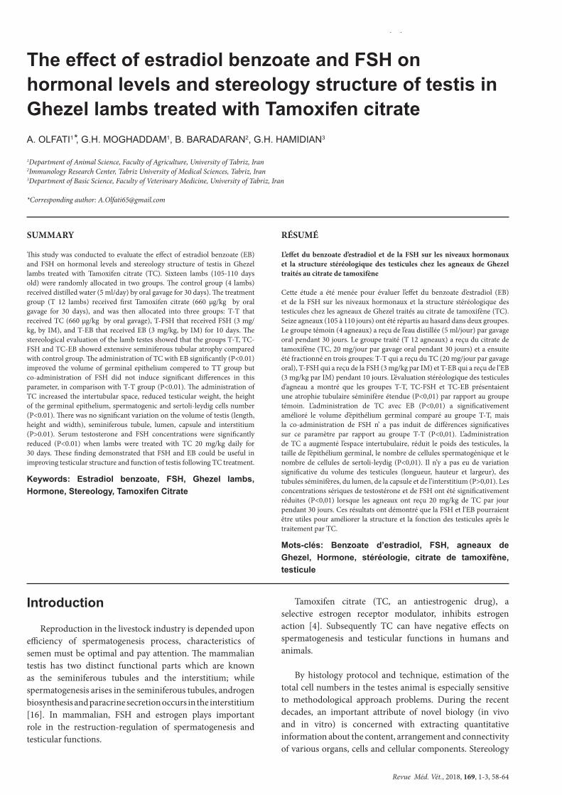
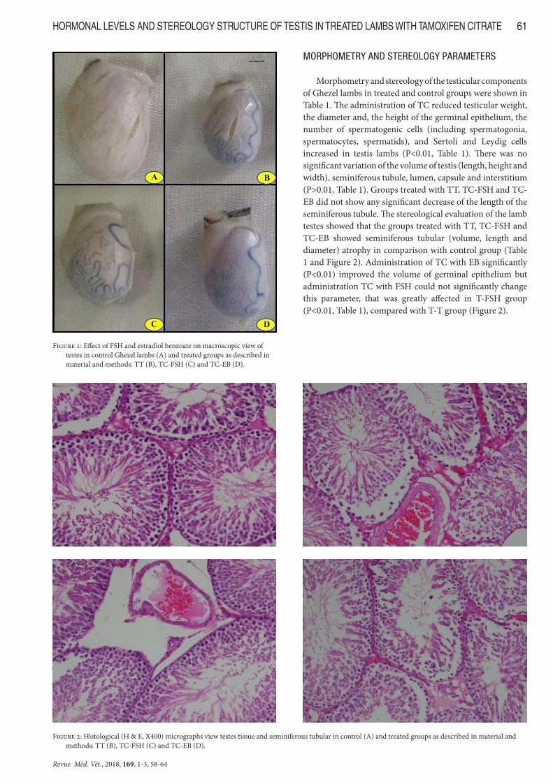
The effects of FSH and EB on macroscopic view of testes, micrographs view testes tissue and seminiferous tubular in treated Ghezel lambs with TC are shown in Figure 1 and 2, respectively. In control group, the macroscopic view and testis structure are normal. In the TT group, the total volume of testis was decreased, but in the TC-FSH and TC-EB groups the total volume of testis was increased when compared to TT group. Testicular vascular hyperemia and swollen were normal in the control and TC-FSH groups, but increased in the TT group and decreased in the TC-EB when compared with control group. Section of testis tissues of lambs groups treated with TT, TC-FSH and TC-EB showed extensive seminiferous tubular atrophy and deformed, destruction of germinal epithelium, increased interstitial space, congested blood vessel and testicular vascular hyperemia compared with control group (Figure 2).
Revue Méd. Vét., 2018, 169, 1-3, 58-64
HORMONAL LEVELS AND STEREOLOGY STRUCTURE OF TESTIS IN TREATED LAMBS WITH TAMOXIFEN CITRATE 61
MORPHOMETRY AND STEREOLOGY PARAMETERS
Morphometry and stereology of the testicular components of Ghezel lambs in treated and control groups were shown in Table 1. The administration of TC reduced testicular weight, the diameter and, the height of the germinal epithelium, the number of spermatogenic cells (including spermatogonia, spermatocytes, spermatids), and Sertoli and Leydig cells increased in testis lambs (P<0.01, Table 1). There was no significant variation of the volume of testis (length, height and width), seminiferous tubule, lumen, capsule and interstitium (P>0.01, Table 1). Groups treated with TT, TC-FSH and TC-EB did not show any significant decrease of the length of the seminiferous tubule. The stereological evaluation of the lamb testes showed that the groups treated with TT, TC-FSH and TC-EB showed seminiferous tubular (volume, length and diameter) atrophy in comparison with control group (Table 1 and Figure 2). Administration of TC with EB significantly (P<0.01) improved the volume of germinal epithelium but administration TC with FSH could not significantly change this parameter, that was greatly affected in T-FSH group (P<0.01, Table 1), compared with T-T group (Figure 2).
Figure 1: Effect of FSH and estradiol benzoate on macroscopic view of testes in control Ghezel lambs (A) and treated groups as described in material and methods: TT (B), TC-FSH (C) and TC-EB (D).
Figure 2: Histological (H & E, X400) micrographs view testes tissue and seminiferous tubular in control (A) and treated groups as described in material and methods: TT (B), TC-FSH (C) and TC-EB (D).
Revue Méd. Vét., 2018, 169, 1-3, 58-64
OLFATI (A.) AND COLLABORATORS62
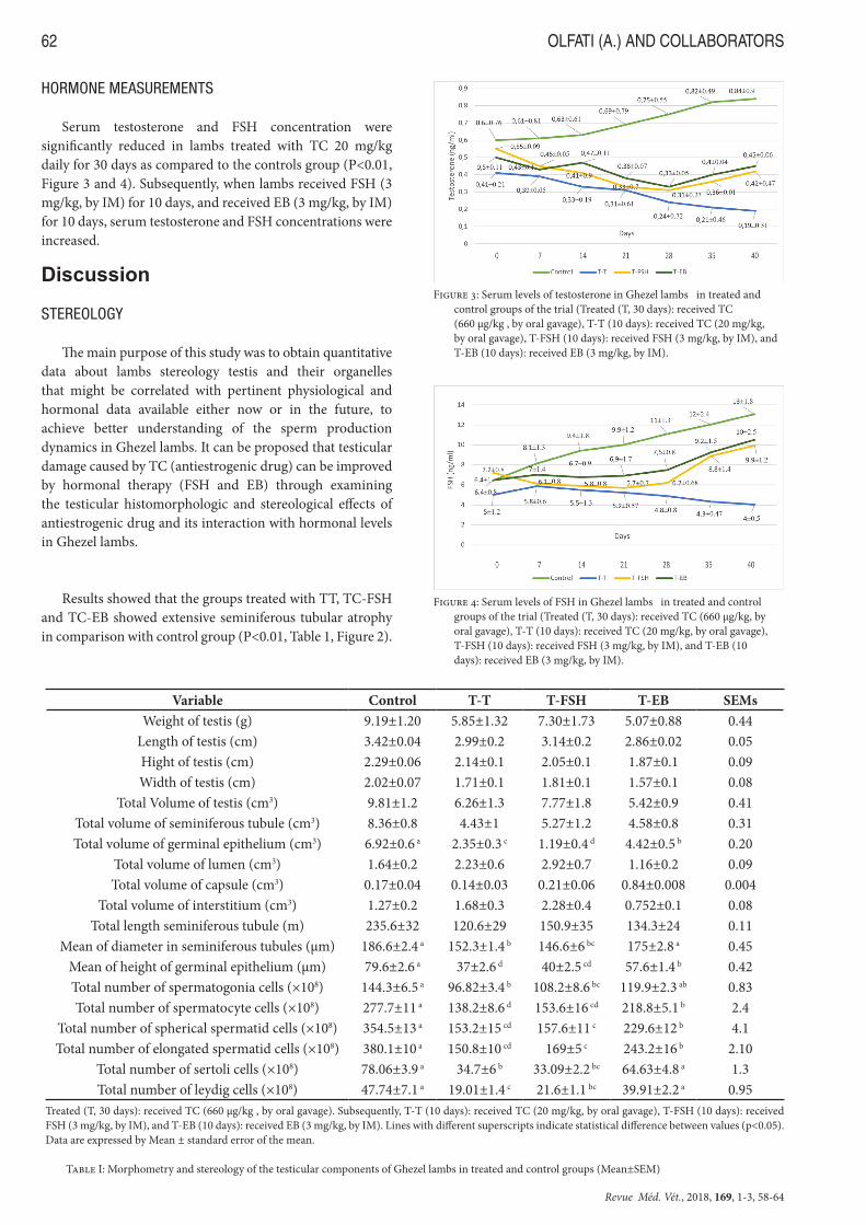
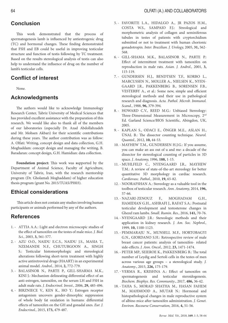
HORMONE MEASUREMENTS
Serum testosterone and FSH concentration were significantly reduced in lambs treated with TC 20 mg/kg daily for 30 days as compared to the controls group (P<0.01, Figure 3 and 4). Subsequently, when lambs received FSH (3 mg/kg, by IM) for 10 days, and received EB (3 mg/kg, by IM) for 10 days, serum testosterone and FSH concentrations were increased.
Discussion
STEREOLOGY
The main purpose of this study was to obtain quantitative data about lambs stereology testis and their organelles that might be correlated with pertinent physiological and hormonal data available either now or in the future, to achieve better understanding of the sperm production dynamics in Ghezel lambs. It can be proposed that testicular damage caused by TC (antiestrogenic drug) can be improved by hormonal therapy (FSH and EB) through examining the testicular histomorphologic and stereological effects of antiestrogenic drug and its interaction with hormonal levels in Ghezel lambs.
Results showed that the groups treated with TT, TC-FSH and TC-EB showed extensive seminiferous tubular atrophy in comparison with control group (P<0.01, Table 1, Figure 2).
Variable Control T-T T-FSH T-EB SEMsWeight of testis (g) 9.19±1.20 5.85±1.32 7.30±1.73 5.07±0.88 0.44
Length of testis (cm) 3.42±0.04 2.99±0.2 3.14±0.2 2.86±0.02 0.05Hight of testis (cm) 2.29±0.06 2.14±0.1 2.05±0.1 1.87±0.1 0.09Width of testis (cm) 2.02±0.07 1.71±0.1 1.81±0.1 1.57±0.1 0.08
Total Volume of testis (cm3) 9.81±1.2 6.26±1.3 7.77±1.8 5.42±0.9 0.41Total volume of seminiferous tubule (cm3) 8.36±0.8 4.43±1 5.27±1.2 4.58±0.8 0.31Total volume of germinal epithelium (cm3) 6.92±0.6 a 2.35±0.3 c 1.19±0.4 d 4.42±0.5 b 0.20
Total volume of lumen (cm3) 1.64±0.2 2.23±0.6 2.92±0.7 1.16±0.2 0.09Total volume of capsule (cm3) 0.17±0.04 0.14±0.03 0.21±0.06 0.84±0.008 0.004
Total volume of interstitium (cm3) 1.27±0.2 1.68±0.3 2.28±0.4 0.752±0.1 0.08Total length seminiferous tubule (m) 235.6±32 120.6±29 150.9±35 134.3±24 0.11
Mean of diameter in seminiferous tubules (µm) 186.6±2.4 a 152.3±1.4 b 146.6±6 bc 175±2.8 a 0.45Mean of height of germinal epithelium (µm) 79.6±2.6 a 37±2.6 d 40±2.5 cd 57.6±1.4 b 0.42Total number of spermatogonia cells (×108) 144.3±6.5 a 96.82±3.4 b 108.2±8.6 bc 119.9±2.3 ab 0.83Total number of spermatocyte cells (×108) 277.7±11 a 138.2±8.6 d 153.6±16 cd 218.8±5.1 b 2.4
Total number of spherical spermatid cells (×108) 354.5±13 a 153.2±15 cd 157.6±11 c 229.6±12 b 4.1Total number of elongated spermatid cells (×108) 380.1±10 a 150.8±10 cd 169±5 c 243.2±16 b 2.10
Total number of sertoli cells (×108) 78.06±3.9 a 34.7±6 b 33.09±2.2 bc 64.63±4.8 a 1.3Total number of leydig cells (×108) 47.74±7.1 a 19.01±1.4 c 21.6±1.1 bc 39.91±2.2 a 0.95
Treated (T, 30 days): received TC (660 µg/kg , by oral gavage). Subsequently, T-T (10 days): received TC (20 mg/kg, by oral gavage), T-FSH (10 days): received FSH (3 mg/kg, by IM), and T-EB (10 days): received EB (3 mg/kg, by IM). Lines with different superscripts indicate statistical difference between values (p<0.05). Data are expressed by Mean ± standard error of the mean.
Table I: Morphometry and stereology of the testicular components of Ghezel lambs in treated and control groups (Mean±SEM)
Figure 3: Serum levels of testosterone in Ghezel lambs in treated and control groups of the trial (Treated (T, 30 days): received TC (660 µg/kg , by oral gavage), T-T (10 days): received TC (20 mg/kg, by oral gavage), T-FSH (10 days): received FSH (3 mg/kg, by IM), and T-EB (10 days): received EB (3 mg/kg, by IM).
Figure 4: Serum levels of FSH in Ghezel lambs in treated and control groups of the trial (Treated (T, 30 days): received TC (660 µg/kg, by oral gavage), T-T (10 days): received TC (20 mg/kg, by oral gavage), T-FSH (10 days): received FSH (3 mg/kg, by IM), and T-EB (10 days): received EB (3 mg/kg, by IM).

Revue Méd. Vét., 2018, 169, 1-3, 58-64
HORMONAL LEVELS AND STEREOLOGY STRUCTURE OF TESTIS IN TREATED LAMBS WITH TAMOXIFEN CITRATE 63
Based on stereological evaluation of the lamb testes, we proposed that the decreased in volume of testicular animals that were exposed to TC in the experiment could be attributed to severe parenchyma atrophy in the seminiferous tubule following TC challenge. The stereological evidences in this research also showed that degenerative changes are characterized by vacuolization of the volume of interstitium, reduced volume of luminal spermatozoa and devoid spermatozoa in cross section of the seminiferous tubules of Ghezel lambs exposed to various treatment of TC (Figure 2). In agreement with the stereology results of the present study, Taha et al. [18] relived that the testes and epididymis of treated mice (TC 0.25 mg/kg daily for 14 days) showed several histopathological changes such as ticking in distortion and deformed in some seminiferous tubules and increased interstitial space and vacuolated cytoplasm of spermatogonia. Also, Verma and Krishna [17] reported that TC (0.2 and 0.4 mg/kg daily for 30 days) inhibits testicular spermatogenesis and steroidogenesis either directly by acting on testis or indirectly through the control gonadotropin secretion in mice.
Therefore, while the mean tubular area was decreased in groups treated with TT compared with other groups, based on stereological evaluation of the lamb testes, it can be verified that some tubules were intensely changed, while others were apparently normal or slightly changed. This finding suggests that the arrest of spermatogenesis may not be so diffused such that there is minimal disruption with some seminiferous tubules in which spermatogenesis is yet sustained in lambs. Studies have been shown interactions between extracellular matrix, tubular wall and germinative cells as important to their normal development [5]. Therefore, although the mean lumen and interstitium area were increased in groups receiving T-T, it was verified that some tubules were intensely changed, while others were apparently normal or slightly changed (Figure 2). The reason for this observation is not yet to be fully understood. Verma and Krishna [17] suggest that in Adult Parkes’ strain mice TC decreased estrogenic action suppresses spermatogenesis by inhibiting germ cells proliferation and survival and by promoting rate of germ cells apoptosis. TC decreased expression of aromatase in the testis may also cause decreased synthesis of estradiol which in turn may be responsible for regressed spermatogenesis. While, Azu et al. [2] suggested that in rats the arrest of spermatogenesis may not be so diffused such that there is minimal disruption with some seminiferous tubules in that spermatogenesis is yet sustained. Finally, results showed that FSH and EB therapy were beneficial in preventing spermatogenesis damage in TC treatments in Ghezel lambs. Despite these well-established negative effects of TC on the lamb testis, there is a need for further investigations in humans and animal model. Again, immunological parameters were not investigated in current study, further research seems necessary.
GERM CELLS
Yet, there has been surprisingly little study (or maybe never) done in lambs to identify TC induced changes in structure or function of the spermatogenesis rates. Permanent and temporary infertility is a main and incurable disease created by destruction and loss of germ cells. TC antiestrogenic drug and elicits estrogen agonist or antagonistic responses, depending on the target tissue. Subsequently, TC can decreases efficiency of spermatogenesis rates in humans and animals. Spermatogenic cycle dynamics allows us to infer more precisely about the reproductive function of the species. Currently, much progress has been conducted on producing male germ cells, such as spermatogonia, spermatocytes, and spermatids, from different types of stem cells. This investigation revealed that the groups treated with TT, TC-FSH and TC-EB showed significant reductions in spermatogoni, spermatocyte, spherical and elongated spermatid concentration when compared to the control groups (P<0.01, Table 1). The presence and increase of vacuoles of the seminiferous epithelium and epithelial cells, reduced volume of luminal spermatozoa, devoid spermatozoa in cross section of the seminiferous tubules, sertoli (which are located in the seminiferous tubules) and leydig (which lie adjacent to the seminiferous tubules) cells damage, and reduction in germinal epithelium could be due to the inhibition of spermatogenesis rates in Ghezel lambs in Iran.
HORMONE
This is the first study to directly assess the effects of TC on sex hormone concentration lambs. Sex hormones and gonadotropins play key roles in the development and maintenance of reproductive performance and fertility. Some studies showed that TC has adverse side effects on the systems of the body one of them the male reproductive system in human or animals such as sexual dysfunction [15] and fertility sex hormone in male [6]. As shown in Figure 3 and 4, we showed that the serum testosterone and FSH concentration in the groups treated with TT, TC-FSH and TC-EB were significantly decreased when compared to control (P<0.01). In addition, we showed that when lambs received FSH (3 mg/kg, by IM) or EB (3 mg/kg, by IM) for 10 days, serum testosterone and FSH concentration tended to increase towards values encountered in controls. These results were in agreement with that reported by Attia [1], who demonstrated that TC administration to mice lowered testosterone and LH-FSH levels. Balasinor et al. [3] reported that the administration of TC resulted in lower testosterone and LH-FSH levels in male rats. Also, Taha et al. [18] reported that in albino mice orally dosed with TC 0.25 mg/kg daily for 14 days, the serum testosterone and FSH concentration were significantly reduced in comparison to controls. The significant decrease in serum testosterone and FSH levels of treated lambs could be due to extensive seminiferous tubular atrophy (Figure 2) and diminished responsiveness of sertoli and leydig cells to FSH.
Revue Méd. Vét., 2018, 169, 1-3, 58-64
OLFATI (A.) AND COLLABORATORS64
Conclusion
This work demonstrated that the process of spermatogenesis lamb is influenced by antiestrogenic drug (TC) and hormonal changes. These finding demonstrated that FSH and EB could be useful in improving testicular structure and function of testis following by TC treatment. Based on the results stereological analysis of testis can also help to understand the influence of drug on the number of lamb’s testicular cells.
Conflict of interest
None.
Acknowledgments
The authors would like to acknowledge Immunology Research Center, Tabriz University of Medical Sciences that has provided excellent assistance with the preparation of this research. We would like also to thank all of the members of our laboratories (especially Dr. Azad Abdollahzadeh and Mr. Mohsen Akbari) for their scientific contributions during these years. The author contribution was as follow: A. Olfati: Writing, concept design and data collection, G.H. Moghaddam: concept design and managing the writing, B. Baradaran: concept design, G.H. Hamidian: data collection.
Foundation project: This work was supported by the Department of Animal Science, Faculty of Agriculture, University of Tabriz, Iran, with the research mentorship program (Dr. Gholamali Moghaddam) of higher education thesis program (grant No. 2015/TUAS/PH03).
Ethical considerations
This article does not contain any studies involving human participants or animals performed by any of the authors.
References
1. - ATTIA A.A.: Light and electron microscopic studies of the effect of tamoxifen on the testes of male mice. J. Biol. Sci., 2003, 3, 561-577.
2. - AZU O.O., NAIDU E.C.S., NAIDU J.S., MASIA T., NZEMANDE N.F., CHUTURGOON A., SINGH S.: Testicular histomorphologic and stereological alterations following short-term treatment with highly active antiretroviral drugs (HAART) in an experimental animal model. Androl., 2014, 2, 772-779.
3. - BALASINOR N., PARTE P., GILL-SHARMA M.K., KINI J.: Mechanism delineating differential effect of an anti-estrogen, tamoxifen, on the serum LH and FSH in adult male rats. J. Endocrinol. Invest., 2006, 29, 485-496.
4. - BIRZNIECE V., KEN K., HO Y.: Estrogen receptor antagonism uncovers gender-dimorphic suppression of whole body fat oxidation in humans: differential effects of tamoxifen on the GH and gonadal axes. Eur. J. Endocrinol., 2015, 173, 479-487.
5. - FAVORITE L.A., HIDALGO A., JR PAZOS H.M., COSTA W.S., SAMPAIO F.J.: Streological and morphometric analysis of collagen and seminiferous tubules in testes of patients with cryptorchidism submitted or not to treatment with human chorionic gonadotropin. Inter. Brazilian. J. Urology, 2005, 31, 562-568.
6. - GILL-SHAMA M.K., BALASINOR N., PARTE P.: Effect of intermittent treatment with tamoxifen on reproduction in male rats. Asian. J. Androl., 2001, 3, 115-119.
7. - GUNDERSEN H.J., BENDTSEN T.F., KORBO L., MARCUSSEN N., MOLLER A., NIELSEN K., NYEN-GAARD J.R., PAKKENBERG B., SORENSEN F.B., VESTERBY A., et al.: Some new, simple and efficient stereological methods and their use in pathological research and diagnosis. Acta. Pathol. Microb. Immunol. Scand., 1988, 96, 379-394.
8. - HOWARD C.V., REED M.G.: Unbiased Stereology: Three-Dimensional Measurement in Microscopy, 2nd Ed. Garland Science/BIOS Scientific, Abingdon, UK, 2005.
9. - KAPLAN S., ODACI E., ÖNGER M.E., ASLAN H., ÜNAL B.: The dissector counting technique. Neurol. Quantol., 2012, 10, 44-53.
10. - MAYHEW T.M., GUNDERSEN H.J.G.: If you assume, you can make an ass out of u and me: a decade of the dissector for stereological counting of particles in 3D space. J. Anatomy, 1996, 188, 1-15.
11. - MUHLFELD C., NYENGAARD J.R., MAYHEW T.M.: A review of state-of-the-art stereology for better quantitative 3D morphology in cardiac research. Cardiovasc. Pathol., 2010, 19, 65-82.
12. - NOORAFSHAN A.: Stereology as a valuable tool in the toolbox of testicular research. Ann. Anatomy, 2014, 196, 57-66.
13. - NAZARI-ZENOUZ F., MOGHADAM G.H., HAMIDIAN G.H., ASHRAFI J., RAFAT S.A.: Postnatal testicular development and testosterone changes in Ghezel ram lambs. Small. Rumin. Res., 2016, 141, 70-76.
14. - NYENGAARD J.R.: Stereologic methods and their application in kidney research. J. Am. Soc. Nephrol., 1999, 10, 1100-1123.
15. - PEMMARAJU N., MUNSELL M.F., HORTOBAGYI G.N., GIORDANO S.H.: Retrospective review of male breast cancer patients: analysis of tamoxifen- related side-effects. J. Ann. Oncol., 2012, 23, 1471-1474.
16. - PETER MP., SEIEROE K., PAKKENBERG B.: The total number of Leydig and Sertoli cells in the testes of men across various age groups – a stereological study. J. Anatomy., 2015, 226, 175-179.
17. - VERMA R., KRISHNA A.: Effect of tamoxifen on spermatogenesis and testicular steroidogenesis. Biochem. Biophys. Res. Community., 2017, 486, 36-42.
18. - TAHA S., MORAD SHATHA M., HASAN FAHEM M., MAHMOOD A., MUTAR N.: Hormonal and histopathological changes in male reproductive system of albino mice after tamoxifen administration. J. Genet. Environ. Recourse Conservation, 2016, 4, 51-56.