Embed Size (px)
DESCRIPTION
Â
Citation preview
�������� ����� ��
The differential effect of trigeminal vs. Peripheral pain stimulation on visualprocessing and memory encoding is influenced by pain-related fear
K. Schmidt, K. Forkmann, C. Sinke, M. Gratz, A. Bitz, U. Bingel
PII: S1053-8119(16)00231-7DOI: doi: 10.1016/j.neuroimage.2016.03.026Reference: YNIMG 13031
To appear in: NeuroImage
Received date: 11 December 2015Accepted date: 12 March 2016
Please cite this article as: Schmidt, K., Forkmann, K., Sinke, C., Gratz, M., Bitz, A.,Bingel, U., The differential effect of trigeminal vs. Peripheral pain stimulation on visualprocessing and memory encoding is influenced by pain-related fear, NeuroImage (2016),doi: 10.1016/j.neuroimage.2016.03.026
This is a PDF file of an unedited manuscript that has been accepted for publication.As a service to our customers we are providing this early version of the manuscript.The manuscript will undergo copyediting, typesetting, and review of the resulting proofbefore it is published in its final form. Please note that during the production processerrors may be discovered which could affect the content, and all legal disclaimers thatapply to the journal pertain.
ACC
EPTE
D M
ANU
SCR
IPT
ACCEPTED MANUSCRIPT
1
The differential effect of trigeminal vs. peripheral pain stimulation on visual processing and
memory encoding is influenced by pain-related fear
K. Schmidt1,2, K. Forkmann1, C. Sinke3, M. Gratz4,5, A. Bitz6 , U. Bingel1,2
1 Department of Neurology, University Hospital Essen, 45122 Essen, Germany
2 Department of Neurology, University Medical Center Hamburg-Eppendorf, 20246 Hamburg, Germany
3Department of Clinical Psychiatry, Social Psychiatry, and Psychotherapy, Hannover Medical School, 30625 Hannover, Germany
4Erwin L. Hahn-Institute for MRI, 45141 Essen, Germany
5Highfield and Hybrid MR-Imaging, University Hospital Essen, 45122 Essen, Germany
6Devision of Medical Physics in Radiology, German Cancer Research Center, 69121 Heidelberg, Germany
Corresponding author:
Katharina Schmidt
Department of Neurology
University Duisburg-Essen
Hufelandstrasse 55
45147 Essen, Germany
Phone: +49 201 / 723-2364
Email: [email protected]
http://www.uk-essen.de/?id=2376
Number of pages: 33
Number of figures: 5
Number of tables: 3
ACC
EPTE
D M
ANU
SCR
IPT
ACCEPTED MANUSCRIPT
2
Abstract
Compared to peripheral pain, trigeminal pain elicits higher levels of fear, which is assumed to
enhance the interruptive effects of pain on concomitant cognitive processes. In this fMRI study we
examined the behavioral and neural effects of trigeminal (forehead) and peripheral (hand) pain on
visual processing and memory encoding. Cerebral activity was measured in 23 healthy subjects
performing a visual categorization task that was immediately followed by a surprise recognition test.
During the categorization task subjects received concomitant noxious electrical stimulation on the
forehead or hand. Our data show that fear ratings were significantly higher for trigeminal pain.
Categorization and recognition performance did not differ between pictures that were presented
with trigeminal and peripheral pain. However, object categorization in the presence of trigeminal
pain was associated with stronger activity in task-relevant visual areas (lateral occipital complex,
LOC), memory encoding areas (hippocampus and parahippocampus) and areas implicated in
emotional processing (amygdala) compared to peripheral pain. Further, individual differences in
neural activation between the trigeminal and the peripheral condition were positively related to
differences in fear rating between both conditions. Functional connectivity between amygdala and
LOC was increased during trigeminal compared to peripheral painful stimulation. Fear-driven
compensatory resource activation seems to be enhanced for trigeminal stimuli, presumably due to
their exceptional biological relevance.
Keywords: Trigeminal pain, facial pain, face pain, pain-related fear, interruptive function of pain,
fMRI
ACC
EPTE
D M
ANU
SCR
IPT
ACCEPTED MANUSCRIPT
3
1. Introduction
Nociceptive signals from the head and facial area are signs of potential danger to essential vital
functions. Facial pain has been proposed to have a higher biological relevance than pain located in
the periphery and might be prioritized over pain in other body parts. Several lines of evidence
support this prioritization hypothesis. We have recently shown increased sensitization for noxious
heat stimuli applied to the face as compared to pain stimuli applied to the hand (Schmidt et al.,
2015). Interestingly, pain-related fear was higher for trigeminal pain as compared to pain on the
extremities despite comparable pain intensity ratings. Moreover, noxious facial stimulation leads to
faster fear conditioning compared to noxious tibial stimulation when used as an unconditioned
stimulus (Meier et al., 2014) and painful stimuli applied to the hand positioned near compared to
further away from the face lead to enhanced defensive hand-blink-reflexes (Sambo et al., 2012).
Due to its biological warning function pain captures attention and interrupts ongoing cognitive
processes. This interruptive effect of pain on cognitive processes (Eccleston and Crombez, 1999) is a
well-established phenomenon induced by both acute and chronic pain states (Eccleston, 1995;
Kuhajda et al., 2002; Grisart et al., 2007; Moore et al., 2012) and has been shown to affect different
cognitive domains such as working memory, short-term memory and visual processing (Bingel et al.,
2007; Oosterman et al., 2011; Forkmann et al., 2013). Interestingly, the interruptive function of pain
is not necessarily linearly related to the intensity of pain, but depends on top-down and bottom-up
factors including personality traits and expectation (Crombez et al., 1998; Van Damme et al., 2004;
Vancleef and Peters, 2006; Tiemann et al., 2010) as well as stimulus characteristics (Crombez et al.,
1994; Buhle and Wager, 2010; Sinke et al., 2015).
In this study we aimed at exploring whether the interruptive function of pain differs between the
head and facial region and peripheral body parts. To this end, we combined a well-established visual
encoding and recognition paradigm known to be sensitive to the interruptive effects of pain
(Forkmann et al., 2013) with noxious electrical stimulation of comparable subjective intensity to the
ACC
EPTE
D M
ANU
SCR
IPT
ACCEPTED MANUSCRIPT
4
forehead or the hand. The effects of trigeminal and peripheral pain (in the following referred to as
face pain and hand pain) on visual processing, memory encoding and retrieval were assessed on the
behavioral level with an object categorization task and a surprise recognition task. Neural effects
were tested using fMRI. Given the crucial biological relevance and higher pain-related fear of face
pain, we assumed a larger interruptive effect of pain when applied to the face compared to stimuli
applied to the hand. Specifically, we predicted lower visual categorization and memory performance
when the noxious stimulation is applied to the face as compared to the hand. On the neural level, we
expected reduced activation in visual and memory-related areas such as hippocampus and
parahippocampus (Forkmann et al., 2013) during face as compared to hand pain. Given the higher
threat value of facial pain compared to pain at the extremities (Meier et al., 2014; Schmidt et al.,
2015), we predicted increased neural activation in brain areas involved in emotional and fear
processing such as the amygdala (Bishop et al., 2004; Sehlmeyer et al., 2009) during face pain. Finally
we aimed at exploring pain- and task-induced connectivity changes between pain-related, task-
related and fear-related brain areas.
ACC
EPTE
D M
ANU
SCR
IPT
ACCEPTED MANUSCRIPT
5
2. Methods
In the following the terms face pain and hand pain refer to the pain stimulation site, i.e. the forehead
and the back of the hand.
2.1. Subjects
Imaging and behavioral data were acquired in 30 healthy subjects. Data from 26 subjects was
included in behavioral data analysis (all right-handed, 11 male; age in years: 27.4 ± 4.2 (M ± SD)).
Four subjects had to be excluded due to the following reasons: n=2 reported no painful sensation
during the task, n=1 due to technical failure of the electrical stimulator and n=1 due to a dislocated
electrode. Three additional subjects had to be excluded from fMRI analyses due to technical failure
of the scanner. Therefore, fMRI data analysis comprised data from 23 subjects (all right-handed, 11
male; age in years: 27.8 ± 4.2 M ± SD). All subjects reported normal or corrected-to-normal eyesight,
normal hearing and no known history of neurological or psychiatric and pain-related diseases. All
subjects gave written informed consent to participate and were free to withdraw from the study at
any time. The study was conducted in accordance with the declaration of Helsinki and had been
approved by the local ethics committee in Essen, Germany. Subjects received a small monetary
compensation for their study participation.
2.2. Experimental Paradigm
The experimental paradigm used here is a modified version of a visual encoding paradigm reported
previously (Forkmann et al., 2013). The study was conducted on two days. On day 1, subjects
underwent the assessment of pain thresholds on both stimulation sites and a calibration procedure
in order to determine individually calibrated electrical pain stimuli to be applied on the hand and the
face. Painful hand and face stimulation was adjusted separately to yield comparable pain intensity
levels of 70 on a 0 – 100 visual analogue scale (VAS, anchors 0 = “not painful at all” and 100 =
“unbearably painful”). Subjects performed a practice run of the visual encoding task. All relevant
behavioral and imaging data were acquired on the second day of the study (1 – 5 days later). The
ACC
EPTE
D M
ANU
SCR
IPT
ACCEPTED MANUSCRIPT
6
main fMRI experiment comprised (1) the simultaneous presentation of visual and painful stimuli
(visual categorization task, memory encoding) and (2) a subsequent surprise recognition task to
investigate differential effects of painful face and hand stimulation on memory performance. Both
tasks were performed inside the 3T fMRI scanner.
2.3. Experimental Procedures
Day 1. On day 1 subjects filled in questionnaires assessing pain-related psychological processing [Pain
Anxiety Symptom Scale (PASS); Pain Catastrophizing Scale (PCS); Pain Vigilance and Awareness
Questionnaire (PVAQ)], anxiety and depression [ADS-K; State Trait Anxiety Inventory (STAI), Trait
Scale]. For details see Psychological Questionnaires. Participants were then familiarized with the
electrical stimuli and underwent calibration procedures with different stimulation intensities that
had to be rated for subjective pain intensity on a VAS. First, we assessed electrical pain thresholds
separately for both sites of stimulus application [(1) back of the left hand, approximately 2 cm away
from the knuckle of the index finger, (2) left side of the forehead, approximately 1 cm above the
outer end of the eyebrow]. Thresholds were obtained using single pulse stimuli with 0.5 ms duration
and by increasing the current by 0.01 mA between consecutive stimuli starting at 0 mA (ascending
method of limits) (Gescheider, 1997). The upper limit was set to 15 mA to avoid tissue damage.
Participants verbally indicated when their perception changed from a tingling to a painful sensation.
This procedure was repeated three times for each stimulation site and the mean mA was defined as
the site-specific pain threshold. Subsequently, subjects underwent a pain calibration procedure to
determine the stimulation intensity corresponding to a level of 70 on a 0 – 100 VAS. To this end,
trains of stimuli with 2.5 s duration and of varying intensity levels around their individual pain
threshold were applied. After each stimulus participants rated their subjective pain intensity on a
VAS that was presented on a computer screen. The stimulation intensity that corresponded to a VAS
level of 70 was chosen to be applied during the experiment. Pain threshold determination and
calibration of the stimulation intensity were performed separately for hand and face in
counterbalanced order.
ACC
EPTE
D M
ANU
SCR
IPT
ACCEPTED MANUSCRIPT
7
Day 2. On day 2 the fMRI experiment was conducted. Before performing the task inside the scanner,
subjects completed the state scale of the STAI. Immediately before scanning, subjects performed a
number of pretests, including a recalibration of pain stimuli and the assessment of fear and
expectation ratings. During recalibration, 5 pain stimuli were applied to the subjects’ hand and
forehead to test whether stimulus intensities determined on day 1 still yielded intensity levels of
approximately 70 when subjects were lying in the MR scanner. In case the intensity ratings differed
largely from 70, stimulation intensity was adjusted. Prior to the main task, subjects were asked to
rate their fear regarding the upcoming stimulation separately for both body sites (presentation of the
word “hand” or “face” before appearance of the VAS) on two 0 – 100 VAS (“Please indicate how
fearful you are about the upcoming stimulation.”, VAS anchors: 0 = “not fearful at all” and 100 =
“extremely fearful”). Further, participants were asked to rate their expectation regarding (1) the
effect of pain on task performance and (2) the effect of task performance on pain perception on a
VAS ((1) “Please indicate how the painful stimulation will influence your task performance.”, VAS
anchors: -50 = “strong performance decrease” and 50 = “strong performance increase”; (2) “Please
indicate how performing the task will influence your pain perception.”, VAS anchors: -50 =
“decreased pain perception” and 50 = “increased pain perception”).
Subsequently, subjects performed the encoding task, which was introduced as a visual categorization
task (duration ~ 10 min). First, all subjects performed 6 practice trials (3 of each condition). These
trials also served to ensure that, while performing the categorization task, pain intensities were still
high and comparable between both stimulation sites. The test trials included 40 images of living and
nonliving objects (see below, Visual stimuli). All pictures were presented with concurrent painful
stimulation either on the (1) forehead (‘face pictures’) or the (2) back of the hand (‘hand pictures’).
Thus, each condition comprised 20 trials (10 living, 10 nonliving objects) that were presented in a
pseudorandomized order with no more than three consecutive pain stimuli of the same type. The
trial structure was as follows: presentation of a white fixation cross (variable duration of 5 – 8 s),
image and concomitant painful stimulation (2.5 s), presentation of a white fixation cross (variable
ACC
EPTE
D M
ANU
SCR
IPT
ACCEPTED MANUSCRIPT
8
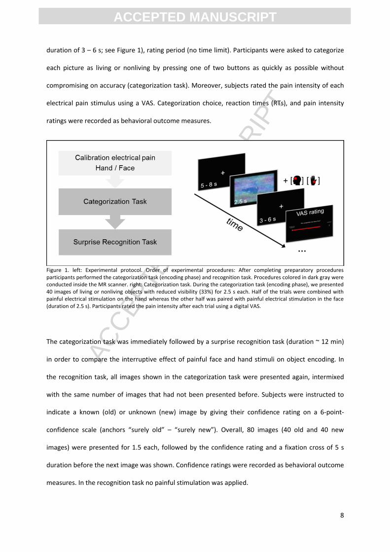
duration of 3 – 6 s; see Figure 1), rating period (no time limit). Participants were asked to categorize
each picture as living or nonliving by pressing one of two buttons as quickly as possible without
compromising on accuracy (categorization task). Moreover, subjects rated the pain intensity of each
electrical pain stimulus using a VAS. Categorization choice, reaction times (RTs), and pain intensity
ratings were recorded as behavioral outcome measures.
Figure 1. left: Experimental protocol. Order of experimental procedures: After completing preparatory procedures participants performed the categorization task (encoding phase) and recognition task. Procedures colored in dark gray were conducted inside the MR scanner. right: Categorization task. During the categorization task (encoding phase), we presented 40 images of living or nonliving objects with reduced visibility (33%) for 2.5 s each. Half of the trials were combined with painful electrical stimulation on the hand whereas the other half was paired with painful electrical stimulation in the face (duration of 2.5 s). Participants rated the pain intensity after each trial using a digital VAS.
The categorization task was immediately followed by a surprise recognition task (duration ~ 12 min)
in order to compare the interruptive effect of painful face and hand stimuli on object encoding. In
the recognition task, all images shown in the categorization task were presented again, intermixed
with the same number of images that had not been presented before. Subjects were instructed to
indicate a known (old) or unknown (new) image by giving their confidence rating on a 6-point-
confidence scale (anchors “surely old” – “surely new”). Overall, 80 images (40 old and 40 new
images) were presented for 1.5 each, followed by the confidence rating and a fixation cross of 5 s
duration before the next image was shown. Confidence ratings were recorded as behavioral outcome
measures. In the recognition task no painful stimulation was applied.
ACC
EPTE
D M
ANU
SCR
IPT
ACCEPTED MANUSCRIPT
9
2.4. Stimuli
The presentation of the visual stimuli, application of the electrical stimuli and recording of the
behavioral data were performed using the software Presentation (www.neurobs.com).
2.4.1. Visual stimuli
The visual stimuli consisted of pictures showing natural scenes with living (e.g. animals) or nonliving
(e.g. cars, buildings) objects. Eighty-eight pictures with neutral valence were selected that have been
used in our lab before (Forkmann et al., 2013). For the categorization task 40 images were randomly
selected, 40 other pictures were chosen for the recognition task as new images and the remaining 8
pictures were used in the practice trials. Within the encoding task all images were presented with a
reduced visibility of 33% to increase the difficulty of the task using a scrambling routine as described
in Rose et al. (2005). Recognition task pictures were presented at full visibility. The outer edges of the
pictures were smoothed (28 mm FWHM isotropic kernel) to embed the pictures into the black
background. Visual stimuli were presented on a back projection screen located behind the MR
scanner. The screen could be seen via a mirror that was attached to the head coil. The pictures had a
visual angle of 11.6° x 8.4°.
2.4.2. Electrical pain stimuli
Painful stimulation was applied using two electrical stimulators (Digitimer DS7A constant current
stimulator, Hertfordshire, UK) and surface electrodes (Specialty Developments, Bexley, UK) with a
diameter of approximately 5 mm that were attached to the skin using medical tape. We applied 82
single pulses of 0.5 ms duration with an inter pulse interval of 30 ms resulting in a train of painful
stimulation with 2.5 s duration. The experimental setup required positioning the electrodes and their
cabling into the center (head electrode) and close to the end rings (hand electrode) of the integrated
radio-frequency (RF) body coil and thus directly into the RF transmit field used for MR excitation.
Since this increases the probability of tissue damage due to currents induced by electro-magnetic
wave coupling, we replaced the standard copper cables by custom-built cables that included special
ACC
EPTE
D M
ANU
SCR
IPT
ACCEPTED MANUSCRIPT
10
non-magnetic chip resistors every 6 cm along the cable as dampers. Timing parameters of the
stimulation that would ensure simultaneous perception of visual and painful stimuli on both body
sites were determined in a preparatory study performed in 17 healthy volunteers (data not shown).
Further we assessed stimulus induced eye blinks to both types of stimulations in this preparatory
study that revealed no difference in eye blink frequency between conditions.
The ecological validity of the pain stimulus is a concern in many experimental studies, especially
studies using electrical stimuli. The choice of this experimental pain model was based on two
reasons: First, the visual task used in our study that allows us to test for the perception, encoding and
recognition of visual objects requires the simultaneous presentation of painful stimuli and visual
objects (presentation time 2.5 sec) on different body parts within the same scanning session. Such
flexible stimulation is most easily implemented by the use of electrical stimulation. Second, when
aiming at investigating CNS responses to facial compared to other pain using fMRI, the choice of the
stimulation device is limited by a number of methodological and physical constrains as the
stimulation has to be performed within the head coil. Again the electrical stimulation used in our
study seemed best suited to meet these requirements.
2.5. Psychological Questionnaires
Subjects completed the German versions of questionnaires assessing anxiety, depression and pain-
related psychological processing, since these variables have been shown to modulate the
interruptive function of pain (Peters et al., 2002; Van Damme et al., 2004). Specifically, participants
completed the following questionnaires: (1) Pain Vigilance and Awareness Questionnaire: PVAQ
(McCracken, 1997), German version (Lautenbacher et al., 2009); (2) Pain Anxiety Symptom Scale:
PASS-D (McCracken et al., 1992), German version (Walter, 2002); (3) Pain Catastrophizing Scale: PCS
(Sullivan et al., 1995), German version (Lautenbacher et al., 2009); (4) Center for Epidemiological
Studies–Depression Scale (Radloff, 1977), German version: ADS-K (Hautzinger, 1993); and (5) State
ACC
EPTE
D M
ANU
SCR
IPT
ACCEPTED MANUSCRIPT
11
Trait Anxiety Inventory: STAI (Spielberger, 1983), German version (Laux, 1992). All questionnaires
were analyzed following the respective manuals.
2.6. Behavioral data
2.6.1. Analysis of behavioral data
Behavioral data were automatically recorded and logged by the stimulation program Presentation
(www.neurobs.com). All behavioral data analyses were conducted using SPSS version 22.0. Results
with a p ≤ 0.05 are considered as statistically significant. All statistical analyses were performed using
two-tailed testing. Means are reported ± standard deviation (SD), if not specified otherwise. To
compare categorization performance (accuracy and RTs) between the conditions ‘hand pictures’ and
‘face pictures’ paired t-tests were performed. Mean RTs were calculated for correctly categorized
images separately for both experimental conditions after excluding extreme outliers (>3 SD above
mean). RTs longer than 2.5 s were not recorded. Previous studies revealed differences in reaction
times between subjects receiving painful stimulation (Tiemann et al., 2010) and an influence of pain-
related and anxiety-related personality traits on disengagement from pain (Van Damme et al., 2004).
Therefore, we performed correlational analyses using Pearson’s correlation coefficient with the
questionnaire and behavioral data (e.g. RTs categorization task) and used Bonferroni correction for
multiple comparisons.
For comparison of recognition accuracy between both experimental conditions a paired t-test was
performed. To this end, we calculated the percentage of images classified as old (pooled across
confidence levels “sure old” – “rather old” (1,2 and 3 of confidence scale)) and new (pooled across
confidence levels “sure new” – “rather new” (6,5 and 4 of confidence scale)). The number of correct
old or new classifications was divided by the number of presented pictures for each condition,
separately. To account for false alarms, we calculated the discrimination index d’ (Stanislaw and
Todorov, 1999) for both experimental conditions separately using the formula d‘ = z(hit rate) – z(false
alarm rate). Higher values of d‘ indicate better discrimination, and therefore better recognition
ACC
EPTE
D M
ANU
SCR
IPT
ACCEPTED MANUSCRIPT
12
memory. Mean pain intensity ratings were calculated for both pain conditions. Differences in pain
intensity ratings between painful face and hand stimulation, pain thresholds and calibrated
stimulation intensities of both stimulation sites, as well as fear and expectation ratings were assessed
using paired t-tests.
2.6.2. Behavioral pilot study
In preparation of the fMRI study we performed a behavioral study using the same experimental
paradigm and procedures without brain imaging to test for possible differences in visual processing
and encoding between conditions at the behavioral level. Categorization and recognition
performance for pictures paired with face pain and pictures paired with hand pain was assessed in 17
healthy volunteers (all right-handed, 8 male; age in years: 25.2 ± 4.6 (M ± SD)). Data analyses were
performed following the same procedures as described before (see Analysis of behavioral data).
Please note that the pilot study was performed on a completely different sample of healthy
volunteers than the fMRI study.
2.7. Imaging data
2.7.1. Image data acquisition
Functional data was acquired on a 3 T MRI system (Siemens Magnetom, Skyra syngo MR D13) with a
standard 32-channel head coil (Skyra, Siemens Healthcare, Erlangen, Germany). Functional imaging
data with a total of 42 axial slices (slice thickness 2.7 mm) per volume were acquired using gradient
multi-echo EPI with three echos (TE 1 13.0 ms, TE 2 28.9 ms, TE 3 44.8 ms, TR 2.89 s; flip angle 90°;
field of view 224 x 224 mm). Structural images were obtained for each participant using a T1-
weighted magnetization-prepared rapid acquisition gradient echo sequence (MPRAGE, slice thickness
1 mm; TR 2.30 s; TE 2.07 ms; flip angle 9°; field of view 256 x 256 mm).
2.7.2. Image preprocessing
ACC
EPTE
D M
ANU
SCR
IPT
ACCEPTED MANUSCRIPT
13
Before data analysis, all imaging data were screened for scanner artifacts using the SPM toolbox
ArtRepair. Image processing and statistical analysis of fMRI data was performed using SPM8
(www.fil.ion.ucl.ac.uk/spm/). The first five volumes were removed to allow for T1 saturation.
Preprocessing included slice timing and realignment to the first volume. To correct for the interaction
of head motion and magnetic field inhomogeneities (susceptibility movement interaction) the
unwarping procedure of SPM8 was used. We assessed stimulation-induced head movement by
comparing translational and rotational movement parameters separately for the hand and face pain
condition. Separate 2X3 rmANOVAs (factor stimulation [hand/face] x factor dimension [x/y/z]) of the
mean movement parameters during different conditions revealed no significant effect of stimulation
neither for translation (F(2,56) = 2.45, p = 0.10) nor rotation (F(2,56) = 1.95, p = 0.15). Thus the two
painful stimulations did not induce significant differences head movement. The maximum amount of
head motion did not exceed 1 mm in any of the participants. The anatomical volume was
coregistered to the mean echo-planar image and segmented with bias regularization set to medium
level. Structural and functional volumes were normalized to standard Montreal Neurological Institute
(MNI) space using the transformation matrix obtained from the segmentation procedure and
resampled to a voxel size of 2 x 2 x 2 mm with an in-plane resolution of 2 mm. Functional images
were finally smoothed with an 8 mm Gaussian kernel with FWHM.
2.7.3. fMRI statistical analyses
Neural data analysis was performed using the general linear model (GLM). On subject level, the
applied model for the categorization task (encoding phase) contained two regressors of interest that
coded for the two experimental conditions: (1) presentation of images with hand pain (‘hand
pictures’) and (2) presentation of images with face pain (‘face pictures’). Two regressors of no
interest were added to the model that coded for the anticipation phase (white fixation cross
presented before the picture) and rating period. Both regressors were modelled with stick function
and convolved as canonical hemodynamic response function. Data were high-pass filtered with a cut-
off period of 128 s. The effects of interest were tested using linear contrasts of the parameter
ACC
EPTE
D M
ANU
SCR
IPT
ACCEPTED MANUSCRIPT
14
estimates for the two regressors of interest, resulting in a t-statistic for each voxel. Further, contrast
images for both conditions and differential contrasts (e.g. [‘face pictures’] > [‘hand pictures’]) were
generated for each participant and included into a second GLM. For group level analyses we used a
random-effects approach using a one sample t-test (Friston et al., 1999) treating inter-subject
variability as a random factor and including non-sphericity.
In order to assess neural activation related to successful memory formation (subsequent memory
effect, SME) (Brewer et al., 1998; Kim, 2011) a second analysis was performed. Images within the
categorization task were classified into remembered (hits) or forgotten images (misses) according to
the results from the recognition task. Thus, this model contained 4 regressors of interest: (1) ‘hand
pictures’ hits, (2) ‘hand pictures’ misses, (3) ‘face pictures’ hits, (4) ‘face pictures’ misses. Two
regressors of no interest coding for the anticipation phase and the rating period were added to the
model. At first level the following contrast c = (-1, 1, 1, -1) ([SME ‘face pictures’ > SME ‘hand
pictures’] = [‘face pictures’ hits > ‘face pictures’ misses] > [‘hand pictures’ hits > ‘hand pictures’
misses]) was generated. In order to test for brain regions showing greater SMEs during face pain
compared to hand pain the individual first level images were then compared against the hypothesis
of no effect using a one-sample t-test at second level. In a second step, this second level model was
extended by including individual difference of fear between face and hand pain as a covariate of
interest. Using this co-variate we then tested for regions showing a correlation between SME ‘face
pictures’ > SME ‘hand pictures’ and fear of face pain – fear of hand pain (c=0, 1 at second level). The
threshold for all statistical analyses was set to p = 0.05, familywise error (FWE) peak-voxel-corrected
for multiple comparisons. Only for visualization purposes in all figures the threshold was set to p =
0.001 uncorrected. Please note that only brain activations surviving correction for multiple
comparisons are reported and discussed in the Results and Discussion section. Correction for multiple
comparisons was based on regions of interest (ROI), which were defined based on previous studies
that had addressed the interaction of pain, visual processing, memory encoding and recognition
(Bingel et al., 2007; Forkmann et al., 2013). Specifically, ROIs included the fusiform gyrus, LOC,
ACC
EPTE
D M
ANU
SCR
IPT
ACCEPTED MANUSCRIPT
15
parahippocampal gyrus and the hippocampus. ROIs with a 20 mm-diameter sphere were centered on
the reported peak-coordinates of these previous studies. Small volume correction of the amygdala
was performed using an anatomical mask (http://neuro.imm.dtu.dk/wiki/Harvard-Oxford_Atlas).
2.7.3.1. Psychophysiological interaction analysis
Compared to hand pain, the face pain led to higher fear ratings in our behavioral pilot study (hand:
21.74 ± 9.81; face: 34.35 ± 23.01 (M ± SD); t(16) = -2.86, p = 0.01) and the fMRI study (see Results).
Furthermore amygdala activation was increased for face compared to hand pain (contrast [‘face
pictures’ > ‘hand pictures’]), see Results, Table 3). Based on these observations, the amygdala’s
established role in (i) affective pain processing (Morris et al., 1998; Sergerie et al., 2008) and (ii)
memory encoding of emotional stimuli (Dolcos et al., 2004; Phelps, 2004), we investigated changes in
functional connectivity between the amygdala and task relevant areas as function of pain-related
fear.
To this end, a psychophysiological interaction analysis (PPI) (Friston et al., 1997) was performed to
explore differences in functional connectivity of the left and right amygdala that might underlie the
differential effects of face and hand painful stimulation on visual processing and visual encoding.
Blood oxygenation level-dependent time series were extracted from a sphere located in the left (10
mm diameter, centered on the peak voxel (x y z) = (-26 -6 -23)) and right amygdala (10 mm diameter,
centered on the peak voxel (x y z) = (22 -6 -23)) as identified in the contrast [‘face pictures’ > ‘hand
pictures’] for each subject individually using the first eigen time series (principal component analysis).
The PPI regressor was calculated using the element-by-element product of the mean-corrected
activation of the left and right amygdala (extracted time series) and the vector coding for the
psychological variable (-1 on regressor ‘hand pictures’, 1 on regressor ‘face pictures’). Individual PPI
regressors were subsequently entered into one sample t-tests to test for condition-specific
differences in functional connectivity between both amygdala peak coordinates and any other brain
regions. We predicted an increased functional connectivity between the amygdala and task-relevant

ACC
EPTE
D M
ANU
SCR
IPT
ACCEPTED MANUSCRIPT
16
brain areas, such as LOC and fusiform gyrus for the facial compared to the hand pain stimulation.
Corrections in these regions were based on anatomical masks and 20-mm-diameter spheres centered
on peak coordinates reported in these previous studies.
ACC
EPTE
D M
ANU
SCR
IPT
ACCEPTED MANUSCRIPT
17
3. Results
3.1. Behavioral results
3.1.1. Behavioral pilot study results
Paired t-tests revealed significantly higher electrical pain thresholds (hand: 1.89 ± 0.98 mA, face: 0.96
± 0.42 mA; t(13) = 4.02, p = 0.001) and stimulation intensities corresponding to VAS 70 (hand: 2.14 ±
2.02 mA, face: 1.39 ± 0.1.28 mA; t(16) = 2.78, p = 0.01) for hand compared to face pain stimulation.
Furthermore, there were significant differences between recalibrated stimulation levels that were
used during the main task (hand: 3.83 ± 3.74 mA, face: 1.93 ± 1.71 mA; t(16) = 3.03, p = 0.008).
However, pain intensity ratings were comparable between both conditions (hand: 59.26 ± 15.59,
face: 56.74 ± 13.24; t(16) = 1.60, p = .13), confirming that the stimulation was perceived as equally
intense at both stimulation sites.
Paired t-tests revealed no significant differences in categorization performance between conditions
(hand: 94.71 ± 5.72 %; face: 90.59 ± 8.64 %; t(16) = 1.53, p = 0.15) but RTs were significantly slower
(hand: 938.47 ± 190.44 ms; face: 1003.17 ± 221.09 ms; t(16) = -2.94, p = 0.01) and omission rates
significantly higher (hand: 0.29 ± 1.21 %; face: 3.53 ± 4.24 %; t(16) = -2.86, p = 0.01) for face
compared to hand pain stimulation in the categorization task (encoding phase; see Figure 2). In the
subsequently performed recognition test we found no significant differences between pictures that
had previously been paired with painful face pain stimulation and those combined with hand stimuli
neither for accuracy (hand: 54.41 ± 14.56 %; face: 55.88 ± 9.56 %; t(16) = -0.37, p = 0.71) nor for d’
(hand: 1.15 ± 0.47; face: 1.18 ± 0.40; t(16) = -0.30, p = 0.77).
3.1.2. fMRI study results
3.1.2.1. Pain thresholds, stimulation intensities and pain intensity ratings
Electrical pain thresholds (hand: 2.46 ± 1.22 mA, face: 0.98 ± 0.41 mA; t(25) = 7.23, p < 0.001) and
stimulation intensities corresponding to VAS 70 (hand: 2.21 ± 1.38 mA, face: 1.09 ± 0.87 mA; t(25) =
4.87, p < 0.001) were significantly higher for hand compared to face stimulation. There were no
ACC
EPTE
D M
ANU
SCR
IPT
ACCEPTED MANUSCRIPT
18
significant differences between recalibrated stimulation levels that were used during the main task
(hand: 4.32 ± 2.72 mA, face: 3.51 ± 2.85 mA; t(25) = 1.90, p = 0.07). Pain intensity ratings were
comparable between both conditions (hand: 56.45 ± 13.75, face: 57.92 ± 10.63; t(25) = -0.51, p =
.61). Note that applied stimulation intensities and VAS ratings did not differ between pilot and fMRI
study.
3.1.2.2. Fear and Expectation Ratings
Fear ratings assessed directly before the encoding task were significantly higher for the upcoming
face pain stimulation than for hand pain stimulation (hand: 15.54 ± 15.70, face: 29.30 ± 19.71; t(25) =
-4.65, p < 0.001). Subjects expected face pain to decrease their task performance significantly (-10.19
± 14.31, t(25) = -3.63, p = 0.001) while hand pain was expected to have no influence (-1.12 ± 9.77,
t(25) = -0.58, p = 0.57). These expectation ratings differed significantly (t(25) = 3.71, p = 0.001).
Moreover, participants expected a decrease in pain intensity for hand pain stimuli (-8.35 ± 11.34,
t(25) = -3.75, p = 0.001) but no change for face pain stimuli (-2.73 ± 12.02, t(25) = -1.16, p = 0.26)
while performing the task. Again, both pain perception expectation ratings differed significantly
(t(25) = -2.34, p = 0.02).
3.1.2.3. Categorization task (encoding phase): accuracy and RT and recognition accuracy
There were no significant differences in categorization hits (‘hand pictures’ hits: 89.04 ± 10.45 %,
‘face pictures’ hits: 92.24 ± 8.06 %; t(25) = -1.67, p = 0.10), misses (‘hand pictures’ misses: 1.54 ± 2.75
%, ‘face pictures’ misses: 1.15 ± 2.57 %; t(25) = 0.63, p = .054; see Figure 2), or false alarms (‘hand
pictures’ false alarms: 9.42 ± 9.73 %, ‘face pictures’ false alarms: 6.42 ± 5.82 %; t(25) = 1.76, p = 0.09)
between painful hand and face stimulation. Furthermore, there were no differences in RTs between
both experimental conditions (RTs ‘hand pictures’: 1088.30 ± 256.37 ms, RTs ‘face pictures’: 1097.67
± 177.88 ms; t(25) = -0.18, p = 0.86; see Figure 2).
ACC
EPTE
D M
ANU
SCR
IPT
ACCEPTED MANUSCRIPT
19
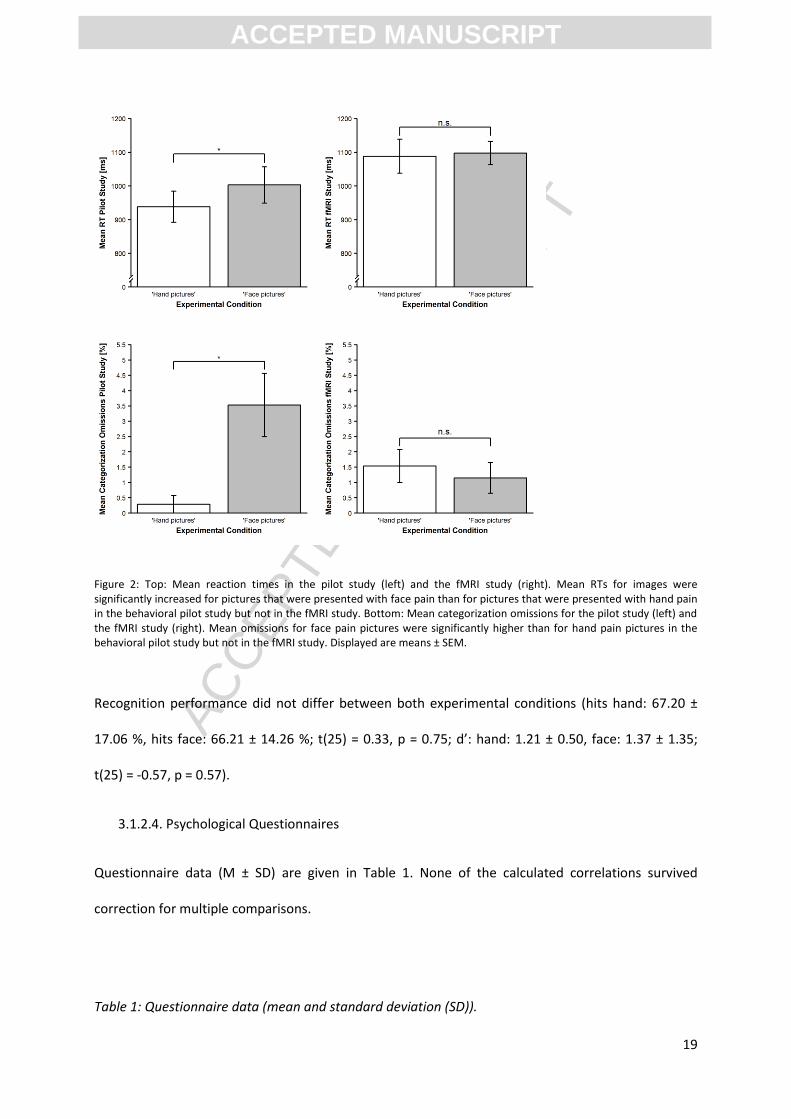
Figure 2: Top: Mean reaction times in the pilot study (left) and the fMRI study (right). Mean RTs for images were significantly increased for pictures that were presented with face pain than for pictures that were presented with hand pain in the behavioral pilot study but not in the fMRI study. Bottom: Mean categorization omissions for the pilot study (left) and the fMRI study (right). Mean omissions for face pain pictures were significantly higher than for hand pain pictures in the behavioral pilot study but not in the fMRI study. Displayed are means ± SEM.
Recognition performance did not differ between both experimental conditions (hits hand: 67.20 ±
17.06 %, hits face: 66.21 ± 14.26 %; t(25) = 0.33, p = 0.75; d’: hand: 1.21 ± 0.50, face: 1.37 ± 1.35;
t(25) = -0.57, p = 0.57).
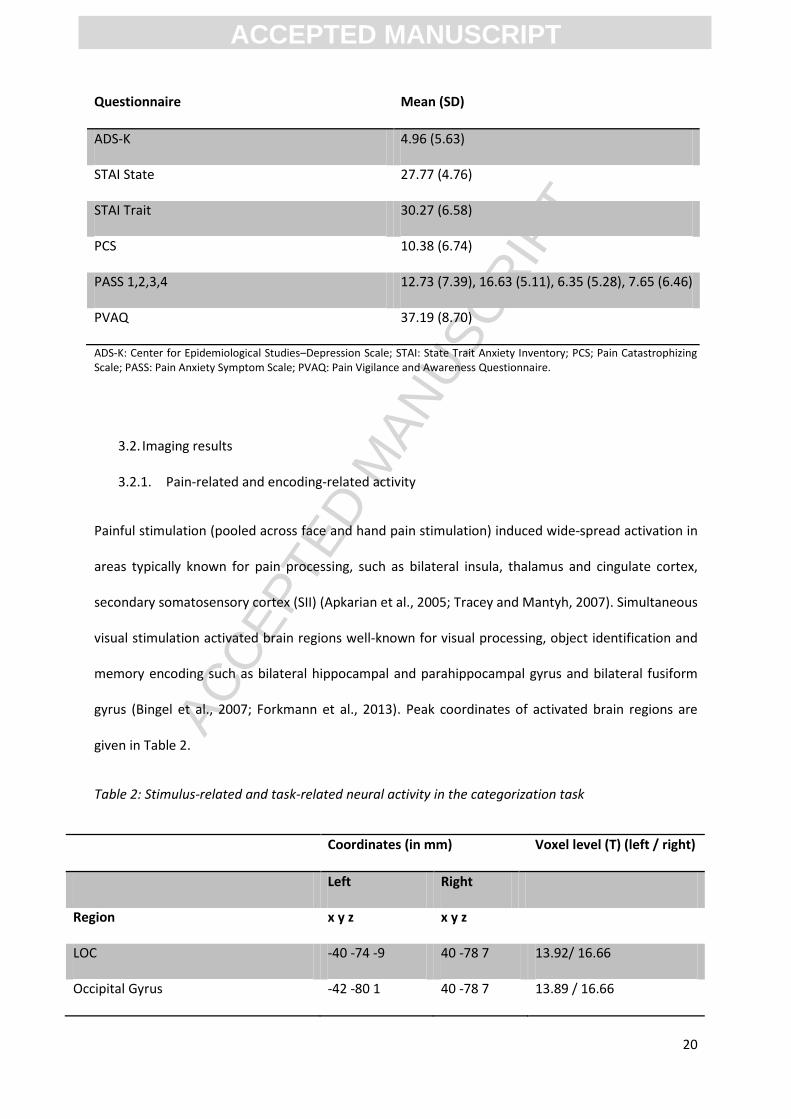
3.1.2.4. Psychological Questionnaires
Questionnaire data (M ± SD) are given in Table 1. None of the calculated correlations survived
correction for multiple comparisons.
Table 1: Questionnaire data (mean and standard deviation (SD)).
ACC
EPTE
D M
ANU
SCR
IPT
ACCEPTED MANUSCRIPT
20
Questionnaire Mean (SD)
ADS-K 4.96 (5.63)
STAI State 27.77 (4.76)
STAI Trait 30.27 (6.58)
PCS 10.38 (6.74)
PASS 1,2,3,4 12.73 (7.39), 16.63 (5.11), 6.35 (5.28), 7.65 (6.46)
PVAQ 37.19 (8.70)
ADS-K: Center for Epidemiological Studies–Depression Scale; STAI: State Trait Anxiety Inventory; PCS; Pain Catastrophizing Scale; PASS: Pain Anxiety Symptom Scale; PVAQ: Pain Vigilance and Awareness Questionnaire.
3.2. Imaging results
3.2.1. Pain-related and encoding-related activity
Painful stimulation (pooled across face and hand pain stimulation) induced wide-spread activation in
areas typically known for pain processing, such as bilateral insula, thalamus and cingulate cortex,
secondary somatosensory cortex (SII) (Apkarian et al., 2005; Tracey and Mantyh, 2007). Simultaneous
visual stimulation activated brain regions well-known for visual processing, object identification and
memory encoding such as bilateral hippocampal and parahippocampal gyrus and bilateral fusiform
gyrus (Bingel et al., 2007; Forkmann et al., 2013). Peak coordinates of activated brain regions are
given in Table 2.
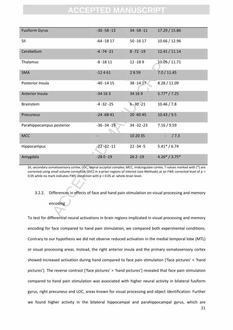
Table 2: Stimulus-related and task-related neural activity in the categorization task
Coordinates (in mm) Voxel level (T) (left / right)
Left Right
Region x y z x y z
LOC -40 -74 -9 40 -78 7 13.92/ 16.66
Occipital Gyrus -42 -80 1 40 -78 7 13.89 / 16.66
ACC
EPTE
D M
ANU
SCR
IPT
ACCEPTED MANUSCRIPT
21
Fusiform Gyrus -30 -58 -13 34 -58 -11 17.29 / 15.86
SII -64 -18 17 50 -16 17 10.66 / 12.96
Cerebellum -4 -74 -21 8 -72 -19 12.41 / 11.14
Thalamus -8 -18 11 12 -18 9 10.05 / 11.71
SMA -12 4 61 2 8 59 7.0 / 11.45
Posterior Insula -40 -14 15 38 -14 17 8.28 / 11.09
Anterior Insula -34 16 3 34 16 9 5.77* / 7.25
Brainstem -4 -32 -25 6 -30 -21 10.46 / 7.8
Precuneus -24 -68 41 20 -60 45 10.43 / 9.5
Parahippocampus posterior -36 -34 -19 34 -32 -23 7.16 / 9.59
MCC - 10 20 35 - / 7.3
Hippocampus -22 -32 -11 22 -34 -5 5.41* / 6.74
Amygdala -28 0 -19 26 2 -19 4.26* / 3.75*
SII, secondary somatosensory cortex; LOC, lateral occipital complex; MCC, midcingulate cortex. T-values marked with (*) are corrected using small volume correction (SVC) in a-priori regions of interest (see Methods) at an FWE corrected level of p < 0.05 while no mark indicates FWE correction with p < 0.05 at whole-brain level.
3.2.2. Differences in effects of face and hand pain stimulation on visual processing and memory
encoding
To test for differential neural activations in brain regions implicated in visual processing and memory
encoding for face compared to hand pain stimulation, we compared both experimental conditions.
Contrary to our hypothesis we did not observe reduced activation in the medial temporal lobe (MTL)
or visual processing areas. Instead, the right anterior insula and the primary somatosensory cortex
showed increased activation during hand compared to face pain stimulation [‘face pictures’ < ‘hand
pictures’]. The reverse contrast [‘face pictures’ > ‘hand pictures’] revealed that face pain stimulation
compared to hand pain stimulation was associated with higher neural activity in bilateral fusiform
gyrus, right precuneus and LOC, areas known for visual processing and object identification. Further
we found higher activity in the bilateral hippocampal and parahippocampal gyrus, which are
ACC
EPTE
D M
ANU
SCR
IPT
ACCEPTED MANUSCRIPT
22
implicated in memory encoding and in the bilateral amygdala, which is well known for the processing
of fear and emotional stimuli, and the left thalamus. Peak coordinates and t-values are given in Table
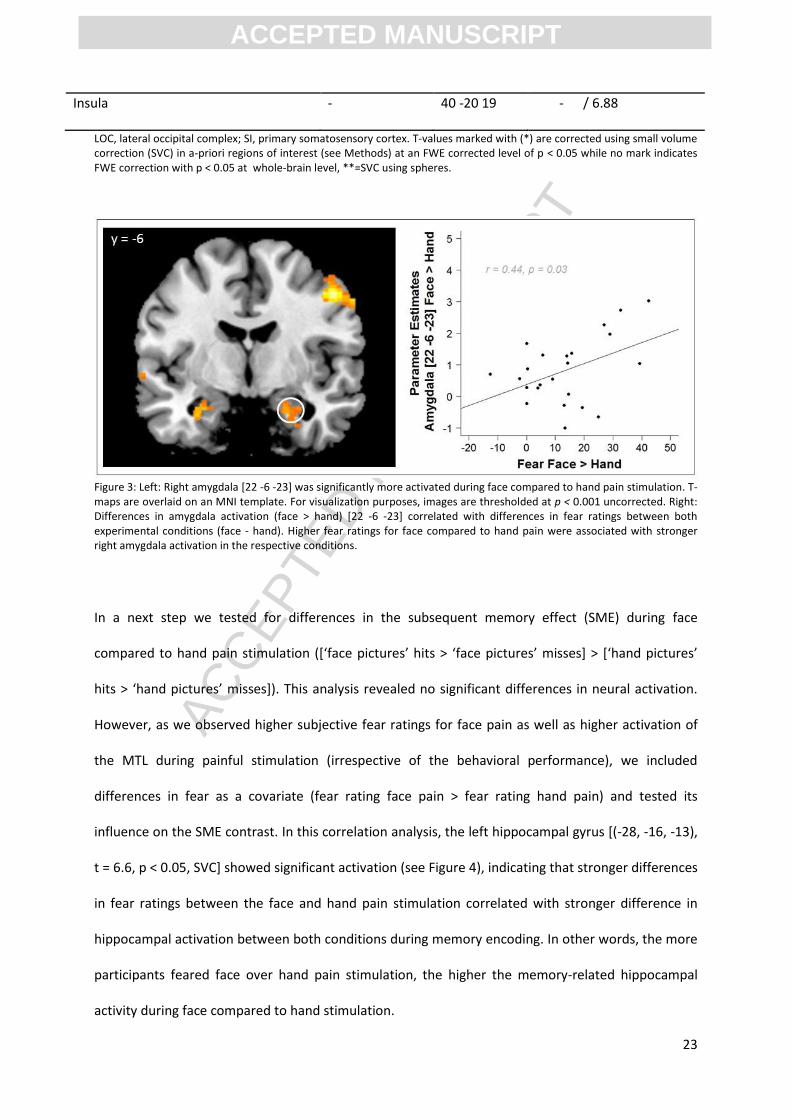
3. To further investigate the influence of fear and the higher amygdala activation for face pain we
extracted parameter estimates from amygdala peak coordinates *‘face pictures’ > ‘hand pictures’+
with 5mm radius spheres using the Marsbar toolbox in Matlab (mathworks.com) and performed
correlation analyses. Across subjects, activation in the right amygdala showed a significant
correlation with subjective differences in fear ratings between face and hand pain condition ([22 -6 -
23], r = 0.44, p = 0.03, see Figure 3) indicating increased amygdala activation for subjects perceiving
face stimulation as more threatening than hand stimulation. Correlation analyses with the left
amygdala peak coordinate revealed no significant results ([-26 -6 -23], r = 0.30, p = 0.16).
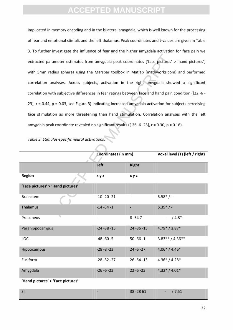
Table 3: Stimulus-specific neural activations.
Coordinates (in mm) Voxel level (T) (left / right)
Left Right
Region x y z x y z
‘Face pictures’ > ‘Hand pictures’
Brainstem -10 -20 -21 - 5.58* / -
Thalamus -14 -34 -1 - 5.39* / -
Precuneus - 8 -54 7 - / 4.8*
Parahippocampus -24 -38 -15 24 -36 -15 4.79* / 3.87*
LOC -48 -60 -5 50 -66 -1 3.83** / 4.36**
Hippocampus -28 -8 -23 24 -6 -27 4.06* / 4.46*
Fusiform -28 -32 -27 26 -54 -13 4.36* / 4.28*
Amygdala -26 -6 -23 22 -6 -23 4.32* / 4.01*
‘Hand pictures’ > ‘Face pictures’
SI - 38 -28 61 - / 7.51
ACC
EPTE
D M
ANU
SCR
IPT
ACCEPTED MANUSCRIPT
23
Insula - 40 -20 19 - / 6.88
LOC, lateral occipital complex; SI, primary somatosensory cortex. T-values marked with (*) are corrected using small volume correction (SVC) in a-priori regions of interest (see Methods) at an FWE corrected level of p < 0.05 while no mark indicates FWE correction with p < 0.05 at whole-brain level, **=SVC using spheres.
Figure 3: Left: Right amygdala [22 -6 -23] was significantly more activated during face compared to hand pain stimulation. T-maps are overlaid on an MNI template. For visualization purposes, images are thresholded at p < 0.001 uncorrected. Right: Differences in amygdala activation (face > hand) [22 -6 -23] correlated with differences in fear ratings between both experimental conditions (face - hand). Higher fear ratings for face compared to hand pain were associated with stronger right amygdala activation in the respective conditions.
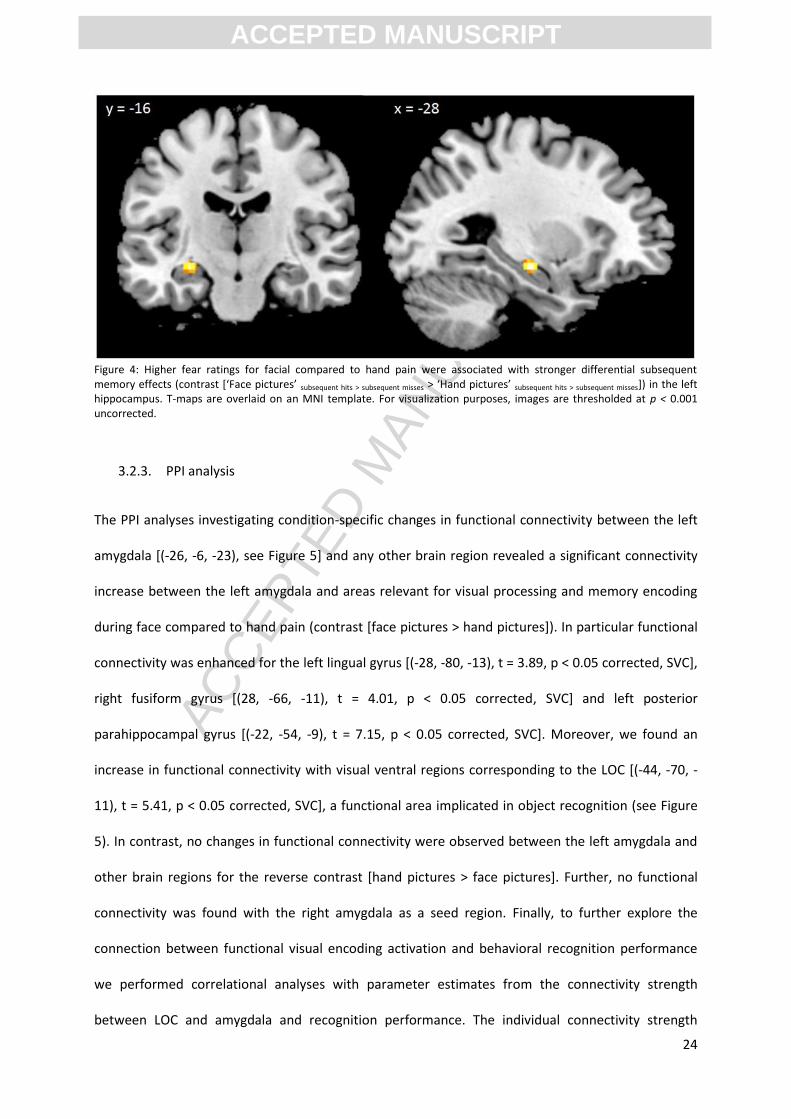
In a next step we tested for differences in the subsequent memory effect (SME) during face
compared to hand pain stimulation ([‘face pictures’ hits > ‘face pictures’ misses] > [‘hand pictures’
hits > ‘hand pictures’ misses]). This analysis revealed no significant differences in neural activation.
However, as we observed higher subjective fear ratings for face pain as well as higher activation of
the MTL during painful stimulation (irrespective of the behavioral performance), we included
differences in fear as a covariate (fear rating face pain > fear rating hand pain) and tested its
influence on the SME contrast. In this correlation analysis, the left hippocampal gyrus [(-28, -16, -13),
t = 6.6, p < 0.05, SVC] showed significant activation (see Figure 4), indicating that stronger differences
in fear ratings between the face and hand pain stimulation correlated with stronger difference in
hippocampal activation between both conditions during memory encoding. In other words, the more
participants feared face over hand pain stimulation, the higher the memory-related hippocampal
activity during face compared to hand stimulation.
ACC
EPTE
D M
ANU
SCR
IPT
ACCEPTED MANUSCRIPT
24
Figure 4: Higher fear ratings for facial compared to hand pain were associated with stronger differential subsequent memory effects (contrast *‘Face pictures’ subsequent hits > subsequent misses > ‘Hand pictures’ subsequent hits > subsequent misses]) in the left hippocampus. T-maps are overlaid on an MNI template. For visualization purposes, images are thresholded at p < 0.001 uncorrected.
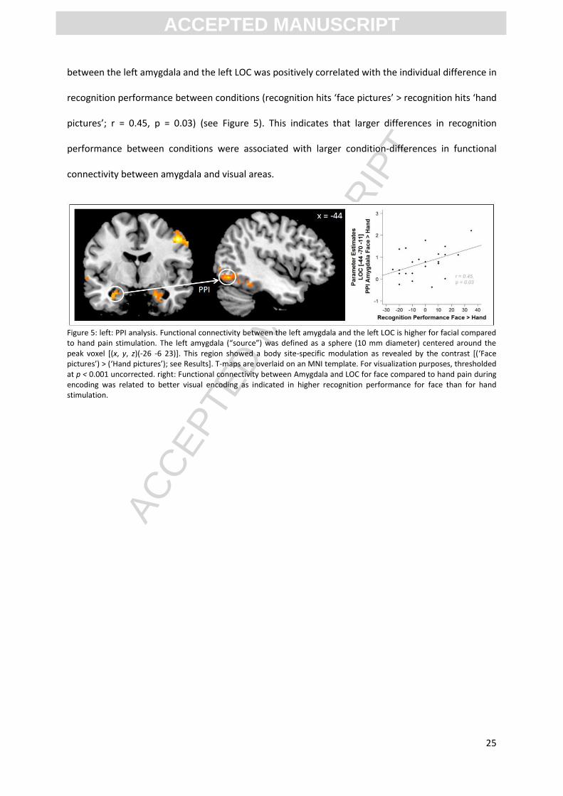
3.2.3. PPI analysis
The PPI analyses investigating condition-specific changes in functional connectivity between the left
amygdala [(-26, -6, -23), see Figure 5] and any other brain region revealed a significant connectivity
increase between the left amygdala and areas relevant for visual processing and memory encoding
during face compared to hand pain (contrast [face pictures > hand pictures]). In particular functional
connectivity was enhanced for the left lingual gyrus [(-28, -80, -13), t = 3.89, p < 0.05 corrected, SVC],
right fusiform gyrus [(28, -66, -11), t = 4.01, p < 0.05 corrected, SVC] and left posterior
parahippocampal gyrus [(-22, -54, -9), t = 7.15, p < 0.05 corrected, SVC]. Moreover, we found an
increase in functional connectivity with visual ventral regions corresponding to the LOC [(-44, -70, -
11), t = 5.41, p < 0.05 corrected, SVC], a functional area implicated in object recognition (see Figure
5). In contrast, no changes in functional connectivity were observed between the left amygdala and
other brain regions for the reverse contrast [hand pictures > face pictures]. Further, no functional
connectivity was found with the right amygdala as a seed region. Finally, to further explore the
connection between functional visual encoding activation and behavioral recognition performance
we performed correlational analyses with parameter estimates from the connectivity strength
between LOC and amygdala and recognition performance. The individual connectivity strength
ACC
EPTE
D M
ANU
SCR
IPT
ACCEPTED MANUSCRIPT
25
between the left amygdala and the left LOC was positively correlated with the individual difference in
recognition performance between conditions (recognition hits ‘face pictures’ > recognition hits ‘hand
pictures’; r = 0.45, p = 0.03) (see Figure 5). This indicates that larger differences in recognition
performance between conditions were associated with larger condition-differences in functional
connectivity between amygdala and visual areas.
Figure 5: left: PPI analysis. Functional connectivity between the left amygdala and the left LOC is higher for facial compared to hand pain stimulation. The left amygdala (“source”) was defined as a sphere (10 mm diameter) centered around the peak voxel [(x, y, z)(-26 -6 23)]. This region showed a body site-specific modulation as revealed by the contrast [(‘Face pictures’) > (‘Hand pictures’); see Results]. T-maps are overlaid on an MNI template. For visualization purposes, thresholded at p < 0.001 uncorrected. right: Functional connectivity between Amygdala and LOC for face compared to hand pain during encoding was related to better visual encoding as indicated in higher recognition performance for face than for hand stimulation.
ACC
EPTE
D M
ANU
SCR
IPT
ACCEPTED MANUSCRIPT
26
4. Discussion
Here we investigated the effects of face compared to hand pain on visual processing, memory
encoding and recognition in young healthy volunteers. As expected, we found higher fear ratings for
face compared to hand pain. On the neural level this result was supported by higher amygdala
activation for the face compared to hand pain condition that scaled with the difference in perceived
pain-related fear. However, contrary to our hypothesis, face pain did not lead to compromised
categorization and recognition performance and did not decrease neural activity in visual and
memory encoding-related areas compared to hand pain. Instead, face pain led to increased brain
activation in areas involved in visual processing (i.e. LOC) and memory encoding in the MTL (i.e.
hippocampus and parahippocampus). Furthermore, hippocampal activity related to greater
recognition performance during face compared to hand stimulation was associated with condition-
specific differences in pain-related fear. Finally, we found enhanced functional connectivity between
the left amygdala and task-relevant areas, such as the LOC, for face compared to hand pain which
was related to less pain-induced recognition impairment during face stimulation. The larger cortical
activation in the primary somatosensory cortex (and insula) for hand pain compared to facial pain is
in line with the known larger cortical representation of the hand area (Penfield and Boldrey, 1937),
compared to the forehead.
4.1. Compensatory neural activation for face pain pictures and behavioral performance
Our behavioral pilot study results had suggested that the face stimulation disturbs visual
categorization more than hand pain. We therefore expected similar behavioral results in our fMRI
study. However, we found no behavioral differences between both experimental conditions, neither
in the categorization nor in the surprise recognition task. Possible explanations for this absence of
behavioral differences might be higher arousal levels in the scanner environment or the overall very
high correct categorization rates, which implies that the task might have been easy to perform, even
in the presence of pain. However, the fact that the same experimental paradigm has been used in a
ACC
EPTE
D M
ANU
SCR
IPT
ACCEPTED MANUSCRIPT
27
previous study that showed a robust interruptive effect of pain on visual categorization, memory
encoding and recognition renders the latter explanation unlikely (Forkmann et al., 2013). Although
the stimulation site had no effect on task performance, we found an interesting difference in the way
participants perceived the two conditions on the emotional level. Across participants, the face
stimulation was perceived as more fear-inducing than the hand stimulation. This finding is
remarkable, particularly in combination with our second unexpected finding of increased instead of
decreased neural activity in task-related and memory encoding-related brain areas, such as the LOC
and sub-regions of the MTL (i.e. hippocampus), for pictures presented with face pain compared to
those presented with hand pain. According to the attentional control theory of anxiety (Eysenck et
al., 2007), compensatory strategies such as enhanced resource usage or increased effort are
activated to counter anxiety-induced performance impairment. Although speculative at this stage,
we suggest with reference to this theoretical framework that the increase in neural activity in the
face pain condition might reflect fear-induced compensatory activation of task-related brain regions
to maintain cognitive performance. The attentional control theory of anxiety has been supported by
various studies (Derakshan et al., 2009; Johnson and Gronlund, 2009; Edwards et al., 2015). For
instance, Ansari and Derakshan (2011) showed increased cognitive effort and neural activation in
high-anxious subjects in an anti- and prosaccade task. Compensatory neural activation and strategies
are also known from neurological disorders and aging. Gour et al. (2011) reported compensatory
brain activity in the anterior temporal network of patients with mild cognitive impairment and
Alzheimer’s disease compared to healthy participants. Anterior temporal network activation
(including hippocampus and amygdala) was enhanced and resting state connectivity within this
network correlated positively with memory performance. Patients with idiopathic Parkinson’s
disease have shown higher neural activation compared to healthy elderly subjects in a working
memory task (Caminiti et al., 2015) and high-performing compared to low-performing older adults
demonstrate a reorganization of neurocognitive networks (Cabeza et al., 2002).
4.2. Fear, memory encoding and the amygdala
ACC
EPTE
D M
ANU
SCR
IPT
ACCEPTED MANUSCRIPT
28
As mentioned in the previous paragraph, pain-related fear ratings were significantly higher for face
pain compared to hand pain. This is in line with findings from our behavioral pilot study and a recent
study investigating increased sensitization for face compared to hand pain (Schmidt et al., 2015). This
higher threat value of face pain might be explained by the exceptional biological relevance of pain
located in the head and facial area which house vital functions such as breathing and food intake. In
support of this notion, Sambo et al. (2012) reported an increased defensive response (i.e. the hand-
blink-reflex) when a painfully stimulated hand was positioned near the face as compared to a
position further away from the face. It is conceivable that unspecific factors such as unpleasantness
or arousal associated with the nociceptive information, which were not assessed in our study, might
have contributed to our results. Future studies should explore the influence of these factors to the
differential effects of facial compared to hand pain on visual processing.
The right amygdala showed fear-related activation that was increased for face compared to hand
pain. This brain area plays an important role in emotional processing and memory encoding
improvement of emotional and fear-related events and threat-related stimuli (LeDoux, 2003; Dolcos
et al., 2004; Phelps, 2004). In line with our findings of increased activity in the amygdala and visual
areas possibly due to higher fear for face pain pictures, Ousdal et al. (2014) reported increased
activation and functional connectivity between amygdala and visual areas in participants with higher
state anxiety. Analyses of the subsequent memory effect (i.e., brain activation during encoding
[recognition hits > recognition misses]) revealed that hippocampal activity related to greater
recognition performance during face compared to hand stimulation was positively correlated with
condition-specific differences in pain-related fear. This result again underlines the influence of threat
on cognitive performance and provides further evidence for fear-driven compensatory neural
activation to achieve stable behavioral performance, since we observed no differences in recognition
performance between both conditions.
In addition, we found enhanced functional connectivity between the left amygdala and the LOC,
lingual, parahippocampal and fusiform gyrus for pictures that were presented with face pain
ACC
EPTE
D M
ANU
SCR
IPT
ACCEPTED MANUSCRIPT
29
compared to hand pain, all areas known to be involved visual processing, memory encoding and
object recognition (Machielsen et al., 2000; Bingel et al., 2007; Forkmann et al., 2013). The LOC was
defined based on coordinates reported by Bingel et al. (2007), who determined this area specifically
for object recognition as they found a main effect of object visibility in their task. With reference to
this previous finding and our observation in the present study we suggest that the increased
functional connectivity between amygdala and LOC contributes to successful memory encoding of
face compared to hand pain pictures. In line with this assumption, we found a significant positive
correlation between activation in the LOC and behavioral performance in the recognition task. More
specifically, we observed higher recognition rates with higher functional connectivity between
amygdala and LOC for face compared to hand pain pictures. Although we found no difference in
recognition accuracy at the behavioral level between both conditions, recent studies using the same
recognition task showed stable effects of experimentally induced pain on recognition accuracy
(Bingel et al., 2007; Forkmann et al., 2013). In support of the idea of fear-driven enhanced functional
connectivity in our study, there are studies showing that functional connectivity between task-
related areas is enhanced for fearful events. Furl et al. (2013) discussed functional connectivity
between amygdala and task-relevant areas, such as the fusiform gyrus, in processing of fearful faces.
A similar influence of the amygdala has been reported in other studies (Morris et al., 1998;
Vuilleumier et al., 2004; Hadj-Bouziane et al., 2012). Furl et al. (2013) concluded that the amygdala
via fear modulates the fusiform gyrus by increasing functional connectivity between both areas.
These interpretations should be seen in light of the fact that the PPI analysis was exploratory in
nature and that our two key amygdala findings were not consistent as the correlation between
condition-specific differences in BOLD activity and pain-related fear was observed in the right
amygdala whereas the condition-related differences in connectivity with task-related areas was
found in the left amygdala. However, we feel that these different pieces of evidence lend support to
the notion that pain-related fear and the engagement of fear-related brain areas such as the
amygdala might be important in the context of facial pain processing.

ACC
EPTE
D M
ANU
SCR
IPT
ACCEPTED MANUSCRIPT
30
5. Conclusion
In this study we show increased neural activity during visual object categorization in the presence of
face compared to hand pain in brain areas involved in visual processing (LOC) and encoding (e.g.
hippocampus) but no difference in behavioral performance. Further, the increased fear for face pain
influenced memory encoding at the neural level and functional connectivity with visual processing
areas. These findings might be interpreted as fear-driven compensatory resource activation. The role
of increased fear (sensitivity) for pain-induced memory disturbances in patients suffering from
recurrent or chronic headache or facial pain warrants further investigation.
Acknowledgements:
The authors declare no competing financial interests. This work was supported by the German
Research Foundation (DFG, SFB 90929-936), EFIC-Grünenthal Research Grant to KF and the Federal
Ministry of Education and Research (01GQ0808). The authors wish to thank Matthias Zunhammer for
helpful comments on this manuscript and Dirk Neumann and Julia Schmid for their help with data
collection.

ACC
EPTE
D M
ANU
SCR
IPT
ACCEPTED MANUSCRIPT
31
6. References
Ansari TL, Derakshan N (2011) The neural correlates of cognitive effort in anxiety: effects on processing efficiency. Biol Psychol 86:337-348.
Apkarian AV, Bushnell MC, Treede RD, Zubieta JK (2005) Human brain mechanisms of pain perception and regulation in health and disease. European journal of pain (London, England) 9:463-484.
Bingel U, Rose M, Gläscher J, Büchel C (2007) fMRI reveals how pain modulates visual object processing in the ventral visual stream. Neuron 55:157-167.
Bishop SJ, Duncan J, Lawrence AD (2004) State anxiety modulation of the amygdala response to unattended threat-related stimuli. The Journal of neuroscience : the official journal of the Society for Neuroscience 24:10364-10368.
Brewer JB, Zhao Z, Desmond JE, Glover GH, Gabrieli JD (1998) Making memories: brain activity that predicts how well visual experience will be remembered. Science (New York, NY) 281:1185-1187.
Buhle J, Wager TD (2010) Performance-dependent inhibition of pain by an executive working memory task. Pain 149:19-26.
Cabeza R, Anderson ND, Locantore JK, McIntosh AR (2002) Aging gracefully: compensatory brain activity in high-performing older adults. NeuroImage 17:1394-1402.
Caminiti SP, Siri C, Guidi L, Antonini A, Perani D (2015) The neural correlates of spatial and object working memory in elderly and Parkinson's disease subjects. Behav Neurol 2015:123636.
Crombez G, Baeyens F, Eelen P (1994) Sensory and temporal information about impending pain: the influence of predictability on pain. Behav Res Ther 32:611-622.
Crombez G, Eccleston C, Baeyens F, Eelen P (1998) When somatic information threatens, catastrophic thinking enhances attentional interference. Pain 75:187-198.
Derakshan N, Ansari TL, Hansard M, Shoker L, Eysenck MW (2009) Anxiety, inhibition, efficiency, and effectiveness. An investigation using antisaccade task. Exp Psychol 56:48-55.
Dolcos F, LaBar KS, Cabeza R (2004) Interaction between the amygdala and the medial temporal lobe memory system predicts better memory for emotional events. Neuron 42:855-863.
Eccleston C (1995) Chronic pain and distraction: an experimental investigation into the role of sustained and shifting attention in the processing of chronic persistent pain. Behav Res Ther 33:391-405.
Eccleston C, Crombez G (1999) Pain demands attention: a cognitive-affective model of the interruptive function of pain. Psychol Bull 125:356-366.
Edwards MS, Moore P, Champion JC, Edwards EJ (2015) Effects of trait anxiety and situational stress on attentional shifting are buffered by working memory capacity. Anxiety Stress Coping 28:1-16.
Eysenck MW, Derakshan N, Santos R, Calvo MG (2007) Anxiety and cognitive performance: attentional control theory. Emotion 7:336-353.
Forkmann K, Wiech K, Ritter C, Sommer T, Rose M, Bingel U (2013) Pain-specific modulation of hippocampal activity and functional connectivity during visual encoding. The Journal of neuroscience: the official journal of the Society for Neuroscience 33:2571-2581.
Friston KJ, Holmes AP, Worsley KJ (1999) How many subjects constitute a study? NeuroImage 10:1-5. Friston KJ, Buechel C, Fink GR, Morris J, Rolls E, Dolan RJ (1997) Psychophysiological and modulatory
interactions in neuroimaging. NeuroImage 6:218-229. Furl N, Henson RN, Friston KJ, Calder AJ (2013) Top-down control of visual responses to fear by the
amygdala. The Journal of neuroscience : the official journal of the Society for Neuroscience 33:17435-17443.
Gescheider G (1997) Psychophysics: the fundamentals. Lawrence Erlbaum Associates. Gour N, Ranjeva JP, Ceccaldi M, Confort-Gouny S, Barbeau E, Soulier E, Guye M, Didic M, Felician O
(2011) Basal functional connectivity within the anterior temporal network is associated with performance on declarative memory tasks. NeuroImage 58:687-697.

ACC
EPTE
D M
ANU
SCR
IPT
ACCEPTED MANUSCRIPT
32
Grisart J, Van der Linden M, Bastin C (2007) The contribution of recollection and familiarity to recognition memory performance in chronic pain patients. Behav Res Ther 45:1077-1084.
Hadj-Bouziane F, Liu N, Bell AH, Gothard KM, Luh WM, Tootell RB, Murray EA, Ungerleider LG (2012) Amygdala lesions disrupt modulation of functional MRI activity evoked by facial expression in the monkey inferior temporal cortex. Proceedings of the National Academy of Sciences of the United States of America 109:E3640-3648.
Hautzinger M, Bailer, M. (1993) Allgemeine Depressionsskala. Weinheim: Beltz. Johnson DR, Gronlund SD (2009) Individuals lower in working memory capacity are particularly
vulnerable to anxiety's disruptive effect on performance. Anxiety Stress Coping 22:201-213. Kim H (2011) Neural activity that predicts subsequent memory and forgetting: a meta-analysis of 74
fMRI studies. NeuroImage 54:2446-2461. Kuhajda MC, Thorn BE, Klinger MR, Rubin NJ (2002) The effect of headache pain on attention
(encoding) and memory (recognition). Pain 97:213-221. Lautenbacher S, Huber C, Kunz M, Parthum A, Weber PG, Griessinger N, Sittl R (2009) Hypervigilance
as predictor of postoperative acute pain: its predictive potency compared with experimental pain sensitivity, cortisol reactivity, and affective state. The Clinical journal of pain 25:92-100.
Laux L, Glanzmann, P., Schaffner, P., Spielberger, C. D. (1992) Das State-Trait-Angstinventar (Testmappe mit Handanweisung, Fragebogen STAI-G Form X1 und Fragebogen STAI-G Form X2). Weinheim: Beltz.
LeDoux J (2003) The emotional brain, fear, and the amygdala. Cell Mol Neurobiol 23:727-738. Machielsen WC, Rombouts SA, Barkhof F, Scheltens P, Witter MP (2000) FMRI of visual encoding:
reproducibility of activation. Human brain mapping 9:156-164. McCracken LM (1997) “Attention” to pain in persons with chronic pain: A behavioral approach.
Behavior Therapy 28:271-284. McCracken LM, Zayfert C, Gross RT (1992) The Pain Anxiety Symptoms Scale: development and
validation of a scale to measure fear of pain. Pain 50:67-73. Meier ML, de Matos NM, Brugger M, Ettlin DA, Lukic N, Cheetham M, Jancke L, Lutz K (2014) Equal
pain-Unequal fear response: enhanced susceptibility of tooth pain to fear conditioning. Front Hum Neurosci 8:526.
Moore DJ, Keogh E, Eccleston C (2012) The interruptive effect of pain on attention. Q J Exp Psychol (Hove) 65:565-586.
Morris JS, Friston KJ, Buchel C, Frith CD, Young AW, Calder AJ, Dolan RJ (1998) A neuromodulatory role for the human amygdala in processing emotional facial expressions. Brain 121 ( Pt 1):47-57.
Oosterman JM, Derksen LC, van Wijck AJ, Veldhuijzen DS, Kessels RP (2011) Memory functions in chronic pain: examining contributions of attention and age to test performance. The Clinical journal of pain 27:70-75.
Ousdal OT, Andreassen OA, Server A, Jensen J (2014) Increased amygdala and visual cortex activity and functional connectivity towards stimulus novelty is associated with state anxiety. PloS one 9:e96146.
Penfield W, Boldrey E (1937) Somatic motor and sensory representation in the cerebral cortex of man as studied by electrical stimulation. Brain 60:389-443.
Peters ML, Vlaeyen JW, Kunnen AM (2002) Is pain-related fear a predictor of somatosensory hypervigilance in chronic low back pain patients? Behav Res Ther 40:85-103.
Phelps EA (2004) Human emotion and memory: interactions of the amygdala and hippocampal complex. Curr Opin Neurobiol 14:198-202.
Radloff LS (1977) The CES-D Scale: A Self-Report Depression Scale for Research in the General Population. Applied Psychological Measurement 1:385-401.
Rose M, Schmid C, Winzen A, Sommer T, Buchel C (2005) The functional and temporal characteristics of top-down modulation in visual selection. Cerebral cortex (New York, NY : 1991) 15:1290-1298.
ACC
EPTE
D M
ANU
SCR
IPT
ACCEPTED MANUSCRIPT
33
Sambo CF, Liang M, Cruccu G, Iannetti GD (2012) Defensive peripersonal space: the blink reflex evoked by hand stimulation is increased when the hand is near the face. Journal of neurophysiology 107:880-889.
Schmidt K, Schunke O, Forkmann K, Bingel U (2015) Enhanced Short-Term Sensitization of Facial Compared With Limb Heat Pain. J Pain 16:781-790.
Sehlmeyer C, Schoning S, Zwitserlood P, Pfleiderer B, Kircher T, Arolt V, Konrad C (2009) Human fear conditioning and extinction in neuroimaging: a systematic review. PloS one 4:e5865.
Sergerie K, Chochol C, Armony JL (2008) The role of the amygdala in emotional processing: a quantitative meta-analysis of functional neuroimaging studies. Neurosci Biobehav Rev 32:811-830.
Sinke C, Schmidt K, Forkmann K, Bingel U (2015) Phasic and tonic pain differentially impact the interruptive function of pain. PloS one 10:e0118363.
Spielberger CD, Gorssuch, R. L., Lushene, P. R., Vagg, P. R., Jacobs, G. A. (1983) Manual for the state-trait anxiety inventory. Palo Alto, CA: Consulting Psychologists Press Inc.
Stanislaw H, Todorov N (1999) Calculation of signal detection theory measures. Behav Res Methods Instrum Comput 31:137-149.
Sullivan MJL, Bishop SR, Pivik J (1995) The Pain Catastrophizing Scale: Development and validation. Psychological Assessment 7:524-532.
Tiemann L, Schulz E, Gross J, Ploner M (2010) Gamma oscillations as a neuronal correlate of the attentional effects of pain. Pain 150:302-308.
Tracey I, Mantyh PW (2007) The Cerebral Signature for Pain Perception and Its Modulation. Neuron 55:377-391.
Van Damme S, Crombez G, Eccleston C (2004) Disengagement from pain: the role of catastrophic thinking about pain. Pain 107:70-76.
Vancleef LM, Peters ML (2006) Pain catastrophizing, but not injury/illness sensitivity or anxiety sensitivity, enhances attentional interference by pain. J Pain 7:23-30.
Vuilleumier P, Richardson MP, Armony JL, Driver J, Dolan RJ (2004) Distant influences of amygdala lesion on visual cortical activation during emotional face processing. Nat Neurosci 7:1271-1278.
Walter B, Hampe, D., Wild, J., Vaitl, D. (2002) Die Erfassung der Angst vor Schmerzen: Eine modifizierte deutsche Version der Pain Anxiety Symptoms Scale (PASS-D). Der Schmerz 16.

ACC
EPTE
D M
ANU
SCR
IPT
ACCEPTED MANUSCRIPT
34
Highlights - Facial pain elicits higher fear, which is assumed to enhance pain-related cognitive disruption. - Facial pain led to increased neural activation in visual and memory-related areas. - Functional connectivity was enhanced between amygdala and LOC during facial pain. - This suggests fear-related compensatory resource activation during facial pain.