Embed Size (px)
Citation preview
The Development of Tools to Study
Carbohydrate-processing Enzymes
Travis Benjamin Coyle BSc(Hons)
Chemistry
School of Chemistry and Biochemistry
This thesis is presented for the degree of Doctor of Philosophy
of the University of Western Australia
2015

.

Candidate Declaration The work described in this thesis was carried out by the author in the School of Chemistry
and Biochemistry at the University of Western Australia under the supervision of Dr.
Keith A. Stubbs, unless specified otherwise. Unless duly referenced, the work described
is original.
___________________
Travis Benjamin Coyle
April 2015

.
i
Contents Summary iii
Acknowledgements v
Glossary vi
Part I: The Development of Tools to Study Glycosidases
Chapter 1
“On the Development of Glycosidase Inhibitors” 1
Chapter 2
“The Development of Some Novel α-L-Fucosidase Inhibitors” 39
Introduction 41
Results and Discussion 61
Experimental 77
Chapter 3
“The Development of Some Novel α-L-Arabinofuranosidase Inhibitors” 99
Introduction 101
Results and Discussion 111
Experimental 127
Part II: The Development of Tools to Study Glycosyltransferases
Chapter 4
“On the Development of Glycosyltransferase Inhibitors” 149
Chapter 5
“The Development of Some Novel Cholesterol α-glucosyltransferase Inhibitors” 197
Introduction 199
Results and Discussion 211
Experimental 267
References 327

.
ii
iii
Summary
Carbohydrates perform a diverse range of functions in biological systems, including roles
in metabolism, modulating structural integrity, and mediating intra-cellular and extra-
cellular signalling. This range of roles can be performed by carbohydrates because of the
overwhelming structural diversity of these molecules, and a variety of enzymes are
required to process carbohydrates because of this structural diversity. The specific
biological roles of carbohydrate-processing enzymes are of significant interest for a better
understanding of the biological systems in which they operate, and as well, many
carbohydrate-processing enzymes are of interest for the disease states that are associated
with their dysfunction.
This thesis is divided into two parts, which detail various efforts to design tools to study
carbohydrate-processing enzymes and demonstrate some applications of these tools. The
first part of this thesis details investigations into the development of some potential
inhibitors and substrates of glycoside hydrolases and is divided into three chapters.
The first chapter provides a general overview of glycoside hydrolases, and outlines
previous efforts to develop inhibitors of these enzymes.
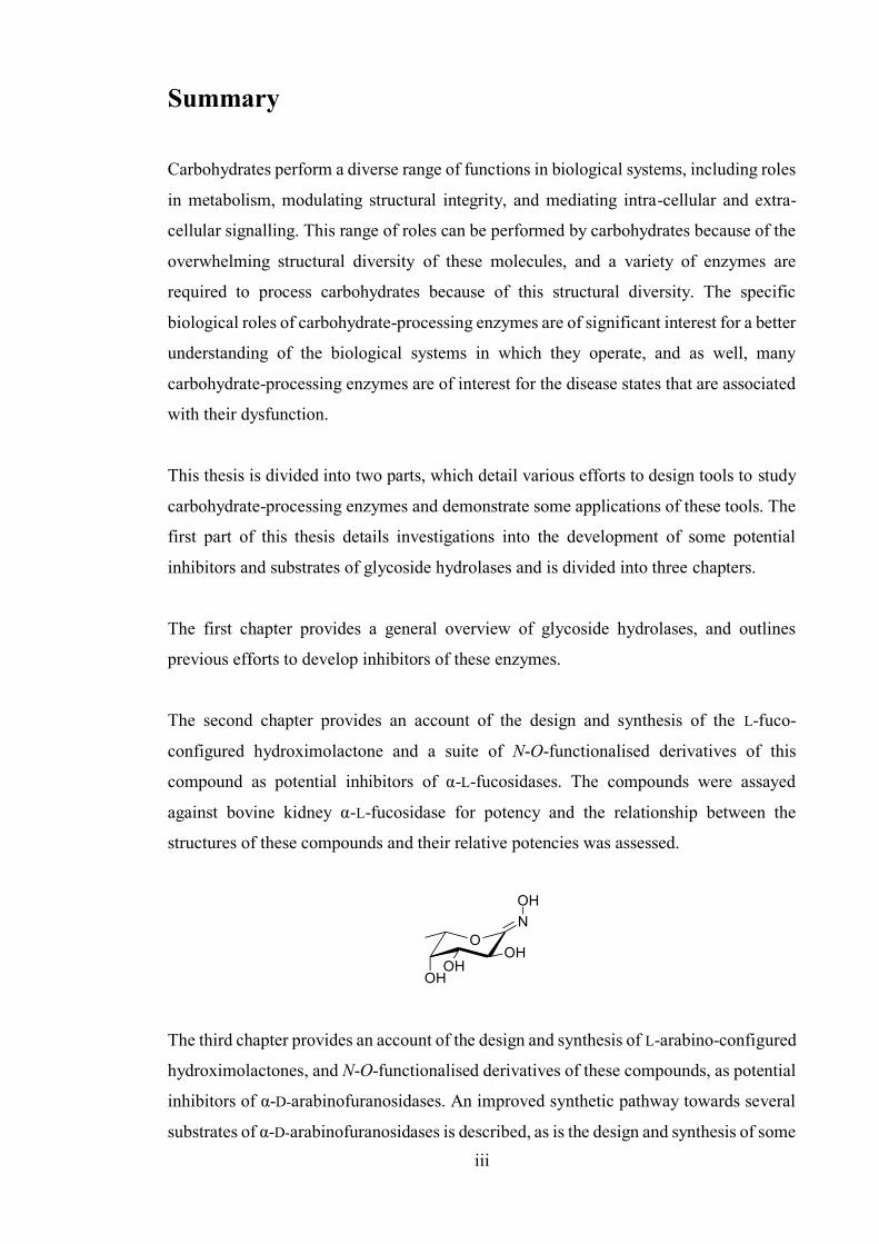
The second chapter provides an account of the design and synthesis of the L-fuco-
configured hydroximolactone and a suite of N-O-functionalised derivatives of this
compound as potential inhibitors of α-L-fucosidases. The compounds were assayed
against bovine kidney α-L-fucosidase for potency and the relationship between the
structures of these compounds and their relative potencies was assessed.
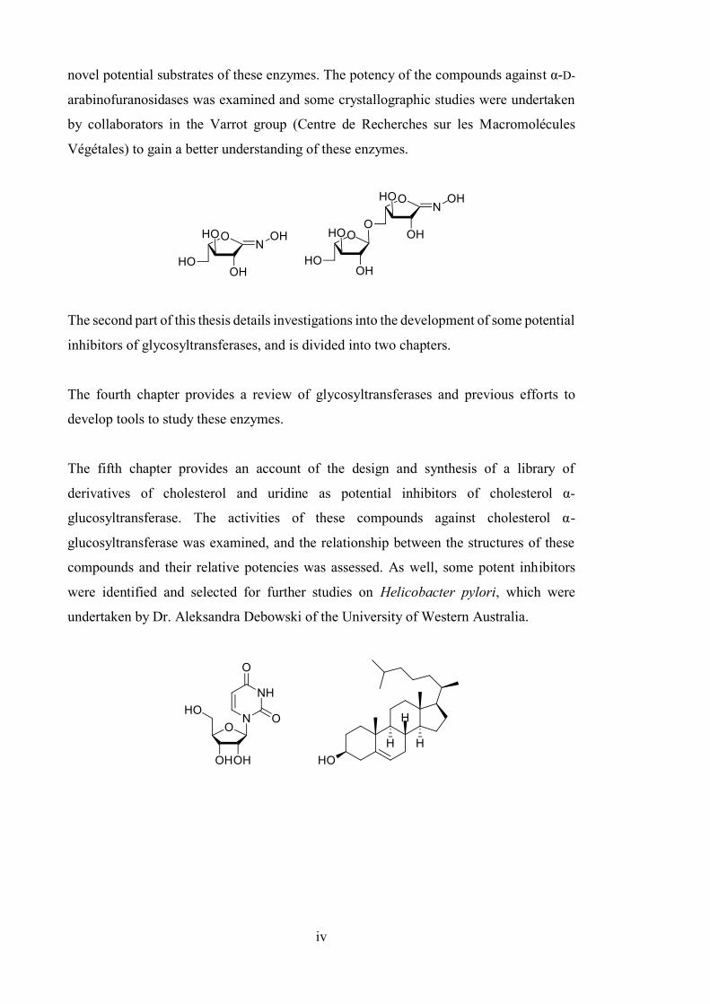
The third chapter provides an account of the design and synthesis of L-arabino-configured
hydroximolactones, and N-O-functionalised derivatives of these compounds, as potential
inhibitors of α-D-arabinofuranosidases. An improved synthetic pathway towards several
substrates of α-D-arabinofuranosidases is described, as is the design and synthesis of some
.
iv
novel potential substrates of these enzymes. The potency of the compounds against α-D-
arabinofuranosidases was examined and some crystallographic studies were undertaken
by collaborators in the Varrot group (Centre de Recherches sur les Macromolécules
Végétales) to gain a better understanding of these enzymes.
The second part of this thesis details investigations into the development of some potential
inhibitors of glycosyltransferases, and is divided into two chapters.
The fourth chapter provides a review of glycosyltransferases and previous efforts to
develop tools to study these enzymes.
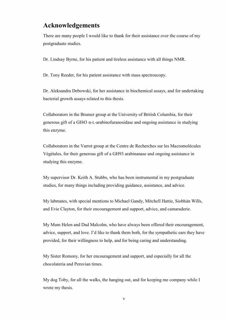
The fifth chapter provides an account of the design and synthesis of a library of
derivatives of cholesterol and uridine as potential inhibitors of cholesterol α-
glucosyltransferase. The activities of these compounds against cholesterol α-
glucosyltransferase was examined, and the relationship between the structures of these
compounds and their relative potencies was assessed. As well, some potent inhibitors
were identified and selected for further studies on Helicobacter pylori, which were
undertaken by Dr. Aleksandra Debowski of the University of Western Australia.
v
Acknowledgements There are many people I would like to thank for their assistance over the course of my
postgraduate studies.
Dr. Lindsay Byrne, for his patient and tireless assistance with all things NMR.
Dr. Tony Reeder, for his patient assistance with mass spectroscopy.
Dr. Aleksandra Debowski, for her assistance in biochemical assays, and for undertaking
bacterial growth assays related to this thesis.
Collaborators in the Brumer group at the University of British Columbia, for their
generous gift of a GH43 α-L-arabinofuranosidase and ongoing assistance in studying
this enzyme.
Collaborators in the Varrot group at the Centre de Recherches sur les Macromolécules
Végétales, for their generous gift of a GH93 arabinanase and ongoing assistance in
studying this enzyme.
My supervisor Dr. Keith A. Stubbs, who has been instrumental in my postgraduate
studies, for many things including providing guidance, assistance, and advice.
My labmates, with special mentions to Michael Gandy, Mitchell Hattie, Siobhán Wills,
and Evie Clayton, for their encouragement and support, advice, and camaraderie.
My Mum Helen and Dad Malcolm, who have always been offered their encouragement,
advice, support, and love. I’d like to thank them both, for the sympathetic ears they have
provided, for their willingness to help, and for being caring and understanding.
My Sister Romony, for her encouragement and support, and especially for all the
chocolateria and Peruvian times.
My dog Toby, for all the walks, the hanging out, and for keeping me company while I
wrote my thesis.
.
vi
My friends, for all the good times. Special mentions to Josh, Jason, Shaked, Dave,
Dane, Shaun, Xiong, Haley, Godwin, Soren and James.
I would also like to acknowledge the financial assistance which I have received in the
form of an Australian Postgraduate Award from the Australian Federal Government,
and a UWA Safety-Net Top-Up Scholarship from the University of Western Australia.
vii
Glossary Ac2O acetic anhydride
AcCl acetyl chloride
AcOH acetic acid
APCI atmospheric pressure chemical ionisation
aq. aqueous
BzCl benzoyl chloride
BTPP (tert-butylimino)tris(pyrrolidino)phosphorane
t-BuOH tert-butyl alcohol
CBM carbohydrate binding module
CG cholesteryl α-glucopyranoside
CGT cholesterol α-glucosyltransferase
conc. concentrated
CuAAC copper-catalysed azide-alkyne cycloaddition
DBU 1,8-diazabicyclo[5.4.0]undec-7-ene
DIPEA diisopropylethylamine
DMAP 4-dimethylaminopyridine
DMF dimethylformamide
DMSO dimethylsulfoxide
DFJ 1-deoxyfuconojirimycin
DMJ 1-deoxymannonojirimycin
DNJ 1-deoxynojirimycin
EDC 1-ethyl-3-(3-dimethylaminopropyl)carbodiimide
ESI electron spray ionisation
equiv. equivalents
GDP guanosine diphosphate
GDP-Man guanosine diphosphate D-mannopyranose
GlcNAc 2-acetamido-2-deoxy-D-glucopyranose
HEWL hen egg-white lysozyme
HIV human immunodeficiency virus
HPLC high-performance liquid chromatography
IR infra-red
MS molecular sieves
MsCl methanesulfonyl chloride
.
viii
NAD nicotinamide adenine dinucleotide
NCS N-chlorosuccinimide
NDP nucleoside diphosphate
NMR nuclear magnetic resonance
Pd/C palladium-on-carbon
iPrOH iso-propyl alcohol
PUL polysaccharide utilisation locus
r.t. room temperature
5SglcNAc 2-acetamido-2-deoxy-5-thio-D-glucopyranose
sat. saturated
THF tetrahydrofuran
TFA trifluoroacetic acid
t.l.c. thin-layer chromatography
TsCl p-toluenesulfonyl chloride
TsOH p-toluenesulfonic acid
UDP uridine diphosphate
UDP-Gal uridine diphosphate D-galactopyranose
UDP-GlcNAc uridine diphosphate 2-acetamido-2-deoxy-D-
- glucopyranose
UDP-5SGlcNAc uridine diphosphate 2-acetamido-2-deoxy-5-thio-D-
- glucopyranose
UTP uridine triphosphate
WT wild-type
1
Part I: The Development of Tools to Study
Glycosidases
Chapter 1
On the Development of Glycosidase Inhibitors

2
Chapter 1
3
Introduction
Carbohydrates perform a variety of essential roles in biological systems. Carbohydrate
polymers form the cell walls of plants and arthropods, and carbohydrates are used for
energy storage within cells.[1] These biomolecules also mediate signalling within a cell
for its growth, development and metastasis, and are involved in signalling between cells,
such as for cellular adhesion and immune responses.[1-2] Myriad polysaccharides coat the
exterior of cells for intercellular signalling and carbohydrates are often attached to
proteins to act as signals within the cell.
Carbohydrates are able to perform such a variety of functions because of the incredibly
complex structures that can be built with them; many monosaccharides have two stable
cyclic forms (pyranose and furanose) and can be linked together through one of several
hydroxyl groups, with different configurations at the anomeric position of each sugar (α,
or β), in different sequences, to form chains which can be branched or linear. Over 70
different disaccharides can be produced from D-glucose alone because each of the stable
pyranose and furanose forms can each be α- or β-configured and a glycosidic linkage with
either α or β configuration can be formed between the anomeric position of a sugar and
any of the five hydroxyl groups present on each anomer of both cyclic forms (Figure
1.1). These cyclic forms interconvert in aqueous solution through the open chain form of
D-glucose 1, which can be represented as a Fischer projection, but this open chain form
is generally only present at very low concentrations in solution. As a result of this
structural complexity, many different types of enzyme are required to produce the
different monosaccharides, combine them and attach them to other biomolecules in
various ways and to remove them. The genes encoding these carbohydrate-processing
enzymes account for 1% of the human genome.[1-2]
Chapter 1 .
4
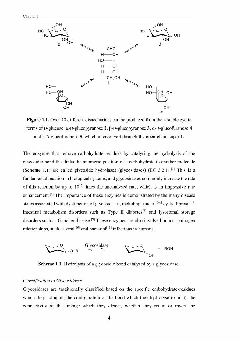
Figure 1.1. Over 70 different disaccharides can be produced from the 4 stable cyclic
forms of D-glucose; α-D-glucopyranose 2, β-D-glucopyranose 3, α-D-glucofuranose 4
and β-D-glucofuranose 5, which interconvert through the open-chain sugar 1.
The enzymes that remove carbohydrate residues by catalysing the hydrolysis of the
glycosidic bond that links the anomeric position of a carbohydrate to another molecule
(Scheme 1.1) are called glycoside hydrolases (glycosidases) (EC 3.2.1).[3] This is a
fundamental reaction in biological systems, and glycosidases commonly increase the rate
of this reaction by up to 1017 times the uncatalysed rate, which is an impressive rate
enhancement.[4] The importance of these enzymes is demonstrated by the many disease
states associated with dysfunction of glycosidases, including cancer,[5-6] cystic fibrosis,[7]
intestinal metabolism disorders such as Type II diabetes[8] and lysosomal storage
disorders such as Gaucher disease.[9] These enzymes are also involved in host-pathogen
relationships, such as viral[10] and bacterial[11] infections in humans.
Scheme 1.1. Hydrolysis of a glycosidic bond catalysed by a glycosidase.
Classification of Glycosidases
Glycosidases are traditionally classified based on the specific carbohydrate-residues
which they act upon, the configuration of the bond which they hydrolyse (α or β), the
connectivity of the linkage which they cleave, whether they retain or invert the
Chapter 1
5
configuration at the anomeric position of the cleaved residue and whether they hydrolyse
a terminal glycosidic bond (exo) or act on a bond in the middle of a chain (endo).[3]
Due to the relationship between the amino acid sequence, the 3-dimensional structure,
and the activity of an enzyme, the activities of glycosidases can be predicted based on
conserved amino acid sequences with known enzymes. The work of Henrissat led to
glycosidases being classified into families based upon amino acid-sequence
homologies,[12] and glycosidases are currently classified into 133 different families.
Comprehensive information about this classification system is catalogued in the CAZy
database,[13] and this sequence-based classification system generally groups glycosidases
with conserved mechanisms together, although within a family there may be glycosidases
which act on different substrates. This is likely to be the result of evolutionary divergence
of enzymes which have a common ancestor and homologous folding.[14]
General Structure and Mechanisms of Glycosidases
Although they exhibit various activities and substrate specificities, most glycosidases are
thought to act through one of a small number of general mechanisms. The general
mechanisms for inverting and retaining glycosidases were first proposed by Koshland in
1953, based upon the mechanisms of equivalent non-enzymatic reactions.[15]
Non-enzymatic substitutions on asymmetric carbons which form a product with inverted
configuration relative to the starting material act through a classic Walden inversion
mechanism. The nucleophilic attack and displacement occur simultaneously in this
single-step reaction and so the attack must occur at the opposite face of the anomeric
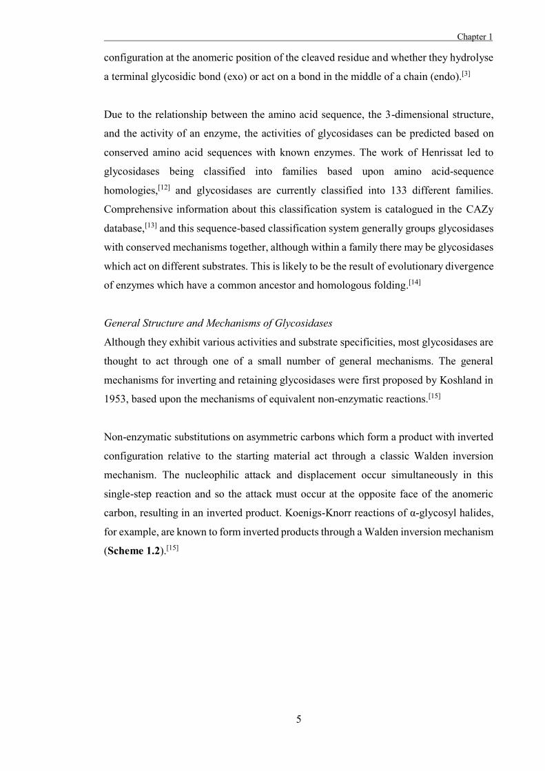
carbon, resulting in an inverted product. Koenigs-Knorr reactions of α-glycosyl halides,
for example, are known to form inverted products through a Walden inversion mechanism
(Scheme 1.2).[15]
Chapter 1 .
6
Scheme 1.2. Walden inversion mechanism for Koenigs-Knorr reactions of glycosyl
halides.
Conversely, 2-O-acetyl glucosyl halides which undergo Koenigs-Knorr reactions retain
the configuration of the starting material. This is because these reactions proceed through
a two-step double Walden-inversion mechanism; in the first step a configurationally
inverted intramolecular intermediate forms from attack of the anomeric position by the
acetoxy oxygen and in the second step this intermediate is displaced by the attack of the
incoming nucleophile, forming a product with the configuration of the starting material
(Scheme 1.3).[15]
Scheme 1.3. Koenigs-Knorr reactions of 2-O-acetyl glucosyl halides pass through an
intramolecular intermediate.
Koshland considered enzymatic reactions where a group is substituted on an asymmetric
carbon with either inversion or retention of the configuration at that position in the context
of these mechanisms.[15] The mechanism proposed by Koshland for inverting
glycosidases was a single step Walden inversion mechanism analogous to that of the
Koenigs-Knorr reactions of α-glucosyl halides, although activation of the leaving group
and the incoming nucleophile are both required in the enzymatic reaction. The glycosidic
oxygen is protonated by an acidic amino acid residue (the catalytic acid), while
simultaneously a water molecule is deprotonated by a basic residue (the catalytic base)
and attacks the anomeric position. This displaces the original aglycon and inverts the
Chapter 1
7
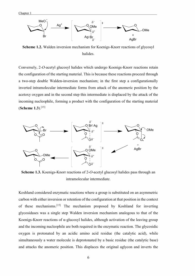
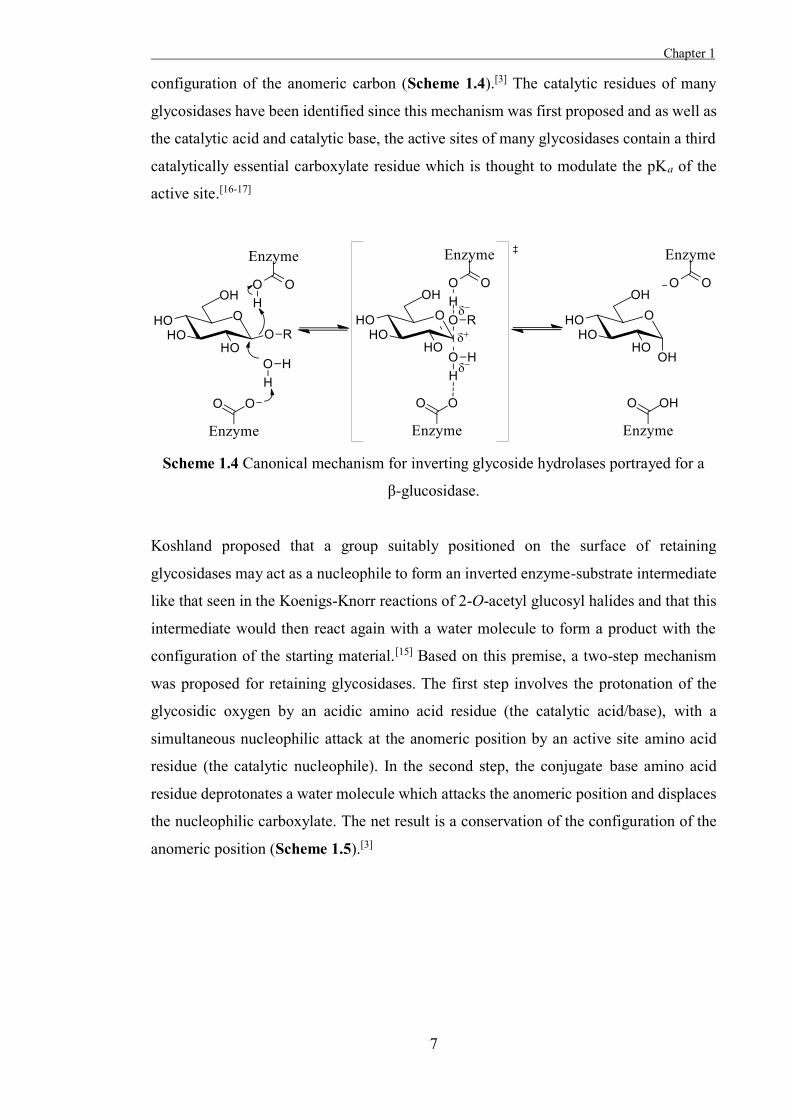
configuration of the anomeric carbon (Scheme 1.4).[3] The catalytic residues of many
glycosidases have been identified since this mechanism was first proposed and as well as
the catalytic acid and catalytic base, the active sites of many glycosidases contain a third
catalytically essential carboxylate residue which is thought to modulate the pKa of the
active site.[16-17]
Scheme 1.4 Canonical mechanism for inverting glycoside hydrolases portrayed for a
β-glucosidase.
Koshland proposed that a group suitably positioned on the surface of retaining
glycosidases may act as a nucleophile to form an inverted enzyme-substrate intermediate
like that seen in the Koenigs-Knorr reactions of 2-O-acetyl glucosyl halides and that this
intermediate would then react again with a water molecule to form a product with the
configuration of the starting material.[15] Based on this premise, a two-step mechanism
was proposed for retaining glycosidases. The first step involves the protonation of the
glycosidic oxygen by an acidic amino acid residue (the catalytic acid/base), with a
simultaneous nucleophilic attack at the anomeric position by an active site amino acid
residue (the catalytic nucleophile). In the second step, the conjugate base amino acid
residue deprotonates a water molecule which attacks the anomeric position and displaces
the nucleophilic carboxylate. The net result is a conservation of the configuration of the
anomeric position (Scheme 1.5).[3]
Chapter 1 .
8
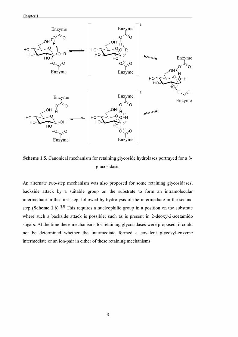
Scheme 1.5. Canonical mechanism for retaining glycoside hydrolases portrayed for a β-
glucosidase.
An alternate two-step mechanism was also proposed for some retaining glycosidases;
backside attack by a suitable group on the substrate to form an intramolecular
intermediate in the first step, followed by hydrolysis of the intermediate in the second
step (Scheme 1.6).[15] This requires a nucleophilic group in a position on the substrate
where such a backside attack is possible, such as is present in 2-deoxy-2-acetamido
sugars. At the time these mechanisms for retaining glycosidases were proposed, it could
not be determined whether the intermediate formed a covalent glycosyl-enzyme
intermediate or an ion-pair in either of these retaining mechanisms.
Chapter 1
9
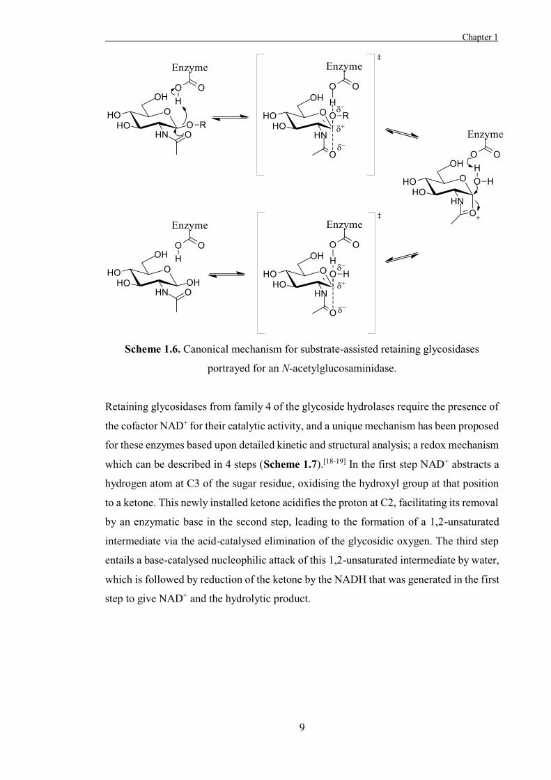
Scheme 1.6. Canonical mechanism for substrate-assisted retaining glycosidases
portrayed for an N-acetylglucosaminidase.
Retaining glycosidases from family 4 of the glycoside hydrolases require the presence of
the cofactor NAD+ for their catalytic activity, and a unique mechanism has been proposed
for these enzymes based upon detailed kinetic and structural analysis; a redox mechanism
which can be described in 4 steps (Scheme 1.7).[18-19] In the first step NAD+ abstracts a
hydrogen atom at C3 of the sugar residue, oxidising the hydroxyl group at that position
to a ketone. This newly installed ketone acidifies the proton at C2, facilitating its removal
by an enzymatic base in the second step, leading to the formation of a 1,2-unsaturated
intermediate via the acid-catalysed elimination of the glycosidic oxygen. The third step
entails a base-catalysed nucleophilic attack of this 1,2-unsaturated intermediate by water,
which is followed by reduction of the ketone by the NADH that was generated in the first
step to give NAD+ and the hydrolytic product.
Chapter 1 .
10
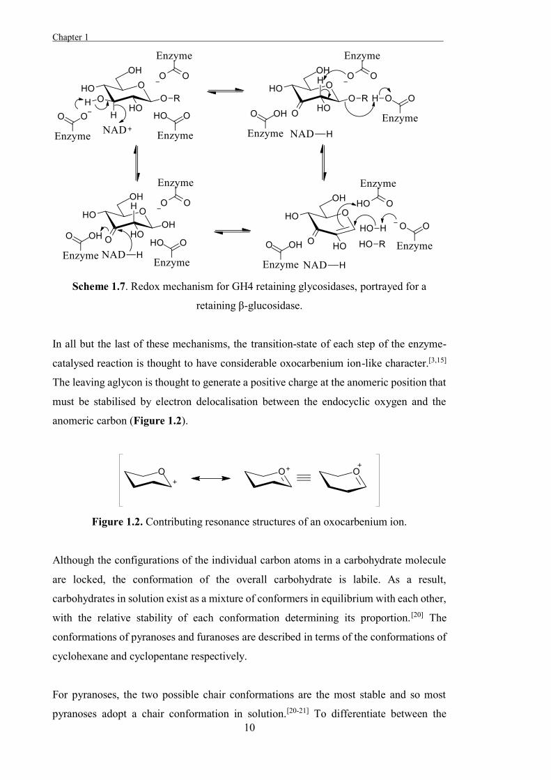
Scheme 1.7. Redox mechanism for GH4 retaining glycosidases, portrayed for a
retaining β-glucosidase.
In all but the last of these mechanisms, the transition-state of each step of the enzyme-
catalysed reaction is thought to have considerable oxocarbenium ion-like character.[3,15]
The leaving aglycon is thought to generate a positive charge at the anomeric position that
must be stabilised by electron delocalisation between the endocyclic oxygen and the
anomeric carbon (Figure 1.2).
Figure 1.2. Contributing resonance structures of an oxocarbenium ion.
Although the configurations of the individual carbon atoms in a carbohydrate molecule
are locked, the conformation of the overall carbohydrate is labile. As a result,
carbohydrates in solution exist as a mixture of conformers in equilibrium with each other,
with the relative stability of each conformation determining its proportion.[20] The
conformations of pyranoses and furanoses are described in terms of the conformations of
cyclohexane and cyclopentane respectively.
For pyranoses, the two possible chair conformations are the most stable and so most
pyranoses adopt a chair conformation in solution.[20-21] To differentiate between the
Chapter 1
11
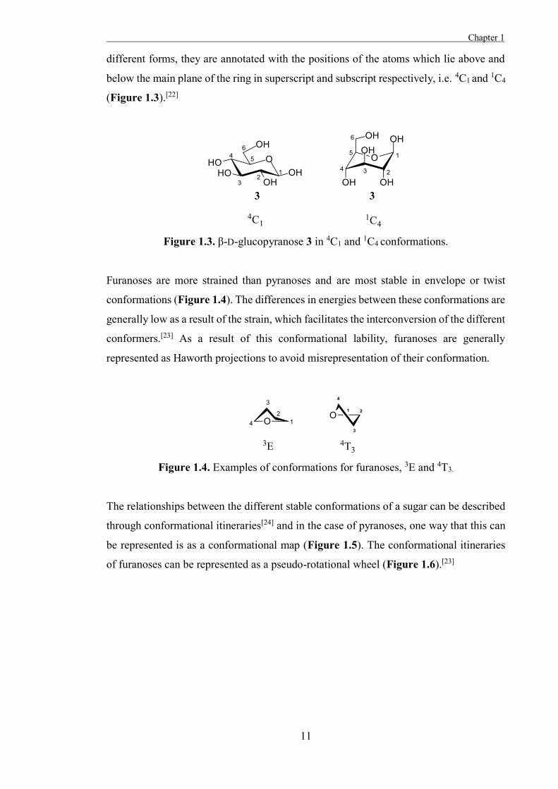
different forms, they are annotated with the positions of the atoms which lie above and
below the main plane of the ring in superscript and subscript respectively, i.e. 4C1 and 1C4
(Figure 1.3).[22]
Figure 1.3. β-D-glucopyranose 3 in 4C1 and 1C4 conformations.
Furanoses are more strained than pyranoses and are most stable in envelope or twist
conformations (Figure 1.4). The differences in energies between these conformations are
generally low as a result of the strain, which facilitates the interconversion of the different
conformers.[23] As a result of this conformational lability, furanoses are generally
represented as Haworth projections to avoid misrepresentation of their conformation.
Figure 1.4. Examples of conformations for furanoses, 3E and 4T3.
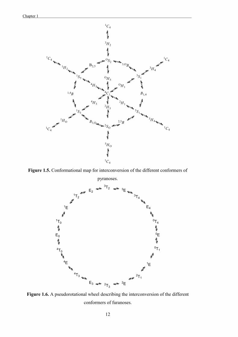
The relationships between the different stable conformations of a sugar can be described
through conformational itineraries[24] and in the case of pyranoses, one way that this can
be represented is as a conformational map (Figure 1.5). The conformational itineraries
of furanoses can be represented as a pseudo-rotational wheel (Figure 1.6).[23]
Chapter 1 .
12
Figure 1.5. Conformational map for interconversion of the different conformers of
pyranoses.
Figure 1.6. A pseudorotational wheel describing the interconversion of the different
conformers of furanoses.
Chapter 1
13
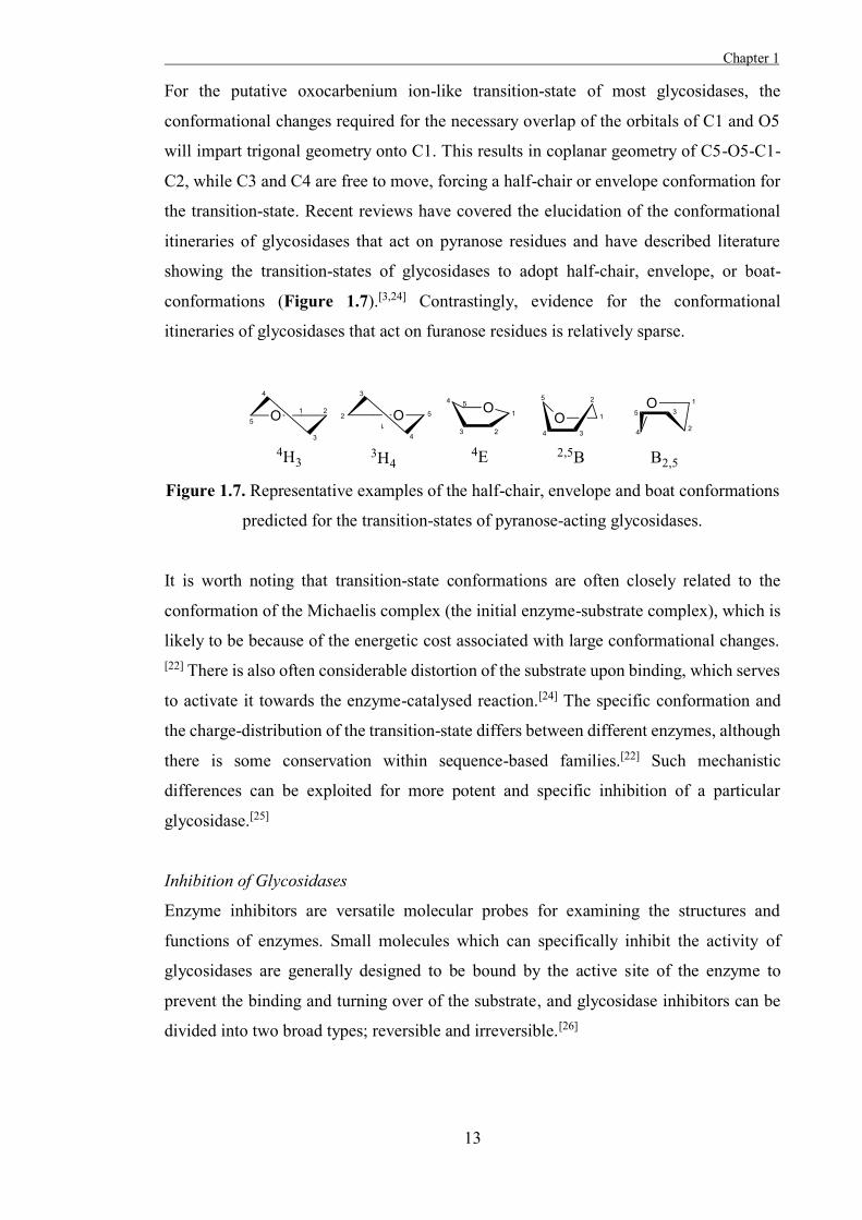
For the putative oxocarbenium ion-like transition-state of most glycosidases, the
conformational changes required for the necessary overlap of the orbitals of C1 and O5
will impart trigonal geometry onto C1. This results in coplanar geometry of C5-O5-C1-
C2, while C3 and C4 are free to move, forcing a half-chair or envelope conformation for
the transition-state. Recent reviews have covered the elucidation of the conformational
itineraries of glycosidases that act on pyranose residues and have described literature
showing the transition-states of glycosidases to adopt half-chair, envelope, or boat-
conformations (Figure 1.7).[3,24] Contrastingly, evidence for the conformational
itineraries of glycosidases that act on furanose residues is relatively sparse.
Figure 1.7. Representative examples of the half-chair, envelope and boat conformations
predicted for the transition-states of pyranose-acting glycosidases.
It is worth noting that transition-state conformations are often closely related to the
conformation of the Michaelis complex (the initial enzyme-substrate complex), which is
likely to be because of the energetic cost associated with large conformational changes. [22] There is also often considerable distortion of the substrate upon binding, which serves
to activate it towards the enzyme-catalysed reaction.[24] The specific conformation and
the charge-distribution of the transition-state differs between different enzymes, although
there is some conservation within sequence-based families.[22] Such mechanistic
differences can be exploited for more potent and specific inhibition of a particular
glycosidase.[25]
Inhibition of Glycosidases
Enzyme inhibitors are versatile molecular probes for examining the structures and
functions of enzymes. Small molecules which can specifically inhibit the activity of
glycosidases are generally designed to be bound by the active site of the enzyme to
prevent the binding and turning over of the substrate, and glycosidase inhibitors can be
divided into two broad types; reversible and irreversible.[26]
Chapter 1 .
14
Irreversible Glycosidase Inhibitors
Irreversible inhibitors form covalent bonds with the glycosidases that they inhibit, which
can inhibit the activity of an enzyme by inactivating a catalytic residue or by blocking
access to the active site. Inhibitors which act this way are useful components of affinity-
based probes for glycosidases, such as for the identification of catalytic residues and
elucidation of the catalytic mechanisms of the enzymes.[27-28] Irreversible glycosidase
inhibitors are generally designed to mimic the carbohydrate substrates of glycosidases in
order to bind the active sites of these enzymes, and also incorporate a moiety that will
react with a nucleophilic side chain spontaneously or can be activated towards
nucleophilic attack either photolytically or by the action of the enzyme.[28]
One approach to developing irreversible glycosidase inhibitors is to functionalise
carbohydrates with chemically reactive moieties that are susceptible to nucleophilic
substitution. The reactive moieties used do not inherently target the active sites of
glycosidases, being susceptible to attack by any nucleophile. Instead, the carbohydrate
moiety is expected to be bound by the active site of the enzyme to bring the reactive
moiety in close proximity to nucleophilic residues in the active site, which it can form
covalent adducts with to inactivate the enzyme. As such, the stability of the inhibitor to
spontaneous hydrolysis and the affinity of the inhibitor for the glycosidase that it targets
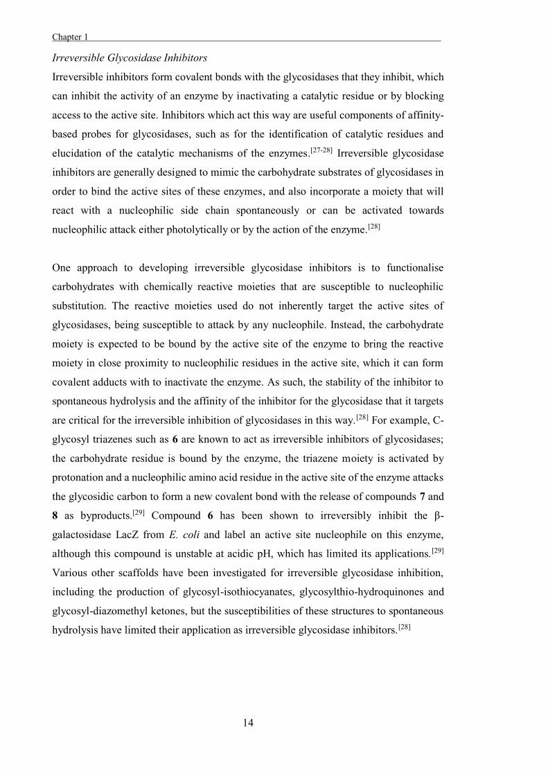
are critical for the irreversible inhibition of glycosidases in this way.[28] For example, C-
glycosyl triazenes such as 6 are known to act as irreversible inhibitors of glycosidases;
the carbohydrate residue is bound by the enzyme, the triazene moiety is activated by
protonation and a nucleophilic amino acid residue in the active site of the enzyme attacks
the glycosidic carbon to form a new covalent bond with the release of compounds 7 and
8 as byproducts.[29] Compound 6 has been shown to irreversibly inhibit the β-
galactosidase LacZ from E. coli and label an active site nucleophile on this enzyme,
although this compound is unstable at acidic pH, which has limited its applications.[29]
Various other scaffolds have been investigated for irreversible glycosidase inhibition,
including the production of glycosyl-isothiocyanates, glycosylthio-hydroquinones and
glycosyl-diazomethyl ketones, but the susceptibilities of these structures to spontaneous
hydrolysis have limited their application as irreversible glycosidase inhibitors.[28]
Chapter 1
15
Figure 1.8. The triazene 6 irreversibly inhibits the E. coli β-galactosidase LacZ.
A similar approach to the development of irreversible glycosidase inhibitors entails
functionalisation of a carbohydrate with a photolabile moiety that will generate a strong
electrophile on photolysis. If the inhibitor is bound by the glycosidase when photolysis
occurs, then the reactive group generated is likely to react with a nucleophile in the active
site of the enzyme and so inactivate the glycosidase. As with the irreversible glycosidase
inhibitors that bear chemically reactive moieties, the reactive moiety of these inhibitors
does not inherently target residues in the active site of the enzyme, and so inactivation of
glycosidases in this way depends upon the inhibitor being bound by the active site when
photolysis occurs and the reactive moiety reacting with an active site residue before it can
diffuse out of the active site.[28]
One example of such a photolabile irreversible glycosidase inhibitor is the
galactopyranose derivative 9, which has been used to label the β-galactosidase LacZ from
E. coli.[30] The inhibitor 9 was expected to be bound in the active site of the enzyme in a
similar fashion to the natural substrate of the enzyme, as this irreversible inhibitor mimics
the configuration of the substrate. The diazirine moiety attached to the anomeric position
generates a highly electrophilic carbene moiety on photolysis and this reacts with
available nucleophiles to form new covalent bonds. LacZ was found to be labelled at two
positions in this way by 9; a residue near the active site and a residue on the outer surface
of the enzyme that was away from the active site. It was suggested that this non-specific
labelling was a result of the low binding affinity of the inhibitor for the enzyme active
site.[31]
Chapter 1 .
16
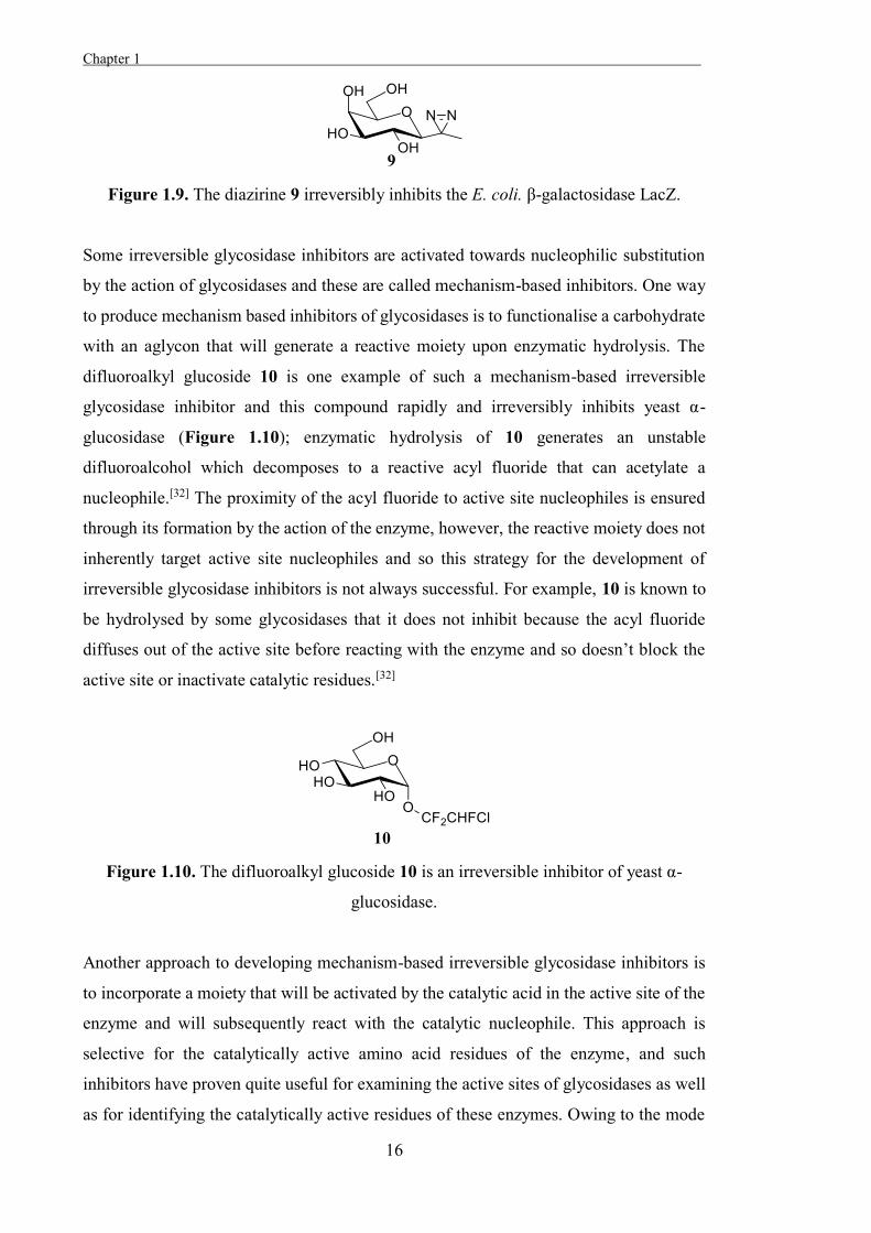
Figure 1.9. The diazirine 9 irreversibly inhibits the E. coli. β-galactosidase LacZ.
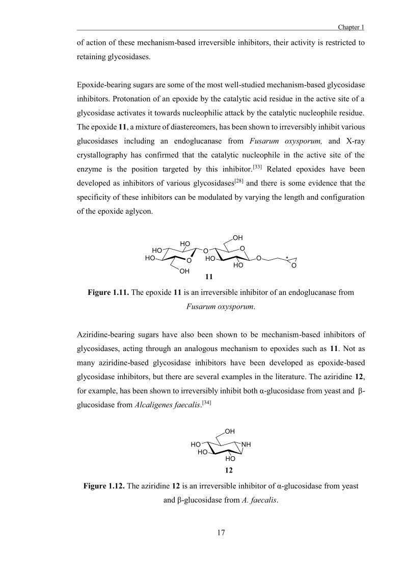
Some irreversible glycosidase inhibitors are activated towards nucleophilic substitution
by the action of glycosidases and these are called mechanism-based inhibitors. One way
to produce mechanism based inhibitors of glycosidases is to functionalise a carbohydrate
with an aglycon that will generate a reactive moiety upon enzymatic hydrolysis. The
difluoroalkyl glucoside 10 is one example of such a mechanism-based irreversible
glycosidase inhibitor and this compound rapidly and irreversibly inhibits yeast α-
glucosidase (Figure 1.10); enzymatic hydrolysis of 10 generates an unstable
difluoroalcohol which decomposes to a reactive acyl fluoride that can acetylate a
nucleophile.[32] The proximity of the acyl fluoride to active site nucleophiles is ensured
through its formation by the action of the enzyme, however, the reactive moiety does not
inherently target active site nucleophiles and so this strategy for the development of
irreversible glycosidase inhibitors is not always successful. For example, 10 is known to
be hydrolysed by some glycosidases that it does not inhibit because the acyl fluoride
diffuses out of the active site before reacting with the enzyme and so doesn’t block the
active site or inactivate catalytic residues.[32]
Figure 1.10. The difluoroalkyl glucoside 10 is an irreversible inhibitor of yeast α-
glucosidase.
Another approach to developing mechanism-based irreversible glycosidase inhibitors is
to incorporate a moiety that will be activated by the catalytic acid in the active site of the
enzyme and will subsequently react with the catalytic nucleophile. This approach is
selective for the catalytically active amino acid residues of the enzyme, and such
inhibitors have proven quite useful for examining the active sites of glycosidases as well
as for identifying the catalytically active residues of these enzymes. Owing to the mode
Chapter 1
17
of action of these mechanism-based irreversible inhibitors, their activity is restricted to
retaining glycosidases.
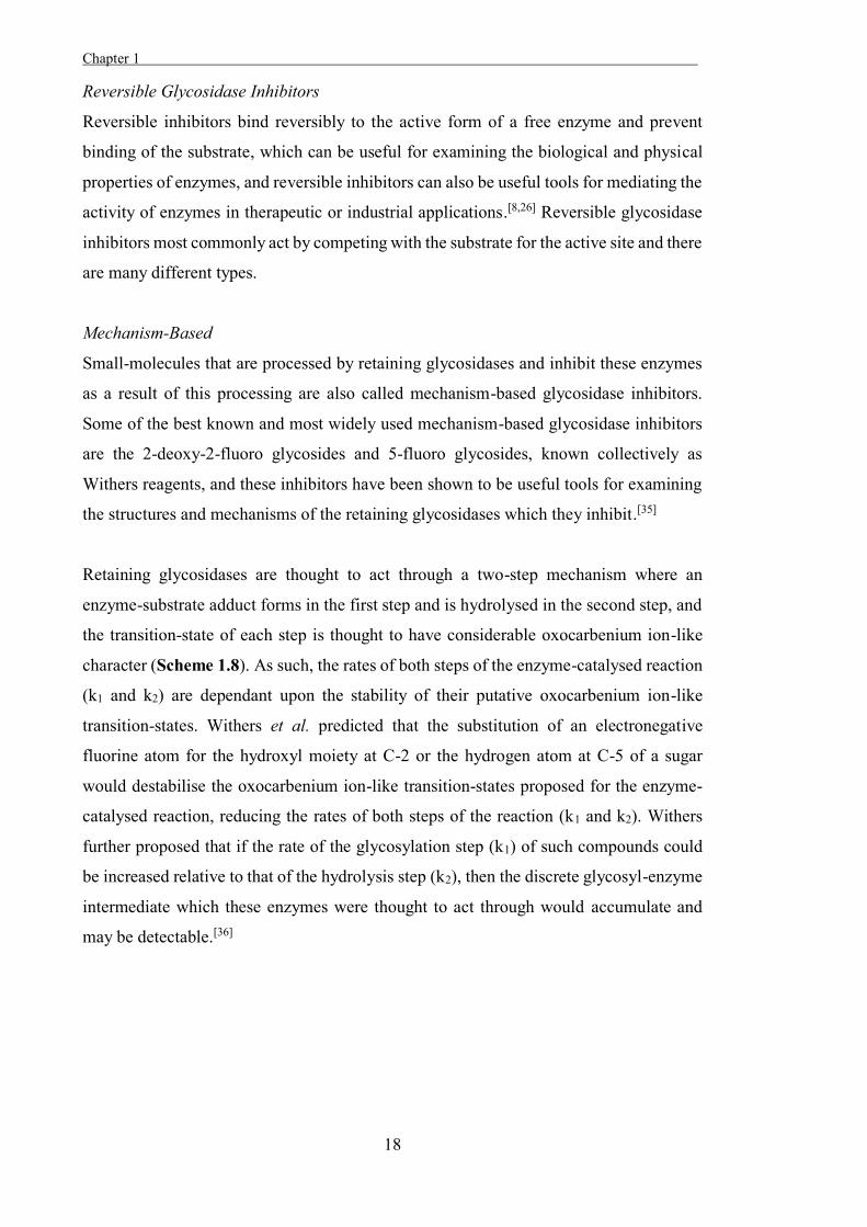
Epoxide-bearing sugars are some of the most well-studied mechanism-based glycosidase
inhibitors. Protonation of an epoxide by the catalytic acid residue in the active site of a
glycosidase activates it towards nucleophilic attack by the catalytic nucleophile residue.
The epoxide 11, a mixture of diastereomers, has been shown to irreversibly inhibit various
glucosidases including an endoglucanase from Fusarum oxysporum, and X-ray
crystallography has confirmed that the catalytic nucleophile in the active site of the
enzyme is the position targeted by this inhibitor.[33] Related epoxides have been
developed as inhibitors of various glycosidases[28] and there is some evidence that the
specificity of these inhibitors can be modulated by varying the length and configuration
of the epoxide aglycon.
Figure 1.11. The epoxide 11 is an irreversible inhibitor of an endoglucanase from
Fusarum oxysporum.
Aziridine-bearing sugars have also been shown to be mechanism-based inhibitors of
glycosidases, acting through an analogous mechanism to epoxides such as 11. Not as
many aziridine-based glycosidase inhibitors have been developed as epoxide-based
glycosidase inhibitors, but there are several examples in the literature. The aziridine 12,
for example, has been shown to irreversibly inhibit both α-glucosidase from yeast and β-
glucosidase from Alcaligenes faecalis.[34]
Figure 1.12. The aziridine 12 is an irreversible inhibitor of α-glucosidase from yeast
and β-glucosidase from A. faecalis.
Chapter 1 .
18
Reversible Glycosidase Inhibitors
Reversible inhibitors bind reversibly to the active form of a free enzyme and prevent
binding of the substrate, which can be useful for examining the biological and physical
properties of enzymes, and reversible inhibitors can also be useful tools for mediating the
activity of enzymes in therapeutic or industrial applications.[8,26] Reversible glycosidase
inhibitors most commonly act by competing with the substrate for the active site and there
are many different types.
Mechanism-Based
Small-molecules that are processed by retaining glycosidases and inhibit these enzymes
as a result of this processing are also called mechanism-based glycosidase inhibitors.
Some of the best known and most widely used mechanism-based glycosidase inhibitors
are the 2-deoxy-2-fluoro glycosides and 5-fluoro glycosides, known collectively as
Withers reagents, and these inhibitors have been shown to be useful tools for examining
the structures and mechanisms of the retaining glycosidases which they inhibit.[35]
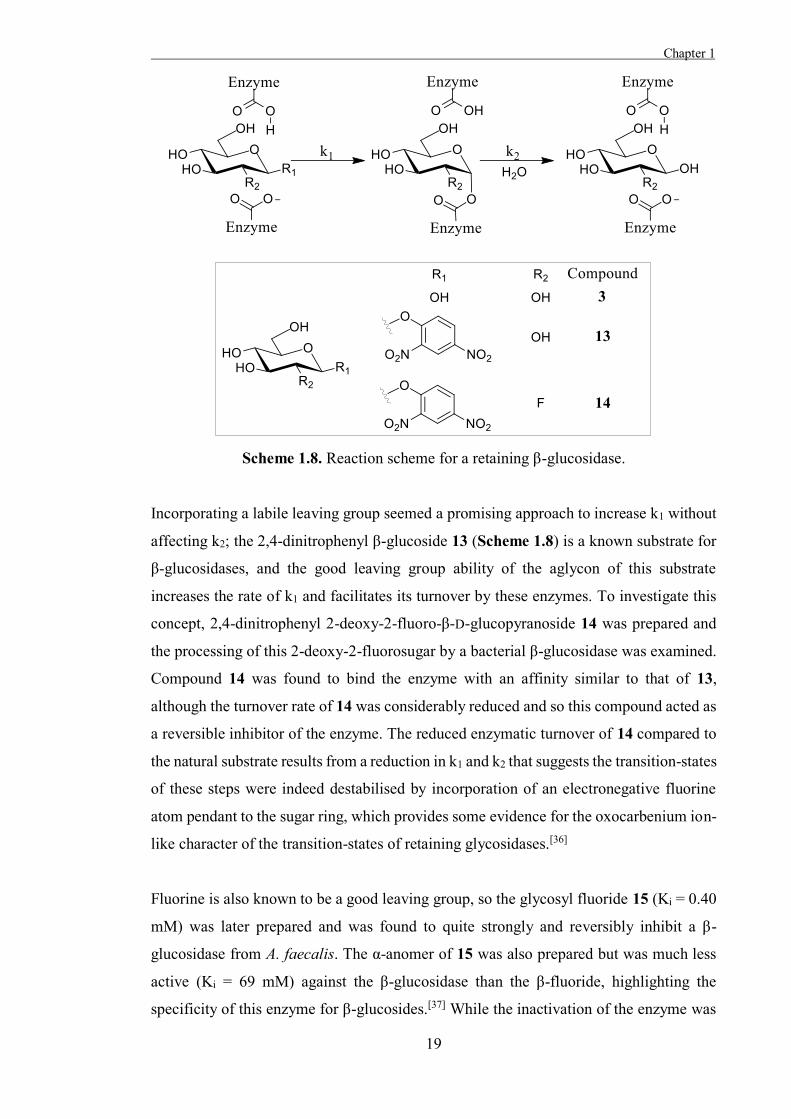
Retaining glycosidases are thought to act through a two-step mechanism where an
enzyme-substrate adduct forms in the first step and is hydrolysed in the second step, and
the transition-state of each step is thought to have considerable oxocarbenium ion-like
character (Scheme 1.8). As such, the rates of both steps of the enzyme-catalysed reaction
(k1 and k2) are dependant upon the stability of their putative oxocarbenium ion-like
transition-states. Withers et al. predicted that the substitution of an electronegative
fluorine atom for the hydroxyl moiety at C-2 or the hydrogen atom at C-5 of a sugar
would destabilise the oxocarbenium ion-like transition-states proposed for the enzyme-
catalysed reaction, reducing the rates of both steps of the reaction (k1 and k2). Withers
further proposed that if the rate of the glycosylation step (k1) of such compounds could
be increased relative to that of the hydrolysis step (k2), then the discrete glycosyl-enzyme
intermediate which these enzymes were thought to act through would accumulate and
may be detectable.[36]
Chapter 1
19
Scheme 1.8. Reaction scheme for a retaining β-glucosidase.
Incorporating a labile leaving group seemed a promising approach to increase k1 without
affecting k2; the 2,4-dinitrophenyl β-glucoside 13 (Scheme 1.8) is a known substrate for
β-glucosidases, and the good leaving group ability of the aglycon of this substrate
increases the rate of k1 and facilitates its turnover by these enzymes. To investigate this
concept, 2,4-dinitrophenyl 2-deoxy-2-fluoro-β-D-glucopyranoside 14 was prepared and
the processing of this 2-deoxy-2-fluorosugar by a bacterial β-glucosidase was examined.
Compound 14 was found to bind the enzyme with an affinity similar to that of 13,
although the turnover rate of 14 was considerably reduced and so this compound acted as
a reversible inhibitor of the enzyme. The reduced enzymatic turnover of 14 compared to
the natural substrate results from a reduction in k1 and k2 that suggests the transition-states
of these steps were indeed destabilised by incorporation of an electronegative fluorine
atom pendant to the sugar ring, which provides some evidence for the oxocarbenium ion-
like character of the transition-states of retaining glycosidases.[36]
Fluorine is also known to be a good leaving group, so the glycosyl fluoride 15 (Ki = 0.40
mM) was later prepared and was found to quite strongly and reversibly inhibit a β-
glucosidase from A. faecalis. The α-anomer of 15 was also prepared but was much less
active (Ki = 69 mM) against the β-glucosidase than the β-fluoride, highlighting the
specificity of this enzyme for β-glucosides.[37] While the inactivation of the enzyme was
Chapter 1 .
20
promising, more direct evidence for the accumulation of a glycosyl-enzyme intermediate
was needed. The release of inorganic fluoride was demonstrated in a 19F NMR study of
the glycosyl fluoride 15 incubated with a bacterial β-glucosidase, and a signal believed to
correspond to the glycosyl-enzyme intermediate was also observed in the spectra, but the
configuration of this intermediate could not be deduced from this experiment.[38]
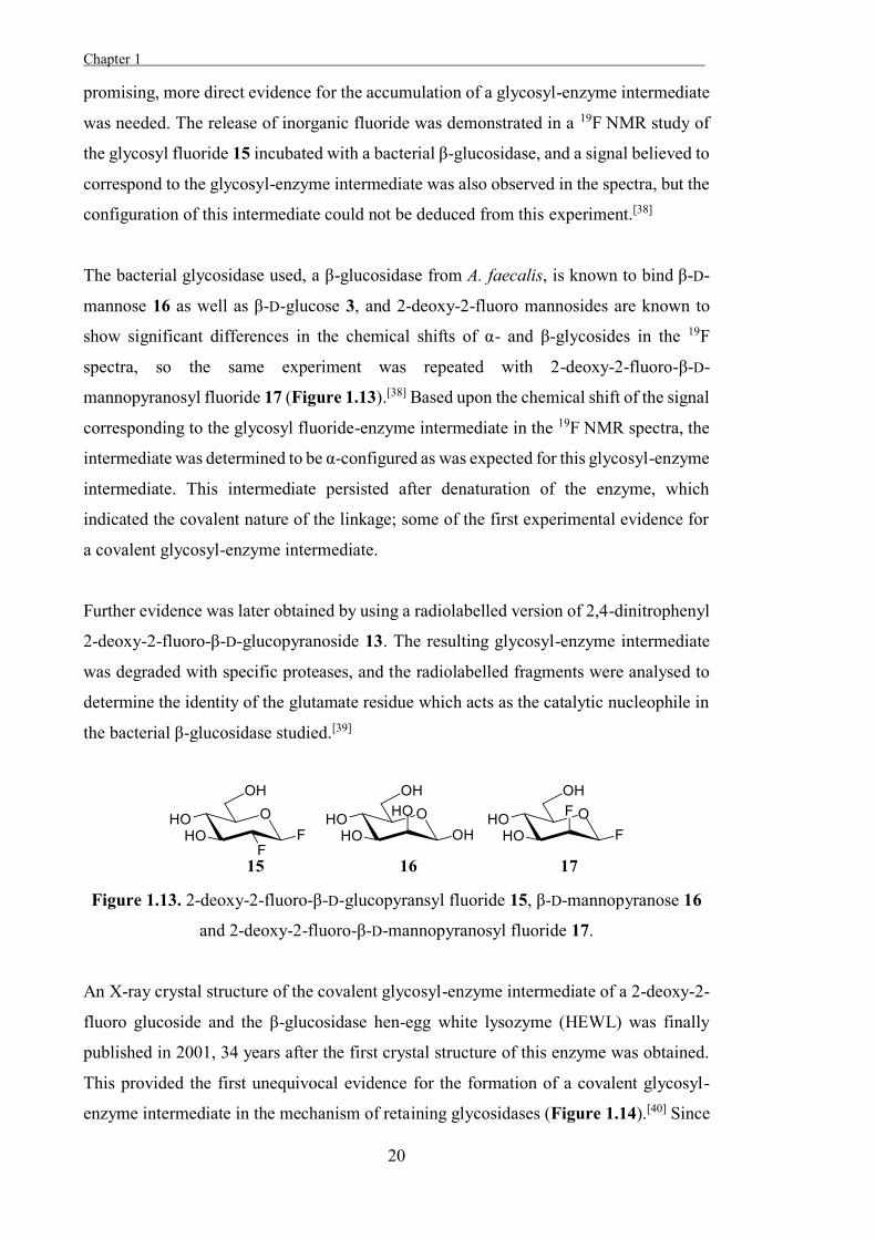
The bacterial glycosidase used, a β-glucosidase from A. faecalis, is known to bind β-D-
mannose 16 as well as β-D-glucose 3, and 2-deoxy-2-fluoro mannosides are known to
show significant differences in the chemical shifts of α- and β-glycosides in the 19F
spectra, so the same experiment was repeated with 2-deoxy-2-fluoro-β-D-
mannopyranosyl fluoride 17 (Figure 1.13).[38] Based upon the chemical shift of the signal
corresponding to the glycosyl fluoride-enzyme intermediate in the 19F NMR spectra, the
intermediate was determined to be α-configured as was expected for this glycosyl-enzyme
intermediate. This intermediate persisted after denaturation of the enzyme, which
indicated the covalent nature of the linkage; some of the first experimental evidence for
a covalent glycosyl-enzyme intermediate.
Further evidence was later obtained by using a radiolabelled version of 2,4-dinitrophenyl
2-deoxy-2-fluoro-β-D-glucopyranoside 13. The resulting glycosyl-enzyme intermediate
was degraded with specific proteases, and the radiolabelled fragments were analysed to
determine the identity of the glutamate residue which acts as the catalytic nucleophile in
the bacterial β-glucosidase studied.[39]
Figure 1.13. 2-deoxy-2-fluoro-β-D-glucopyransyl fluoride 15, β-D-mannopyranose 16
and 2-deoxy-2-fluoro-β-D-mannopyranosyl fluoride 17.
An X-ray crystal structure of the covalent glycosyl-enzyme intermediate of a 2-deoxy-2-
fluoro glucoside and the β-glucosidase hen-egg white lysozyme (HEWL) was finally
published in 2001, 34 years after the first crystal structure of this enzyme was obtained.
This provided the first unequivocal evidence for the formation of a covalent glycosyl-
enzyme intermediate in the mechanism of retaining glycosidases (Figure 1.14).[40] Since
Chapter 1
21
this seminal work, many other glycosyl-enzyme intermediates and catalytic nucleophiles
of retaining glycosidases have been identified.[28,41]
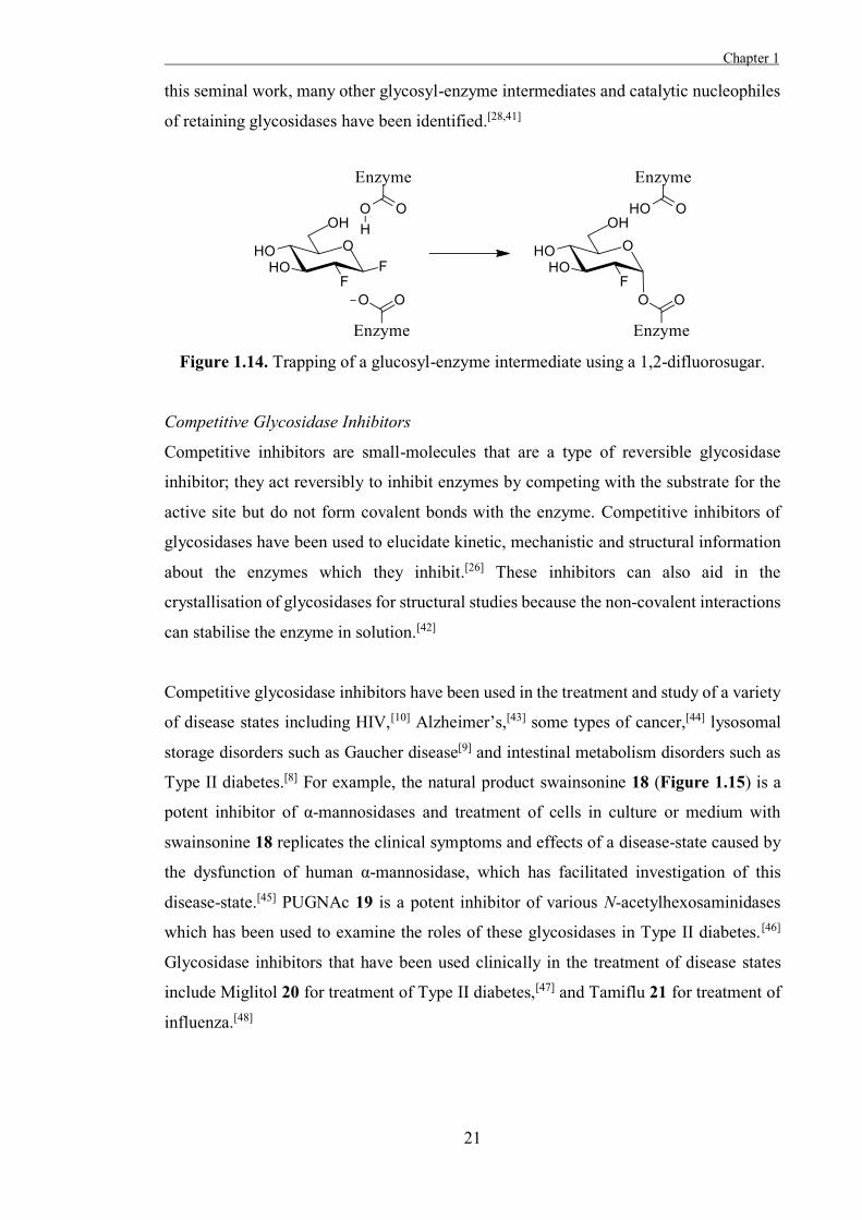
Figure 1.14. Trapping of a glucosyl-enzyme intermediate using a 1,2-difluorosugar.
Competitive Glycosidase Inhibitors
Competitive inhibitors are small-molecules that are a type of reversible glycosidase
inhibitor; they act reversibly to inhibit enzymes by competing with the substrate for the
active site but do not form covalent bonds with the enzyme. Competitive inhibitors of
glycosidases have been used to elucidate kinetic, mechanistic and structural information
about the enzymes which they inhibit.[26] These inhibitors can also aid in the
crystallisation of glycosidases for structural studies because the non-covalent interactions
can stabilise the enzyme in solution.[42]
Competitive glycosidase inhibitors have been used in the treatment and study of a variety
of disease states including HIV,[10] Alzheimer’s,[43] some types of cancer,[44] lysosomal
storage disorders such as Gaucher disease[9] and intestinal metabolism disorders such as
Type II diabetes.[8] For example, the natural product swainsonine 18 (Figure 1.15) is a
potent inhibitor of α-mannosidases and treatment of cells in culture or medium with
swainsonine 18 replicates the clinical symptoms and effects of a disease-state caused by
the dysfunction of human α-mannosidase, which has facilitated investigation of this
disease-state.[45] PUGNAc 19 is a potent inhibitor of various N-acetylhexosaminidases
which has been used to examine the roles of these glycosidases in Type II diabetes.[46]
Glycosidase inhibitors that have been used clinically in the treatment of disease states
include Miglitol 20 for treatment of Type II diabetes,[47] and Tamiflu 21 for treatment of
influenza.[48]

Chapter 1 .
22
Figure 1.15. Some glycosidase inhibitors which have been used to treat or study disease
states.
Competitive glycosidase inhibitors have been developed through a variety of approaches, [8,26,49-50] but generally designs have focused on mimicking the interactions between the
substrate and the active site of the enzyme during the enzyme-catalysed reaction. This is
most commonly achieved by producing analogues of the substrate of the enzyme, or
analogues of the transition-state of the enzyme-catalysed reaction which are detailed
below.
Substrate-Based
A common approach to developing inhibitors of glycosidases is to produce analogues of
the carbohydrate substrate that are resistant to the enzymes action.[51] Stable analogues of
the substrates of glycosidases can be produced by replacing the O-glycosidic bond with
a more stable linkage that can mimic the interactions between the O-glycoside and the
active site of a glycosidase. This strategy is commonly used to prepare pseudo-di- and
oligosaccharides which mimic the substrates of glycosidases.[51] More binding
interactions are possible between a pseudo-disaccharide and the active site of an enzyme
compared to a monosaccharide mimic, which can result in more potent and specific
inhibition by pseudo-disaccharides than pseudo-monosaccharides.[52] As a result, pseudo-
disaccharides are of interest as biological probes and in the development of more potent
and selective inhibitors of glycosidases.
S-Glycosides are known to be more stable to chemical or enzymatic hydrolysis than O-
glycosides due to the weak basicity of the sulfur-atom and the poor leaving group ability
of the S-glycoside and so compounds based on these have been investigated as substrate-
mimic glycosidase inhibitors.[51] Many S-glycosides are relatively weak inhibitors of the
Chapter 1
23
glycosidases they are designed to target, which may be the result of the different geometry
of the S-glycosidic bond compared to an O-glycoside and the weaker binding of the
substrate relative to the binding that occurs at the transition-state of the enzyme-catalysed
reaction. Nonetheless, these molecules have proven quite useful in the study of
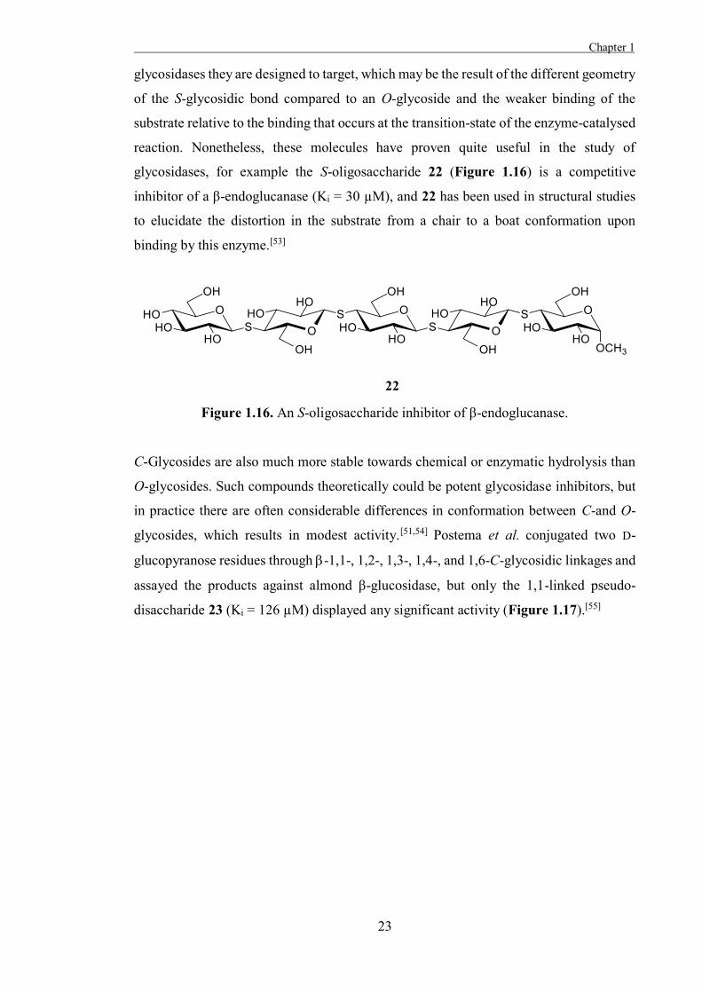
glycosidases, for example the S-oligosaccharide 22 (Figure 1.16) is a competitive
inhibitor of a β-endoglucanase (Ki = 30 µM), and 22 has been used in structural studies
to elucidate the distortion in the substrate from a chair to a boat conformation upon
binding by this enzyme.[53]
Figure 1.16. An S-oligosaccharide inhibitor of β-endoglucanase.
C-Glycosides are also much more stable towards chemical or enzymatic hydrolysis than
O-glycosides. Such compounds theoretically could be potent glycosidase inhibitors, but
in practice there are often considerable differences in conformation between C-and O-
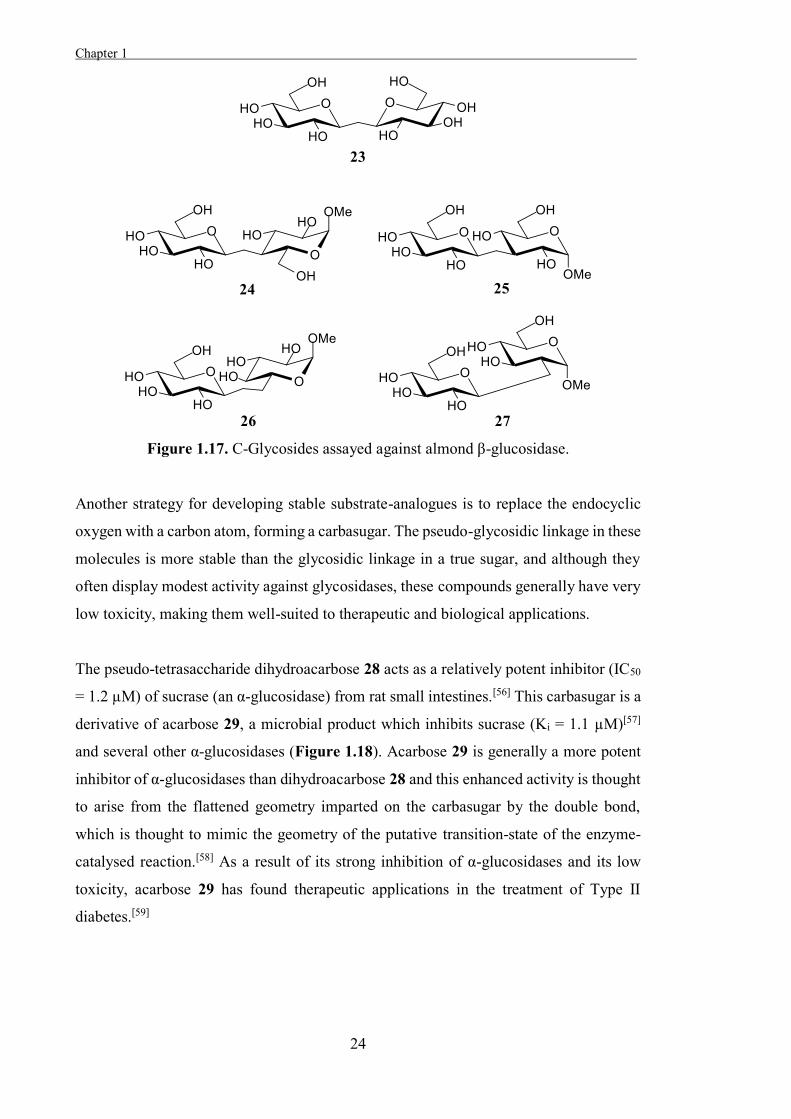
glycosides, which results in modest activity.[51,54] Postema et al. conjugated two D-
glucopyranose residues through -1,1-, 1,2-, 1,3-, 1,4-, and 1,6-C-glycosidic linkages and
assayed the products against almond β-glucosidase, but only the 1,1-linked pseudo-
disaccharide 23 (Ki = 126 µM) displayed any significant activity (Figure 1.17).[55]
Chapter 1 .
24
Figure 1.17. C-Glycosides assayed against almond β-glucosidase.
Another strategy for developing stable substrate-analogues is to replace the endocyclic
oxygen with a carbon atom, forming a carbasugar. The pseudo-glycosidic linkage in these
molecules is more stable than the glycosidic linkage in a true sugar, and although they
often display modest activity against glycosidases, these compounds generally have very
low toxicity, making them well-suited to therapeutic and biological applications.
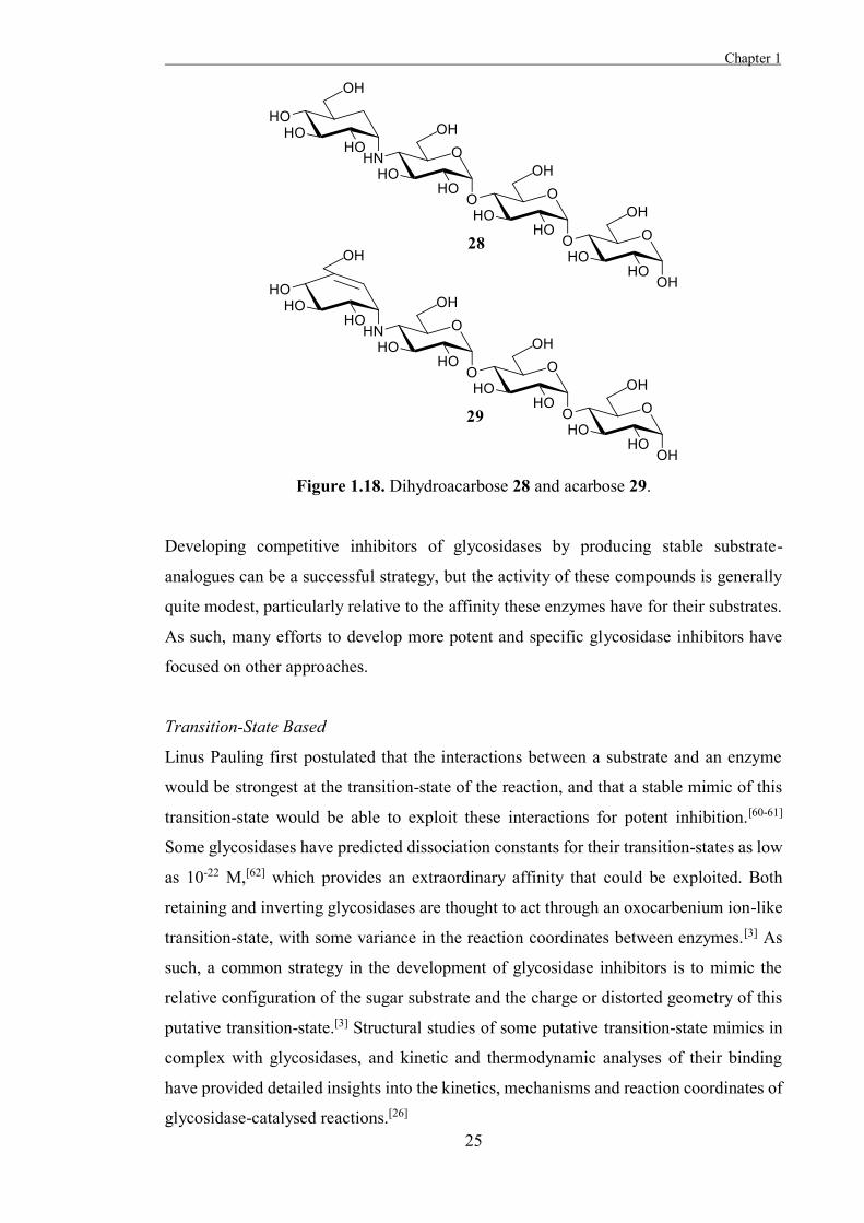
The pseudo-tetrasaccharide dihydroacarbose 28 acts as a relatively potent inhibitor (IC50
= 1.2 µM) of sucrase (an α-glucosidase) from rat small intestines.[56] This carbasugar is a
derivative of acarbose 29, a microbial product which inhibits sucrase (Ki = 1.1 µM)[57]
and several other α-glucosidases (Figure 1.18). Acarbose 29 is generally a more potent
inhibitor of α-glucosidases than dihydroacarbose 28 and this enhanced activity is thought
to arise from the flattened geometry imparted on the carbasugar by the double bond,
which is thought to mimic the geometry of the putative transition-state of the enzyme-
catalysed reaction.[58] As a result of its strong inhibition of α-glucosidases and its low
toxicity, acarbose 29 has found therapeutic applications in the treatment of Type II
diabetes.[59]
Chapter 1
25
Figure 1.18. Dihydroacarbose 28 and acarbose 29.
Developing competitive inhibitors of glycosidases by producing stable substrate-
analogues can be a successful strategy, but the activity of these compounds is generally
quite modest, particularly relative to the affinity these enzymes have for their substrates.
As such, many efforts to develop more potent and specific glycosidase inhibitors have
focused on other approaches.
Transition-State Based
Linus Pauling first postulated that the interactions between a substrate and an enzyme
would be strongest at the transition-state of the reaction, and that a stable mimic of this
transition-state would be able to exploit these interactions for potent inhibition.[60-61]
Some glycosidases have predicted dissociation constants for their transition-states as low
as 10-22 M,[62] which provides an extraordinary affinity that could be exploited. Both
retaining and inverting glycosidases are thought to act through an oxocarbenium ion-like
transition-state, with some variance in the reaction coordinates between enzymes.[3] As
such, a common strategy in the development of glycosidase inhibitors is to mimic the
relative configuration of the sugar substrate and the charge or distorted geometry of this
putative transition-state.[3] Structural studies of some putative transition-state mimics in
complex with glycosidases, and kinetic and thermodynamic analyses of their binding
have provided detailed insights into the kinetics, mechanisms and reaction coordinates of
glycosidase-catalysed reactions.[26]
Chapter 1 .
26
Many potent glycosidase inhibitors that have been developed attempt to mimic the
putative transition-state of the enzyme catalysed reaction, which has led to considerable
discussion over whether such compounds are true transition-state analogues, ground-state
analogues, or simply bind fortuitously. Several criteria have been proposed for a true
transition-state mimic, including tight binding, enthalpy-driven binding, slow-onset of
binding and binding by the active form of the enzyme,[63] and these criteria can be
assessed by examining free energy correlations, X-ray structure analysis and computer
modelling.[62] Very few of the many potent glycosidase inhibitors developed by
attempting to mimic the charge and shape of the putative transition-state have been shown
to be true transition-state mimics, but this has not hindered the use of these inhibitors in
biological studies or therapeutic applications.[42,64-65] Examples of inhibitors that attempt
to mimic an aspect of the transition-state are listed below.
Charge-Based
Glycosidase inhibitors designed to mimic the charged ring-oxygen and anomeric
positions of the transition-state generally incorporate a heteroatom that can be positively
charged at one or both of these positions.[8,66] Some of the most potent and specific
charge-based glycosidase inhibitors are those bearing a nitrogen atom in place of the
anomeric carbon (azasugars), or the endocyclic oxygen (iminosugars) of the carbohydrate
substrate. The nitrogen is thought to be protonated when bound by the enzyme, and this
acts to mimic the putative positively charged oxocarbenium ion-like transition-state for
glycosidases.[67] This electrostatic attraction is the main driving force behind binding of
azasugars and iminosugars by glycosidases.[68]
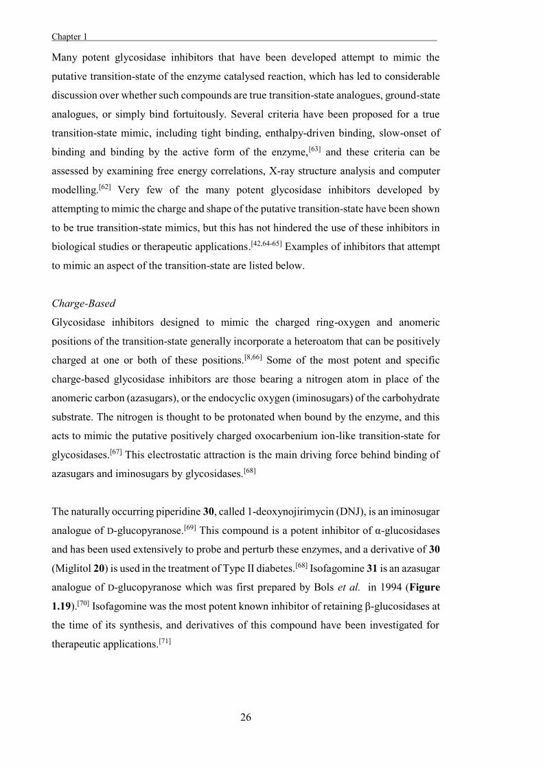
The naturally occurring piperidine 30, called 1-deoxynojirimycin (DNJ), is an iminosugar
analogue of D-glucopyranose.[69] This compound is a potent inhibitor of α-glucosidases
and has been used extensively to probe and perturb these enzymes, and a derivative of 30
(Miglitol 20) is used in the treatment of Type II diabetes.[68] Isofagomine 31 is an azasugar
analogue of D-glucopyranose which was first prepared by Bols et al. in 1994 (Figure
1.19).[70] Isofagomine was the most potent known inhibitor of retaining β-glucosidases at
the time of its synthesis, and derivatives of this compound have been investigated for
therapeutic applications.[71]
Chapter 1
27
Figure 1.19. Competitive inhibitors of α- and β-glucosidases.
The relative activities of DNJ 30 and isofagomine 31 against α-glucosidases and β-
glucosidases are notable. DNJ 30 is more active against yeast α-glucosidase (Ki = 27 µM)
than almond β-glucosidase (Ki = 49 µM), while isofagomine 31 is considerably more
potent against the β-glucosidase (Ki = 0.11 µM) than the α-glucosidase (Ki = 86 µM).[67]
It has been determined that the position of the charged nitrogen centre affects the relative
activities of these inhibitors against α- and β-glucosidases because of the differing charge
distributions in the transition-states of these enzymes,[72] and that the presence or absence
of the hydroxyl moiety at C2 also affects the specificity of these inhibitors.[66]
Analogues of DNJ 30 and isofagomine 31 that match the configurations of other
monosaccharides are generally potent inhibitors of the glycosidases which process those
sugars,[73] and these inhibitors have been used in kinetic, mechanistic and structural
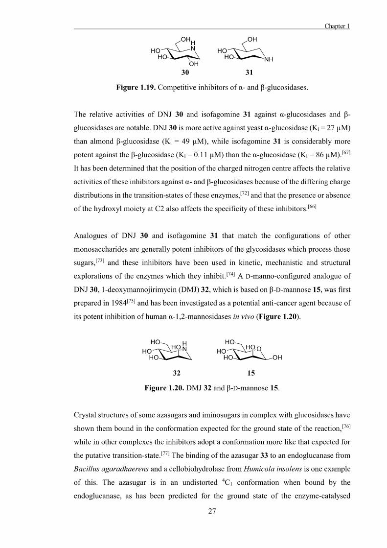
explorations of the enzymes which they inhibit.[74] A D-manno-configured analogue of
DNJ 30, 1-deoxymannojirimycin (DMJ) 32, which is based on β-D-mannose 15, was first
prepared in 1984[75] and has been investigated as a potential anti-cancer agent because of
its potent inhibition of human α-1,2-mannosidases in vivo (Figure 1.20).
Figure 1.20. DMJ 32 and β-D-mannose 15.
Crystal structures of some azasugars and iminosugars in complex with glucosidases have
shown them bound in the conformation expected for the ground state of the reaction,[76]
while in other complexes the inhibitors adopt a conformation more like that expected for
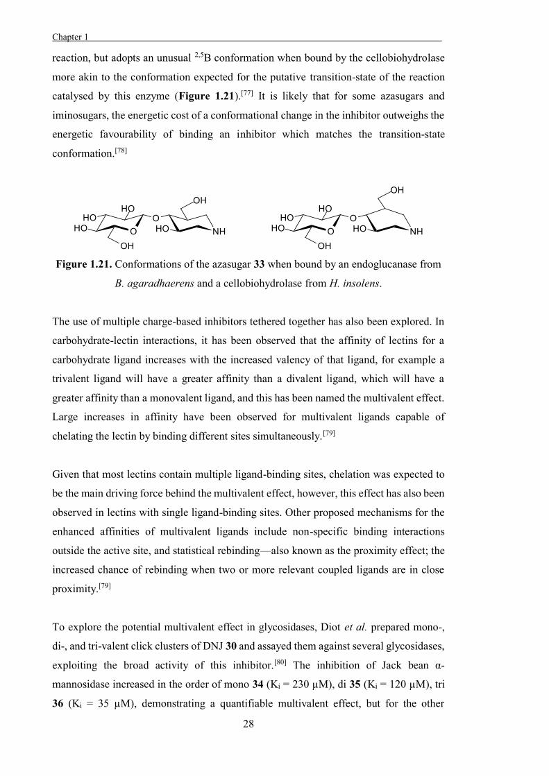
the putative transition-state.[77] The binding of the azasugar 33 to an endoglucanase from
Bacillus agaradhaerens and a cellobiohydrolase from Humicola insolens is one example
of this. The azasugar is in an undistorted 4C1 conformation when bound by the
endoglucanase, as has been predicted for the ground state of the enzyme-catalysed
Chapter 1 .
28
reaction, but adopts an unusual 2,5B conformation when bound by the cellobiohydrolase
more akin to the conformation expected for the putative transition-state of the reaction
catalysed by this enzyme (Figure 1.21).[77] It is likely that for some azasugars and
iminosugars, the energetic cost of a conformational change in the inhibitor outweighs the
energetic favourability of binding an inhibitor which matches the transition-state
conformation.[78]
Figure 1.21. Conformations of the azasugar 33 when bound by an endoglucanase from
B. agaradhaerens and a cellobiohydrolase from H. insolens.
The use of multiple charge-based inhibitors tethered together has also been explored. In
carbohydrate-lectin interactions, it has been observed that the affinity of lectins for a
carbohydrate ligand increases with the increased valency of that ligand, for example a
trivalent ligand will have a greater affinity than a divalent ligand, which will have a
greater affinity than a monovalent ligand, and this has been named the multivalent effect.
Large increases in affinity have been observed for multivalent ligands capable of
chelating the lectin by binding different sites simultaneously.[79]
Given that most lectins contain multiple ligand-binding sites, chelation was expected to
be the main driving force behind the multivalent effect, however, this effect has also been
observed in lectins with single ligand-binding sites. Other proposed mechanisms for the
enhanced affinities of multivalent ligands include non-specific binding interactions
outside the active site, and statistical rebinding—also known as the proximity effect; the
increased chance of rebinding when two or more relevant coupled ligands are in close
proximity.[79]
To explore the potential multivalent effect in glycosidases, Diot et al. prepared mono-,
di-, and tri-valent click clusters of DNJ 30 and assayed them against several glycosidases,
exploiting the broad activity of this inhibitor.[80] The inhibition of Jack bean α-
mannosidase increased in the order of mono 34 (Ki = 230 µM), di 35 (Ki = 120 µM), tri
36 (Ki = 35 µM), demonstrating a quantifiable multivalent effect, but for the other
Chapter 1
29
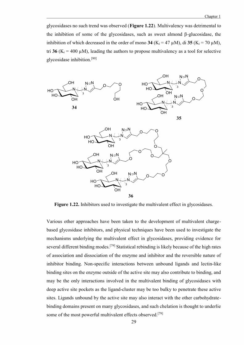
glycosidases no such trend was observed (Figure 1.22). Multivalency was detrimental to
the inhibition of some of the glycosidases, such as sweet almond β-glucosidase, the
inhibition of which decreased in the order of mono 34 (Ki = 47 µM), di 35 (Ki = 70 µM),
tri 36 (Ki = 400 µM), leading the authors to propose multivalency as a tool for selective
glycosidase inhibition.[80]
Figure 1.22. Inhibitors used to investigate the multivalent effect in glycosidases.
Various other approaches have been taken to the development of multivalent charge-
based glycosidase inhibitors, and physical techniques have been used to investigate the
mechanisms underlying the multivalent effect in glycosidases, providing evidence for
several different binding modes.[79] Statistical rebinding is likely because of the high rates
of association and dissociation of the enzyme and inhibitor and the reversible nature of
inhibitor binding. Non-specific interactions between unbound ligands and lectin-like
binding sites on the enzyme outside of the active site may also contribute to binding, and
may be the only interactions involved in the multivalent binding of glycosidases with
deep active site pockets as the ligand-cluster may be too bulky to penetrate these active
sites. Ligands unbound by the active site may also interact with the other carbohydrate-
binding domains present on many glycosidases, and such chelation is thought to underlie
some of the most powerful multivalent effects observed.[79]
Chapter 1 .
30
Based on these studies, multivalent ligands seem well suited to the inhibition of some
specific glycosidases for which the multivalent effect has been observed, although the
requirement of multiple synthetically costly ligands for each multivalent inhibitor is
disadvantageous.
Shape-Based
As well as attempting to mimic the charge of the transition-state, attempting to mimic the
shape expected for the transition-state is an effective strategy for developing glycosidase
inhibitors. As enzymes tend to have the highest affinity for their transition-states, a
compound which matches the conformation of the substrate at the transition-state will
have a higher affinity than one which matches the ground state conformation.[81]
Glycosidase inhibitors which have been designed to mimic the distorted shape of the
oxocarbenium ion-like transition-state of these enzymes often incorporate an sp2
hybridised pseudo-anomeric centre.[50] This sp2 hybridisation enforces coplanar geometry
around C5-O5-C1-C2, as is expected for some oxocarbenium ion-like transition-states.[3]
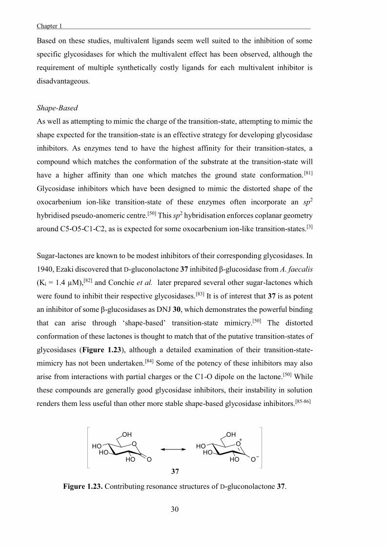
Sugar-lactones are known to be modest inhibitors of their corresponding glycosidases. In
1940, Ezaki discovered that D-gluconolactone 37 inhibited β-glucosidase from A. faecalis
(Ki = 1.4 µM),[82] and Conchie et al. later prepared several other sugar-lactones which
were found to inhibit their respective glycosidases.[83] It is of interest that 37 is as potent
an inhibitor of some β-glucosidases as DNJ 30, which demonstrates the powerful binding
that can arise through ‘shape-based’ transition-state mimicry.[50] The distorted
conformation of these lactones is thought to match that of the putative transition-states of
glycosidases (Figure 1.23), although a detailed examination of their transition-state-
mimicry has not been undertaken.[84] Some of the potency of these inhibitors may also
arise from interactions with partial charges or the C1-O dipole on the lactone.[50] While
these compounds are generally good glycosidase inhibitors, their instability in solution
renders them less useful than other more stable shape-based glycosidase inhibitors.[85-86]
Figure 1.23. Contributing resonance structures of D-gluconolactone 37.
Chapter 1
31
Another strategy to developing glycosidase inhibitors which mimic the shape of the
putative transition-state is to incorporate a fused ring in the sugar, such as with the
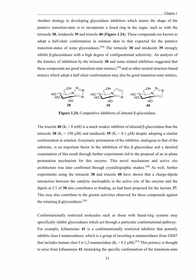
tetrazole 38, imidazole 39 and triazole 40 (Figure 1.24). These compounds are known to
adopt a half-chair conformation in solution akin to that expected for the putative
transition-states of some glycosidases.[50] The tetrazole 38 and imidazole 39 strongly
inhibit β-glucosidases with a high degree of configurational selectivity. An analysis of
the kinetics of inhibition by the tetrazole 38 and some related inhibitors suggested that
these compounds are good transition-state mimics,[50] and so other neutral structure-based
mimics which adopt a half-chair conformation may also be good transition-state mimics.
Figure 1.24. Competitive inhibitors of almond β-glucosidase.
The triazole 40 (Ki > 8 mM) is a much weaker inhibitor of almond β-glucosidase than the
tetrazole 38 (Ki = 150 µM) and imidazole 39 (Ki = 0.1 µM) despite adopting a similar
conformation in solution. Enzymatic protonation of the inhibitor, analogous to that of the
substrate, is an important factor in the inhibition of this β-glucosidase and a detailed
examination of this result through further experiments led to the proposal of an in-plane
protonation mechanism for this enzyme. This novel mechanism and active site
architecture was later confirmed through crystallographic studies.[50] As well, further
experiments using the tetrazole 38 and triazole 40 have shown that a charge-dipole
interaction between the catalytic nucleophile in the active site of the enzyme and the
dipole at C1 of 38 also contributes to binding, as had been proposed for the lactone 37.
This may also contribute to the greater activities observed for these compounds against
the retaining β-glycosidases.[50]
Conformationally restricted molecules such as those with fused-ring systems may
specifically inhibit glycosidases which act through a particular conformational pathway.
For example, kifunensine 41 is a conformationally restricted inhibitor that potently
inhibits class I mannosidases, which is a group of inverting α-mannosidases from GH47
that includes human class I α-1,2-mannosidase (Ki = 0.2 µM).[87] This potency is thought
to arise from kifunensine 41 mimicking the specific conformation of the transition-state
Chapter 1 .
32
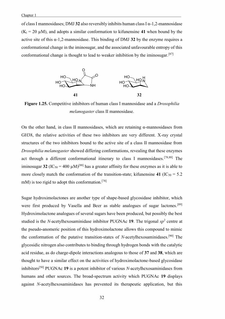
of class I mannosidases; DMJ 32 also reversibly inhibits human class I α-1,2-mannosidase
(Ki = 20 µM), and adopts a similar conformation to kifunensine 41 when bound by the
active site of this α-1,2-mannosidase. This binding of DMJ 32 by the enzyme requires a
conformational change in the iminosugar, and the associated unfavourable entropy of this
conformational change is thought to lead to weaker inhibition by the iminosugar.[87]
Figure 1.25. Competitive inhibitors of human class I mannosidase and a Drosophilia
melanogaster class II mannosidase.
On the other hand, in class II mannosidases, which are retaining α-mannosidases from
GH38, the relative activities of these two inhibitors are very different. X-ray crystal
structures of the two inhibitors bound to the active site of a class II mannosidase from
Drosophilia melanogaster showed differing conformations, revealing that these enzymes
act through a different conformational itinerary to class I mannosidases.[78,88] The
iminosugar 32 (IC50 = 400 µM)[88] has a greater affinity for these enzymes as it is able to
more closely match the conformation of the transition-state; kifunensine 41 (IC50 = 5.2
mM) is too rigid to adopt this conformation.[78]
Sugar hydroximolactones are another type of shape-based glycosidase inhibitor, which
were first produced by Vasella and Beer as stable analogues of sugar lactones.[89]
Hydroximolactone analogues of several sugars have been produced, but possibly the best
studied is the N-acetylhexosaminidase inhibitor PUGNAc 19. The trigonal sp2 centre at
the pseudo-anomeric position of this hydroximolactone allows this compound to mimic
the conformation of the putative transition-states of N-acetylhexosaminidases.[90] The
glycosidic nitrogen also contributes to binding through hydrogen bonds with the catalytic
acid residue, as do charge-dipole interactions analogous to those of 37 and 38, which are
thought to have a similar effect on the activities of hydroximolactone-based glycosidase
inhibitors[50] PUGNAc 19 is a potent inhibitor of various N-acetylhexosaminidases from
humans and other sources. The broad-spectrum activity which PUGNAc 19 displays
against N-acetylhexosaminidases has prevented its therapeutic application, but this
Chapter 1
33
inhibitor and other molecular probes have been used to identify the functions of these
enzymes at the cellular level and examine their roles in these disease states.[91-92]
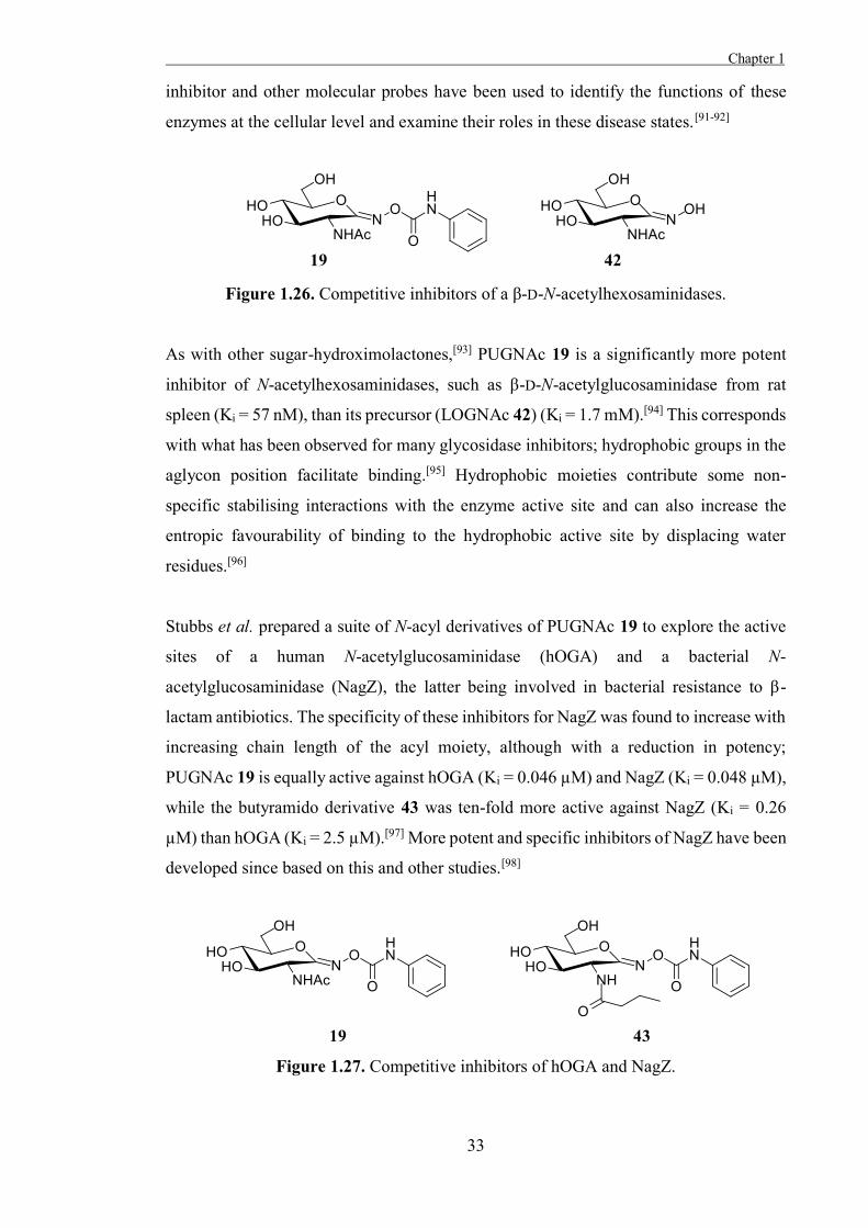
Figure 1.26. Competitive inhibitors of a β-D-N-acetylhexosaminidases.
As with other sugar-hydroximolactones,[93] PUGNAc 19 is a significantly more potent
inhibitor of N-acetylhexosaminidases, such as β-D-N-acetylglucosaminidase from rat
spleen (Ki = 57 nM), than its precursor (LOGNAc 42) (Ki = 1.7 mM).[94] This corresponds
with what has been observed for many glycosidase inhibitors; hydrophobic groups in the
aglycon position facilitate binding.[95] Hydrophobic moieties contribute some non-
specific stabilising interactions with the enzyme active site and can also increase the
entropic favourability of binding to the hydrophobic active site by displacing water
residues.[96]
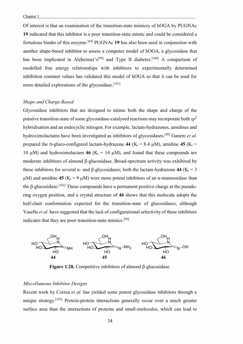
Stubbs et al. prepared a suite of N-acyl derivatives of PUGNAc 19 to explore the active
sites of a human N-acetylglucosaminidase (hOGA) and a bacterial N-
acetylglucosaminidase (NagZ), the latter being involved in bacterial resistance to β-
lactam antibiotics. The specificity of these inhibitors for NagZ was found to increase with
increasing chain length of the acyl moiety, although with a reduction in potency;
PUGNAc 19 is equally active against hOGA (Ki = 0.046 µM) and NagZ (Ki = 0.048 µM),
while the butyramido derivative 43 was ten-fold more active against NagZ (Ki = 0.26
µM) than hOGA (Ki = 2.5 µM).[97] More potent and specific inhibitors of NagZ have been
developed since based on this and other studies.[98]
Figure 1.27. Competitive inhibitors of hOGA and NagZ.
Chapter 1 .
34
Of interest is that an examination of the transition-state mimicry of hOGA by PUGNAc
19 indicated that this inhibitor is a poor transition-state mimic and could be considered a
fortuitous binder of this enzyme.[64] PUGNAc 19 has also been used in conjunction with
another shape-based inhibitor to assess a computer model of hOGA, a glycosidase that
has been implicated in Alzheimer’s[99] and Type II diabetes.[100] A comparison of
modelled free energy relationships with inhibitors to experimentally determined
inhibition constant values has validated this model of hOGA so that it can be used for
more detailed explorations of the glycosidase.[101]
Shape and Charge-Based
Glycosidase inhibitors that are designed to mimic both the shape and charge of the
putative transition-state of some glycosidase-catalysed reactions may incorporate both sp2
hybridisation and an endocyclic nitrogen. For example, lactam-hydrazones, amidines and
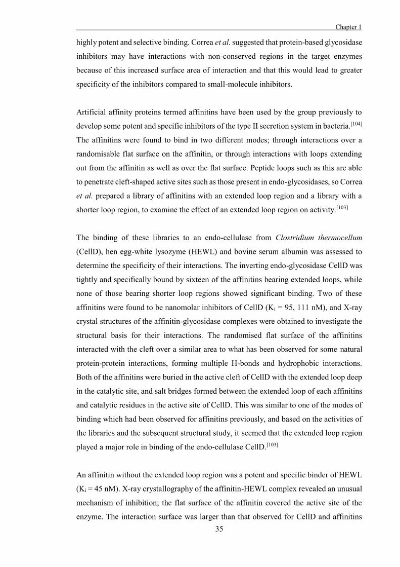
hydroximolactams have been investigated as inhibitors of glycosidases.[50] Ganem et al.
prepared the D-gluco-configured lactam-hydrazone 44 (Ki = 8.4 µM), amidine 45 (Ki =
10 µM) and hydroximolactam 46 (Ki = 14 µM), and found that these compounds are
moderate inhibitors of almond β-glucosidase. Broad-spectrum activity was exhibited by
these inhibitors for several α- and β-glycosidases; both the lactam-hydrazone 44 (Ki = 3
µM) and amidine 45 (Ki = 9 µM) were more potent inhibitors of an α-mannosidase than
the β-glucosidase.[102] These compounds have a permanent positive charge at the pseudo-
ring oxygen position, and a crystal structure of 46 shows that this molecule adopts the
half-chair conformation expected for the transition-state of glucosidases, although
Vasella et al. have suggested that the lack of configurational selectivity of these inhibitors
indicates that they are poor transition-state mimics.[50]
Figure 1.28. Competitive inhibitors of almond β-glucosidase.
Miscellaneous Inhibitor Designs
Recent work by Correa et al. has yielded some potent glycosidase inhibitors through a
unique strategy.[103] Protein-protein interactions generally occur over a much greater
surface area than the interactions of proteins and small-molecules, which can lead to
Chapter 1
35
highly potent and selective binding. Correa et al. suggested that protein-based glycosidase
inhibitors may have interactions with non-conserved regions in the target enzymes
because of this increased surface area of interaction and that this would lead to greater
specificity of the inhibitors compared to small-molecule inhibitors.
Artificial affinity proteins termed affinitins have been used by the group previously to
develop some potent and specific inhibitors of the type II secretion system in bacteria.[104]
The affinitins were found to bind in two different modes; through interactions over a
randomisable flat surface on the affinitin, or through interactions with loops extending
out from the affinitin as well as over the flat surface. Peptide loops such as this are able
to penetrate cleft-shaped active sites such as those present in endo-glycosidases, so Correa
et al. prepared a library of affinitins with an extended loop region and a library with a
shorter loop region, to examine the effect of an extended loop region on activity.[103]
The binding of these libraries to an endo-cellulase from Clostridium thermocellum
(CellD), hen egg-white lysozyme (HEWL) and bovine serum albumin was assessed to
determine the specificity of their interactions. The inverting endo-glycosidase CellD was
tightly and specifically bound by sixteen of the affinitins bearing extended loops, while
none of those bearing shorter loop regions showed significant binding. Two of these
affinitins were found to be nanomolar inhibitors of CellD (Ki = 95, 111 nM), and X-ray
crystal structures of the affinitin-glycosidase complexes were obtained to investigate the
structural basis for their interactions. The randomised flat surface of the affinitins
interacted with the cleft over a similar area to what has been observed for some natural
protein-protein interactions, forming multiple H-bonds and hydrophobic interactions.
Both of the affinitins were buried in the active cleft of CellD with the extended loop deep
in the catalytic site, and salt bridges formed between the extended loop of each affinitins
and catalytic residues in the active site of CellD. This was similar to one of the modes of
binding which had been observed for affinitins previously, and based on the activities of
the libraries and the subsequent structural study, it seemed that the extended loop region
played a major role in binding of the endo-cellulase CellD.[103]
An affinitin without the extended loop region was a potent and specific binder of HEWL
(Ki = 45 nM). X-ray crystallography of the affinitin-HEWL complex revealed an unusual
mechanism of inhibition; the flat surface of the affinitin covered the active site of the
enzyme. The interaction surface was larger than that observed for CellD and affinitins
Chapter 1 .
36
bearing extended loops, and included multiple hydrogen bonds and hydrophobic
interactions, as well as a salt-bridge with the catalytic nucleophile of the glycosidase. [103]
Potent and specific inhibitors of HEWL and CellD were developed using this novel and
intriguing approach. Both enzymes were bound by the affinitins in the active site region
without selection for active-site binding in the screening process, leading the authors to
suggest that affinitins have a propensity to bind where the curvature of the enzyme surface
changes, such as in the active sites. The powerful interactions observed outside the active
site were also promising for the development of specific glycosidase inhibitors using this
approach. A modelling study of seventeen glycosidases indicated that many of the
glycosidase residues involved in interactions with the affinitins were not conserved,
although this requires further exploration.[103]
Summation
Glycosidases perform a range of essential biological roles, and their dysfunction can lead
to a number of disease states. In the study of the structures, functions and mechanisms of
these enzymes, small-molecule inhibitors have proven invaluable, and some have found
therapeutic applications as a result of their activity. A variety of strategies exist for the
rational design of glycosidase inhibitors, however, the most successful approach has been
to attempt to mimic the charge or shape of the putative transition-state of the enzyme-
catalysed reaction. Although a multitude of inhibitor designs have been examined, potent
and specific inhibitors of many glycosidases are lacking and in many cases the factors
which contribute to potent and specific binding remain elusive. This work aims to make
a contribution to the body of literature on glycosidases through the rational design and
synthesis of novel glycosidase inhibitors, and their subsequent application as tools to
study glycosidases.

37

38
39
Chapter 2
The Development of Some Novel α-L-Fucosidase
Inhibitors

40
Chapter 2
41
Introduction
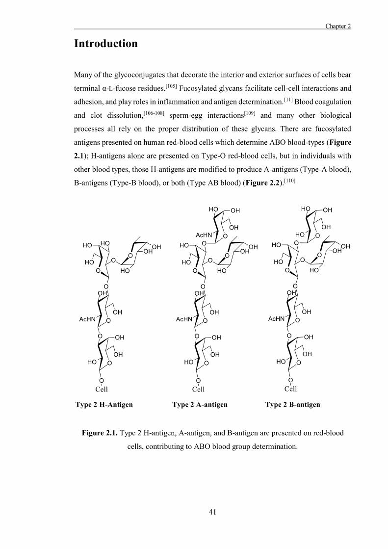
Many of the glycoconjugates that decorate the interior and exterior surfaces of cells bear
terminal α-L-fucose residues.[105] Fucosylated glycans facilitate cell-cell interactions and
adhesion, and play roles in inflammation and antigen determination.[11] Blood coagulation
and clot dissolution,[106-108] sperm-egg interactions[109] and many other biological
processes all rely on the proper distribution of these glycans. There are fucosylated
antigens presented on human red-blood cells which determine ABO blood-types (Figure
2.1); H-antigens alone are presented on Type-O red-blood cells, but in individuals with
other blood types, those H-antigens are modified to produce A-antigens (Type-A blood),
B-antigens (Type-B blood), or both (Type AB blood) (Figure 2.2).[110]
Figure 2.1. Type 2 H-antigen, A-antigen, and B-antigen are presented on red-blood
cells, contributing to ABO blood group determination.
Chapter 2 .
42
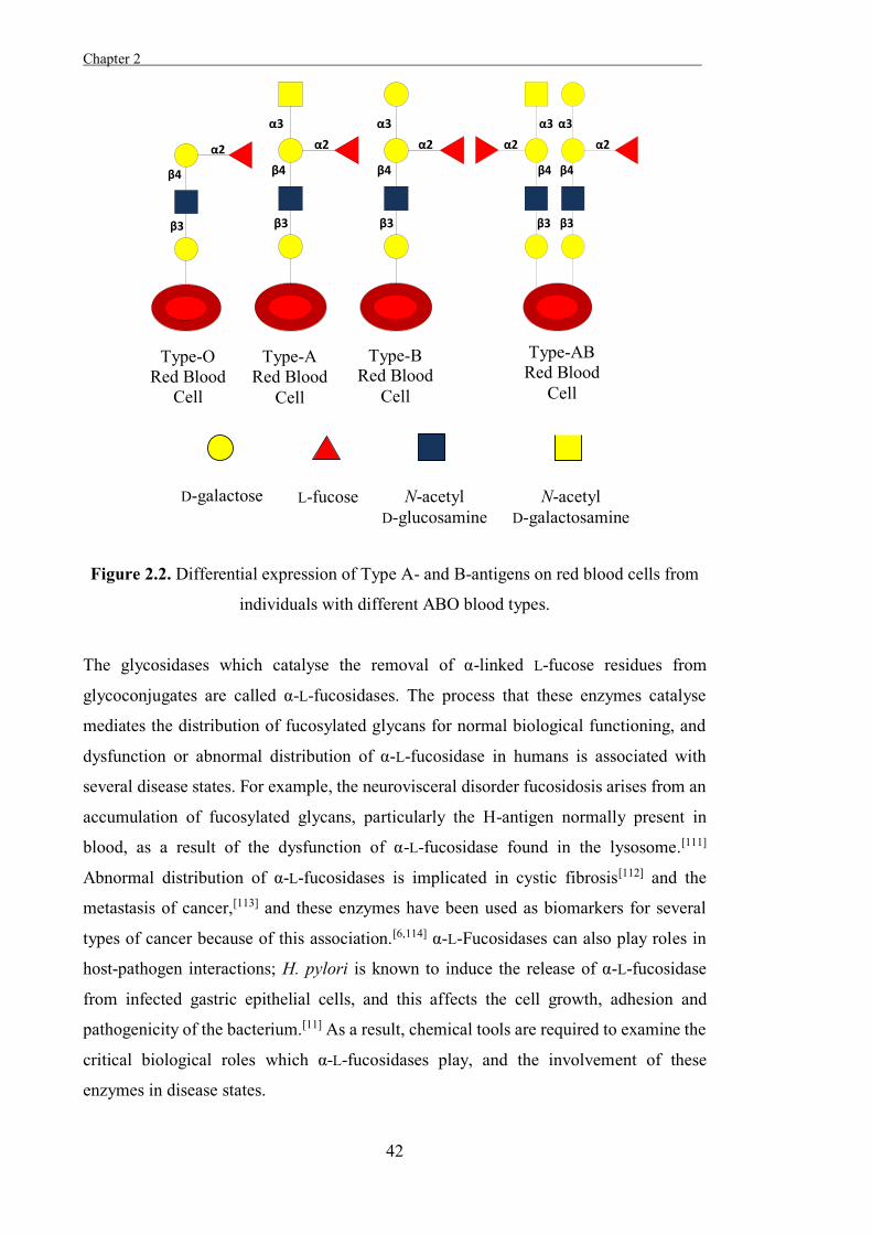
Legend
Figure 2.2. Differential expression of Type A- and B-antigens on red blood cells from
individuals with different ABO blood types.
The glycosidases which catalyse the removal of α-linked L-fucose residues from
glycoconjugates are called α-L-fucosidases. The process that these enzymes catalyse
mediates the distribution of fucosylated glycans for normal biological functioning, and
dysfunction or abnormal distribution of α-L-fucosidase in humans is associated with
several disease states. For example, the neurovisceral disorder fucosidosis arises from an
accumulation of fucosylated glycans, particularly the H-antigen normally present in
blood, as a result of the dysfunction of α-L-fucosidase found in the lysosome.[111]
Abnormal distribution of α-L-fucosidases is implicated in cystic fibrosis[112] and the
metastasis of cancer,[113] and these enzymes have been used as biomarkers for several
types of cancer because of this association.[6,114] α-L-Fucosidases can also play roles in
host-pathogen interactions; H. pylori is known to induce the release of α-L-fucosidase
from infected gastric epithelial cells, and this affects the cell growth, adhesion and
pathogenicity of the bacterium.[11] As a result, chemical tools are required to examine the
critical biological roles which α-L-fucosidases play, and the involvement of these
enzymes in disease states.
α2
β4
β3
α2
β4
β3
α3
α2
β4
β3
α3
β4
β3
α3
α2
β4
β3
α3
α2
Type-O Red Blood
Cell
Type-A Red Blood
Cell
Type-B Red Blood
Cell
Type-AB Red Blood
Cell
N-acetyl D-glucosamine
N-acetyl D-galactosamine
L-fucose D-galactose
Chapter 2
43
Classification and Mechanisms of α-L-Fucosidases
As with other glycosidases, α-L-fucosidases have been classified into families based upon
amino acid sequence homologies. All known α-L-fucosidases fall into the families GH29
or GH95 (EC 3.2.1.51), and there are no enzymes currently classified in these families
which lack α-L-fucosidase activity.[13] All known GH29 α-L-fucosidases retain the α-
configuration of their substrates, while GH95 α-L-fucosidases invert the configuration of
their substrates at the anomeric position.
There are two α-L-fucosidases present in human tissue: an extracellular α-L-fucosidase
(FucA2), and a better studied intracellular α-L-fucosidase (FucA1).[115] These two
enzymes have nearly identical amino acid sequences and both belong to family GH29.
Human α-L-fucosidases hydrolyse α-configured 1,2-, 1,3-, 1,4- and 1,6- linkages,[116] and
broad spectrum activity is required of these enzymes because of the varied fucosylated
glycans which they act upon. GH29 α-L-fucosidases have been studied in more depth than
GH95 α-L-fucosidases, possibly because of their involvement in human disease states.[111-
113,117] The limited availability of human α-L-fucosidases has hampered their direct study,
and so closely related GH29 α-L-fucosidase bacterial homologues are often used as
models to study the human enzymes.[105]
GH29 α-L-fucosidases are thought to act through a double-displacement mechanism with
a covalent β-glycosyl-enzyme intermediate, as with other retaining glycosidases.[116] X-
ray crystal structures have been obtained of this glycosyl-enzyme intermediate in α-L-
fucosidases from Thermotoga maritima[118] and Bacteroides thetaiotaomicron,[17] using
mutant enzymes and difluorinated L-fucose derivatives. The ‘trapped’ glycosyl-enzyme
intermediates observed were in the 3S1 conformation, and Michaelis complex structures
showed the substrate bound in a 1C4 conformation. From the results, a half-chair
conformation was expected for the putative oxocarbenium ion-like transition-state based
upon the conformational itineraries for pyranoses and as such, a 1C4-3H4-3S1
conformational itinerary has been predicted. These results have also been further ratified
by quantum mechanical analysis (Figure 2.3).[17,118]
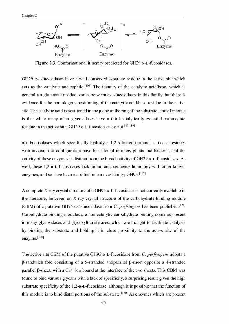
Chapter 2 .
44
Figure 2.3. Conformational itinerary predicted for GH29 α-L-fucosidases.
GH29 α-L-fucosidases have a well conserved aspartate residue in the active site which
acts as the catalytic nucleophile.[105] The identity of the catalytic acid/base, which is
generally a glutamate residue, varies between α-L-fucosidases in this family, but there is
evidence for the homologous positioning of the catalytic acid/base residue in the active
site. The catalytic acid is positioned in the plane of the ring of the substrate, and of interest
is that while many other glycosidases have a third catalytically essential carboxylate
residue in the active site, GH29 α-L-fucosidases do not.[17,119]
α-L-Fucosidases which specifically hydrolyse 1,2-α-linked terminal L-fucose residues
with inversion of configuration have been found in many plants and bacteria, and the
activity of these enzymes is distinct from the broad activity of GH29 α-L-fucosidases. As
well, these 1,2-α-L-fucosidases lack amino acid sequence homology with other known
enzymes, and so have been classified into a new family; GH95.[117]
A complete X-ray crystal structure of a GH95 α-L-fucosidase is not currently available in
the literature, however, an X-ray crystal structure of the carbohydrate-binding-module
(CBM) of a putative GH95 α-L-fucosidase from C. perfringens has been published.[120]
Carbohydrate-binding-modules are non-catalytic carbohydrate-binding domains present
in many glycosidases and glycosyltransferases, which are thought to facilitate catalysis
by binding the substrate and holding it in close proximity to the active site of the
enzyme.[120]
The active site CBM of the putative GH95 α-L-fucosidase from C. perfringens adopts a
β-sandwich fold consisting of a 5-stranded antiparallel β-sheet opposite a 4-stranded
parallel β-sheet, with a Ca2+ ion bound at the interface of the two sheets. This CBM was
found to bind various glycans with a lack of specificity, a surprising result given the high
substrate specificity of the 1,2-α-L-fucosidase, although it is possible that the function of
this module is to bind distal portions of the substrate.[120] As enzymes which are present
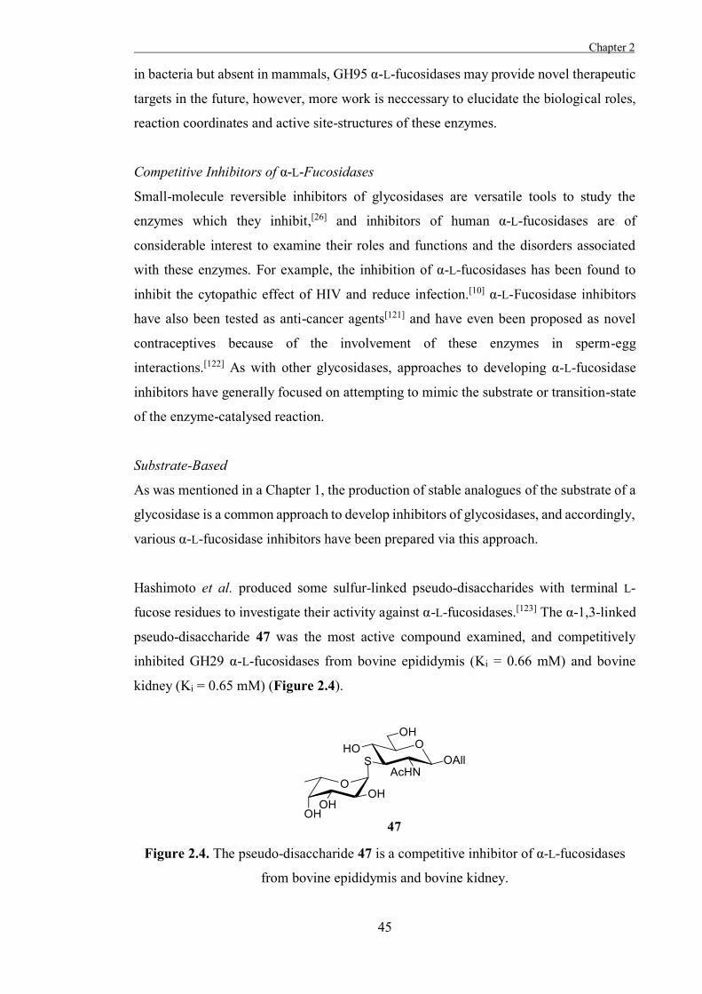
Chapter 2
45
in bacteria but absent in mammals, GH95 α-L-fucosidases may provide novel therapeutic
targets in the future, however, more work is neccessary to elucidate the biological roles,
reaction coordinates and active site-structures of these enzymes.
Competitive Inhibitors of α-L-Fucosidases
Small-molecule reversible inhibitors of glycosidases are versatile tools to study the
enzymes which they inhibit,[26] and inhibitors of human α-L-fucosidases are of
considerable interest to examine their roles and functions and the disorders associated
with these enzymes. For example, the inhibition of α-L-fucosidases has been found to
inhibit the cytopathic effect of HIV and reduce infection.[10] α-L-Fucosidase inhibitors
have also been tested as anti-cancer agents[121] and have even been proposed as novel
contraceptives because of the involvement of these enzymes in sperm-egg
interactions.[122] As with other glycosidases, approaches to developing α-L-fucosidase
inhibitors have generally focused on attempting to mimic the substrate or transition-state
of the enzyme-catalysed reaction.
Substrate-Based
As was mentioned in a Chapter 1, the production of stable analogues of the substrate of a
glycosidase is a common approach to develop inhibitors of glycosidases, and accordingly,
various α-L-fucosidase inhibitors have been prepared via this approach.
Hashimoto et al. produced some sulfur-linked pseudo-disaccharides with terminal L-
fucose residues to investigate their activity against α-L-fucosidases.[123] The α-1,3-linked
pseudo-disaccharide 47 was the most active compound examined, and competitively
inhibited GH29 α-L-fucosidases from bovine epididymis (Ki = 0.66 mM) and bovine
kidney (Ki = 0.65 mM) (Figure 2.4).
Figure 2.4. The pseudo-disaccharide 47 is a competitive inhibitor of α-L-fucosidases
from bovine epididymis and bovine kidney.
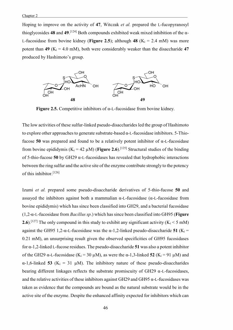
Chapter 2 .
46
Hoping to improve on the activity of 47, Witczak et al. prepared the L-fucopyranosyl
thioglycosides 48 and 49.[124] Both compounds exhibited weak mixed inhibition of the α-
L-fucosidase from bovine kidney (Figure 2.5); although 48 (Ki = 2.4 mM) was more
potent than 49 (Ki = 4.0 mM), both were considerably weaker than the disaccharide 47
produced by Hashimoto’s group.
Figure 2.5. Competitive inhibitors of α-L-fucosidase from bovine kidney.
The low activities of these sulfur-linked pseudo-disaccharides led the group of Hashimoto
to explore other approaches to generate substrate-based α-L-fucosidase inhibitors. 5-Thio-
fucose 50 was prepared and found to be a relatively potent inhibitor of α-L-fucosidase
from bovine epididymis (Ki = 42 µM) (Figure 2.6).[125] Structural studies of the binding
of 5-thio-fucose 50 by GH29 α-L-fucosidases has revealed that hydrophobic interactions
between the ring sulfur and the active site of the enzyme contribute strongly to the potency
of this inhibitor.[126]
Izumi et al. prepared some pseudo-disaccharide derivatives of 5-thio-fucose 50 and
assayed the inhibitors against both a mammalian α-L-fucosidase (α-L-fucosidase from
bovine epididymis) which has since been classified into GH29, and a bacterial fucosidase
(1,2-α-L-fucosidase from Bacillus sp.) which has since been classified into GH95 (Figure
2.6).[127] The only compound in this study to exhibit any significant activity (Ki < 5 mM)
against the GH95 1,2-α-L-fucosidase was the α-1,2-linked pseudo-disaccharide 51 (Ki =
0.21 mM), an unsurprising result given the observed specificities of GH95 fucosidases
for α-1,2-linked L-fucose residues. The pseudo-disaccharide 51 was also a potent inhibitor
of the GH29 α-L-fucosidase (Ki = 30 µM), as were the α-1,3-linked 52 (Ki = 91 µM) and
α-1,6-linked 53 (Ki = 31 µM). The inhibitory nature of these pseudo-disaccharides
bearing different linkages reflects the substrate promiscuity of GH29 α-L-fucosidases,
and the relative activities of these inhibitors against GH29 and GH95 α-L-fucosidases was
taken as evidence that the compounds are bound as the natural substrate would be in the
active site of the enzyme. Despite the enhanced affinity expected for inhibitors which can
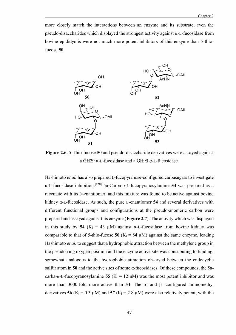
Chapter 2
47
more closely match the interactions between an enzyme and its substrate, even the
pseudo-disaccharides which displayed the strongest activity against α-L-fucosidase from
bovine epididymis were not much more potent inhibitors of this enzyme than 5-thio-
fucose 50.
Figure 2.6. 5-Thio-fucose 50 and pseudo-disaccharide derivatives were assayed against
a GH29 α-L-fucosidase and a GH95 α-L-fucosidase.
Hashimoto et al. has also prepared L-fucopyranose-configured carbasugars to investigate
α-L-fucosidase inhibition.[128] 5a-Carba-α-L-fucopyranosylamine 54 was prepared as a
racemate with its D-enantiomer, and this mixture was found to be active against bovine
kidney α-L-fucosidase. As such, the pure L-enantiomer 54 and several derivatives with
different functional groups and configurations at the pseudo-anomeric carbon were
prepared and assayed against this enzyme (Figure 2.7). The activity which was displayed
in this study by 54 (Ki = 43 µM) against α-L-fucosidase from bovine kidney was
comparable to that of 5-thio-fucose 50 (Ki = 84 µM) against the same enzyme, leading
Hashimoto et al. to suggest that a hydrophobic attraction between the methylene group in
the pseudo-ring oxygen position and the enzyme active site was contributing to binding,
somewhat analogous to the hydrophobic attraction observed between the endocyclic
sulfur atom in 50 and the active sites of some α-fucosidases. Of these compounds, the 5a-
carba-α-L-fucopyranosylamine 55 (Ki = 12 nM) was the most potent inhibitor and was
more than 3000-fold more active than 54. The α- and β- configured aminomethyl
derivatives 56 (Ki = 0.3 µM) and 57 (Ki = 2.8 µM) were also relatively potent, with the
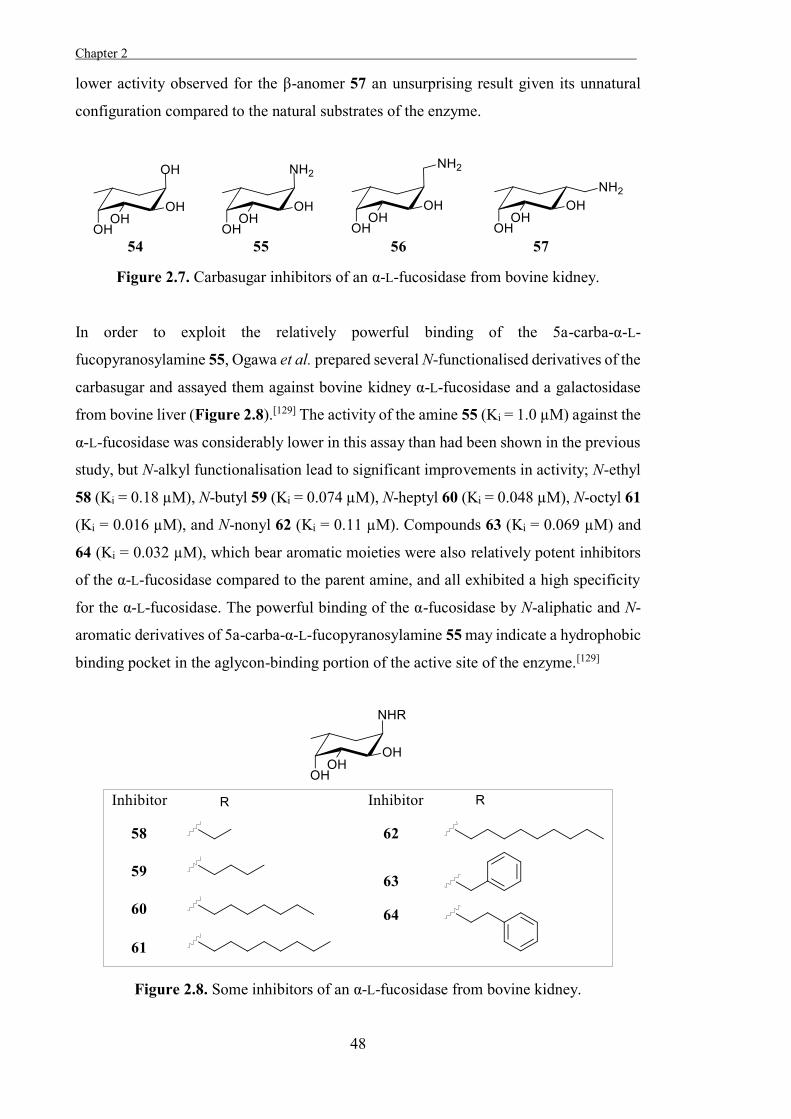
Chapter 2 .
48
lower activity observed for the β-anomer 57 an unsurprising result given its unnatural
configuration compared to the natural substrates of the enzyme.
Figure 2.7. Carbasugar inhibitors of an α-L-fucosidase from bovine kidney.
In order to exploit the relatively powerful binding of the 5a-carba-α-L-
fucopyranosylamine 55, Ogawa et al. prepared several N-functionalised derivatives of the
carbasugar and assayed them against bovine kidney α-L-fucosidase and a galactosidase
from bovine liver (Figure 2.8).[129] The activity of the amine 55 (Ki = 1.0 µM) against the
α-L-fucosidase was considerably lower in this assay than had been shown in the previous
study, but N-alkyl functionalisation lead to significant improvements in activity; N-ethyl
58 (Ki = 0.18 µM), N-butyl 59 (Ki = 0.074 µM), N-heptyl 60 (Ki = 0.048 µM), N-octyl 61
(Ki = 0.016 µM), and N-nonyl 62 (Ki = 0.11 µM). Compounds 63 (Ki = 0.069 µM) and
64 (Ki = 0.032 µM), which bear aromatic moieties were also relatively potent inhibitors
of the α-L-fucosidase compared to the parent amine, and all exhibited a high specificity
for the α-L-fucosidase. The powerful binding of the α-fucosidase by N-aliphatic and N-
aromatic derivatives of 5a-carba-α-L-fucopyranosylamine 55 may indicate a hydrophobic
binding pocket in the aglycon-binding portion of the active site of the enzyme.[129]
Figure 2.8. Some inhibitors of an α-L-fucosidase from bovine kidney.
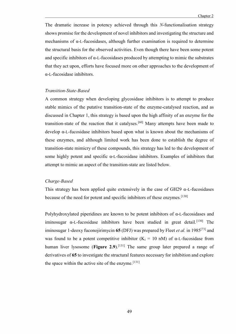
Chapter 2
49
The dramatic increase in potency achieved through this N-functionalisation strategy
shows promise for the development of novel inhibitors and investigating the structure and
mechanisms of α-L-fucosidases, although further examination is required to determine
the structural basis for the observed activities. Even though there have been some potent
and specific inhibitors of α-L-fucosidases produced by attempting to mimic the substrates
that they act upon, efforts have focused more on other approaches to the development of
α-L-fucosidase inhibitors.
Transition-State-Based
A common strategy when developing glycosidase inhibitors is to attempt to produce
stable mimics of the putative transition-state of the enzyme-catalysed reaction, and as
discussed in Chapter 1, this strategy is based upon the high affinity of an enzyme for the
transition-state of the reaction that it catalyses.[60] Many attempts have been made to
develop α-L-fucosidase inhibitors based upon what is known about the mechanisms of
these enzymes, and although limited work has been done to establish the degree of
transition-state mimicry of these compounds, this strategy has led to the development of
some highly potent and specific α-L-fucosidase inhibitors. Examples of inhibitors that
attempt to mimic an aspect of the transition-state are listed below.
Charge-Based
This strategy has been applied quite extensively in the case of GH29 α-L-fucosidases
because of the need for potent and specific inhibitors of these enzymes.[130]
Polyhydroxylated piperidines are known to be potent inhibitors of α-L-fucosidases and
iminosugar α-L-fucosidase inhibitors have been studied in great detail.[130] The
iminosugar 1-deoxy fuconojirimycin 65 (DFJ) was prepared by Fleet et al. in 1985[73] and
was found to be a potent competitive inhibitor (Ki = 10 nM) of α-L-fucosidase from
human liver lysosome (Figure 2.9).[131] The same group later prepared a range of
derivatives of 65 to investigate the structural features necessary for inhibition and explore
the space within the active site of the enzyme.[131]
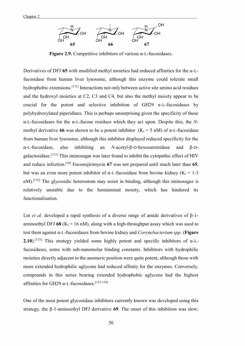
Chapter 2 .
50
Figure 2.9. Competitive inhibitors of various α-L-fucosidases.
Derivatives of DFJ 65 with modified methyl moieties had reduced affinities for the α-L-
fucosidase from human liver lysosome, although this enzyme could tolerate small
hydrophobic extensions.[131] Interactions not only between active site amino acid residues
and the hydroxyl moieties at C2, C3 and C4, but also the methyl moiety appear to be
crucial for the potent and selective inhibition of GH29 α-L-fucosidases by
polyhydroxylated piperidines. This is perhaps unsurprising given the specificity of these
α-L-fucosidases for the α-L-fucose residues which they act upon. Despite this, the N-
methyl derivative 66 was shown to be a potent inhibitor (Ki = 5 nM) of α-L-fucosidase
from human liver lysosome, although this inhibitor displayed reduced specificity for the
α-L-fucosidase, also inhibiting an N-acetyl-β-D-hexosaminidase and β-D-
galactosidase.[131] This iminosugar was later found to inhibit the cytopathic effect of HIV
and reduce infection.[10] Fuconojirimycin 67 was not prepared until much later than 65,
but was an even more potent inhibitor of α-L-fucosidase from bovine kidney (Ki = 1-3
nM).[132] The glycosidic heteroatom may assist in binding, although this iminosugar is
relatively unstable due to the hemiaminal moiety, which has hindered its
functionalisation.
Lin et al. developed a rapid synthesis of a diverse range of amide derivatives of β-1-
aminoethyl DFJ 68 (Ki = 16 nM), along with a high-throughput assay which was used to
test them against α-L-fucosidases from bovine kidney and Corynebacterium spp. (Figure
2.10).[133] This strategy yielded some highly potent and specific inhibitors of α-L-
fucosidases, some with sub-nanomolar binding constants. Inhibitors with hydrophilic
moieties directly adjacent to the anomeric position were quite potent, although those with
more extended hydrophilic aglycons had reduced affinity for the enzymes. Conversely,
compounds in this series bearing extended hydrophobic aglycons had the highest
affinities for GH29 α-L-fucosidases.[133-134]
One of the most potent glycosidase inhibitors currently known was developed using this
strategy, the β-1-aminoethyl DFJ derivative 69. The onset of this inhibition was slow;
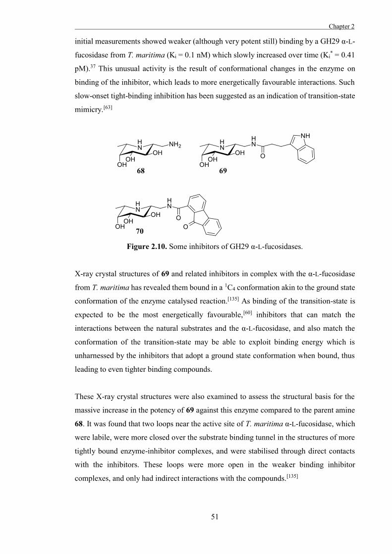
Chapter 2
51
initial measurements showed weaker (although very potent still) binding by a GH29 α-L-
fucosidase from T. maritima (Ki = 0.1 nM) which slowly increased over time (Ki* = 0.41
pM).37 This unusual activity is the result of conformational changes in the enzyme on
binding of the inhibitor, which leads to more energetically favourable interactions. Such
slow-onset tight-binding inhibition has been suggested as an indication of transition-state
mimicry.[63]
Figure 2.10. Some inhibitors of GH29 α-L-fucosidases.
X-ray crystal structures of 69 and related inhibitors in complex with the α-L-fucosidase
from T. maritima has revealed them bound in a 1C4 conformation akin to the ground state
conformation of the enzyme catalysed reaction.[135] As binding of the transition-state is
expected to be the most energetically favourable,[60] inhibitors that can match the
interactions between the natural substrates and the α-L-fucosidase, and also match the
conformation of the transition-state may be able to exploit binding energy which is
unharnessed by the inhibitors that adopt a ground state conformation when bound, thus
leading to even tighter binding compounds.
These X-ray crystal structures were also examined to assess the structural basis for the
massive increase in the potency of 69 against this enzyme compared to the parent amine
68. It was found that two loops near the active site of T. maritima α-L-fucosidase, which
were labile, were more closed over the substrate binding tunnel in the structures of more
tightly bound enzyme-inhibitor complexes, and were stabilised through direct contacts
with the inhibitors. These loops were more open in the weaker binding inhibitor
complexes, and only had indirect interactions with the compounds.[135]
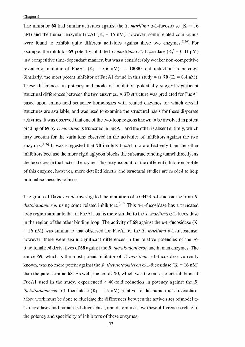
Chapter 2 .
52
The inhibitor 68 had similar activities against the T. maritima α-L-fucosidase (Ki = 16
nM) and the human enzyme FucA1 (Ki = 15 nM), however, some related compounds
were found to exhibit quite different activities against these two enzymes.[136] For
example, the inhibitor 69 potently inhibited T. maritima α-L-fucosidase (Ki* = 0.41 pM)
in a competitive time-dependant manner, but was a considerably weaker non-competitive
reversible inhibitor of FucA1 (Ki = 5.6 nM)—a 10000-fold reduction in potency.
Similarly, the most potent inhibitor of FucA1 found in this study was 70 (Ki = 0.4 nM).
These differences in potency and mode of inhibition potentially suggest significant
structural differences between the two enzymes. A 3D structure was predicted for FucA1
based upon amino acid sequence homologies with related enzymes for which crystal
structures are available, and was used to examine the structural basis for these disparate
activities. It was observed that one of the two-loop regions known to be involved in potent
binding of 69 by T. maritima is truncated in FucA1, and the other is absent entirely, which
may account for the variations observed in the activities of inhibitors against the two
enzymes.[136] It was suggested that 70 inhibits FucA1 more effectively than the other
inhibitors because the more rigid aglycon blocks the substrate binding tunnel directly, as
the loop does in the bacterial enzyme. This may account for the different inhibition profile
of this enzyme, however, more detailed kinetic and structural studies are needed to help
rationalise these hypotheses.
The group of Davies et al. investigated the inhibition of a GH29 α-L-fucosidase from B.
thetaiotaomicron using some related inhibitors.[119] This α-L-fucosidase has a truncated
loop region similar to that in FucA1, but is more similar to the T. maritima α-L-fucosidase
in the region of the other binding loop. The activity of 68 against the α-L-fucosidase (Ki
= 16 nM) was similar to that observed for FucA1 or the T. maritima α-L-fucosidase,
however, there were again significant differences in the relative potencies of the N-
functionalised derivatives of 68 against the B. thetaiotaomicron and human enzymes. The
amide 69, which is the most potent inhibitor of T. maritima α-L-fucosidase currently
known, was no more potent against the B. thetaiotaomicron α-L-fucosidase (Ki = 16 nM)
than the parent amine 68. As well, the amide 70, which was the most potent inhibitor of
FucA1 used in the study, experienced a 40-fold reduction in potency against the B.
thetaiotaomicron α-L-fucosidase (Ki = 16 nM) relative to the human α-L-fucosidase.
More work must be done to elucidate the differences between the active sites of model α-
L-fucosidases and human α-L-fucosidase, and determine how these differences relate to
the potency and specificity of inhibitors of these enzymes.
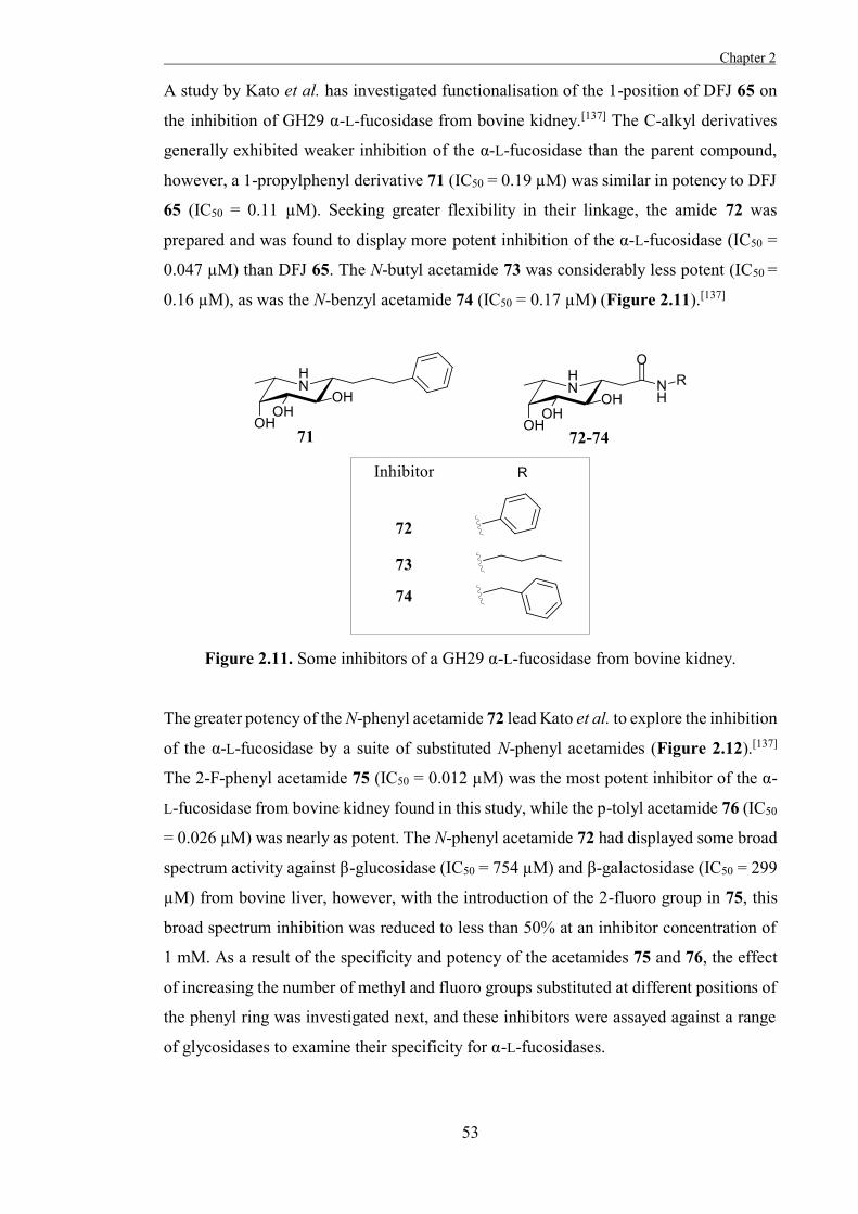
Chapter 2
53
A study by Kato et al. has investigated functionalisation of the 1-position of DFJ 65 on
the inhibition of GH29 α-L-fucosidase from bovine kidney.[137] The C-alkyl derivatives
generally exhibited weaker inhibition of the α-L-fucosidase than the parent compound,
however, a 1-propylphenyl derivative 71 (IC50 = 0.19 µM) was similar in potency to DFJ
65 (IC50 = 0.11 µM). Seeking greater flexibility in their linkage, the amide 72 was
prepared and was found to display more potent inhibition of the α-L-fucosidase (IC50 =
0.047 µM) than DFJ 65. The N-butyl acetamide 73 was considerably less potent (IC50 =
0.16 µM), as was the N-benzyl acetamide 74 (IC50 = 0.17 µM) (Figure 2.11).[137]
Figure 2.11. Some inhibitors of a GH29 α-L-fucosidase from bovine kidney.
The greater potency of the N-phenyl acetamide 72 lead Kato et al. to explore the inhibition
of the α-L-fucosidase by a suite of substituted N-phenyl acetamides (Figure 2.12).[137]
The 2-F-phenyl acetamide 75 (IC50 = 0.012 µM) was the most potent inhibitor of the α-
L-fucosidase from bovine kidney found in this study, while the p-tolyl acetamide 76 (IC50
= 0.026 µM) was nearly as potent. The N-phenyl acetamide 72 had displayed some broad
spectrum activity against β-glucosidase (IC50 = 754 µM) and β-galactosidase (IC50 = 299
µM) from bovine liver, however, with the introduction of the 2-fluoro group in 75, this
broad spectrum inhibition was reduced to less than 50% at an inhibitor concentration of
1 mM. As a result of the specificity and potency of the acetamides 75 and 76, the effect
of increasing the number of methyl and fluoro groups substituted at different positions of
the phenyl ring was investigated next, and these inhibitors were assayed against a range
of glycosidases to examine their specificity for α-L-fucosidases.
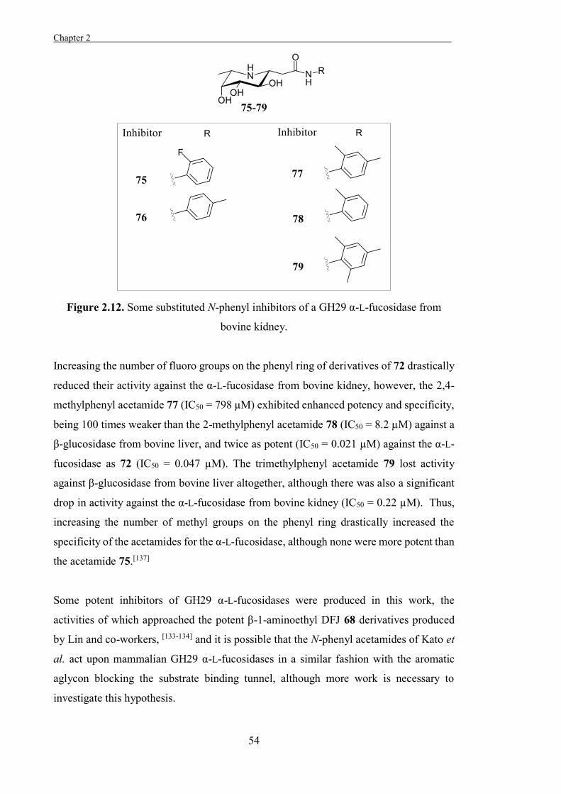
Chapter 2 .
54
Figure 2.12. Some substituted N-phenyl inhibitors of a GH29 α-L-fucosidase from
bovine kidney.
Increasing the number of fluoro groups on the phenyl ring of derivatives of 72 drastically
reduced their activity against the α-L-fucosidase from bovine kidney, however, the 2,4-
methylphenyl acetamide 77 (IC50 = 798 µM) exhibited enhanced potency and specificity,
being 100 times weaker than the 2-methylphenyl acetamide 78 (IC50 = 8.2 µM) against a
β-glucosidase from bovine liver, and twice as potent (IC50 = 0.021 µM) against the α-L-
fucosidase as 72 (IC50 = 0.047 µM). The trimethylphenyl acetamide 79 lost activity
against β-glucosidase from bovine liver altogether, although there was also a significant
drop in activity against the α-L-fucosidase from bovine kidney (IC50 = 0.22 µM). Thus,
increasing the number of methyl groups on the phenyl ring drastically increased the
specificity of the acetamides for the α-L-fucosidase, although none were more potent than
the acetamide 75.[137]
Some potent inhibitors of GH29 α-L-fucosidases were produced in this work, the
activities of which approached the potent β-1-aminoethyl DFJ 68 derivatives produced
by Lin and co-workers, [133-134] and it is possible that the N-phenyl acetamides of Kato et
al. act upon mammalian GH29 α-L-fucosidases in a similar fashion with the aromatic
aglycon blocking the substrate binding tunnel, although more work is necessary to
investigate this hypothesis.
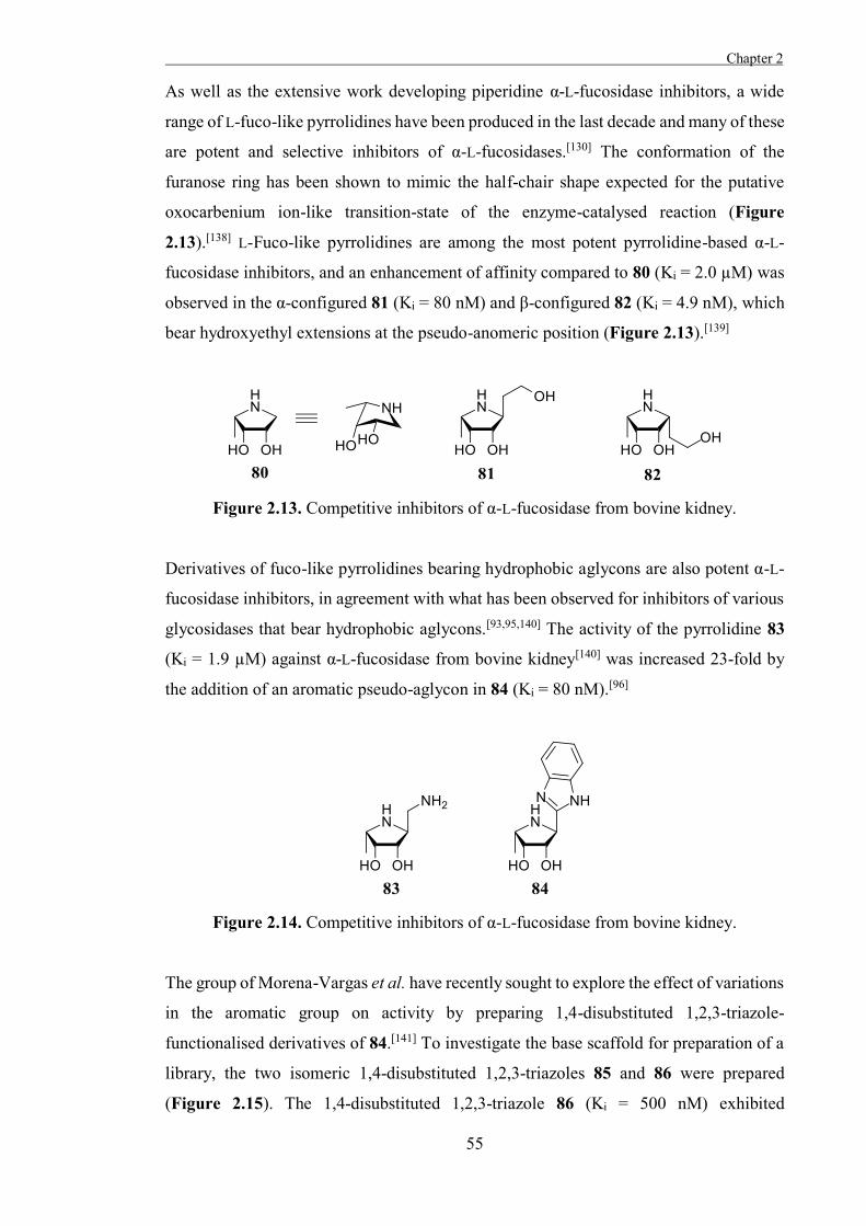
Chapter 2
55
As well as the extensive work developing piperidine α-L-fucosidase inhibitors, a wide
range of L-fuco-like pyrrolidines have been produced in the last decade and many of these
are potent and selective inhibitors of α-L-fucosidases.[130] The conformation of the
furanose ring has been shown to mimic the half-chair shape expected for the putative
oxocarbenium ion-like transition-state of the enzyme-catalysed reaction (Figure
2.13).[138] L-Fuco-like pyrrolidines are among the most potent pyrrolidine-based α-L-
fucosidase inhibitors, and an enhancement of affinity compared to 80 (Ki = 2.0 µM) was
observed in the α-configured 81 (Ki = 80 nM) and β-configured 82 (Ki = 4.9 nM), which
bear hydroxyethyl extensions at the pseudo-anomeric position (Figure 2.13).[139]
Figure 2.13. Competitive inhibitors of α-L-fucosidase from bovine kidney.
Derivatives of fuco-like pyrrolidines bearing hydrophobic aglycons are also potent α-L-
fucosidase inhibitors, in agreement with what has been observed for inhibitors of various
glycosidases that bear hydrophobic aglycons.[93,95,140] The activity of the pyrrolidine 83
(Ki = 1.9 µM) against α-L-fucosidase from bovine kidney[140] was increased 23-fold by
the addition of an aromatic pseudo-aglycon in 84 (Ki = 80 nM).[96]
Figure 2.14. Competitive inhibitors of α-L-fucosidase from bovine kidney.
The group of Morena-Vargas et al. have recently sought to explore the effect of variations
in the aromatic group on activity by preparing 1,4-disubstituted 1,2,3-triazole-
functionalised derivatives of 84.[141] To investigate the base scaffold for preparation of a
library, the two isomeric 1,4-disubstituted 1,2,3-triazoles 85 and 86 were prepared
(Figure 2.15). The 1,4-disubstituted 1,2,3-triazole 86 (Ki = 500 nM) exhibited
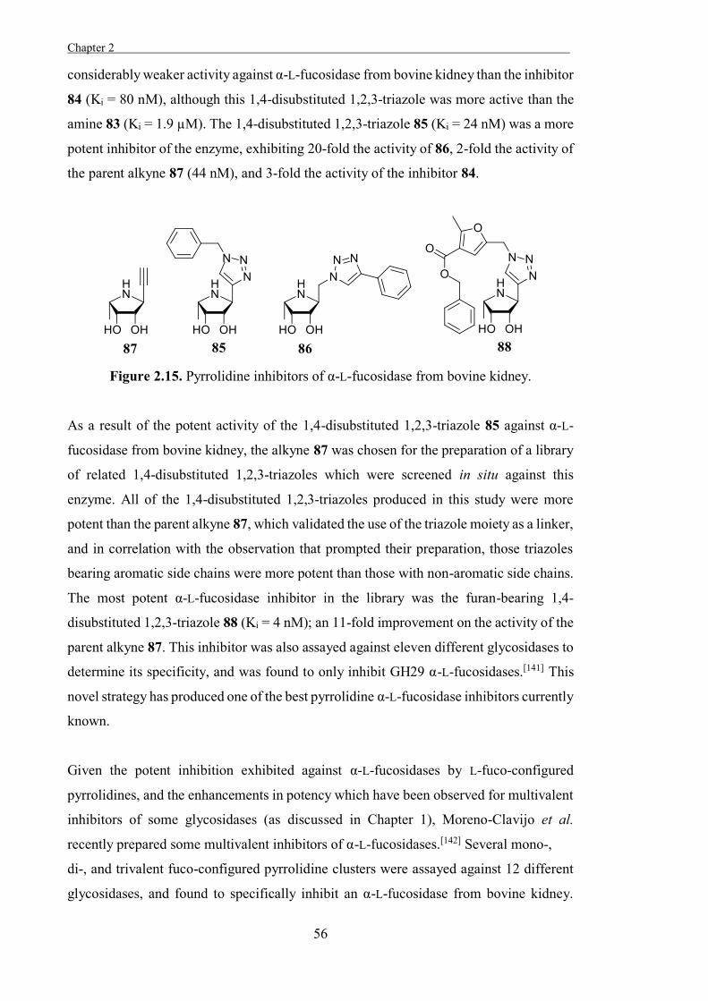
Chapter 2 .
56
considerably weaker activity against α-L-fucosidase from bovine kidney than the inhibitor
84 (Ki = 80 nM), although this 1,4-disubstituted 1,2,3-triazole was more active than the
amine 83 (Ki = 1.9 µM). The 1,4-disubstituted 1,2,3-triazole 85 (Ki = 24 nM) was a more
potent inhibitor of the enzyme, exhibiting 20-fold the activity of 86, 2-fold the activity of
the parent alkyne 87 (44 nM), and 3-fold the activity of the inhibitor 84.
Figure 2.15. Pyrrolidine inhibitors of α-L-fucosidase from bovine kidney.
As a result of the potent activity of the 1,4-disubstituted 1,2,3-triazole 85 against α-L-
fucosidase from bovine kidney, the alkyne 87 was chosen for the preparation of a library
of related 1,4-disubstituted 1,2,3-triazoles which were screened in situ against this
enzyme. All of the 1,4-disubstituted 1,2,3-triazoles produced in this study were more
potent than the parent alkyne 87, which validated the use of the triazole moiety as a linker,
and in correlation with the observation that prompted their preparation, those triazoles
bearing aromatic side chains were more potent than those with non-aromatic side chains.
The most potent α-L-fucosidase inhibitor in the library was the furan-bearing 1,4-
disubstituted 1,2,3-triazole 88 (Ki = 4 nM); an 11-fold improvement on the activity of the
parent alkyne 87. This inhibitor was also assayed against eleven different glycosidases to
determine its specificity, and was found to only inhibit GH29 α-L-fucosidases.[141] This
novel strategy has produced one of the best pyrrolidine α-L-fucosidase inhibitors currently
known.
Given the potent inhibition exhibited against α-L-fucosidases by L-fuco-configured
pyrrolidines, and the enhancements in potency which have been observed for multivalent
inhibitors of some glycosidases (as discussed in Chapter 1), Moreno-Clavijo et al.
recently prepared some multivalent inhibitors of α-L-fucosidases.[142] Several mono-,
di-, and trivalent fuco-configured pyrrolidine clusters were assayed against 12 different
glycosidases, and found to specifically inhibit an α-L-fucosidase from bovine kidney.
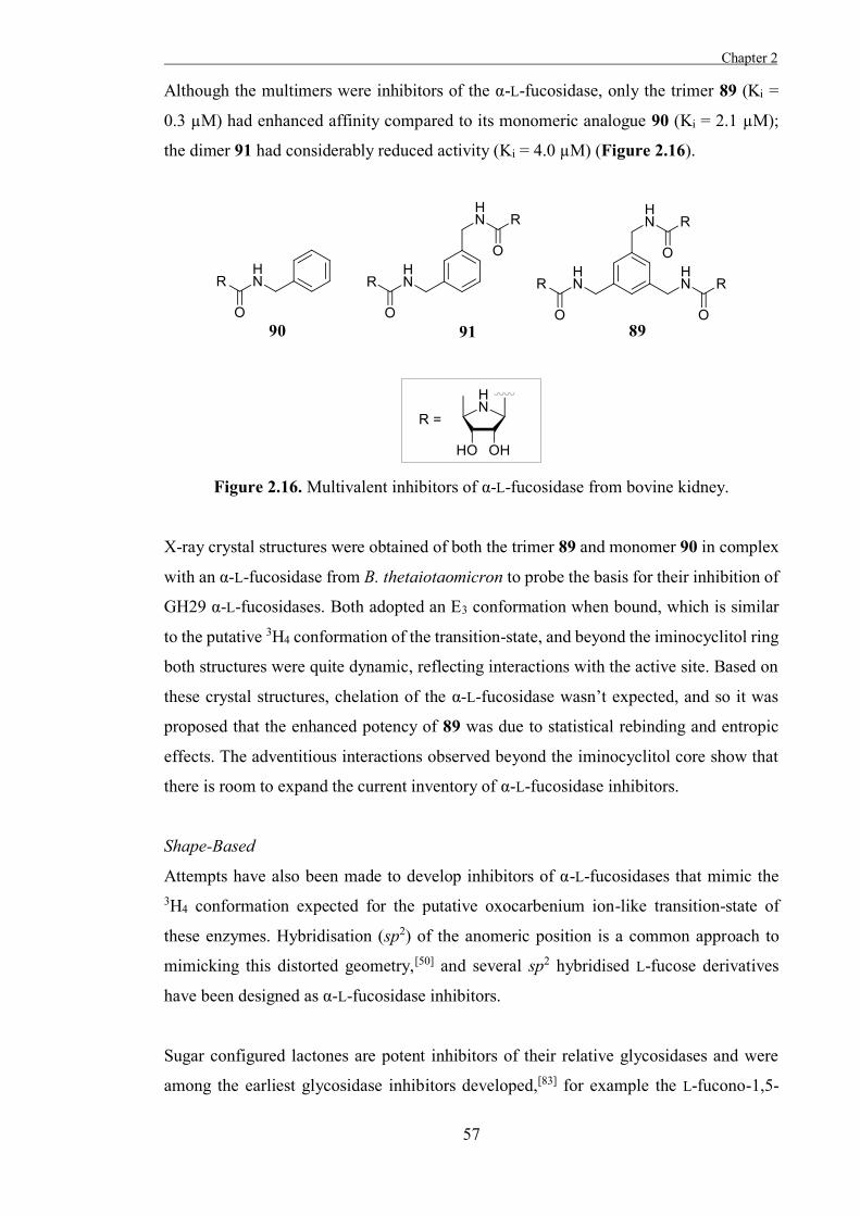
Chapter 2
57
Although the multimers were inhibitors of the α-L-fucosidase, only the trimer 89 (Ki =
0.3 µM) had enhanced affinity compared to its monomeric analogue 90 (Ki = 2.1 µM);
the dimer 91 had considerably reduced activity (Ki = 4.0 µM) (Figure 2.16).
Figure 2.16. Multivalent inhibitors of α-L-fucosidase from bovine kidney.
X-ray crystal structures were obtained of both the trimer 89 and monomer 90 in complex
with an α-L-fucosidase from B. thetaiotaomicron to probe the basis for their inhibition of
GH29 α-L-fucosidases. Both adopted an E3 conformation when bound, which is similar
to the putative 3H4 conformation of the transition-state, and beyond the iminocyclitol ring
both structures were quite dynamic, reflecting interactions with the active site. Based on
these crystal structures, chelation of the α-L-fucosidase wasn’t expected, and so it was
proposed that the enhanced potency of 89 was due to statistical rebinding and entropic
effects. The adventitious interactions observed beyond the iminocyclitol core show that
there is room to expand the current inventory of α-L-fucosidase inhibitors.
Shape-Based
Attempts have also been made to develop inhibitors of α-L-fucosidases that mimic the 3H4 conformation expected for the putative oxocarbenium ion-like transition-state of
these enzymes. Hybridisation (sp2) of the anomeric position is a common approach to
mimicking this distorted geometry,[50] and several sp2 hybridised L-fucose derivatives
have been designed as α-L-fucosidase inhibitors.
Sugar configured lactones are potent inhibitors of their relative glycosidases and were
among the earliest glycosidase inhibitors developed,[83] for example the L-fucono-1,5-
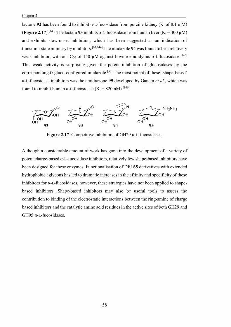
Chapter 2 .
58
lactone 92 has been found to inhibit α-L-fucosidase from porcine kidney (Ki of 8.1 mM)
(Figure 2.17).[143] The lactam 93 inhibits α-L-fucosidase from human liver (Ki = 400 µM)
and exhibits slow-onset inhibition, which has been suggested as an indication of
transition-state mimicry by inhibitors.[63,144] The imidazole 94 was found to be a relatively
weak inhibitor, with an IC50 of 150 µM against bovine epididymis α-L-fucosidase.[145]
This weak activity is surprising given the potent inhibition of glucosidases by the
corresponding D-gluco-configured imidazole.[50] The most potent of these ‘shape-based’
α-L-fucosidase inhibitors was the amidrazone 95 developed by Ganem et al., which was
found to inhibit human α-L-fucosidase (Ki = 820 nM).[146]
Figure 2.17. Competitive inhibitors of GH29 α-L-fucosidases.
Although a considerable amount of work has gone into the development of a variety of
potent charge-based α-L-fucosidase inhibitors, relatively few shape-based inhibitors have
been designed for these enzymes. Functionalisation of DFJ 65 derivatives with extended
hydrophobic aglycons has led to dramatic increases in the affinity and specificity of these
inhibitors for α-L-fucosidases, however, these strategies have not been applied to shape-
based inhibitors. Shape-based inhibitors may also be useful tools to assess the
contribution to binding of the electrostatic interactions between the ring-amine of charge
based inhibitors and the catalytic amino acid residues in the active sites of both GH29 and
GH95 α-L-fucosidases.
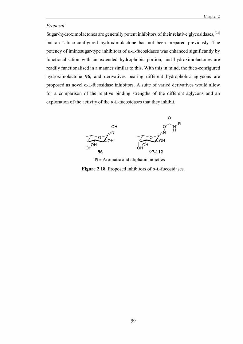
Chapter 2
59
Proposal
Sugar-hydroximolactones are generally potent inhibitors of their relative glycosidases,[93]
but an L-fuco-configured hydroximolactone has not been prepared previously. The
potency of iminosugar-type inhibitors of α-L-fucosidases was enhanced significantly by
functionalisation with an extended hydrophobic portion, and hydroximolactones are
readily functionalised in a manner similar to this. With this in mind, the fuco-configured
hydroximolactone 96, and derivatives bearing different hydrophobic aglycons are
proposed as novel α-L-fucosidase inhibitors. A suite of varied derivatives would allow
for a comparison of the relative binding strengths of the different aglycons and an
exploration of the activity of the α-L-fucosidases that they inhibit.
Figure 2.18. Proposed inhibitors of α-L-fucosidases.

Chapter 2 .
60
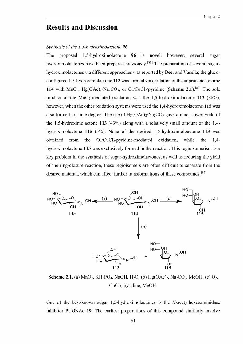
Chapter 2
61
Results and Discussion
Synthesis of the 1,5-hydroximolactone 96
The proposed 1,5-hydroximolactone 96 is novel, however, several sugar
hydroximolactones have been prepared previously.[89] The preparation of several sugar-
hydroximolactones via different approaches was reported by Beer and Vasella; the gluco-
configured 1,5-hydroximolactone 113 was formed via oxidation of the unprotected oxime
114 with MnO2, Hg(OAc)2/Na2CO3, or O2/CuCl2/pyridine (Scheme 2.1).[89] The sole
product of the MnO2-mediated oxidation was the 1,5-hydroximolactone 113 (86%),
however, when the other oxidation systems were used the 1,4-hydroximolactone 115 was
also formed to some degree. The use of Hg(OAc)2/Na2CO3 gave a much lower yield of
the 1,5-hydroximolactone 113 (43%) along with a relatively small amount of the 1,4-
hydroximolactone 115 (5%). None of the desired 1,5-hydroximoloactone 113 was
obtained from the O2/CuCl2/pyridine-mediated oxidation, while the 1,4-
hydroximolactone 115 was exclusively formed in the reaction. This regioisomerism is a
key problem in the synthesis of sugar-hydroximolactones; as well as reducing the yield
of the ring-closure reaction, these regioisomers are often difficult to separate from the
desired material, which can affect further transformations of these compounds.[97]
Scheme 2.1. (a) MnO2, KH2PO4, NaOH, H2O; (b) Hg(OAc)2, Na2CO3, MeOH; (c) O2,
CuCl2, pyridine, MeOH.
One of the best-known sugar 1,5-hydroximolactones is the N-acetylhexosaminidase
inhibitor PUGNAc 19. The earliest preparations of this compound similarly involve
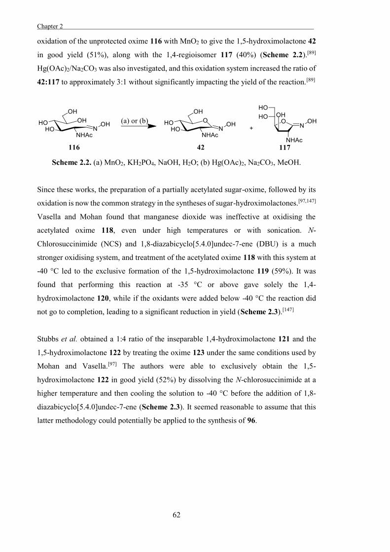
Chapter 2 .
62
oxidation of the unprotected oxime 116 with MnO2 to give the 1,5-hydroximolactone 42
in good yield (51%), along with the 1,4-regioisomer 117 (40%) (Scheme 2.2).[89]
Hg(OAc)2/Na2CO3 was also investigated, and this oxidation system increased the ratio of
42:117 to approximately 3:1 without significantly impacting the yield of the reaction.[89]
Scheme 2.2. (a) MnO2, KH2PO4, NaOH, H2O; (b) Hg(OAc)2, Na2CO3, MeOH.
Since these works, the preparation of a partially acetylated sugar-oxime, followed by its
oxidation is now the common strategy in the syntheses of sugar-hydroximolactones.[97,147]
Vasella and Mohan found that manganese dioxide was ineffective at oxidising the
acetylated oxime 118, even under high temperatures or with sonication. N-
Chlorosuccinimide (NCS) and 1,8-diazabicyclo[5.4.0]undec-7-ene (DBU) is a much
stronger oxidising system, and treatment of the acetylated oxime 118 with this system at
-40 °C led to the exclusive formation of the 1,5-hydroximolactone 119 (59%). It was
found that performing this reaction at -35 °C or above gave solely the 1,4-
hydroximolactone 120, while if the oxidants were added below -40 °C the reaction did
not go to completion, leading to a significant reduction in yield (Scheme 2.3).[147]
Stubbs et al. obtained a 1:4 ratio of the inseparable 1,4-hydroximolactone 121 and the
1,5-hydroximolactone 122 by treating the oxime 123 under the same conditions used by
Mohan and Vasella.[97] The authors were able to exclusively obtain the 1,5-
hydroximolactone 122 in good yield (52%) by dissolving the N-chlorosuccinimide at a
higher temperature and then cooling the solution to -40 °C before the addition of 1,8-
diazabicyclo[5.4.0]undec-7-ene (Scheme 2.3). It seemed reasonable to assume that this
latter methodology could potentially be applied to the synthesis of 96.
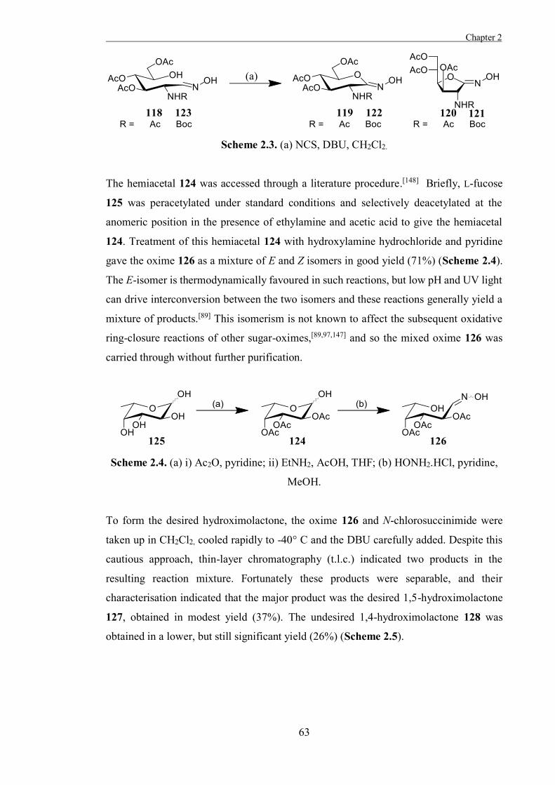
Chapter 2
63
Scheme 2.3. (a) NCS, DBU, CH2Cl2.
The hemiacetal 124 was accessed through a literature procedure.[148] Briefly, L-fucose
125 was peracetylated under standard conditions and selectively deacetylated at the
anomeric position in the presence of ethylamine and acetic acid to give the hemiacetal
124. Treatment of this hemiacetal 124 with hydroxylamine hydrochloride and pyridine
gave the oxime 126 as a mixture of E and Z isomers in good yield (71%) (Scheme 2.4).
The E-isomer is thermodynamically favoured in such reactions, but low pH and UV light
can drive interconversion between the two isomers and these reactions generally yield a
mixture of products.[89] This isomerism is not known to affect the subsequent oxidative
ring-closure reactions of other sugar-oximes,[89,97,147] and so the mixed oxime 126 was
carried through without further purification.
Scheme 2.4. (a) i) Ac2O, pyridine; ii) EtNH2, AcOH, THF; (b) HONH2.HCl, pyridine,
MeOH.
To form the desired hydroximolactone, the oxime 126 and N-chlorosuccinimide were
taken up in CH2Cl2, cooled rapidly to -40° C and the DBU carefully added. Despite this
cautious approach, thin-layer chromatography (t.l.c.) indicated two products in the
resulting reaction mixture. Fortunately these products were separable, and their
characterisation indicated that the major product was the desired 1,5-hydroximolactone
127, obtained in modest yield (37%). The undesired 1,4-hydroximolactone 128 was
obtained in a lower, but still significant yield (26%) (Scheme 2.5).
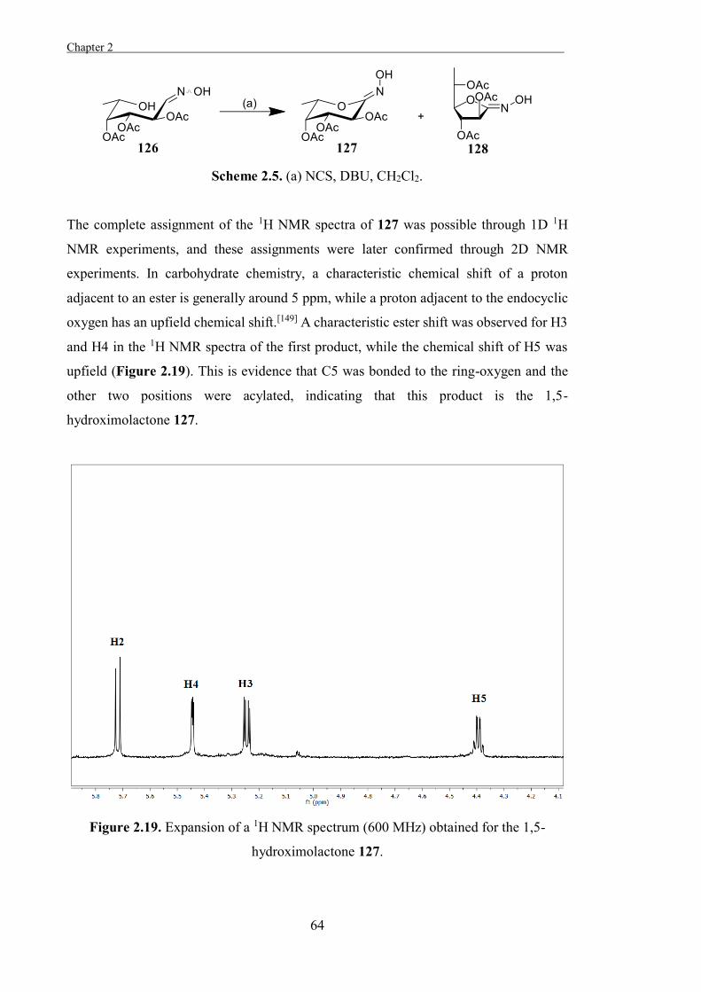
Chapter 2 .
64
Scheme 2.5. (a) NCS, DBU, CH2Cl2.
The complete assignment of the 1H NMR spectra of 127 was possible through 1D 1H
NMR experiments, and these assignments were later confirmed through 2D NMR
experiments. In carbohydrate chemistry, a characteristic chemical shift of a proton
adjacent to an ester is generally around 5 ppm, while a proton adjacent to the endocyclic
oxygen has an upfield chemical shift.[149] A characteristic ester shift was observed for H3
and H4 in the 1H NMR spectra of the first product, while the chemical shift of H5 was
upfield (Figure 2.19). This is evidence that C5 was bonded to the ring-oxygen and the
other two positions were acylated, indicating that this product is the 1,5-
hydroximolactone 127.
Figure 2.19. Expansion of a 1H NMR spectrum (600 MHz) obtained for the 1,5-
hydroximolactone 127.
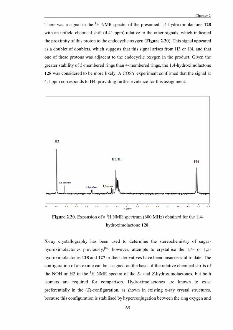
Chapter 2
65
There was a signal in the 1H NMR spectra of the presumed 1,4-hydroximolactone 128
with an upfield chemical shift (4.41 ppm) relative to the other signals, which indicated
the proximity of this proton to the endocyclic oxygen (Figure 2.20). This signal appeared
as a doublet of doublets, which suggests that this signal arises from H3 or H4, and that
one of these protons was adjacent to the endocyclic oxygen in the product. Given the
greater stability of 5-membered rings than 4-membered rings, the 1,4-hydroximolactone
128 was considered to be more likely. A COSY experiment confirmed that the signal at
4.1 ppm corresponds to H4, providing further evidence for this assignment.
Figure 2.20. Expansion of a 1H NMR spectrum (600 MHz) obtained for the 1,4-
hydroximolactone 128.
X-ray crystallography has been used to determine the stereochemistry of sugar-
hydroximolactones previously,[89] however, attempts to crystallise the 1,4- or 1,5-
hydroximolactones 128 and 127 or their derivatives have been unsuccessful to date. The
configuration of an oxime can be assigned on the basis of the relative chemical shifts of
the NOH or H2 in the 1H NMR spectra of the E- and Z-hydroximolactones, but both
isomers are required for comparison. Hydroximolactones are known to exist
preferentially in the (Z)-configuration, as shown in existing x-ray crystal structures,
because this configuration is stabilised by hyperconjugation between the ring oxygen and
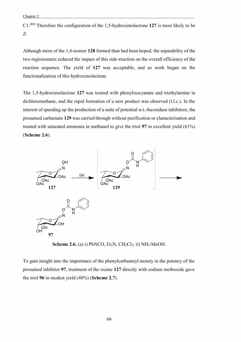
Chapter 2 .
66
C1.[89] Therefore the configuration of the 1,5-hydroximolactone 127 is most likely to be
Z.
Although more of the 1,4-isomer 128 formed than had been hoped, the separability of the
two regioisomers reduced the impact of this side-reaction on the overall efficiency of the
reaction sequence. The yield of 127 was acceptable, and so work began on the
functionalization of this hydroximolactone.
The 1,5-hydroximolactone 127 was treated with phenylisocyanate and triethylamine in
dichloromethane, and the rapid formation of a new product was observed (t.l.c.). In the
interest of speeding up the production of a suite of potential α-L-fucosidase inhibitors, the
presumed carbamate 129 was carried through without purification or characterisation and
treated with saturated ammonia in methanol to give the triol 97 in excellent yield (61%)
(Scheme 2.6).
Scheme 2.6. (a) i) PhNCO, Et3N, CH2Cl2; ii) NH3/MeOH.
To gain insight into the importance of the phenylcarbamoyl moiety in the potency of the
presumed inhibitor 97, treatment of the oxime 127 directly with sodium methoxide gave
the triol 96 in modest yield (40%) (Scheme 2.7).
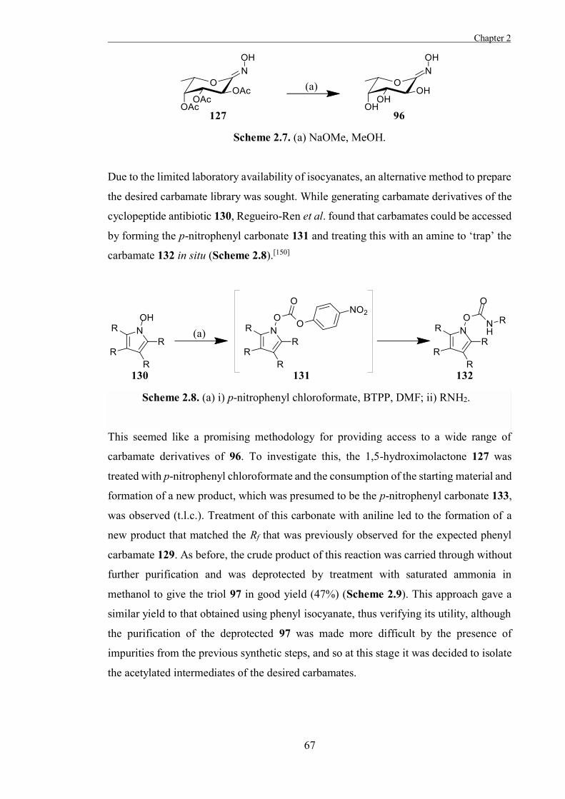
Chapter 2
67
Scheme 2.7. (a) NaOMe, MeOH.
Due to the limited laboratory availability of isocyanates, an alternative method to prepare
the desired carbamate library was sought. While generating carbamate derivatives of the
cyclopeptide antibiotic 130, Regueiro-Ren et al. found that carbamates could be accessed
by forming the p-nitrophenyl carbonate 131 and treating this with an amine to ‘trap’ the
carbamate 132 in situ (Scheme 2.8).[150]
Scheme 2.8. (a) i) p-nitrophenyl chloroformate, BTPP, DMF; ii) RNH2.
This seemed like a promising methodology for providing access to a wide range of
carbamate derivatives of 96. To investigate this, the 1,5-hydroximolactone 127 was
treated with p-nitrophenyl chloroformate and the consumption of the starting material and
formation of a new product, which was presumed to be the p-nitrophenyl carbonate 133,
was observed (t.l.c.). Treatment of this carbonate with aniline led to the formation of a
new product that matched the Rf that was previously observed for the expected phenyl
carbamate 129. As before, the crude product of this reaction was carried through without
further purification and was deprotected by treatment with saturated ammonia in
methanol to give the triol 97 in good yield (47%) (Scheme 2.9). This approach gave a
similar yield to that obtained using phenyl isocyanate, thus verifying its utility, although
the purification of the deprotected 97 was made more difficult by the presence of
impurities from the previous synthetic steps, and so at this stage it was decided to isolate
the acetylated intermediates of the desired carbamates.
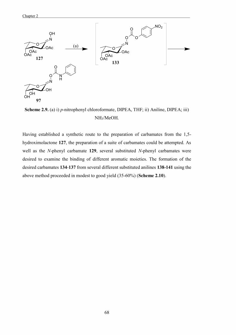
Chapter 2 .
68
Scheme 2.9. (a) i) p-nitrophenyl chloroformate, DIPEA, THF; ii) Aniline, DIPEA; iii)
NH3/MeOH.
Having established a synthetic route to the preparation of carbamates from the 1,5-
hydroximolactone 127, the preparation of a suite of carbamates could be attempted. As
well as the N-phenyl carbamate 129, several substituted N-phenyl carbamates were
desired to examine the binding of different aromatic moieties. The formation of the
desired carbamates 134-137 from several different substituted anilines 138-141 using the
above method proceeded in modest to good yield (35-60%) (Scheme 2.10).
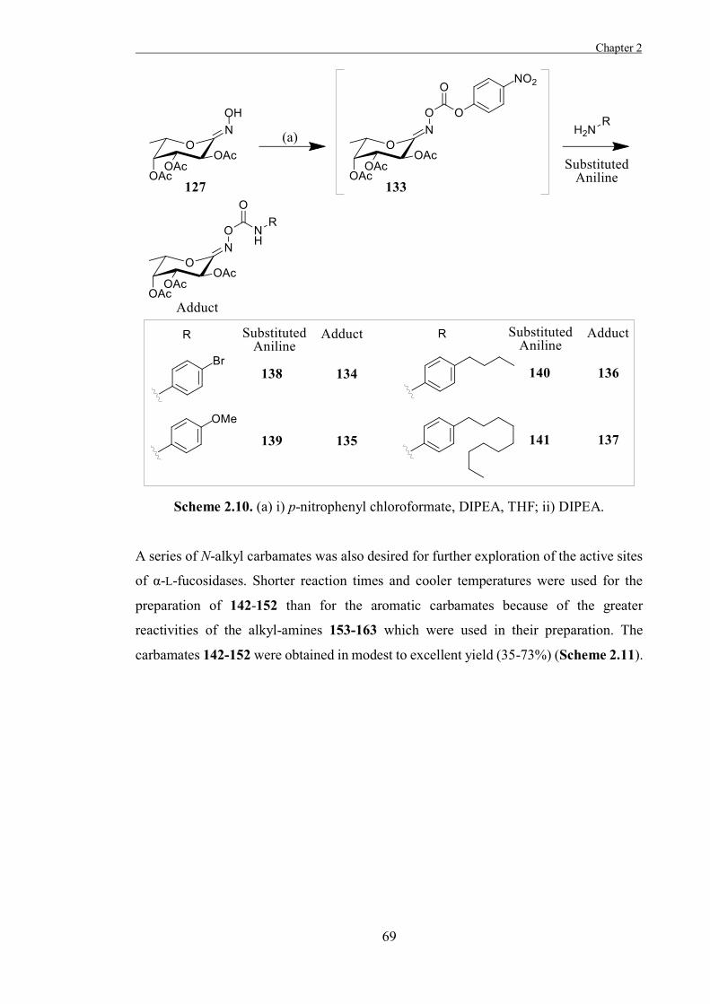
Chapter 2
69
Scheme 2.10. (a) i) p-nitrophenyl chloroformate, DIPEA, THF; ii) DIPEA.
A series of N-alkyl carbamates was also desired for further exploration of the active sites
of α-L-fucosidases. Shorter reaction times and cooler temperatures were used for the
preparation of 142-152 than for the aromatic carbamates because of the greater
reactivities of the alkyl-amines 153-163 which were used in their preparation. The
carbamates 142-152 were obtained in modest to excellent yield (35-73%) (Scheme 2.11).
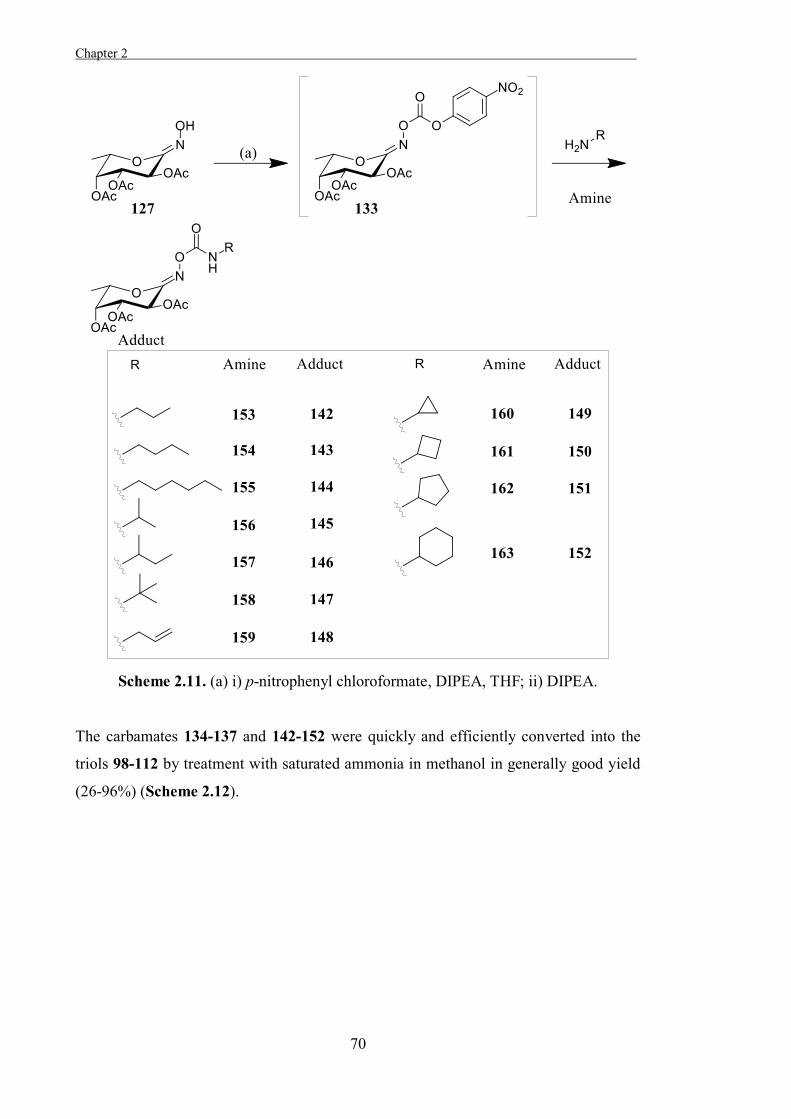
Chapter 2 .
70
Scheme 2.11. (a) i) p-nitrophenyl chloroformate, DIPEA, THF; ii) DIPEA.
The carbamates 134-137 and 142-152 were quickly and efficiently converted into the
triols 98-112 by treatment with saturated ammonia in methanol in generally good yield
(26-96%) (Scheme 2.12).
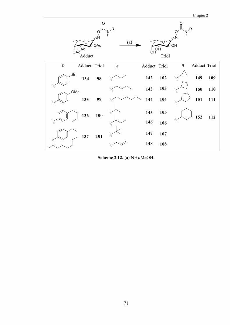
Chapter 2
71
Scheme 2.12. (a) NH3/MeOH.
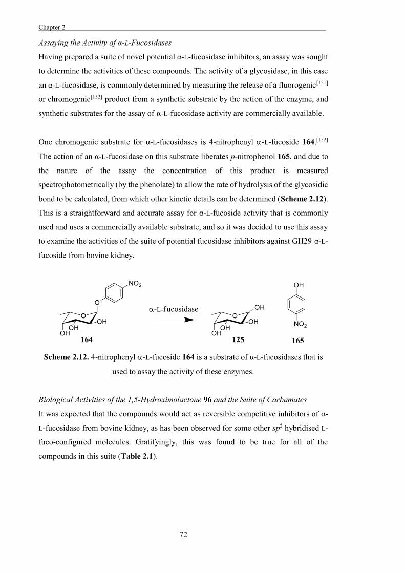
Chapter 2 .
72
Assaying the Activity of α-L-Fucosidases
Having prepared a suite of novel potential α-L-fucosidase inhibitors, an assay was sought
to determine the activities of these compounds. The activity of a glycosidase, in this case
an α-L-fucosidase, is commonly determined by measuring the release of a fluorogenic[151]
or chromogenic[152] product from a synthetic substrate by the action of the enzyme, and
synthetic substrates for the assay of α-L-fucosidase activity are commercially available.
One chromogenic substrate for α-L-fucosidases is 4-nitrophenyl -L-fucoside 164.[152]
The action of an α-L-fucosidase on this substrate liberates p-nitrophenol 165, and due to
the nature of the assay the concentration of this product is measured
spectrophotometrically (by the phenolate) to allow the rate of hydrolysis of the glycosidic
bond to be calculated, from which other kinetic details can be determined (Scheme 2.12).
This is a straightforward and accurate assay for α-L-fucoside activity that is commonly
used and uses a commercially available substrate, and so it was decided to use this assay
to examine the activities of the suite of potential fucosidase inhibitors against GH29 α-L-
fucoside from bovine kidney.
Scheme 2.12. 4-nitrophenyl -L-fucoside 164 is a substrate of α-L-fucosidases that is
used to assay the activity of these enzymes.
Biological Activities of the 1,5-Hydroximolactone 96 and the Suite of Carbamates
It was expected that the compounds would act as reversible competitive inhibitors of α-
L-fucosidase from bovine kidney, as has been observed for some other sp2 hybridised L-
fuco-configured molecules. Gratifyingly, this was found to be true for all of the
compounds in this suite (Table 2.1).
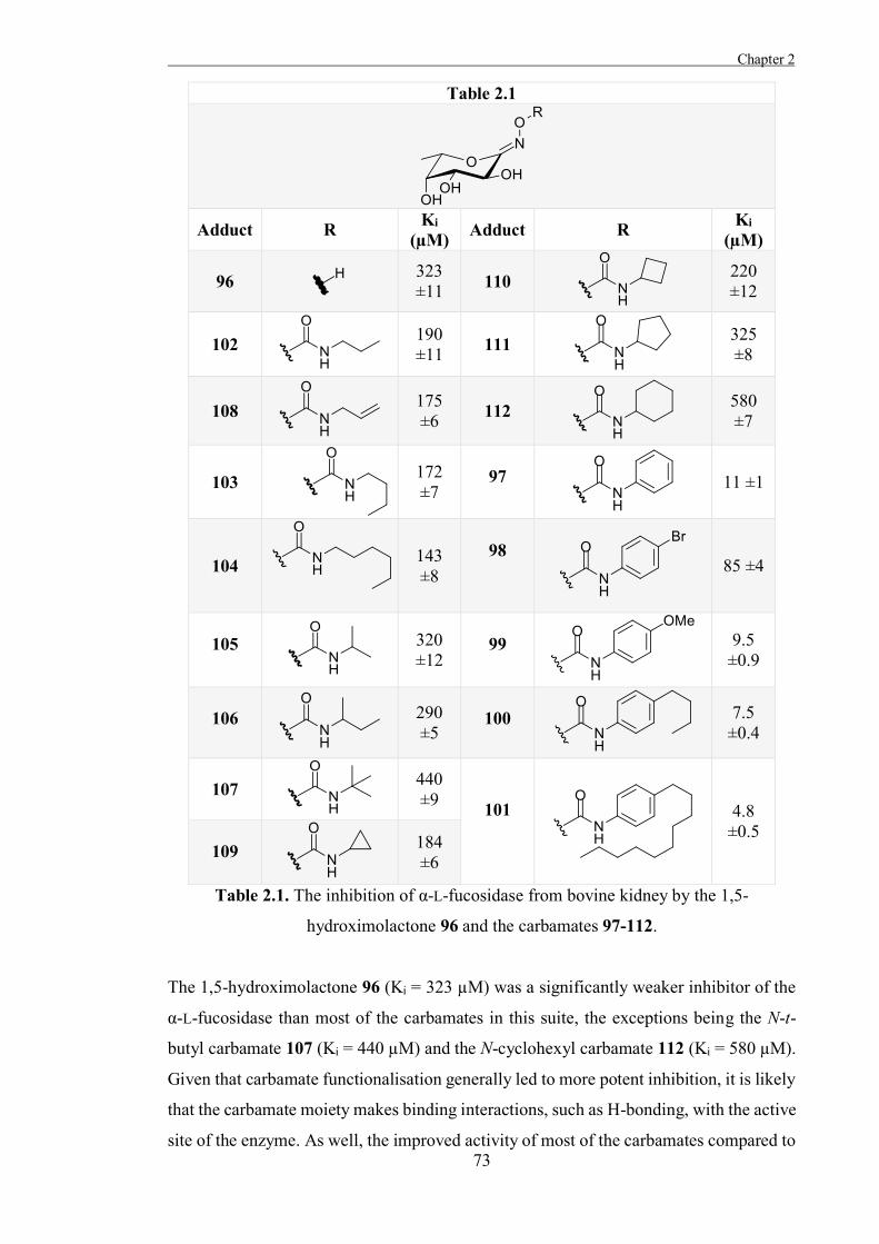
Chapter 2
73
Table 2.1
Adduct R Ki
(µM) Adduct R Ki (µM)
96 323 ±11 110
220 ±12
102
190 ±11 111
325 ±8
108
175 ±6 112
580 ±7
103
172 ±7
97
11 ±1
104
143 ±8
98
85 ±4
105
320 ±12
99
9.5 ±0.9
106
290 ±5
100
7.5 ±0.4
107
440 ±9
101
4.8 ±0.5
109
184 ±6
Table 2.1. The inhibition of α-L-fucosidase from bovine kidney by the 1,5-
hydroximolactone 96 and the carbamates 97-112.
The 1,5-hydroximolactone 96 (Ki = 323 µM) was a significantly weaker inhibitor of the
α-L-fucosidase than most of the carbamates in this suite, the exceptions being the N-t-
butyl carbamate 107 (Ki = 440 µM) and the N-cyclohexyl carbamate 112 (Ki = 580 µM).
Given that carbamate functionalisation generally led to more potent inhibition, it is likely
that the carbamate moiety makes binding interactions, such as H-bonding, with the active
site of the enzyme. As well, the improved activity of most of the carbamates compared to
Chapter 2 .
74
the parent hydroximolactone indicates the suitability of the functionalisation strategy used
for the development of potential α-L-fucosidase inhibitors.
Trends were apparent in the relative activities of carbamates in this suite bearing different
pendant moieties. One such trend was that the relative activities of the n-alkyl chain
carbamates 102-104 increased with increasing chain length. This correlates with what has
been observed for some carbasugar inhibitors of GH29 α-L-fucosidases[129] and may
indicate a hydrophobic binding pocket in the active site of the enzyme. Conversely, the
activities of alkyl chain carbamates decreased with increasing steric bulk; the t-butyl
carbamate 107 (Ki = 440 µM), for example, was one of the weakest inhibitors in this suite.
The relative activities of the cyclic alkyl carbamates 109-112 were in agreement with this
observation; those with larger pendant rings were weaker inhibitors of the α-L-fucosidase.
The reduction in activity associated with increasing steric bulk may arise through steric
effects, indicating tight binding by the enzyme of its substrates, or through unfavourable
interactions between the bulky hydrophobic groups and a hydrophilic region in the active
site of the enzyme.
Significant increases in potency were observed for the N-aryl carbamates compared to
the parent hydroximolactone and the aliphatic carbamates. This was most apparent in the
50-fold increase in the activity of the N-phenyl carbamate 97 (Ki = 11 µM) compared to
the N-cyclohexyl carbamate 112 (Ki = 580 µM). Given the similar shape, size, and
hydrophobicity of the pendant moieties in these two inhibitors, the drastic increase in
activity seems to be associated with the aromaticity of the pendant moiety. As such, it is
likely that specific interactions with the aromatic ring, such as π-stacking, may contribute
to the strong activity of the inhibitors bearing pendant N-aryl carbamate moieties. A trend
apparent within the set of N-aryl carbamates was that para-substituted aliphatic chains on
the aromatic rings led to stronger inhibition of the α-L-fucosidase; the activities of the N-
aryl carbamates increased in the order of N-phenyl (Ki = 11 µM), N-p-butylphenyl (Ki =
7.5 µM), and N-p-decylphenyl (Ki = 4.8 µM). The adduct 101 was the most potent
inhibitor found in this study, with an activity against α-L-fucosidase from bovine kidney
67-fold greater than that of the parent compound 96. This provides further evidence for a
hydrophobic binding pocket within the active site of this α-L-fucosidase, which may be
exploited for the development of more potent inhibitors of the enzyme.
Chapter 2
75
As was discussed earlier in this Chapter, significant differences have been observed in
the activities of inhibitors against the medically relevant human α-L-fucosidase FucA1
and GH29 bacterial α-L-fucosidase commonly used as models for this enzyme. This is
thought to arise largely because of structural variations in particular regions of these
different enzymes. A comparison using the basic local alignment search tool[153] showed
that the bovine and human enzymes have far greater homology (83%) than the human
and T. maritima enzymes (35%), which suggests that bovine α-L-fucosidase is potentially
a more suitable model for FucA1 than bacterial GH29 fucosidases. As well, many
inhibitors which have been assayed against both human and bovine α-L-fucosidases
exhibit similar activity against both enzymes. For example, in a study by Kato et al., C1-
functionalised derivatives of DFJ 65 showed similar trends in their activity against FucA1
and α-L-fucosidase from bovine kidney.[137] This suggests that the trends observed for the
activities of the compounds in Table 2.1 may also apply to the human enzyme.
It is notable that the inhibitors in Table 2.1 are considerably weaker inhibitors of α-L-
fucosidase from bovine kidneys than many charge-based inhibitors reported in the
literature. For example, DFJ 65 exhibits over 11000-fold greater activity (Ki = 0.029 µM)
against α-L-fucosidase from bovine kidney than the 1,5-hydroximolactone 96. This may
indicate that the electrostatic attraction between the endocyclic nitrogen in DFJ 65 is a
greater driving force for binding by GH29 α-L-fucosidases than structural similarity to
the putative transition-state.
The carbasugar 54 (Ki = 43 µM)[128] is also a more potent inhibitor of α-L-fucosidase from
bovine kidney than 96 and many of the carbamates in Table 2.1, despite not mimicking
the charge or shape of the putative transition-state of GH29 α-L-fucosidases. It has been
suggested that a hydrophobic interaction with the methylene group of 54 contributed to
binding by the α-L-fucosidase, as has been observed for thiofucose 50 and the inhibitors
in Table 2.1, which bear an endocyclic oxygen, would lack this hydrophobic interaction.
This also potentially indicates the importance of charge in the binding of inhibitors by
GH29 α-L-fucosidases.
Chapter 2 .
76
Conclusions and Future work
A considerable amount of effort has gone into the development of charge-based inhibitors
of GH29 α-L-fucosidases, but relatively few inhibitors have been designed to mimic the
putative shape of the transition-states of these enzymes. The 1,5-hydroximolactone 96
and its derivatives were expected to generally mimic the distorted shape of the putative
transition-states of GH29 α-L-fucosidases as a result of their sp2 hybridisation at C1, but
were found to be considerably weaker inhibitors of α-L-fucosidase from bovine kidney
than the charge-based inhibitor DFJ 65. As such, it could potentially be concluded that
charge is a more important consideration than shape when designing inhibitors of GH29
α-L-fucosidases, at least with regards to potency. A combination of the two approaches
may potentially lead to improvements upon the already impressive activities of charge-
based GH29 α-L-fucosidase inhibitors, and given the lack of specificity exhibited by
many charge-based glycosidase inhibitors,[80] this strategy could potentially lead to more
potent and specific inhibitors of α-L-fucosidases.
Functionalisation with extended aliphatic chains led to more potent inhibition of the
enzyme, as did functionalisation with aromatic moieties, but the most impressive activity
was observed when these two functionalisation strategies were combined, as in the
carbamate 101. Given the close sequence homologies of the bovine -L-fucosidase and
the human enzyme, it is likely that these trends will apply similarly, so this may be
exploited in future designs of inhibitors of the enzyme.
Although there have been many studies into the inhibition of GH29 retaining fucosidases,
less work has been undertaken on GH95 inverting fucosidases. Some hydroximolactone-
based inhibitors have been found to be more potent inhibitors of retaining glycosidases
than inverting glycosidases,[154] but there is also some evidence that hydroximolactones
are more potent inhibitors of glycosidases in which the catalytic acid is in the plane of the
sugar-ring than those in which the catalytic acid is perpendicular to the ring.[50] GH95
inverting fucosidases belong to the former group, and so an examination of the activities
of the compounds in Table 2.1 against a GH95 fucosidase would be of interest.
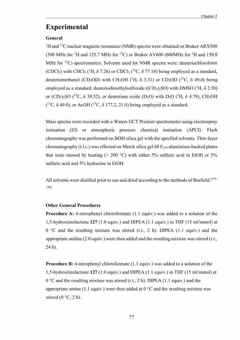
Chapter 2
77
Experimental General 1H and 13C nuclear magnetic resonance (NMR) spectra were obtained on Bruker ARX500
(500 MHz for 1H and 125.7 MHz for 13C) or Bruker AV600 (600MHz for 1H and 150.8
MHz for 13C) spectrometers. Solvents used for NMR spectra were: deuteriochloroform
(CDCl3) with CHCl3 (1H, δ 7.26) or CDCl3 (13C, δ 77.16) being employed as a standard,
deuteriomethanol (CD3OD) with CH3OH (1H, δ 3.31) or CD3OD (13C, δ 49.0) being
employed as a standard, deuteriodimethylsulfoxide ((CD3)2SO) with DMSO (1H, δ 2.50)
or (CD3)2SO (13C, δ 39.52), or deuterium oxide (D2O) with D2O (1H, δ 4.79), CH3OH
(13C, δ 49.0), or AcOH (13C, δ 177.2, 21.0) being employed as a standard.
Mass spectra were recorded with a Waters GCT Premier spectrometer using electrospray
ionisation (EI) or atmospheric pressure chemical ionisation (APCI). Flash
chromatography was performed on BDH silica gel with the specfied solvents. Thin-layer
chromatography (t.l.c.) was effected on Merck silica gel 60 F254 aluminium-backed plates
that were stained by heating (> 200 °C) with either 5% sulfuric acid in EtOH or 5%
sulfuric acid and 5% hydrazine in EtOH.
All solvents were distilled prior to use and dried according to the methods of Burfield.[155-
158]
Other General Procedures
Procedure A: 4-nitrophenyl chloroformate (1.1 equiv.) was added to a solution of the
1,5-hydroximolactone 127 (1.0 equiv.) and DIPEA (1.1 equiv.) in THF (15 ml/mmol) at
0 °C and the resulting mixture was stirred (r.t., 2 h). DIPEA (1.1 equiv.) and the
appropriate aniline (2.0 equiv.) were then added and the resulting mixture was stirred (r.t.,
24 h).
Procedure B: 4-nitrophenyl chloroformate (1.1 equiv.) was added to a solution of the
1,5-hydroximolactone 127 (1.0 equiv.) and DIPEA (1.1 equiv.) in THF (15 ml/mmol) at
0 °C and the resulting mixture was stirred (r.t., 2 h). DIPEA (1.1 equiv.) and the
appropriate amine (1.1 equiv.) were then added at 0 °C and the resulting mixture was
stirred (0 °C, 2 h).
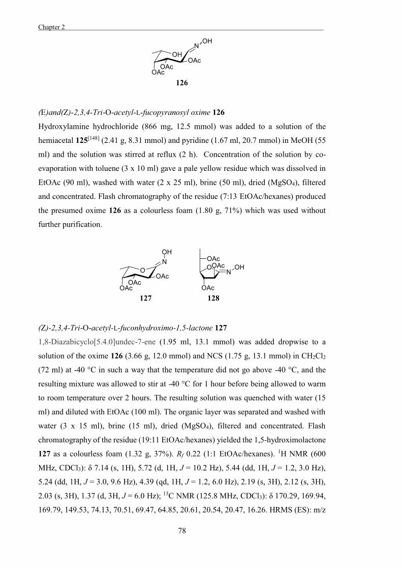
Chapter 2 .
78
(E)and(Z)-2,3,4-Tri-O-acetyl-L-fucopyranosyl oxime 126
Hydroxylamine hydrochloride (866 mg, 12.5 mmol) was added to a solution of the
hemiacetal 125[148] (2.41 g, 8.31 mmol) and pyridine (1.67 ml, 20.7 mmol) in MeOH (55
ml) and the solution was stirred at reflux (2 h). Concentration of the solution by co-
evaporation with toluene (3 x 10 ml) gave a pale yellow residue which was dissolved in
EtOAc (90 ml), washed with water (2 x 25 ml), brine (50 ml), dried (MgSO4), filtered
and concentrated. Flash chromatography of the residue (7:13 EtOAc/hexanes) produced
the presumed oxime 126 as a colourless foam (1.80 g, 71%) which was used without
further purification.
(Z)-2,3,4-Tri-O-acetyl-L-fuconhydroximo-1,5-lactone 127
1,8-Diazabicyclo[5.4.0]undec-7-ene (1.95 ml, 13.1 mmol) was added dropwise to a
solution of the oxime 126 (3.66 g, 12.0 mmol) and NCS (1.75 g, 13.1 mmol) in CH2Cl2
(72 ml) at -40 °C in such a way that the temperature did not go above -40 °C, and the
resulting mixture was allowed to stir at -40 °C for 1 hour before being allowed to warm
to room temperature over 2 hours. The resulting solution was quenched with water (15
ml) and diluted with EtOAc (100 ml). The organic layer was separated and washed with
water (3 x 15 ml), brine (15 ml), dried (MgSO4), filtered and concentrated. Flash
chromatography of the residue (19:11 EtOAc/hexanes) yielded the 1,5-hydroximolactone
127 as a colourless foam (1.32 g, 37%). Rf 0.22 (1:1 EtOAc/hexanes). 1H NMR (600
MHz, CDCl3): δ 7.14 (s, 1H), 5.72 (d, 1H, J = 10.2 Hz), 5.44 (dd, 1H, J = 1.2, 3.0 Hz),
5.24 (dd, 1H, J = 3.0, 9.6 Hz), 4.39 (qd, 1H, J = 1.2, 6.0 Hz), 2.19 (s, 3H), 2.12 (s, 3H),
2.03 (s, 3H), 1.37 (d, 3H, J = 6.0 Hz); 13C NMR (125.8 MHz, CDCl3): δ 170.29, 169.94,
169.79, 149.53, 74.13, 70.51, 69.47, 64.85, 20.61, 20.54, 20.47, 16.26. HRMS (ES): m/z
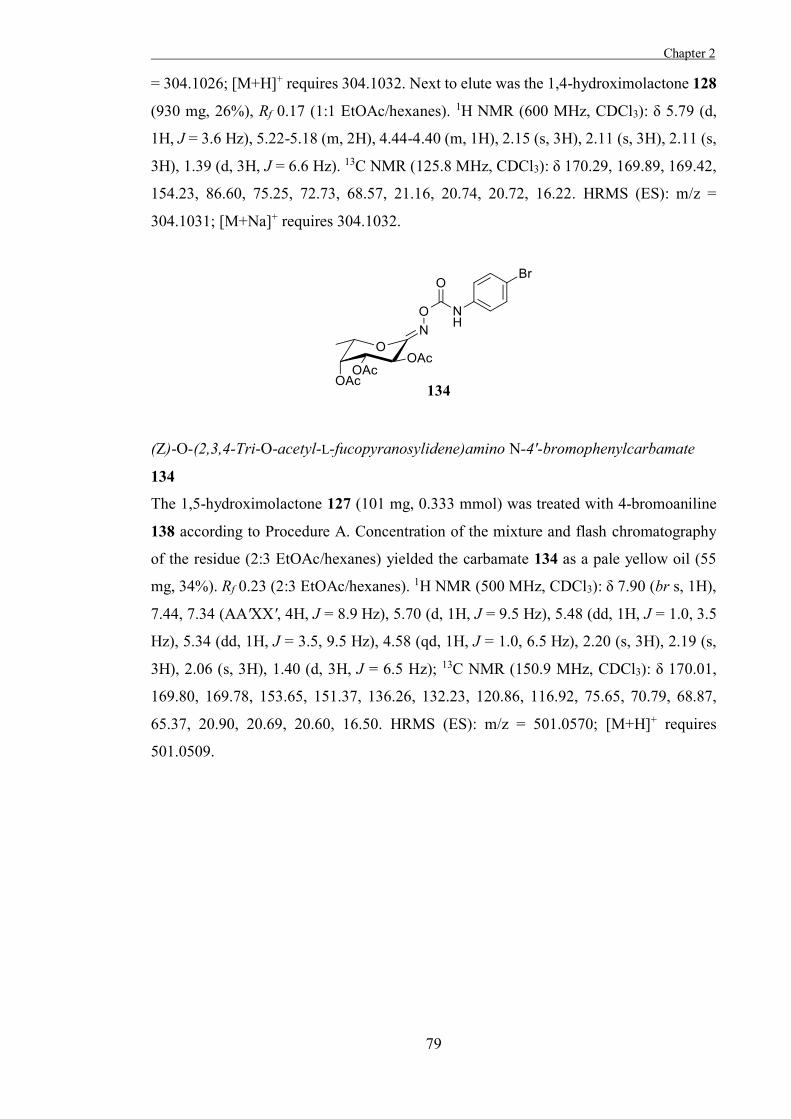
Chapter 2
79
= 304.1026; [M+H]+ requires 304.1032. Next to elute was the 1,4-hydroximolactone 128
(930 mg, 26%), Rf 0.17 (1:1 EtOAc/hexanes). 1H NMR (600 MHz, CDCl3): δ 5.79 (d,
1H, J = 3.6 Hz), 5.22-5.18 (m, 2H), 4.44-4.40 (m, 1H), 2.15 (s, 3H), 2.11 (s, 3H), 2.11 (s,
3H), 1.39 (d, 3H, J = 6.6 Hz). 13C NMR (125.8 MHz, CDCl3): δ 170.29, 169.89, 169.42,
154.23, 86.60, 75.25, 72.73, 68.57, 21.16, 20.74, 20.72, 16.22. HRMS (ES): m/z =
304.1031; [M+Na]+ requires 304.1032.
(Z)-O-(2,3,4-Tri-O-acetyl-L-fucopyranosylidene)amino N-4'-bromophenylcarbamate
134
The 1,5-hydroximolactone 127 (101 mg, 0.333 mmol) was treated with 4-bromoaniline
138 according to Procedure A. Concentration of the mixture and flash chromatography
of the residue (2:3 EtOAc/hexanes) yielded the carbamate 134 as a pale yellow oil (55
mg, 34%). Rf 0.23 (2:3 EtOAc/hexanes). 1H NMR (500 MHz, CDCl3): δ 7.90 (br s, 1H),
7.44, 7.34 (AA'XX', 4H, J = 8.9 Hz), 5.70 (d, 1H, J = 9.5 Hz), 5.48 (dd, 1H, J = 1.0, 3.5
Hz), 5.34 (dd, 1H, J = 3.5, 9.5 Hz), 4.58 (qd, 1H, J = 1.0, 6.5 Hz), 2.20 (s, 3H), 2.19 (s,
3H), 2.06 (s, 3H), 1.40 (d, 3H, J = 6.5 Hz); 13C NMR (150.9 MHz, CDCl3): δ 170.01,
169.80, 169.78, 153.65, 151.37, 136.26, 132.23, 120.86, 116.92, 75.65, 70.79, 68.87,
65.37, 20.90, 20.69, 20.60, 16.50. HRMS (ES): m/z = 501.0570; [M+H]+ requires
501.0509.
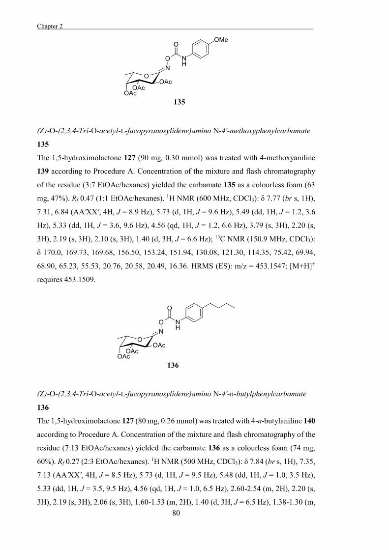
Chapter 2 .
80
(Z)-O-(2,3,4-Tri-O-acetyl-L-fucopyranosylidene)amino N-4'-methoxyphenylcarbamate
135
The 1,5-hydroximolactone 127 (90 mg, 0.30 mmol) was treated with 4-methoxyaniline
139 according to Procedure A. Concentration of the mixture and flash chromatography
of the residue (3:7 EtOAc/hexanes) yielded the carbamate 135 as a colourless foam (63
mg, 47%). Rf 0.47 (1:1 EtOAc/hexanes). 1H NMR (600 MHz, CDCl3): δ 7.77 (br s, 1H),
7.31, 6.84 (AA'XX', 4H, J = 8.9 Hz), 5.73 (d, 1H, J = 9.6 Hz), 5.49 (dd, 1H, J = 1.2, 3.6
Hz), 5.33 (dd, 1H, J = 3.6, 9.6 Hz), 4.56 (qd, 1H, J = 1.2, 6.6 Hz), 3.79 (s, 3H), 2.20 (s,
3H), 2.19 (s, 3H), 2.10 (s, 3H), 1.40 (d, 3H, J = 6.6 Hz); 13C NMR (150.9 MHz, CDCl3):
δ 170.0, 169.73, 169.68, 156.50, 153.24, 151.94, 130.08, 121.30, 114.35, 75.42, 69.94,
68.90, 65.23, 55.53, 20.76, 20.58, 20.49, 16.36. HRMS (ES): m/z = 453.1547; [M+H]+
requires 453.1509.
(Z)-O-(2,3,4-Tri-O-acetyl-L-fucopyranosylidene)amino N-4'-n-butylphenylcarbamate
136
The 1,5-hydroximolactone 127 (80 mg, 0.26 mmol) was treated with 4-n-butylaniline 140
according to Procedure A. Concentration of the mixture and flash chromatography of the
residue (7:13 EtOAc/hexanes) yielded the carbamate 136 as a colourless foam (74 mg,
60%). Rf 0.27 (2:3 EtOAc/hexanes). 1H NMR (500 MHz, CDCl3): δ 7.84 (br s, 1H), 7.35,
7.13 (AA'XX', 4H, J = 8.5 Hz), 5.73 (d, 1H, J = 9.5 Hz), 5.48 (dd, 1H, J = 1.0, 3.5 Hz),
5.33 (dd, 1H, J = 3.5, 9.5 Hz), 4.56 (qd, 1H, J = 1.0, 6.5 Hz), 2.60-2.54 (m, 2H), 2.20 (s,
3H), 2.19 (s, 3H), 2.06 (s, 3H), 1.60-1.53 (m, 2H), 1.40 (d, 3H, J = 6.5 Hz), 1.38-1.30 (m,
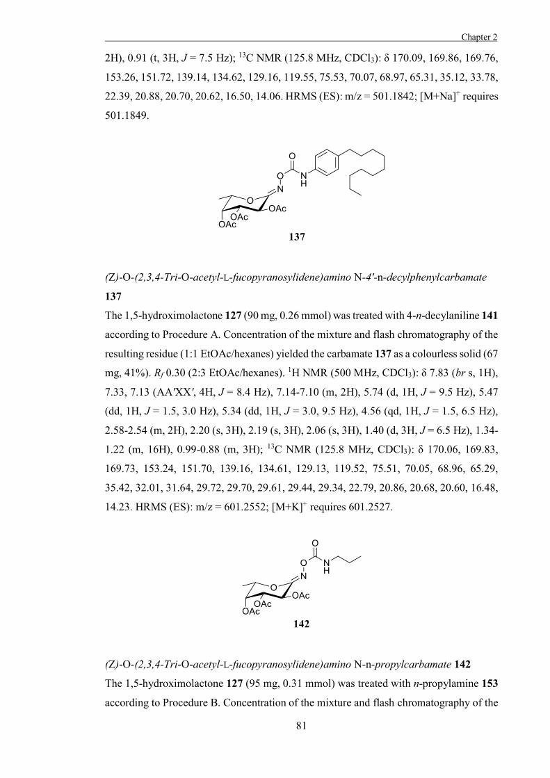
Chapter 2
81
2H), 0.91 (t, 3H, J = 7.5 Hz); 13C NMR (125.8 MHz, CDCl3): δ 170.09, 169.86, 169.76,
153.26, 151.72, 139.14, 134.62, 129.16, 119.55, 75.53, 70.07, 68.97, 65.31, 35.12, 33.78,
22.39, 20.88, 20.70, 20.62, 16.50, 14.06. HRMS (ES): m/z = 501.1842; [M+Na]+ requires
501.1849.
(Z)-O-(2,3,4-Tri-O-acetyl-L-fucopyranosylidene)amino N-4'-n-decylphenylcarbamate
137
The 1,5-hydroximolactone 127 (90 mg, 0.26 mmol) was treated with 4-n-decylaniline 141
according to Procedure A. Concentration of the mixture and flash chromatography of the
resulting residue (1:1 EtOAc/hexanes) yielded the carbamate 137 as a colourless solid (67
mg, 41%). Rf 0.30 (2:3 EtOAc/hexanes). 1H NMR (500 MHz, CDCl3): δ 7.83 (br s, 1H),
7.33, 7.13 (AA'XX', 4H, J = 8.4 Hz), 7.14-7.10 (m, 2H), 5.74 (d, 1H, J = 9.5 Hz), 5.47
(dd, 1H, J = 1.5, 3.0 Hz), 5.34 (dd, 1H, J = 3.0, 9.5 Hz), 4.56 (qd, 1H, J = 1.5, 6.5 Hz),
2.58-2.54 (m, 2H), 2.20 (s, 3H), 2.19 (s, 3H), 2.06 (s, 3H), 1.40 (d, 3H, J = 6.5 Hz), 1.34-
1.22 (m, 16H), 0.99-0.88 (m, 3H); 13C NMR (125.8 MHz, CDCl3): δ 170.06, 169.83,
169.73, 153.24, 151.70, 139.16, 134.61, 129.13, 119.52, 75.51, 70.05, 68.96, 65.29,
35.42, 32.01, 31.64, 29.72, 29.70, 29.61, 29.44, 29.34, 22.79, 20.86, 20.68, 20.60, 16.48,
14.23. HRMS (ES): m/z = 601.2552; [M+K]+ requires 601.2527.
(Z)-O-(2,3,4-Tri-O-acetyl-L-fucopyranosylidene)amino N-n-propylcarbamate 142
The 1,5-hydroximolactone 127 (95 mg, 0.31 mmol) was treated with n-propylamine 153
according to Procedure B. Concentration of the mixture and flash chromatography of the
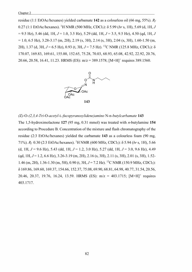
Chapter 2 .
82
residue (1:1 EtOAc/hexanes) yielded carbamate 142 as a colourless oil (66 mg, 55%). Rf
0.27 (1:1 EtOAc/hexanes). 1H NMR (500 MHz, CDCl3): δ 5.99 (br s, 1H), 5.69 (d, 1H, J
= 9.5 Hz), 5.46 (dd, 1H, J = 1.0, 3.5 Hz), 5.29 (dd, 1H, J = 3.5, 9.5 Hz), 4.50 (qd, 1H, J
= 1.0, 6.5 Hz), 3.28-3.17 (m, 2H), 2.19 (s, 3H), 2.14 (s, 3H), 2.04 (s, 3H), 1.60-1.50 (m,
2H), 1.37 (d, 3H, J = 6.5 Hz), 0.93 (t, 3H, J = 7.5 Hz). 13C NMR (125.8 MHz, CDCl3): δ
170.07, 169.83, 169.61, 155.00, 152.65, 75.28, 70.03, 68.93, 65.08, 42.92, 22.92, 20.76,
20.66, 20.58, 16.41, 11.23. HRMS (ES): m/z = 389.1578; [M+H]+ requires 389.1560.
(Z)-O-(2,3,4-Tri-O-acetyl-L-fucopyranosylidene)amino N-n-butylcarbamate 143
The 1,5-hydroximolactone 127 (95 mg, 0.31 mmol) was treated with n-butylamine 154
according to Procedure B. Concentration of the mixture and flash chromatography of the
residue (2:3 EtOAc/hexanes) yielded the carbamate 143 as a colourless foam (90 mg,
71%). Rf 0.30 (2:3 EtOAc/hexanes). 1H NMR (600 MHz, CDCl3): δ 5.94 (br s, 1H), 5.66
(d, 1H, J = 9.6 Hz), 5.43 (dd, 1H, J = 1.2, 3.0 Hz), 5.27 (dd, 1H, J = 3.0, 9.6 Hz), 4.49
(qd, 1H, J = 1.2, 6.6 Hz), 3.26-3.19 (m, 2H), 2.16 (s, 3H), 2.11 (s, 3H), 2.01 (s, 3H), 1.52-
1.46 (m, 2H), 1.36-1.30 (m, 5H), 0.90 (t, 3H, J = 7.2 Hz). 13C NMR (150.9 MHz, CDCl3):
δ 169.86, 169.60, 169.37, 154.66, 152.37, 75.08, 69.90, 68.81, 64.98, 40.77, 31.54, 20.56,
20.46, 20.37, 19.76, 16.24, 13.59. HRMS (ES): m/z = 403.1715; [M+H]+ requires
403.1717.
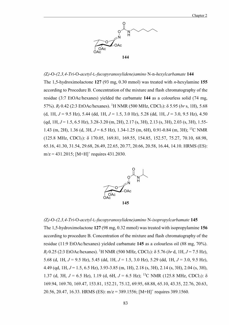
Chapter 2
83
(Z)-O-(2,3,4-Tri-O-acetyl-L-fucopyranosylidene)amino N-n-hexylcarbamate 144
The 1,5-hydroximolactone 127 (93 mg, 0.30 mmol) was treated with n-hexylamine 155
according to Procedure B. Concentration of the mixture and flash chromatography of the
residue (3:7 EtOAc/hexanes) yielded the carbamate 144 as a colourless solid (74 mg,
57%). Rf 0.42 (2:3 EtOAc/hexanes). 1H NMR (500 MHz, CDCl3): δ 5.95 (br s, 1H), 5.68
(d, 1H, J = 9.5 Hz), 5.44 (dd, 1H, J = 1.5, 3.0 Hz), 5.28 (dd, 1H, J = 3.0, 9.5 Hz), 4.50
(qd, 1H, J = 1.5, 6.5 Hz), 3.28-3.20 (m, 2H), 2.17 (s, 3H), 2.13 (s, 3H), 2.03 (s, 3H), 1.55-
1.43 (m, 2H), 1.36 (d, 3H, J = 6.5 Hz), 1.34-1.25 (m, 6H), 0.91-0.84 (m, 3H); 13C NMR
(125.8 MHz, CDCl3): δ 170.05, 169.81, 169.55, 154.85, 152.57, 75.27, 70.10, 68.98,
65.16, 41.30, 31.54, 29.68, 26.49, 22.65, 20.77, 20.66, 20.58, 16.44, 14.10. HRMS (ES):
m/z = 431.2015; [M+H]+ requires 431.2030.
(Z)-O-(2,3,4-Tri-O-acetyl-L-fucopyranosylidene)amino N-isopropylcarbamate 145
The 1,5-hydroximolactone 127 (98 mg, 0.32 mmol) was treated with isopropylamine 156
according to procedure B. Concentration of the mixture and flash chromatography of the
residue (11:9 EtOAc/hexanes) yielded carbamate 145 as a colourless oil (88 mg, 70%).
Rf 0.25 (2:3 EtOAc/hexanes). 1H NMR (500 MHz, CDCl3): δ 5.76 (br d, 1H, J = 7.5 Hz),
5.68 (d, 1H, J = 9.5 Hz), 5.45 (dd, 1H, J = 1.5, 3.0 Hz), 5.29 (dd, 1H, J = 3.0, 9.5 Hz),
4.49 (qd, 1H, J = 1.5, 6.5 Hz), 3.93-3.85 (m, 1H), 2.18 (s, 3H), 2.14 (s, 3H), 2.04 (s, 3H),
1.37 (d, 3H, J = 6.5 Hz), 1.19 (d, 6H, J = 6.5 Hz); 13C NMR (125.8 MHz, CDCl3): δ
169.94, 169.70, 169.47, 153.81, 152.21, 75.12, 69.95, 68.88, 65.10, 43.35, 22.76, 20.63,
20.56, 20.47, 16.33. HRMS (ES): m/z = 389.1556; [M+H]+ requires 389.1560.
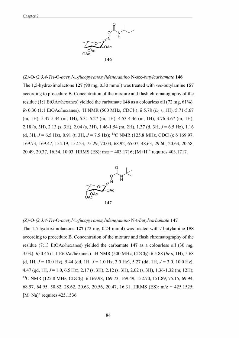
Chapter 2 .
84
(Z)-O-(2,3,4-Tri-O-acetyl-L-fucopyranosylidene)amino N-sec-butylcarbamate 146
The 1,5-hydroximolactone 127 (90 mg, 0.30 mmol) was treated with sec-butylamine 157
according to procedure B. Concentration of the mixture and flash chromatography of the
residue (1:1 EtOAc/hexanes) yielded the carbamate 146 as a colourless oil (72 mg, 61%).
Rf 0.30 (1:1 EtOAc/hexanes). 1H NMR (500 MHz, CDCl3): δ 5.78 (br s, 1H), 5.71-5.67
(m, 1H), 5.47-5.44 (m, 1H), 5.31-5.27 (m, 1H), 4.53-4.46 (m, 1H), 3.76-3.67 (m, 1H),
2.18 (s, 3H), 2.13 (s, 3H), 2.04 (s, 3H), 1.46-1.54 (m, 2H), 1.37 (d, 3H, J = 6.5 Hz), 1.16
(d, 3H, J = 6.5 Hz), 0.91 (t, 3H, J = 7.5 Hz); 13C NMR (125.8 MHz, CDCl3): δ 169.97,
169.73, 169.47, 154.19, 152.23, 75.29, 70.03, 68.92, 65.07, 48.63, 29.60, 20.63, 20.58,
20.49, 20.37, 16.34, 10.03. HRMS (ES): m/z = 403.1716; [M+H]+ requires 403.1717.
(Z)-O-(2,3,4-Tri-O-acetyl-L-fucopyranosylidene)amino N-t-butylcarbamate 147
The 1,5-hydroximolactone 127 (72 mg, 0.24 mmol) was treated with t-butylamine 158
according to procedure B. Concentration of the mixture and flash chromatography of the
residue (7:13 EtOAc/hexanes) yielded the carbamate 147 as a colourless oil (30 mg,
35%). Rf 0.45 (1:1 EtOAc/hexanes). 1H NMR (500 MHz, CDCl3): δ 5.88 (br s, 1H), 5.68
(d, 1H, J = 10.0 Hz), 5.44 (dd, 1H, J = 1.0 Hz, 3.0 Hz), 5.27 (dd, 1H, J = 3.0, 10.0 Hz),
4.47 (qd, 1H, J = 1.0, 6.5 Hz), 2.17 (s, 3H), 2.12 (s, 3H), 2.02 (s, 3H), 1.36-1.32 (m, 12H); 13C NMR (125.8 MHz, CDCl3): δ 169.98, 169.73, 169.49, 152.70, 151.89, 75.15, 69.94,
68.97, 64.95, 50.82, 28.62, 20.63, 20.56, 20.47, 16.31. HRMS (ES): m/z = 425.1525;
[M+Na]+ requires 425.1536.
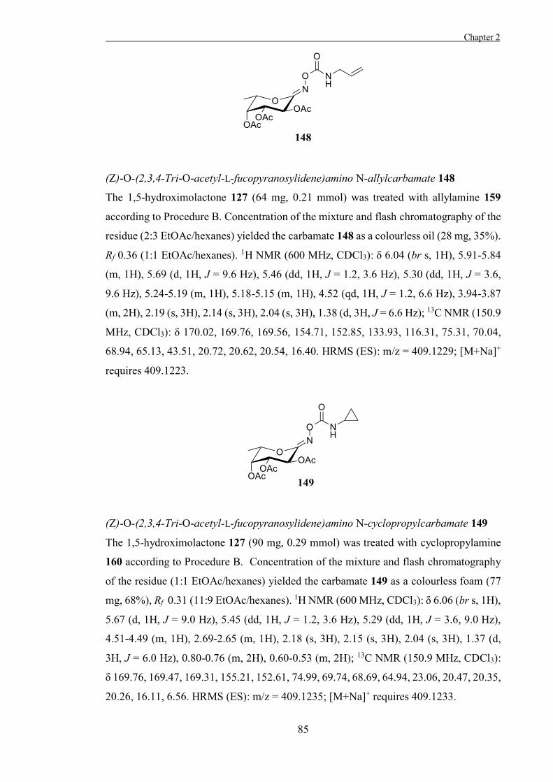
Chapter 2
85
(Z)-O-(2,3,4-Tri-O-acetyl-L-fucopyranosylidene)amino N-allylcarbamate 148
The 1,5-hydroximolactone 127 (64 mg, 0.21 mmol) was treated with allylamine 159
according to Procedure B. Concentration of the mixture and flash chromatography of the
residue (2:3 EtOAc/hexanes) yielded the carbamate 148 as a colourless oil (28 mg, 35%).
Rf 0.36 (1:1 EtOAc/hexanes). 1H NMR (600 MHz, CDCl3): δ 6.04 (br s, 1H), 5.91-5.84
(m, 1H), 5.69 (d, 1H, J = 9.6 Hz), 5.46 (dd, 1H, J = 1.2, 3.6 Hz), 5.30 (dd, 1H, J = 3.6,
9.6 Hz), 5.24-5.19 (m, 1H), 5.18-5.15 (m, 1H), 4.52 (qd, 1H, J = 1.2, 6.6 Hz), 3.94-3.87
(m, 2H), 2.19 (s, 3H), 2.14 (s, 3H), 2.04 (s, 3H), 1.38 (d, 3H, J = 6.6 Hz); 13C NMR (150.9
MHz, CDCl3): δ 170.02, 169.76, 169.56, 154.71, 152.85, 133.93, 116.31, 75.31, 70.04,
68.94, 65.13, 43.51, 20.72, 20.62, 20.54, 16.40. HRMS (ES): m/z = 409.1229; [M+Na]+
requires 409.1223.
(Z)-O-(2,3,4-Tri-O-acetyl-L-fucopyranosylidene)amino N-cyclopropylcarbamate 149
The 1,5-hydroximolactone 127 (90 mg, 0.29 mmol) was treated with cyclopropylamine
160 according to Procedure B. Concentration of the mixture and flash chromatography
of the residue (1:1 EtOAc/hexanes) yielded the carbamate 149 as a colourless foam (77
mg, 68%), Rf 0.31 (11:9 EtOAc/hexanes). 1H NMR (600 MHz, CDCl3): δ 6.06 (br s, 1H),
5.67 (d, 1H, J = 9.0 Hz), 5.45 (dd, 1H, J = 1.2, 3.6 Hz), 5.29 (dd, 1H, J = 3.6, 9.0 Hz),
4.51-4.49 (m, 1H), 2.69-2.65 (m, 1H), 2.18 (s, 3H), 2.15 (s, 3H), 2.04 (s, 3H), 1.37 (d,
3H, J = 6.0 Hz), 0.80-0.76 (m, 2H), 0.60-0.53 (m, 2H); 13C NMR (150.9 MHz, CDCl3):
δ 169.76, 169.47, 169.31, 155.21, 152.61, 74.99, 69.74, 68.69, 64.94, 23.06, 20.47, 20.35,
20.26, 16.11, 6.56. HRMS (ES): m/z = 409.1235; [M+Na]+ requires 409.1233.
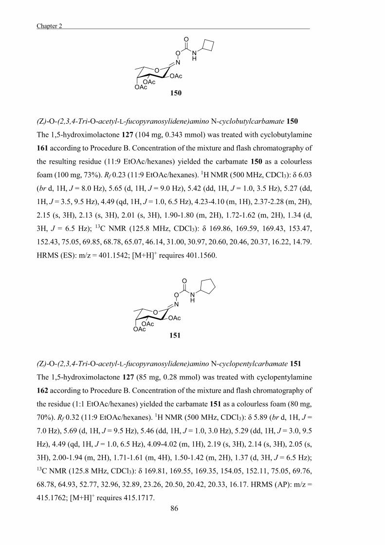
Chapter 2 .
86
(Z)-O-(2,3,4-Tri-O-acetyl-L-fucopyranosylidene)amino N-cyclobutylcarbamate 150
The 1,5-hydroximolactone 127 (104 mg, 0.343 mmol) was treated with cyclobutylamine
161 according to Procedure B. Concentration of the mixture and flash chromatography of
the resulting residue (11:9 EtOAc/hexanes) yielded the carbamate 150 as a colourless
foam (100 mg, 73%). Rf 0.23 (11:9 EtOAc/hexanes). 1H NMR (500 MHz, CDCl3): δ 6.03
(br d, 1H, J = 8.0 Hz), 5.65 (d, 1H, J = 9.0 Hz), 5.42 (dd, 1H, J = 1.0, 3.5 Hz), 5.27 (dd,
1H, J = 3.5, 9.5 Hz), 4.49 (qd, 1H, J = 1.0, 6.5 Hz), 4.23-4.10 (m, 1H), 2.37-2.28 (m, 2H),
2.15 (s, 3H), 2.13 (s, 3H), 2.01 (s, 3H), 1.90-1.80 (m, 2H), 1.72-1.62 (m, 2H), 1.34 (d,
3H, J = 6.5 Hz); 13C NMR (125.8 MHz, CDCl3): δ 169.86, 169.59, 169.43, 153.47,
152.43, 75.05, 69.85, 68.78, 65.07, 46.14, 31.00, 30.97, 20.60, 20.46, 20.37, 16.22, 14.79.
HRMS (ES): m/z = 401.1542; [M+H]+ requires 401.1560.
(Z)-O-(2,3,4-Tri-O-acetyl-L-fucopyranosylidene)amino N-cyclopentylcarbamate 151
The 1,5-hydroximolactone 127 (85 mg, 0.28 mmol) was treated with cyclopentylamine
162 according to Procedure B. Concentration of the mixture and flash chromatography of
the residue (1:1 EtOAc/hexanes) yielded the carbamate 151 as a colourless foam (80 mg,
70%). Rf 0.32 (11:9 EtOAc/hexanes). 1H NMR (500 MHz, CDCl3): δ 5.89 (br d, 1H, J =
7.0 Hz), 5.69 (d, 1H, J = 9.5 Hz), 5.46 (dd, 1H, J = 1.0, 3.0 Hz), 5.29 (dd, 1H, J = 3.0, 9.5
Hz), 4.49 (qd, 1H, J = 1.0, 6.5 Hz), 4.09-4.02 (m, 1H), 2.19 (s, 3H), 2.14 (s, 3H), 2.05 (s,
3H), 2.00-1.94 (m, 2H), 1.71-1.61 (m, 4H), 1.50-1.42 (m, 2H), 1.37 (d, 3H, J = 6.5 Hz); 13C NMR (125.8 MHz, CDCl3): δ 169.81, 169.55, 169.35, 154.05, 152.11, 75.05, 69.76,
68.78, 64.93, 52.77, 32.96, 32.89, 23.26, 20.50, 20.42, 20.33, 16.17. HRMS (AP): m/z =
415.1762; [M+H]+ requires 415.1717.
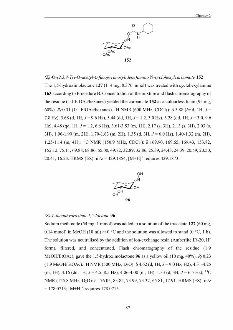
Chapter 2
87
(Z)-O-(2,3,4-Tri-O-acetyl-L-fucopyranosylidene)amino N-cyclohexylcarbamate 152
The 1,5-hydroximolactone 127 (114 mg, 0.376 mmol) was treated with cyclohexylamine
163 according to Procedure B. Concentration of the mixture and flash chromatography of
the residue (1:1 EtOAc/hexanes) yielded the carbamate 152 as a colourless foam (95 mg,
60%). Rf 0.31 (1:1 EtOAc/hexanes). 1H NMR (600 MHz, CDCl3): δ 5.88 (br d, 1H, J =
7.8 Hz), 5.68 (d, 1H, J = 9.6 Hz), 5.44 (dd, 1H, J = 1.2, 3.0 Hz), 5.28 (dd, 1H, J = 3.0, 9.6
Hz), 4.48 (qd, 1H, J = 1.2, 6.6 Hz), 3.61-3.53 (m, 1H), 2.17 (s, 3H), 2.13 (s, 3H), 2.03 (s,
3H), 1.96-1.90 (m, 2H), 1.70-1.63 (m, 2H), 1.35 (d, 3H, J = 6.0 Hz), 1.40-1.32 (m, 2H),
1.25-1.14 (m, 4H); 13C NMR (150.9 MHz, CDCl3): δ 169.90, 169.65, 169.43, 153.82,
152.12, 75.11, 69.88, 68.86, 65.00, 49.72, 32.89, 32.86, 25.39, 24.43, 24.39, 20.59, 20.50,
20.41, 16.23. HRMS (ES): m/z = 429.1854; [M+H]+ requires 429.1873.
(Z)-L-fuconhydroximo-1,5-lactone 96
Sodium methoxide (54 mg, 1 mmol) was added to a solution of the triacetate 127 (60 mg,
0.14 mmol) in MeOH (10 ml) at 0 °C and the solution was allowed to stand (0 °C, 1 h).
The solution was neutralised by the addition of ion-exchange resin (Amberlite IR-20, H+
form), filtered, and concentrated. Flash chromatography of the residue (1:9
MeOH/EtOAc), gave the 1,5-hydroximolactone 96 as a yellow oil (10 mg, 40%). Rf 0.23
(1:9 MeOH/EtOAc). 1H NMR (500 MHz, D2O): δ 4.62 (d, 1H, J = 9.0 Hz, H2), 4.31-4.25
(m, 1H), 4.16 (dd, 1H, J = 4.5, 8.5 Hz), 4.06-4.00 (m, 1H), 1.33 (d, 3H, J = 6.5 Hz); 13C
NMR (125.8 MHz, D2O): δ 176.05, 83.82, 73.99, 73.37, 65.81, 17.91. HRMS (ES): m/z
= 178.0713; [M+H]+ requires 178.0715.
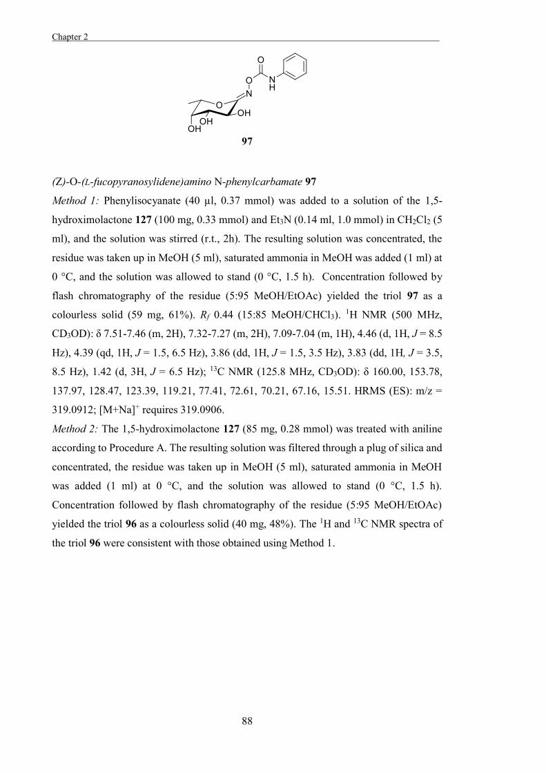
Chapter 2 .
88
(Z)-O-(L-fucopyranosylidene)amino N-phenylcarbamate 97
Method 1: Phenylisocyanate (40 µl, 0.37 mmol) was added to a solution of the 1,5-
hydroximolactone 127 (100 mg, 0.33 mmol) and Et3N (0.14 ml, 1.0 mmol) in CH2Cl2 (5
ml), and the solution was stirred (r.t., 2h). The resulting solution was concentrated, the
residue was taken up in MeOH (5 ml), saturated ammonia in MeOH was added (1 ml) at
0 °C, and the solution was allowed to stand (0 °C, 1.5 h). Concentration followed by
flash chromatography of the residue (5:95 MeOH/EtOAc) yielded the triol 97 as a
colourless solid (59 mg, 61%). Rf 0.44 (15:85 MeOH/CHCl3). 1H NMR (500 MHz,
CD3OD): δ 7.51-7.46 (m, 2H), 7.32-7.27 (m, 2H), 7.09-7.04 (m, 1H), 4.46 (d, 1H, J = 8.5
Hz), 4.39 (qd, 1H, J = 1.5, 6.5 Hz), 3.86 (dd, 1H, J = 1.5, 3.5 Hz), 3.83 (dd, 1H, J = 3.5,
8.5 Hz), 1.42 (d, 3H, J = 6.5 Hz); 13C NMR (125.8 MHz, CD3OD): δ 160.00, 153.78,
137.97, 128.47, 123.39, 119.21, 77.41, 72.61, 70.21, 67.16, 15.51. HRMS (ES): m/z =
319.0912; [M+Na]+ requires 319.0906.
Method 2: The 1,5-hydroximolactone 127 (85 mg, 0.28 mmol) was treated with aniline
according to Procedure A. The resulting solution was filtered through a plug of silica and
concentrated, the residue was taken up in MeOH (5 ml), saturated ammonia in MeOH
was added (1 ml) at 0 °C, and the solution was allowed to stand (0 °C, 1.5 h).
Concentration followed by flash chromatography of the residue (5:95 MeOH/EtOAc)
yielded the triol 96 as a colourless solid (40 mg, 48%). The 1H and 13C NMR spectra of
the triol 96 were consistent with those obtained using Method 1.
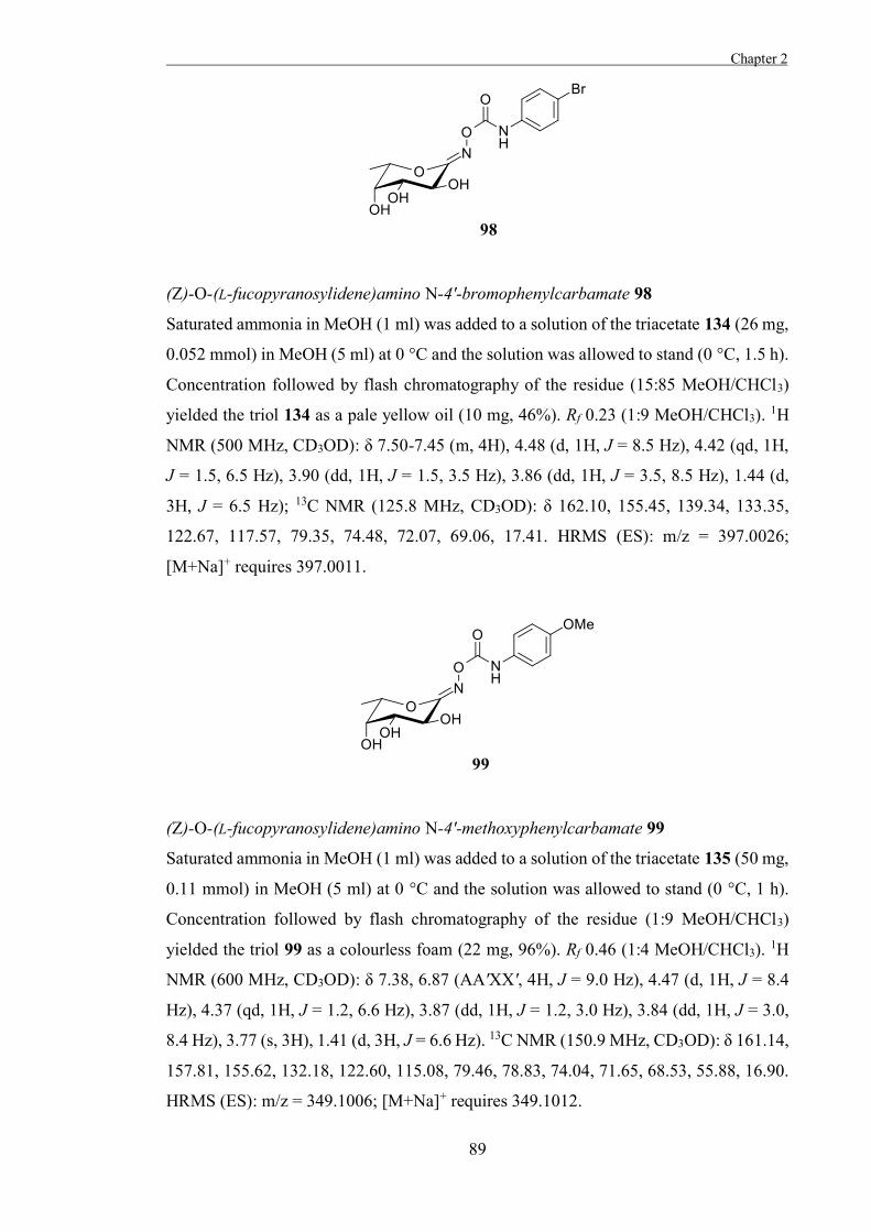
Chapter 2
89
(Z)-O-(L-fucopyranosylidene)amino N-4'-bromophenylcarbamate 98
Saturated ammonia in MeOH (1 ml) was added to a solution of the triacetate 134 (26 mg,
0.052 mmol) in MeOH (5 ml) at 0 °C and the solution was allowed to stand (0 °C, 1.5 h).
Concentration followed by flash chromatography of the residue (15:85 MeOH/CHCl3)
yielded the triol 134 as a pale yellow oil (10 mg, 46%). Rf 0.23 (1:9 MeOH/CHCl3). 1H
NMR (500 MHz, CD3OD): δ 7.50-7.45 (m, 4H), 4.48 (d, 1H, J = 8.5 Hz), 4.42 (qd, 1H,
J = 1.5, 6.5 Hz), 3.90 (dd, 1H, J = 1.5, 3.5 Hz), 3.86 (dd, 1H, J = 3.5, 8.5 Hz), 1.44 (d,
3H, J = 6.5 Hz); 13C NMR (125.8 MHz, CD3OD): δ 162.10, 155.45, 139.34, 133.35,
122.67, 117.57, 79.35, 74.48, 72.07, 69.06, 17.41. HRMS (ES): m/z = 397.0026;
[M+Na]+ requires 397.0011.
(Z)-O-(L-fucopyranosylidene)amino N-4'-methoxyphenylcarbamate 99
Saturated ammonia in MeOH (1 ml) was added to a solution of the triacetate 135 (50 mg,
0.11 mmol) in MeOH (5 ml) at 0 °C and the solution was allowed to stand (0 °C, 1 h).
Concentration followed by flash chromatography of the residue (1:9 MeOH/CHCl3)
yielded the triol 99 as a colourless foam (22 mg, 96%). Rf 0.46 (1:4 MeOH/CHCl3). 1H
NMR (600 MHz, CD3OD): δ 7.38, 6.87 (AA'XX', 4H, J = 9.0 Hz), 4.47 (d, 1H, J = 8.4
Hz), 4.37 (qd, 1H, J = 1.2, 6.6 Hz), 3.87 (dd, 1H, J = 1.2, 3.0 Hz), 3.84 (dd, 1H, J = 3.0,
8.4 Hz), 3.77 (s, 3H), 1.41 (d, 3H, J = 6.6 Hz). 13C NMR (150.9 MHz, CD3OD): δ 161.14,
157.81, 155.62, 132.18, 122.60, 115.08, 79.46, 78.83, 74.04, 71.65, 68.53, 55.88, 16.90.
HRMS (ES): m/z = 349.1006; [M+Na]+ requires 349.1012.
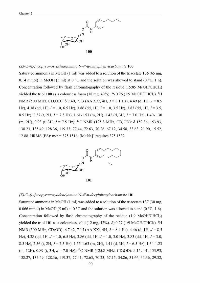
Chapter 2 .
90
(Z)-O-(L-fucopyranosylidene)amino N-4'-n-butylphenylcarbamate 100
Saturated ammonia in MeOH (1 ml) was added to a solution of the triacetate 136 (65 mg,
0.14 mmol) in MeOH (5 ml) at 0 °C and the solution was allowed to stand (0 °C, 1 h).
Concentration followed by flash chromatography of the residue (15:85 MeOH/CHCl3)
yielded the triol 100 as a colourless foam (18 mg, 40%). Rf 0.26 (1:9 MeOH/CHCl3). 1H
NMR (500 MHz, CD3OD): δ 7.40, 7.13 (AA'XX', 4H, J = 8.1 Hz), 4.49 (d, 1H, J = 8.5
Hz), 4.38 (qd, 1H, J = 1.0, 6.5 Hz), 3.86 (dd, 1H, J = 1.0, 3.5 Hz), 3.83 (dd, 1H, J = 3.5,
8.5 Hz), 2.57 (t, 2H, J = 7.5 Hz), 1.61-1.53 (m, 2H), 1.42 (d, 3H, J = 7.0 Hz), 1.40-1.30
(m, 2H), 0.93 (t, 3H, J = 7.5 Hz); 13C NMR (125.8 MHz, CD3OD): δ 159.86, 153.93,
138.23, 135.49, 128.36, 119.33, 77.44, 72.63, 70.26, 67.12, 34.58, 33.63, 21.90, 15.52,
12.88. HRMS (ES): m/z = 375.1516; [M+Na]+ requires 375.1532.
(Z)-O-(L-fucopyranosylidene)amino N-4'-n-decylphenylcarbamate 101
Saturated ammonia in MeOH (1 ml) was added to a solution of the triacetate 137 (30 mg,
0.066 mmol) in MeOH (5 ml) at 0 °C and the solution was allowed to stand (0 °C, 1 h).
Concentration followed by flash chromatography of the residue (1:9 MeOH/CHCl3)
yielded the triol 101 as a colourless solid (12 mg, 42%). Rf 0.27 (1:9 MeOH/CHCl3). 1H
NMR (500 MHz, CD3OD): δ 7.42, 7.15 (AA'XX', 4H, J = 8.4 Hz), 4.46 (d, 1H, J = 8.5
Hz), 4.38 (qd, 1H, J = 1.0, 6.5 Hz), 3.86 (dd, 1H, J = 1.0, 3.0 Hz), 3.83 (dd, 1H, J = 3.0,
8.5 Hz), 2.56 (t, 2H, J = 7.5 Hz), 1.55-1.63 (m, 2H), 1.41 (d, 3H, J = 6.5 Hz), 1.34-1.23
(m, 12H), 0.89 (t, 3H, J = 7.0 Hz); 13C NMR (125.8 MHz, CD3OD): δ 159.01, 153.93,
138.27, 135.49, 128.36, 119.37, 77.41, 72.63, 70.23, 67.15, 34.86, 31.66, 31.36, 29.32,

Chapter 2
91
29.18, 29.04, 28.87, 22.32, 20.65, 15.51, 13.02. HRMS (ES): m/z = 459.2468; [M+Na]+
requires 459.2471.
(Z)-O-(L-fucopyranosylidene)amino N-n-propylcarbamate 102
Saturated ammonia in MeOH (1 ml) was added to a solution of the triacetate 142 (48 mg,
0.12 mmol) in MeOH (5 ml) at 0 °C and the solution was allowed to stand (0 °C, 1 h).
Concentration followed by flash chromatography of the residue (15:85 MeOH/CHCl3)
yielded the triol 102 as a colourless oil (8 mg, 26%). Rf 0.14 (1:9 MeOH/CHCl3). 1H NMR
(500 MHz, CD3OD): δ 4.44 (d, 1H, J = 8.8 Hz), 4.38-4.34 (m, 1H), 3.90-3.85 (m, 1H),
3.71 (dd, 1H, J = 3.0, 8.8 Hz), 3.20-3.16 (m, 2H), 1.63-1.54 (m, 2H), 1.42 (d, 3H, J = 6.5
Hz), 0.96 (t, 3H, J = 7.4 Hz); 13C NMR (125.8 MHz, CD3OD): δ 160.48, 158.55, 78.76,
74.09, 71.73, 68.42, 43.77, 23.88, 16.87, 11.51. HRMS (ES): m/z = 285.1060; [M+Na]+
requires 285.1063.
(Z)-O-(L-fucopyranosylidene)amino N-n-butylcarbamate 103
Saturated ammonia in MeOH (1 ml) was added to a solution of the triacetate 143 (71 mg,
0.18 mmol) in MeOH (5 ml) at 0 °C and the solution was allowed to stand (0 °C, 2 h).
Concentration followed by flash chromatography of the residue (1:9 MeOH/CHCl3)
yielded the triol 103 as a colourless foam (47 mg, 96%). Rf 0.30 (1:9 MeOH/CHCl3). 1H
NMR (500 MHz, CD3OD): δ 4.44 (d, 1H, J = 9.0 Hz), 4.36-4.33 (m, 1H), 3.89-3.85 (m,
1H), 3.82 (dd, 1H, J = 2.0, 9.0 Hz), 3.22 (t, 2H, J = 7.0 Hz), 1.58-1.52 (m, 2H), 1.45-1.35
(m, 5H), 0.97 (t, 3H, J = 7.5 Hz); 13C NMR (125.8 MHz, CD3OD): δ 160.43, 158.49,
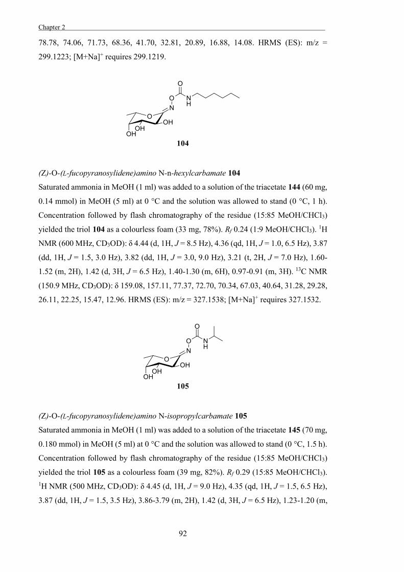
Chapter 2 .
92
78.78, 74.06, 71.73, 68.36, 41.70, 32.81, 20.89, 16.88, 14.08. HRMS (ES): m/z =
299.1223; [M+Na]+ requires 299.1219.
(Z)-O-(L-fucopyranosylidene)amino N-n-hexylcarbamate 104
Saturated ammonia in MeOH (1 ml) was added to a solution of the triacetate 144 (60 mg,
0.14 mmol) in MeOH (5 ml) at 0 °C and the solution was allowed to stand (0 °C, 1 h).
Concentration followed by flash chromatography of the residue (15:85 MeOH/CHCl3)
yielded the triol 104 as a colourless foam (33 mg, 78%). Rf 0.24 (1:9 MeOH/CHCl3). 1H
NMR (600 MHz, CD3OD): δ 4.44 (d, 1H, J = 8.5 Hz), 4.36 (qd, 1H, J = 1.0, 6.5 Hz), 3.87
(dd, 1H, J = 1.5, 3.0 Hz), 3.82 (dd, 1H, J = 3.0, 9.0 Hz), 3.21 (t, 2H, J = 7.0 Hz), 1.60-
1.52 (m, 2H), 1.42 (d, 3H, J = 6.5 Hz), 1.40-1.30 (m, 6H), 0.97-0.91 (m, 3H). 13C NMR
(150.9 MHz, CD3OD): δ 159.08, 157.11, 77.37, 72.70, 70.34, 67.03, 40.64, 31.28, 29.28,
26.11, 22.25, 15.47, 12.96. HRMS (ES): m/z = 327.1538; [M+Na]+ requires 327.1532.
(Z)-O-(L-fucopyranosylidene)amino N-isopropylcarbamate 105
Saturated ammonia in MeOH (1 ml) was added to a solution of the triacetate 145 (70 mg,
0.180 mmol) in MeOH (5 ml) at 0 °C and the solution was allowed to stand (0 °C, 1.5 h).
Concentration followed by flash chromatography of the residue (15:85 MeOH/CHCl3)
yielded the triol 105 as a colourless foam (39 mg, 82%). Rf 0.29 (15:85 MeOH/CHCl3). 1H NMR (500 MHz, CD3OD): δ 4.45 (d, 1H, J = 9.0 Hz), 4.35 (qd, 1H, J = 1.5, 6.5 Hz),
3.87 (dd, 1H, J = 1.5, 3.5 Hz), 3.86-3.79 (m, 2H), 1.42 (d, 3H, J = 6.5 Hz), 1.23-1.20 (m,
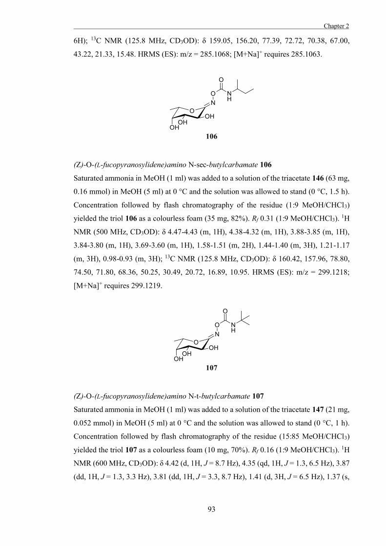
Chapter 2
93
6H); 13C NMR (125.8 MHz, CD3OD): δ 159.05, 156.20, 77.39, 72.72, 70.38, 67.00,
43.22, 21.33, 15.48. HRMS (ES): m/z = 285.1068; [M+Na]+ requires 285.1063.
(Z)-O-(L-fucopyranosylidene)amino N-sec-butylcarbamate 106
Saturated ammonia in MeOH (1 ml) was added to a solution of the triacetate 146 (63 mg,
0.16 mmol) in MeOH (5 ml) at 0 °C and the solution was allowed to stand (0 °C, 1.5 h).
Concentration followed by flash chromatography of the residue (1:9 MeOH/CHCl3)
yielded the triol 106 as a colourless foam (35 mg, 82%). Rf 0.31 (1:9 MeOH/CHCl3). 1H
NMR (500 MHz, CD3OD): δ 4.47-4.43 (m, 1H), 4.38-4.32 (m, 1H), 3.88-3.85 (m, 1H),
3.84-3.80 (m, 1H), 3.69-3.60 (m, 1H), 1.58-1.51 (m, 2H), 1.44-1.40 (m, 3H), 1.21-1.17
(m, 3H), 0.98-0.93 (m, 3H); 13C NMR (125.8 MHz, CD3OD): δ 160.42, 157.96, 78.80,
74.50, 71.80, 68.36, 50.25, 30.49, 20.72, 16.89, 10.95. HRMS (ES): m/z = 299.1218;
[M+Na]+ requires 299.1219.
(Z)-O-(L-fucopyranosylidene)amino N-t-butylcarbamate 107
Saturated ammonia in MeOH (1 ml) was added to a solution of the triacetate 147 (21 mg,
0.052 mmol) in MeOH (5 ml) at 0 °C and the solution was allowed to stand (0 °C, 1 h).
Concentration followed by flash chromatography of the residue (15:85 MeOH/CHCl3)
yielded the triol 107 as a colourless foam (10 mg, 70%). Rf 0.16 (1:9 MeOH/CHCl3). 1H
NMR (600 MHz, CD3OD): δ 4.42 (d, 1H, J = 8.7 Hz), 4.35 (qd, 1H, J = 1.3, 6.5 Hz), 3.87
(dd, 1H, J = 1.3, 3.3 Hz), 3.81 (dd, 1H, J = 3.3, 8.7 Hz), 1.41 (d, 3H, J = 6.5 Hz), 1.37 (s,
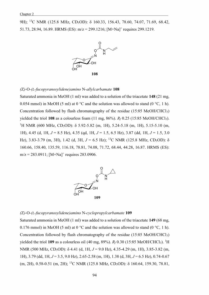
Chapter 2 .
94
9H); 13C NMR (125.8 MHz, CD3OD): δ 160.33, 156.43, 78.60, 74.07, 71.69, 68.42,
51.73, 28.94, 16.89. HRMS (ES): m/z = 299.1216; [M+Na]+ requires 299.1219.
(Z)-O-(L-fucopyranosylidene)amino N-allylcarbamate 108
Saturated ammonia in MeOH (1 ml) was added to a solution of the triacetate 148 (21 mg,
0.054 mmol) in MeOH (5 ml) at 0 °C and the solution was allowed to stand (0 °C, 1 h).
Concentration followed by flash chromatography of the residue (15:85 MeOH/CHCl3)
yielded the triol 108 as a colourless foam (11 mg, 86%). Rf 0.25 (15:85 MeOH/CHCl3). 1H NMR (600 MHz, CD3OD): δ 5.92-5.82 (m, 1H), 5.24-5.18 (m, 1H), 5.15-5.10 (m,
1H), 4.45 (d, 1H, J = 8.5 Hz), 4.35 (qd, 1H, J = 1.5, 6.5 Hz), 3.87 (dd, 1H, J = 1.5, 3.0
Hz), 3.83-3.79 (m, 3H), 1.42 (d, 3H, J = 6.5 Hz); 13C NMR (125.8 MHz, CD3OD): δ
160.66, 158.40, 135.59, 116.18, 78.81, 74.08, 71.72, 68.44, 44.28, 16.87. HRMS (ES):
m/z = 283.0911; [M+Na]+ requires 283.0906.
(Z)-O-(L-fucopyranosylidene)amino N-cyclopropylcarbamate 109
Saturated ammonia in MeOH (1 ml) was added to a solution of the triacetate 149 (68 mg,
0.176 mmol) in MeOH (5 ml) at 0 °C and the solution was allowed to stand (0 °C, 1 h).
Concentration followed by flash chromatography of the residue (15:85 MeOH/CHCl3)
yielded the triol 109 as a colourless oil (40 mg, 89%). Rf 0.30 (15:85 MeOH/CHCl3). 1H
NMR (500 MHz, CD3OD): δ 4.41 (d, 1H, J = 9.0 Hz), 4.35-4.29 (m, 1H), 3.85-3.82 (m,
1H), 3.79 (dd, 1H, J = 3.5, 9.0 Hz), 2.65-2.58 (m, 1H), 1.38 (d, 3H, J = 6.5 Hz), 0.74-0.67
(m, 2H), 0.58-0.51 (m, 2H); 13C NMR (125.8 MHz, CD3OD): δ 160.64, 159.30, 78.81,
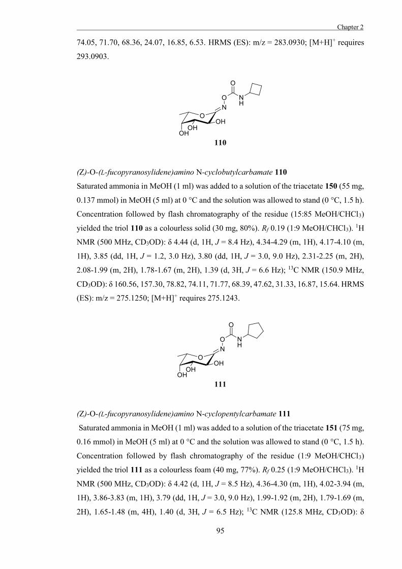
Chapter 2
95
74.05, 71.70, 68.36, 24.07, 16.85, 6.53. HRMS (ES): m/z = 283.0930; [M+H]+ requires
293.0903.
(Z)-O-(L-fucopyranosylidene)amino N-cyclobutylcarbamate 110
Saturated ammonia in MeOH (1 ml) was added to a solution of the triacetate 150 (55 mg,
0.137 mmol) in MeOH (5 ml) at 0 °C and the solution was allowed to stand (0 °C, 1.5 h).
Concentration followed by flash chromatography of the residue (15:85 MeOH/CHCl3)
yielded the triol 110 as a colourless solid (30 mg, 80%). Rf 0.19 (1:9 MeOH/CHCl3). 1H
NMR (500 MHz, CD3OD): δ 4.44 (d, 1H, J = 8.4 Hz), 4.34-4.29 (m, 1H), 4.17-4.10 (m,
1H), 3.85 (dd, 1H, J = 1.2, 3.0 Hz), 3.80 (dd, 1H, J = 3.0, 9.0 Hz), 2.31-2.25 (m, 2H),
2.08-1.99 (m, 2H), 1.78-1.67 (m, 2H), 1.39 (d, 3H, J = 6.6 Hz); 13C NMR (150.9 MHz,
CD3OD): δ 160.56, 157.30, 78.82, 74.11, 71.77, 68.39, 47.62, 31.33, 16.87, 15.64. HRMS
(ES): m/z = 275.1250; [M+H]+ requires 275.1243.
(Z)-O-(L-fucopyranosylidene)amino N-cyclopentylcarbamate 111
Saturated ammonia in MeOH (1 ml) was added to a solution of the triacetate 151 (75 mg,
0.16 mmol) in MeOH (5 ml) at 0 °C and the solution was allowed to stand (0 °C, 1.5 h).
Concentration followed by flash chromatography of the residue (1:9 MeOH/CHCl3)
yielded the triol 111 as a colourless foam (40 mg, 77%). Rf 0.25 (1:9 MeOH/CHCl3). 1H
NMR (500 MHz, CD3OD): δ 4.42 (d, 1H, J = 8.5 Hz), 4.36-4.30 (m, 1H), 4.02-3.94 (m,
1H), 3.86-3.83 (m, 1H), 3.79 (dd, 1H, J = 3.0, 9.0 Hz), 1.99-1.92 (m, 2H), 1.79-1.69 (m,
2H), 1.65-1.48 (m, 4H), 1.40 (d, 3H, J = 6.5 Hz); 13C NMR (125.8 MHz, CD3OD): δ
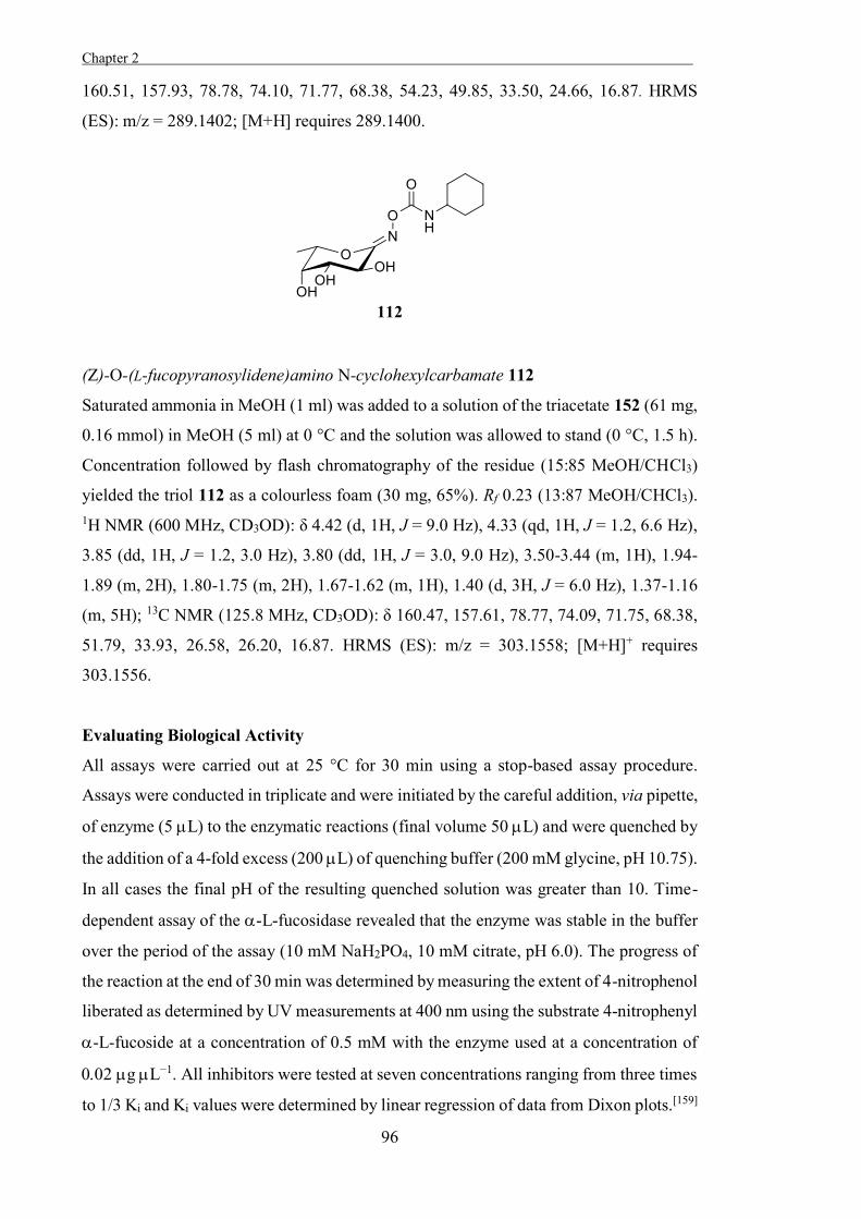
Chapter 2 .
96
160.51, 157.93, 78.78, 74.10, 71.77, 68.38, 54.23, 49.85, 33.50, 24.66, 16.87. HRMS
(ES): m/z = 289.1402; [M+H] requires 289.1400.
(Z)-O-(L-fucopyranosylidene)amino N-cyclohexylcarbamate 112
Saturated ammonia in MeOH (1 ml) was added to a solution of the triacetate 152 (61 mg,
0.16 mmol) in MeOH (5 ml) at 0 °C and the solution was allowed to stand (0 °C, 1.5 h).
Concentration followed by flash chromatography of the residue (15:85 MeOH/CHCl3)
yielded the triol 112 as a colourless foam (30 mg, 65%). Rf 0.23 (13:87 MeOH/CHCl3). 1H NMR (600 MHz, CD3OD): δ 4.42 (d, 1H, J = 9.0 Hz), 4.33 (qd, 1H, J = 1.2, 6.6 Hz),
3.85 (dd, 1H, J = 1.2, 3.0 Hz), 3.80 (dd, 1H, J = 3.0, 9.0 Hz), 3.50-3.44 (m, 1H), 1.94-
1.89 (m, 2H), 1.80-1.75 (m, 2H), 1.67-1.62 (m, 1H), 1.40 (d, 3H, J = 6.0 Hz), 1.37-1.16
(m, 5H); 13C NMR (125.8 MHz, CD3OD): δ 160.47, 157.61, 78.77, 74.09, 71.75, 68.38,
51.79, 33.93, 26.58, 26.20, 16.87. HRMS (ES): m/z = 303.1558; [M+H]+ requires
303.1556.
Evaluating Biological Activity
All assays were carried out at 25 °C for 30 min using a stop-based assay procedure.
Assays were conducted in triplicate and were initiated by the careful addition, via pipette,
of enzyme (5 L) to the enzymatic reactions (final volume 50 L) and were quenched by
the addition of a 4-fold excess (200 L) of quenching buffer (200 mM glycine, pH 10.75).
In all cases the final pH of the resulting quenched solution was greater than 10. Time-
dependent assay of the -L-fucosidase revealed that the enzyme was stable in the buffer
over the period of the assay (10 mM NaH2PO4, 10 mM citrate, pH 6.0). The progress of
the reaction at the end of 30 min was determined by measuring the extent of 4-nitrophenol
liberated as determined by UV measurements at 400 nm using the substrate 4-nitrophenyl
-L-fucoside at a concentration of 0.5 mM with the enzyme used at a concentration of
g L−1. All inhibitors were tested at seven concentrations ranging from three times
to 1/3 Ki and Ki values were determined by linear regression of data from Dixon plots.[159]

97

98
99
Chapter 3
The Development of Some Novel α-L-
Arabinofuranosidase Inhibitors

100
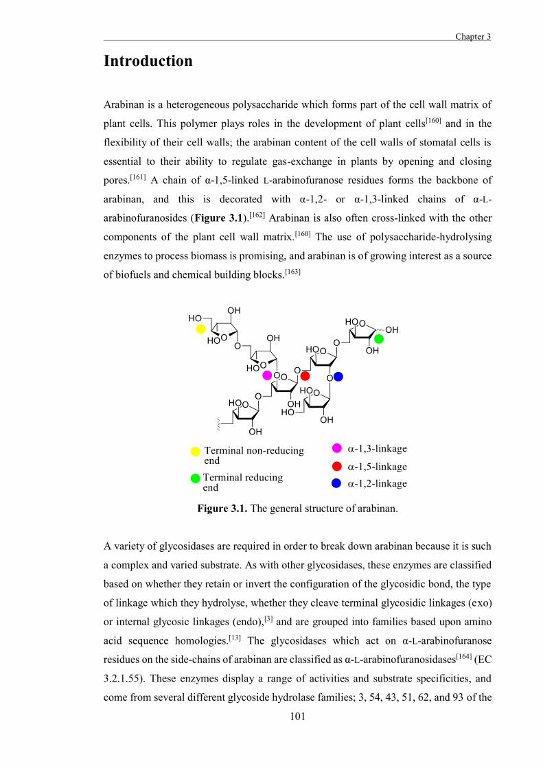
Chapter 3
101
Introduction
Arabinan is a heterogeneous polysaccharide which forms part of the cell wall matrix of
plant cells. This polymer plays roles in the development of plant cells[160] and in the
flexibility of their cell walls; the arabinan content of the cell walls of stomatal cells is
essential to their ability to regulate gas-exchange in plants by opening and closing
pores.[161] A chain of α-1,5-linked L-arabinofuranose residues forms the backbone of
arabinan, and this is decorated with α-1,2- or α-1,3-linked chains of α-L-
arabinofuranosides (Figure 3.1).[162] Arabinan is also often cross-linked with the other
components of the plant cell wall matrix.[160] The use of polysaccharide-hydrolysing
enzymes to process biomass is promising, and arabinan is of growing interest as a source
of biofuels and chemical building blocks.[163]
Figure 3.1. The general structure of arabinan.
A variety of glycosidases are required in order to break down arabinan because it is such
a complex and varied substrate. As with other glycosidases, these enzymes are classified
based on whether they retain or invert the configuration of the glycosidic bond, the type
of linkage which they hydrolyse, whether they cleave terminal glycosidic linkages (exo)
or internal glycosic linkages (endo),[3] and are grouped into families based upon amino
acid sequence homologies.[13] The glycosidases which act on α-L-arabinofuranose
residues on the side-chains of arabinan are classified as α-L-arabinofuranosidases[164] (EC
3.2.1.55). These enzymes display a range of activities and substrate specificities, and
come from several different glycoside hydrolase families; 3, 54, 43, 51, 62, and 93 of the
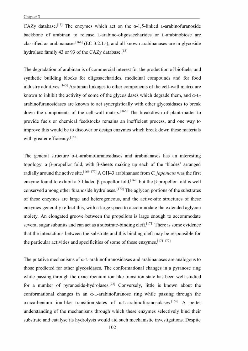
Chapter 3 .
102
CAZy database.[13] The enzymes which act on the α-1,5-linked L-arabinofuranoside
backbone of arabinan to release L-arabino-oligosaccharides or L-arabinobiose are
classified as arabinanases[164] (EC 3.2.1.-), and all known arabinanases are in glycoside
hydrolase family 43 or 93 of the CAZy database.[13]
The degradation of arabinan is of commercial interest for the production of biofuels, and
synthetic building blocks for oligosaccharides, medicinal compounds and for food
industry additives.[165] Arabinan linkages to other components of the cell-wall matrix are
known to inhibit the activity of some of the glycosidases which degrade them, and α-L-
arabinofuranosidases are known to act synergistically with other glycosidases to break
down the components of the cell-wall matrix.[165] The breakdown of plant-matter to
provide fuels or chemical feedstocks remains an inefficient process, and one way to
improve this would be to discover or design enzymes which break down these materials
with greater efficiency.[165]
The general structure α-L-arabinofuranosidases and arabinanases has an interesting
topology; a β-propellor fold, with β-sheets making up each of the ‘blades’ arranged
radially around the active site.[166-170] A GH43 arabinanase from C. japonicus was the first
enzyme found to exhibit a 5-bladed β-propellor fold,[169] but the β-propellor fold is well
conserved among other furanoside hydrolases.[170] The aglycon portions of the substrates
of these enzymes are large and heterogeneous, and the active-site structures of these
enzymes generally reflect this, with a large space to accommodate the extended aglycon
moiety. An elongated groove between the propellors is large enough to accommodate
several sugar subunits and can act as a substrate-binding cleft.[171] There is some evidence
that the interactions between the substrate and this binding cleft may be responsible for
the particular activities and specificities of some of these enzymes.[171-172]
The putative mechanisms of α-L-arabinofuranosidases and arabinanases are analogous to
those predicted for other glycosidases. The conformational changes in a pyranose ring
while passing through the oxacarbenium ion-like transition-state has been well-studied
for a number of pyranoside-hydrolases.[22] Conversely, little is known about the
conformational changes in an α-L-arabinofuranose ring while passing through the
oxacarbenium ion-like transition-states of α-L-arabinofuranosidases.[166] A better
understanding of the mechanisms through which these enzymes selectively bind their
substrate and catalyse its hydrolysis would aid such mechanistic investigations. Despite
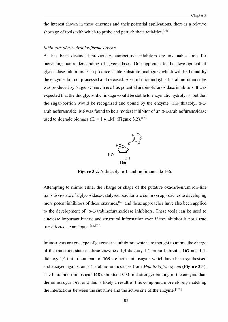
Chapter 3
103
the interest shown in these enzymes and their potential applications, there is a relative
shortage of tools with which to probe and perturb their activities.[166]
Inhibitors of α-L-Arabinofuranosidases
As has been discussed previously, competitive inhibitors are invaluable tools for
increasing our understanding of glycosidases. One approach to the development of
glycosidase inhibitors is to produce stable substrate-analogues which will be bound by
the enzyme, but not processed and released. A set of thioimidoyl α-L-arabinofuranosides
was produced by Nugier-Chauvin et al. as potential arabinofuranosidase inhibitors. It was
expected that the thioglycosidic linkage would be stable to enzymatic hydrolysis, but that
the sugar-portion would be recognised and bound by the enzyme. The thiazolyl α-L-
arabinofuranoside 166 was found to be a modest inhibitor of an α-L-arabinofuranosidase
used to degrade biomass (Ki = 1.4 µM) (Figure 3.2).[173]
Figure 3.2. A thiazolyl α-L-arabinofuranoside 166.
Attempting to mimic either the charge or shape of the putative oxacarbenium ion-like
transition-state of a glycosidase-catalysed reaction are common approaches to developing
more potent inhibitors of these enzymes,[62] and these approaches have also been applied
to the development of α-L-arabinofuranosidase inhibitors. These tools can be used to
elucidate important kinetic and structural information even if the inhibitor is not a true
transition-state analogue.[62,174]
Iminosugars are one type of glycosidase inhibitors which are thought to mimic the charge
of the transition-state of these enzymes. 1,4-dideoxy-1,4-imino-L-threitol 167 and 1,4-
dideoxy-1,4-imino-L-arabanitol 168 are both iminosugars which have been synthesised
and assayed against an α-L-arabinofuranosidase from Monilinia fructigena (Figure 3.3).
The L-arabino-iminosugar 168 exhibited 1000-fold stronger binding of the enzyme than
the iminosugar 167, and this is likely a result of this compound more closely matching
the interactions between the substrate and the active site of the enzyme.[175]
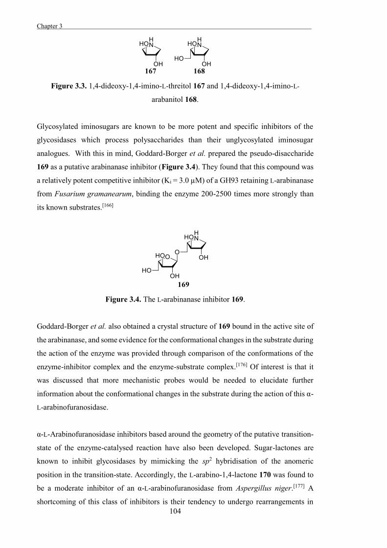
Chapter 3 .
104
Figure 3.3. 1,4-dideoxy-1,4-imino-L-threitol 167 and 1,4-dideoxy-1,4-imino-L-
arabanitol 168.
Glycosylated iminosugars are known to be more potent and specific inhibitors of the
glycosidases which process polysaccharides than their unglycosylated iminosugar
analogues. With this in mind, Goddard-Borger et al. prepared the pseudo-disaccharide
169 as a putative arabinanase inhibitor (Figure 3.4). They found that this compound was
a relatively potent competitive inhibitor (Ki = 3.0 µM) of a GH93 retaining L-arabinanase
from Fusarium gramanearum, binding the enzyme 200-2500 times more strongly than
its known substrates.[166]
Figure 3.4. The L-arabinanase inhibitor 169.
Goddard-Borger et al. also obtained a crystal structure of 169 bound in the active site of
the arabinanase, and some evidence for the conformational changes in the substrate during
the action of the enzyme was provided through comparison of the conformations of the
enzyme-inhibitor complex and the enzyme-substrate complex.[176] Of interest is that it
was discussed that more mechanistic probes would be needed to elucidate further
information about the conformational changes in the substrate during the action of this α-
L-arabinofuranosidase.
α-L-Arabinofuranosidase inhibitors based around the geometry of the putative transition-
state of the enzyme-catalysed reaction have also been developed. Sugar-lactones are
known to inhibit glycosidases by mimicking the sp2 hybridisation of the anomeric
position in the transition-state. Accordingly, the L-arabino-1,4-lactone 170 was found to
be a moderate inhibitor of an α-L-arabinofuranosidase from Aspergillus niger.[177] A
shortcoming of this class of inhibitors is their tendency to undergo rearrangements in
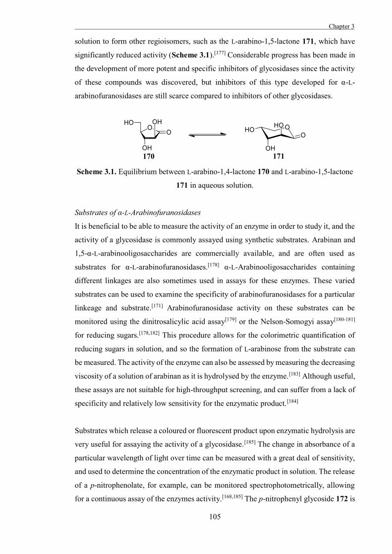
Chapter 3
105
solution to form other regioisomers, such as the L-arabino-1,5-lactone 171, which have
significantly reduced activity (Scheme 3.1).[177] Considerable progress has been made in
the development of more potent and specific inhibitors of glycosidases since the activity
of these compounds was discovered, but inhibitors of this type developed for α-L-
arabinofuranosidases are still scarce compared to inhibitors of other glycosidases.
Scheme 3.1. Equilibrium between L-arabino-1,4-lactone 170 and L-arabino-1,5-lactone
171 in aqueous solution.
Substrates of α-L-Arabinofuranosidases
It is beneficial to be able to measure the activity of an enzyme in order to study it, and the
activity of a glycosidase is commonly assayed using synthetic substrates. Arabinan and
1,5-α-L-arabinooligosaccharides are commercially available, and are often used as
substrates for α-L-arabinofuranosidases.[178] α-L-Arabinooligosaccharides containing
different linkages are also sometimes used in assays for these enzymes. These varied
substrates can be used to examine the specificity of arabinofuranosidases for a particular
linkeage and substrate.[171] Arabinofuranosidase activity on these substrates can be
monitored using the dinitrosalicylic acid assay[179] or the Nelson-Somogyi assay[180-181]
for reducing sugars.[178,182] This procedure allows for the colorimetric quantification of
reducing sugars in solution, and so the formation of L-arabinose from the substrate can
be measured. The activity of the enzyme can also be assessed by measuring the decreasing
viscosity of a solution of arabinan as it is hydrolysed by the enzyme.[183] Although useful,
these assays are not suitable for high-throughput screening, and can suffer from a lack of
specificity and relatively low sensitivity for the enzymatic product.[184]
Substrates which release a coloured or fluorescent product upon enzymatic hydrolysis are
very useful for assaying the activity of a glycosidase.[185] The change in absorbance of a
particular wavelength of light over time can be measured with a great deal of sensitivity,
and used to determine the concentration of the enzymatic product in solution. The release
of a p-nitrophenolate, for example, can be monitored spectrophotometrically, allowing
for a continuous assay of the enzymes activity.[168,185] The p-nitrophenyl glycoside 172 is
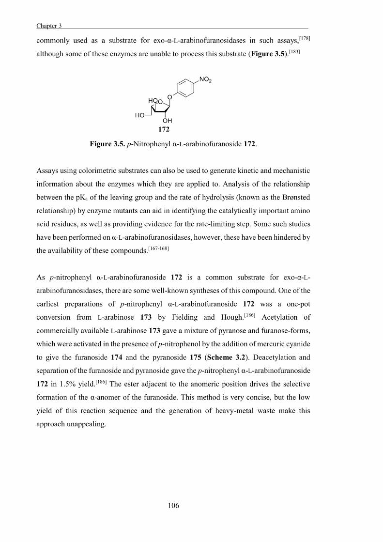
Chapter 3 .
106
commonly used as a substrate for exo-α-L-arabinofuranosidases in such assays,[178]
although some of these enzymes are unable to process this substrate (Figure 3.5).[183]
Figure 3.5. p-Nitrophenyl α-L-arabinofuranoside 172.
Assays using colorimetric substrates can also be used to generate kinetic and mechanistic
information about the enzymes which they are applied to. Analysis of the relationship
between the pKa of the leaving group and the rate of hydrolysis (known as the Brønsted
relationship) by enzyme mutants can aid in identifying the catalytically important amino
acid residues, as well as providing evidence for the rate-limiting step. Some such studies
have been performed on α-L-arabinofuranosidases, however, these have been hindered by
the availability of these compounds.[167-168]
As p-nitrophenyl α-L-arabinofuranoside 172 is a common substrate for exo-α-L-
arabinofuranosidases, there are some well-known syntheses of this compound. One of the
earliest preparations of p-nitrophenyl α-L-arabinofuranoside 172 was a one-pot
conversion from L-arabinose 173 by Fielding and Hough.[186] Acetylation of
commercially available L-arabinose 173 gave a mixture of pyranose and furanose-forms,
which were activated in the presence of p-nitrophenol by the addition of mercuric cyanide
to give the furanoside 174 and the pyranoside 175 (Scheme 3.2). Deacetylation and
separation of the furanoside and pyranoside gave the p-nitrophenyl α-L-arabinofuranoside
172 in 1.5% yield.[186] The ester adjacent to the anomeric position drives the selective
formation of the α-anomer of the furanoside. This method is very concise, but the low
yield of this reaction sequence and the generation of heavy-metal waste make this
approach unappealing.
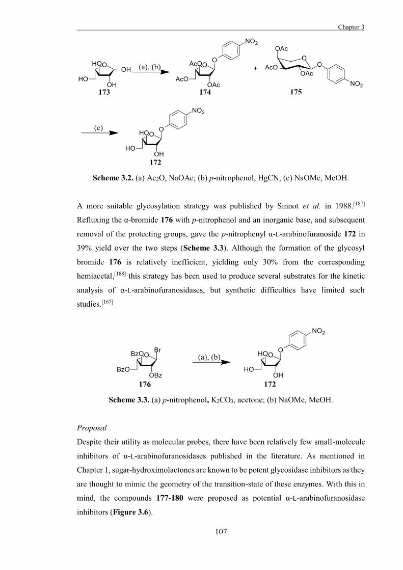
Chapter 3
107
Scheme 3.2. (a) Ac2O, NaOAc; (b) p-nitrophenol, HgCN; (c) NaOMe, MeOH.
A more suitable glycosylation strategy was published by Sinnot et al. in 1988.[187]
Refluxing the α-bromide 176 with p-nitrophenol and an inorganic base, and subsequent
removal of the protecting groups, gave the p-nitrophenyl α-L-arabinofuranoside 172 in
39% yield over the two steps (Scheme 3.3). Although the formation of the glycosyl
bromide 176 is relatively inefficient, yielding only 30% from the corresponding
hemiacetal,[188] this strategy has been used to produce several substrates for the kinetic
analysis of α-L-arabinofuranosidases, but synthetic difficulties have limited such
studies.[167]
Scheme 3.3. (a) p-nitrophenol, K2CO3, acetone; (b) NaOMe, MeOH.
Proposal
Despite their utility as molecular probes, there have been relatively few small-molecule
inhibitors of α-L-arabinofuranosidases published in the literature. As mentioned in
Chapter 1, sugar-hydroximolactones are known to be potent glycosidase inhibitors as they
are thought to mimic the geometry of the transition-state of these enzymes. With this in
mind, the compounds 177-180 were proposed as potential α-L-arabinofuranosidase
inhibitors (Figure 3.6).
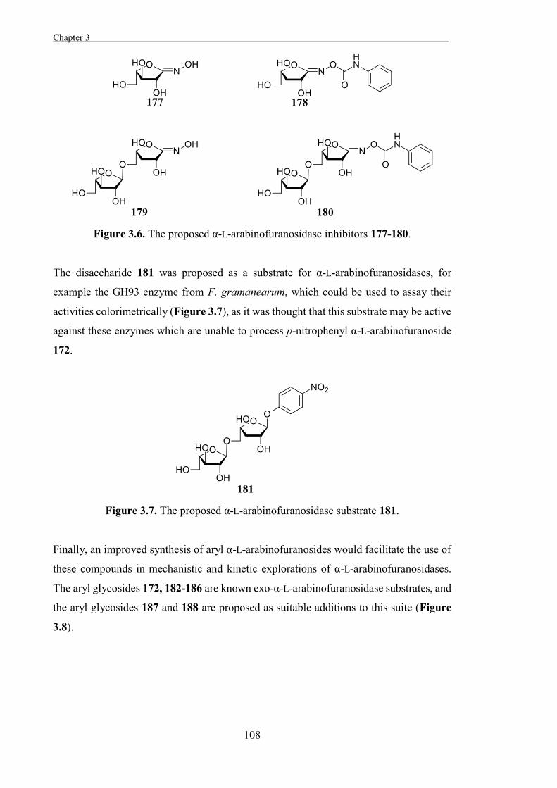
Chapter 3 .
108
Figure 3.6. The proposed α-L-arabinofuranosidase inhibitors 177-180.
The disaccharide 181 was proposed as a substrate for α-L-arabinofuranosidases, for
example the GH93 enzyme from F. gramanearum, which could be used to assay their
activities colorimetrically (Figure 3.7), as it was thought that this substrate may be active
against these enzymes which are unable to process p-nitrophenyl α-L-arabinofuranoside
172.
Figure 3.7. The proposed α-L-arabinofuranosidase substrate 181.
Finally, an improved synthesis of aryl α-L-arabinofuranosides would facilitate the use of
these compounds in mechanistic and kinetic explorations of α-L-arabinofuranosidases.
The aryl glycosides 172, 182-186 are known exo-α-L-arabinofuranosidase substrates, and
the aryl glycosides 187 and 188 are proposed as suitable additions to this suite (Figure
3.8).
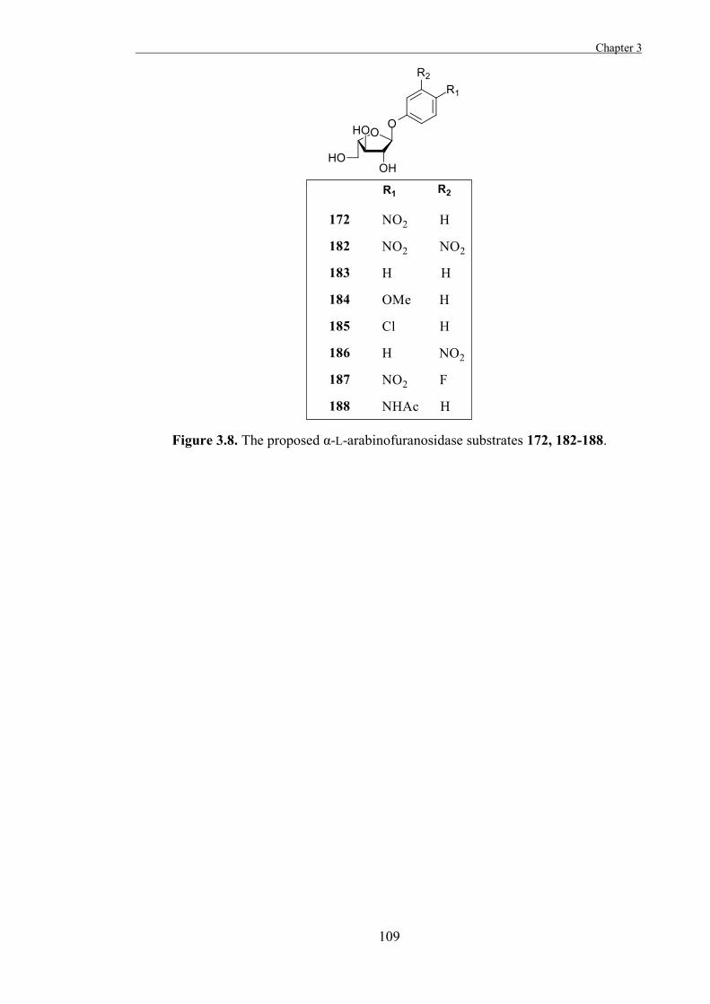
Chapter 3
109
Figure 3.8. The proposed α-L-arabinofuranosidase substrates 172, 182-188.

Chapter 3 .
110
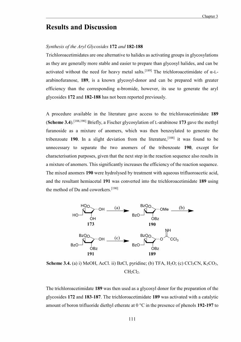
Chapter 3
111
Results and Discussion
Synthesis of the Aryl Glycosides 172 and 182-188
Trichloroacetimidates are one alternative to halides as activating groups in glycosylations
as they are generally more stable and easier to prepare than glycosyl halides, and can be
activated without the need for heavy metal salts.[189] The trichloroacetimidate of α-L-
arabinofuranose, 189, is a known glycosyl-donor and can be prepared with greater
efficiency than the corresponding α-bromide, however, its use to generate the aryl
glycosides 172 and 182-188 has not been reported previously.
A procedure available in the literature gave access to the trichloroacetimidate 189
(Scheme 3.4).[188,190] Briefly, a Fischer glycosylation of L-arabinose 173 gave the methyl
furanoside as a mixture of anomers, which was then benzoylated to generate the
tribenzoate 190. In a slight deviation from the literature,[188] it was found to be
unnecessary to separate the two anomers of the tribenzoate 190, except for
characterisation purposes, given that the next step in the reaction sequence also results in
a mixture of anomers. This significantly increases the efficiency of the reaction sequence.
The mixed anomers 190 were hydrolysed by treatment with aqueous trifluoroacetic acid,
and the resultant hemiacetal 191 was converted into the trichloroacetimidate 189 using
the method of Du and coworkers.[190]
Scheme 3.4. (a) i) MeOH, AcCl. ii) BzCl, pyridine; (b) TFA, H2O; (c) CCl3CN, K2CO3,
CH2Cl2.
The trichloroacetimidate 189 was then used as a glycosyl donor for the preparation of the
glycosides 172 and 183-187. The trichloroacetimidate 189 was activated with a catalytic
amount of boron trifluoride diethyl etherate at 0 °C in the presence of phenols 192-197 to
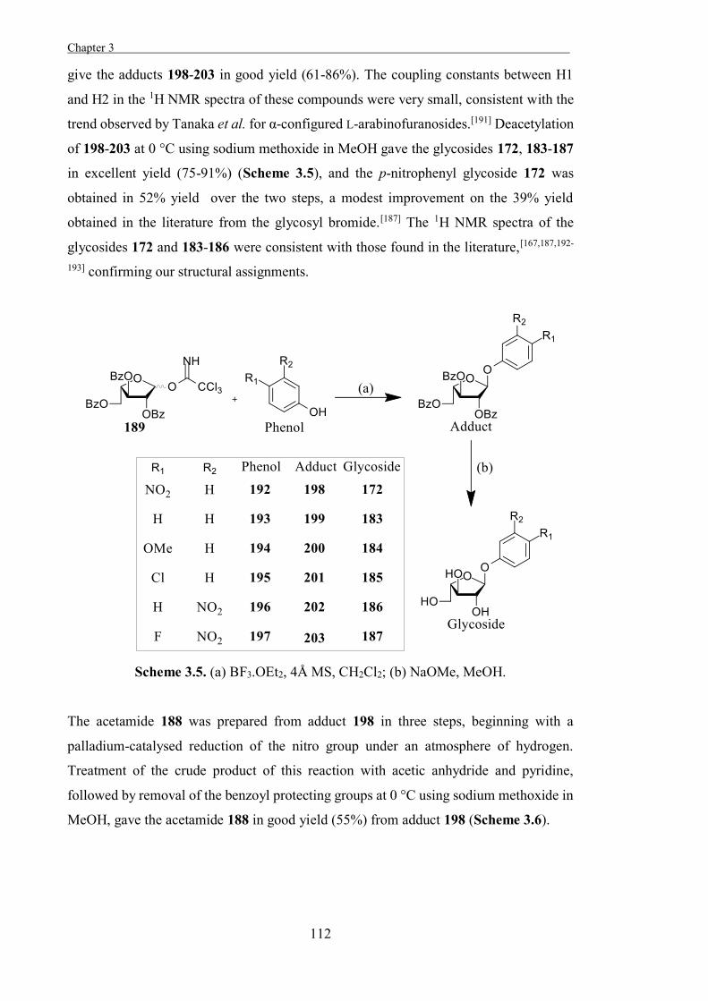
Chapter 3 .
112
give the adducts 198-203 in good yield (61-86%). The coupling constants between H1
and H2 in the 1H NMR spectra of these compounds were very small, consistent with the
trend observed by Tanaka et al. for α-configured L-arabinofuranosides.[191] Deacetylation
of 198-203 at 0 °C using sodium methoxide in MeOH gave the glycosides 172, 183-187
in excellent yield (75-91%) (Scheme 3.5), and the p-nitrophenyl glycoside 172 was
obtained in 52% yield over the two steps, a modest improvement on the 39% yield
obtained in the literature from the glycosyl bromide.[187] The 1H NMR spectra of the
glycosides 172 and 183-186 were consistent with those found in the literature,[167,187,192-
193] confirming our structural assignments.
Scheme 3.5. (a) BF3.OEt2, 4Å MS, CH2Cl2; (b) NaOMe, MeOH.
The acetamide 188 was prepared from adduct 198 in three steps, beginning with a
palladium-catalysed reduction of the nitro group under an atmosphere of hydrogen.
Treatment of the crude product of this reaction with acetic anhydride and pyridine,
followed by removal of the benzoyl protecting groups at 0 °C using sodium methoxide in
MeOH, gave the acetamide 188 in good yield (55%) from adduct 198 (Scheme 3.6).
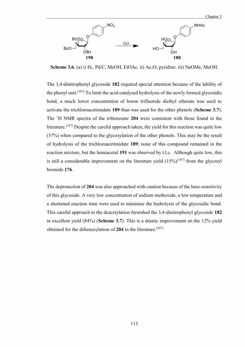
Chapter 3
113
Scheme 3.6. (a) i) H2, Pd/C, MeOH, EtOAc. ii) Ac2O, pyridine. iii) NaOMe, MeOH.
The 3,4-dinitrophenyl glycoside 182 required special attention because of the lability of
the phenyl unit.[187] To limit the acid-catalysed hydrolysis of the newly formed glycosidic
bond, a much lower concentration of boron trifluoride diethyl etherate was used to
activate the trichloroacetimidate 189 than was used for the other phenols (Scheme 3.7).
The 1H NMR spectra of the tribenzoate 204 were consistent with those found in the
literature.[187] Despite the careful approach taken, the yield for this reaction was quite low
(37%) when compared to the glycosylation of the other phenols. This may be the result
of hydrolysis of the trichloroacetimidate 189; none of this compound remained in the
reaction mixture, but the hemiacetal 191 was observed by t.l.c. Although quite low, this
is still a considerable improvement on the literature yield (15%)[187] from the glycosyl
bromide 176.
The deprotection of 204 was also approached with caution because of the base-sensitivity
of this glycoside. A very low concentration of sodium methoxide, a low temperature and
a shortened reaction time were used to minimise the hydrolysis of the glycosidic bond.
This careful approach to the deacetylation furnished the 3,4-dinitrophenyl glycoside 182
in excellent yield (84%) (Scheme 3.7). This is a drastic improvement on the 12% yield
obtained for the debenzoylation of 204 in the literature.[187]
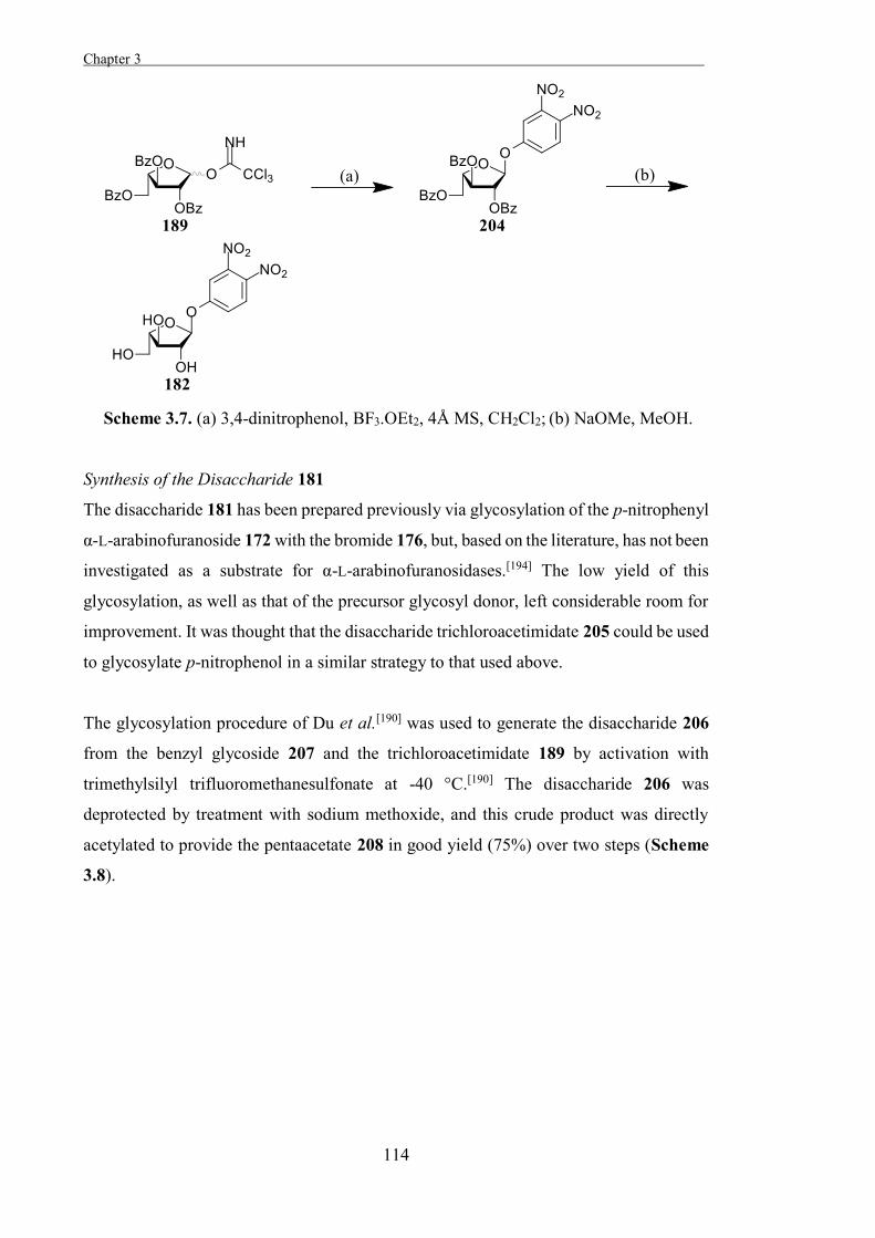
Chapter 3 .
114
Scheme 3.7. (a) 3,4-dinitrophenol, BF3.OEt2, 4Å MS, CH2Cl2; (b) NaOMe, MeOH.
Synthesis of the Disaccharide 181
The disaccharide 181 has been prepared previously via glycosylation of the p-nitrophenyl
α-L-arabinofuranoside 172 with the bromide 176, but, based on the literature, has not been
investigated as a substrate for α-L-arabinofuranosidases.[194] The low yield of this
glycosylation, as well as that of the precursor glycosyl donor, left considerable room for
improvement. It was thought that the disaccharide trichloroacetimidate 205 could be used
to glycosylate p-nitrophenol in a similar strategy to that used above.
The glycosylation procedure of Du et al.[190] was used to generate the disaccharide 206
from the benzyl glycoside 207 and the trichloroacetimidate 189 by activation with
trimethylsilyl trifluoromethanesulfonate at -40 °C.[190] The disaccharide 206 was
deprotected by treatment with sodium methoxide, and this crude product was directly
acetylated to provide the pentaacetate 208 in good yield (75%) over two steps (Scheme
3.8).
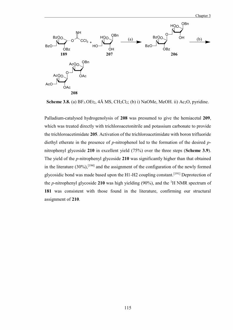
Chapter 3
115
Scheme 3.8. (a) BF3.OEt2, 4Å MS, CH2Cl2; (b) i) NaOMe, MeOH. ii) Ac2O, pyridine.
Palladium-catalysed hydrogenolysis of 208 was presumed to give the hemiacetal 209,
which was treated directly with trichloroacetonitrile and potassium carbonate to provide
the trichloroacetimidate 205. Activation of the trichloroacetimidate with boron trifluoride
diethyl etherate in the presence of p-nitrophenol led to the formation of the desired p-
nitrophenyl glycoside 210 in excellent yield (75%) over the three steps (Scheme 3.9).
The yield of the p-nitrophenyl glycoside 210 was significantly higher than that obtained
in the literature (30%),[194] and the assignment of the configuration of the newly formed
glycosidic bond was made based upon the H1-H2 coupling constant.[191] Deprotection of
the p-nitrophenyl glycoside 210 was high yielding (90%), and the 1H NMR spectrum of
181 was consistent with those found in the literature, confirming our structural
assignment of 210.
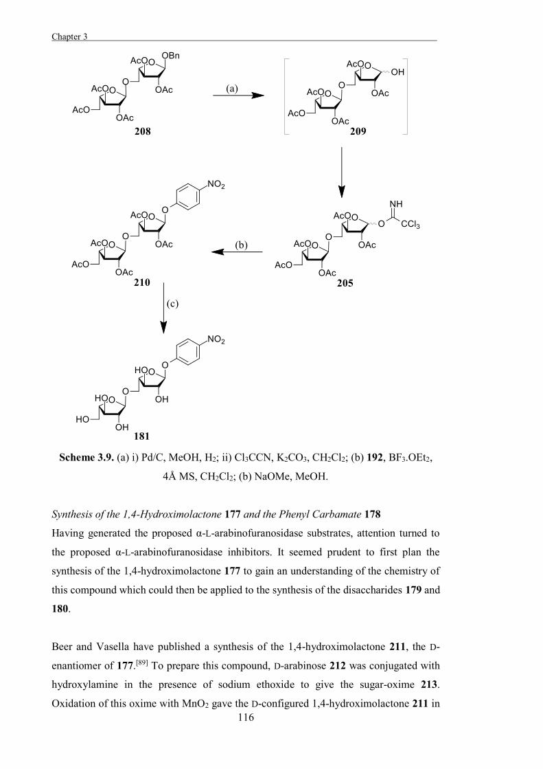
Chapter 3 .
116
Scheme 3.9. (a) i) Pd/C, MeOH, H2; ii) Cl3CCN, K2CO3, CH2Cl2; (b) 192, BF3.OEt2,
4Å MS, CH2Cl2; (b) NaOMe, MeOH.
Synthesis of the 1,4-Hydroximolactone 177 and the Phenyl Carbamate 178
Having generated the proposed α-L-arabinofuranosidase substrates, attention turned to
the proposed α-L-arabinofuranosidase inhibitors. It seemed prudent to first plan the
synthesis of the 1,4-hydroximolactone 177 to gain an understanding of the chemistry of
this compound which could then be applied to the synthesis of the disaccharides 179 and
180.
Beer and Vasella have published a synthesis of the 1,4-hydroximolactone 211, the D-
enantiomer of 177.[89] To prepare this compound, D-arabinose 212 was conjugated with
hydroxylamine in the presence of sodium ethoxide to give the sugar-oxime 213.
Oxidation of this oxime with MnO2 gave the D-configured 1,4-hydroximolactone 211 in
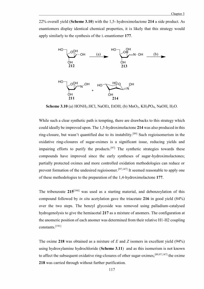
Chapter 3
117
22% overall yield (Scheme 3.10) with the 1,5- hydroximolactone 214 a side product. As
enantiomers display identical chemical properties, it is likely that this strategy would
apply similarly to the synthesis of the L-enantiomer 177.
Scheme 3.10 (a) HONH2.HCl, NaOEt, EtOH; (b) MnO2, KH2PO4, NaOH, H2O.
While such a clear synthetic path is tempting, there are drawbacks to this strategy which
could ideally be improved upon. The 1,5-hydroximolactone 214 was also produced in this
ring-closure, but wasn’t quantified due to its instability.[89] Such regioisomerism in the
oxidative ring-closures of sugar-oximes is a significant issue, reducing yields and
impairing efforts to purify the products.[97] The synthetic strategies towards these
compounds have improved since the early syntheses of sugar-hydroximolactones;
partially protected oximes and more controlled oxidation methodologies can reduce or
prevent formation of the undesired regioisomer.[97,147] It seemed reasonable to apply one
of these methodologies to the preparation of the 1,4-hydroximolactone 177.
The tribenzoate 215[166] was used as a starting material, and debenzoylation of this
compound followed by in situ acetylation gave the triacetate 216 in good yield (84%)
over the two steps. The benzyl glycoside was removed using palladium-catalysed
hydrogenolysis to give the hemiacetal 217 as a mixture of anomers. The configuration at
the anomeric position of each anomer was determined from their relative H1-H2 coupling
constants.[191]
The oxime 218 was obtained as a mixture of E and Z isomers in excellent yield (94%)
using hydroxylamine hydrochloride (Scheme 3.11) and as this isomerism is not known
to affect the subsequent oxidative ring-closures of other sugar-oximes,[89,97,147] the oxime
218 was carried through without further purification.
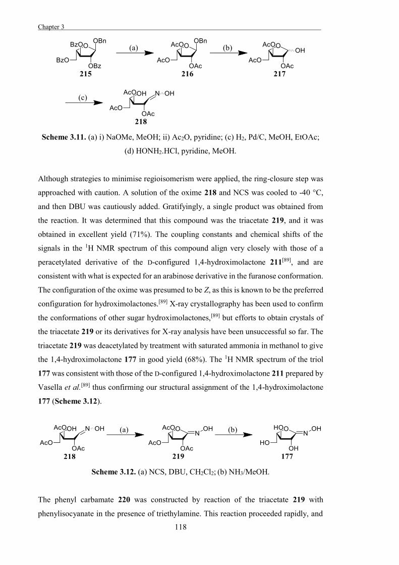
Chapter 3 .
118
Scheme 3.11. (a) i) NaOMe, MeOH; ii) Ac2O, pyridine; (c) H2, Pd/C, MeOH, EtOAc;
(d) HONH2.HCl, pyridine, MeOH.
Although strategies to minimise regioisomerism were applied, the ring-closure step was
approached with caution. A solution of the oxime 218 and NCS was cooled to -40 °C,
and then DBU was cautiously added. Gratifyingly, a single product was obtained from
the reaction. It was determined that this compound was the triacetate 219, and it was
obtained in excellent yield (71%). The coupling constants and chemical shifts of the
signals in the 1H NMR spectrum of this compound align very closely with those of a
peracetylated derivative of the D-configured 1,4-hydroximolactone 211[89], and are
consistent with what is expected for an arabinose derivative in the furanose conformation.
The configuration of the oxime was presumed to be Z, as this is known to be the preferred
configuration for hydroximolactones.[89] X-ray crystallography has been used to confirm
the conformations of other sugar hydroximolactones,[89] but efforts to obtain crystals of
the triacetate 219 or its derivatives for X-ray analysis have been unsuccessful so far. The
triacetate 219 was deacetylated by treatment with saturated ammonia in methanol to give
the 1,4-hydroximolactone 177 in good yield (68%). The 1H NMR spectrum of the triol
177 was consistent with those of the D-configured 1,4-hydroximolactone 211 prepared by
Vasella et al.[89] thus confirming our structural assignment of the 1,4-hydroximolactone
177 (Scheme 3.12).
Scheme 3.12. (a) NCS, DBU, CH2Cl2; (b) NH3/MeOH.
The phenyl carbamate 220 was constructed by reaction of the triacetate 219 with
phenylisocyanate in the presence of triethylamine. This reaction proceeded rapidly, and
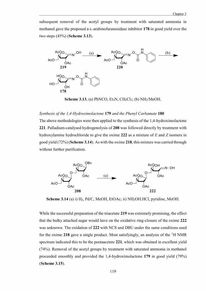
Chapter 3
119
subsequent removal of the acetyl groups by treatment with saturated ammonia in
methanol gave the proposed a-L-arabinofuranosidase inhibitor 178 in good yield over the
two steps (45%) (Scheme 3.13).
Scheme 3.13. (a) PhNCO, Et3N, CH2Cl2; (b) NH3/MeOH.
Synthesis of the 1,4-Hydroximolactone 179 and the Phenyl Carbamate 180
The above methodologies were then applied to the synthesis of the 1,4-hydroximolactone
221. Palladium-catalysed hydrogenolysis of 208 was followed directly by treatment with
hydroxylamine hydrochloride to give the oxime 222 as a mixture of E and Z isomers in
good yield (72%) (Scheme 3.14). As with the oxime 218, this mixture was carried through
without further purification.
Scheme 3.14 (a) i) H2, Pd/C, MeOH, EtOAc; ii) NH2OH.HCl, pyridine, MeOH.
While the successful preparation of the triacetate 219 was extremely promising, the effect
that the bulky attached sugar would have on the oxidative ring-closure of the oxime 222
was unknown. The oxidation of 222 with NCS and DBU under the same conditions used
for the oxime 218 gave a single product. Most satisfyingly, an analysis of the 1H NMR
spectrum indicated this to be the pentaacetate 221, which was obtained in excellent yield
(74%). Removal of the acetyl groups by treatment with saturated ammonia in methanol
proceeded smoothly and provided the 1,4-hydroximolactone 179 in good yield (79%)
(Scheme 3.15).
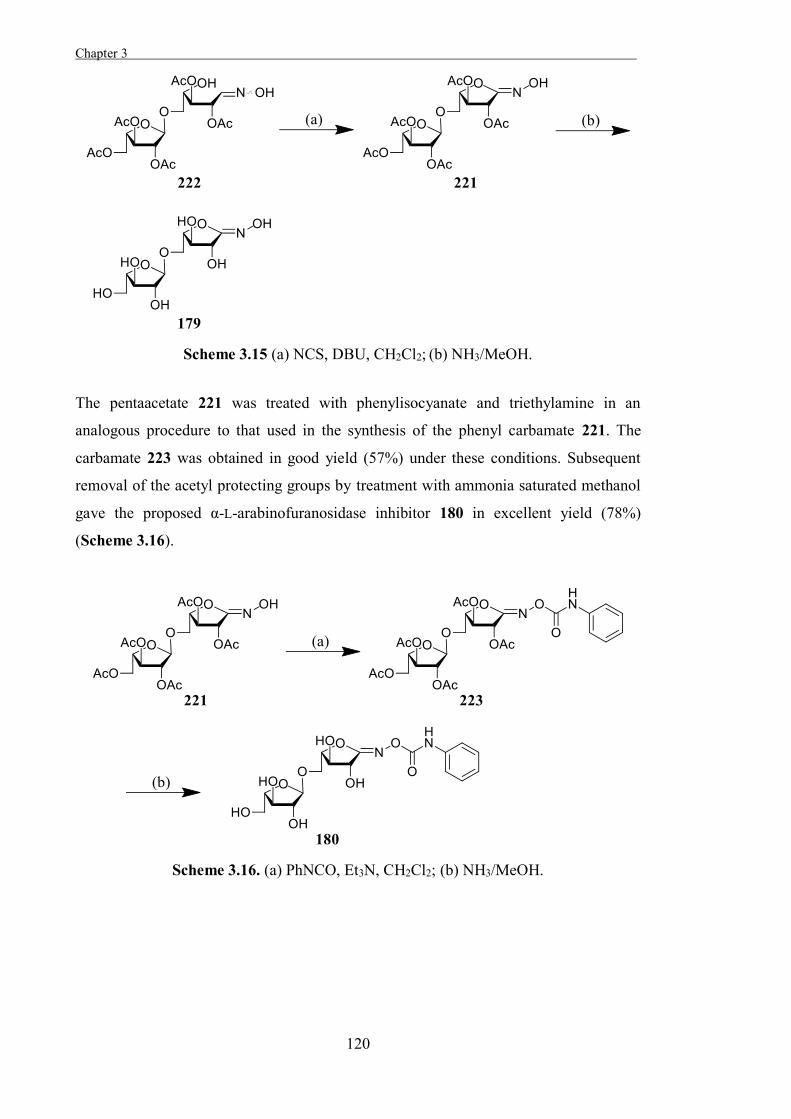
Chapter 3 .
120
Scheme 3.15 (a) NCS, DBU, CH2Cl2; (b) NH3/MeOH.
The pentaacetate 221 was treated with phenylisocyanate and triethylamine in an
analogous procedure to that used in the synthesis of the phenyl carbamate 221. The
carbamate 223 was obtained in good yield (57%) under these conditions. Subsequent
removal of the acetyl protecting groups by treatment with ammonia saturated methanol
gave the proposed α-L-arabinofuranosidase inhibitor 180 in excellent yield (78%)
(Scheme 3.16).
Scheme 3.16. (a) PhNCO, Et3N, CH2Cl2; (b) NH3/MeOH.
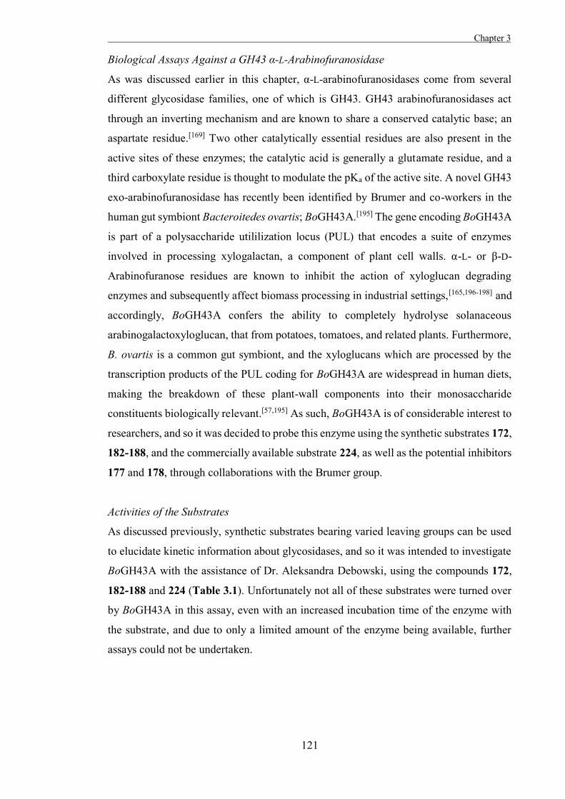
Chapter 3
121
Biological Assays Against a GH43 α-L-Arabinofuranosidase
As was discussed earlier in this chapter, α-L-arabinofuranosidases come from several
different glycosidase families, one of which is GH43. GH43 arabinofuranosidases act
through an inverting mechanism and are known to share a conserved catalytic base; an
aspartate residue.[169] Two other catalytically essential residues are also present in the
active sites of these enzymes; the catalytic acid is generally a glutamate residue, and a
third carboxylate residue is thought to modulate the pKa of the active site. A novel GH43
exo-arabinofuranosidase has recently been identified by Brumer and co-workers in the
human gut symbiont Bacteroitedes ovartis; BoGH43A.[195] The gene encoding BoGH43A
is part of a polysaccharide utililization locus (PUL) that encodes a suite of enzymes
involved in processing xylogalactan, a component of plant cell walls. α-L- or β-D-
Arabinofuranose residues are known to inhibit the action of xyloglucan degrading
enzymes and subsequently affect biomass processing in industrial settings,[165,196-198] and
accordingly, BoGH43A confers the ability to completely hydrolyse solanaceous
arabinogalactoxyloglucan, that from potatoes, tomatoes, and related plants. Furthermore,
B. ovartis is a common gut symbiont, and the xyloglucans which are processed by the
transcription products of the PUL coding for BoGH43A are widespread in human diets,
making the breakdown of these plant-wall components into their monosaccharide
constituents biologically relevant.[57,195] As such, BoGH43A is of considerable interest to
researchers, and so it was decided to probe this enzyme using the synthetic substrates 172,
182-188, and the commercially available substrate 224, as well as the potential inhibitors
177 and 178, through collaborations with the Brumer group.
Activities of the Substrates
As discussed previously, synthetic substrates bearing varied leaving groups can be used
to elucidate kinetic information about glycosidases, and so it was intended to investigate
BoGH43A with the assistance of Dr. Aleksandra Debowski, using the compounds 172,
182-188 and 224 (Table 3.1). Unfortunately not all of these substrates were turned over
by BoGH43A in this assay, even with an increased incubation time of the enzyme with
the substrate, and due to only a limited amount of the enzyme being available, further
assays could not be undertaken.
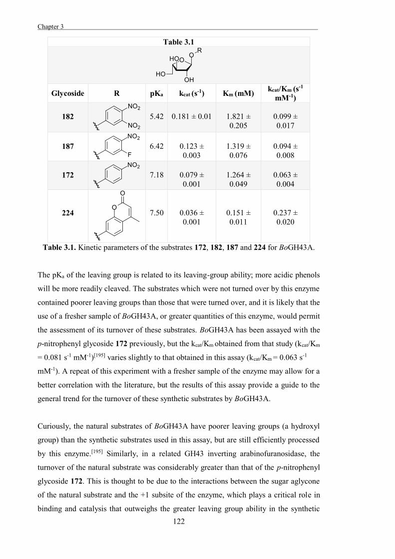
Chapter 3 .
122
Table 3.1
Glycoside R pKa kcat (s-1) Km (mM) kcat/Km (s-1
mM-1)
182
5.42
0.181 ± 0.01
1.821 ± 0.205
0.099 ± 0.017
187
6.42
0.123 ± 0.003
1.319 ± 0.076
0.094 ± 0.008
172
7.18
0.079 ± 0.001
1.264 ± 0.049
0.063 ± 0.004
224
7.50
0.036 ± 0.001
0.151 ± 0.011
0.237 ± 0.020
Table 3.1. Kinetic parameters of the substrates 172, 182, 187 and 224 for BoGH43A.
The pKa of the leaving group is related to its leaving-group ability; more acidic phenols
will be more readily cleaved. The substrates which were not turned over by this enzyme
contained poorer leaving groups than those that were turned over, and it is likely that the
use of a fresher sample of BoGH43A, or greater quantities of this enzyme, would permit
the assessment of its turnover of these substrates. BoGH43A has been assayed with the
p-nitrophenyl glycoside 172 previously, but the kcat/Km obtained from that study (kcat/Km
= 0.081 s-1 mM-1)[195] varies slightly to that obtained in this assay (kcat/Km = 0.063 s-1
mM-1). A repeat of this experiment with a fresher sample of the enzyme may allow for a
better correlation with the literature, but the results of this assay provide a guide to the
general trend for the turnover of these synthetic substrates by BoGH43A.
Curiously, the natural substrates of BoGH43A have poorer leaving groups (a hydroxyl
group) than the synthetic substrates used in this assay, but are still efficiently processed
by this enzyme.[195] Similarly, in a related GH43 inverting arabinofuranosidase, the
turnover of the natural substrate was considerably greater than that of the p-nitrophenyl
glycoside 172. This is thought to be due to the interactions between the sugar aglycone
of the natural substrate and the +1 subsite of the enzyme, which plays a critical role in
binding and catalysis that outweighs the greater leaving group ability in the synthetic
Chapter 3
123
substrates.[172] This sugar-binding +1 subsite is highly conserved among GH43 inverting
arabinofuranosidases, and so it is likely that a similar phenomenon is driving the results
obtained in the assay with BoGH43A.
Activities of the 1,4-Hydroximolactone 177 and the Phenyl Carbamate 178
Following on from this, the activity of BoGH43A in the presence of the 1,4-
hydroximolactone 177 and the phenyl carbamate 178 was examined. Disappointingly, the
1,4-hydroximolactone 177 exhibited no inhibition against BoGH43A even at 1 mM
concentration, and the phenyl carbamate 178 exhibited only weak inhibition of this
enzyme (25% inhibition at 1 mM). As such, these inhibitors are not well suited to further
examination of BoGH43A. It was expected that the phenyl carbamate 178 would exhibit
greater activity than the 1,4-hydroximolactone 177, as this trend has been observed in
other hydroximolactone-based glycosidase inhibitors, but it is unclear why these
compounds exhibit such weak activity against the enzyme. Some hydroximolactone-
based inhibitors have been found to be more potent inhibitors of retaining glycosidases
than inverting glycosidases, and this may also be true of the compounds 177 and
178.[50,154]
Biological Assays Against a GH93 Arabinanase
As described earlier, all known arabinanases belong to either GH43 or GH93. All known
GH93 arabinanases are exo-acting glycosidases which act through a retaining mechanism,
and it was thought that the activities of the 1,4-hydroximolactone 179 and the phenyl
carbamate 180 against a GH93 arabinanase would be of interest. One characterised GH93
arabinanase is Arb93A, isolated from the fungus F. graminearum. X-ray crystal structures
of this enzyme are available, and the catalytic residues have been identified, with a
glutamate residue acting as the catalytic nucleophile, and another glutamate residue acting
as the catalytic acid/base. As well, this enzyme has been structurally and biochemically
examined using the pseudo-disaccharide 169, but it has been stated that more tools are
required to examine this enzyme.[166] As such, this enzyme was a promising candidate for
investigation using the potential inhibitors 179 and 180 and this was done through
collaboration with the Varrot group (France) for the X-ray crystallographic aspect of the
project (Table 3.3).
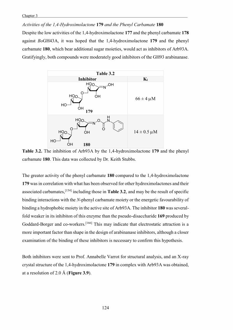
Chapter 3 .
124
Activities of the 1,4-Hydroximolactone 179 and the Phenyl Carbamate 180
Despite the low activities of the 1,4-hydroximolactone 177 and the phenyl carbamate 178
against BoGH43A, it was hoped that the 1,4-hydroximolactone 179 and the phenyl
carbamate 180, which bear additional sugar moieties, would act as inhibitors of Arb93A.
Gratifyingly, both compounds were moderately good inhibitors of the GH93 arabinanase.
Table 3.2 Inhibitor Ki
66 ± 4 M
14 ± 0.5 M
Table 3.2. The inhibition of Arb93A by the 1,4-hydroximolactone 179 and the phenyl
carbamate 180. This data was collected by Dr. Keith Stubbs.
The greater activity of the phenyl carbamate 180 compared to the 1,4-hydroximolactone
179 was in correlation with what has been observed for other hydroximolactones and their
associated carbamates,[154] including those in Table 3.2, and may be the result of specific
binding interactions with the N-phenyl carbamate moiety or the energetic favourability of
binding a hydrophobic moiety in the active site of Arb93A. The inhibitor 180 was several-
fold weaker in its inhibiton of this enzyme than the pseudo-disaccharide 169 produced by
Goddard-Borger and co-workers.[166] This may indicate that electrostatic attraction is a
more important factor than shape in the design of arabinanase inhibitors, although a closer
examination of the binding of these inhibitors is necessary to confirm this hypothesis.
Both inhibitors were sent to Prof. Annabelle Varrot for structural analysis, and an X-ray
crystal structure of the 1,4-hydroximolactone 179 in complex with Arb93A was obtained,
at a resolution of 2.0 Å (Figure 3.9).
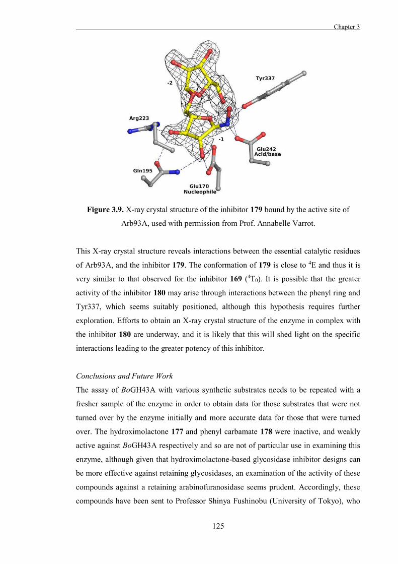
Chapter 3
125
Figure 3.9. X-ray crystal structure of the inhibitor 179 bound by the active site of
Arb93A, used with permission from Prof. Annabelle Varrot.
This X-ray crystal structure reveals interactions between the essential catalytic residues
of Arb93A, and the inhibitor 179. The conformation of 179 is close to 4E and thus it is
very similar to that observed for the inhibitor 169 (4T0). It is possible that the greater
activity of the inhibitor 180 may arise through interactions between the phenyl ring and
Tyr337, which seems suitably positioned, although this hypothesis requires further
exploration. Efforts to obtain an X-ray crystal structure of the enzyme in complex with
the inhibitor 180 are underway, and it is likely that this will shed light on the specific
interactions leading to the greater potency of this inhibitor.
Conclusions and Future Work
The assay of BoGH43A with various synthetic substrates needs to be repeated with a
fresher sample of the enzyme in order to obtain data for those substrates that were not
turned over by the enzyme initially and more accurate data for those that were turned
over. The hydroximolactone 177 and phenyl carbamate 178 were inactive, and weakly
active against BoGH43A respectively and so are not of particular use in examining this
enzyme, although given that hydroximolactone-based glycosidase inhibitor designs can
be more effective against retaining glycosidases, an examination of the activity of these
compounds against a retaining arabinofuranosidase seems prudent. Accordingly, these
compounds have been sent to Professor Shinya Fushinobu (University of Tokyo), who
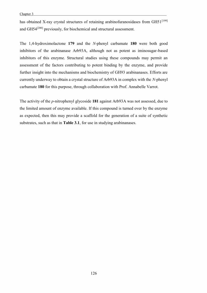
Chapter 3 .
126
has obtained X-ray crystal structures of retaining arabinofuranosidases from GH51[199]
and GH54[200] previously, for biochemical and structural assessment.
The 1,4-hydroximolactone 179 and the N-phenyl carbamate 180 were both good
inhibitors of the arabinanase Arb93A, although not as potent as iminosugar-based
inhibitors of this enzyme. Structural studies using these compounds may permit an
assessment of the factors contributing to potent binding by the enzyme, and provide
further insight into the mechanisms and biochemistry of GH93 arabinanases. Efforts are
currently underway to obtain a crystal structure of Arb93A in complex with the N-phenyl
carbamate 180 for this purpose, through collaboration with Prof. Annabelle Varrot.
The activity of the p-nitrophenyl glycoside 181 against Arb93A was not assessed, due to
the limited amount of enzyme available. If this compound is turned over by the enzyme
as expected, then this may provide a scaffold for the generation of a suite of synthetic
substrates, such as that in Table 3.1, for use in studying arabinanases.
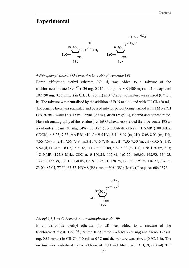
Chapter 3
127
Experimental
4-Nitrophenyl 2,3,5-tri-O-benzoyl-α-L-arabinofuranoside 198
Boron trifluoride diethyl etherate (60 µl) was added to a mixture of the
trichloroacetimidate 189[190] (130 mg, 0.215 mmol), 4Å MS (400 mg) and 4-nitrophenol
192 (90 mg, 0.65 mmol) in CH2Cl2 (20 ml) at 0 °C and the mixture was stirred (0 °C, 1
h). The mixture was neutralised by the addition of Et3N and diluted with CH2Cl2 (20 ml).
The organic layer was separated and poured into ice before being washed with 1 M NaOH
(3 x 20 ml), water (3 x 15 ml), brine (20 ml), dried (MgSO4), filtered and concentrated.
Flash chromatography of the residue (1:3 EtOAc/hexanes) yielded the tribenzoate 198 as
a colourless foam (80 mg, 64%). Rf 0.25 (1:3 EtOAc/hexanes). 1H NMR (500 MHz,
CDCl3): δ 8.23, 7.22 (AA'BB', 4H, J = 9.5 Hz), 8.14-8.09 (m, 2H), 8.08-8.01 (m, 4H),
7.66-7.58 (m, 2H), 7.56-7.48 (m, 3H), 7.45-7.40 (m, 2H), 7.35-7.30 (m, 2H), 6.05 (s, 1H),
5.82 (d, 1H, J = 1.0 Hz), 5.71 (d, 1H, J = 4.0 Hz), 4.87-4.80 (m, 1H), 4.78-4.70 (m, 2H); 13C NMR (125.8 MHz, CDCl3): δ 166.28, 165.81, 165.55, 160.95, 142.93, 134.03,
133.96, 133.39, 130.10, 130.08, 129.91, 128.81, 128.78, 128.55, 125.98, 116.72, 104.05,
83.00, 82.05, 77.59, 63.52. HRMS (ES): m/z = 606.1381; [M+Na]+ requires 606.1376.
Phenyl 2,3,5-tri-O-benzoyl-α-L-arabinofuranoside 199
Boron trifluoride diethyl etherate (40 µl) was added to a mixture of the
trichloroacetimidate 189[190] (180 mg, 0.297 mmol), 4Å MS (250 mg) and phenol 193 (80
mg, 0.85 mmol) in CH2Cl2 (10 ml) at 0 °C and the mixture was stirred (0 °C, 1 h). The
mixture was neutralised by the addition of Et3N and diluted with CH2Cl2 (20 ml). The
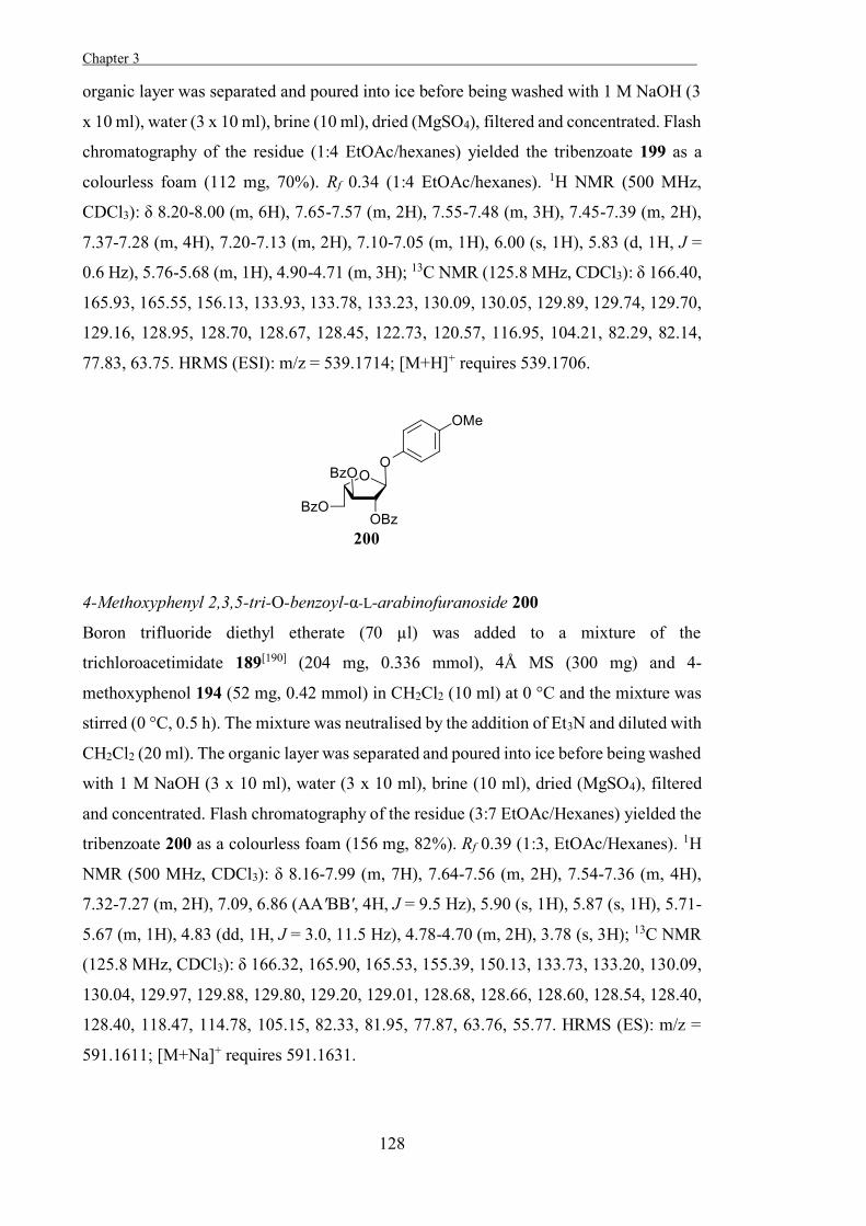
Chapter 3 .
128
organic layer was separated and poured into ice before being washed with 1 M NaOH (3
x 10 ml), water (3 x 10 ml), brine (10 ml), dried (MgSO4), filtered and concentrated. Flash
chromatography of the residue (1:4 EtOAc/hexanes) yielded the tribenzoate 199 as a
colourless foam (112 mg, 70%). Rf 0.34 (1:4 EtOAc/hexanes). 1H NMR (500 MHz,
CDCl3): δ 8.20-8.00 (m, 6H), 7.65-7.57 (m, 2H), 7.55-7.48 (m, 3H), 7.45-7.39 (m, 2H),
7.37-7.28 (m, 4H), 7.20-7.13 (m, 2H), 7.10-7.05 (m, 1H), 6.00 (s, 1H), 5.83 (d, 1H, J =
0.6 Hz), 5.76-5.68 (m, 1H), 4.90-4.71 (m, 3H); 13C NMR (125.8 MHz, CDCl3): δ 166.40,
165.93, 165.55, 156.13, 133.93, 133.78, 133.23, 130.09, 130.05, 129.89, 129.74, 129.70,
129.16, 128.95, 128.70, 128.67, 128.45, 122.73, 120.57, 116.95, 104.21, 82.29, 82.14,
77.83, 63.75. HRMS (ESI): m/z = 539.1714; [M+H]+ requires 539.1706.
4-Methoxyphenyl 2,3,5-tri-O-benzoyl-α-L-arabinofuranoside 200
Boron trifluoride diethyl etherate (70 µl) was added to a mixture of the
trichloroacetimidate 189[190] (204 mg, 0.336 mmol), 4Å MS (300 mg) and 4-
methoxyphenol 194 (52 mg, 0.42 mmol) in CH2Cl2 (10 ml) at 0 °C and the mixture was
stirred (0 °C, 0.5 h). The mixture was neutralised by the addition of Et3N and diluted with
CH2Cl2 (20 ml). The organic layer was separated and poured into ice before being washed
with 1 M NaOH (3 x 10 ml), water (3 x 10 ml), brine (10 ml), dried (MgSO4), filtered
and concentrated. Flash chromatography of the residue (3:7 EtOAc/Hexanes) yielded the
tribenzoate 200 as a colourless foam (156 mg, 82%). Rf 0.39 (1:3, EtOAc/Hexanes). 1H
NMR (500 MHz, CDCl3): δ 8.16-7.99 (m, 7H), 7.64-7.56 (m, 2H), 7.54-7.36 (m, 4H),
7.32-7.27 (m, 2H), 7.09, 6.86 (AA'BB', 4H, J = 9.5 Hz), 5.90 (s, 1H), 5.87 (s, 1H), 5.71-
5.67 (m, 1H), 4.83 (dd, 1H, J = 3.0, 11.5 Hz), 4.78-4.70 (m, 2H), 3.78 (s, 3H); 13C NMR
(125.8 MHz, CDCl3): δ 166.32, 165.90, 165.53, 155.39, 150.13, 133.73, 133.20, 130.09,
130.04, 129.97, 129.88, 129.80, 129.20, 129.01, 128.68, 128.66, 128.60, 128.54, 128.40,
128.40, 118.47, 114.78, 105.15, 82.33, 81.95, 77.87, 63.76, 55.77. HRMS (ES): m/z =
591.1611; [M+Na]+ requires 591.1631.
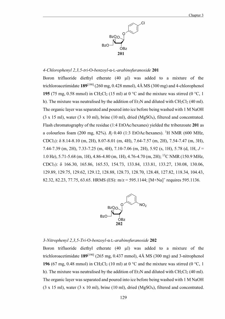
Chapter 3
129
4-Chlorophenyl 2,3,5-tri-O-benzoyl-α-L-arabinofuranoside 201
Boron trifluoride diethyl etherate (40 µl) was added to a mixture of the
trichloroacetimidate 189[190] (260 mg, 0.428 mmol), 4Å MS (300 mg) and 4-chlorophenol
195 (75 mg, 0.58 mmol) in CH2Cl2 (15 ml) at 0 °C and the mixture was stirred (0 °C, 1
h). The mixture was neutralised by the addition of Et3N and diluted with CH2Cl2 (40 ml).
The organic layer was separated and poured into ice before being washed with 1 M NaOH
(3 x 15 ml), water (3 x 10 ml), brine (10 ml), dried (MgSO4), filtered and concentrated.
Flash chromatography of the residue (1:4 EtOAc/hexanes) yielded the tribenzoate 201 as
a colourless foam (200 mg, 82%). Rf 0.40 (1:3 EtOAc/hexanes). 1H NMR (600 MHz,
CDCl3): δ 8.14-8.10 (m, 2H), 8.07-8.01 (m, 4H), 7.64-7.57 (m, 2H), 7.54-7.47 (m, 3H),
7.44-7.39 (m, 2H), 7.33-7.25 (m, 4H), 7.10-7.06 (m, 2H), 5.92 (s, 1H), 5.78 (d, 1H, J =
1.0 Hz), 5.71-5.68 (m, 1H), 4.86-4.80 (m, 1H), 4.76-4.70 (m, 2H); 13C NMR (150.9 MHz,
CDCl3): δ 166.30, 165.86, 165.53, 154.73, 133.84, 133.81, 133.27, 130.08, 130.06,
129.89, 129.75, 129.62, 129.12, 128.88, 128.73, 128.70, 128.48, 127.82, 118.34, 104.43,
82.32, 82.23, 77.75, 63.65. HRMS (ES): m/z = 595.1144; [M+Na]+ requires 595.1136.
3-Nitrophenyl 2,3,5-Tri-O-benzoyl-α-L-arabinofuranoside 202
Boron trifluoride diethyl etherate (40 µl) was added to a mixture of the
trichloroacetimidate 189[190] (265 mg, 0.437 mmol), 4Å MS (300 mg) and 3-nitrophenol
196 (67 mg, 0.48 mmol) in CH2Cl2 (10 ml) at 0 °C and the mixture was stirred (0 °C, 1
h). The mixture was neutralised by the addition of Et3N and diluted with CH2Cl2 (40 ml).
The organic layer was separated and poured into ice before being washed with 1 M NaOH
(3 x 15 ml), water (3 x 10 ml), brine (10 ml), dried (MgSO4), filtered and concentrated.

Chapter 3 .
130
Flash chromatography of the residue (3:7 EtOAc/hexanes) yielded the tribenzoate 202 as
a colourless foam (210 mg, 86%). Rf 0.20 (1:4 EtOAc/hexanes). 1H NMR (500 MHz,
CDCl3): δ 8.14-8.10 (m, 2H), 8.07-8.00 (m, 5H), 7.94-7.91 (m, 1H), 7.65-7.59 (m, 2H),
7.55-7.40 (m, 7H), 7.34-7.30 (m, 2H), 6.04 (s, 1H), 5.82 (d, 1H, J = 1.0 Hz), 5.73-5.70
(m, 1H), 4.83 (dd, 1H, J = 5.5, 13.5 Hz), 4.77-4.72 (m, 2H); 13C NMR (125.8 MHz,
CDCl3): δ 166.31, 165.85, 165.58, 156.57, 149.27, 133.97, 133.93, 133.34, 130.08,
129.91, 129.69, 129.01, 128.78, 128.73, 128.53, 123.18, 117.63, 112.09, 104.39, 82.76,
82.18, 77.63, 63.56. HRMS (ES): m/z = 606.1354; [M+Na]+ requires 606.1376.
3-Fluoro-4-nitrophenyl 2,3,5-tri-O-benzoyl-α-L-arabinofuranoside 203
Boron trifluoride diethyl etherate (70 µl) was added to a mixture of the
trichloroacetimidate 189[190] (198 mg, 0.326 mmol), 4Å MS (300 mg) and 3-fluoro-4-
nitrophenol 197 (60 mg, 0.38 mmol) in CH2Cl2 (10 ml) at 0 °C and the mixture was stirred
(0 °C, 0.5 h). The mixture was neutralised by the addition of Et3N and diluted with CH2Cl2
(20 ml). The organic layer was separated and poured into ice before being washed with 1
M NaOH (3 x 10 ml), water (3 x 10 ml), brine (10 ml), dried (MgSO4), filtered and
concentrated. Flash chromatography of the residue (3:7 EtOAc/hexanes) yielded the
tribenzoate 203 as a colourless foam (120 mg, 61%), Rf 0.21 (1:3 EtOAc/hexanes). 1H
NMR (500 MHz, CDCl3): δ 8.15-8.01 (m, 7H), 7.66-7.60 (m, 2H), 7.56-7.47 (m, 3H),
7.45-7.40 (m, 2H), 7.36-7.31 (m, 2H), 7.07-7.00 (m, 2H), 6.02 (s, 1H), 5.81 (d, 1H, J =
1.0 Hz), 5.73-5.70 (m, 1H), 4.84 (dd, 1H, J = 5.5, 18.0 Hz), 4.77-4.72 (m, 2H); 13C NMR
(125.8 MHz, CDCl3): δ 166.16, 165.69, 165.44, 161.34 (d, J = 10.8 Hz), 157.12 (d, J =
265.7 Hz), 133.99, 133.92, 133.32, 132.03 (d, J = 7 Hz), 130.0, 129.96, 129.81, 129.54,
128.83, 128.74, 128.71, 128.47, 127.93, 112.46 (d, J = 12 Hz), 106.13 (d, J = 25 Hz),
104.14, 83.20, 81.86, 77.38, 63.35. HRMS (ES): m/z = 602.1472; [M+H]+ requires
602.1462.
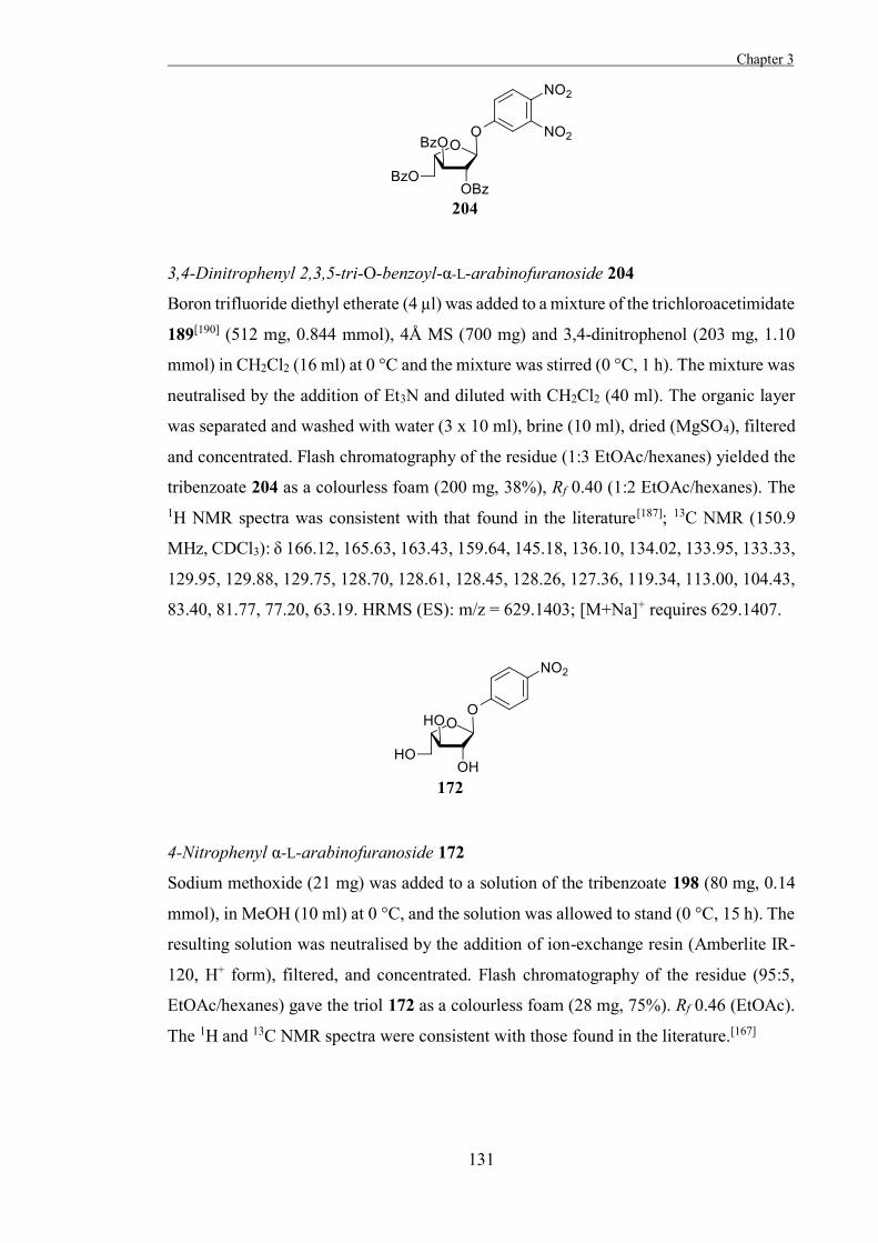
Chapter 3
131
3,4-Dinitrophenyl 2,3,5-tri-O-benzoyl-α-L-arabinofuranoside 204
Boron trifluoride diethyl etherate (4 µl) was added to a mixture of the trichloroacetimidate
189[190] (512 mg, 0.844 mmol), 4Å MS (700 mg) and 3,4-dinitrophenol (203 mg, 1.10
mmol) in CH2Cl2 (16 ml) at 0 °C and the mixture was stirred (0 °C, 1 h). The mixture was
neutralised by the addition of Et3N and diluted with CH2Cl2 (40 ml). The organic layer
was separated and washed with water (3 x 10 ml), brine (10 ml), dried (MgSO4), filtered
and concentrated. Flash chromatography of the residue (1:3 EtOAc/hexanes) yielded the
tribenzoate 204 as a colourless foam (200 mg, 38%), Rf 0.40 (1:2 EtOAc/hexanes). The 1H NMR spectra was consistent with that found in the literature[187]; 13C NMR (150.9
MHz, CDCl3): δ 166.12, 165.63, 163.43, 159.64, 145.18, 136.10, 134.02, 133.95, 133.33,
129.95, 129.88, 129.75, 128.70, 128.61, 128.45, 128.26, 127.36, 119.34, 113.00, 104.43,
83.40, 81.77, 77.20, 63.19. HRMS (ES): m/z = 629.1403; [M+Na]+ requires 629.1407.
4-Nitrophenyl α-L-arabinofuranoside 172
Sodium methoxide (21 mg) was added to a solution of the tribenzoate 198 (80 mg, 0.14
mmol), in MeOH (10 ml) at 0 °C, and the solution was allowed to stand (0 °C, 15 h). The
resulting solution was neutralised by the addition of ion-exchange resin (Amberlite IR-
120, H+ form), filtered, and concentrated. Flash chromatography of the residue (95:5,
EtOAc/hexanes) gave the triol 172 as a colourless foam (28 mg, 75%). Rf 0.46 (EtOAc).
The 1H and 13C NMR spectra were consistent with those found in the literature.[167]
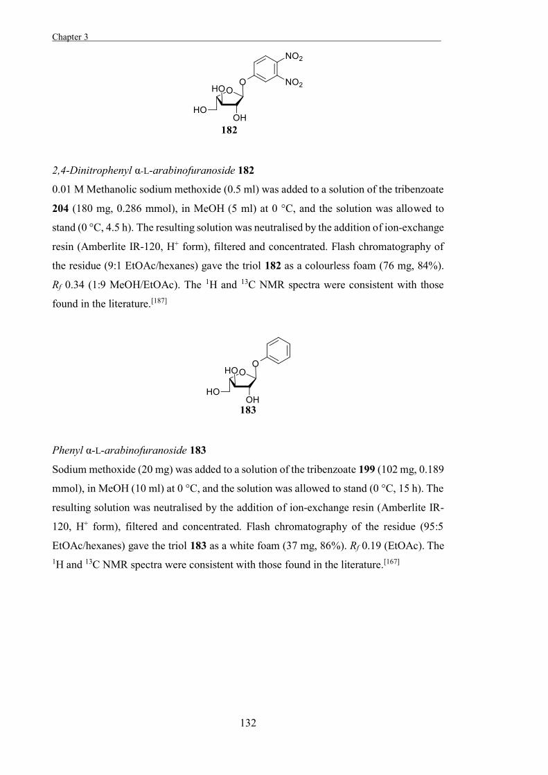
Chapter 3 .
132
2,4-Dinitrophenyl α-L-arabinofuranoside 182
0.01 M Methanolic sodium methoxide (0.5 ml) was added to a solution of the tribenzoate
204 (180 mg, 0.286 mmol), in MeOH (5 ml) at 0 °C, and the solution was allowed to
stand (0 °C, 4.5 h). The resulting solution was neutralised by the addition of ion-exchange
resin (Amberlite IR-120, H+ form), filtered and concentrated. Flash chromatography of
the residue (9:1 EtOAc/hexanes) gave the triol 182 as a colourless foam (76 mg, 84%).
Rf 0.34 (1:9 MeOH/EtOAc). The 1H and 13C NMR spectra were consistent with those
found in the literature.[187]
Phenyl α-L-arabinofuranoside 183
Sodium methoxide (20 mg) was added to a solution of the tribenzoate 199 (102 mg, 0.189
mmol), in MeOH (10 ml) at 0 °C, and the solution was allowed to stand (0 °C, 15 h). The
resulting solution was neutralised by the addition of ion-exchange resin (Amberlite IR-
120, H+ form), filtered and concentrated. Flash chromatography of the residue (95:5
EtOAc/hexanes) gave the triol 183 as a white foam (37 mg, 86%). Rf 0.19 (EtOAc). The 1H and 13C NMR spectra were consistent with those found in the literature.[167]
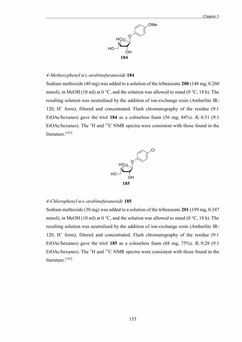
Chapter 3
133
4-Methoxyphenyl α-L-arabinofuranoside 184
Sodium methoxide (40 mg) was added to a solution of the tribenzoate 200 (148 mg, 0.260
mmol), in MeOH (10 ml) at 0 °C, and the solution was allowed to stand (0 °C, 18 h). The
resulting solution was neutralised by the addition of ion-exchange resin (Amberlite IR-
120, H+ form), filtered and concentrated. Flash chromatography of the residue (9:1
EtOAc/hexanes) gave the triol 184 as a colourless foam (56 mg, 84%). Rf 0.31 (9:1
EtOAc/hexanes). The 1H and 13C NMR spectra were consistent with those found in the
literature.[193]
4-Chlorophenyl α-L-arabinofuranoside 185
Sodium methoxide (30 mg) was added to a solution of the tribenzoate 201 (199 mg, 0.347
mmol), in MeOH (10 ml) at 0 °C, and the solution was allowed to stand (0 °C, 18 h). The
resulting solution was neutralised by the addition of ion-exchange resin (Amberlite IR-
120, H+ form), filtered and concentrated. Flash chromatography of the residue (9:1
EtOAc/hexanes) gave the triol 185 as a colourless foam (68 mg, 75%). Rf 0.28 (9:1
EtOAc/hexanes). The 1H and 13C NMR spectra were consistent with those found in the
literature.[192]
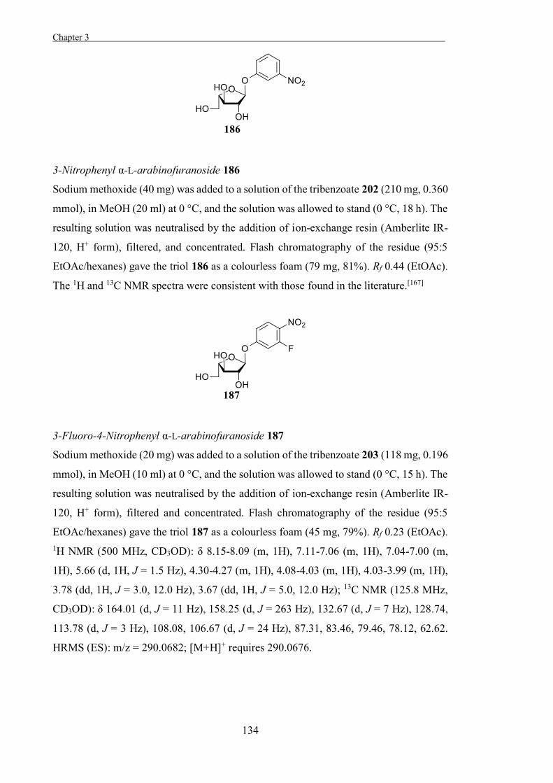
Chapter 3 .
134
3-Nitrophenyl α-L-arabinofuranoside 186
Sodium methoxide (40 mg) was added to a solution of the tribenzoate 202 (210 mg, 0.360
mmol), in MeOH (20 ml) at 0 °C, and the solution was allowed to stand (0 °C, 18 h). The
resulting solution was neutralised by the addition of ion-exchange resin (Amberlite IR-
120, H+ form), filtered, and concentrated. Flash chromatography of the residue (95:5
EtOAc/hexanes) gave the triol 186 as a colourless foam (79 mg, 81%). Rf 0.44 (EtOAc).
The 1H and 13C NMR spectra were consistent with those found in the literature.[167]
3-Fluoro-4-Nitrophenyl α-L-arabinofuranoside 187
Sodium methoxide (20 mg) was added to a solution of the tribenzoate 203 (118 mg, 0.196
mmol), in MeOH (10 ml) at 0 °C, and the solution was allowed to stand (0 °C, 15 h). The
resulting solution was neutralised by the addition of ion-exchange resin (Amberlite IR-
120, H+ form), filtered and concentrated. Flash chromatography of the residue (95:5
EtOAc/hexanes) gave the triol 187 as a colourless foam (45 mg, 79%). Rf 0.23 (EtOAc). 1H NMR (500 MHz, CD3OD): δ 8.15-8.09 (m, 1H), 7.11-7.06 (m, 1H), 7.04-7.00 (m,
1H), 5.66 (d, 1H, J = 1.5 Hz), 4.30-4.27 (m, 1H), 4.08-4.03 (m, 1H), 4.03-3.99 (m, 1H),
3.78 (dd, 1H, J = 3.0, 12.0 Hz), 3.67 (dd, 1H, J = 5.0, 12.0 Hz); 13C NMR (125.8 MHz,
CD3OD): δ 164.01 (d, J = 11 Hz), 158.25 (d, J = 263 Hz), 132.67 (d, J = 7 Hz), 128.74,
113.78 (d, J = 3 Hz), 108.08, 106.67 (d, J = 24 Hz), 87.31, 83.46, 79.46, 78.12, 62.62.
HRMS (ES): m/z = 290.0682; [M+H]+ requires 290.0676.
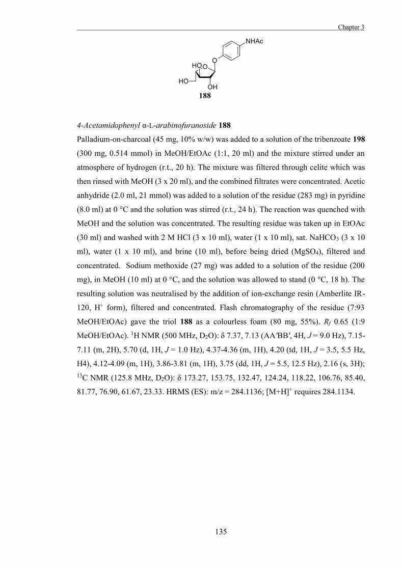
Chapter 3
135
4-Acetamidophenyl α-L-arabinofuranoside 188
Palladium-on-charcoal (45 mg, 10% w/w) was added to a solution of the tribenzoate 198
(300 mg, 0.514 mmol) in MeOH/EtOAc (1:1, 20 ml) and the mixture stirred under an
atmosphere of hydrogen (r.t., 20 h). The mixture was filtered through celite which was
then rinsed with MeOH (3 x 20 ml), and the combined filtrates were concentrated. Acetic
anhydride (2.0 ml, 21 mmol) was added to a solution of the residue (283 mg) in pyridine
(8.0 ml) at 0 °C and the solution was stirred (r.t., 24 h). The reaction was quenched with
MeOH and the solution was concentrated. The resulting residue was taken up in EtOAc
(30 ml) and washed with 2 M HCl (3 x 10 ml), water (1 x 10 ml), sat. NaHCO3 (3 x 10
ml), water (1 x 10 ml), and brine (10 ml), before being dried (MgSO4), filtered and
concentrated. Sodium methoxide (27 mg) was added to a solution of the residue (200
mg), in MeOH (10 ml) at 0 °C, and the solution was allowed to stand (0 °C, 18 h). The
resulting solution was neutralised by the addition of ion-exchange resin (Amberlite IR-
120, H+ form), filtered and concentrated. Flash chromatography of the residue (7:93
MeOH/EtOAc) gave the triol 188 as a colourless foam (80 mg, 55%). Rf 0.65 (1:9
MeOH/EtOAc). 1H NMR (500 MHz, D2O): δ 7.37, 7.13 (AA'BB', 4H, J = 9.0 Hz), 7.15-
7.11 (m, 2H), 5.70 (d, 1H, J = 1.0 Hz), 4.37-4.36 (m, 1H), 4.20 (td, 1H, J = 3.5, 5.5 Hz,
H4), 4.12-4.09 (m, 1H), 3.86-3.81 (m, 1H), 3.75 (dd, 1H, J = 5.5, 12.5 Hz), 2.16 (s, 3H); 13C NMR (125.8 MHz, D2O): δ 173.27, 153.75, 132.47, 124.24, 118.22, 106.76, 85.40,
81.77, 76.90, 61.67, 23.33. HRMS (ES): m/z = 284.1136; [M+H]+ requires 284.1134.
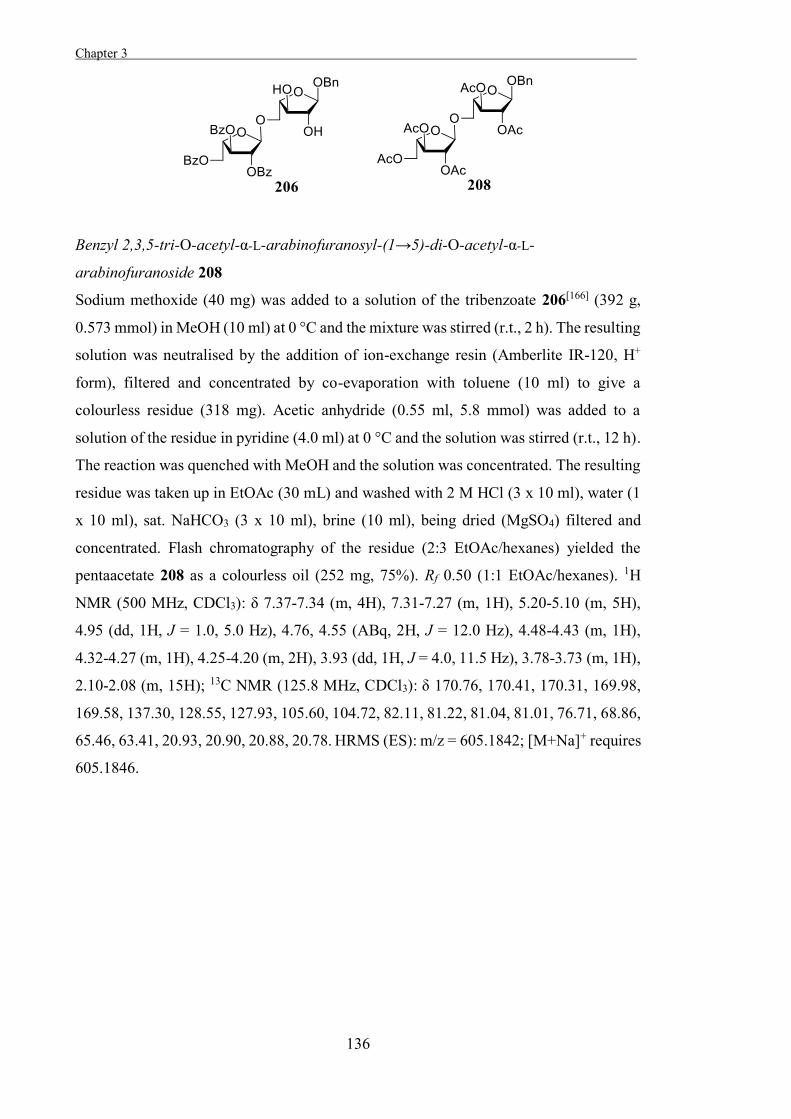
Chapter 3 .
136
Benzyl 2,3,5-tri-O-acetyl-α-L-arabinofuranosyl-(1→5)-di-O-acetyl-α-L-
arabinofuranoside 208
Sodium methoxide (40 mg) was added to a solution of the tribenzoate 206[166] (392 g,
0.573 mmol) in MeOH (10 ml) at 0 °C and the mixture was stirred (r.t., 2 h). The resulting
solution was neutralised by the addition of ion-exchange resin (Amberlite IR-120, H+
form), filtered and concentrated by co-evaporation with toluene (10 ml) to give a
colourless residue (318 mg). Acetic anhydride (0.55 ml, 5.8 mmol) was added to a
solution of the residue in pyridine (4.0 ml) at 0 °C and the solution was stirred (r.t., 12 h).
The reaction was quenched with MeOH and the solution was concentrated. The resulting
residue was taken up in EtOAc (30 mL) and washed with 2 M HCl (3 x 10 ml), water (1
x 10 ml), sat. NaHCO3 (3 x 10 ml), brine (10 ml), being dried (MgSO4) filtered and
concentrated. Flash chromatography of the residue (2:3 EtOAc/hexanes) yielded the
pentaacetate 208 as a colourless oil (252 mg, 75%). Rf 0.50 (1:1 EtOAc/hexanes). 1H
NMR (500 MHz, CDCl3): δ 7.37-7.34 (m, 4H), 7.31-7.27 (m, 1H), 5.20-5.10 (m, 5H),
4.95 (dd, 1H, J = 1.0, 5.0 Hz), 4.76, 4.55 (ABq, 2H, J = 12.0 Hz), 4.48-4.43 (m, 1H),
4.32-4.27 (m, 1H), 4.25-4.20 (m, 2H), 3.93 (dd, 1H, J = 4.0, 11.5 Hz), 3.78-3.73 (m, 1H),
2.10-2.08 (m, 15H); 13C NMR (125.8 MHz, CDCl3): δ 170.76, 170.41, 170.31, 169.98,
169.58, 137.30, 128.55, 127.93, 105.60, 104.72, 82.11, 81.22, 81.04, 81.01, 76.71, 68.86,
65.46, 63.41, 20.93, 20.90, 20.88, 20.78. HRMS (ES): m/z = 605.1842; [M+Na]+ requires
605.1846.
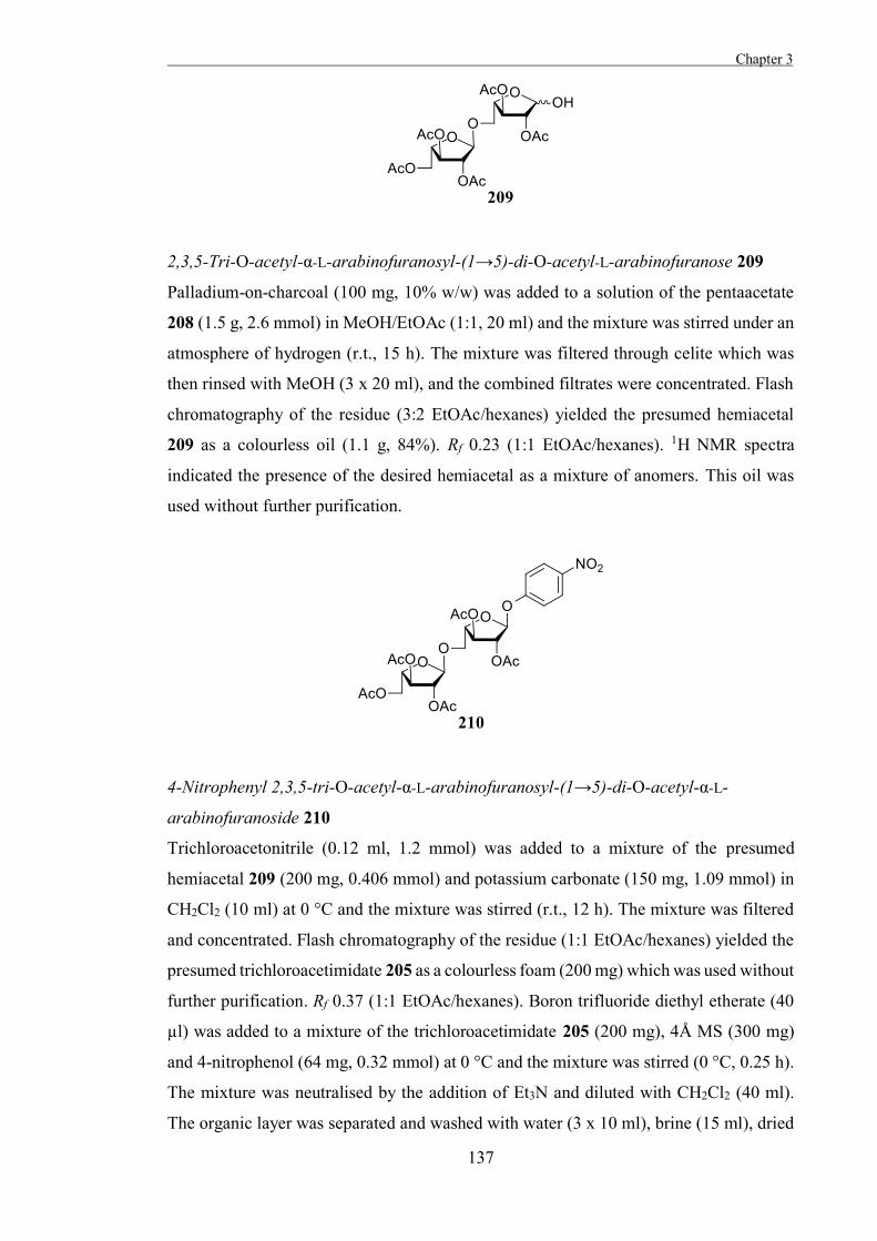
Chapter 3
137
2,3,5-Tri-O-acetyl-α-L-arabinofuranosyl-(1→5)-di-O-acetyl-L-arabinofuranose 209
Palladium-on-charcoal (100 mg, 10% w/w) was added to a solution of the pentaacetate
208 (1.5 g, 2.6 mmol) in MeOH/EtOAc (1:1, 20 ml) and the mixture was stirred under an
atmosphere of hydrogen (r.t., 15 h). The mixture was filtered through celite which was
then rinsed with MeOH (3 x 20 ml), and the combined filtrates were concentrated. Flash
chromatography of the residue (3:2 EtOAc/hexanes) yielded the presumed hemiacetal
209 as a colourless oil (1.1 g, 84%). Rf 0.23 (1:1 EtOAc/hexanes). 1H NMR spectra
indicated the presence of the desired hemiacetal as a mixture of anomers. This oil was
used without further purification.
4-Nitrophenyl 2,3,5-tri-O-acetyl-α-L-arabinofuranosyl-(1→5)-di-O-acetyl-α-L-
arabinofuranoside 210
Trichloroacetonitrile (0.12 ml, 1.2 mmol) was added to a mixture of the presumed
hemiacetal 209 (200 mg, 0.406 mmol) and potassium carbonate (150 mg, 1.09 mmol) in
CH2Cl2 (10 ml) at 0 °C and the mixture was stirred (r.t., 12 h). The mixture was filtered
and concentrated. Flash chromatography of the residue (1:1 EtOAc/hexanes) yielded the
presumed trichloroacetimidate 205 as a colourless foam (200 mg) which was used without
further purification. Rf 0.37 (1:1 EtOAc/hexanes). Boron trifluoride diethyl etherate (40
µl) was added to a mixture of the trichloroacetimidate 205 (200 mg), 4Å MS (300 mg)
and 4-nitrophenol (64 mg, 0.32 mmol) at 0 °C and the mixture was stirred (0 °C, 0.25 h).
The mixture was neutralised by the addition of Et3N and diluted with CH2Cl2 (40 ml).
The organic layer was separated and washed with water (3 x 10 ml), brine (15 ml), dried
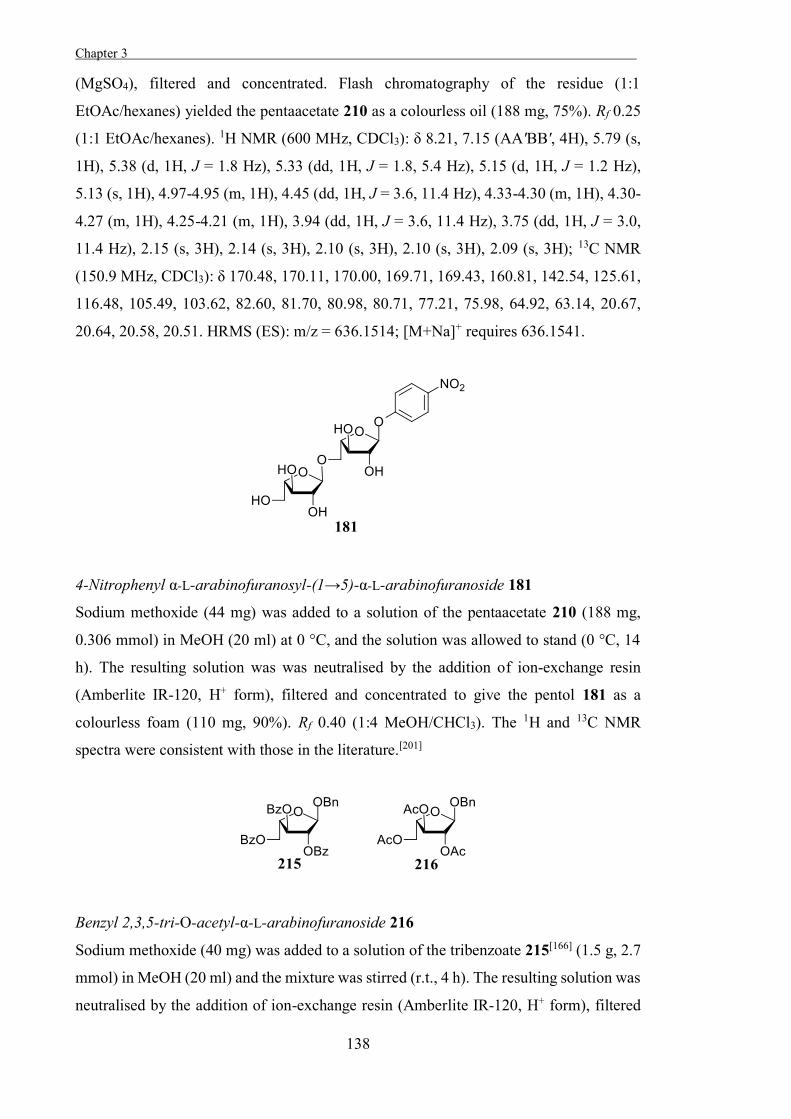
Chapter 3 .
138
(MgSO4), filtered and concentrated. Flash chromatography of the residue (1:1
EtOAc/hexanes) yielded the pentaacetate 210 as a colourless oil (188 mg, 75%). Rf 0.25
(1:1 EtOAc/hexanes). 1H NMR (600 MHz, CDCl3): δ 8.21, 7.15 (AA'BB', 4H), 5.79 (s,
1H), 5.38 (d, 1H, J = 1.8 Hz), 5.33 (dd, 1H, J = 1.8, 5.4 Hz), 5.15 (d, 1H, J = 1.2 Hz),
5.13 (s, 1H), 4.97-4.95 (m, 1H), 4.45 (dd, 1H, J = 3.6, 11.4 Hz), 4.33-4.30 (m, 1H), 4.30-
4.27 (m, 1H), 4.25-4.21 (m, 1H), 3.94 (dd, 1H, J = 3.6, 11.4 Hz), 3.75 (dd, 1H, J = 3.0,
11.4 Hz), 2.15 (s, 3H), 2.14 (s, 3H), 2.10 (s, 3H), 2.10 (s, 3H), 2.09 (s, 3H); 13C NMR
(150.9 MHz, CDCl3): δ 170.48, 170.11, 170.00, 169.71, 169.43, 160.81, 142.54, 125.61,
116.48, 105.49, 103.62, 82.60, 81.70, 80.98, 80.71, 77.21, 75.98, 64.92, 63.14, 20.67,
20.64, 20.58, 20.51. HRMS (ES): m/z = 636.1514; [M+Na]+ requires 636.1541.
4-Nitrophenyl α-L-arabinofuranosyl-(1→5)-α-L-arabinofuranoside 181
Sodium methoxide (44 mg) was added to a solution of the pentaacetate 210 (188 mg,
0.306 mmol) in MeOH (20 ml) at 0 °C, and the solution was allowed to stand (0 °C, 14
h). The resulting solution was was neutralised by the addition of ion-exchange resin
(Amberlite IR-120, H+ form), filtered and concentrated to give the pentol 181 as a
colourless foam (110 mg, 90%). Rf 0.40 (1:4 MeOH/CHCl3). The 1H and 13C NMR
spectra were consistent with those in the literature.[201]
Benzyl 2,3,5-tri-O-acetyl-α-L-arabinofuranoside 216
Sodium methoxide (40 mg) was added to a solution of the tribenzoate 215[166] (1.5 g, 2.7
mmol) in MeOH (20 ml) and the mixture was stirred (r.t., 4 h). The resulting solution was
neutralised by the addition of ion-exchange resin (Amberlite IR-120, H+ form), filtered
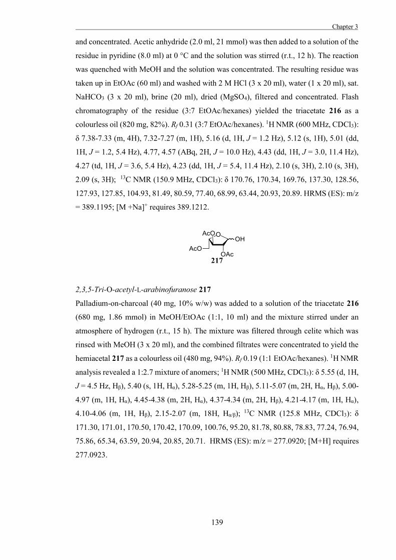
Chapter 3
139
and concentrated. Acetic anhydride (2.0 ml, 21 mmol) was then added to a solution of the
residue in pyridine (8.0 ml) at 0 °C and the solution was stirred (r.t., 12 h). The reaction
was quenched with MeOH and the solution was concentrated. The resulting residue was
taken up in EtOAc (60 ml) and washed with 2 M HCl (3 x 20 ml), water (1 x 20 ml), sat.
NaHCO3 (3 x 20 ml), brine (20 ml), dried (MgSO4), filtered and concentrated. Flash
chromatography of the residue (3:7 EtOAc/hexanes) yielded the triacetate 216 as a
colourless oil (820 mg, 82%). Rf 0.31 (3:7 EtOAc/hexanes). 1H NMR (600 MHz, CDCl3):
δ 7.38-7.33 (m, 4H), 7.32-7.27 (m, 1H), 5.16 (d, 1H, J = 1.2 Hz), 5.12 (s, 1H), 5.01 (dd,
1H, J = 1.2, 5.4 Hz), 4.77, 4.57 (ABq, 2H, J = 10.0 Hz), 4.43 (dd, 1H, J = 3.0, 11.4 Hz),
4.27 (td, 1H, J = 3.6, 5.4 Hz), 4.23 (dd, 1H, J = 5.4, 11.4 Hz), 2.10 (s, 3H), 2.10 (s, 3H),
2.09 (s, 3H); 13C NMR (150.9 MHz, CDCl3): δ 170.76, 170.34, 169.76, 137.30, 128.56,
127.93, 127.85, 104.93, 81.49, 80.59, 77.40, 68.99, 63.44, 20.93, 20.89. HRMS (ES): m/z
= 389.1195; [M +Na]+ requires 389.1212.
2,3,5-Tri-O-acetyl-L-arabinofuranose 217
Palladium-on-charcoal (40 mg, 10% w/w) was added to a solution of the triacetate 216
(680 mg, 1.86 mmol) in MeOH/EtOAc (1:1, 10 ml) and the mixture stirred under an
atmosphere of hydrogen (r.t., 15 h). The mixture was filtered through celite which was
rinsed with MeOH (3 x 20 ml), and the combined filtrates were concentrated to yield the
hemiacetal 217 as a colourless oil (480 mg, 94%). Rf 0.19 (1:1 EtOAc/hexanes). 1H NMR
analysis revealed a 1:2.7 mixture of anomers; 1H NMR (500 MHz, CDCl3): δ 5.55 (d, 1H,
J = 4.5 Hz, Hβ), 5.40 (s, 1H, Hα), 5.28-5.25 (m, 1H, Hβ), 5.11-5.07 (m, 2H, Hα, Hβ), 5.00-
4.97 (m, 1H, Hα), 4.45-4.38 (m, 2H, Hα), 4.37-4.34 (m, 2H, Hβ), 4.21-4.17 (m, 1H, Hα),
4.10-4.06 (m, 1H, Hβ), 2.15-2.07 (m, 18H, Hα/β); 13C NMR (125.8 MHz, CDCl3): δ
171.30, 171.01, 170.50, 170.42, 170.09, 100.76, 95.20, 81.78, 80.88, 78.83, 77.24, 76.94,
75.86, 65.34, 63.59, 20.94, 20.85, 20.71. HRMS (ES): m/z = 277.0920; [M+H] requires
277.0923.
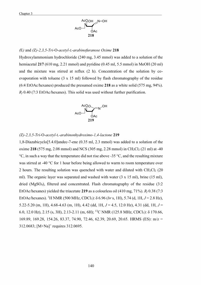
Chapter 3 .
140
(E) and (Z)-2,3,5-Tri-O-acetyl-L-arabinofuranose Oxime 218
Hydroxylammonium hydrochloride (240 mg, 3.45 mmol) was added to a solution of the
hemiacetal 217 (610 mg, 2.21 mmol) and pyridine (0.45 ml, 5.5 mmol) in MeOH (20 ml)
and the mixture was stirred at reflux (2 h). Concentration of the solution by co-
evaporation with toluene (3 x 15 ml) followed by flash chromatography of the residue
(6:4 EtOAc/hexanes) produced the presumed oxime 218 as a white solid (575 mg, 94%).
Rf 0.40 (7:3 EtOAc/hexanes). This solid was used without further purification.
(Z)-2,3,5-Tri-O-acetyl-L-arabinonhydroximo-1,4-lactone 219
1,8-Diazabicyclo[5.4.0]undec-7-ene (0.35 ml, 2.3 mmol) was added to a solution of the
oxime 218 (575 mg, 2.08 mmol) and NCS (305 mg, 2.28 mmol) in CH2Cl2 (21 ml) at -40
°C, in such a way that the temperature did not rise above -35 °C, and the resulting mixture
was stirred at -40 °C for 1 hour before being allowed to warm to room temperature over
2 hours. The resulting solution was quenched with water and diluted with CH2Cl2 (20
ml). The organic layer was separated and washed with water (3 x 15 ml), brine (15 ml),
dried (MgSO4), filtered and concentrated. Flash chromatography of the residue (3:2
EtOAc/hexanes) yielded the triacetate 219 as a colourless oil (410 mg, 71%). Rf 0.38 (7:3
EtOAc/hexanes). 1H NMR (500 MHz, CDCl3): δ 6.96 (br s, 1H), 5.74 (d, 1H, J = 2.8 Hz),
5.22-5.20 (m, 1H), 4.68-4.63 (m, 1H), 4.42 (dd, 1H, J = 4.5, 12.0 Hz), 4.31 (dd, 1H, J =
6.0, 12.0 Hz), 2.15 (s, 3H), 2.13-2.11 (m, 6H); 13C NMR (125.8 MHz, CDCl3): δ 170.66,
169.89, 169.28, 154.26, 83.37, 74.90, 72.46, 62.39, 20.69, 20.65. HRMS (ES): m/z =
312.0683; [M+Na]+ requires 312.0695.
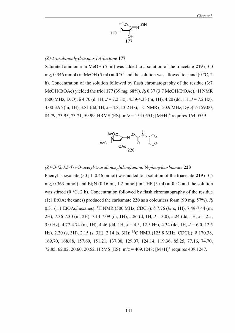
Chapter 3
141
(Z)-L-arabinonhydroximo-1,4-lactone 177
Saturated ammonia in MeOH (5 ml) was added to a solution of the triacetate 219 (100
mg, 0.346 mmol) in MeOH (5 ml) at 0 °C and the solution was allowed to stand (0 °C, 2
h). Concentration of the solution followed by flash chromatography of the residue (3:7
MeOH/EtOAc) yielded the triol 177 (39 mg, 68%). Rf 0.37 (3:7 MeOH/EtOAc). 1H NMR
(600 MHz, D2O): δ 4.70 (d, 1H, J = 7.2 Hz), 4.39-4.33 (m, 1H), 4.20 (dd, 1H, J = 7.2 Hz),
4.00-3.95 (m, 1H), 3.81 (dd, 1H, J = 4.8, 13.2 Hz); 13C NMR (150.9 MHz, D2O): δ 159.00,
84.79, 73.95, 73.71, 59.99. HRMS (ES): m/z = 154.0551; [M+H]+ requires 164.0559.
(Z)-O-(2,3,5-Tri-O-acetyl-L-arabinosylidene)amino N-phenylcarbamate 220
Phenyl isocyanate (50 µl, 0.46 mmol) was added to a solution of the triacetate 219 (105
mg, 0.363 mmol) and Et3N (0.16 ml, 1.2 mmol) in THF (5 ml) at 0 °C and the solution
was stirred (0 °C, 2 h). Concentration followed by flash chromatography of the residue
(1:1 EtOAc/hexanes) produced the carbamate 220 as a colourless foam (90 mg, 57%). Rf
0.31 (1:1 EtOAc/hexanes). 1H NMR (500 MHz, CDCl3): δ 7.76 (br s, 1H), 7.49-7.44 (m,
2H), 7.36-7.30 (m, 2H), 7.14-7.09 (m, 1H), 5.86 (d, 1H, J = 3.0), 5.24 (dd, 1H, J = 2.5,
3.0 Hz), 4.77-4.74 (m, 1H), 4.46 (dd, 1H, J = 4.5, 12.5 Hz), 4.34 (dd, 1H, J = 6.0, 12.5
Hz), 2.20 (s, 3H), 2.15 (s, 3H), 2.14 (s, 3H); 13C NMR (125.8 MHz, CDCl3): δ 170.38,
169.70, 168.88, 157.69, 151.21, 137.00, 129.07, 124.14, 119.36, 85.25, 77.16, 74.70,
72.85, 62.02, 20.60, 20.52. HRMS (ES): m/z = 409.1248; [M+H]+ requires 409.1247.
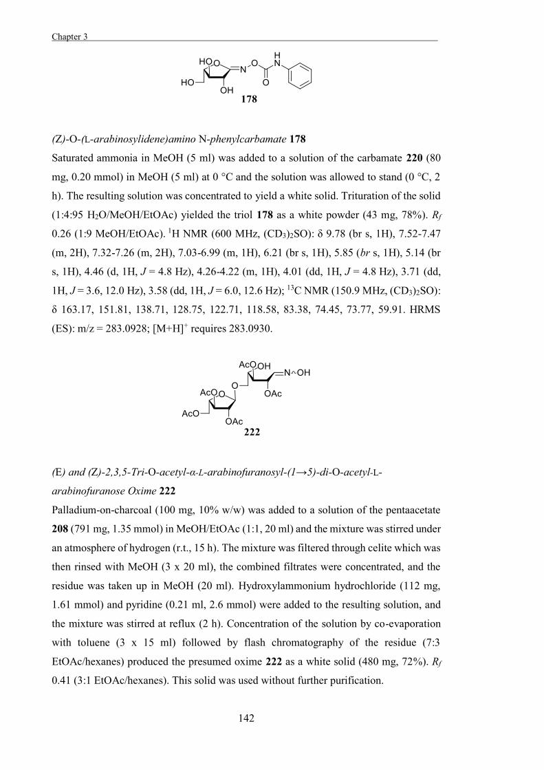
Chapter 3 .
142
(Z)-O-(L-arabinosylidene)amino N-phenylcarbamate 178
Saturated ammonia in MeOH (5 ml) was added to a solution of the carbamate 220 (80
mg, 0.20 mmol) in MeOH (5 ml) at 0 °C and the solution was allowed to stand (0 °C, 2
h). The resulting solution was concentrated to yield a white solid. Trituration of the solid
(1:4:95 H2O/MeOH/EtOAc) yielded the triol 178 as a white powder (43 mg, 78%). Rf
0.26 (1:9 MeOH/EtOAc). 1H NMR (600 MHz, (CD3)2SO): δ 9.78 (br s, 1H), 7.52-7.47
(m, 2H), 7.32-7.26 (m, 2H), 7.03-6.99 (m, 1H), 6.21 (br s, 1H), 5.85 (br s, 1H), 5.14 (br
s, 1H), 4.46 (d, 1H, J = 4.8 Hz), 4.26-4.22 (m, 1H), 4.01 (dd, 1H, J = 4.8 Hz), 3.71 (dd,
1H, J = 3.6, 12.0 Hz), 3.58 (dd, 1H, J = 6.0, 12.6 Hz); 13C NMR (150.9 MHz, (CD3)2SO):
δ 163.17, 151.81, 138.71, 128.75, 122.71, 118.58, 83.38, 74.45, 73.77, 59.91. HRMS
(ES): m/z = 283.0928; [M+H]+ requires 283.0930.
(E) and (Z)-2,3,5-Tri-O-acetyl-α-L-arabinofuranosyl-(1→5)-di-O-acetyl-L-
arabinofuranose Oxime 222
Palladium-on-charcoal (100 mg, 10% w/w) was added to a solution of the pentaacetate
208 (791 mg, 1.35 mmol) in MeOH/EtOAc (1:1, 20 ml) and the mixture was stirred under
an atmosphere of hydrogen (r.t., 15 h). The mixture was filtered through celite which was
then rinsed with MeOH (3 x 20 ml), the combined filtrates were concentrated, and the
residue was taken up in MeOH (20 ml). Hydroxylammonium hydrochloride (112 mg,
1.61 mmol) and pyridine (0.21 ml, 2.6 mmol) were added to the resulting solution, and
the mixture was stirred at reflux (2 h). Concentration of the solution by co-evaporation
with toluene (3 x 15 ml) followed by flash chromatography of the residue (7:3
EtOAc/hexanes) produced the presumed oxime 222 as a white solid (480 mg, 72%). Rf
0.41 (3:1 EtOAc/hexanes). This solid was used without further purification.
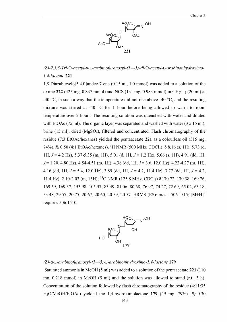
Chapter 3
143
(Z)-2,3,5-Tri-O-acetyl-α-L-arabinofuranosyl-(1→5)-di-O-acetyl-L-arabinonhydroximo-
1,4-lactone 221
1,8-Diazabicyclo[5.4.0]undec-7-ene (0.15 ml, 1.0 mmol) was added to a solution of the
oxime 222 (425 mg, 0.837 mmol) and NCS (131 mg, 0.983 mmol) in CH2Cl2 (20 ml) at
-40 °C, in such a way that the temperature did not rise above -40 °C, and the resulting
mixture was stirred at -40 °C for 1 hour before being allowed to warm to room
temperature over 2 hours. The resulting solution was quenched with water and diluted
with EtOAc (75 ml). The organic layer was separated and washed with water (3 x 15 ml),
brine (15 ml), dried (MgSO4), filtered and concentrated. Flash chromatography of the
residue (7:3 EtOAc/hexanes) yielded the pentaacetate 221 as a colourless oil (315 mg,
74%). Rf 0.50 (4:1 EtOAc/hexanes). 1H NMR (500 MHz, CDCl3): δ 8.16 (s, 1H), 5.73 (d,
1H, J = 4.2 Hz), 5.37-5.35 (m, 1H), 5.01 (d, 1H, J = 1.2 Hz), 5.06 (s, 1H), 4.91 (dd, 1H,
J = 1.20, 4.80 Hz), 4.54-4.51 (m, 1H), 4.38 (dd, 1H, J = 3.6, 12.0 Hz), 4.22-4.27 (m, 1H),
4.16 (dd, 1H, J = 5.4, 12.0 Hz), 3.89 (dd, 1H, J = 4.2, 11.4 Hz), 3.77 (dd, 1H, J = 4.2,
11.4 Hz), 2.10-2.03 (m, 15H); 13C NMR (125.8 MHz, CDCl3) δ 170.72, 170.38, 169.76,
169.59, 169.37, 153.98, 105.57, 83.49, 81.06, 80.68, 76.97, 74.27, 72.69, 65.02, 63.18,
53.48, 29.57, 20.75, 20.67, 20.60, 20.59, 20.57. HRMS (ES): m/z = 506.1515; [M+H]+
requires 506.1510.
(Z)-α-L-arabinofuranosyl-(1→5)-L-arabinonhydroximo-1,4-lactone 179
Saturated ammonia in MeOH (5 ml) was added to a solution of the pentaacetate 221 (110
mg, 0.218 mmol) in MeOH (5 ml) and the solution was allowed to stand (r.t., 3 h).
Concentration of the solution followed by flash chromatography of the residue (4:11:35
H2O/MeOH/EtOAc) yielded the 1,4-hydroximolactone 179 (49 mg, 79%). Rf 0.30
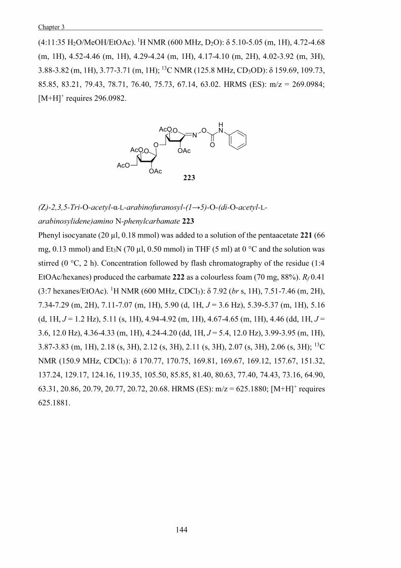
Chapter 3 .
144
(4:11:35 H2O/MeOH/EtOAc). 1H NMR (600 MHz, D2O): δ 5.10-5.05 (m, 1H), 4.72-4.68
(m, 1H), 4.52-4.46 (m, 1H), 4.29-4.24 (m, 1H), 4.17-4.10 (m, 2H), 4.02-3.92 (m, 3H),
3.88-3.82 (m, 1H), 3.77-3.71 (m, 1H); 13C NMR (125.8 MHz, CD3OD): δ 159.69, 109.73,
85.85, 83.21, 79.43, 78.71, 76.40, 75.73, 67.14, 63.02. HRMS (ES): m/z = 269.0984;
[M+H]+ requires 296.0982.
(Z)-2,3,5-Tri-O-acetyl-α-L-arabinofuranosyl-(1→5)-O-(di-O-acetyl-L-
arabinosylidene)amino N-phenylcarbamate 223
Phenyl isocyanate (20 µl, 0.18 mmol) was added to a solution of the pentaacetate 221 (66
mg, 0.13 mmol) and Et3N (70 µl, 0.50 mmol) in THF (5 ml) at 0 °C and the solution was
stirred (0 °C, 2 h). Concentration followed by flash chromatography of the residue (1:4
EtOAc/hexanes) produced the carbamate 222 as a colourless foam (70 mg, 88%). Rf 0.41
(3:7 hexanes/EtOAc). 1H NMR (600 MHz, CDCl3): δ 7.92 (br s, 1H), 7.51-7.46 (m, 2H),
7.34-7.29 (m, 2H), 7.11-7.07 (m, 1H), 5.90 (d, 1H, J = 3.6 Hz), 5.39-5.37 (m, 1H), 5.16
(d, 1H, J = 1.2 Hz), 5.11 (s, 1H), 4.94-4.92 (m, 1H), 4.67-4.65 (m, 1H), 4.46 (dd, 1H, J =
3.6, 12.0 Hz), 4.36-4.33 (m, 1H), 4.24-4.20 (dd, 1H, J = 5.4, 12.0 Hz), 3.99-3.95 (m, 1H),
3.87-3.83 (m, 1H), 2.18 (s, 3H), 2.12 (s, 3H), 2.11 (s, 3H), 2.07 (s, 3H), 2.06 (s, 3H); 13C
NMR (150.9 MHz, CDCl3): δ 170.77, 170.75, 169.81, 169.67, 169.12, 157.67, 151.32,
137.24, 129.17, 124.16, 119.35, 105.50, 85.85, 81.40, 80.63, 77.40, 74.43, 73.16, 64.90,
63.31, 20.86, 20.79, 20.77, 20.72, 20.68. HRMS (ES): m/z = 625.1880; [M+H]+ requires
625.1881.
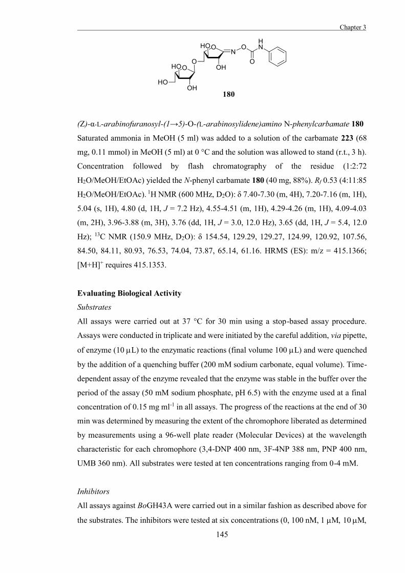
Chapter 3
145
(Z)-α-L-arabinofuranosyl-(1→5)-O-(L-arabinosylidene)amino N-phenylcarbamate 180
Saturated ammonia in MeOH (5 ml) was added to a solution of the carbamate 223 (68
mg, 0.11 mmol) in MeOH (5 ml) at 0 °C and the solution was allowed to stand (r.t., 3 h).
Concentration followed by flash chromatography of the residue (1:2:72
H2O/MeOH/EtOAc) yielded the N-phenyl carbamate 180 (40 mg, 88%). Rf 0.53 (4:11:85
H2O/MeOH/EtOAc). 1H NMR (600 MHz, D2O): δ 7.40-7.30 (m, 4H), 7.20-7.16 (m, 1H),
5.04 (s, 1H), 4.80 (d, 1H, J = 7.2 Hz), 4.55-4.51 (m, 1H), 4.29-4.26 (m, 1H), 4.09-4.03
(m, 2H), 3.96-3.88 (m, 3H), 3.76 (dd, 1H, J = 3.0, 12.0 Hz), 3.65 (dd, 1H, J = 5.4, 12.0
Hz); 13C NMR (150.9 MHz, D2O): δ 154.54, 129.29, 129.27, 124.99, 120.92, 107.56,
84.50, 84.11, 80.93, 76.53, 74.04, 73.87, 65.14, 61.16. HRMS (ES): m/z = 415.1366;
[M+H]+ requires 415.1353.
Evaluating Biological Activity
Substrates
All assays were carried out at 37 °C for 30 min using a stop-based assay procedure.
Assays were conducted in triplicate and were initiated by the careful addition, via pipette,
of enzyme (10 L) to the enzymatic reactions (final volume 100 L) and were quenched
by the addition of a quenching buffer (200 mM sodium carbonate, equal volume). Time-
dependent assay of the enzyme revealed that the enzyme was stable in the buffer over the
period of the assay (50 mM sodium phosphate, pH 6.5) with the enzyme used at a final
concentration of 0.15 mg ml-1 in all assays. The progress of the reactions at the end of 30
min was determined by measuring the extent of the chromophore liberated as determined
by measurements using a 96-well plate reader (Molecular Devices) at the wavelength
characteristic for each chromophore (3,4-DNP 400 nm, 3F-4NP 388 nm, PNP 400 nm,
UMB 360 nm). All substrates were tested at ten concentrations ranging from 0-4 mM.
Inhibitors
All assays against BoGH43A were carried out in a similar fashion as described above for
the substrates. The inhibitors were tested at six concentrations (0, 100 nM, 1 10
Chapter 3 .
146
100 1mM) using the substrate 4-nitrophenyl -L-arabinopyranoside at a
concentration of 1 mM with the enzyme used at a final concentration of0.15 mg ml-1 in
all assays.
All assays against Arb93A were carried out as described by Carapito et al.[202] using
inhibitor concentrations of 50 and 100 M. These experiments were conducted by Dr.
Keith Stubbs.

147

148
149
Part II: The Development of Tools to Study
Glycosyltransferases
Chapter 4
On the Development of Glycosyltransferase Inhibitors

150
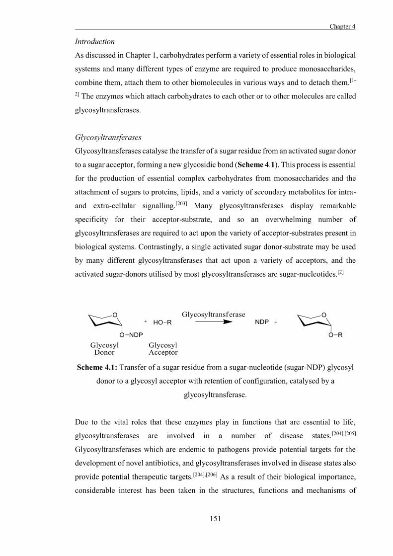
Chapter 4
151
Introduction
As discussed in Chapter 1, carbohydrates perform a variety of essential roles in biological
systems and many different types of enzyme are required to produce monosaccharides,
combine them, attach them to other biomolecules in various ways and to detach them.[1-
2] The enzymes which attach carbohydrates to each other or to other molecules are called
glycosyltransferases.
Glycosyltransferases
Glycosyltransferases catalyse the transfer of a sugar residue from an activated sugar donor
to a sugar acceptor, forming a new glycosidic bond (Scheme 4.1). This process is essential
for the production of essential complex carbohydrates from monosaccharides and the
attachment of sugars to proteins, lipids, and a variety of secondary metabolites for intra-
and extra-cellular signalling.[203] Many glycosyltransferases display remarkable
specificity for their acceptor-substrate, and so an overwhelming number of
glycosyltransferases are required to act upon the variety of acceptor-substrates present in
biological systems. Contrastingly, a single activated sugar donor-substrate may be used
by many different glycosyltransferases that act upon a variety of acceptors, and the
activated sugar-donors utilised by most glycosyltransferases are sugar-nucleotides.[2]
Scheme 4.1: Transfer of a sugar residue from a sugar-nucleotide (sugar-NDP) glycosyl
donor to a glycosyl acceptor with retention of configuration, catalysed by a
glycosyltransferase.
Due to the vital roles that these enzymes play in functions that are essential to life,
glycosyltransferases are involved in a number of disease states.[204],[205]
Glycosyltransferases which are endemic to pathogens provide potential targets for the
development of novel antibiotics, and glycosyltransferases involved in disease states also
provide potential therapeutic targets.[204],[206] As a result of their biological importance,
considerable interest has been taken in the structures, functions and mechanisms of
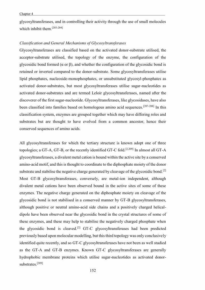
Chapter 4 .
152
glycosyltransferases, and in controlling their activity through the use of small molecules
which inhibit them.[203-204]
Classification and General Mechanisms of Glycosyltransferases
Glycosyltransferases are classified based on the activated donor-substrate utilised, the
acceptor-substrate utilised, the topology of the enzyme, the configuration of the
glycosidic bond formed (α or β), and whether the configuration of the glycosidic bond is
retained or inverted compared to the donor-substrate. Some glycosyltransferases utilise
lipid phosphates, nucleoside-monophosphates, or unsubstituted glycosyl-phosphates as
activated donor-substrates, but most glycosyltransferases utilise sugar-nucleotides as
activated donor-substrates and are termed Leloir glycosyltransferases, named after the
discoverer of the first sugar-nucleotide. Glycosyltransferases, like glycosidases, have also
been classified into families based on homologous amino acid sequences.[207-208] In this
classification system, enzymes are grouped together which may have differing roles and
substrates but are thought to have evolved from a common ancestor, hence their
conserved sequences of amino acids.
All glycosyltransferases for which the tertiary structure is known adopt one of three
topologies; a GT-A, GT-B, or the recently identified GT-C fold.[2,209] In almost all GT-A
glycosyltransferases, a divalent metal cation is bound within the active site by a conserved
amino-acid motif, and this is thought to coordinate to the diphosphate moiety of the donor
substrate and stabilise the negative charge generated by cleavage of the glycosidic bond.[2]
Most GT-B glycosyltransferases, conversely, are metal-ion independent, although
divalent metal cations have been observed bound in the active sites of some of these
enzymes. The negative charge generated on the diphosphate moiety on cleavage of the
glycosidic bond is not stabilised in a conserved manner by GT-B glycosyltransferases,
although positive or neutral amino-acid side chains and a positively charged helical-
dipole have been observed near the glycosidic bond in the crystal structures of some of
these enzymes, and these may help to stabilise the negatively charged phosphate when
the glycosidic bond is cleaved.[2] GT-C glycosyltransferases had been predicted
previously based upon molecular modelling, but this third topology was only conclusively
identified quite recently, and so GT-C glycosyltransferases have not been as well studied
as the GT-A and GT-B enzymes. Known GT-C glycosyltransferases are generally
hydrophobic membrane proteins which utilise sugar-nucleotides as activated donor-
substrates.[209]
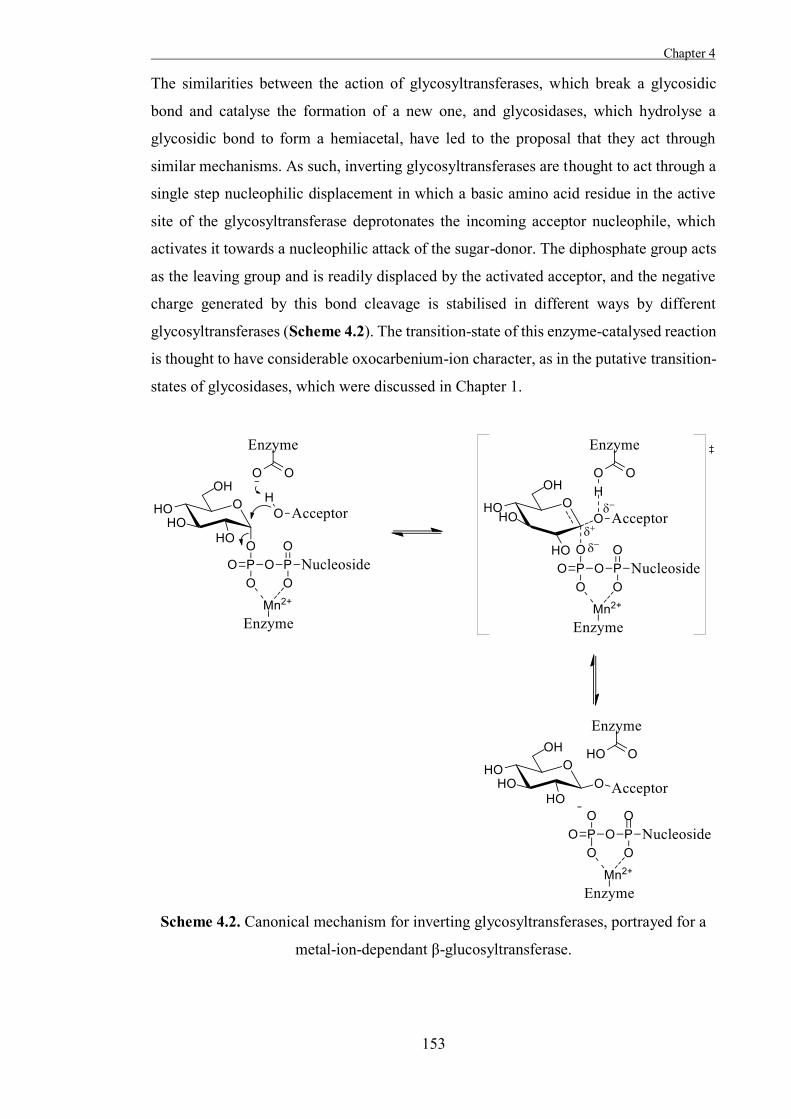
Chapter 4
153
The similarities between the action of glycosyltransferases, which break a glycosidic
bond and catalyse the formation of a new one, and glycosidases, which hydrolyse a
glycosidic bond to form a hemiacetal, have led to the proposal that they act through
similar mechanisms. As such, inverting glycosyltransferases are thought to act through a
single step nucleophilic displacement in which a basic amino acid residue in the active
site of the glycosyltransferase deprotonates the incoming acceptor nucleophile, which
activates it towards a nucleophilic attack of the sugar-donor. The diphosphate group acts
as the leaving group and is readily displaced by the activated acceptor, and the negative
charge generated by this bond cleavage is stabilised in different ways by different
glycosyltransferases (Scheme 4.2). The transition-state of this enzyme-catalysed reaction
is thought to have considerable oxocarbenium-ion character, as in the putative transition-
states of glycosidases, which were discussed in Chapter 1.
Scheme 4.2. Canonical mechanism for inverting glycosyltransferases, portrayed for a
metal-ion-dependant β-glucosyltransferase.
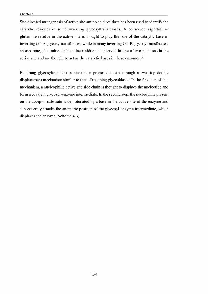
Chapter 4 .
154
Site directed mutagenesis of active site amino acid residues has been used to identify the
catalytic residues of some inverting glycosyltransferases. A conserved aspartate or
glutamine residue in the active site is thought to play the role of the catalytic base in
inverting GT-A glycosyltransferases, while in many inverting GT-B glycosyltransferases,
an aspartate, glutamine, or histidine residue is conserved in one of two positions in the
active site and are thought to act as the catalytic bases in these enzymes.[2]
Retaining glycosyltransferases have been proposed to act through a two-step double
displacement mechanism similar to that of retaining glycosidases. In the first step of this
mechanism, a nucleophilic active site side chain is thought to displace the nucleotide and
form a covalent glycosyl-enzyme intermediate. In the second step, the nucleophile present
on the acceptor substrate is deprotonated by a base in the active site of the enzyme and
subsequently attacks the anomeric position of the glycosyl-enzyme intermediate, which
displaces the enzyme (Scheme 4.3).
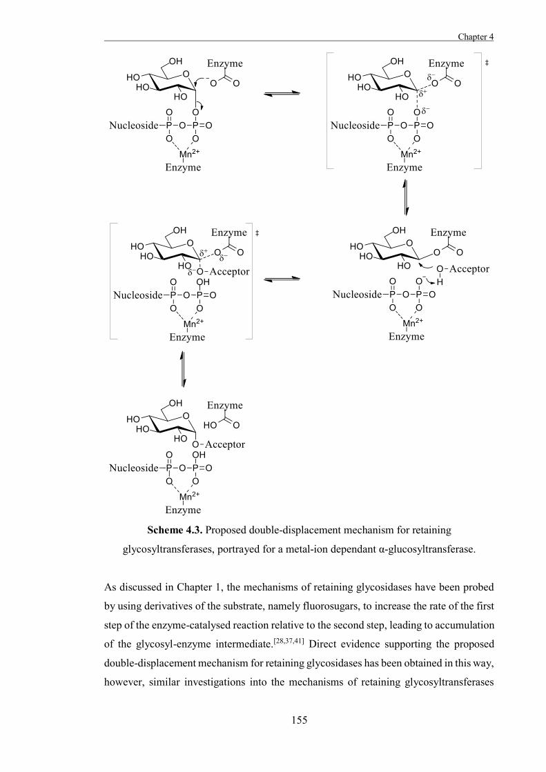
Chapter 4
155
Scheme 4.3. Proposed double-displacement mechanism for retaining
glycosyltransferases, portrayed for a metal-ion dependant α-glucosyltransferase.
As discussed in Chapter 1, the mechanisms of retaining glycosidases have been probed
by using derivatives of the substrate, namely fluorosugars, to increase the rate of the first
step of the enzyme-catalysed reaction relative to the second step, leading to accumulation
of the glycosyl-enzyme intermediate.[28,37,41] Direct evidence supporting the proposed
double-displacement mechanism for retaining glycosidases has been obtained in this way,
however, similar investigations into the mechanisms of retaining glycosyltransferases
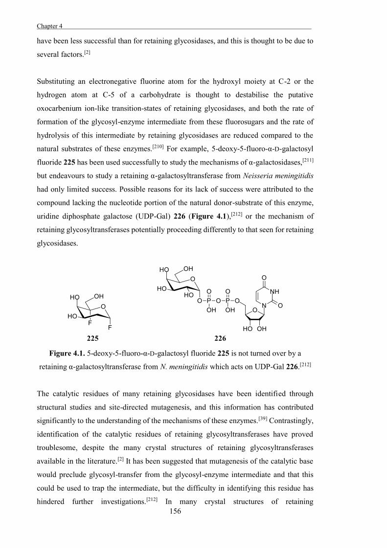
Chapter 4 .
156
have been less successful than for retaining glycosidases, and this is thought to be due to
several factors.[2]
Substituting an electronegative fluorine atom for the hydroxyl moiety at C-2 or the
hydrogen atom at C-5 of a carbohydrate is thought to destabilise the putative
oxocarbenium ion-like transition-states of retaining glycosidases, and both the rate of
formation of the glycosyl-enzyme intermediate from these fluorosugars and the rate of
hydrolysis of this intermediate by retaining glycosidases are reduced compared to the
natural substrates of these enzymes.[210] For example, 5-deoxy-5-fluoro-α-D-galactosyl
fluoride 225 has been used successfully to study the mechanisms of α-galactosidases,[211]
but endeavours to study a retaining α-galactosyltransferase from Neisseria meningitidis
had only limited success. Possible reasons for its lack of success were attributed to the
compound lacking the nucleotide portion of the natural donor-substrate of this enzyme,
uridine diphosphate galactose (UDP-Gal) 226 (Figure 4.1),[212] or the mechanism of
retaining glycosyltransferases potentially proceeding differently to that seen for retaining
glycosidases.
Figure 4.1. 5-deoxy-5-fluoro-α-D-galactosyl fluoride 225 is not turned over by a
retaining α-galactosyltransferase from N. meningitidis which acts on UDP-Gal 226.[212]
The catalytic residues of many retaining glycosidases have been identified through
structural studies and site-directed mutagenesis, and this information has contributed
significantly to the understanding of the mechanisms of these enzymes.[39] Contrastingly,
identification of the catalytic residues of retaining glycosyltransferases have proved
troublesome, despite the many crystal structures of retaining glycosyltransferases
available in the literature.[2] It has been suggested that mutagenesis of the catalytic base
would preclude glycosyl-transfer from the glycosyl-enzyme intermediate and that this
could be used to trap the intermediate, but the difficulty in identifying this residue has
hindered further investigations.[212] In many crystal structures of retaining
Chapter 4
157
glycosyltransferases, the only basic group that is seen to be in the correct position in the
enzyme to deprotonate the incoming acceptor molecule is the metal-bound diphosphate
group, and it is possible that this moiety acts as the catalytic base in these enzymes. In
addition, a conserved residue which may act as the catalytic nucleophile has not been
observed in retaining glycosyltransferases,[2] which may be because the catalytic
nucleophile is not well conserved between glycosyltransferases, or because these
enzymes act through a different mechanistic pathway.
In the absence of conclusive evidence for the double-displacement mechanism, an
alternative mechanism has been proposed for retaining glycosyltransferases; a single-step
internal return mechanism. In this mechanism, the incoming nucleophile approaches from
the same face as the leaving group. The glycosidic bond decomposes to form an ion-pair
(a carbocation and a phosphoanion) and the incoming nucleophile is deprotonated by the
phosphoanion to form a new ion-pair, and then attacks the carbocation to form a new
bond with retention of configuration (Scheme 4.4). This putative mechanism mimics that
proposed for the solvolysis of glycosyl fluorides, which sometimes occurs with a
retention of configuration.[2]
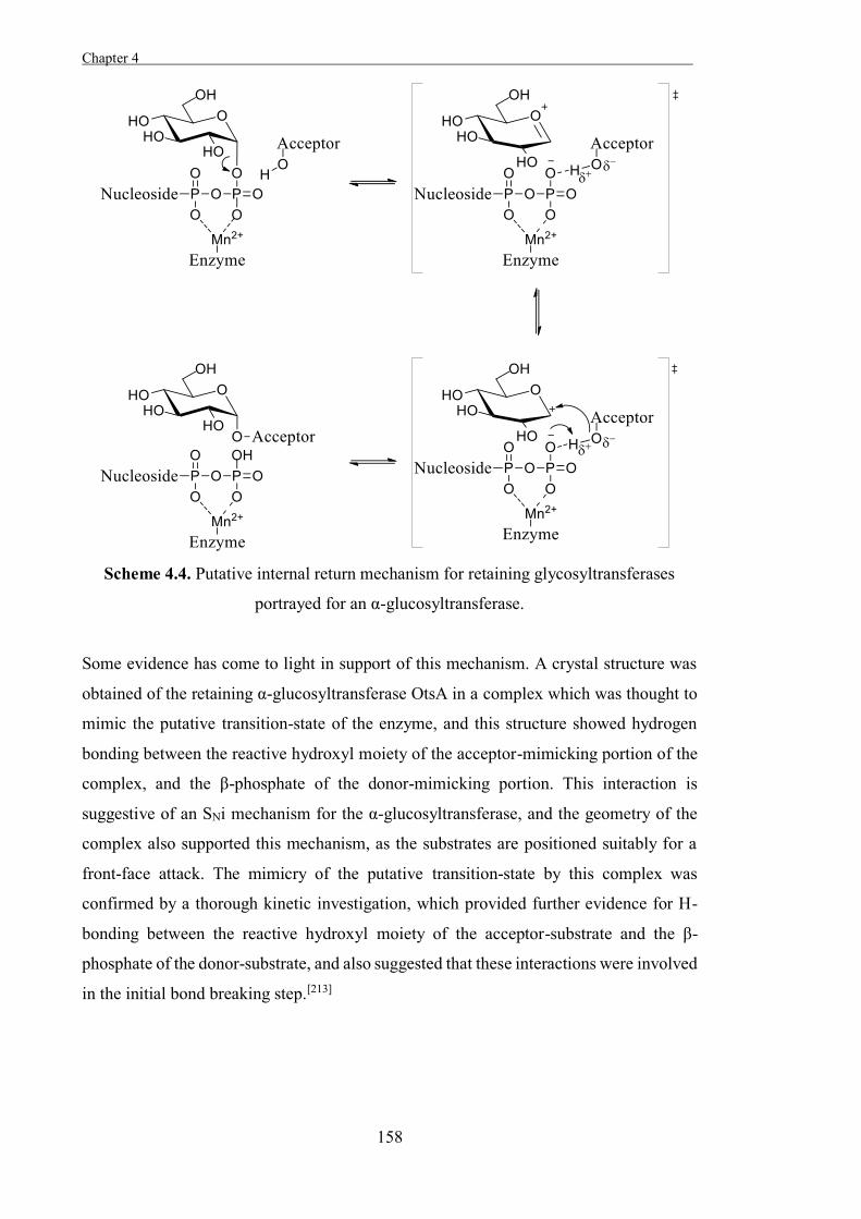
Chapter 4 .
158
Scheme 4.4. Putative internal return mechanism for retaining glycosyltransferases
portrayed for an α-glucosyltransferase.
Some evidence has come to light in support of this mechanism. A crystal structure was
obtained of the retaining α-glucosyltransferase OtsA in a complex which was thought to
mimic the putative transition-state of the enzyme, and this structure showed hydrogen
bonding between the reactive hydroxyl moiety of the acceptor-mimicking portion of the
complex, and the β-phosphate of the donor-mimicking portion. This interaction is
suggestive of an SNi mechanism for the α-glucosyltransferase, and the geometry of the
complex also supported this mechanism, as the substrates are positioned suitably for a
front-face attack. The mimicry of the putative transition-state by this complex was
confirmed by a thorough kinetic investigation, which provided further evidence for H-
bonding between the reactive hydroxyl moiety of the acceptor-substrate and the β-
phosphate of the donor-substrate, and also suggested that these interactions were involved
in the initial bond breaking step.[213]
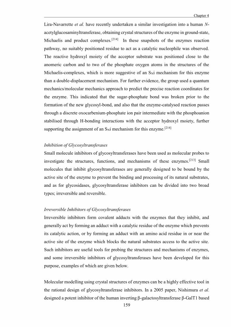
Chapter 4
159
Lira-Navarrette et al. have recently undertaken a similar investigation into a human N-
acetylglucosaminyltransferase, obtaining crystal structures of the enzyme in ground-state,
Michaelis and product complexes.[214] In these snapshots of the enzymes reaction
pathway, no suitably positioned residue to act as a catalytic nucleophile was observed.
The reactive hydroxyl moiety of the acceptor substrate was positioned close to the
anomeric carbon and to two of the phosphate oxygen atoms in the structures of the
Michaelis-complexes, which is more suggestive of an SNi mechanism for this enzyme
than a double-displacement mechanism. For further evidence, the group used a quantum
mechanics/molecular mechanics approach to predict the precise reaction coordinates for
the enzyme. This indicated that the sugar-phosphate bond was broken prior to the
formation of the new glycosyl-bond, and also that the enzyme-catalysed reaction passes
through a discrete oxocarbenium-phosphate ion pair intermediate with the phosphoanion
stabilised through H-bonding interactions with the acceptor hydroxyl moiety, further
supporting the assignment of an SNi mechanism for this enzyme.[214]
Inhibition of Glycosyltransferases
Small molecule inhibitors of glycosyltransferases have been used as molecular probes to
investigate the structures, functions, and mechanisms of these enzymes.[213] Small
molecules that inhibit glycosyltransferases are generally designed to be bound by the
active site of the enzyme to prevent the binding and processing of its natural substrates,
and as for glycosidases, glycosyltransferase inhibitors can be divided into two broad
types; irreversible and reversible.
Irreversible Inhibitors of Glycosyltransferases
Irreversible inhibitors form covalent adducts with the enzymes that they inhibit, and
generally act by forming an adduct with a catalytic residue of the enzyme which prevents
its catalytic action, or by forming an adduct with an amino acid residue in or near the
active site of the enzyme which blocks the natural substrates access to the active site.
Such inhibitors are useful tools for probing the structures and mechanisms of enzymes,
and some irreversible inhibitors of glycosyltransferases have been developed for this
purpose, examples of which are given below.
Molecular modelling using crystal structures of enzymes can be a highly effective tool in
the rational design of glycosyltransferase inhibitors. In a 2005 paper, Nishimura et al.
designed a potent inhibitor of the human inverting β-galactosyltransferase β-GalT1 based
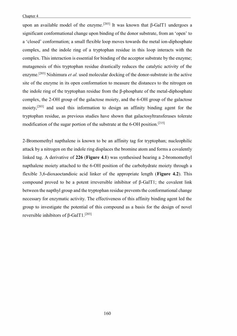
Chapter 4 .
160
upon an available model of the enzyme.[203] It was known that β-GalT1 undergoes a
significant conformational change upon binding of the donor substrate, from an ‘open’ to
a ‘closed’ conformation; a small flexible loop moves towards the metal ion-diphosphate
complex, and the indole ring of a tryptophan residue in this loop interacts with the
complex. This interaction is essential for binding of the acceptor substrate by the enzyme;
mutagenesis of this tryptophan residue drastically reduces the catalytic activity of the
enzyme.[203] Nishimura et al. used molecular docking of the donor-substrate in the active
site of the enzyme in its open conformation to measure the distances to the nitrogen on
the indole ring of the tryptophan residue from the β-phosphate of the metal-diphosphate
complex, the 2-OH group of the galactose moiety, and the 6-OH group of the galactose
moiety,[203] and used this information to design an affinity binding agent for the
tryptophan residue, as previous studies have shown that galactosyltransferases tolerate
modification of the sugar portion of the substrate at the 6-OH position.[215]
2-Bromomethyl napthalene is known to be an affinity tag for tryptophan; nucleophilic
attack by a nitrogen on the indole ring displaces the bromine atom and forms a covalently
linked tag. A derivative of 226 (Figure 4.1) was synthesised bearing a 2-bromomethyl
napthalene moiety attached to the 6-OH position of the carbohydrate moiety through a
flexible 3,6-dioxaoctandioic acid linker of the appropriate length (Figure 4.2). This
compound proved to be a potent irreversible inhibitor of β-GalT1; the covalent link
between the napthyl group and the tryptophan residue prevents the conformational change
necessary for enzymatic activity. The effectiveness of this affinity binding agent led the
group to investigate the potential of this compound as a basis for the design of novel
reversible inhibitors of β-GalT1.[203]
Chapter 4
161
Figure 4.2. The human inverting galactosyltransferase β-GalT1 is irreversibly inhibited
by 227.
Jiang and coworkers[216] used a high-throughput screen to assay the ability of a library of
small-molecules to inhibit human O-linked N-acetylglucosaminyltransferase (hOGT). A
compound with a benzoxazolinone core, 228, was found to be an irreversible inhibitor of
the enzyme (Figure 4.3), and so some derivatives of this compound were produced to
identify structural features important for binding by the enzyme. The p-methoxy
derivative 229 was a much more potent irreversible inhibitor of hOGT, and so this
inhibitor was used to investigate the mechanism of inhibition.
Figure 4.3. Irreversible inhibitors of hOGT.
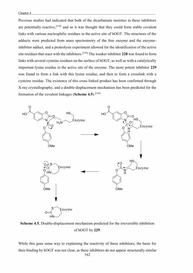
Chapter 4 .
162
Previous studies had indicated that both of the dicarbamate moieties in these inhibitors
are potentially reactive,[210] and so it was thought that they could form stable covalent
links with various nucleophilic residues in the active site of hOGT. The structures of the
adducts were predicted from mass spectrometry of the free enzyme and the enzyme-
inhibitor adduct, and a proteolysis experiment allowed for the identification of the active
site residues that react with the inhibitors.[216] The weaker inhibitor 228 was found to form
links with several cysteine residues on the surface of hOGT, as well as with a catalytically
important lysine residue in the active site of the enzyme. The more potent inhibitor 229
was found to form a link with this lysine residue, and then to form a crosslink with a
cysteine residue. The existence of this cross-linked product has been confirmed through
X-ray crystallography, and a double-displacement mechanism has been predicted for the
formation of the covalent linkages (Scheme 4.5).[216]
Scheme 4.5. Double-displacement mechanism predicted for the irreversible inhibition
of hOGT by 229.
While this goes some way to explaining the reactivity of these inhibitors, the basis for
their binding by hOGT was not clear, as these inhibitors do not appear structurally similar
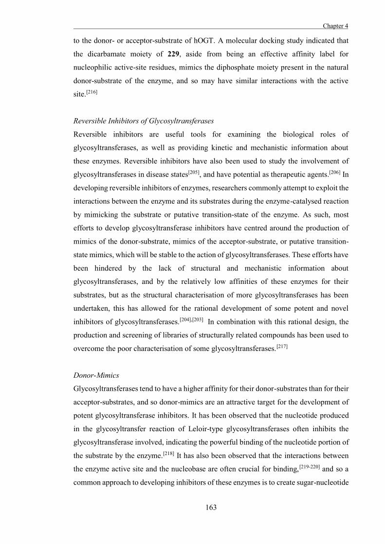
Chapter 4
163
to the donor- or acceptor-substrate of hOGT. A molecular docking study indicated that
the dicarbamate moiety of 229, aside from being an effective affinity label for
nucleophilic active-site residues, mimics the diphosphate moiety present in the natural
donor-substrate of the enzyme, and so may have similar interactions with the active
site.[216]
Reversible Inhibitors of Glycosyltransferases
Reversible inhibitors are useful tools for examining the biological roles of
glycosyltransferases, as well as providing kinetic and mechanistic information about
these enzymes. Reversible inhibitors have also been used to study the involvement of
glycosyltransferases in disease states[205], and have potential as therapeutic agents.[206] In
developing reversible inhibitors of enzymes, researchers commonly attempt to exploit the
interactions between the enzyme and its substrates during the enzyme-catalysed reaction
by mimicking the substrate or putative transition-state of the enzyme. As such, most
efforts to develop glycosyltransferase inhibitors have centred around the production of
mimics of the donor-substrate, mimics of the acceptor-substrate, or putative transition-
state mimics, which will be stable to the action of glycosyltransferases. These efforts have
been hindered by the lack of structural and mechanistic information about
glycosyltransferases, and by the relatively low affinities of these enzymes for their
substrates, but as the structural characterisation of more glycosyltransferases has been
undertaken, this has allowed for the rational development of some potent and novel
inhibitors of glycosyltransferases.[204],[203] In combination with this rational design, the
production and screening of libraries of structurally related compounds has been used to
overcome the poor characterisation of some glycosyltransferases.[217]
Donor-Mimics
Glycosyltransferases tend to have a higher affinity for their donor-substrates than for their
acceptor-substrates, and so donor-mimics are an attractive target for the development of
potent glycosyltransferase inhibitors. It has been observed that the nucleotide produced
in the glycosyltransfer reaction of Leloir-type glycosyltransferases often inhibits the
glycosyltransferase involved, indicating the powerful binding of the nucleotide portion of
the substrate by the enzyme.[218] It has also been observed that the interactions between
the enzyme active site and the nucleobase are often crucial for binding,[219-220] and so a
common approach to developing inhibitors of these enzymes is to create sugar-nucleotide
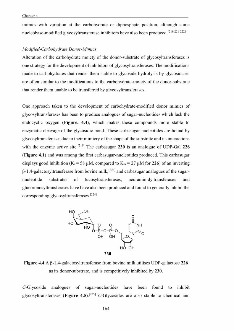
Chapter 4 .
164
mimics with variation at the carbohydrate or diphosphate position, although some
nucleobase-modified glycosyltransferase inhibitors have also been produced.[219,221-222]
Modified-Carbohydrate Donor-Mimics
Alteration of the carbohydrate moiety of the donor-substrate of glycosyltransferases is
one strategy for the development of inhibitors of glycosyltransferases. The modifications
made to carbohydrates that render them stable to glycoside hydrolysis by glycosidases
are often similar to the modifications to the carbohydrate-moiety of the donor-substrate
that render them unable to be transferred by glycosyltransferases.
One approach taken to the development of carbohydrate-modified donor mimics of
glycosyltransferases has been to produce analogues of sugar-nucleotides which lack the
endocyclic oxygen (Figure. 4.4), which makes these compounds more stable to
enzymatic cleavage of the glycosidic bond. These carbasugar-nucleotides are bound by
glycosyltransferases due to their mimicry of the shape of the substrate and its interactions
with the enzyme active site.[219] The carbasugar 230 is an analogue of UDP-Gal 226
(Figure 4.1) and was among the first carbasugar-nucleotides produced. This carbasugar
displays good inhibition (Ki = 58 µM, compared to Km = 27 µM for 226) of an inverting
β-1,4-galactosyltransferase from bovine milk,[223] and carbasugar analogues of the sugar-
nucleotide substrates of fucosyltransferases, neuraminidyltransferases and
glucoronosyltransferases have have also been produced and found to generally inhibit the
corresponding glycosyltransferases.[224]
Figure 4.4 A β-1,4-galactosyltransferase from bovine milk utilises UDP-galactose 226
as its donor-substrate, and is competitively inhibited by 230.
C-Glycoside analogues of sugar-nucleotides have been found to inhibit
glycosyltransferases (Figure 4.5).[225] C-Glycosides are also stable to chemical and
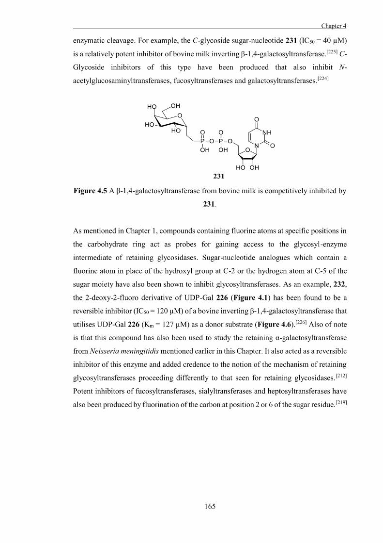
Chapter 4
165
enzymatic cleavage. For example, the C-glycoside sugar-nucleotide 231 (IC50 = 40 µM)
is a relatively potent inhibitor of bovine milk inverting β-1,4-galactosyltransferase.[225] C-
Glycoside inhibitors of this type have been produced that also inhibit N-
acetylglucosaminyltransferases, fucosyltransferases and galactosyltransferases.[224]
Figure 4.5 A β-1,4-galactosyltransferase from bovine milk is competitively inhibited by
231.
As mentioned in Chapter 1, compounds containing fluorine atoms at specific positions in
the carbohydrate ring act as probes for gaining access to the glycosyl-enzyme
intermediate of retaining glycosidases. Sugar-nucleotide analogues which contain a
fluorine atom in place of the hydroxyl group at C-2 or the hydrogen atom at C-5 of the
sugar moiety have also been shown to inhibit glycosyltransferases. As an example, 232,
the 2-deoxy-2-fluoro derivative of UDP-Gal 226 (Figure 4.1) has been found to be a
reversible inhibitor (IC50 = 120 µM) of a bovine inverting β-1,4-galactosyltransferase that
utilises UDP-Gal 226 (Km = 127 µM) as a donor substrate (Figure 4.6).[226] Also of note
is that this compound has also been used to study the retaining α-galactosyltransferase
from Neisseria meningitidis mentioned earlier in this Chapter. It also acted as a reversible
inhibitor of this enzyme and added credence to the notion of the mechanism of retaining
glycosyltransferases proceeding differently to that seen for retaining glycosidases.[212]
Potent inhibitors of fucosyltransferases, sialyltransferases and heptosyltransferases have
also been produced by fluorination of the carbon at position 2 or 6 of the sugar residue.[219]
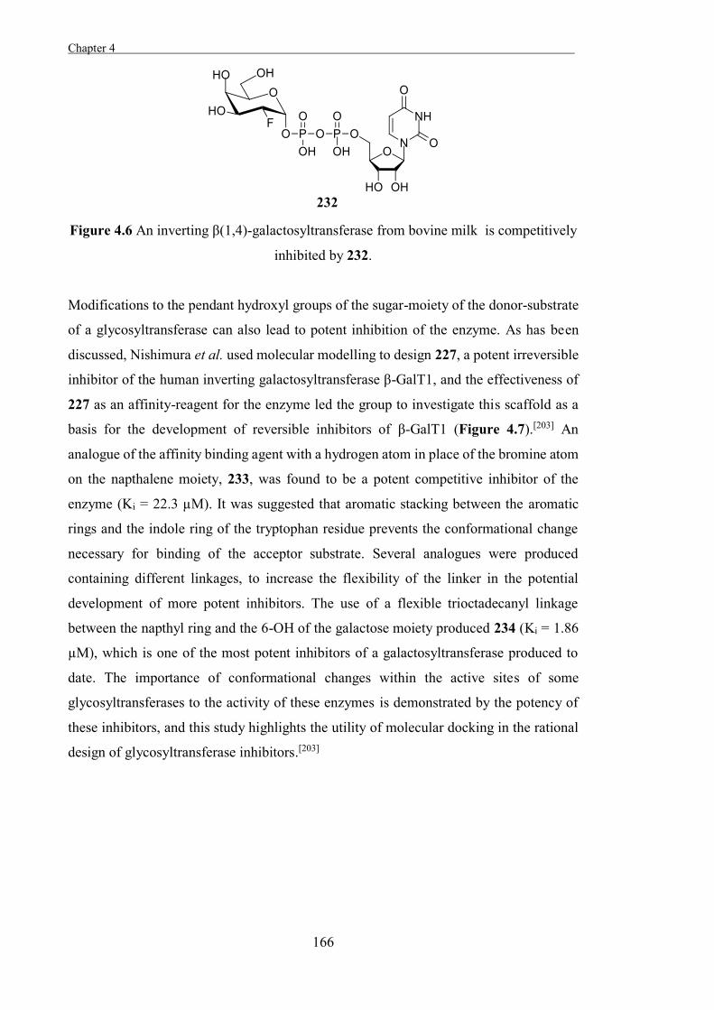
Chapter 4 .
166
Figure 4.6 An inverting β(1,4)-galactosyltransferase from bovine milk is competitively
inhibited by 232.
Modifications to the pendant hydroxyl groups of the sugar-moiety of the donor-substrate
of a glycosyltransferase can also lead to potent inhibition of the enzyme. As has been
discussed, Nishimura et al. used molecular modelling to design 227, a potent irreversible
inhibitor of the human inverting galactosyltransferase β-GalT1, and the effectiveness of
227 as an affinity-reagent for the enzyme led the group to investigate this scaffold as a
basis for the development of reversible inhibitors of β-GalT1 (Figure 4.7).[203] An
analogue of the affinity binding agent with a hydrogen atom in place of the bromine atom
on the napthalene moiety, 233, was found to be a potent competitive inhibitor of the
enzyme (Ki = 22.3 µM). It was suggested that aromatic stacking between the aromatic
rings and the indole ring of the tryptophan residue prevents the conformational change
necessary for binding of the acceptor substrate. Several analogues were produced
containing different linkages, to increase the flexibility of the linker in the potential
development of more potent inhibitors. The use of a flexible trioctadecanyl linkage
between the napthyl ring and the 6-OH of the galactose moiety produced 234 (Ki = 1.86
µM), which is one of the most potent inhibitors of a galactosyltransferase produced to
date. The importance of conformational changes within the active sites of some
glycosyltransferases to the activity of these enzymes is demonstrated by the potency of
these inhibitors, and this study highlights the utility of molecular docking in the rational
design of glycosyltransferase inhibitors.[203]
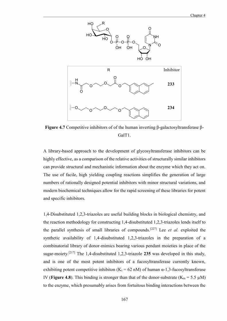
Chapter 4
167
Figure 4.7 Competitive inhibitors of of the human inverting β-galactosyltransferase β-
GalT1.
A library-based approach to the development of glycosyltransferase inhibitors can be
highly effective, as a comparison of the relative activities of structurally similar inhibitors
can provide structural and mechanistic information about the enzyme which they act on.
The use of facile, high yielding coupling reactions simplifies the generation of large
numbers of rationally designed potential inhibitors with minor structural variations, and
modern biochemical techniques allow for the rapid screening of these libraries for potent
and specific inhibitors.
1,4-Disubstituted 1,2,3-triazoles are useful building blocks in biological chemistry, and
the reaction methodology for constructing 1,4-disubstituted 1,2,3-triazoles lends itself to
the parallel synthesis of small libraries of compounds.[227] Lee et al. exploited the
synthetic availability of 1,4-disubstituted 1,2,3-triazoles in the preparation of a
combinatorial library of donor-mimics bearing various pendant moieties in place of the
sugar-moiety.[217] The 1,4-disubstituted 1,2,3-triazole 235 was developed in this study,
and is one of the most potent inhibitors of a fucosyltransferase currently known,
exhibiting potent competitive inhibiton (Ki = 62 nM) of human α-1,3-fucosyltransferase
IV (Figure 4.8). This binding is stronger than that of the donor-substrate (Km = 5.5 µM)
to the enzyme, which presumably arises from fortuitous binding interactions between the
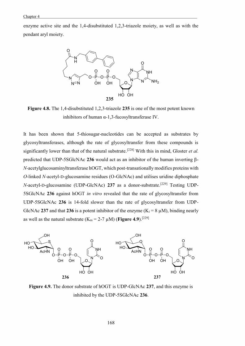
Chapter 4 .
168
enzyme active site and the 1,4-disubstituted 1,2,3-triazole moiety, as well as with the
pendant aryl moiety.
Figure 4.8. The 1,4-disubstituted 1,2,3-triazole 235 is one of the most potent known
inhibitors of human α-1,3-fucosyltransferase IV.
It has been shown that 5-thiosugar-nucleotides can be accepted as substrates by
glycosyltransferases, although the rate of glycosyltransfer from these compounds is
significantly lower than that of the natural substrate.[228] With this in mind, Gloster et al.
predicted that UDP-5SGlcNAc 236 would act as an inhibitor of the human inverting β-
N-acetylglucosaminyltransferase hOGT, which post-transationally modifies proteins with
O-linked N-acetyl-D-glucosamine residues (O-GlcNAc) and utilises uridine diphosphate
N-acetyl-D-glucosamine (UDP-GlcNAc) 237 as a donor-substrate.[229] Testing UDP-
5SGlcNAc 236 against hOGT in vitro revealed that the rate of glycosyltransfer from
UDP-5SGlcNAc 236 is 14-fold slower than the rate of glycosyltransfer from UDP-
GlcNAc 237 and that 236 is a potent inhibitor of the enzyme (Ki = 8 µM), binding nearly
as well as the natural substrate (Km = 2-7 µM) (Figure 4.9).[229]
Figure 4.9. The donor substrate of hOGT is UDP-GlcNAc 237, and this enzyme is
inhibited by the UDP-5SGlcNAc 236.
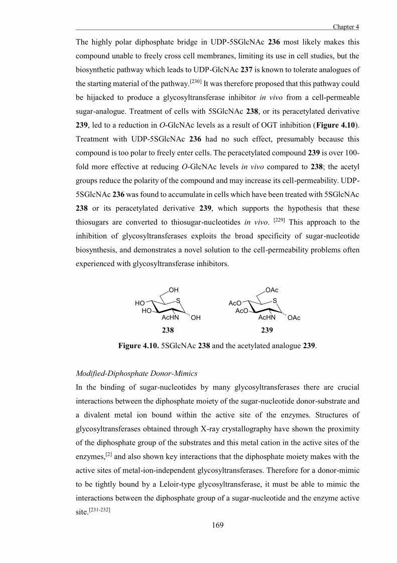
Chapter 4
169
The highly polar diphosphate bridge in UDP-5SGlcNAc 236 most likely makes this
compound unable to freely cross cell membranes, limiting its use in cell studies, but the
biosynthetic pathway which leads to UDP-GlcNAc 237 is known to tolerate analogues of
the starting material of the pathway.[230] It was therefore proposed that this pathway could
be hijacked to produce a glycosyltransferase inhibitor in vivo from a cell-permeable
sugar-analogue. Treatment of cells with 5SGlcNAc 238, or its peracetylated derivative
239, led to a reduction in O-GlcNAc levels as a result of OGT inhibition (Figure 4.10).
Treatment with UDP-5SGlcNAc 236 had no such effect, presumably because this
compound is too polar to freely enter cells. The peracetylated compound 239 is over 100-
fold more effective at reducing O-GlcNAc levels in vivo compared to 238; the acetyl
groups reduce the polarity of the compound and may increase its cell-permeability. UDP-
5SGlcNAc 236 was found to accumulate in cells which have been treated with 5SGlcNAc
238 or its peracetylated derivative 239, which supports the hypothesis that these
thiosugars are converted to thiosugar-nucleotides in vivo. [229] This approach to the
inhibition of glycosyltransferases exploits the broad specificity of sugar-nucleotide
biosynthesis, and demonstrates a novel solution to the cell-permeability problems often
experienced with glycosyltransferase inhibitors.
Figure 4.10. 5SGlcNAc 238 and the acetylated analogue 239.
Modified-Diphosphate Donor-Mimics
In the binding of sugar-nucleotides by many glycosyltransferases there are crucial
interactions between the diphosphate moiety of the sugar-nucleotide donor-substrate and
a divalent metal ion bound within the active site of the enzymes. Structures of
glycosyltransferases obtained through X-ray crystallography have shown the proximity
of the diphosphate group of the substrates and this metal cation in the active sites of the
enzymes,[2] and also shown key interactions that the diphosphate moiety makes with the
active sites of metal-ion-independent glycosyltransferases. Therefore for a donor-mimic
to be tightly bound by a Leloir-type glycosyltransferase, it must be able to mimic the
interactions between the diphosphate group of a sugar-nucleotide and the enzyme active
site.[231-232]
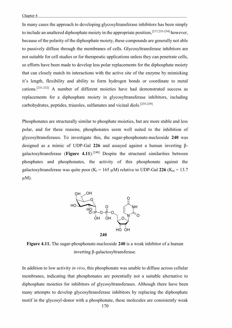
Chapter 4 .
170
In many cases the approach to developing glycosyltransferase inhibitors has been simply
to include an unaltered diphosphate moiety in the appropriate position,[217,233-234] however,
because of the polarity of the diphosphate moiety, these compounds are generally not able
to passively diffuse through the membranes of cells. Glycosyltransferase inhibitors are
not suitable for cell studies or for therapeutic applications unless they can penetrate cells,
so efforts have been made to develop less polar replacements for the diphosphate moiety
that can closely match its interactions with the active site of the enzyme by mimicking
it’s length, flexibility and ability to form hydrogen bonds or coordinate to metal
cations.[231-232] A number of different moieties have had demonstrated success as
replacements for a diphosphate moiety in glycosyltransferase inhibitors, including
carbohydrates, peptides, triazoles, sulfamates and vicinal diols.[235-239]
Phosphonates are structurally similar to phosphate moieties, but are more stable and less
polar, and for these reasons, phosphonates seem well suited to the inhibition of
glycosyltransferases. To investigate this, the sugar-phosphonate-nucleoside 240 was
designed as a mimic of UDP-Gal 226 and assayed against a human inverting β-
galactosyltransferase (Figure 4.11).[240] Despite the structural similarities between
phosphates and phosphonates, the activity of this phosphonate against the
galactosyltransferase was quite poor (Ki = 165 µM) relative to UDP-Gal 226 (Km = 13.7
µM).
Figure 4.11. The sugar-phosphonate-nucleoside 240 is a weak inhibitor of a human
inverting β-galactosyltransferase.
In addition to low activity in vivo, this phosphonate was unable to diffuse across cellular
membranes, indicating that phosphonates are potentially not a suitable alternative to
diphosphate moieties for inhibitors of glycosyltransferases. Although there have been
many attempts to develop glycosyltransferase inhibitors by replacing the diphosphate
motif in the glycosyl-donor with a phosphonate, these molecules are consistently weak
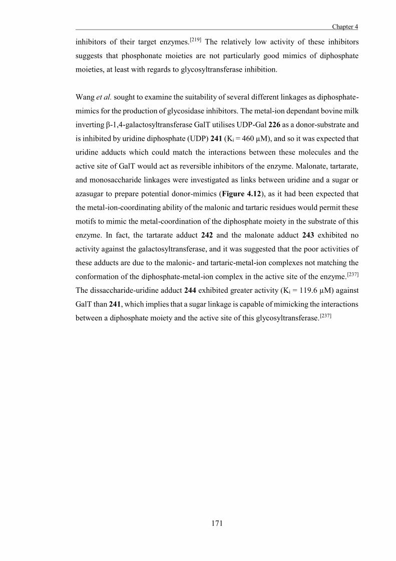
Chapter 4
171
inhibitors of their target enzymes.[219] The relatively low activity of these inhibitors
suggests that phosphonate moieties are not particularly good mimics of diphosphate
moieties, at least with regards to glycosyltransferase inhibition.
Wang et al. sought to examine the suitability of several different linkages as diphosphate-
mimics for the production of glycosidase inhibitors. The metal-ion dependant bovine milk
inverting β-1,4-galactosyltransferase GalT utilises UDP-Gal 226 as a donor-substrate and
is inhibited by uridine diphosphate (UDP) 241 (Ki = 460 µM), and so it was expected that
uridine adducts which could match the interactions between these molecules and the
active site of GalT would act as reversible inhibitors of the enzyme. Malonate, tartarate,
and monosaccharide linkages were investigated as links between uridine and a sugar or
azasugar to prepare potential donor-mimics (Figure 4.12), as it had been expected that
the metal-ion-coordinating ability of the malonic and tartaric residues would permit these
motifs to mimic the metal-coordination of the diphosphate moiety in the substrate of this
enzyme. In fact, the tartarate adduct 242 and the malonate adduct 243 exhibited no
activity against the galactosyltransferase, and it was suggested that the poor activities of
these adducts are due to the malonic- and tartaric-metal-ion complexes not matching the
conformation of the diphosphate-metal-ion complex in the active site of the enzyme.[237]
The dissaccharide-uridine adduct 244 exhibited greater activity (Ki = 119.6 µM) against
GalT than 241, which implies that a sugar linkage is capable of mimicking the interactions
between a diphosphate moiety and the active site of this glycosyltransferase.[237]
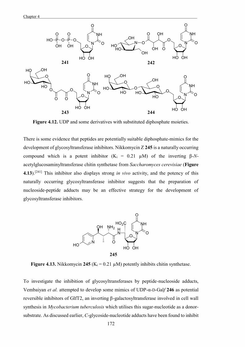
Chapter 4 .
172
Figure 4.12. UDP and some derivatives with substituted diphosphate moieties.
There is some evidence that peptides are potentially suitable diphosphate-mimics for the
development of glycosyltransferase inhibitors. Nikkomycin Z 245 is a naturally occurring
compound which is a potent inhibitor (Ki = 0.21 µM) of the inverting β-N-
acetylglucosaminyltransferase chitin synthetase from Saccharomyces cerevisiae (Figure
4.13).[241] This inhibitor also displays strong in vivo activity, and the potency of this
naturally occurring glycosyltransferase inhibitor suggests that the preparation of
nucleoside-peptide adducts may be an effective strategy for the development of
glycosyltransferase inhibitors.
Figure 4.13. Nikkomycin 245 (Ki = 0.21 µM) potently inhibits chitin synthetase.
To investigate the inhibition of glycosyltransferases by peptide-nucleoside adducts,
Vembaiyan et al. attempted to develop some mimics of UDP-α-D-Galf 246 as potential
reversible inhibitors of GlfT2, an inverting β-galactosyltransferase involved in cell wall
synthesis in Mycobacterium tuberculosis which utilises this sugar-nucleotide as a donor-
substrate. As discussed earlier, C-glycoside-nucleotide adducts have been found to inhibit
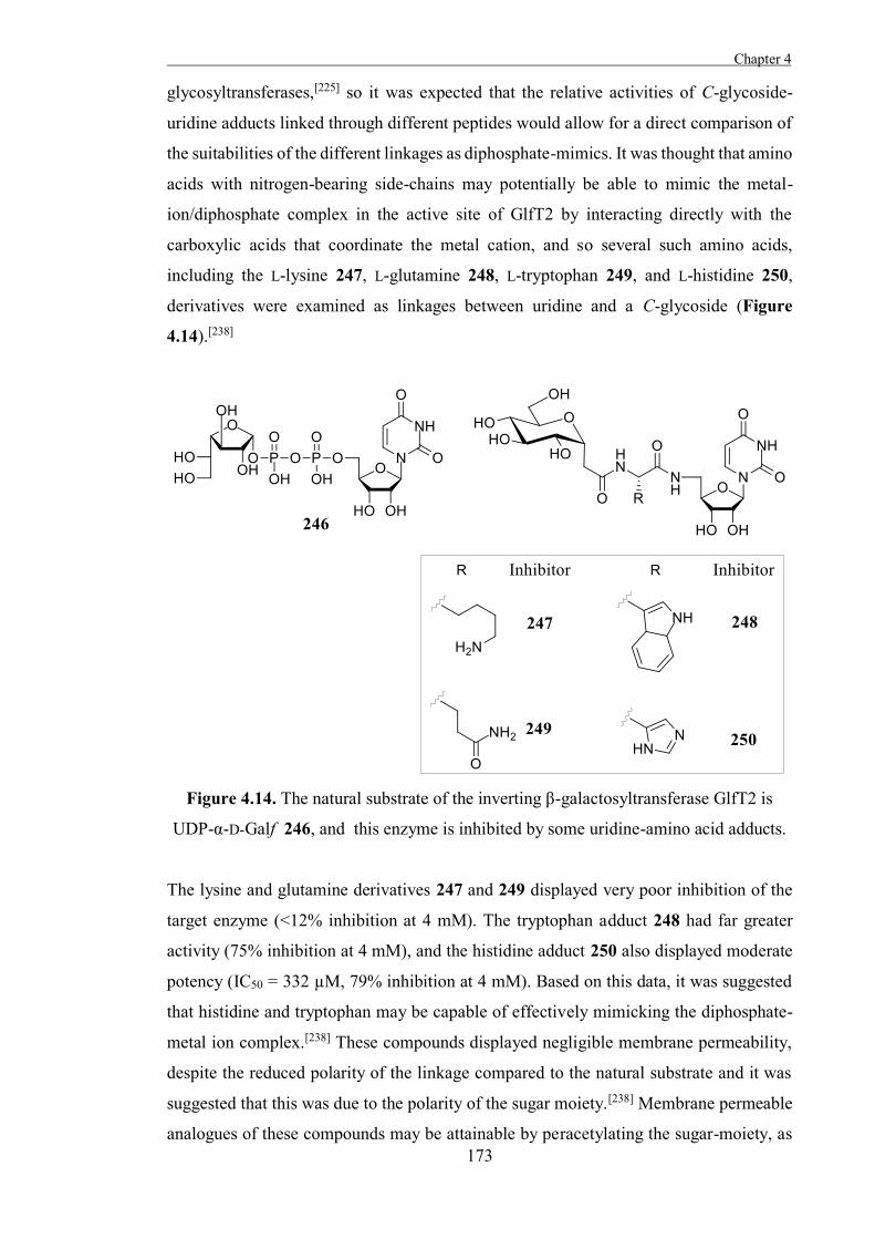
Chapter 4
173
glycosyltransferases,[225] so it was expected that the relative activities of C-glycoside-
uridine adducts linked through different peptides would allow for a direct comparison of
the suitabilities of the different linkages as diphosphate-mimics. It was thought that amino
acids with nitrogen-bearing side-chains may potentially be able to mimic the metal-
ion/diphosphate complex in the active site of GlfT2 by interacting directly with the
carboxylic acids that coordinate the metal cation, and so several such amino acids,
including the L-lysine 247, L-glutamine 248, L-tryptophan 249, and L-histidine 250,
derivatives were examined as linkages between uridine and a C-glycoside (Figure
4.14).[238]
Figure 4.14. The natural substrate of the inverting β-galactosyltransferase GlfT2 is
UDP-α-D-Galf 246, and this enzyme is inhibited by some uridine-amino acid adducts.
The lysine and glutamine derivatives 247 and 249 displayed very poor inhibition of the
target enzyme (<12% inhibition at 4 mM). The tryptophan adduct 248 had far greater
activity (75% inhibition at 4 mM), and the histidine adduct 250 also displayed moderate
potency (IC50 = 332 µM, 79% inhibition at 4 mM). Based on this data, it was suggested
that histidine and tryptophan may be capable of effectively mimicking the diphosphate-
metal ion complex.[238] These compounds displayed negligible membrane permeability,
despite the reduced polarity of the linkage compared to the natural substrate and it was
suggested that this was due to the polarity of the sugar moiety.[238] Membrane permeable
analogues of these compounds may be attainable by peracetylating the sugar-moiety, as
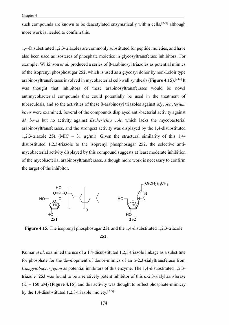
Chapter 4 .
174
such compounds are known to be deacetylated enzymatically within cells,[229] although
more work is needed to confirm this.
1,4-Disubstituted 1,2,3-triazoles are commonly substituted for peptide moieties, and have
also been used as isosteres of phosphate moieties in glycosyltransferase inhibitors. For
example, Wilkinson et al. produced a series of β-arabinosyl triazoles as potential mimics
of the isoprenyl phosphosugar 252, which is used as a glycosyl donor by non-Leloir type
arabinosyltransferases involved in mycobacterial cell-wall synthesis (Figure 4.15).[242] It
was thought that inhibitors of these arabinosyltransferases would be novel
antimycobacterial compounds that could potentially be used in the treatment of
tuberculosis, and so the activities of these β-arabinosyl triazoles against Mycobacterium
bovis were examined. Several of the compounds displayed anti-bacterial activity against
M. bovis but no activity against Escherichia coli, which lacks the mycobacterial
arabinosyltransferases, and the strongest activity was displayed by the 1,4-disubstituted
1,2,3-triazole 251 (MIC = 31 µg/ml). Given the structural similarity of this 1,4-
disubstituted 1,2,3-triazole to the isoprenyl phosphosugar 252, the selective anti-
mycobacterial activity displayed by this compound suggests at least moderate inhibition
of the mycobacterial arabinosyltransferases, although more work is necessary to confirm
the target of the inhibitor.
Figure 4.15. The isoprenyl phosphosugar 251 and the 1,4-disubstituted 1,2,3-triazole
252.
Kumar et al. examined the use of a 1,4-disubstituted 1,2,3-triazole linkage as a substitute
for phosphate for the development of donor-mimics of an α-2,3-sialyltransferase from
Campylobacter jejuni as potential inhibitors of this enzyme. The 1,4-disubstituted 1,2,3-
triazole 253 was found to be a relatively potent inhibitor of this α-2,3-sialyltransferase
(Ki = 160 µM) (Figure 4.16), and this activity was thought to reflect phosphate-mimicry
by the 1,4-disubstituted 1,2,3-triazole moiety.[239]
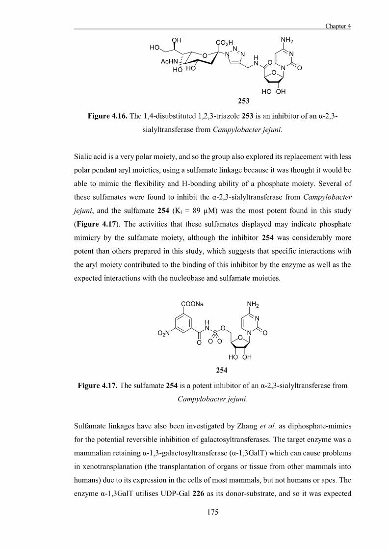
Chapter 4
175
Figure 4.16. The 1,4-disubstituted 1,2,3-triazole 253 is an inhibitor of an α-2,3-
sialyltransferase from Campylobacter jejuni.
Sialic acid is a very polar moiety, and so the group also explored its replacement with less
polar pendant aryl moieties, using a sulfamate linkage because it was thought it would be
able to mimic the flexibility and H-bonding ability of a phosphate moiety. Several of
these sulfamates were found to inhibit the α-2,3-sialyltransferase from Campylobacter
jejuni, and the sulfamate 254 (Ki = 89 µM) was the most potent found in this study
(Figure 4.17). The activities that these sulfamates displayed may indicate phosphate
mimicry by the sulfamate moiety, although the inhibitor 254 was considerably more
potent than others prepared in this study, which suggests that specific interactions with
the aryl moiety contributed to the binding of this inhibitor by the enzyme as well as the
expected interactions with the nucleobase and sulfamate moieties.
Figure 4.17. The sulfamate 254 is a potent inhibitor of an α-2,3-sialyltransferase from
Campylobacter jejuni.
Sulfamate linkages have also been investigated by Zhang et al. as diphosphate-mimics
for the potential reversible inhibition of galactosyltransferases. The target enzyme was a
mammalian retaining α-1,3-galactosyltransferase (α-1,3GalT) which can cause problems
in xenotransplanation (the transplantation of organs or tissue from other mammals into
humans) due to its expression in the cells of most mammals, but not humans or apes. The
enzyme α-1,3GalT utilises UDP-Gal 226 as its donor-substrate, and so it was expected
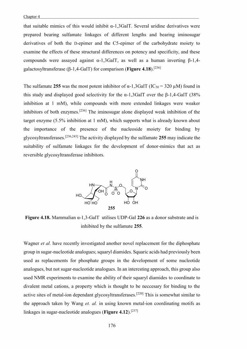
Chapter 4 .
176
that suitable mimics of this would inhibit α-1,3GalT. Several uridine derivatives were
prepared bearing sulfamate linkages of different lengths and bearing iminosugar
derivatives of both the D-epimer and the C5-epimer of the carbohydrate moiety to
examine the effects of these structural differences on potency and specificity, and these
compounds were assayed against α-1,3GalT, as well as a human inverting β-1,4-
galactosyltransferase (β-1,4-GalT) for comparison (Figure 4.18).[236]
The sulfamate 255 was the most potent inhibitor of α-1,3GalT (IC50 = 320 µM) found in
this study and displayed good selectivity for the α-1,3GalT over the β-1,4-GalT (38%
inhibition at 1 mM), while compounds with more extended linkages were weaker
inhibitors of both enzymes.[236] The iminosugar alone displayed weak inhibition of the
target enzyme (5.5% inhibition at 1 mM), which supports what is already known about
the importance of the presence of the nucleoside moiety for binding by
glycosyltransferases.[236,243] The activity displayed by the sulfamate 255 may indicate the
suitability of sulfamate linkages for the development of donor-mimics that act as
reversible glycosyltransferase inhibitors.
Figure 4.18. Mammalian α-1,3-GalT utilises UDP-Gal 226 as a donor substrate and is
inhibited by the sulfamate 255.
Wagner et al. have recently investigated another novel replacement for the diphosphate
group in sugar-nucleotide analogues; squaryl diamides. Squaric acids had previously been
used as replacements for phosphate groups in the development of some nucleotide
analogues, but not sugar-nucleotide analogues. In an interesting approach, this group also
used NMR experiments to examine the ability of their squaryl diamides to coordinate to
divalent metal cations, a property which is thought to be neccesary for binding to the
active sites of metal-ion dependant glycosyltransferases.[220] This is somewhat similar to
the approach taken by Wang et. al. in using known metal-ion coordinating motifs as
linkages in sugar-nucleotide analogues (Figure 4.12).[237]
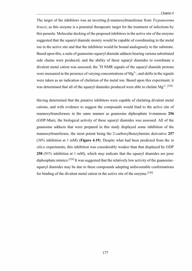
Chapter 4
177
The target of the inhibitors was an inverting β-mannosyltransferase from Trypanosoma
brucei, as this enzyme is a potential therapeutic target for the treatment of infections by
this parasite. Molecular docking of the proposed inhibitors in the active site of the enzyme
suggested that the squaryl diamide moiety would be capable of coordinating to the metal
ion in the active site and that the inhibitors would be bound analogously to the substrate.
Based upon this, a suite of guanosine-squaryl diamide adducts bearing various substituted
side chains were produced, and the ability of these squaryl diamides to coordinate a
divalent metal cation was assessed; the 1H NMR signals of the squaryl diamide protons
were measured in the presence of varying concentrations of Mg2+, and shifts in the signals
were taken as an indication of chelation of the metal ion. Based upon this experiment, it
was determined that all of the squaryl diamides produced were able to chelate Mg2+.[220]
Having determined that the putative inhibitors were capable of chelating divalent metal
cations, and with evidence to suggest the compounds would bind to the active site of
mannosyltransferases in the same manner as guanosine diphosphate D-mannose 256
(GDP-Man), the biological activity of these squaryl diamides was assessed. All of the
guanosine adducts that were prepared in this study displayed some inhibition of the
mannosyltransferase, the most potent being the 2-carboxylbenzylamino derivative 257
(36% inhibition at 1 mM) (Figure 4.19). Despite what had been predicted from the in
silico experiments, this inhibition was considerably weaker than that displayed by GDP
258 (91% inhibition at 1 mM), which may indicate that the squaryl diamides are poor
diphosphate mimics.[220] It was suggested that the relatively low activity of the guanosine-
squaryl diamides may be due to these compounds adopting unfavourable conformations
for binding of the divalent metal cation in the active site of the enzyme.[220]
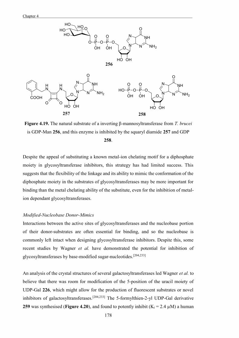
Chapter 4 .
178
Figure 4.19. The natural substrate of a inverting β-mannosyltransferase from T. brucei
is GDP-Man 256, and this enzyme is inhibited by the squaryl diamide 257 and GDP
258.
Despite the appeal of substituting a known metal-ion chelating motif for a diphosphate
moiety in glycosyltransferase inhibitors, this strategy has had limited success. This
suggests that the flexibility of the linkage and its ability to mimic the conformation of the
diphosphate moiety in the substrates of glycosyltransferases may be more important for
binding than the metal chelating ability of the substitute, even for the inhibition of metal-
ion dependant glycosyltransferases.
Modified-Nucleobase Donor-Mimics
Interactions between the active sites of glycosyltransferases and the nucleobase portion
of their donor-substrates are often essential for binding, and so the nucleobase is
commonly left intact when designing glycosyltransferase inhibitors. Despite this, some
recent studies by Wagner et al. have demonstrated the potential for inhibition of
glycosyltransferases by base-modified sugar-nucleotides.[204,233]
An analysis of the crystal structures of several galactosyltransferases led Wagner et al. to
believe that there was room for modification of the 5-position of the uracil moiety of
UDP-Gal 226, which might allow for the production of fluorescent substrates or novel
inhibitors of galactosyltransferases.[204,233] The 5-formylthien-2-yl UDP-Gal derivative
259 was synthesised (Figure 4.20), and found to potently inhibit (Ki = 2.4 µM) a human
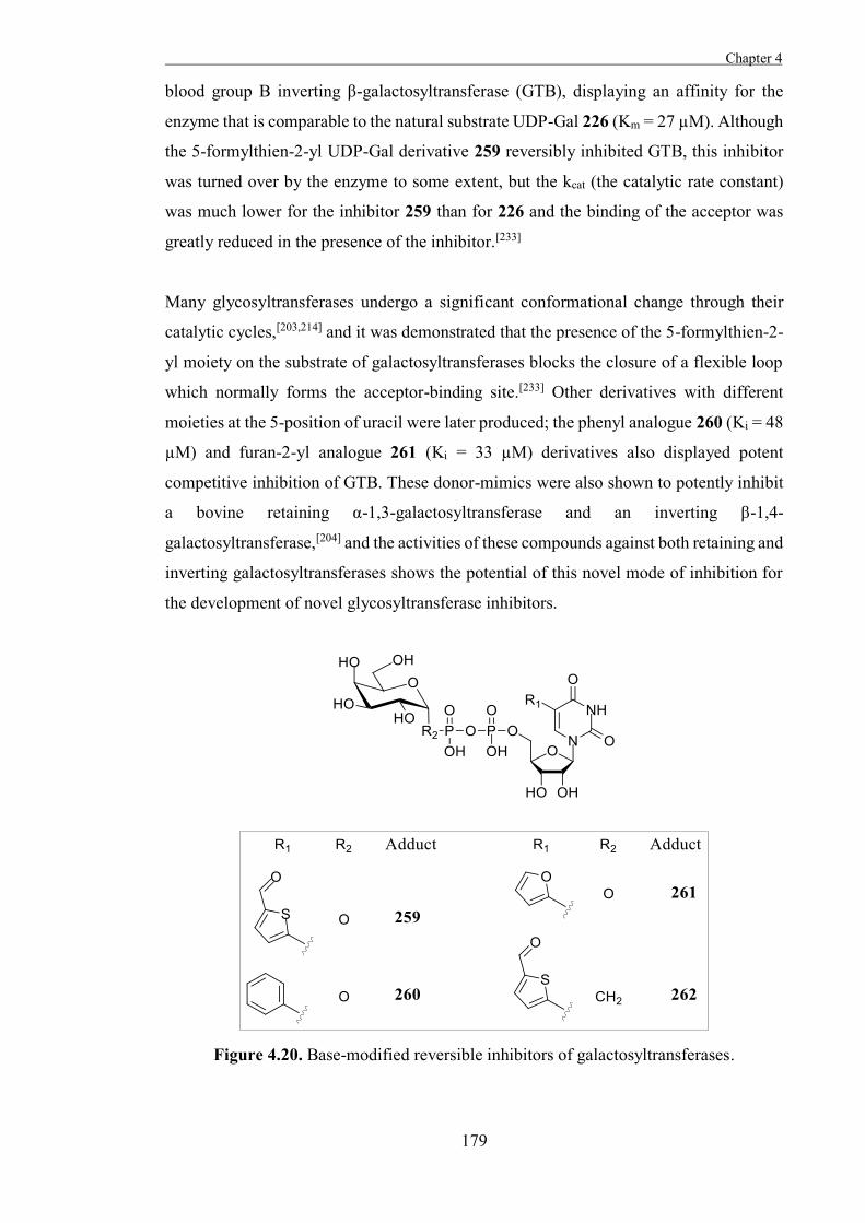
Chapter 4
179
blood group B inverting β-galactosyltransferase (GTB), displaying an affinity for the
enzyme that is comparable to the natural substrate UDP-Gal 226 (Km = 27 µM). Although
the 5-formylthien-2-yl UDP-Gal derivative 259 reversibly inhibited GTB, this inhibitor
was turned over by the enzyme to some extent, but the kcat (the catalytic rate constant)
was much lower for the inhibitor 259 than for 226 and the binding of the acceptor was
greatly reduced in the presence of the inhibitor.[233]
Many glycosyltransferases undergo a significant conformational change through their
catalytic cycles,[203,214] and it was demonstrated that the presence of the 5-formylthien-2-
yl moiety on the substrate of galactosyltransferases blocks the closure of a flexible loop
which normally forms the acceptor-binding site.[233] Other derivatives with different
moieties at the 5-position of uracil were later produced; the phenyl analogue 260 (Ki = 48
µM) and furan-2-yl analogue 261 (Ki = 33 µM) derivatives also displayed potent
competitive inhibition of GTB. These donor-mimics were also shown to potently inhibit
a bovine retaining α-1,3-galactosyltransferase and an inverting β-1,4-
galactosyltransferase,[204] and the activities of these compounds against both retaining and
inverting galactosyltransferases shows the potential of this novel mode of inhibition for
the development of novel glycosyltransferase inhibitors.
Figure 4.20. Base-modified reversible inhibitors of galactosyltransferases.
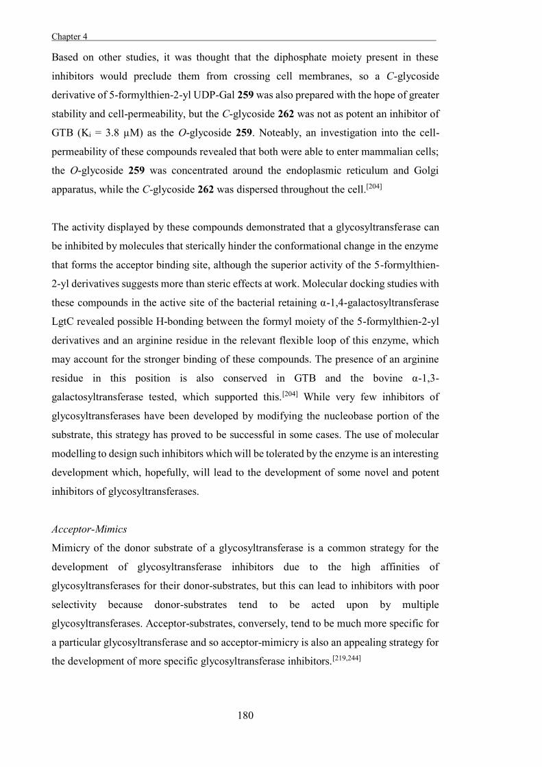
Chapter 4 .
180
Based on other studies, it was thought that the diphosphate moiety present in these
inhibitors would preclude them from crossing cell membranes, so a C-glycoside
derivative of 5-formylthien-2-yl UDP-Gal 259 was also prepared with the hope of greater
stability and cell-permeability, but the C-glycoside 262 was not as potent an inhibitor of
GTB (Ki = 3.8 µM) as the O-glycoside 259. Noteably, an investigation into the cell-
permeability of these compounds revealed that both were able to enter mammalian cells;
the O-glycoside 259 was concentrated around the endoplasmic reticulum and Golgi
apparatus, while the C-glycoside 262 was dispersed throughout the cell.[204]
The activity displayed by these compounds demonstrated that a glycosyltransferase can
be inhibited by molecules that sterically hinder the conformational change in the enzyme
that forms the acceptor binding site, although the superior activity of the 5-formylthien-
2-yl derivatives suggests more than steric effects at work. Molecular docking studies with
these compounds in the active site of the bacterial retaining α-1,4-galactosyltransferase
LgtC revealed possible H-bonding between the formyl moiety of the 5-formylthien-2-yl
derivatives and an arginine residue in the relevant flexible loop of this enzyme, which
may account for the stronger binding of these compounds. The presence of an arginine
residue in this position is also conserved in GTB and the bovine α-1,3-
galactosyltransferase tested, which supported this.[204] While very few inhibitors of
glycosyltransferases have been developed by modifying the nucleobase portion of the
substrate, this strategy has proved to be successful in some cases. The use of molecular
modelling to design such inhibitors which will be tolerated by the enzyme is an interesting
development which, hopefully, will lead to the development of some novel and potent
inhibitors of glycosyltransferases.
Acceptor-Mimics
Mimicry of the donor substrate of a glycosyltransferase is a common strategy for the
development of glycosyltransferase inhibitors due to the high affinities of
glycosyltransferases for their donor-substrates, but this can lead to inhibitors with poor
selectivity because donor-substrates tend to be acted upon by multiple
glycosyltransferases. Acceptor-substrates, conversely, tend to be much more specific for
a particular glycosyltransferase and so acceptor-mimicry is also an appealing strategy for
the development of more specific glycosyltransferase inhibitors.[219,244]
Chapter 4
181
Producing analogues of the acceptor-substrates which lack a hydroxyl group at the
necessary position for enzymatic glycosylation is one approach which has shown some
success in the development of acceptor-mimicking glycosyltransferase inhibitors. Such
molecules would be expected to bind to the enzymes similarly to the natural substrates,
but will not be glycosylated because they lack the reactive hydroxyl moiety.
For example, Sinha et al. prepared 4-deoxy-4-azido D-glucose 263 and 4-deoxy-D-xylo-
hexose 264 as mimics of D-glucopyranose 2/3, the acceptor-substrate of 4-β-D-
galactopyranosyltransferase (lactose synthase) (Figure 4.21).[245] These acceptor-mimics
were not glycosylated (turned over) by lactose synthase, instead acting as reversible
inhibitors of the enzyme. Both inhibitors were found to bind lactose synthase more
weakly than the natural substrate, indicating that interactions with the 4-OH moiety are
important for substrate recognition by this enzyme.
Figure 4.21. 4-deoxy-4-azido-D-glucose 263 and 4-deoxy-D-xylo-hexose 264.
The group of Hindsgaul has prepared acceptor-mimics of several glycosyltransferases
which lack the hydroxyl moiety acted upon by the respective enzyme and in all cases the
deoxygenated acceptor-mimics were inactive as substrates for their relative
glycosyltransferases.[246] Deoxygenated mimics of the acceptor-substrates of mouse
kidney and hamster kidney inverting β-1,6-N-acetyl-D-glucosaminyltransferases were
prepared (Figure 4.22) and assayed against these enzymes. The trisaccharide 265 was a
relatively potent inhibitor (Ki = 0.063 mM) of a hamster kidney β-1,6-N-acetyl-D-
glucosamine transferase, and the disaccharide 266 was a moderate inhibitor (Ki = 0.56
mM) of a mouse kidney β-1,6-N-acetyl-D-glucosaminyltransferase.
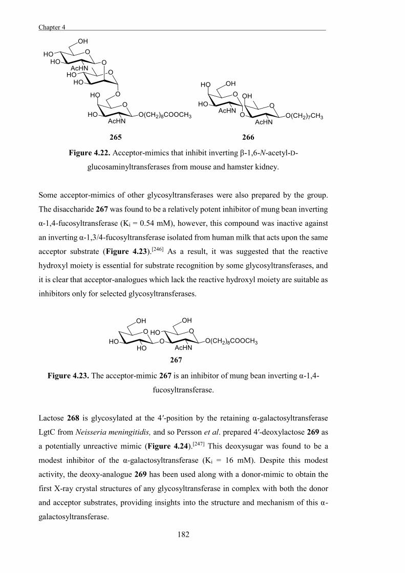
Chapter 4 .
182
Figure 4.22. Acceptor-mimics that inhibit inverting β-1,6-N-acetyl-D-
glucosaminyltransferases from mouse and hamster kidney.
Some acceptor-mimics of other glycosyltransferases were also prepared by the group.
The disaccharide 267 was found to be a relatively potent inhibitor of mung bean inverting
α-1,4-fucosyltransferase (Ki = 0.54 mM), however, this compound was inactive against
an inverting α-1,3/4-fucosyltransferase isolated from human milk that acts upon the same
acceptor substrate (Figure 4.23).[246] As a result, it was suggested that the reactive
hydroxyl moiety is essential for substrate recognition by some glycosyltransferases, and
it is clear that acceptor-analogues which lack the reactive hydroxyl moiety are suitable as
inhibitors only for selected glycosyltransferases.
Figure 4.23. The acceptor-mimic 267 is an inhibitor of mung bean inverting α-1,4-
fucosyltransferase.
Lactose 268 is glycosylated at the 4′-position by the retaining α-galactosyltransferase
LgtC from Neisseria meningitidis, and so Persson et al. prepared 4′-deoxylactose 269 as
a potentially unreactive mimic (Figure 4.24).[247] This deoxysugar was found to be a
modest inhibitor of the α-galactosyltransferase (Ki = 16 mM). Despite this modest
activity, the deoxy-analogue 269 has been used along with a donor-mimic to obtain the
first X-ray crystal structures of any glycosyltransferase in complex with both the donor
and acceptor substrates, providing insights into the structure and mechanism of this α-
galactosyltransferase.
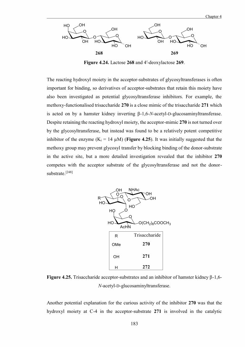
Chapter 4
183
Figure 4.24. Lactose 268 and 4′-deoxylactose 269.
The reacting hydroxyl moiety in the acceptor-substrates of glycosyltransferases is often
important for binding, so derivatives of acceptor-substrates that retain this moiety have
also been investigated as potential glycosyltransferase inhibitors. For example, the
methoxy-functionalised trisaccharide 270 is a close mimic of the trisaccharide 271 which
is acted on by a hamster kidney inverting β-1,6-N-acetyl-D-glucosaminyltransferase.
Despite retaining the reacting hydroxyl moiety, the acceptor-mimic 270 is not turned over
by the glycosyltransferase, but instead was found to be a relatively potent competitive
inhibitor of the enzyme (Ki = 14 µM) (Figure 4.25). It was initially suggested that the
methoxy group may prevent glycosyl transfer by blocking binding of the donor-substrate
in the active site, but a more detailed investigation revealed that the inhibitor 270
competes with the acceptor substrate of the glycosyltransferase and not the donor-
substrate.[248]
Figure 4.25. Trisaccharide acceptor-substrates and an inhibitor of hamster kidney β-1,6-
N-acetyl-D-glucosaminyltransferase.
Another potential explanation for the curious activity of the inhibitor 270 was that the
hydroxyl moiety at C-4 in the acceptor-substrate 271 is involved in the catalytic
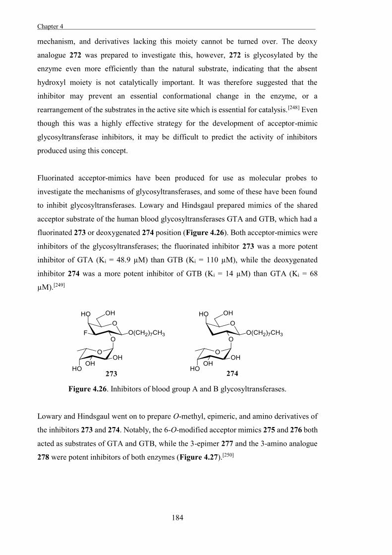
Chapter 4 .
184
mechanism, and derivatives lacking this moiety cannot be turned over. The deoxy
analogue 272 was prepared to investigate this, however, 272 is glycosylated by the
enzyme even more efficiently than the natural substrate, indicating that the absent
hydroxyl moiety is not catalytically important. It was therefore suggested that the
inhibitor may prevent an essential conformational change in the enzyme, or a
rearrangement of the substrates in the active site which is essential for catalysis.[248] Even
though this was a highly effective strategy for the development of acceptor-mimic
glycosyltransferase inhibitors, it may be difficult to predict the activity of inhibitors
produced using this concept.
Fluorinated acceptor-mimics have been produced for use as molecular probes to
investigate the mechanisms of glycosyltransferases, and some of these have been found
to inhibit glycosyltransferases. Lowary and Hindsgaul prepared mimics of the shared
acceptor substrate of the human blood glycosyltransferases GTA and GTB, which had a
fluorinated 273 or deoxygenated 274 position (Figure 4.26). Both acceptor-mimics were
inhibitors of the glycosyltransferases; the fluorinated inhibitor 273 was a more potent
inhibitor of GTA (Ki = 48.9 µM) than GTB (Ki = 110 µM), while the deoxygenated
inhibitor 274 was a more potent inhibitor of GTB (Ki = 14 µM) than GTA (Ki = 68
µM).[249]
Figure 4.26. Inhibitors of blood group A and B glycosyltransferases.
Lowary and Hindsgaul went on to prepare O-methyl, epimeric, and amino derivatives of
the inhibitors 273 and 274. Notably, the 6-O-modified acceptor mimics 275 and 276 both
acted as substrates of GTA and GTB, while the 3-epimer 277 and the 3-amino analogue
278 were potent inhibitors of both enzymes (Figure 4.27).[250]
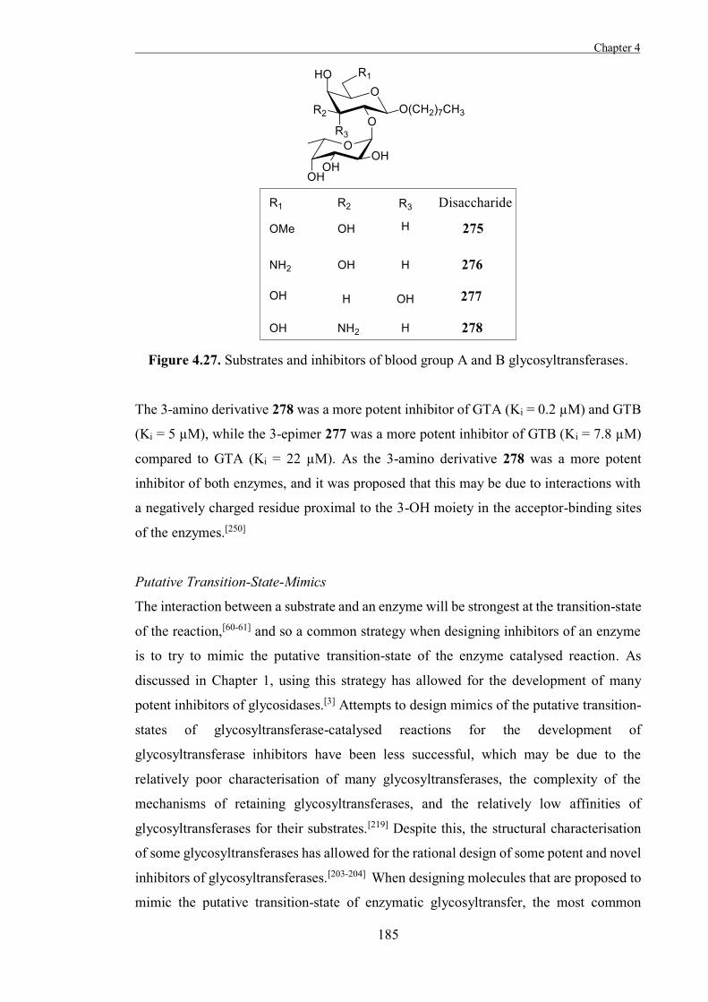
Chapter 4
185
Figure 4.27. Substrates and inhibitors of blood group A and B glycosyltransferases.
The 3-amino derivative 278 was a more potent inhibitor of GTA (Ki = 0.2 µM) and GTB
(Ki = 5 µM), while the 3-epimer 277 was a more potent inhibitor of GTB (Ki = 7.8 µM)
compared to GTA (Ki = 22 µM). As the 3-amino derivative 278 was a more potent
inhibitor of both enzymes, and it was proposed that this may be due to interactions with
a negatively charged residue proximal to the 3-OH moiety in the acceptor-binding sites
of the enzymes.[250]
Putative Transition-State-Mimics
The interaction between a substrate and an enzyme will be strongest at the transition-state
of the reaction,[60-61] and so a common strategy when designing inhibitors of an enzyme
is to try to mimic the putative transition-state of the enzyme catalysed reaction. As
discussed in Chapter 1, using this strategy has allowed for the development of many
potent inhibitors of glycosidases.[3] Attempts to design mimics of the putative transition-
states of glycosyltransferase-catalysed reactions for the development of
glycosyltransferase inhibitors have been less successful, which may be due to the
relatively poor characterisation of many glycosyltransferases, the complexity of the
mechanisms of retaining glycosyltransferases, and the relatively low affinities of
glycosyltransferases for their substrates.[219] Despite this, the structural characterisation
of some glycosyltransferases has allowed for the rational design of some potent and novel
inhibitors of glycosyltransferases.[203-204] When designing molecules that are proposed to
mimic the putative transition-state of enzymatic glycosyltransfer, the most common
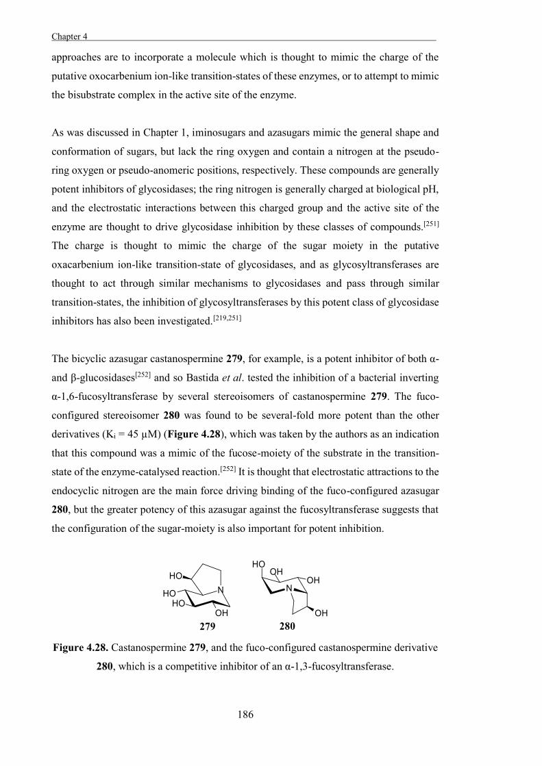
Chapter 4 .
186
approaches are to incorporate a molecule which is thought to mimic the charge of the
putative oxocarbenium ion-like transition-states of these enzymes, or to attempt to mimic
the bisubstrate complex in the active site of the enzyme.
As was discussed in Chapter 1, iminosugars and azasugars mimic the general shape and
conformation of sugars, but lack the ring oxygen and contain a nitrogen at the pseudo-
ring oxygen or pseudo-anomeric positions, respectively. These compounds are generally
potent inhibitors of glycosidases; the ring nitrogen is generally charged at biological pH,
and the electrostatic interactions between this charged group and the active site of the
enzyme are thought to drive glycosidase inhibition by these classes of compounds.[251]
The charge is thought to mimic the charge of the sugar moiety in the putative
oxacarbenium ion-like transition-state of glycosidases, and as glycosyltransferases are
thought to act through similar mechanisms to glycosidases and pass through similar
transition-states, the inhibition of glycosyltransferases by this potent class of glycosidase
inhibitors has also been investigated.[219,251]
The bicyclic azasugar castanospermine 279, for example, is a potent inhibitor of both α-
and β-glucosidases[252] and so Bastida et al. tested the inhibition of a bacterial inverting
α-1,6-fucosyltransferase by several stereoisomers of castanospermine 279. The fuco-
configured stereoisomer 280 was found to be several-fold more potent than the other
derivatives (Ki = 45 µM) (Figure 4.28), which was taken by the authors as an indication
that this compound was a mimic of the fucose-moiety of the substrate in the transition-
state of the enzyme-catalysed reaction.[252] It is thought that electrostatic attractions to the
endocyclic nitrogen are the main force driving binding of the fuco-configured azasugar
280, but the greater potency of this azasugar against the fucosyltransferase suggests that
the configuration of the sugar-moiety is also important for potent inhibition.
Figure 4.28. Castanospermine 279, and the fuco-configured castanospermine derivative
280, which is a competitive inhibitor of an α-1,3-fucosyltransferase.
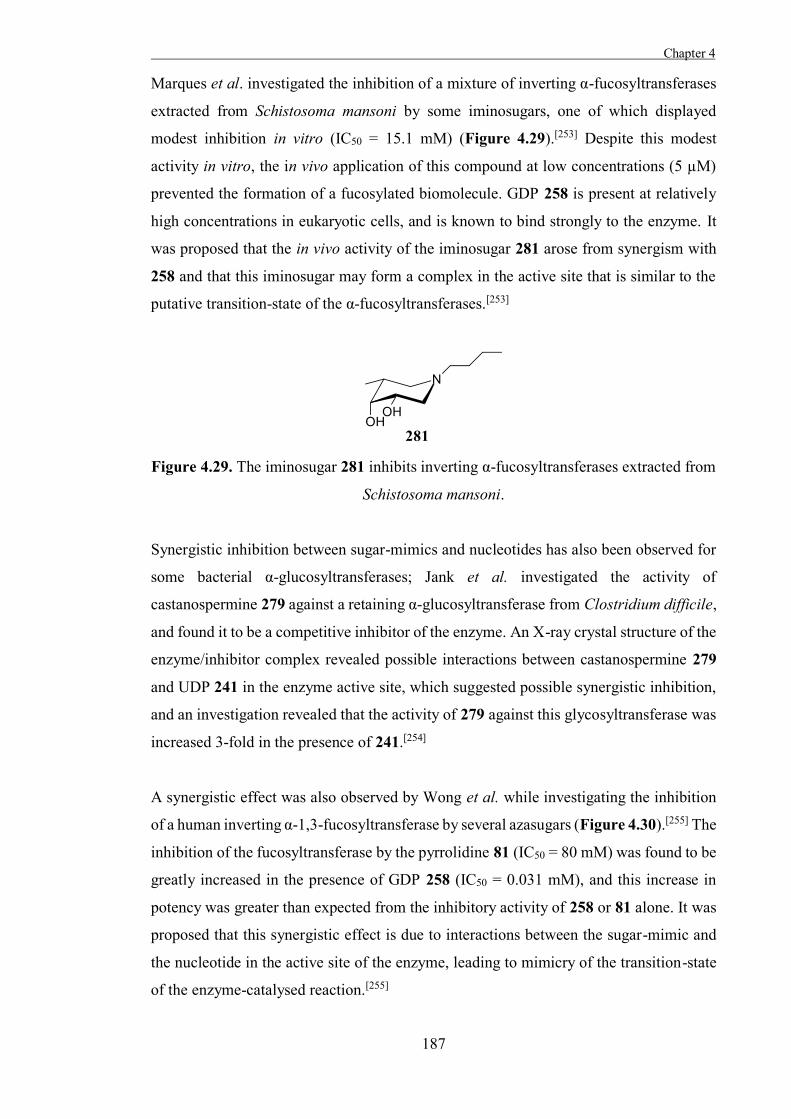
Chapter 4
187
Marques et al. investigated the inhibition of a mixture of inverting α-fucosyltransferases
extracted from Schistosoma mansoni by some iminosugars, one of which displayed
modest inhibition in vitro (IC50 = 15.1 mM) (Figure 4.29).[253] Despite this modest
activity in vitro, the in vivo application of this compound at low concentrations (5 µM)
prevented the formation of a fucosylated biomolecule. GDP 258 is present at relatively
high concentrations in eukaryotic cells, and is known to bind strongly to the enzyme. It
was proposed that the in vivo activity of the iminosugar 281 arose from synergism with
258 and that this iminosugar may form a complex in the active site that is similar to the
putative transition-state of the α-fucosyltransferases.[253]
Figure 4.29. The iminosugar 281 inhibits inverting α-fucosyltransferases extracted from
Schistosoma mansoni.
Synergistic inhibition between sugar-mimics and nucleotides has also been observed for
some bacterial α-glucosyltransferases; Jank et al. investigated the activity of
castanospermine 279 against a retaining α-glucosyltransferase from Clostridium difficile,
and found it to be a competitive inhibitor of the enzyme. An X-ray crystal structure of the
enzyme/inhibitor complex revealed possible interactions between castanospermine 279
and UDP 241 in the enzyme active site, which suggested possible synergistic inhibition,
and an investigation revealed that the activity of 279 against this glycosyltransferase was
increased 3-fold in the presence of 241.[254]
A synergistic effect was also observed by Wong et al. while investigating the inhibition
of a human inverting α-1,3-fucosyltransferase by several azasugars (Figure 4.30).[255] The
inhibition of the fucosyltransferase by the pyrrolidine 81 (IC50 = 80 mM) was found to be
greatly increased in the presence of GDP 258 (IC50 = 0.031 mM), and this increase in
potency was greater than expected from the inhibitory activity of 258 or 81 alone. It was
proposed that this synergistic effect is due to interactions between the sugar-mimic and
the nucleotide in the active site of the enzyme, leading to mimicry of the transition-state
of the enzyme-catalysed reaction.[255]
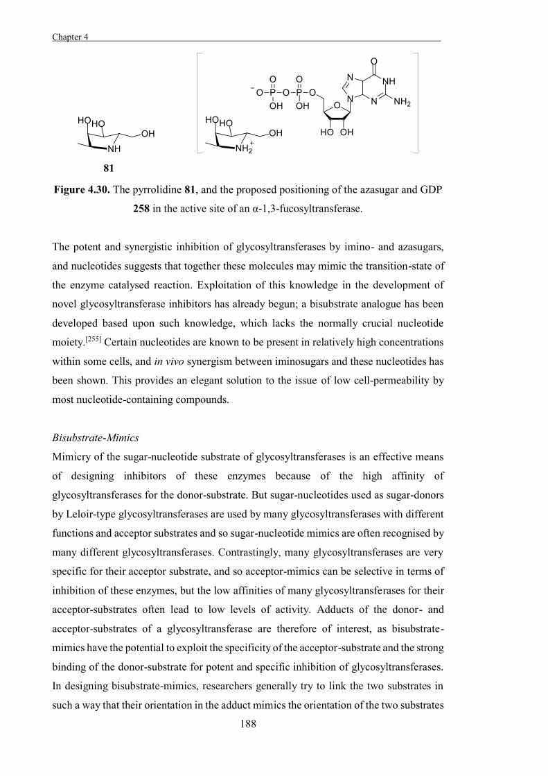
Chapter 4 .
188
Figure 4.30. The pyrrolidine 81, and the proposed positioning of the azasugar and GDP
258 in the active site of an α-1,3-fucosyltransferase.
The potent and synergistic inhibition of glycosyltransferases by imino- and azasugars,
and nucleotides suggests that together these molecules may mimic the transition-state of
the enzyme catalysed reaction. Exploitation of this knowledge in the development of
novel glycosyltransferase inhibitors has already begun; a bisubstrate analogue has been
developed based upon such knowledge, which lacks the normally crucial nucleotide
moiety.[255] Certain nucleotides are known to be present in relatively high concentrations
within some cells, and in vivo synergism between iminosugars and these nucleotides has
been shown. This provides an elegant solution to the issue of low cell-permeability by
most nucleotide-containing compounds.
Bisubstrate-Mimics
Mimicry of the sugar-nucleotide substrate of glycosyltransferases is an effective means
of designing inhibitors of these enzymes because of the high affinity of
glycosyltransferases for the donor-substrate. But sugar-nucleotides used as sugar-donors
by Leloir-type glycosyltransferases are used by many glycosyltransferases with different
functions and acceptor substrates and so sugar-nucleotide mimics are often recognised by
many different glycosyltransferases. Contrastingly, many glycosyltransferases are very
specific for their acceptor substrate, and so acceptor-mimics can be selective in terms of
inhibition of these enzymes, but the low affinities of many glycosyltransferases for their
acceptor-substrates often lead to low levels of activity. Adducts of the donor- and
acceptor-substrates of a glycosyltransferase are therefore of interest, as bisubstrate-
mimics have the potential to exploit the specificity of the acceptor-substrate and the strong
binding of the donor-substrate for potent and specific inhibition of glycosyltransferases.
In designing bisubstrate-mimics, researchers generally try to link the two substrates in
such a way that their orientation in the adduct mimics the orientation of the two substrates
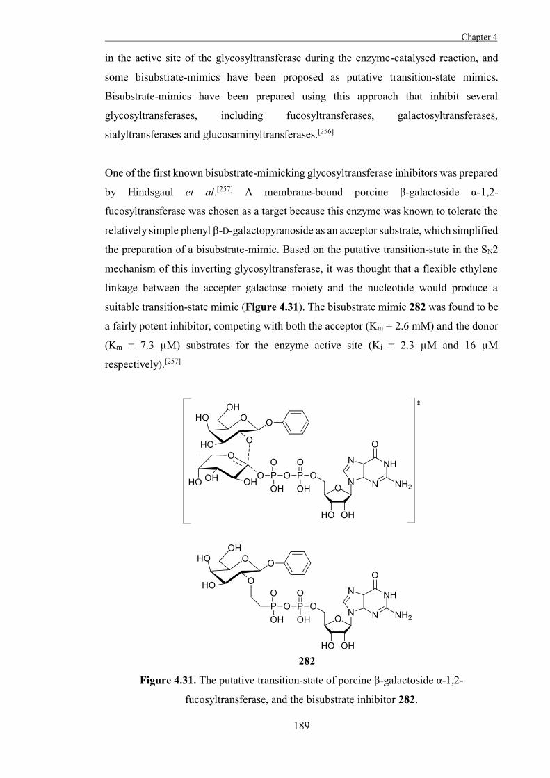
Chapter 4
189
in the active site of the glycosyltransferase during the enzyme-catalysed reaction, and
some bisubstrate-mimics have been proposed as putative transition-state mimics.
Bisubstrate-mimics have been prepared using this approach that inhibit several
glycosyltransferases, including fucosyltransferases, galactosyltransferases,
sialyltransferases and glucosaminyltransferases.[256]
One of the first known bisubstrate-mimicking glycosyltransferase inhibitors was prepared
by Hindsgaul et al.[257] A membrane-bound porcine β-galactoside α-1,2-
fucosyltransferase was chosen as a target because this enzyme was known to tolerate the
relatively simple phenyl β-D-galactopyranoside as an acceptor substrate, which simplified
the preparation of a bisubstrate-mimic. Based on the putative transition-state in the SN2
mechanism of this inverting glycosyltransferase, it was thought that a flexible ethylene
linkage between the accepter galactose moiety and the nucleotide would produce a
suitable transition-state mimic (Figure 4.31). The bisubstrate mimic 282 was found to be
a fairly potent inhibitor, competing with both the acceptor (Km = 2.6 mM) and the donor
(Km = 7.3 µM) substrates for the enzyme active site (Ki = 2.3 µM and 16 µM
respectively).[257]
Figure 4.31. The putative transition-state of porcine β-galactoside α-1,2-
fucosyltransferase, and the bisubstrate inhibitor 282.
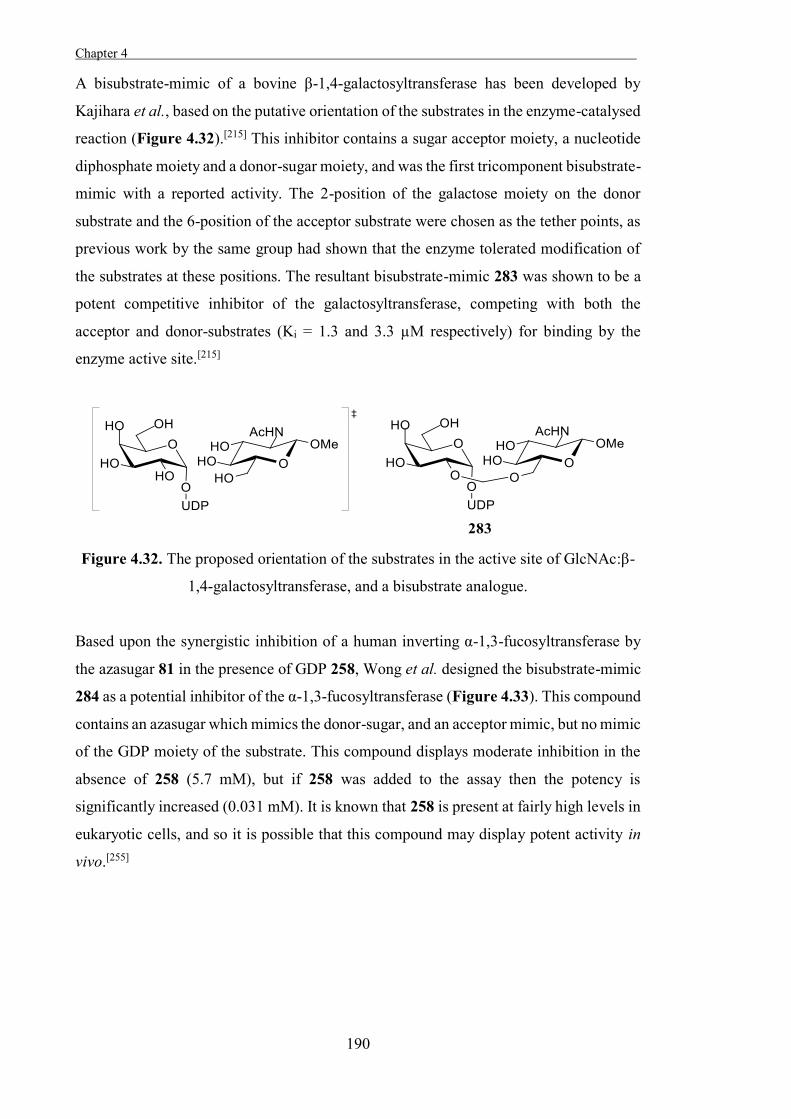
Chapter 4 .
190
A bisubstrate-mimic of a bovine β-1,4-galactosyltransferase has been developed by
Kajihara et al., based on the putative orientation of the substrates in the enzyme-catalysed
reaction (Figure 4.32).[215] This inhibitor contains a sugar acceptor moiety, a nucleotide
diphosphate moiety and a donor-sugar moiety, and was the first tricomponent bisubstrate-
mimic with a reported activity. The 2-position of the galactose moiety on the donor
substrate and the 6-position of the acceptor substrate were chosen as the tether points, as
previous work by the same group had shown that the enzyme tolerated modification of
the substrates at these positions. The resultant bisubstrate-mimic 283 was shown to be a
potent competitive inhibitor of the galactosyltransferase, competing with both the
acceptor and donor-substrates (Ki = 1.3 and 3.3 µM respectively) for binding by the
enzyme active site.[215]
Figure 4.32. The proposed orientation of the substrates in the active site of GlcNAc:β-
1,4-galactosyltransferase, and a bisubstrate analogue.
Based upon the synergistic inhibition of a human inverting α-1,3-fucosyltransferase by
the azasugar 81 in the presence of GDP 258, Wong et al. designed the bisubstrate-mimic
284 as a potential inhibitor of the α-1,3-fucosyltransferase (Figure 4.33). This compound
contains an azasugar which mimics the donor-sugar, and an acceptor mimic, but no mimic
of the GDP moiety of the substrate. This compound displays moderate inhibition in the
absence of 258 (5.7 mM), but if 258 was added to the assay then the potency is
significantly increased (0.031 mM). It is known that 258 is present at fairly high levels in
eukaryotic cells, and so it is possible that this compound may display potent activity in
vivo.[255]
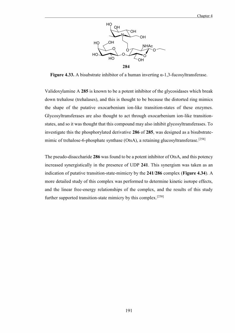
Chapter 4
191
Figure 4.33. A bisubstrate inhibitor of a human inverting α-1,3-fucosyltransferase.
Validoxylamine A 285 is known to be a potent inhibitor of the glycosidases which break
down trehalose (trehalases), and this is thought to be because the distorted ring mimics
the shape of the putative oxocarbenium ion-like transition-states of these enzymes.
Glycosyltransferases are also thought to act through oxocarbenium ion-like transition-
states, and so it was thought that this compound may also inhibit glycosyltransferases. To
investigate this the phosphorylated derivative 286 of 285, was designed as a bisubstrate-
mimic of trehalose-6-phosphate synthase (OtsA), a retaining glucosyltransferase.[258]
The pseudo-disaccharide 286 was found to be a potent inhibitor of OtsA, and this potency
increased synergistically in the presence of UDP 241. This synergism was taken as an
indication of putative transition-state-mimicry by the 241/286 complex (Figure 4.34). A
more detailed study of this complex was performed to determine kinetic isotope effects,
and the linear free-energy relationships of the complex, and the results of this study
further supported transition-state mimicry by this complex.[258]
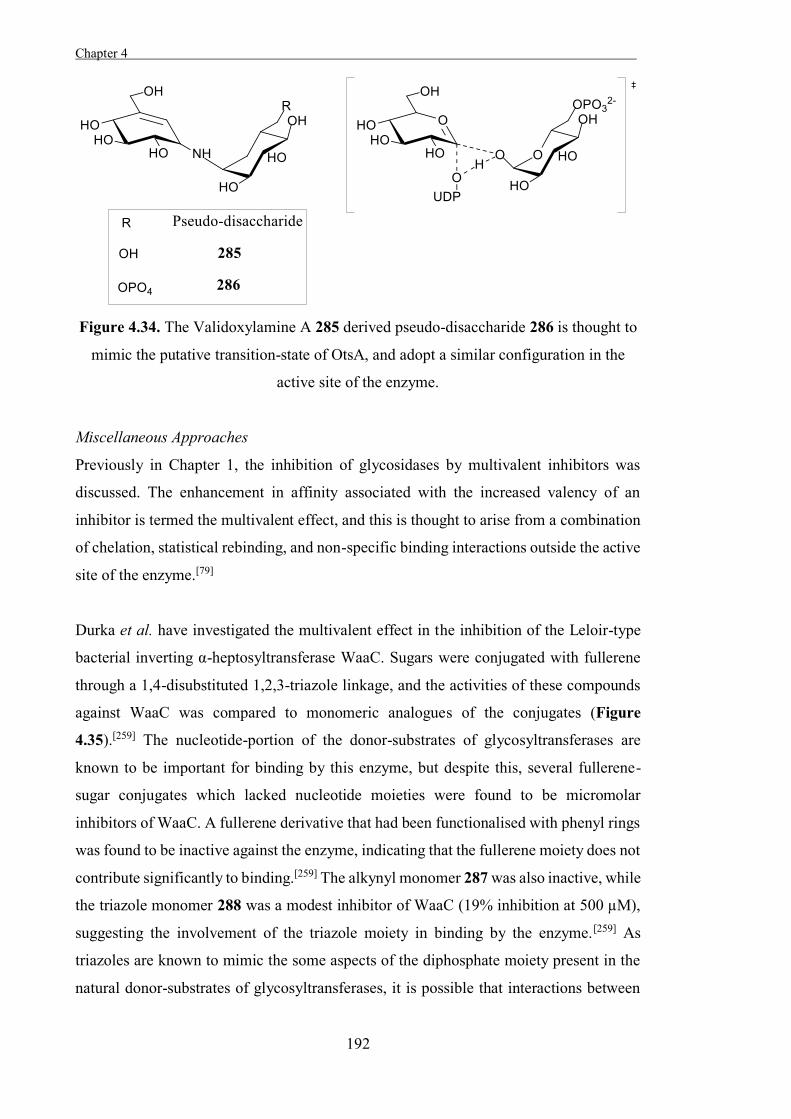
Chapter 4 .
192
Figure 4.34. The Validoxylamine A 285 derived pseudo-disaccharide 286 is thought to
mimic the putative transition-state of OtsA, and adopt a similar configuration in the
active site of the enzyme.
Miscellaneous Approaches
Previously in Chapter 1, the inhibition of glycosidases by multivalent inhibitors was
discussed. The enhancement in affinity associated with the increased valency of an
inhibitor is termed the multivalent effect, and this is thought to arise from a combination
of chelation, statistical rebinding, and non-specific binding interactions outside the active
site of the enzyme.[79]
Durka et al. have investigated the multivalent effect in the inhibition of the Leloir-type
bacterial inverting α-heptosyltransferase WaaC. Sugars were conjugated with fullerene
through a 1,4-disubstituted 1,2,3-triazole linkage, and the activities of these compounds
against WaaC was compared to monomeric analogues of the conjugates (Figure
4.35).[259] The nucleotide-portion of the donor-substrates of glycosyltransferases are
known to be important for binding by this enzyme, but despite this, several fullerene-
sugar conjugates which lacked nucleotide moieties were found to be micromolar
inhibitors of WaaC. A fullerene derivative that had been functionalised with phenyl rings
was found to be inactive against the enzyme, indicating that the fullerene moiety does not
contribute significantly to binding.[259] The alkynyl monomer 287 was also inactive, while
the triazole monomer 288 was a modest inhibitor of WaaC (19% inhibition at 500 µM),
suggesting the involvement of the triazole moiety in binding by the enzyme.[259] As
triazoles are known to mimic the some aspects of the diphosphate moiety present in the
natural donor-substrates of glycosyltransferases, it is possible that interactions between
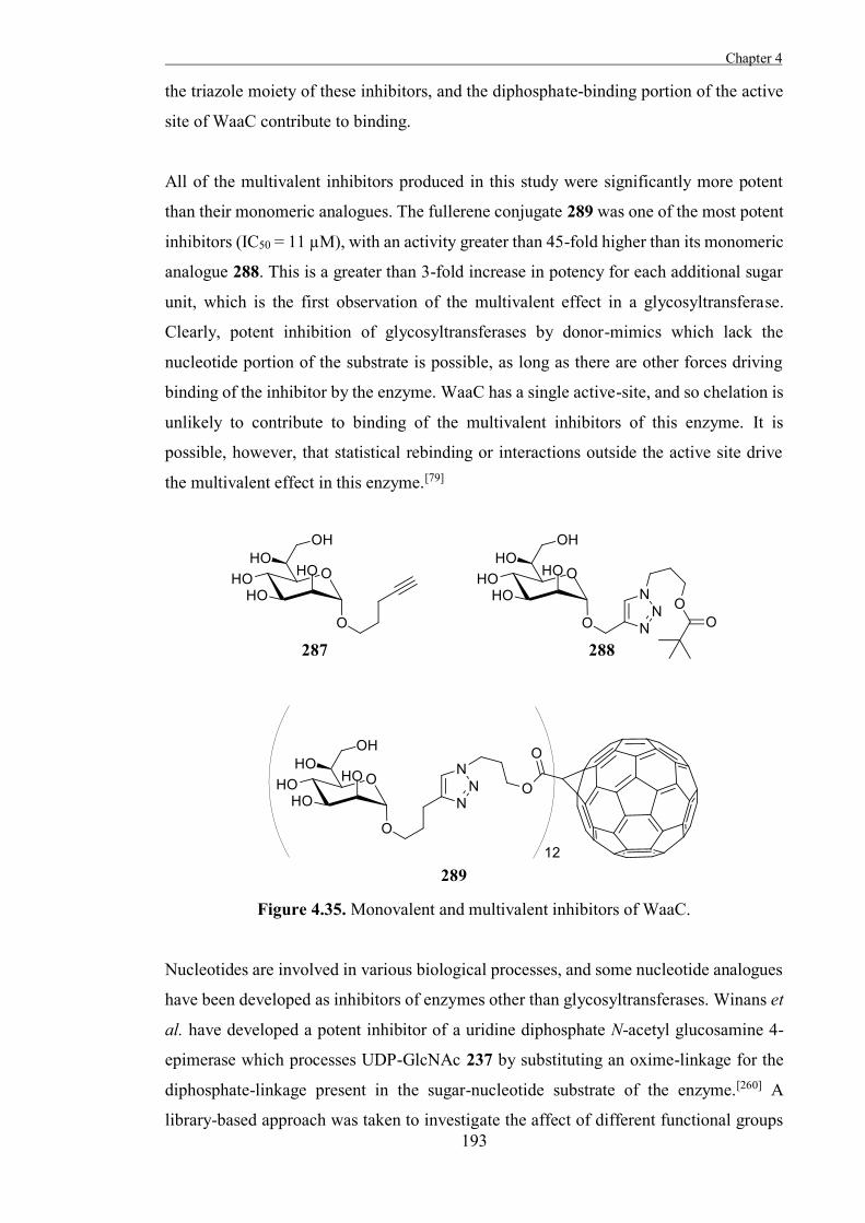
Chapter 4
193
the triazole moiety of these inhibitors, and the diphosphate-binding portion of the active
site of WaaC contribute to binding.
All of the multivalent inhibitors produced in this study were significantly more potent
than their monomeric analogues. The fullerene conjugate 289 was one of the most potent
inhibitors (IC50 = 11 µM), with an activity greater than 45-fold higher than its monomeric
analogue 288. This is a greater than 3-fold increase in potency for each additional sugar
unit, which is the first observation of the multivalent effect in a glycosyltransferase.
Clearly, potent inhibition of glycosyltransferases by donor-mimics which lack the
nucleotide portion of the substrate is possible, as long as there are other forces driving
binding of the inhibitor by the enzyme. WaaC has a single active-site, and so chelation is
unlikely to contribute to binding of the multivalent inhibitors of this enzyme. It is
possible, however, that statistical rebinding or interactions outside the active site drive
the multivalent effect in this enzyme.[79]
Figure 4.35. Monovalent and multivalent inhibitors of WaaC.
Nucleotides are involved in various biological processes, and some nucleotide analogues
have been developed as inhibitors of enzymes other than glycosyltransferases. Winans et
al. have developed a potent inhibitor of a uridine diphosphate N-acetyl glucosamine 4-
epimerase which processes UDP-GlcNAc 237 by substituting an oxime-linkage for the
diphosphate-linkage present in the sugar-nucleotide substrate of the enzyme.[260] A
library-based approach was taken to investigate the affect of different functional groups
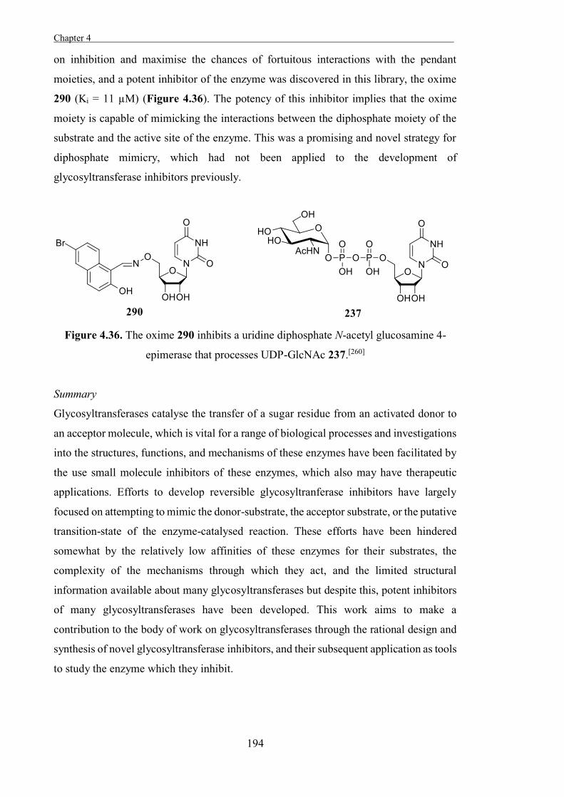
Chapter 4 .
194
on inhibition and maximise the chances of fortuitous interactions with the pendant
moieties, and a potent inhibitor of the enzyme was discovered in this library, the oxime
290 (Ki = 11 µM) (Figure 4.36). The potency of this inhibitor implies that the oxime
moiety is capable of mimicking the interactions between the diphosphate moiety of the
substrate and the active site of the enzyme. This was a promising and novel strategy for
diphosphate mimicry, which had not been applied to the development of
glycosyltransferase inhibitors previously.
Figure 4.36. The oxime 290 inhibits a uridine diphosphate N-acetyl glucosamine 4-
epimerase that processes UDP-GlcNAc 237.[260]
Summary
Glycosyltransferases catalyse the transfer of a sugar residue from an activated donor to
an acceptor molecule, which is vital for a range of biological processes and investigations
into the structures, functions, and mechanisms of these enzymes have been facilitated by
the use small molecule inhibitors of these enzymes, which also may have therapeutic
applications. Efforts to develop reversible glycosyltranferase inhibitors have largely
focused on attempting to mimic the donor-substrate, the acceptor substrate, or the putative
transition-state of the enzyme-catalysed reaction. These efforts have been hindered
somewhat by the relatively low affinities of these enzymes for their substrates, the
complexity of the mechanisms through which they act, and the limited structural
information available about many glycosyltransferases but despite this, potent inhibitors
of many glycosyltransferases have been developed. This work aims to make a
contribution to the body of work on glycosyltransferases through the rational design and
synthesis of novel glycosyltransferase inhibitors, and their subsequent application as tools
to study the enzyme which they inhibit.

195

196
197
Chapter 5
The Development of Some Novel Cholesterol α-
glucosyltransferase Inhibitors

198
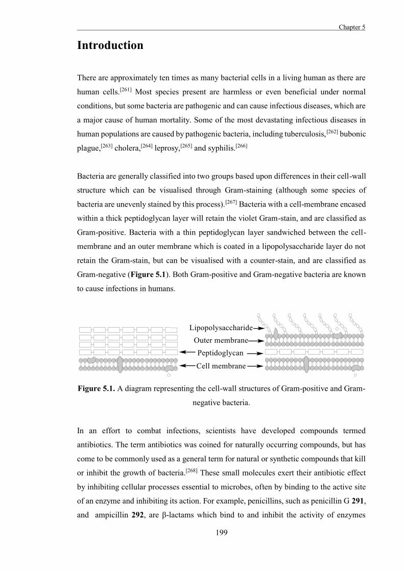
Chapter 5
199
Introduction
There are approximately ten times as many bacterial cells in a living human as there are
human cells.[261] Most species present are harmless or even beneficial under normal
conditions, but some bacteria are pathogenic and can cause infectious diseases, which are
a major cause of human mortality. Some of the most devastating infectious diseases in
human populations are caused by pathogenic bacteria, including tuberculosis,[262] bubonic
plague,[263] cholera,[264] leprosy,[265] and syphilis.[266]
Bacteria are generally classified into two groups based upon differences in their cell-wall
structure which can be visualised through Gram-staining (although some species of
bacteria are unevenly stained by this process).[267] Bacteria with a cell-membrane encased
within a thick peptidoglycan layer will retain the violet Gram-stain, and are classified as
Gram-positive. Bacteria with a thin peptidoglycan layer sandwiched between the cell-
membrane and an outer membrane which is coated in a lipopolysaccharide layer do not
retain the Gram-stain, but can be visualised with a counter-stain, and are classified as
Gram-negative (Figure 5.1). Both Gram-positive and Gram-negative bacteria are known
to cause infections in humans.
Figure 5.1. A diagram representing the cell-wall structures of Gram-positive and Gram-
negative bacteria.
In an effort to combat infections, scientists have developed compounds termed
antibiotics. The term antibiotics was coined for naturally occurring compounds, but has
come to be commonly used as a general term for natural or synthetic compounds that kill
or inhibit the growth of bacteria.[268] These small molecules exert their antibiotic effect
by inhibiting cellular processes essential to microbes, often by binding to the active site
of an enzyme and inhibiting its action. For example, penicillins, such as penicillin G 291,
and ampicillin 292, are β-lactams which bind to and inhibit the activity of enzymes
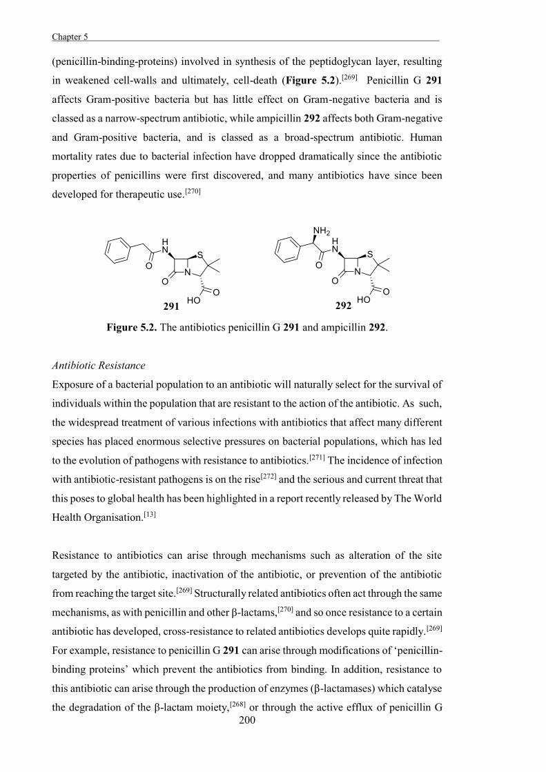
Chapter 5 .
200
(penicillin-binding-proteins) involved in synthesis of the peptidoglycan layer, resulting
in weakened cell-walls and ultimately, cell-death (Figure 5.2).[269] Penicillin G 291
affects Gram-positive bacteria but has little effect on Gram-negative bacteria and is
classed as a narrow-spectrum antibiotic, while ampicillin 292 affects both Gram-negative
and Gram-positive bacteria, and is classed as a broad-spectrum antibiotic. Human
mortality rates due to bacterial infection have dropped dramatically since the antibiotic
properties of penicillins were first discovered, and many antibiotics have since been
developed for therapeutic use.[270]
Figure 5.2. The antibiotics penicillin G 291 and ampicillin 292.
Antibiotic Resistance
Exposure of a bacterial population to an antibiotic will naturally select for the survival of
individuals within the population that are resistant to the action of the antibiotic. As such,
the widespread treatment of various infections with antibiotics that affect many different
species has placed enormous selective pressures on bacterial populations, which has led
to the evolution of pathogens with resistance to antibiotics.[271] The incidence of infection
with antibiotic-resistant pathogens is on the rise[272] and the serious and current threat that
this poses to global health has been highlighted in a report recently released by The World
Health Organisation.[13]
Resistance to antibiotics can arise through mechanisms such as alteration of the site
targeted by the antibiotic, inactivation of the antibiotic, or prevention of the antibiotic
from reaching the target site.[269] Structurally related antibiotics often act through the same
mechanisms, as with penicillin and other β-lactams,[270] and so once resistance to a certain
antibiotic has developed, cross-resistance to related antibiotics develops quite rapidly.[269]
For example, resistance to penicillin G 291 can arise through modifications of ‘penicillin-
binding proteins’ which prevent the antibiotics from binding. In addition, resistance to
this antibiotic can arise through the production of enzymes (β-lactamases) which catalyse
the degradation of the β-lactam moiety,[268] or through the active efflux of penicillin G
Chapter 5
201
from bacterial cells.[273] Pathogens which display resistance to penicillin G 291 are
generally also resistant to the activity of other penicillins, such as ampicillin 292, because
of their shared structural features and common target-site.[271,274] Resistance mechanisms
can also be transferred between organisms through horizontal gene transfer, which allows
for resistance to be rapidly acquired by unrelated bacterium once it has arisen in one
species, further increasing the rate at which resistance can develop and spread.[269]
Overcoming Antibiotic Resistance
Antibiotic resistance is not a new phenomenon; resistance to penicillins was observed
within a few years of the discovery of their antibiotic properties, and rapidly became
widespread.[268] This was circumvented by the introduction of broad-spectrum macrolide
antibiotics which act through a different mechanism to penicillins, until resistance to
macrolides also became widespread among bacterial populations. For example, a
macrolide antibiotic began being used as an alternative to penicillins for the treatment of
Staphylococcus aureus in Boston city hospital in 1950, and within a year, 70% of isolates
of S. aureus from within the hospital exhibited resistance to the macrolide.[268] Controlled
treatment strategies and restrictions on the application of antibiotics have slowed the
development of antibiotic resistance somewhat by reducing the exposure of bacterial
populations to certain antibiotics, but this cycle of development of antibiotic resistance
by pathogens and the development of novel antibiotics to overcome this resistance has
continued through the middle part of the 20th century. As such, overcoming antibiotic
resistance has been an ongoing campaign requiring the development of novel antibiotics
before resistance to those currently in use can become widespread. Despite this, only a
few novel classes of antibiotic were introduced in the latter half of the 20 th century,[275-
276] while resistance has continued to develop for the antibiotics in use, with the end result
being that the pathogens are catching up.
One major problem with antibiotic development is that although many antibiotics are
known, they are based upon relatively few chemical scaffolds, and clinically significant
resistance has developed to all therapeutically applied antibiotics.[271,277] Due to emerging
antibiotic-resistant pathogens, new antibiotics with novel modes of action are required to
circumvent the growing resistance. Resistance to existing antibiotics that affect a variety
of pathogens has developed quite rapidly, however, resistance would potentially develop
more slowly to antibiotics which target a specific pathogen. The total size of the bacterial
population affected by these antibiotics will be smaller than that affected by broad-
Chapter 5 .
202
spectrum antibiotics, which will reduce the evolutionary driving force behind the
development of resistance.
Another strategy which has received increasing attention is the targeting of virulence
mechanisms of a pathogen.[277] Virulence mechanisms in pathogens facilitate the
development of disease states and allow them to overcome the natural defences of the
host, and treating infections by inhibiting virulence mechanisms may render pathogens
vulnerable to elimination by the immune system of the host, or prevent the progression
of disease, rather than eliminating pathogens directly.[278] Pathogens would be expected
to adapt more slowly to these virulence mechanism inhibitors than to traditional
antibiotics because of their indirect mode of action. The development of virulence
mechanism inhibitors has been hindered by the difficulty of identifying virulence
mechanisms in pathogens, but some efforts in this area have shown promise, for example
a novel virulence factor has recently been identified in Helicobacter pylori.
Helicobacter pylori
H. pylori is a Gram-negative bacterial pathogen which has been colonising the gastric
mucosa of humans for over 60000 years.[279] Over half of the world’s population is
estimated to be infected with this bacterium, and this infection leads to a range of
gastrointestinal complications including chronic gastritis, peptic ulcers, and gastric
cancers.[280] Peptic ulcers were considered to be practically untreatable until work by
Warren and Marshall revealed the causal role of H. pylori in their formation and showed
that the disorder could be treated by eliminating the bacterium.[281-282] The 2005 Nobel
Prize for Medicine or Physiology was awarded for this momentous discovery, and
research on H. pylori has flourished since; there is even a peer-reviewed journal,
Helicobacter, dedicated solely to the study of this genus.[283]
Infection with H. pylori is thought to most commonly occur during childhood, although
the rate of infection increases with age in industrialised countries, and this infection can
persist for decades, if not life.[284] All individuals infected with H. pylori will develop
gastritis, an inflammation of the stomach lining, although most will not experience any
symptoms of this disorder.[285] A further 10-20% of those infected will go on to develop
peptic ulcer disease and 1-2% will develop gastric cancer, and while this is a small
proportion of those infected, H. pylori is a major cause of these diseases as a result of the
large percentage of the human population infected with the bacterium.[285-286] In order to
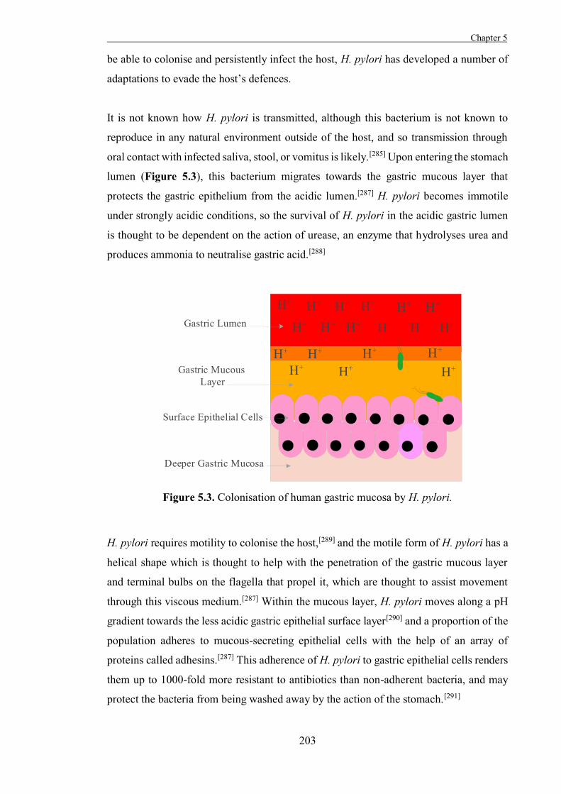
Chapter 5
203
be able to colonise and persistently infect the host, H. pylori has developed a number of
adaptations to evade the host’s defences.
It is not known how H. pylori is transmitted, although this bacterium is not known to
reproduce in any natural environment outside of the host, and so transmission through
oral contact with infected saliva, stool, or vomitus is likely.[285] Upon entering the stomach
lumen (Figure 5.3), this bacterium migrates towards the gastric mucous layer that
protects the gastric epithelium from the acidic lumen.[287] H. pylori becomes immotile
under strongly acidic conditions, so the survival of H. pylori in the acidic gastric lumen
is thought to be dependent on the action of urease, an enzyme that hydrolyses urea and
produces ammonia to neutralise gastric acid.[288]
Figure 5.3. Colonisation of human gastric mucosa by H. pylori.
H. pylori requires motility to colonise the host,[289] and the motile form of H. pylori has a
helical shape which is thought to help with the penetration of the gastric mucous layer
and terminal bulbs on the flagella that propel it, which are thought to assist movement
through this viscous medium.[287] Within the mucous layer, H. pylori moves along a pH
gradient towards the less acidic gastric epithelial surface layer[290] and a proportion of the
population adheres to mucous-secreting epithelial cells with the help of an array of
proteins called adhesins.[287] This adherence of H. pylori to gastric epithelial cells renders
them up to 1000-fold more resistant to antibiotics than non-adherent bacteria, and may
protect the bacteria from being washed away by the action of the stomach.[291]
Chapter 5 .
204
The precise mechanisms of colonisation of the gastric mucosa by H. pylori are not known,
but it is apparent that this bacterium is well adapted to the environmental niche which it
occupies within the host. Adhesins in H. pylori are known to target various moieties
presented on epithelial cells, including Lewis blood-group antigens,[292] the sialylated
glycoprotein laminin and heavily sulfated molecules.[293] The lipopolysaccharide outer
layer of this Gram-negative bacterium is also known to present antigens that mimic the
Lewis blood group antigens presented by human cells, which is known to facilitate
adhesion[291] and may also facilitate immune evasion,[294] although a relationship between
the specific antigens presented by the pathogen and those of the host has yet to be
demonstrated.[295]
H. pylori that is adhered to the epithelium is usually clustered around the junctions
between cells[287] and cholesterol-rich membrane regions such as lipid rafts,[296] and this
adherence results in degradation of the epithelial surface layer, causing loss of microvilli,
irregularity of the luminal border, and intracellular changes such as vacuolysis. [297] It is
generally considered to be rare for H. pylori to invade epithelial cells, although there is
some evidence that the presentation of Lewis blood group antigens by the bacteria
facilitates this.[285] Colonisation of the submucosa is also uncommon, however, secretions
of H. pylori do gain access to the submucosa, and these can stimulate the release of
cytokine messengers, which results in mucosal infiltration by neutrophils, monocytes and
lymphocytes, and leads to gastritis.[298] This can then develop into more serious disorders,
however, eliminating the infection halts the progression of peptic ulcer disease.[299]
The standard treatment for H. pylori infection generally involves a triple therapy using
two antibiotics with different mechanisms of action, and a proton pump inhibitor.
Commonly used antibiotics for this therapy include amoxicillin 293, a penicillin that
inhibits peptidoglycan synthesis, and clarithromycin 294, a macrolide that inhibits
bacterial protein synthesis by binding the 50S subunit of the bacterial ribosome (Figure
5.4).[274] Many drugs are inactivated by the acidic conditions of the stomach, so a proton
pump inhibitor is used to reduce this acidity and extend the active lifetime of antibiotics
in the stomach. None of the antibiotics used to treat infection with H. pylori in
combination therapies are effective alone, and even triple therapy sometimes proves
ineffective. Antibiotic resistance in H. pylori is often the cause of the failure of this
therapy.[272,274,300]
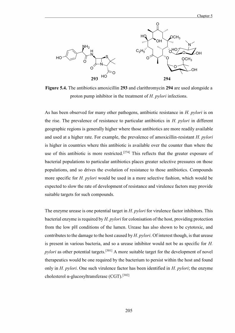
Chapter 5
205
Figure 5.4. The antibiotics amoxicillin 293 and clarithromycin 294 are used alongside a
proton pump inhibitor in the treatment of H. pylori infections.
As has been observed for many other pathogens, antibiotic resistance in H. pylori is on
the rise. The prevalence of resistance to particular antibiotics in H. pylori in different
geographic regions is generally higher where those antibiotics are more readily available
and used at a higher rate. For example, the prevalence of amoxicillin-resistant H. pylori
is higher in countries where this antibiotic is available over the counter than where the
use of this antibiotic is more restricted.[274] This reflects that the greater exposure of
bacterial populations to particular antibiotics places greater selective pressures on those
populations, and so drives the evolution of resistance to those antibiotics. Compounds
more specific for H. pylori would be used in a more selective fashion, which would be
expected to slow the rate of development of resistance and virulence factors may provide
suitable targets for such compounds.
The enzyme urease is one potential target in H. pylori for virulence factor inhibitors. This
bacterial enzyme is required by H. pylori for colonisation of the host, providing protection
from the low pH conditions of the lumen. Urease has also shown to be cytotoxic, and
contributes to the damage to the host caused by H. pylori. Of interest though, is that urease
is present in various bacteria, and so a urease inhibitor would not be as specific for H.
pylori as other potential targets.[301] A more suitable target for the development of novel
therapeutics would be one required by the bacterium to persist within the host and found
only in H. pylori. One such virulence factor has been identified in H. pylori; the enzyme
cholesterol α-glucosyltransferase (CGT).[302]
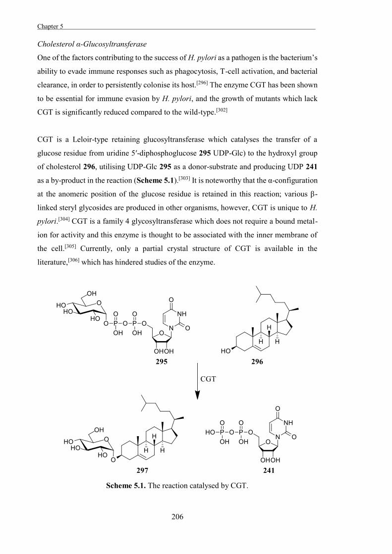
Chapter 5 .
206
Cholesterol α-Glucosyltransferase
One of the factors contributing to the success of H. pylori as a pathogen is the bacterium’s
ability to evade immune responses such as phagocytosis, T-cell activation, and bacterial
clearance, in order to persistently colonise its host.[296] The enzyme CGT has been shown
to be essential for immune evasion by H. pylori, and the growth of mutants which lack
CGT is significantly reduced compared to the wild-type.[302]
CGT is a Leloir-type retaining glucosyltransferase which catalyses the transfer of a
glucose residue from uridine 5′-diphosphoglucose 295 UDP-Glc) to the hydroxyl group
of cholesterol 296, utilising UDP-Glc 295 as a donor-substrate and producing UDP 241
as a by-product in the reaction (Scheme 5.1).[303] It is noteworthy that the α-configuration
at the anomeric position of the glucose residue is retained in this reaction; various β-
linked steryl glycosides are produced in other organisms, however, CGT is unique to H.
pylori.[304] CGT is a family 4 glycosyltransferase which does not require a bound metal-
ion for activity and this enzyme is thought to be associated with the inner membrane of
the cell.[305] Currently, only a partial crystal structure of CGT is available in the
literature,[306] which has hindered studies of the enzyme.
Scheme 5.1. The reaction catalysed by CGT.
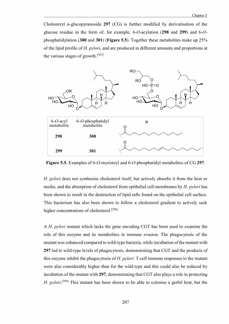
Chapter 5
207
Cholesteryl α-glucopyranoside 297 (CG) is further modified by derivatisation of the
glucose residue in the form of, for example, 6-O-acylation (298 and 299) and 6-O-
phosphatidylation (300 and 301) (Figure 5.5). Together these metabolites make up 25%
of the lipid profile of H. pylori, and are produced in different amounts and proportions at
the various stages of growth.[307]
Figure 5.5. Examples of 6-O-myristoyl and 6-O-phosphatidyl metabolites of CG 297.
H. pylori does not synthesise cholesterol itself, but actively absorbs it from the host or
media, and the absorption of cholesterol from epithelial cell-membranes by H. pylori has
been shown to result in the destruction of lipid rafts found on the epithelial cell surface.
This bacterium has also been shown to follow a cholesterol gradient to actively seek
higher concentrations of cholesterol.[296]
A H. pylori mutant which lacks the gene encoding CGT has been used to examine the
role of this enzyme and its metabolites in immune evasion. The phagocytosis of the
mutant was enhanced compared to wild-type bacteria, while incubation of the mutant with
297 led to wild-type levels of phagocytosis, demonstrating that CGT and the products of
this enzyme inhibit the phagocytosis of H. pylori. T-cell immune responses to the mutant
were also considerably higher than for the wild-type and this could also be reduced by
incubation of the mutant with 297, demonstrating that CGT also plays a role in protecting
H. pylori.[296] This mutant has been shown to be able to colonise a gerbil host, but the
Chapter 5 .
208
bacterial loading is drastically reduced compared to wild-type, suggesting that the lack of
the gene encoding CGT reduces the overall fitness of the organism.[308]
Given the roles that CGT plays in the evasion of immune responses by H. pylori, this
virulence factor is a promising target for the development of inhibitors, and because CGT
is unique to H. pylori,[304] a specific inhibitor of this enzyme would only affect H. pylori.
It is likely that CGT inhibitors would reduce the bacterial load to a level similar to that of
the mutant bacteria, and facilitate an immune response to the infection. This may permit
clearance of the infection by the immune system of the host, or increase the effectiveness
of antibiotic regimes by weakening the bacteria, and as such, small-molecule inhibitors
of CGT are desirable as potential therapeutic agents and as tools to investigate the
functions and activities of the enzyme and its metabolites.
Inhibitors of CGT
It has been observed that H. pylori rarely colonises the deeper portions of the gastric
mucosa, and several α-1,4-N-acetylglucosamine-capped (α-1,4-GlcNAc) mucin-type O-
glycans that have been isolated from the deeper gastric mucosa of humans have been
found to suppress the growth of the bacterium. It was initially suggested that these mucins
may disrupt cell-wall biosynthesis, given that exposure to them induced similar
morphological abnormalities to exposure to β-lactam antibiotics, which disrupt cell-wall
synthesis. Upon further investigation, the activity of CGT in H. pylori was found to be
suppressed by these mucins.[309]
Lee et al. assayed the activity of some of these mucin-type O-glycans against CGT in-
vitro and found that several of these oligosaccharides inhibited CGT,[310] the most potent
of which was the α-1,4-GlcNAc-capped O-glycan 302 (IC50 = 440 µM). Despite the
promising biological activities of these mucin-type O-glycans, further examination of
these molecules for therapeutic applications has been hindered by their relatively low
activity and difficult syntheses (Figure 5.6).
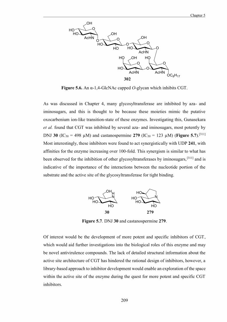
Chapter 5
209
Figure 5.6. An α-1,4-GlcNAc capped O-glycan which inhibits CGT.
As was discussed in Chapter 4, many glycosyltransferase are inhibited by aza- and
iminosugars, and this is thought to be because these moieties mimic the putative
oxocarbenium ion-like transition-state of these enzymes. Investigating this, Gunasekara
et al. found that CGT was inhibited by several aza- and iminosugars, most potently by
DNJ 30 (IC50 = 498 µM) and castanospermine 279 (IC50 = 123 µM) (Figure 5.7).[311]
Most interestingly, these inhibitors were found to act synergistically with UDP 241, with
affinities for the enzyme increasing over 100-fold. This synergism is similar to what has
been observed for the inhibition of other glycosyltransferases by iminosugars,[311] and is
indicative of the importance of the interactions between the nucleotide portion of the
substrate and the active site of the glycosyltransferase for tight binding.
Figure 5.7. DNJ 30 and castanospermine 279.
Of interest would be the development of more potent and specific inhibitors of CGT,
which would aid further investigations into the biological roles of this enzyme and may
be novel antivirulence compounds. The lack of detailed structural information about the
active site architecture of CGT has hindered the rational design of inhibitors, however, a
library-based approach to inhibitor development would enable an exploration of the space
within the active site of the enzyme during the quest for more potent and specific CGT
inhibitors.
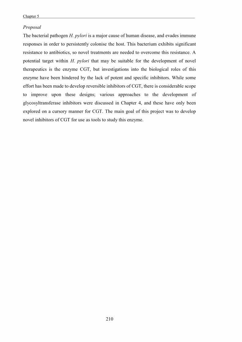
Chapter 5 .
210
Proposal
The bacterial pathogen H. pylori is a major cause of human disease, and evades immune
responses in order to persistently colonise the host. This bacterium exhibits significant
resistance to antibiotics, so novel treatments are needed to overcome this resistance. A
potential target within H. pylori that may be suitable for the development of novel
therapeutics is the enzyme CGT, but investigations into the biological roles of this
enzyme have been hindered by the lack of potent and specific inhibitors. While some
effort has been made to develop reversible inhibitors of CGT, there is considerable scope
to improve upon these designs; various approaches to the development of
glycosyltransferase inhibitors were discussed in Chapter 4, and these have only been
explored on a cursory manner for CGT. The main goal of this project was to develop
novel inhibitors of CGT for use as tools to study this enzyme.
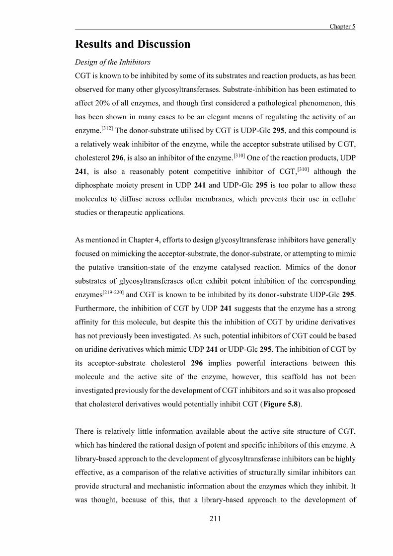
Chapter 5
211
Results and Discussion Design of the Inhibitors
CGT is known to be inhibited by some of its substrates and reaction products, as has been
observed for many other glycosyltransferases. Substrate-inhibition has been estimated to
affect 20% of all enzymes, and though first considered a pathological phenomenon, this
has been shown in many cases to be an elegant means of regulating the activity of an
enzyme.[312] The donor-substrate utilised by CGT is UDP-Glc 295, and this compound is
a relatively weak inhibitor of the enzyme, while the acceptor substrate utilised by CGT,
cholesterol 296, is also an inhibitor of the enzyme.[310] One of the reaction products, UDP
241, is also a reasonably potent competitive inhibitor of CGT,[310] although the
diphosphate moiety present in UDP 241 and UDP-Glc 295 is too polar to allow these
molecules to diffuse across cellular membranes, which prevents their use in cellular
studies or therapeutic applications.
As mentioned in Chapter 4, efforts to design glycosyltransferase inhibitors have generally
focused on mimicking the acceptor-substrate, the donor-substrate, or attempting to mimic
the putative transition-state of the enzyme catalysed reaction. Mimics of the donor
substrates of glycosyltransferases often exhibit potent inhibition of the corresponding
enzymes[219-220] and CGT is known to be inhibited by its donor-substrate UDP-Glc 295.
Furthermore, the inhibition of CGT by UDP 241 suggests that the enzyme has a strong
affinity for this molecule, but despite this the inhibition of CGT by uridine derivatives
has not previously been investigated. As such, potential inhibitors of CGT could be based
on uridine derivatives which mimic UDP 241 or UDP-Glc 295. The inhibition of CGT by
its acceptor-substrate cholesterol 296 implies powerful interactions between this
molecule and the active site of the enzyme, however, this scaffold has not been
investigated previously for the development of CGT inhibitors and so it was also proposed
that cholesterol derivatives would potentially inhibit CGT (Figure 5.8).
There is relatively little information available about the active site structure of CGT,
which has hindered the rational design of potent and specific inhibitors of this enzyme. A
library-based approach to the development of glycosyltransferase inhibitors can be highly
effective, as a comparison of the relative activities of structurally similar inhibitors can
provide structural and mechanistic information about the enzymes which they inhibit. It
was thought, because of this, that a library-based approach to the development of
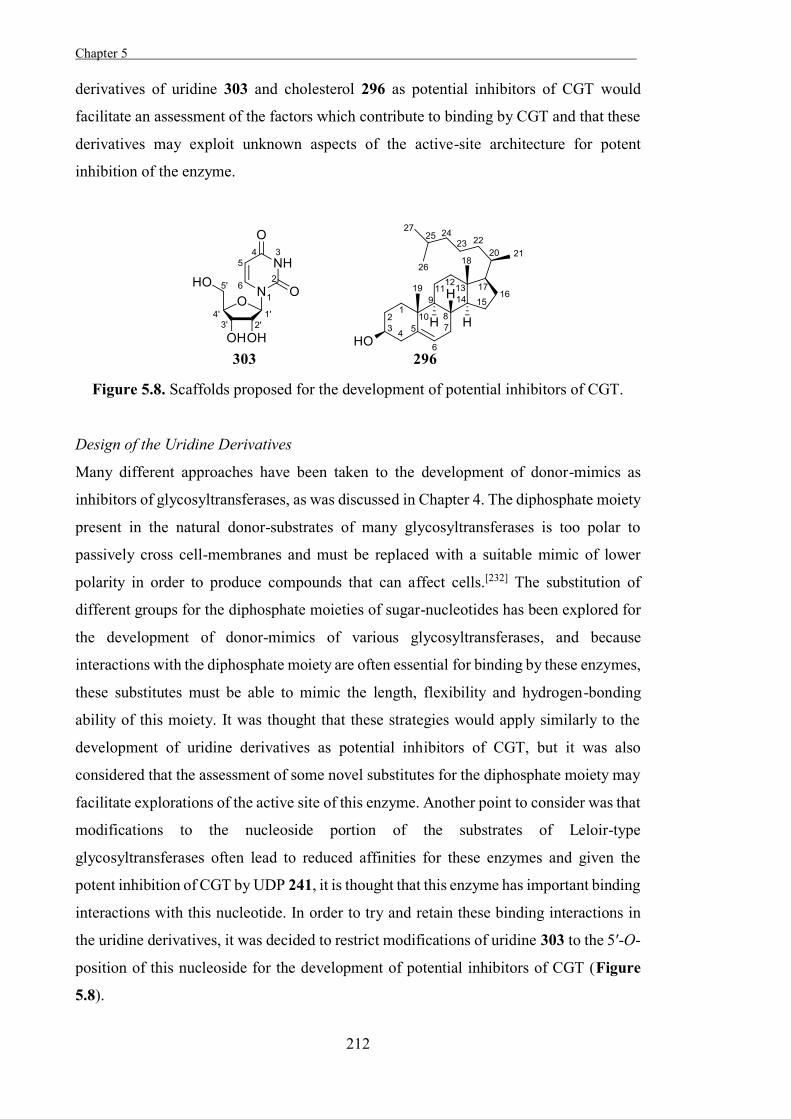
Chapter 5 .
212
derivatives of uridine 303 and cholesterol 296 as potential inhibitors of CGT would
facilitate an assessment of the factors which contribute to binding by CGT and that these
derivatives may exploit unknown aspects of the active-site architecture for potent
inhibition of the enzyme.
Figure 5.8. Scaffolds proposed for the development of potential inhibitors of CGT.
Design of the Uridine Derivatives
Many different approaches have been taken to the development of donor-mimics as
inhibitors of glycosyltransferases, as was discussed in Chapter 4. The diphosphate moiety
present in the natural donor-substrates of many glycosyltransferases is too polar to
passively cross cell-membranes and must be replaced with a suitable mimic of lower
polarity in order to produce compounds that can affect cells.[232] The substitution of
different groups for the diphosphate moieties of sugar-nucleotides has been explored for
the development of donor-mimics of various glycosyltransferases, and because
interactions with the diphosphate moiety are often essential for binding by these enzymes,
these substitutes must be able to mimic the length, flexibility and hydrogen-bonding
ability of this moiety. It was thought that these strategies would apply similarly to the
development of uridine derivatives as potential inhibitors of CGT, but it was also
considered that the assessment of some novel substitutes for the diphosphate moiety may
facilitate explorations of the active site of this enzyme. Another point to consider was that
modifications to the nucleoside portion of the substrates of Leloir-type
glycosyltransferases often lead to reduced affinities for these enzymes and given the
potent inhibition of CGT by UDP 241, it is thought that this enzyme has important binding
interactions with this nucleotide. In order to try and retain these binding interactions in
the uridine derivatives, it was decided to restrict modifications of uridine 303 to the 5′-O-
position of this nucleoside for the development of potential inhibitors of CGT (Figure
5.8).
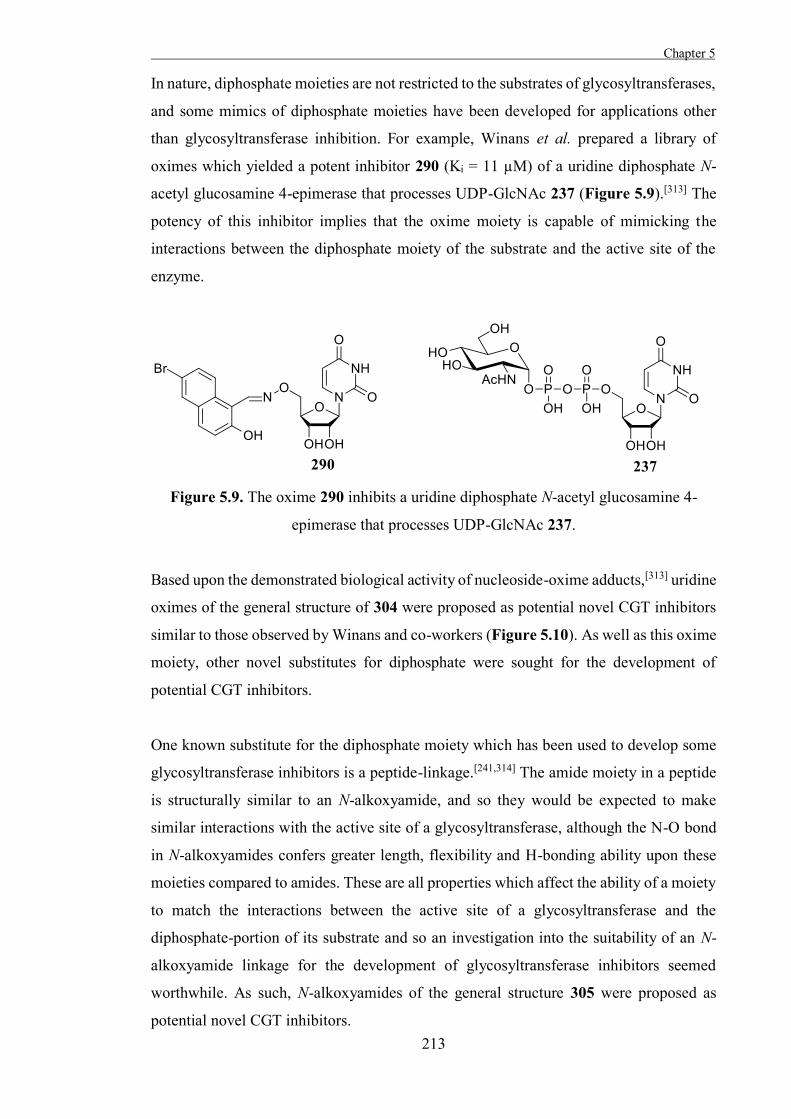
Chapter 5
213
In nature, diphosphate moieties are not restricted to the substrates of glycosyltransferases,
and some mimics of diphosphate moieties have been developed for applications other
than glycosyltransferase inhibition. For example, Winans et al. prepared a library of
oximes which yielded a potent inhibitor 290 (Ki = 11 µM) of a uridine diphosphate N-
acetyl glucosamine 4-epimerase that processes UDP-GlcNAc 237 (Figure 5.9).[313] The
potency of this inhibitor implies that the oxime moiety is capable of mimicking the
interactions between the diphosphate moiety of the substrate and the active site of the
enzyme.
Figure 5.9. The oxime 290 inhibits a uridine diphosphate N-acetyl glucosamine 4-
epimerase that processes UDP-GlcNAc 237.
Based upon the demonstrated biological activity of nucleoside-oxime adducts,[313] uridine
oximes of the general structure of 304 were proposed as potential novel CGT inhibitors
similar to those observed by Winans and co-workers (Figure 5.10). As well as this oxime
moiety, other novel substitutes for diphosphate were sought for the development of
potential CGT inhibitors.
One known substitute for the diphosphate moiety which has been used to develop some
glycosyltransferase inhibitors is a peptide-linkage.[241,314] The amide moiety in a peptide
is structurally similar to an N-alkoxyamide, and so they would be expected to make
similar interactions with the active site of a glycosyltransferase, although the N-O bond
in N-alkoxyamides confers greater length, flexibility and H-bonding ability upon these
moieties compared to amides. These are all properties which affect the ability of a moiety
to match the interactions between the active site of a glycosyltransferase and the
diphosphate-portion of its substrate and so an investigation into the suitability of an N-
alkoxyamide linkage for the development of glycosyltransferase inhibitors seemed
worthwhile. As such, N-alkoxyamides of the general structure 305 were proposed as
potential novel CGT inhibitors.
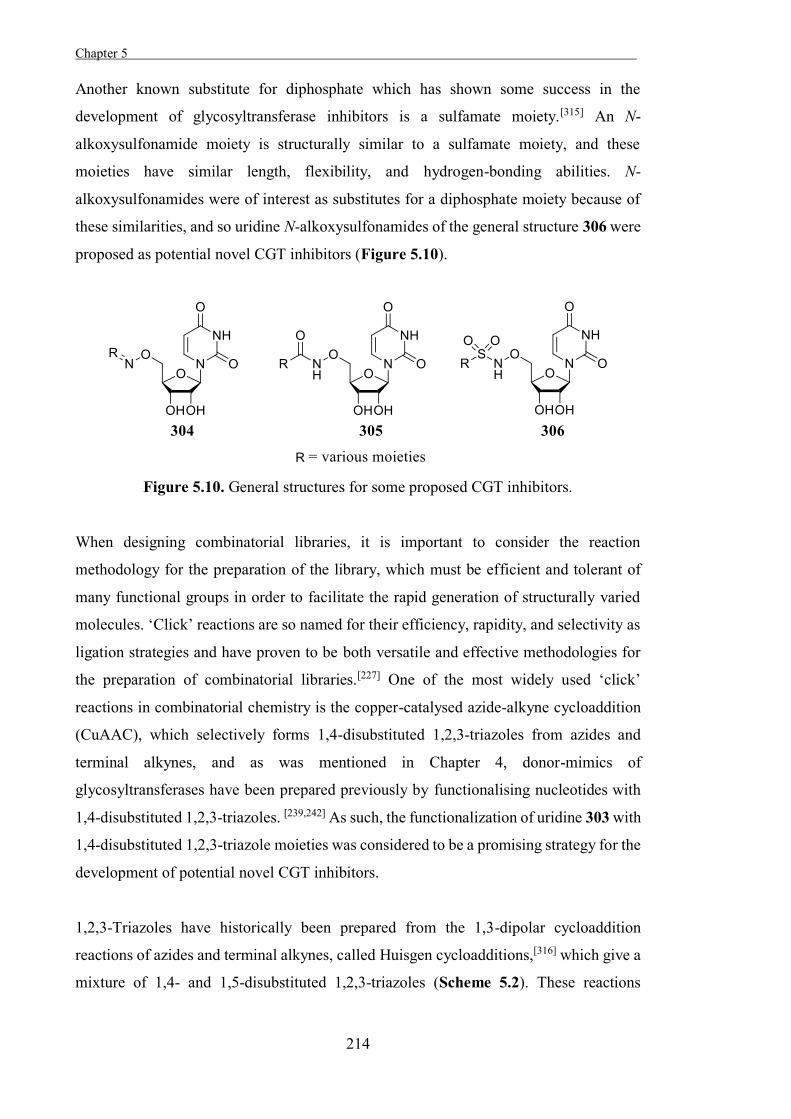
Chapter 5 .
214
Another known substitute for diphosphate which has shown some success in the
development of glycosyltransferase inhibitors is a sulfamate moiety.[315] An N-
alkoxysulfonamide moiety is structurally similar to a sulfamate moiety, and these
moieties have similar length, flexibility, and hydrogen-bonding abilities. N-
alkoxysulfonamides were of interest as substitutes for a diphosphate moiety because of
these similarities, and so uridine N-alkoxysulfonamides of the general structure 306 were
proposed as potential novel CGT inhibitors (Figure 5.10).
Figure 5.10. General structures for some proposed CGT inhibitors.
When designing combinatorial libraries, it is important to consider the reaction
methodology for the preparation of the library, which must be efficient and tolerant of
many functional groups in order to facilitate the rapid generation of structurally varied
molecules. ‘Click’ reactions are so named for their efficiency, rapidity, and selectivity as
ligation strategies and have proven to be both versatile and effective methodologies for
the preparation of combinatorial libraries.[227] One of the most widely used ‘click’
reactions in combinatorial chemistry is the copper-catalysed azide-alkyne cycloaddition
(CuAAC), which selectively forms 1,4-disubstituted 1,2,3-triazoles from azides and
terminal alkynes, and as was mentioned in Chapter 4, donor-mimics of
glycosyltransferases have been prepared previously by functionalising nucleotides with
1,4-disubstituted 1,2,3-triazoles. [239,242] As such, the functionalization of uridine 303 with
1,4-disubstituted 1,2,3-triazole moieties was considered to be a promising strategy for the
development of potential novel CGT inhibitors.
1,2,3-Triazoles have historically been prepared from the 1,3-dipolar cycloaddition
reactions of azides and terminal alkynes, called Huisgen cycloadditions,[316] which give a
mixture of 1,4- and 1,5-disubstituted 1,2,3-triazoles (Scheme 5.2). These reactions
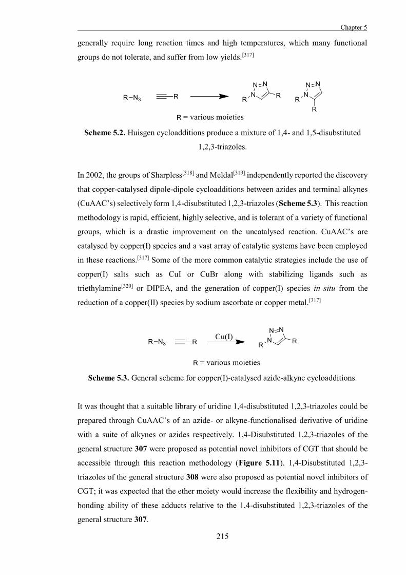
Chapter 5
215
generally require long reaction times and high temperatures, which many functional
groups do not tolerate, and suffer from low yields.[317]
Scheme 5.2. Huisgen cycloadditions produce a mixture of 1,4- and 1,5-disubstituted
1,2,3-triazoles.
In 2002, the groups of Sharpless[318] and Meldal[319] independently reported the discovery
that copper-catalysed dipole-dipole cycloadditions between azides and terminal alkynes
(CuAAC’s) selectively form 1,4-disubstituted 1,2,3-triazoles (Scheme 5.3). This reaction
methodology is rapid, efficient, highly selective, and is tolerant of a variety of functional
groups, which is a drastic improvement on the uncatalysed reaction. CuAAC’s are
catalysed by copper(I) species and a vast array of catalytic systems have been employed
in these reactions.[317] Some of the more common catalytic strategies include the use of
copper(I) salts such as CuI or CuBr along with stabilizing ligands such as
triethylamine[320] or DIPEA, and the generation of copper(I) species in situ from the
reduction of a copper(II) species by sodium ascorbate or copper metal.[317]
Scheme 5.3. General scheme for copper(I)-catalysed azide-alkyne cycloadditions.
It was thought that a suitable library of uridine 1,4-disubstituted 1,2,3-triazoles could be
prepared through CuAAC’s of an azide- or alkyne-functionalised derivative of uridine
with a suite of alkynes or azides respectively. 1,4-Disubstituted 1,2,3-triazoles of the
general structure 307 were proposed as potential novel inhibitors of CGT that should be
accessible through this reaction methodology (Figure 5.11). 1,4-Disubstituted 1,2,3-
triazoles of the general structure 308 were also proposed as potential novel inhibitors of
CGT; it was expected that the ether moiety would increase the flexibility and hydrogen-
bonding ability of these adducts relative to the 1,4-disubstituted 1,2,3-triazoles of the
general structure 307.

Chapter 5 .
216
Figure 5.11. Proposed scaffolds for library of 1,4-disubstituted 1,2,3-triazole
derivatives of uridine.
Synthesis of 5′-Aminooxy Uridine
5′-Aminooxyuridine 309, required as a starting material for the preparation of the oxime,
N-alkoxyamide, and N-alkoxysulfonamide derivatives of uridine, was readily accessible
through a literature preparation (Scheme 5.4).[231] Briefly, uridine 303 was treated with
2,2-dimethoxypropane under acidic conditions to give the acetonide 310 that was then
tosylated selectively at the 5′-position to give the tosylate 311. The aminooxy moiety was
installed by conjugation of the tosylate 311 with N-hydroxysuccinimide to give the imide
312 and cleavage of the imide and subsequent deprotection with aqueous trifluoroacetic
acid provided 5′-aminooxy uridine 309. In a slight variation from the literature, 5′-
aminooxyuridine 309 was purified through flash-chromatography rather than HPLC, and
the NMR spectra of 5′-aminooxyuridine 309 obtained in this fashion were consistent with
those found in the literature.[231]
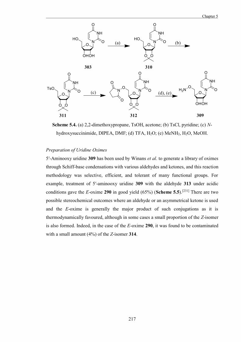
Chapter 5
217
Scheme 5.4. (a) 2,2-dimethoxypropane, TsOH, acetone; (b) TsCl, pyridine; (c) N-
hydroxysuccinimide, DIPEA, DMF; (d) TFA, H2O; (e) MeNH2, H2O, MeOH.
Preparation of Uridine Oximes
5′-Aminooxy uridine 309 has been used by Winans et al. to generate a library of oximes
through Schiff-base condensations with various aldehydes and ketones, and this reaction
methodology was selective, efficient, and tolerant of many functional groups. For
example, treatment of 5′-aminooxy uridine 309 with the aldehyde 313 under acidic
conditions gave the E-oxime 290 in good yield (65%) (Scheme 5.5).[231] There are two
possible stereochemical outcomes where an aldehyde or an asymmetrical ketone is used
and the E-oxime is generally the major product of such conjugations as it is
thermodynamically favoured, although in some cases a small proportion of the Z-isomer
is also formed. Indeed, in the case of the E-oxime 290, it was found to be contaminated
with a small amount (4%) of the Z-isomer 314.
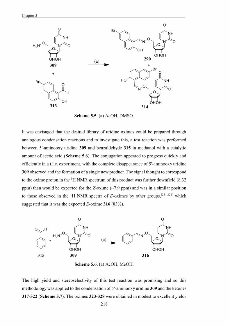
Chapter 5 .
218
Scheme 5.5. (a) AcOH, DMSO.
It was envisaged that the desired library of uridine oximes could be prepared through
analogous condensation reactions and to investigate this, a test reaction was performed
between 5′-aminooxy uridine 309 and benzaldehyde 315 in methanol with a catalytic
amount of acetic acid (Scheme 5.6). The conjugation appeared to progress quickly and
efficiently in a t.l.c. experiment, with the complete disappearance of 5′-aminooxy uridine
309 observed and the formation of a single new product. The signal thought to correspond
to the oxime proton in the 1H NMR spectrum of this product was further downfield (8.32
ppm) than would be expected for the Z-oxime (~7.9 ppm) and was in a similar position
to those observed in the 1H NMR spectra of E-oximes by other groups,[231,321] which
suggested that it was the expected E-oxime 316 (83%).
Scheme 5.6. (a) AcOH, MeOH.
The high yield and stereoselectivity of this test reaction was promising and so this
methodology was applied to the condensation of 5′-aminooxy uridine 309 and the ketones
317-322 (Scheme 5.7). The oximes 323-328 were obtained in modest to excellent yields
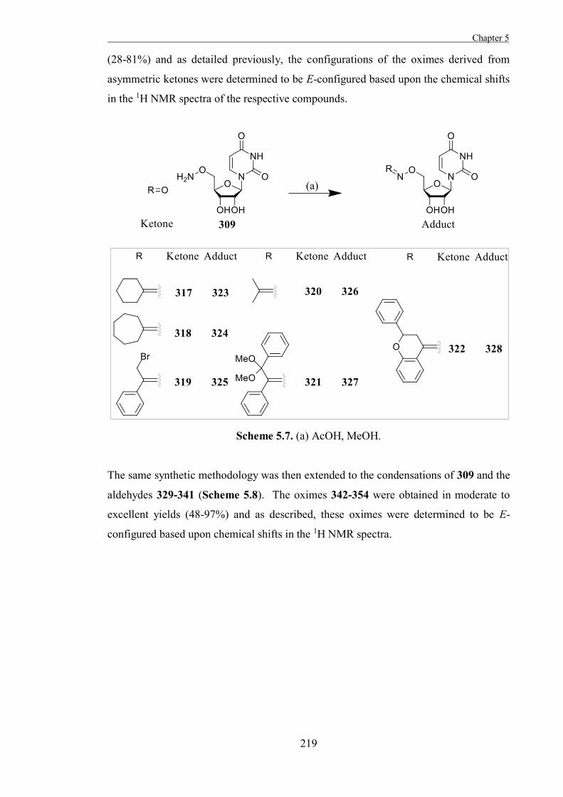
Chapter 5
219
(28-81%) and as detailed previously, the configurations of the oximes derived from
asymmetric ketones were determined to be E-configured based upon the chemical shifts
in the 1H NMR spectra of the respective compounds.
Scheme 5.7. (a) AcOH, MeOH.
The same synthetic methodology was then extended to the condensations of 309 and the
aldehydes 329-341 (Scheme 5.8). The oximes 342-354 were obtained in moderate to
excellent yields (48-97%) and as described, these oximes were determined to be E-
configured based upon chemical shifts in the 1H NMR spectra.
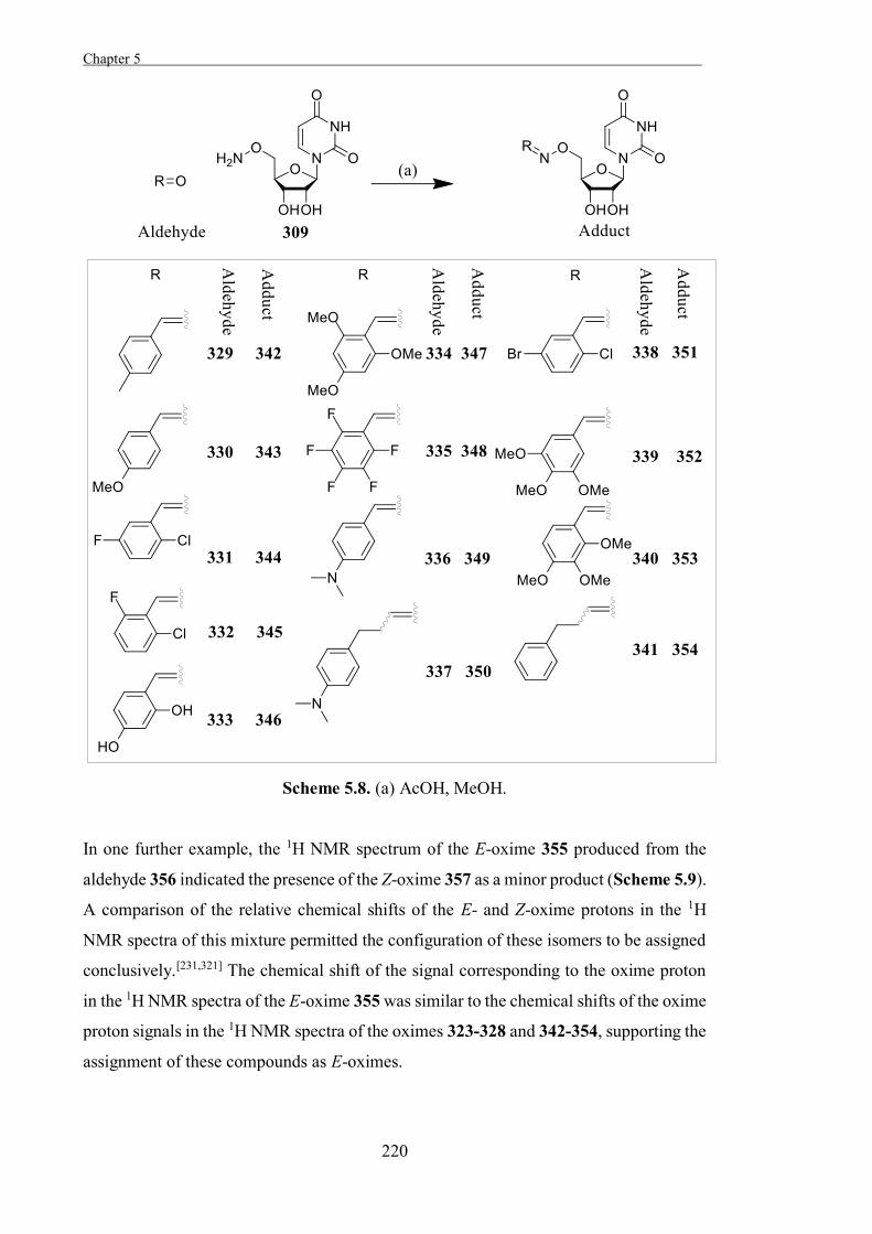
Chapter 5 .
220
Scheme 5.8. (a) AcOH, MeOH.
In one further example, the 1H NMR spectrum of the E-oxime 355 produced from the
aldehyde 356 indicated the presence of the Z-oxime 357 as a minor product (Scheme 5.9).
A comparison of the relative chemical shifts of the E- and Z-oxime protons in the 1H
NMR spectra of this mixture permitted the configuration of these isomers to be assigned
conclusively.[231,321] The chemical shift of the signal corresponding to the oxime proton
in the 1H NMR spectra of the E-oxime 355 was similar to the chemical shifts of the oxime
proton signals in the 1H NMR spectra of the oximes 323-328 and 342-354, supporting the
assignment of these compounds as E-oximes.
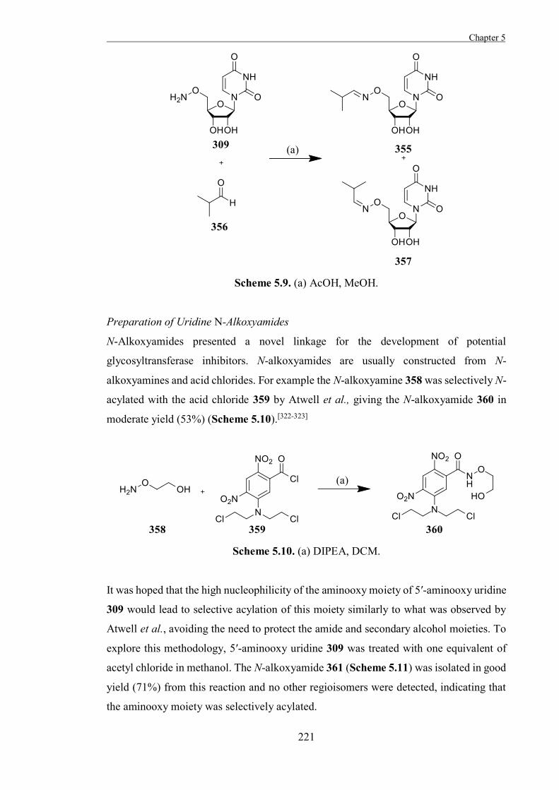
Chapter 5
221
Scheme 5.9. (a) AcOH, MeOH.
Preparation of Uridine N-Alkoxyamides
N-Alkoxyamides presented a novel linkage for the development of potential
glycosyltransferase inhibitors. N-alkoxyamides are usually constructed from N-
alkoxyamines and acid chlorides. For example the N-alkoxyamine 358 was selectively N-
acylated with the acid chloride 359 by Atwell et al., giving the N-alkoxyamide 360 in
moderate yield (53%) (Scheme 5.10).[322-323]
Scheme 5.10. (a) DIPEA, DCM.
It was hoped that the high nucleophilicity of the aminooxy moiety of 5′-aminooxy uridine
309 would lead to selective acylation of this moiety similarly to what was observed by
Atwell et al., avoiding the need to protect the amide and secondary alcohol moieties. To
explore this methodology, 5′-aminooxy uridine 309 was treated with one equivalent of
acetyl chloride in methanol. The N-alkoxyamide 361 (Scheme 5.11) was isolated in good
yield (71%) from this reaction and no other regioisomers were detected, indicating that
the aminooxy moiety was selectively acylated.
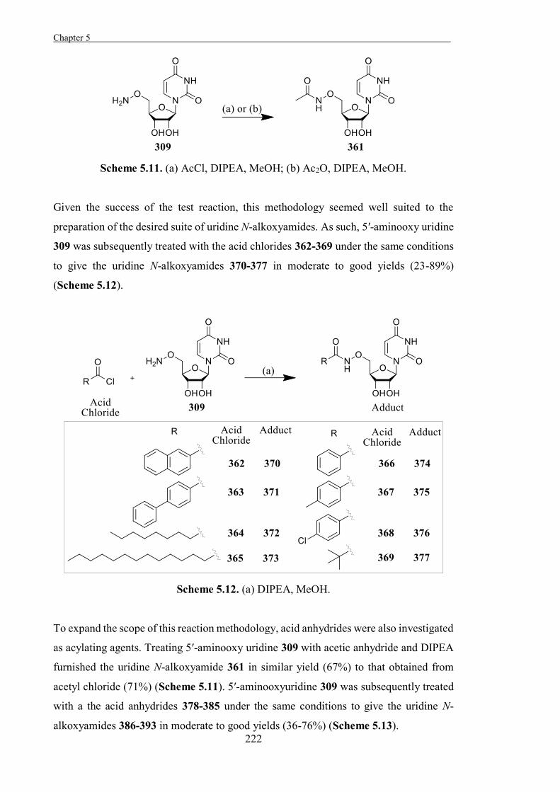
Chapter 5 .
222
Scheme 5.11. (a) AcCl, DIPEA, MeOH; (b) Ac2O, DIPEA, MeOH.
Given the success of the test reaction, this methodology seemed well suited to the
preparation of the desired suite of uridine N-alkoxyamides. As such, 5′-aminooxy uridine
309 was subsequently treated with the acid chlorides 362-369 under the same conditions
to give the uridine N-alkoxyamides 370-377 in moderate to good yields (23-89%)
(Scheme 5.12).
Scheme 5.12. (a) DIPEA, MeOH.
To expand the scope of this reaction methodology, acid anhydrides were also investigated
as acylating agents. Treating 5′-aminooxy uridine 309 with acetic anhydride and DIPEA
furnished the uridine N-alkoxyamide 361 in similar yield (67%) to that obtained from
acetyl chloride (71%) (Scheme 5.11). 5′-aminooxyuridine 309 was subsequently treated
with a the acid anhydrides 378-385 under the same conditions to give the uridine N-
alkoxyamides 386-393 in moderate to good yields (36-76%) (Scheme 5.13).
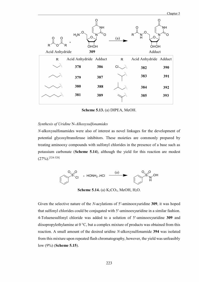
Chapter 5
223
Scheme 5.13. (a) DIPEA, MeOH.
Synthesis of Uridine N-Alkoxysulfonamides
N-alkoxysulfonamides were also of interest as novel linkages for the development of
potential glycosyltransferase inhibitors. These moieties are commonly prepared by
treating aminooxy compounds with sulfonyl chlorides in the presence of a base such as
potassium carbonate (Scheme 5.14), although the yield for this reaction are modest
(27%).[324-328]
Scheme 5.14. (a) K2CO3, MeOH, H2O.
Given the selective nature of the N-acylations of 5′-aminooxyuridine 309, it was hoped
that sulfonyl chlorides could be conjugated with 5′-aminooxyuridine in a similar fashion.
4-Toluenesulfonyl chloride was added to a solution of 5′-aminooxyuridine 309 and
diisopropylethylamine at 0 °C, but a complex mixture of products was obtained from this
reaction. A small amount of the desired uridine N-alkoxysulfonamide 394 was isolated
from this mixture upon repeated flash chromatography, however, the yield was unfeasibly
low (9%) (Scheme 5.15).
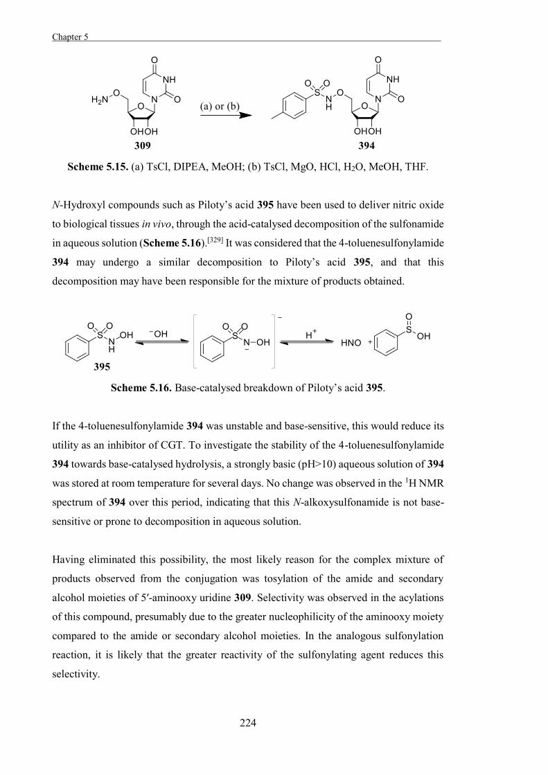
Chapter 5 .
224
Scheme 5.15. (a) TsCl, DIPEA, MeOH; (b) TsCl, MgO, HCl, H2O, MeOH, THF.
N-Hydroxyl compounds such as Piloty’s acid 395 have been used to deliver nitric oxide
to biological tissues in vivo, through the acid-catalysed decomposition of the sulfonamide
in aqueous solution (Scheme 5.16).[329] It was considered that the 4-toluenesulfonylamide
394 may undergo a similar decomposition to Piloty’s acid 395, and that this
decomposition may have been responsible for the mixture of products obtained.
Scheme 5.16. Base-catalysed breakdown of Piloty’s acid 395.
If the 4-toluenesulfonylamide 394 was unstable and base-sensitive, this would reduce its
utility as an inhibitor of CGT. To investigate the stability of the 4-toluenesulfonylamide
394 towards base-catalysed hydrolysis, a strongly basic (pH>10) aqueous solution of 394
was stored at room temperature for several days. No change was observed in the 1H NMR
spectrum of 394 over this period, indicating that this N-alkoxysulfonamide is not base-
sensitive or prone to decomposition in aqueous solution.
Having eliminated this possibility, the most likely reason for the complex mixture of
products observed from the conjugation was tosylation of the amide and secondary
alcohol moieties of 5′-aminooxy uridine 309. Selectivity was observed in the acylations
of this compound, presumably due to the greater nucleophilicity of the aminooxy moiety
compared to the amide or secondary alcohol moieties. In the analogous sulfonylation
reaction, it is likely that the greater reactivity of the sulfonylating agent reduces this
selectivity.
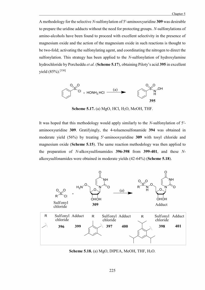
Chapter 5
225
A methodology for the selective N-sulfonylation of 5′-aminooxyuridine 309 was desirable
to prepare the uridine adducts without the need for protecting groups. N-sulfonylations of
amino-alcohols have been found to proceed with excellent selectivity in the presence of
magnesium oxide and the action of the magnesium oxide in such reactions is thought to
be two-fold; activating the sulfonylating agent, and coordinating the nitrogen to direct the
sulfonylation. This strategy has been applied to the N-sulfonylation of hydroxylamine
hydrochloride by Porcheddu et al. (Scheme 5.17), obtaining Piloty’s acid 395 in excellent
yield (85%).[330]
Scheme 5.17. (a) MgO, HCl, H2O, MeOH, THF.
It was hoped that this methodology would apply similarly to the N-sulfonylation of 5′-
aminooxyuridine 309. Gratifyingly, the 4-toluenesulfonamide 394 was obtained in
moderate yield (56%) by treating 5′-aminooxyuridine 309 with tosyl chloride and
magnesium oxide (Scheme 5.15). The same reaction methodology was then applied to
the preparation of N-alkoxysulfonamides 396-398 from 399-401, and these N-
alkoxysulfonamides were obtained in moderate yields (42-64%) (Scheme 5.18).
Scheme 5.18. (a) MgO, DIPEA, MeOH, THF, H2O.
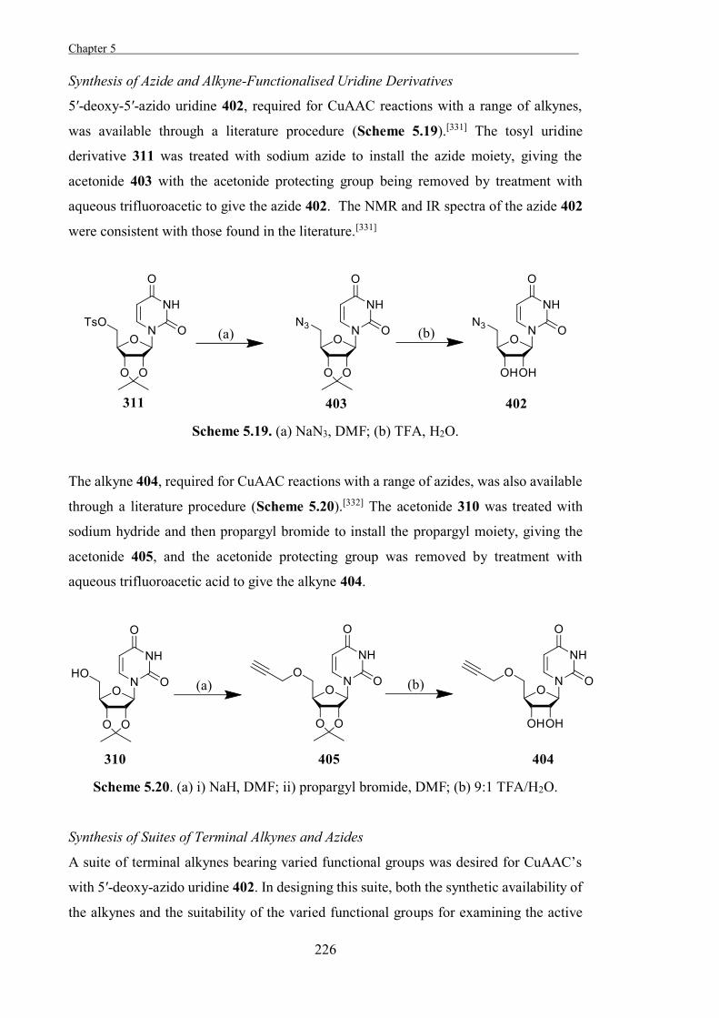
Chapter 5 .
226
Synthesis of Azide and Alkyne-Functionalised Uridine Derivatives
5′-deoxy-5′-azido uridine 402, required for CuAAC reactions with a range of alkynes,
was available through a literature procedure (Scheme 5.19).[331] The tosyl uridine
derivative 311 was treated with sodium azide to install the azide moiety, giving the
acetonide 403 with the acetonide protecting group being removed by treatment with
aqueous trifluoroacetic to give the azide 402. The NMR and IR spectra of the azide 402
were consistent with those found in the literature.[331]
Scheme 5.19. (a) NaN3, DMF; (b) TFA, H2O.
The alkyne 404, required for CuAAC reactions with a range of azides, was also available
through a literature procedure (Scheme 5.20).[332] The acetonide 310 was treated with
sodium hydride and then propargyl bromide to install the propargyl moiety, giving the
acetonide 405, and the acetonide protecting group was removed by treatment with
aqueous trifluoroacetic acid to give the alkyne 404.
Scheme 5.20. (a) i) NaH, DMF; ii) propargyl bromide, DMF; (b) 9:1 TFA/H2O.
Synthesis of Suites of Terminal Alkynes and Azides
A suite of terminal alkynes bearing varied functional groups was desired for CuAAC’s
with 5′-deoxy-azido uridine 402. In designing this suite, both the synthetic availability of
the alkynes and the suitability of the varied functional groups for examining the active
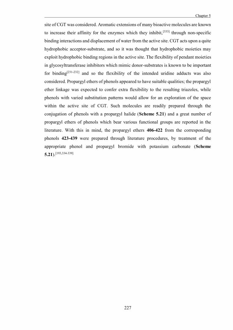
Chapter 5
227
site of CGT was considered. Aromatic extensions of many bioactive molecules are known
to increase their affinity for the enzymes which they inhibit,[333] through non-specific
binding interactions and displacement of water from the active site. CGT acts upon a quite
hydrophobic acceptor-substrate, and so it was thought that hydrophobic moieties may
exploit hydrophobic binding regions in the active site. The flexibility of pendant moieties
in glycosyltransferase inhibitors which mimic donor-substrates is known to be important
for binding[231-232] and so the flexibility of the intended uridine adducts was also
considered. Propargyl ethers of phenols appeared to have suitable qualities; the propargyl
ether linkage was expected to confer extra flexibility to the resulting triazoles, while
phenols with varied substitution patterns would allow for an exploration of the space
within the active site of CGT. Such molecules are readily prepared through the
conjugation of phenols with a propargyl halide (Scheme 5.21) and a great number of
propargyl ethers of phenols which bear various functional groups are reported in the
literature. With this in mind, the propargyl ethers 406-422 from the corresponding
phenols 423-439 were prepared through literature procedures, by treatment of the
appropriate phenol and propargyl bromide with potassium carbonate (Scheme
5.21).[193,334-339]
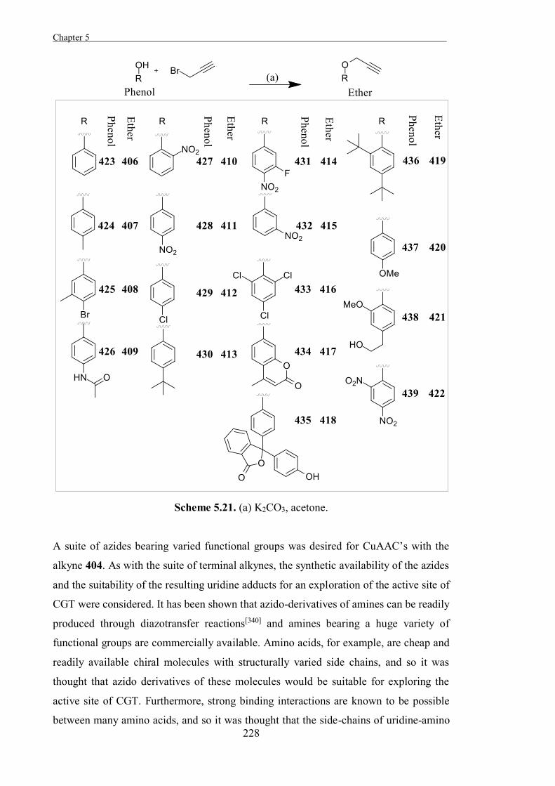
Chapter 5 .
228
Scheme 5.21. (a) K2CO3, acetone.
A suite of azides bearing varied functional groups was desired for CuAAC’s with the
alkyne 404. As with the suite of terminal alkynes, the synthetic availability of the azides
and the suitability of the resulting uridine adducts for an exploration of the active site of
CGT were considered. It has been shown that azido-derivatives of amines can be readily
produced through diazotransfer reactions[340] and amines bearing a huge variety of
functional groups are commercially available. Amino acids, for example, are cheap and
readily available chiral molecules with structurally varied side chains, and so it was
thought that azido derivatives of these molecules would be suitable for exploring the
active site of CGT. Furthermore, strong binding interactions are known to be possible
between many amino acids, and so it was thought that the side-chains of uridine-amino
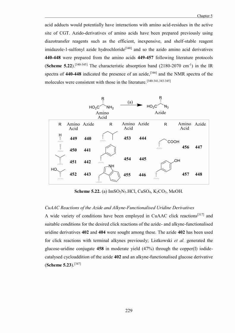
Chapter 5
229
acid adducts would potentially have interactions with amino acid-residues in the active
site of CGT. Azido-derivatives of amino acids have been prepared previously using
diazotransfer reagents such as the efficient, inexpensive, and shelf-stable reagent
imidazole-1-sulfonyl azide hydrochloride[340] and so the azido amino acid derivatives
440-448 were prepared from the amino acids 449-457 following literature protocols
(Scheme 5.22).[340-345] The characteristic absorption band (2180-2070 cm-1) in the IR
spectra of 440-448 indicated the presence of an azide,[346] and the NMR spectra of the
molecules were consistent with those in the literature.[340-341,343-345]
Scheme 5.22. (a) ImSO2N3.HCl, CuSO4, K2CO3, MeOH.
CuAAC Reactions of the Azide and Alkyne-Functionalised Uridine Derivatives
A wide variety of conditions have been employed in CuAAC click reactions[317] and
suitable conditions for the desired click reactions of the azide- and alkyne-functionalised
uridine derivatives 402 and 404 were sought among these. The azide 402 has been used
for click reactions with terminal alkynes previously; Listkowski et al. generated the
glucose-uridine conjugate 458 in moderate yield (47%) through the copper(I) iodide-
catalysed cycloaddition of the azide 402 and an alkyne-functionalised glucose derivative
(Scheme 5.23).[347]
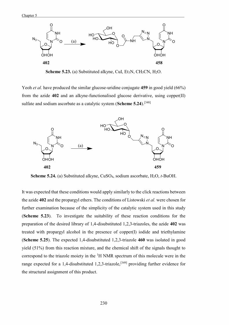
Chapter 5 .
230
Scheme 5.23. (a) Substituted alkyne, CuI, Et3N, CH3CN, H2O.
Yeoh et al. have produced the similar glucose-uridine conjugate 459 in good yield (66%)
from the azide 402 and an alkyne-functionalised glucose derivative, using copper(II)
sulfate and sodium ascorbate as a catalytic system (Scheme 5.24).[348]
Scheme 5.24. (a) Substituted alkyne, CuSO4, sodium ascorbate, H2O, t-BuOH.
It was expected that these conditions would apply similarly to the click reactions between
the azide 402 and the propargyl ethers. The conditions of Listowski et al. were chosen for
further examination because of the simplicity of the catalytic system used in this study
(Scheme 5.23). To investigate the suitability of these reaction conditions for the
preparation of the desired library of 1,4-disubstituted 1,2,3-triazoles, the azide 402 was
treated with propargyl alcohol in the presence of copper(I) iodide and triethylamine
(Scheme 5.25). The expected 1,4-disubstituted 1,2,3-triazole 460 was isolated in good
yield (51%) from this reaction mixture, and the chemical shift of the signals thought to
correspond to the triazole moiety in the 1H NMR spectrum of this molecule were in the
range expected for a 1,4-disubstituted 1,2,3-triazole,[349] providing further evidence for
the structural assignment of this product.
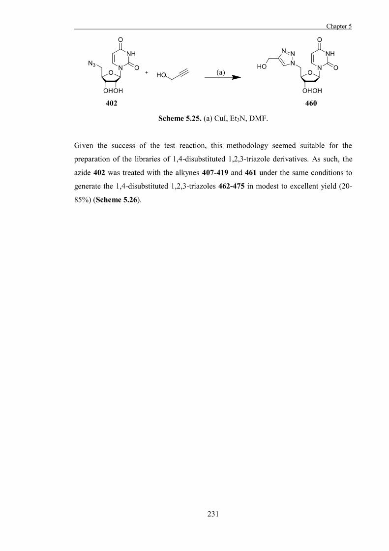
Chapter 5
231
Scheme 5.25. (a) CuI, Et3N, DMF.
Given the success of the test reaction, this methodology seemed suitable for the
preparation of the libraries of 1,4-disubstituted 1,2,3-triazole derivatives. As such, the
azide 402 was treated with the alkynes 407-419 and 461 under the same conditions to
generate the 1,4-disubstituted 1,2,3-triazoles 462-475 in modest to excellent yield (20-
85%) (Scheme 5.26).
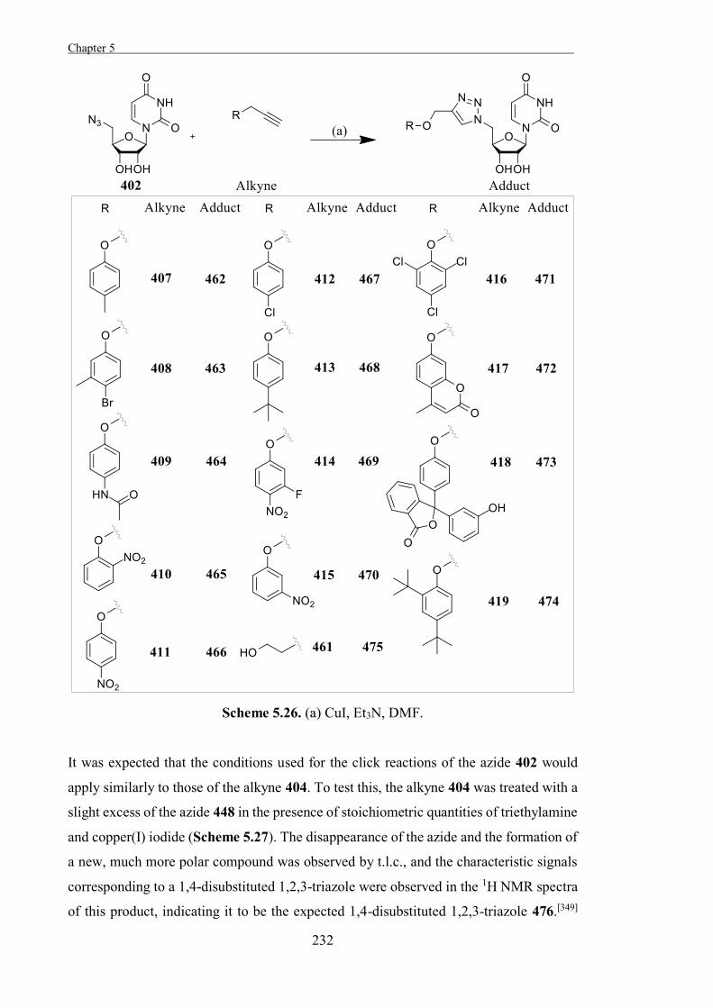
Chapter 5 .
232
Scheme 5.26. (a) CuI, Et3N, DMF.
It was expected that the conditions used for the click reactions of the azide 402 would
apply similarly to those of the alkyne 404. To test this, the alkyne 404 was treated with a
slight excess of the azide 448 in the presence of stoichiometric quantities of triethylamine
and copper(I) iodide (Scheme 5.27). The disappearance of the azide and the formation of
a new, much more polar compound was observed by t.l.c., and the characteristic signals
corresponding to a 1,4-disubstituted 1,2,3-triazole were observed in the 1H NMR spectra
of this product, indicating it to be the expected 1,4-disubstituted 1,2,3-triazole 476.[349]
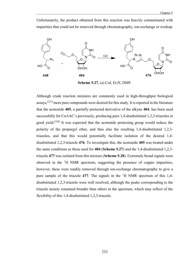
Chapter 5
233
Unfortunately, the product obtained from this reaction was heavily contaminated with
impurities that could not be removed through chromatography, ion-exchange or workup.
Scheme 5.27. (a) CuI, Et3N, DMF.
Although crude reaction mixtures are commonly used in high-throughput biological
assays,[231] more pure compounds were desired for this study. It is reported in the literature
that the acetonide 405, a partially protected derivative of the alkyne 404, has been used
successfully for CuAAC’s previously, producing pure 1,4-disubstituted 1,2,3-triazoles in
good yield.[350] It was expected that the acetonide protecting group would reduce the
polarity of the propargyl ether, and thus also the resulting 1,4-disubstituted 1,2,3-
triazoles, and that this would potentially facilitate isolation of the desired 1,4-
disubstituted 1,2,3-triazole 476. To investigate this, the acetonide 405 was treated under
the same conditions as those used for 404 (Scheme 5.27) and the 1,4-disubstituted 1,2,3-
triazole 477 was isolated from this mixture (Scheme 5.28). Extremely broad signals were
observed in the 1H NMR spectrum, suggesting the presence of copper impurities,
however, these were readily removed through ion-exchange chromatography to give a
pure sample of the triazole 477. The signals in the 1H NMR spectrum of this 1,4-
disubstituted 1,2,3-triazole were well resolved, although the peaks corresponding to the
triazole moiety remained broader than others in the spectrum, which may reflect of the
flexibility of this 1,4-disubstituted 1,2,3-triazole.
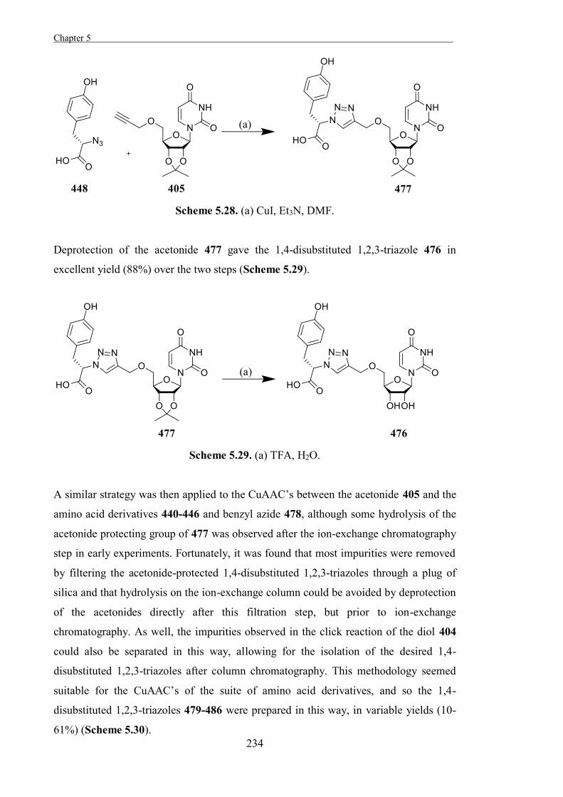
Chapter 5 .
234
Scheme 5.28. (a) CuI, Et3N, DMF.
Deprotection of the acetonide 477 gave the 1,4-disubstituted 1,2,3-triazole 476 in
excellent yield (88%) over the two steps (Scheme 5.29).
Scheme 5.29. (a) TFA, H2O.
A similar strategy was then applied to the CuAAC’s between the acetonide 405 and the
amino acid derivatives 440-446 and benzyl azide 478, although some hydrolysis of the
acetonide protecting group of 477 was observed after the ion-exchange chromatography
step in early experiments. Fortunately, it was found that most impurities were removed
by filtering the acetonide-protected 1,4-disubstituted 1,2,3-triazoles through a plug of
silica and that hydrolysis on the ion-exchange column could be avoided by deprotection
of the acetonides directly after this filtration step, but prior to ion-exchange
chromatography. As well, the impurities observed in the click reaction of the diol 404
could also be separated in this way, allowing for the isolation of the desired 1,4-
disubstituted 1,2,3-triazoles after column chromatography. This methodology seemed
suitable for the CuAAC’s of the suite of amino acid derivatives, and so the 1,4-
disubstituted 1,2,3-triazoles 479-486 were prepared in this way, in variable yields (10-
61%) (Scheme 5.30).
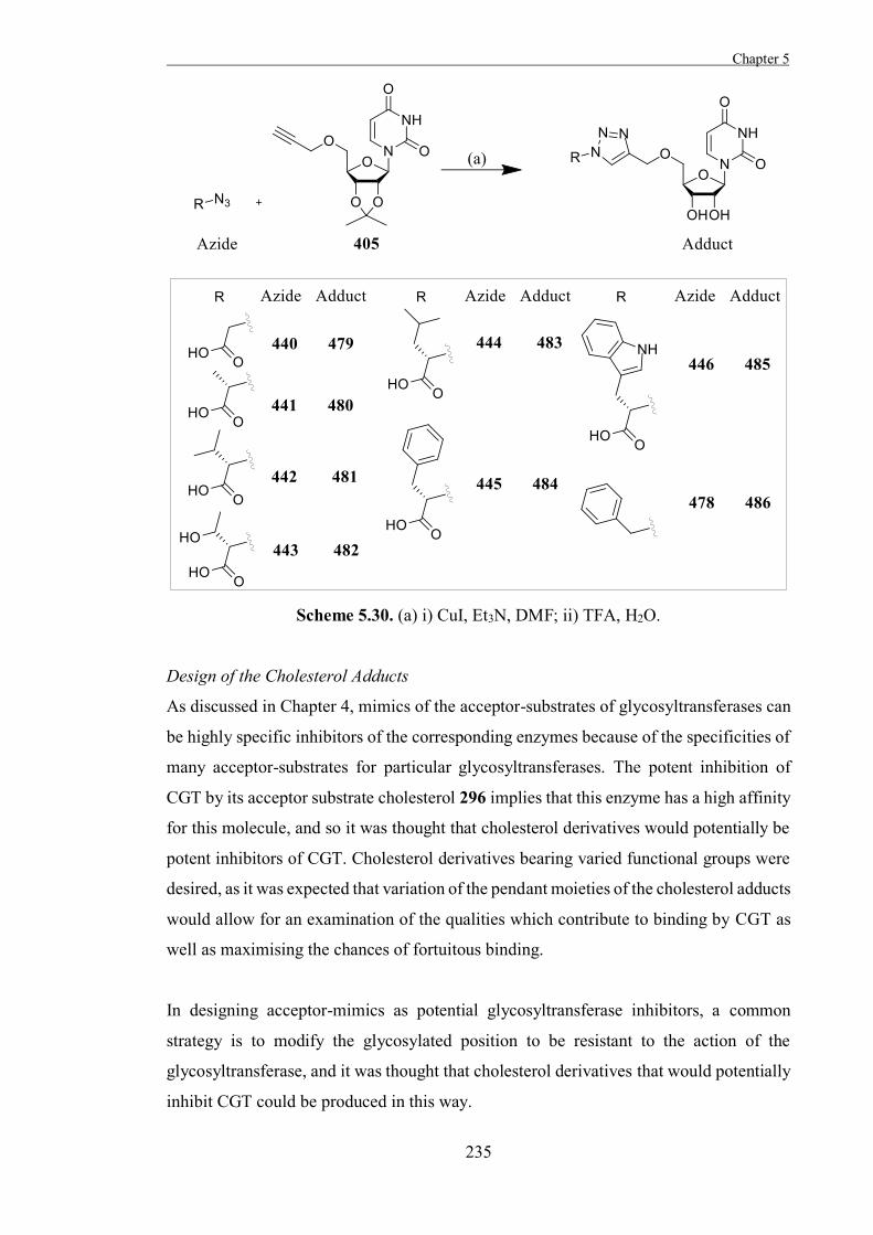
Chapter 5
235
Scheme 5.30. (a) i) CuI, Et3N, DMF; ii) TFA, H2O.
Design of the Cholesterol Adducts
As discussed in Chapter 4, mimics of the acceptor-substrates of glycosyltransferases can
be highly specific inhibitors of the corresponding enzymes because of the specificities of
many acceptor-substrates for particular glycosyltransferases. The potent inhibition of
CGT by its acceptor substrate cholesterol 296 implies that this enzyme has a high affinity
for this molecule, and so it was thought that cholesterol derivatives would potentially be
potent inhibitors of CGT. Cholesterol derivatives bearing varied functional groups were
desired, as it was expected that variation of the pendant moieties of the cholesterol adducts
would allow for an examination of the qualities which contribute to binding by CGT as
well as maximising the chances of fortuitous binding.
In designing acceptor-mimics as potential glycosyltransferase inhibitors, a common
strategy is to modify the glycosylated position to be resistant to the action of the
glycosyltransferase, and it was thought that cholesterol derivatives that would potentially
inhibit CGT could be produced in this way.
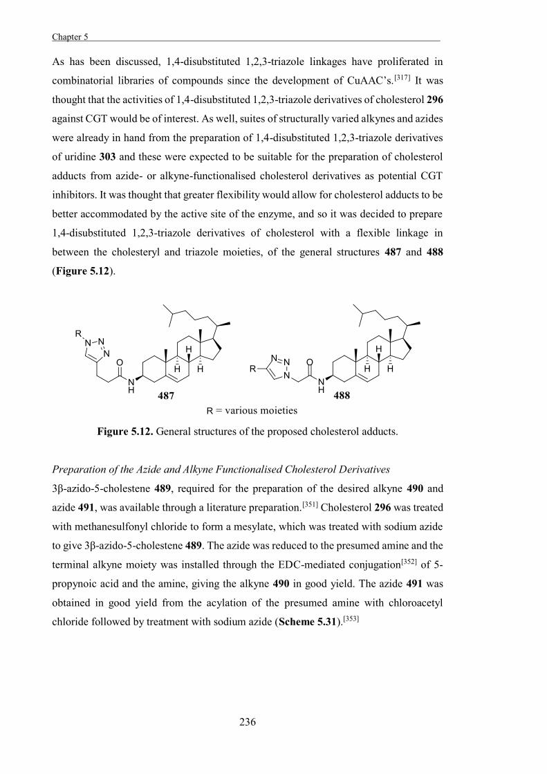
Chapter 5 .
236
As has been discussed, 1,4-disubstituted 1,2,3-triazole linkages have proliferated in
combinatorial libraries of compounds since the development of CuAAC’s.[317] It was
thought that the activities of 1,4-disubstituted 1,2,3-triazole derivatives of cholesterol 296
against CGT would be of interest. As well, suites of structurally varied alkynes and azides
were already in hand from the preparation of 1,4-disubstituted 1,2,3-triazole derivatives
of uridine 303 and these were expected to be suitable for the preparation of cholesterol
adducts from azide- or alkyne-functionalised cholesterol derivatives as potential CGT
inhibitors. It was thought that greater flexibility would allow for cholesterol adducts to be
better accommodated by the active site of the enzyme, and so it was decided to prepare
1,4-disubstituted 1,2,3-triazole derivatives of cholesterol with a flexible linkage in
between the cholesteryl and triazole moieties, of the general structures 487 and 488
(Figure 5.12).
Figure 5.12. General structures of the proposed cholesterol adducts.
Preparation of the Azide and Alkyne Functionalised Cholesterol Derivatives
3β-azido-5-cholestene 489, required for the preparation of the desired alkyne 490 and
azide 491, was available through a literature preparation.[351] Cholesterol 296 was treated
with methanesulfonyl chloride to form a mesylate, which was treated with sodium azide
to give 3β-azido-5-cholestene 489. The azide was reduced to the presumed amine and the
terminal alkyne moiety was installed through the EDC-mediated conjugation[352] of 5-
propynoic acid and the amine, giving the alkyne 490 in good yield. The azide 491 was
obtained in good yield from the acylation of the presumed amine with chloroacetyl
chloride followed by treatment with sodium azide (Scheme 5.31).[353]
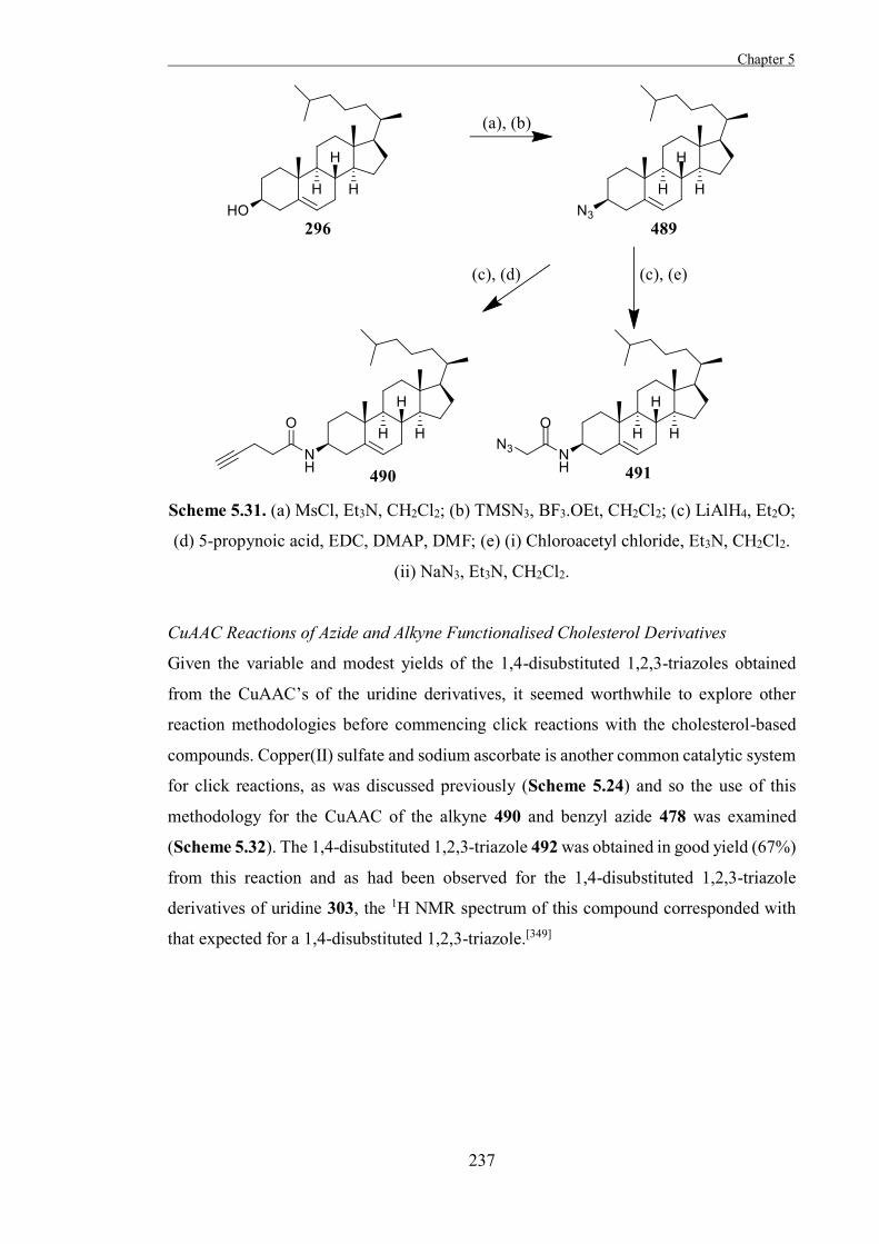
Chapter 5
237
Scheme 5.31. (a) MsCl, Et3N, CH2Cl2; (b) TMSN3, BF3.OEt, CH2Cl2; (c) LiAlH4, Et2O;
(d) 5-propynoic acid, EDC, DMAP, DMF; (e) (i) Chloroacetyl chloride, Et3N, CH2Cl2.
(ii) NaN3, Et3N, CH2Cl2.
CuAAC Reactions of Azide and Alkyne Functionalised Cholesterol Derivatives
Given the variable and modest yields of the 1,4-disubstituted 1,2,3-triazoles obtained
from the CuAAC’s of the uridine derivatives, it seemed worthwhile to explore other
reaction methodologies before commencing click reactions with the cholesterol-based
compounds. Copper(II) sulfate and sodium ascorbate is another common catalytic system
for click reactions, as was discussed previously (Scheme 5.24) and so the use of this
methodology for the CuAAC of the alkyne 490 and benzyl azide 478 was examined
(Scheme 5.32). The 1,4-disubstituted 1,2,3-triazole 492 was obtained in good yield (67%)
from this reaction and as had been observed for the 1,4-disubstituted 1,2,3-triazole
derivatives of uridine 303, the 1H NMR spectrum of this compound corresponded with
that expected for a 1,4-disubstituted 1,2,3-triazole.[349]
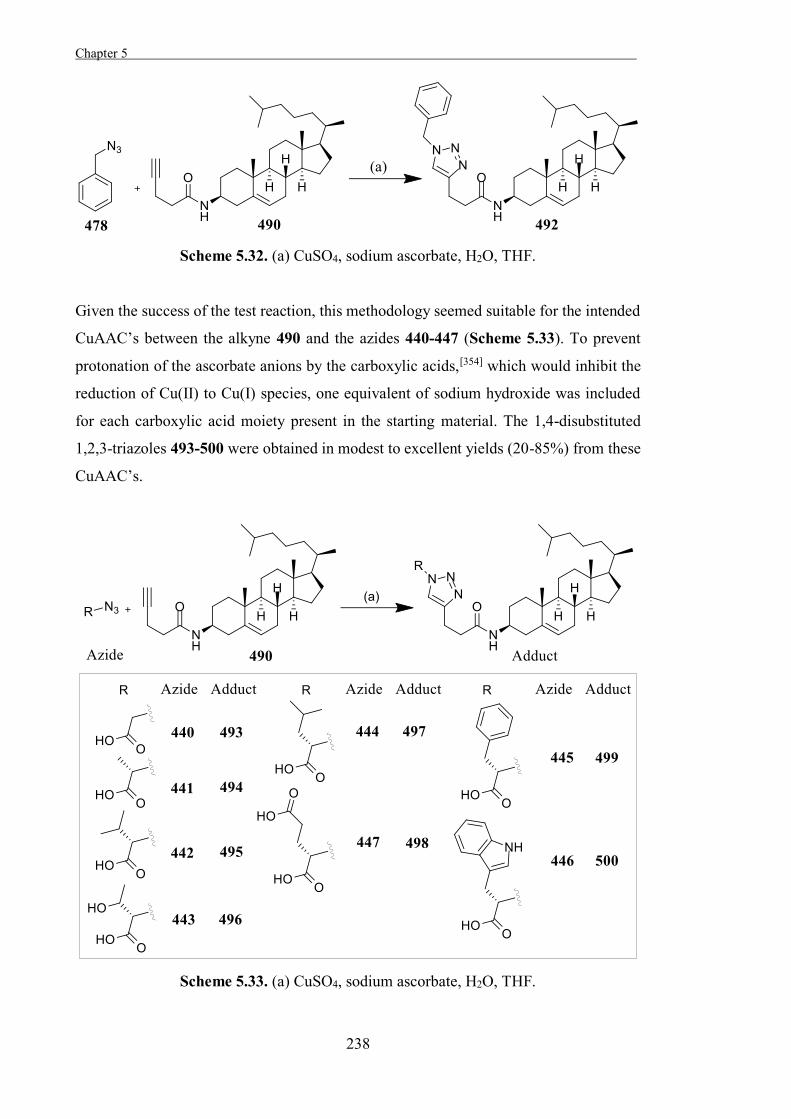
Chapter 5 .
238
Scheme 5.32. (a) CuSO4, sodium ascorbate, H2O, THF.
Given the success of the test reaction, this methodology seemed suitable for the intended
CuAAC’s between the alkyne 490 and the azides 440-447 (Scheme 5.33). To prevent
protonation of the ascorbate anions by the carboxylic acids,[354] which would inhibit the
reduction of Cu(II) to Cu(I) species, one equivalent of sodium hydroxide was included
for each carboxylic acid moiety present in the starting material. The 1,4-disubstituted
1,2,3-triazoles 493-500 were obtained in modest to excellent yields (20-85%) from these
CuAAC’s.
Scheme 5.33. (a) CuSO4, sodium ascorbate, H2O, THF.
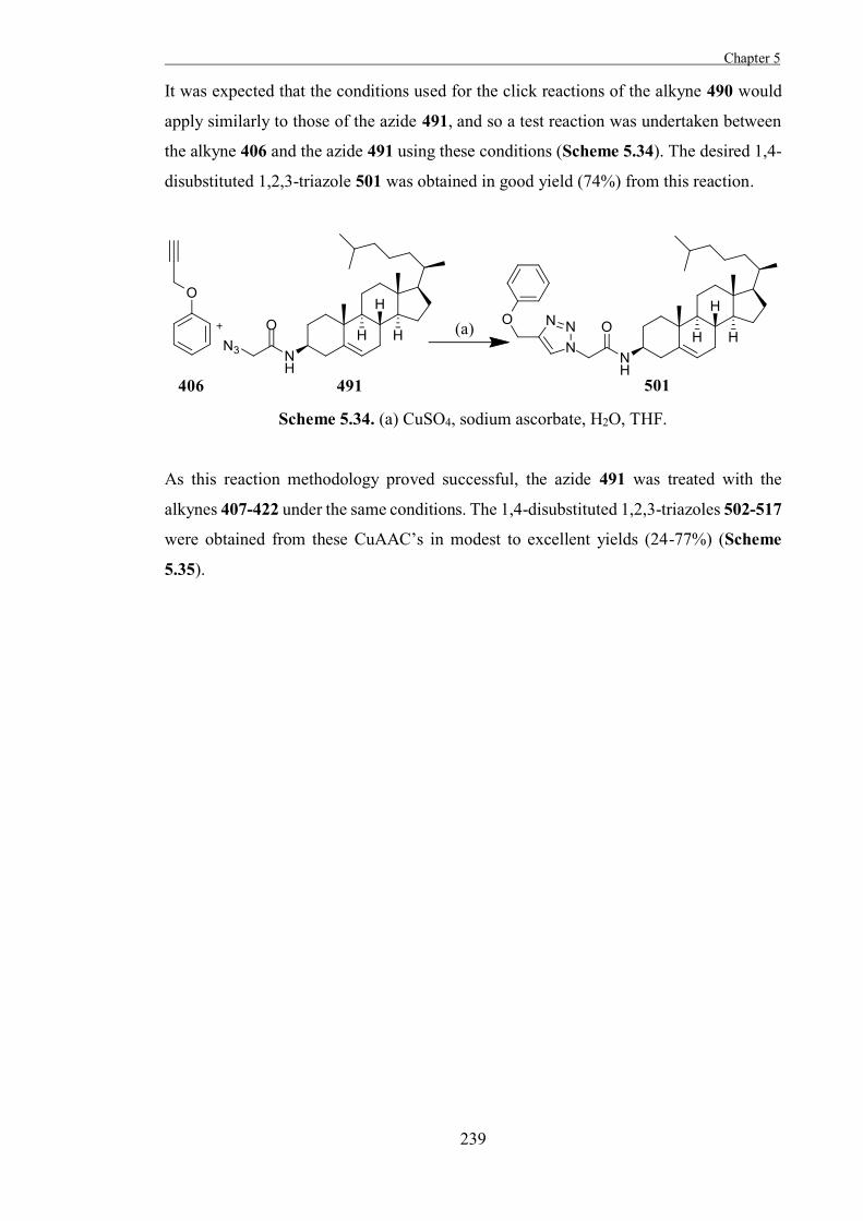
Chapter 5
239
It was expected that the conditions used for the click reactions of the alkyne 490 would
apply similarly to those of the azide 491, and so a test reaction was undertaken between
the alkyne 406 and the azide 491 using these conditions (Scheme 5.34). The desired 1,4-
disubstituted 1,2,3-triazole 501 was obtained in good yield (74%) from this reaction.
Scheme 5.34. (a) CuSO4, sodium ascorbate, H2O, THF.
As this reaction methodology proved successful, the azide 491 was treated with the
alkynes 407-422 under the same conditions. The 1,4-disubstituted 1,2,3-triazoles 502-517
were obtained from these CuAAC’s in modest to excellent yields (24-77%) (Scheme
5.35).
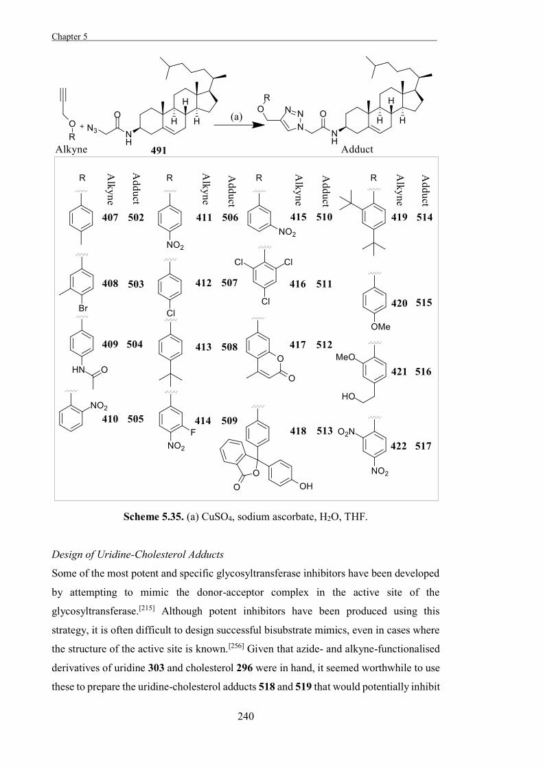
Chapter 5 .
240
Scheme 5.35. (a) CuSO4, sodium ascorbate, H2O, THF.
Design of Uridine-Cholesterol Adducts
Some of the most potent and specific glycosyltransferase inhibitors have been developed
by attempting to mimic the donor-acceptor complex in the active site of the
glycosyltransferase.[215] Although potent inhibitors have been produced using this
strategy, it is often difficult to design successful bisubstrate mimics, even in cases where
the structure of the active site is known.[256] Given that azide- and alkyne-functionalised
derivatives of uridine 303 and cholesterol 296 were in hand, it seemed worthwhile to use
these to prepare the uridine-cholesterol adducts 518 and 519 that would potentially inhibit
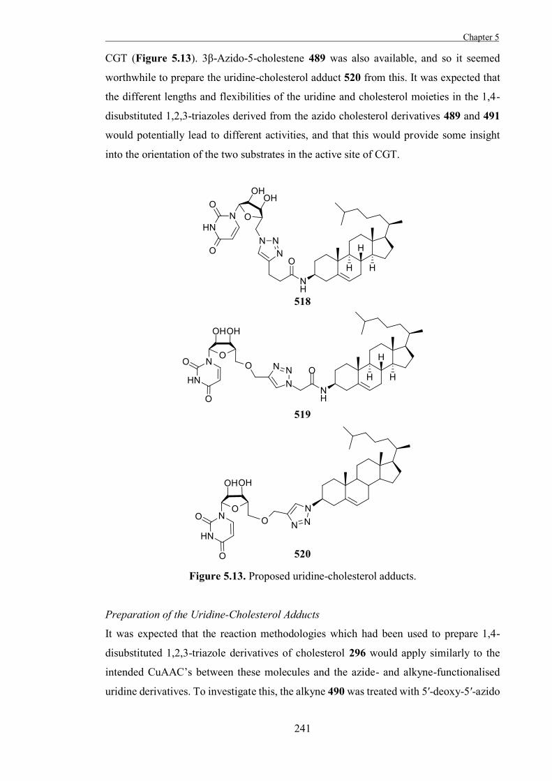
Chapter 5
241
CGT (Figure 5.13). 3β-Azido-5-cholestene 489 was also available, and so it seemed
worthwhile to prepare the uridine-cholesterol adduct 520 from this. It was expected that
the different lengths and flexibilities of the uridine and cholesterol moieties in the 1,4-
disubstituted 1,2,3-triazoles derived from the azido cholesterol derivatives 489 and 491
would potentially lead to different activities, and that this would provide some insight
into the orientation of the two substrates in the active site of CGT.
Figure 5.13. Proposed uridine-cholesterol adducts.
Preparation of the Uridine-Cholesterol Adducts
It was expected that the reaction methodologies which had been used to prepare 1,4-
disubstituted 1,2,3-triazole derivatives of cholesterol 296 would apply similarly to the
intended CuAAC’s between these molecules and the azide- and alkyne-functionalised
uridine derivatives. To investigate this, the alkyne 490 was treated with 5′-deoxy-5′-azido
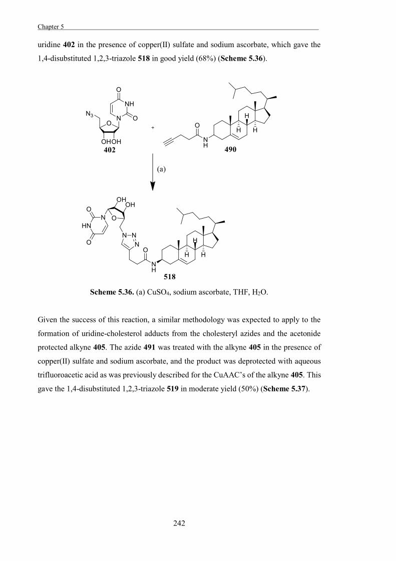
Chapter 5 .
242
uridine 402 in the presence of copper(II) sulfate and sodium ascorbate, which gave the
1,4-disubstituted 1,2,3-triazole 518 in good yield (68%) (Scheme 5.36).
Scheme 5.36. (a) CuSO4, sodium ascorbate, THF, H2O.
Given the success of this reaction, a similar methodology was expected to apply to the
formation of uridine-cholesterol adducts from the cholesteryl azides and the acetonide
protected alkyne 405. The azide 491 was treated with the alkyne 405 in the presence of
copper(II) sulfate and sodium ascorbate, and the product was deprotected with aqueous
trifluoroacetic acid as was previously described for the CuAAC’s of the alkyne 405. This
gave the 1,4-disubstituted 1,2,3-triazole 519 in moderate yield (50%) (Scheme 5.37).
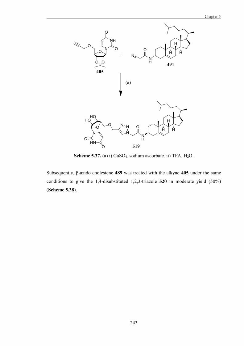
Chapter 5
243
Scheme 5.37. (a) i) CuSO4, sodium ascorbate. ii) TFA, H2O.
Subsequently, β-azido cholestene 489 was treated with the alkyne 405 under the same
conditions to give the 1,4-disubstituted 1,2,3-triazole 520 in moderate yield (50%)
(Scheme 5.38).
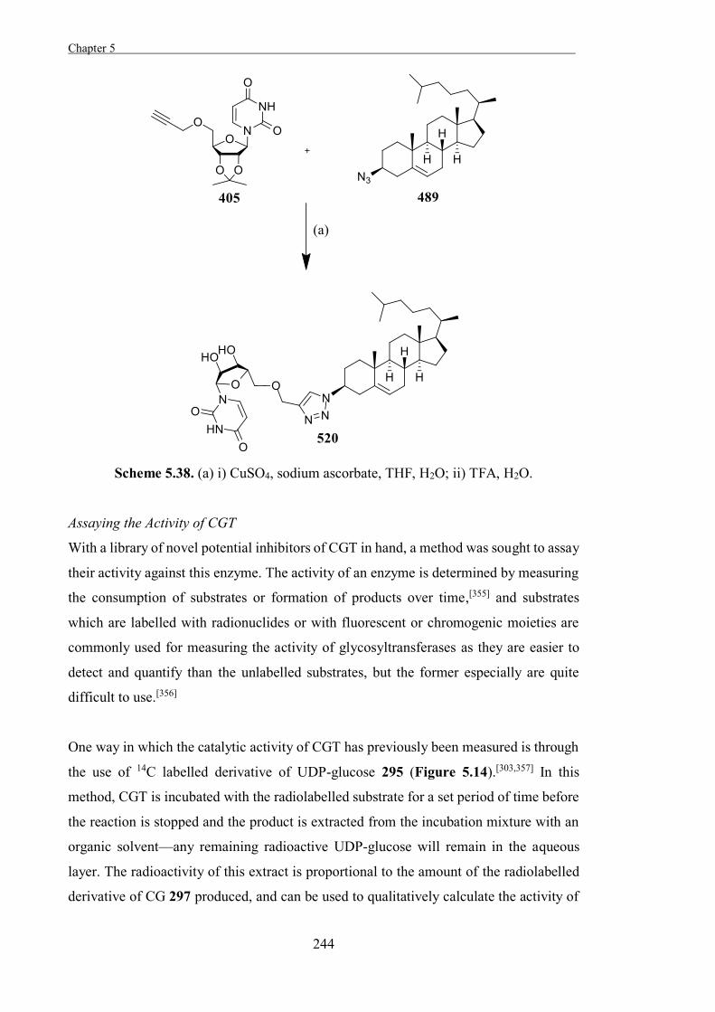
Chapter 5 .
244
Scheme 5.38. (a) i) CuSO4, sodium ascorbate, THF, H2O; ii) TFA, H2O.
Assaying the Activity of CGT
With a library of novel potential inhibitors of CGT in hand, a method was sought to assay
their activity against this enzyme. The activity of an enzyme is determined by measuring
the consumption of substrates or formation of products over time,[355] and substrates
which are labelled with radionuclides or with fluorescent or chromogenic moieties are
commonly used for measuring the activity of glycosyltransferases as they are easier to
detect and quantify than the unlabelled substrates, but the former especially are quite
difficult to use.[356]
One way in which the catalytic activity of CGT has previously been measured is through
the use of 14C labelled derivative of UDP-glucose 295 (Figure 5.14).[303,357] In this
method, CGT is incubated with the radiolabelled substrate for a set period of time before
the reaction is stopped and the product is extracted from the incubation mixture with an
organic solvent—any remaining radioactive UDP-glucose will remain in the aqueous
layer. The radioactivity of this extract is proportional to the amount of the radiolabelled
derivative of CG 297 produced, and can be used to qualitatively calculate the activity of
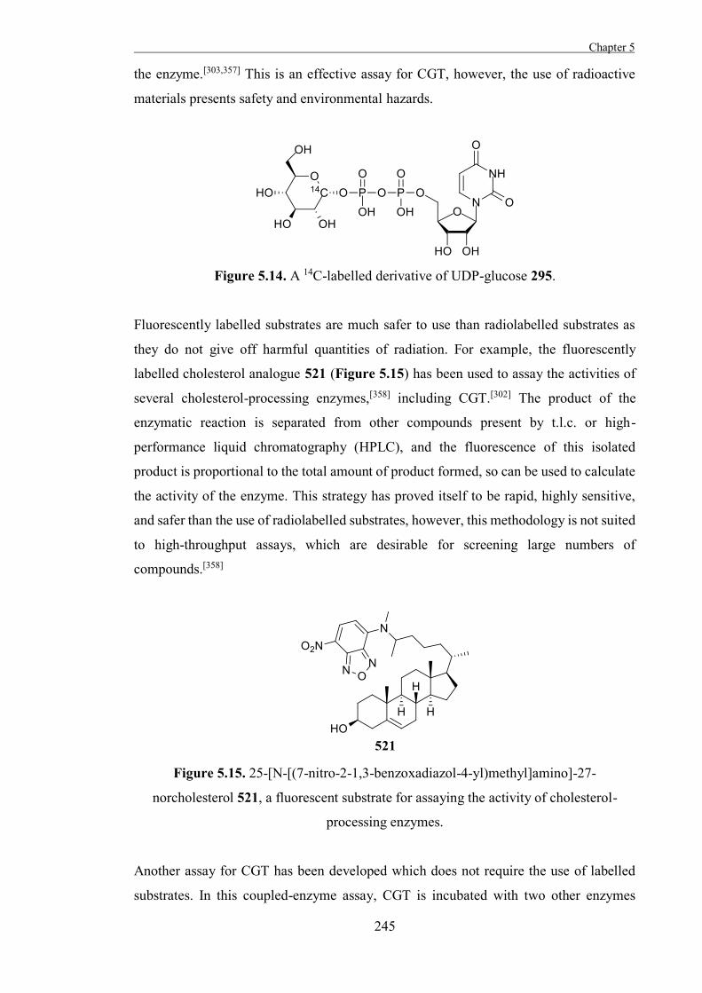
Chapter 5
245
the enzyme.[303,357] This is an effective assay for CGT, however, the use of radioactive
materials presents safety and environmental hazards.
Figure 5.14. A 14C-labelled derivative of UDP-glucose 295.
Fluorescently labelled substrates are much safer to use than radiolabelled substrates as
they do not give off harmful quantities of radiation. For example, the fluorescently
labelled cholesterol analogue 521 (Figure 5.15) has been used to assay the activities of
several cholesterol-processing enzymes,[358] including CGT.[302] The product of the
enzymatic reaction is separated from other compounds present by t.l.c. or high-
performance liquid chromatography (HPLC), and the fluorescence of this isolated
product is proportional to the total amount of product formed, so can be used to calculate
the activity of the enzyme. This strategy has proved itself to be rapid, highly sensitive,
and safer than the use of radiolabelled substrates, however, this methodology is not suited
to high-throughput assays, which are desirable for screening large numbers of
compounds.[358]
Figure 5.15. 25-[N-[(7-nitro-2-1,3-benzoxadiazol-4-yl)methyl]amino]-27-
norcholesterol 521, a fluorescent substrate for assaying the activity of cholesterol-
processing enzymes.
Another assay for CGT has been developed which does not require the use of labelled
substrates. In this coupled-enzyme assay, CGT is incubated with two other enzymes
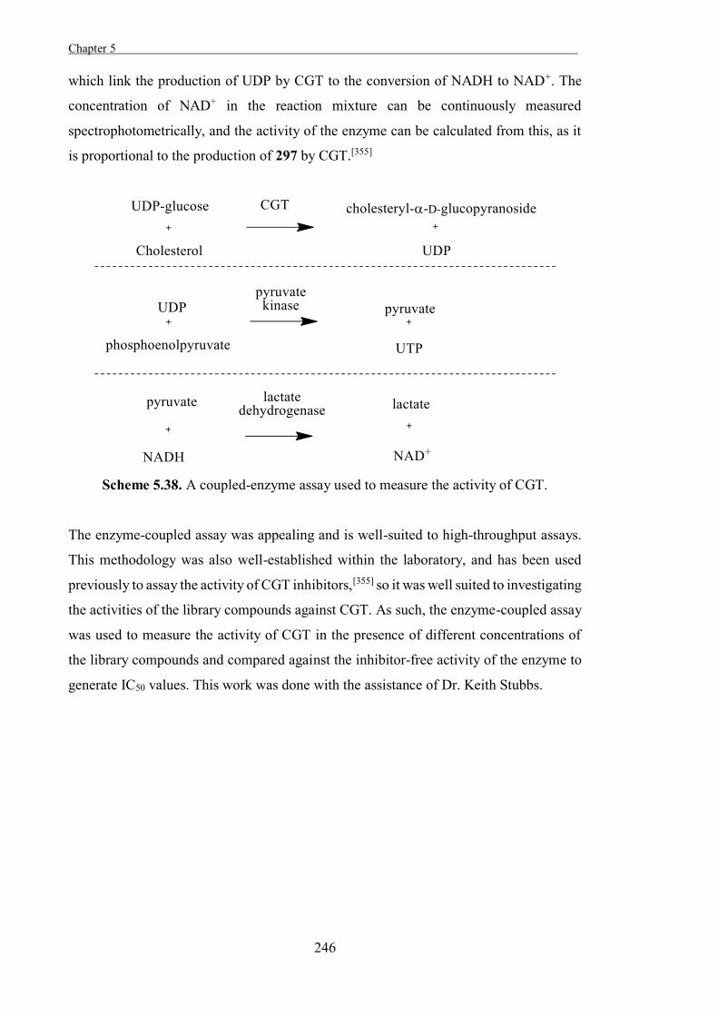
Chapter 5 .
246
which link the production of UDP by CGT to the conversion of NADH to NAD+. The
concentration of NAD+ in the reaction mixture can be continuously measured
spectrophotometrically, and the activity of the enzyme can be calculated from this, as it
is proportional to the production of 297 by CGT.[355]
Scheme 5.38. A coupled-enzyme assay used to measure the activity of CGT.
The enzyme-coupled assay was appealing and is well-suited to high-throughput assays.
This methodology was also well-established within the laboratory, and has been used
previously to assay the activity of CGT inhibitors,[355] so it was well suited to investigating
the activities of the library compounds against CGT. As such, the enzyme-coupled assay
was used to measure the activity of CGT in the presence of different concentrations of
the library compounds and compared against the inhibitor-free activity of the enzyme to
generate IC50 values. This work was done with the assistance of Dr. Keith Stubbs.
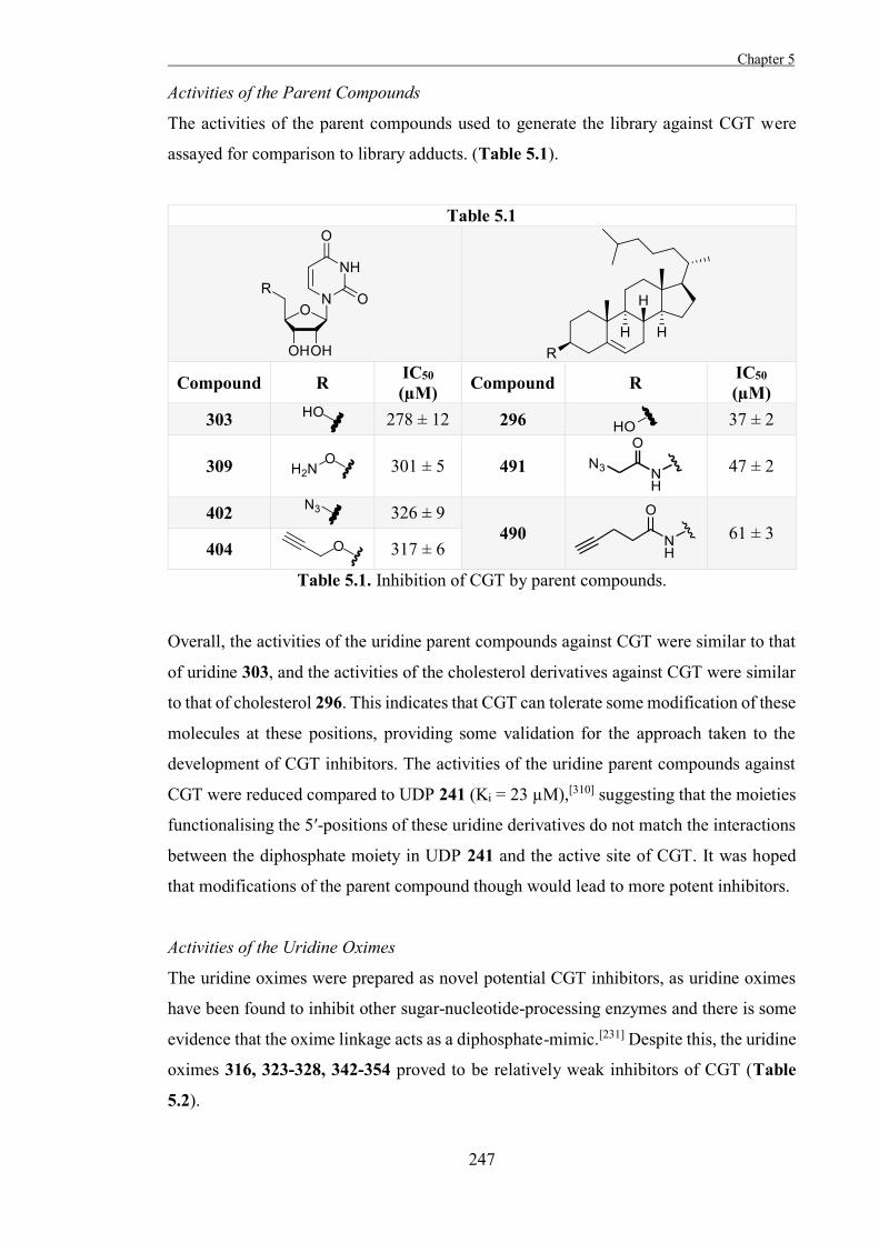
Chapter 5
247
Activities of the Parent Compounds
The activities of the parent compounds used to generate the library against CGT were
assayed for comparison to library adducts. (Table 5.1).
Table 5.1
Compound R IC50
(µM) Compound R IC50 (µM)
303 278 ± 12 296 37 ± 2
309 301 ± 5 491
47 ± 2
402 326 ± 9 490
61 ± 3
404
317 ± 6
Table 5.1. Inhibition of CGT by parent compounds.
Overall, the activities of the uridine parent compounds against CGT were similar to that
of uridine 303, and the activities of the cholesterol derivatives against CGT were similar
to that of cholesterol 296. This indicates that CGT can tolerate some modification of these
molecules at these positions, providing some validation for the approach taken to the
development of CGT inhibitors. The activities of the uridine parent compounds against
CGT were reduced compared to UDP 241 (Ki = 23 µM),[310] suggesting that the moieties
functionalising the 5′-positions of these uridine derivatives do not match the interactions
between the diphosphate moiety in UDP 241 and the active site of CGT. It was hoped
that modifications of the parent compound though would lead to more potent inhibitors.
Activities of the Uridine Oximes
The uridine oximes were prepared as novel potential CGT inhibitors, as uridine oximes
have been found to inhibit other sugar-nucleotide-processing enzymes and there is some
evidence that the oxime linkage acts as a diphosphate-mimic.[231] Despite this, the uridine
oximes 316, 323-328, 342-354 proved to be relatively weak inhibitors of CGT (Table
5.2).
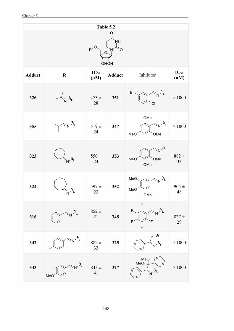
Chapter 5 .
248
Table 5.2
Adduct R IC50 (µM) Adduct Inhibitor IC50
(µM)
326
473 ±
28 351
> 1000
355
519 ±
24 347
> 1000
323
550 ±
24 353
882 ±
33
324
597 ±
23 352
904 ±
48
316
852 ± 21
348
827 ±
29
342
882 ±
33 325
> 1000
343
843 ±
41 327
> 1000
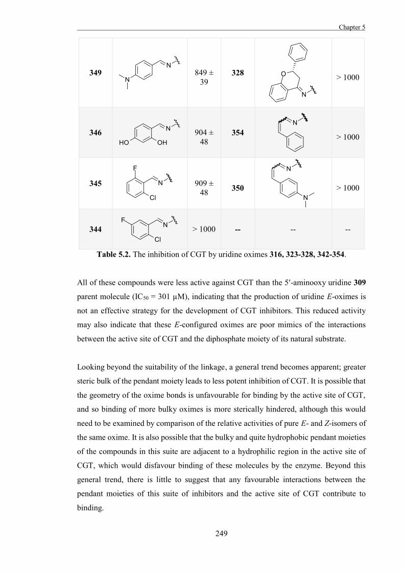
Chapter 5
249
349
849 ±
39 328
> 1000
346
904 ±
48 354
> 1000
345
909 ±
48
350
> 1000
344
> 1000 -- -- --
Table 5.2. The inhibition of CGT by uridine oximes 316, 323-328, 342-354.
All of these compounds were less active against CGT than the 5′-aminooxy uridine 309
parent molecule (IC50 = 301 µM), indicating that the production of uridine E-oximes is
not an effective strategy for the development of CGT inhibitors. This reduced activity
may also indicate that these E-configured oximes are poor mimics of the interactions
between the active site of CGT and the diphosphate moiety of its natural substrate.
Looking beyond the suitability of the linkage, a general trend becomes apparent; greater
steric bulk of the pendant moiety leads to less potent inhibition of CGT. It is possible that
the geometry of the oxime bonds is unfavourable for binding by the active site of CGT,
and so binding of more bulky oximes is more sterically hindered, although this would
need to be examined by comparison of the relative activities of pure E- and Z-isomers of
the same oxime. It is also possible that the bulky and quite hydrophobic pendant moieties
of the compounds in this suite are adjacent to a hydrophilic region in the active site of
CGT, which would disfavour binding of these molecules by the enzyme. Beyond this
general trend, there is little to suggest that any favourable interactions between the
pendant moieties of this suite of inhibitors and the active site of CGT contribute to
binding.
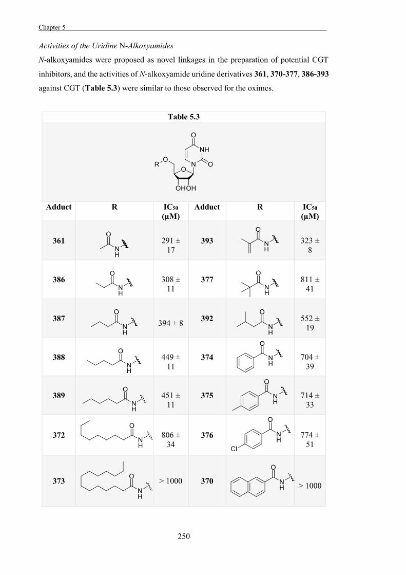
Chapter 5 .
250
Activities of the Uridine N-Alkoxyamides
N-alkoxyamides were proposed as novel linkages in the preparation of potential CGT
inhibitors, and the activities of N-alkoxyamide uridine derivatives 361, 370-377, 386-393
against CGT (Table 5.3) were similar to those observed for the oximes.
Table 5.3
Adduct R IC50 (µM)
Adduct R IC50 (µM)
361
291 ±
17 393
323 ±
8
386
308 ±
11 377
811 ±
41
387
394 ± 8 392
552 ±
19
388
449 ±
11 374
704 ±
39
389
451 ±
11 375
714 ±
33
372
806 ±
34 376
774 ±
51
373
> 1000
370
> 1000
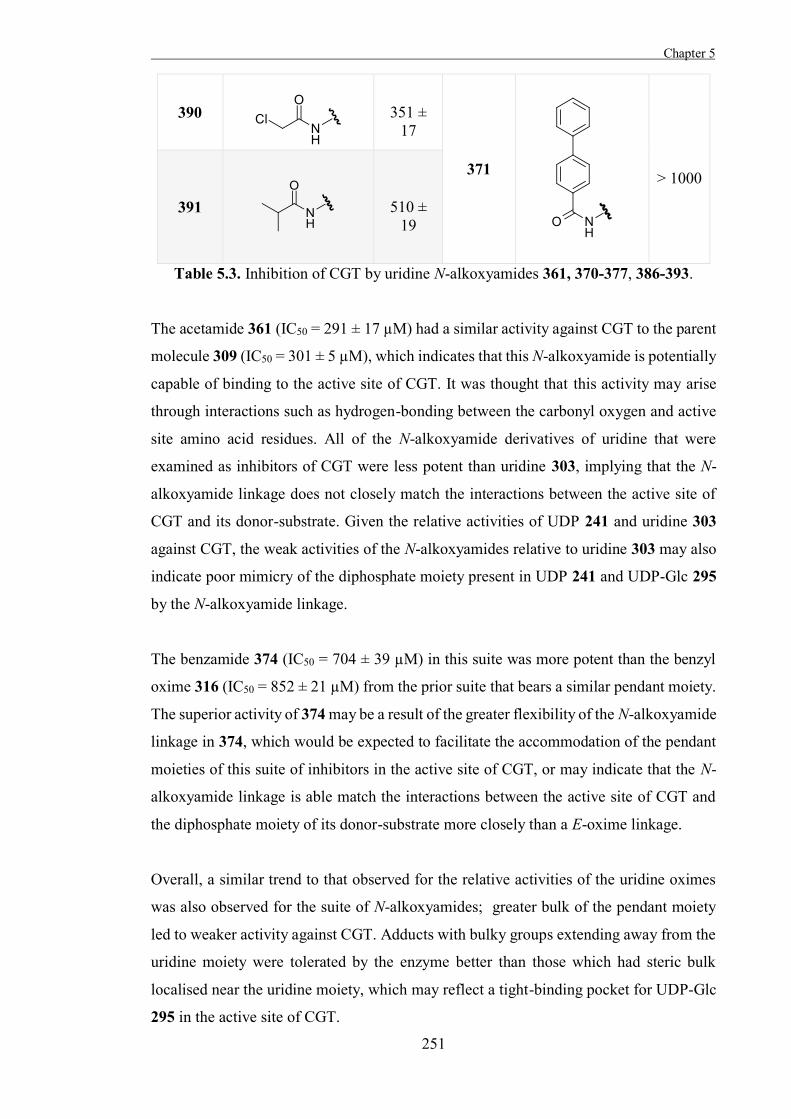
Chapter 5
251
390
351 ±
17
371
> 1000
391
510 ±
19
Table 5.3. Inhibition of CGT by uridine N-alkoxyamides 361, 370-377, 386-393.
The acetamide 361 (IC50 = 291 ± 17 µM) had a similar activity against CGT to the parent
molecule 309 (IC50 = 301 ± 5 µM), which indicates that this N-alkoxyamide is potentially
capable of binding to the active site of CGT. It was thought that this activity may arise
through interactions such as hydrogen-bonding between the carbonyl oxygen and active
site amino acid residues. All of the N-alkoxyamide derivatives of uridine that were
examined as inhibitors of CGT were less potent than uridine 303, implying that the N-
alkoxyamide linkage does not closely match the interactions between the active site of
CGT and its donor-substrate. Given the relative activities of UDP 241 and uridine 303
against CGT, the weak activities of the N-alkoxyamides relative to uridine 303 may also
indicate poor mimicry of the diphosphate moiety present in UDP 241 and UDP-Glc 295
by the N-alkoxyamide linkage.
The benzamide 374 (IC50 = 704 ± 39 µM) in this suite was more potent than the benzyl
oxime 316 (IC50 = 852 ± 21 µM) from the prior suite that bears a similar pendant moiety.
The superior activity of 374 may be a result of the greater flexibility of the N-alkoxyamide
linkage in 374, which would be expected to facilitate the accommodation of the pendant
moieties of this suite of inhibitors in the active site of CGT, or may indicate that the N-
alkoxyamide linkage is able match the interactions between the active site of CGT and
the diphosphate moiety of its donor-substrate more closely than a E-oxime linkage.
Overall, a similar trend to that observed for the relative activities of the uridine oximes
was also observed for the suite of N-alkoxyamides; greater bulk of the pendant moiety
led to weaker activity against CGT. Adducts with bulky groups extending away from the
uridine moiety were tolerated by the enzyme better than those which had steric bulk
localised near the uridine moiety, which may reflect a tight-binding pocket for UDP-Glc
295 in the active site of CGT.
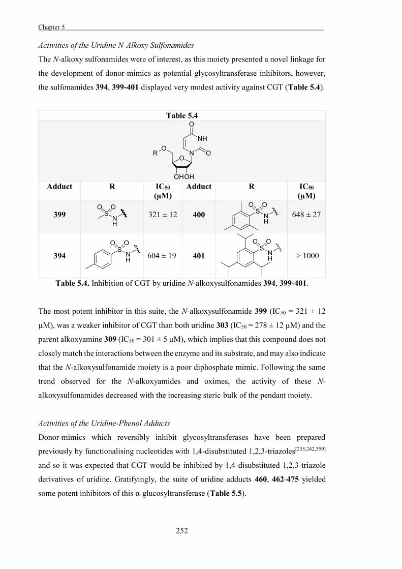
Chapter 5 .
252
Activities of the Uridine N-Alkoxy Sulfonamides
The N-alkoxy sulfonamides were of interest, as this moiety presented a novel linkage for
the development of donor-mimics as potential glycosyltransferase inhibitors, however,
the sulfonamides 394, 399-401 displayed very modest activity against CGT (Table 5.4).
Table 5.4
Adduct R IC50
(µM) Adduct R IC50
(µM)
399
321 ± 12
400
648 ± 27
394
604 ± 19
401
> 1000
Table 5.4. Inhibition of CGT by uridine N-alkoxysulfonamides 394, 399-401.
The most potent inhibitor in this suite, the N-alkoxysulfonamide 399 (IC50 = 321 ± 12
µM), was a weaker inhibitor of CGT than both uridine 303 (IC50 = 278 ± 12 µM) and the
parent alkoxyamine 309 (IC50 = 301 ± 5 µM), which implies that this compound does not
closely match the interactions between the enzyme and its substrate, and may also indicate
that the N-alkoxysulfonamide moiety is a poor diphosphate mimic. Following the same
trend observed for the N-alkoxyamides and oximes, the activity of these N-
alkoxysulfonamides decreased with the increasing steric bulk of the pendant moiety.
Activities of the Uridine-Phenol Adducts
Donor-mimics which reversibly inhibit glycosyltransferases have been prepared
previously by functionalising nucleotides with 1,4-disubstituted 1,2,3-triazoles[235,242,359]
and so it was expected that CGT would be inhibited by 1,4-disubstituted 1,2,3-triazole
derivatives of uridine. Gratifyingly, the suite of uridine adducts 460, 462-475 yielded
some potent inhibitors of this α-glucosyltransferase (Table 5.5).
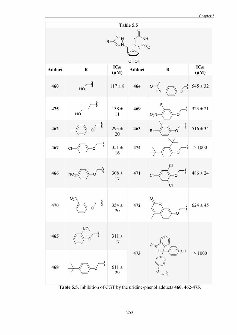
Chapter 5
253
Table 5.5
Adduct R IC50
(µM) Adduct R IC50 (µM)
460
117 ± 8 464
545 ± 32
475
138 ±
11 469
323 ± 21
462
293 ±
20 463
516 ± 34
467
351 ±
16 474
> 1000
466
308 ±
17 471
486 ± 24
470
354 ±
20 472
624 ± 45
465
311 ±
17
473
> 1000
468
611 ±
29
Table 5.5. Inhibition of CGT by the uridine-phenol adducts 460, 462-475.
Chapter 5 .
254
The most potent inhibitors of CGT found in this series were the hydroxymethyl adduct
460 (IC50 = 117 ± 8 µM) and the hydroxypropyl adduct 475 (IC50 = 138 ± 11 µM), both
of which were significantly more potent than 5′-deoxy 5′-azido uridine 402 (IC50 = 326 ±
9 µM), or uridine 303 (IC50 = 278 ± 12 µM). This potency implies closer mimicry of the
donor-substrate of CGT by these adducts than by uridine 303 itself, and may be an
indication of the suitability of a 1,4-disubstituted triazole as a substitute for the
diphosphate moiety in sugar-nucleotides for the preparation of donor-mimics as potential
glycosyltransferase inhibitors.
As had been observed for the other suites of uridine adducts, the activity of these adducts
generally decreased with increased steric bulk. As well as steric effects, it is possible that
more specific interactions with amino acid residues in the active site of CGT account for
the relative potency of the aliphatic uridine adducts against this enzyme compared to the
aromatic adducts. It is possible that repulsive forces between a hydrophilic region in the
active site of CGT and the hydrophobic aromatic rings of the aryl triazoles reduce the
affinities of these adducts for CGT, as was suggested for the uridine oximes, N-
alkoxyamides, and N-alkoxysulfonamides. It was also considered that the terminal
hydroxyl moieties of the two aliphatic adducts may potentially contribute binding
interactions with hydrophilic active site amino acid residues in CGT which the aromatic
adducts do not, which would also account for the increased affinities of these uridine
adducts for CGT compared to the aromatic adducts.
Activities of the Uridine-Amino Acid Adducts
The suite of 1,4-disubstituted 1,2,3-triazole uridine derivatives 476, 479-486 also yielded
some highly potent inhibitors of CGT (Table 5.6).
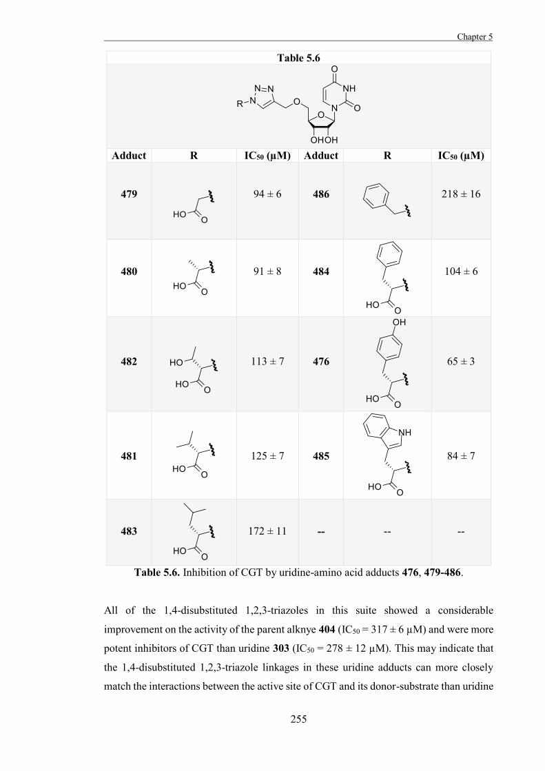
Chapter 5
255
Table 5.6
Adduct R IC50 (µM) Adduct R IC50 (µM)
479
94 ± 6
486
218 ± 16
480
91 ± 8
484
104 ± 6
482
113 ± 7
476
65 ± 3
481
125 ± 7
485
84 ± 7
483
172 ± 11
--
--
--
Table 5.6. Inhibition of CGT by uridine-amino acid adducts 476, 479-486.
All of the 1,4-disubstituted 1,2,3-triazoles in this suite showed a considerable
improvement on the activity of the parent alknye 404 (IC50 = 317 ± 6 µM) and were more
potent inhibitors of CGT than uridine 303 (IC50 = 278 ± 12 µM). This may indicate that
the 1,4-disubstituted 1,2,3-triazole linkages in these uridine adducts can more closely
match the interactions between the active site of CGT and its donor-substrate than uridine
Chapter 5 .
256
303, and that this linkage may be able to effectively mimic the diphosphate moiety present
in the substrate.
One structural feature common to most of the uridine adducts in this suite is a carboxylic
acid moiety. The benzyl adduct 486, which is considerably weaker than the other 1,4-
disubstituted 1,2,3-triazoles in this suite, is the only adduct in this suite to lack a
carboxylic acid moiety. The phenylalanine adduct 484 (IC50 = 104 ± 6 µM) is structurally
very similar to the benzyl adduct 486, but has a significantly higher affinity for CGT,
suggesting that the pendant carboxylic acid moiety in the adduct 484 contributes to the
strong binding of this molecule by the active site of CGT. It is not clear whether the
carboxylic acid moiety contributes to binding through interactions with the diphosphate-
binding region of the active site of CGT or through interactions with other portions of the
active site. The carboxylic acid moieties of these adducts would be expected to be
deprotonated for the most part under the conditions of the assay (pH 7) and so would be
charged. Electrostatic interactions between charged moieties can significantly contribute
to binding,[360] and such interactions may underlie the potent inhibition of CGT by these
uridine adducts.
The benzyl adduct 486 is significantly more potent than aryl triazoles with similar pendant
moieties from the previous suite of inhibitors, such as the 4-toluoyl adduct 462 (IC50 =
293 ± 20 µM). It is possible that the ether-linkage between the 1,4-disubstituted 1,2,3-
triazole and uridine moieties in the suite of inhibitors from Table 5.6 confers greater
flexibility on these pendant moieties, which allows these adducts to be more easily
accommodated into the active site, or that the linkage in 486 more closely matches the
interactions between the active site of CGT and the diphosphate moiety of its donor-
substrate than the linkage in the previous suite of 1,4-disubstituted 1,2,3-triazole
functionalised uridine derivatives.
Aside from the apparent affinity-enhancing effect of a pendant carboxylic acid moiety, a
clear relationship between the relative activities of adducts within this suite against and
their pendant moieties was not evident, and this may indicate that there are fortuitous
binding interactions between the active site of CGT and some of these pendant moieties.
The tyrosine adduct 476 was the most potent CGT inhibitor in this suite (IC50 = 65 ± 3
µM), and was significantly more potent than the structurally similar phenylalanine adduct
484 (IC50 = 104 ± 6 µM). As the only difference between the pendant moieties of these
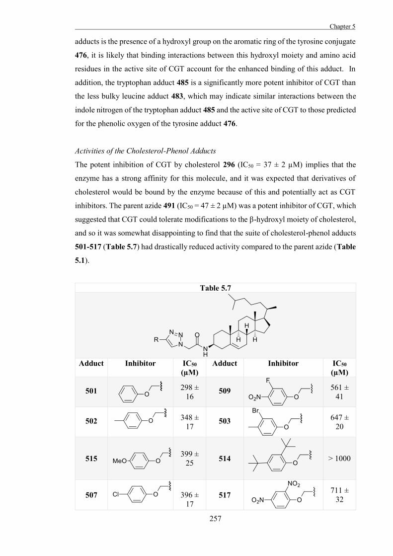
Chapter 5
257
adducts is the presence of a hydroxyl group on the aromatic ring of the tyrosine conjugate
476, it is likely that binding interactions between this hydroxyl moiety and amino acid
residues in the active site of CGT account for the enhanced binding of this adduct. In
addition, the tryptophan adduct 485 is a significantly more potent inhibitor of CGT than
the less bulky leucine adduct 483, which may indicate similar interactions between the
indole nitrogen of the tryptophan adduct 485 and the active site of CGT to those predicted
for the phenolic oxygen of the tyrosine adduct 476.
Activities of the Cholesterol-Phenol Adducts
The potent inhibition of CGT by cholesterol 296 (IC50 = 37 ± 2 µM) implies that the
enzyme has a strong affinity for this molecule, and it was expected that derivatives of
cholesterol would be bound by the enzyme because of this and potentially act as CGT
inhibitors. The parent azide 491 (IC50 = 47 ± 2 µM) was a potent inhibitor of CGT, which
suggested that CGT could tolerate modifications to the β-hydroxyl moiety of cholesterol,
and so it was somewhat disappointing to find that the suite of cholesterol-phenol adducts
501-517 (Table 5.7) had drastically reduced activity compared to the parent azide (Table
5.1).
Table 5.7
Adduct Inhibitor IC50
(µM) Adduct Inhibitor IC50
(µM)
501
298 ± 16
509
561 ± 41
502
348 ±
17
503
647 ± 20
515
399 ± 25 514
> 1000
507
396 ±
17 517
711 ± 32
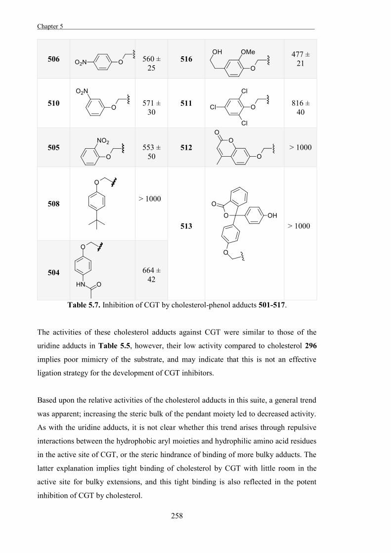
Chapter 5 .
258
506
560 ±
25 516
477 ± 21
510
571 ±
30 511
816 ±
40
505
553 ±
50 512
> 1000
508
> 1000
513
> 1000
504
664 ±
42
Table 5.7. Inhibition of CGT by cholesterol-phenol adducts 501-517.
The activities of these cholesterol adducts against CGT were similar to those of the
uridine adducts in Table 5.5, however, their low activity compared to cholesterol 296
implies poor mimicry of the substrate, and may indicate that this is not an effective
ligation strategy for the development of CGT inhibitors.
Based upon the relative activities of the cholesterol adducts in this suite, a general trend
was apparent; increasing the steric bulk of the pendant moiety led to decreased activity.
As with the uridine adducts, it is not clear whether this trend arises through repulsive
interactions between the hydrophobic aryl moieties and hydrophilic amino acid residues
in the active site of CGT, or the steric hindrance of binding of more bulky adducts. The
latter explanation implies tight binding of cholesterol by CGT with little room in the
active site for bulky extensions, and this tight binding is also reflected in the potent
inhibition of CGT by cholesterol.
Chapter 5
259
The homovanillin adduct 516 is one exception to this trend, as this 1,4-disubstituted 1,2,3-
triazole was a considerably more potent inhibitor (IC50 = 477 ± 21 µM), of CGT than
several less bulky adducts such as the 4-nitrophenyl triazole 506 (IC50 = 560 ± 25 µM).
It is possible that fortuitous interactions between the homovannilin moiety and CGT
confers this relative potency on 516, although this adduct is still a weak inhibitor of the
enzyme.
Activities of the Cholesterol-Amino Acid Adducts
It was hoped that the cholesterol amino acid adducts would exhibit interesting activities
against CGT, as was observed for the suite of uridine amino acid adducts 492-500.
Gratifyingly, the compounds in this suite of 1,4-disubstituted 1,2,3-triazoles displayed
potent inhibition of CGT (Table 5.8).
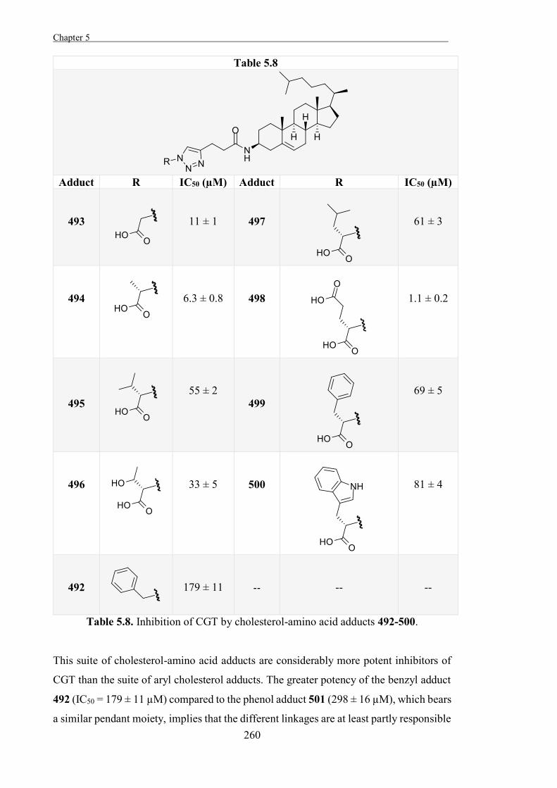
Chapter 5 .
260
Table 5.8
Adduct R IC50 (µM) Adduct R IC50 (µM)
493
11 ± 1
497
61 ± 3
494
6.3 ± 0.8
498
1.1 ± 0.2
495
55 ± 2
499
69 ± 5
496
33 ± 5
500
81 ± 4
492
179 ± 11
--
--
--
Table 5.8. Inhibition of CGT by cholesterol-amino acid adducts 492-500.
This suite of cholesterol-amino acid adducts are considerably more potent inhibitors of
CGT than the suite of aryl cholesterol adducts. The greater potency of the benzyl adduct
492 (IC50 = 179 ± 11 µM) compared to the phenol adduct 501 (298 ± 16 µM), which bears
a similar pendant moiety, implies that the different linkages are at least partly responsible
Chapter 5
261
for these differences in activity. This suite utilises a slightly extended linkage, which may
position the triazole and pendant moieties more favourably in the active site of CGT, and
also may confer greater flexibility upon these adducts to allow the pendant moieties to be
accommodated more readily in the active site of CGT.
As had been observed for the corresponding suite of uridine adducts (Table 5.6), pendant
carboxylic acid moieties conferred greater affinity for CGT upon the cholesterol adducts;
the phenylalanine adduct 499 (IC50 = 69 ± 5 µM) was significantly more potent than the
benzyl adduct 492 (IC50 = 179 ± 11 µM). It is unclear whether the pendant carboxylic
acid moieties of these inhibitors interact with the same region of the active site of CGT
as the carboxylic acid moieties of the uridine adducts.
For those adducts in this series bearing hydrophobic pendant moieties, the steric bulk of
the pendant moiety also affected their activity. As had been observed for the previous
suite of inhibitors, increasing the bulk of the hydrophobic moiety generally decreased the
affinity for CGT. As with the other inhibitors, it is likely that this trend is the result of the
steric hindrance of binding a bulkier molecule, repulsive forces between the hydrophobic
moieties and the active site of CGT, or a combination of these factors. It is notable that
the alanine adduct 494 is a more potent inhibitor of CGT than the glycine adduct 493,
going against this trend. It is possible that the pendant methyl moiety of the alanine adduct
494 interacts with a hydrophobic region in the active site of CGT, increasing the affinity
of this inhibitor relative to the glycine adduct 493. The valine adduct 495 is a considerably
less potent inhibitor of CGT than the glycine or alanine adducts, suggesting that this
hydrophobic region must be too small to accommodate the pendant isopropyl moiety of
the adduct 495. That the threonine adduct 496 is a more potent inhibitor than the valine
adduct 495 supports this, as the greater affinity of the adduct 496 for CGT likely arises
from interactions between the hydroxyl moiety and the active site of the enzyme,
implying a hydrophilic region in the active site.
The glutamate adduct 498 (IC50 = 1.1 ± 0.2 µM), bearing two pendant carboxylic acids,
was the most potent CGT inhibitor developed in this study. This activity must arise
through favourable interactions with amino acid residues in the active site of CGT. It
would be expected that these carboxylic acids would be largely ionised under the assay
conditions (pH 7), and so it is possible that electrostatic attractions and hydrogen bonding
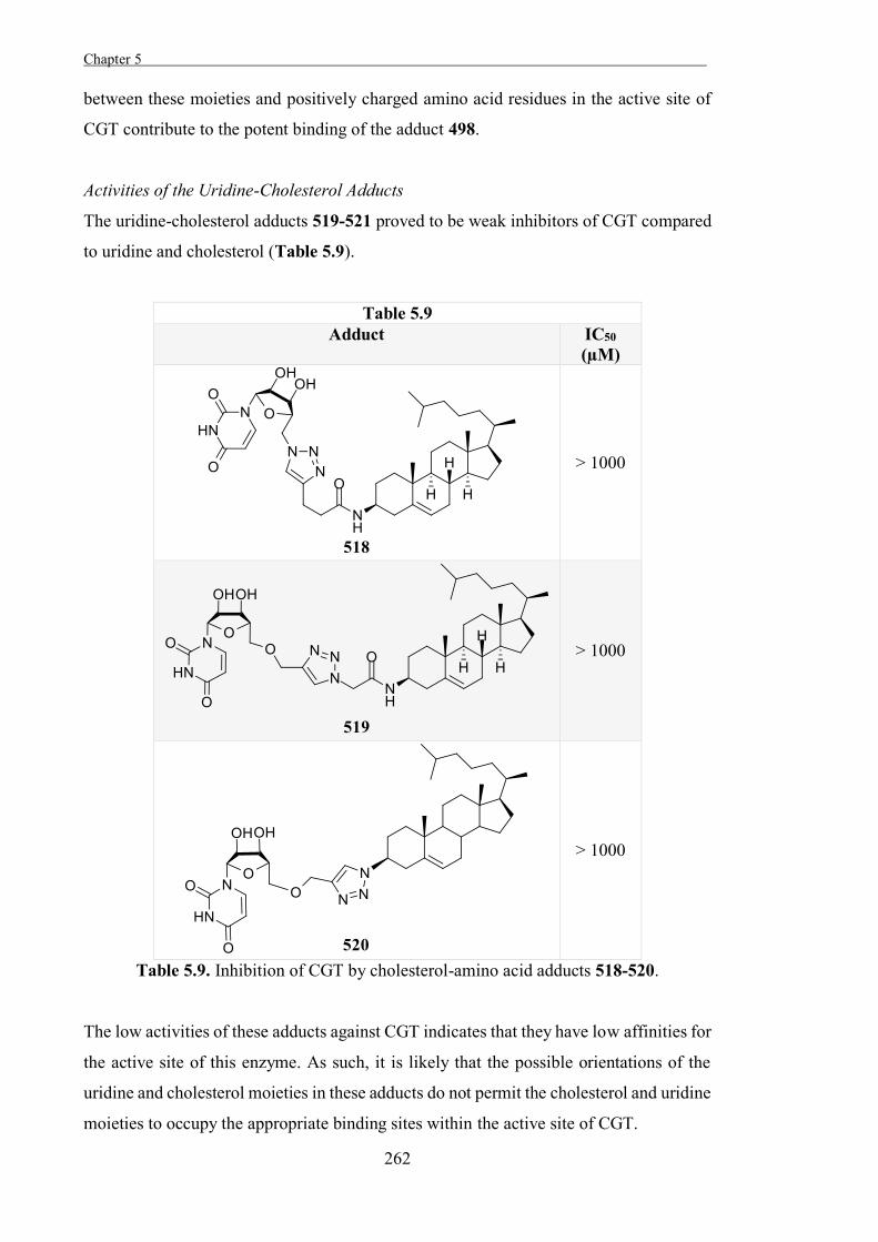
Chapter 5 .
262
between these moieties and positively charged amino acid residues in the active site of
CGT contribute to the potent binding of the adduct 498.
Activities of the Uridine-Cholesterol Adducts
The uridine-cholesterol adducts 519-521 proved to be weak inhibitors of CGT compared
to uridine and cholesterol (Table 5.9).
Table 5.9 Adduct IC50
(µM)
> 1000
> 1000
> 1000
Table 5.9. Inhibition of CGT by cholesterol-amino acid adducts 518-520.
The low activities of these adducts against CGT indicates that they have low affinities for
the active site of this enzyme. As such, it is likely that the possible orientations of the
uridine and cholesterol moieties in these adducts do not permit the cholesterol and uridine
moieties to occupy the appropriate binding sites within the active site of CGT.
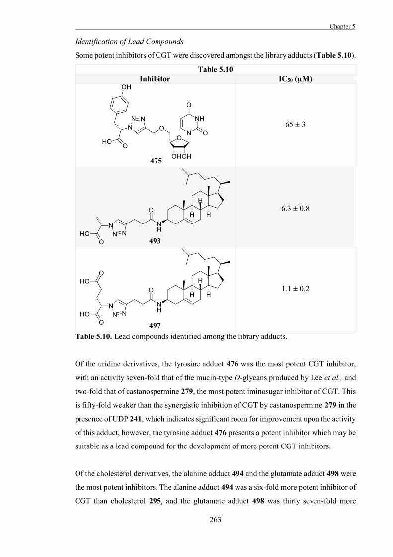
Chapter 5
263
Identification of Lead Compounds
Some potent inhibitors of CGT were discovered amongst the library adducts (Table 5.10).
Table 5.10 Inhibitor IC50 (µM)
65 ± 3
6.3 ± 0.8
1.1 ± 0.2
Table 5.10. Lead compounds identified among the library adducts.
Of the uridine derivatives, the tyrosine adduct 476 was the most potent CGT inhibitor,
with an activity seven-fold that of the mucin-type O-glycans produced by Lee et al., and
two-fold that of castanospermine 279, the most potent iminosugar inhibitor of CGT. This
is fifty-fold weaker than the synergistic inhibition of CGT by castanospermine 279 in the
presence of UDP 241, which indicates significant room for improvement upon the activity
of this adduct, however, the tyrosine adduct 476 presents a potent inhibitor which may be
suitable as a lead compound for the development of more potent CGT inhibitors.
Of the cholesterol derivatives, the alanine adduct 494 and the glutamate adduct 498 were
the most potent inhibitors. The alanine adduct 494 was a six-fold more potent inhibitor of
CGT than cholesterol 295, and the glutamate adduct 498 was thirty seven-fold more
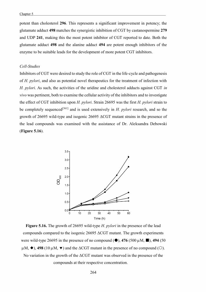
Chapter 5 .
264
potent than cholesterol 296. This represents a significant improvement in potency; the
glutamate adduct 498 matches the synergistic inhibition of CGT by castanospermine 279
and UDP 241, making this the most potent inhibitor of CGT reported to date. Both the
glutamate adduct 498 and the alanine adduct 494 are potent enough inhibitors of the
enzyme to be suitable leads for the development of more potent CGT inhibitors.
Cell-Studies
Inhibitors of CGT were desired to study the role of CGT in the life-cycle and pathogenesis
of H. pylori, and also as potential novel therapeutics for the treatment of infection with
H. pylori. As such, the activities of the uridine and cholesterol adducts against CGT in
vivo was pertinent, both to examine the cellular activity of the inhibitors and to investigate
the effect of CGT inhibition upon H. pylori. Strain 26695 was the first H. pylori strain to
be completely sequenced[361] and is used extensively in H. pylori research, and so the
growth of 26695 wild-type and isogenic 26695 ΔCGT mutant strains in the presence of
the lead compounds was examined with the assistance of Dr. Aleksandra Debowski
(Figure 5.16).
Figure 5.16. The growth of 26695 wild-type H. pylori in the presence of the lead
compounds compared to the isogenic 26695 ΔCGT mutant. The growth experiments
were wild-type 26695 in the presence of no compound (), 476 (500 M, ), 494 (50
M, ), 498 (10 M, ) and the ΔCGT mutant in the presence of no compound ().
No variation in the growth of the ΔCGT mutant was observed in the presence of the
compounds at their respective concentration.
Chapter 5
265
The growth of the knockout strain was significantly reduced compared to the wild-type,
reflecting the importance of CGT for this bacterium. As well, the growth of the wild-type
strain was significantly reduced in the presence of the inhibitors, demonstrating that their
action impedes the growth of H. pylori, and implying that the inhibitors can enter cells.
The growth of the knockout strain was unaffected by the inhibitors, which provides
evidence that CGT is the in vivo target of the inhibitors and that they are selective. The
glutamate adduct 498, the most potent CGT inhibitor used, reduced the growth of the
wild-type strain to almost that of the knockout, suggesting strong inhibition of CGT in-
vivo.
Conclusions and Future Work
The bacterium H. pylori is a major cause of human disease which exhibits significant
resistance to available antibiotics. As such, there is a need for novel treatments for
infection with H. pylori, as well as tools to study the biology of this organism in more
detail. It is thought that the virulence factor CGT, which is specific to H. pylori and
facilitates immune evasion by this bacterium, is a potential target for the development of
novel therapeutics, but more potent and specific inhibitors of this enzyme were needed to
investigate this and examine the role that CGT plays in the life-cycle of the bacterium. A
library of potential novel inhibitors of CGT was prepared based upon several scaffolds
which were expected to mimic the donor-substrate or acceptor-substrate of this enzyme,
and some potent inhibitors of the enzyme were identified within this library.
The most potent CGT inhibitor produced in this study based upon a donor-substrate-like
scaffold was the adduct 476. Based upon the relative activities of the different scaffolds
which were expected to act as donor-mimics, the ether-linked 1,4-disubstituted 1,2,3-
triazole linkage was determined to be the best match, out of the scaffolds examined. As
such, this linkage may also be useful in developing potential novel inhibitors of other
glycosyltransferases. It was also apparent that the pendant carboxylate moiety found in
most of the inhibitors lead to an increased affinity for CGT and as well the potency of the
tyrosine adduct 476 compared to the other amino acid-uridine adducts implies specific
and fortuitous interactions between the pendant phenol moiety of this inhibitor and the
active site of CGT. The 1,4-disubstituted 1,2,3-triazole 476 was subsequently assayed
against cell cultures of H. pylori, and was found to reduce the growth of the wild-type
bacterium, but not affect the growth of a CGT knockout, which suggested that the mode
of action of this anti-pylori agent was through inhibition of CGT. While the reduction in
Chapter 5 .
266
growth of the wild-type bacterium upon incubation with this CGT inhibitor was modest,
it provides further evidence that this enzyme is essential for normal functioning of H.
pylori. This also indicates that this compound is able to cross cell-membranes, which is
crucial for therapeutic applications or cellular studies, and when considered alongside the
activity of the adduct 476 against CGT, it suggests that this compound would be a good
lead compound for the development of potentially more potent and specific inhibitors of
CGT.
Of the acceptor-mimics produced in this study, the two most potent inhibitors of CGT
produced were 498 and 494. The potent inhibition of CGT by these cholesterol adducts,
and others found within the same suite, shows that β-functionalised cholesterol
derivatives are tolerated by the active site of CGT, and also implies that the 1,4-
disubstituted 1,2,3-triazole linkage in these inhibitors is able to make favourable
interactions with the active-site. As with the uridine adducts, it was apparent that the
presence of a pendant carboxylate moiety leads to enhanced binding by CGT, and that
the potencies of adducts 498 and 494 compared to the rest of the inhibitors arose through
fortuitous binding interactions with the varied pendant moieties. Both of these inhibitors
were assayed against H. pylori, and were found to significantly reduce the growth of the
wild-type bacterium through the inhibition of CGT. In particular, the alanine adduct
reduced the growth almost to the level of growth of the knockout, suggesting significant
inhibition of CGT activity. This provides further evidence that the inhibition of CGT
reduces the growth of H. pylori, and these inhibitors may also provide lead compounds
for the development of more potent and specific CGT inhibitors.
The potency of the glutamate adduct 498 against CGT suggests that this compound may
be useful for a further investigation of the biological roles of this enzyme in the life-cycle
of H. pylori and the ability to evade immune responses of the host which this enzyme
confers upon the bacterium. This compound may also be suitable for further investigation
of the suitability of CGT as a potential target for novel therapeutics, possibly in
combination with existing therapies, although more evidence is needed to confirm this.
With this in mind, further studies are intended to investigate the effect of the glutamate
adduct 498 on the colonisation of a mouse model host by H. pylori.
Chapter 5
267
Experimental General Procedures
Procedure A: Acetic acid (0.08ml/mmol) was added to a solution of the hydroxylamine
309[313] (1.0 equiv.) in a 2 M methanolic solution of the appropriate aldehyde or ketone
(10 ml/mmol), and the solution was stirred in the dark (r.t., 15-18 h). Concentration of the
solution and flash chromatography of the residue gave the desired oxime.
Procedure B: The appropriate acid chloride (2.0 equiv.) was added to a solution of the
hydroxylamine 309[313] (1.0 equiv.) and DIPEA (2.2 equiv.) in MeOH (15 ml/mmol) at 0
°C and the resulting mixture was stirred (r.t., 3 h).
Procedure C: The appropriate acid anhydride (1.1 equiv.) was added to a solution of the
hydroxylamine 309[313] (1.0 equiv.) in MeOH (12 ml/mmol) and the resulting mixture
was stirred (r.t., 3 h).
Procedure A: Acetic acid (0.08ml/mmol) was added to a solution of the hydroxylamine
309[313] (1.0 equiv.) in a 2 M methanolic solution of the appropriate aldehyde or ketone
(10 ml/mmol), and the solution was stirred in the dark (r.t., 15-18 h). Concentration of the
solution and flash chromatography of the residue gave the desired oxime.
Procedure D: Copper (I) iodide (1.1 equiv.) was added to a solution of the azide 402[362]
(1.0 equiv.), Et3N (1.2 equiv.), and the appropriate alkyne (1.1 equiv.) in CHCl3 (20
ml/mmol), and this mixture was stirred (r.t., 12-24 h). The resulting mixture was diluted
with MeOH, acidified by the addition of conc. HCl, and concentrated. The residue was
dissolved in MeOH (30 ml/mmol) and passed through an ion-exchange column
(Amberlite IR-120, H+ form).
Procedure E: Copper (I) iodide (1.1 equiv.) was added to a solution of the alkyne 405[332]
(1.0 equiv.), Et3N (1.2 equiv.) and the relevant azide (1.1 equiv.) in DMF (10ml/mmol),
and the resulting mixture was stirred (r.t., 24 h). This mixture was concentrated, taken up
in MeOH (30 ml/mmol), filtered through a plug of silica, and concentrated to yield the
presumed triazole, which was taken up in a mixture of TFA and H2O (9:1, 10 ml/mmol)
and stirred (r.t., 1 h). This mixture was concentrated, taken up in a mixture of water and
MeOH (2:1, 20 ml/mmol), and filtered through an ion-exchange column (Amberlite IR-
120, H+ form).
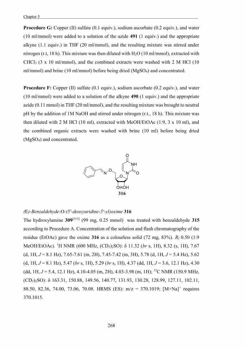
Chapter 5 .
268
Procedure G: Copper (II) sulfate (0.1 equiv.), sodium ascorbate (0.2 equiv.), and water
(10 ml/mmol) were added to a solution of the azide 491 (1 equiv.) and the appropriate
alkyne (1.1 equiv.) in THF (20 ml/mmol), and the resulting mixture was stirred under
nitrogen (r.t, 18 h). This mixture was then diluted with H2O (10 ml/mmol), extracted with
CHCl3 (3 x 10 ml/mmol), and the combined extracts were washed with 2 M HCl (10
ml/mmol) and brine (10 ml/mmol) before being dried (MgSO4) and concentrated.
Procedure F: Copper (II) sulfate (0.1 equiv.), sodium ascorbate (0.2 equiv.), and water
(10 ml/mmol) were added to a solution of the alkyne 490 (1 equiv.) and the appropriate
azide (0.11 mmol) in THF (20 ml/mmol), and the resulting mixture was brought to neutral
pH by the addition of 1M NaOH and stirred under nitrogen (r.t., 18 h). This mixture was
then diluted with 2 M HCl (10 ml), extracted with MeOH/EtOAc (1:9, 3 x 10 ml), and
the combined organic extracts were washed with brine (10 ml) before being dried
(MgSO4) and concentrated.
(E)-Benzaldehyde-O-(5'-deoxyuridine-5'-yl)oxime 316
The hydroxylamine 309[313] (99 mg, 0.25 mmol) was treated with benzaldehyde 315
according to Procedure A. Concentration of the solution and flash chromatography of the
residue (EtOAc) gave the oxime 316 as a colourless solid (72 mg, 83%). Rf 0.50 (1:9
MeOH/EtOAc). 1H NMR (600 MHz, (CD3)2SO): δ 11.32 (br s, 1H), 8.32 (s, 1H), 7.67
(d, 1H, J = 8.1 Hz), 7.65-7.61 (m, 2H), 7.45-7.42 (m, 3H), 5.78 (d, 1H, J = 5.4 Hz), 5.62
(d, 1H, J = 8.1 Hz), 5.47 (br s, 1H), 5.29 (br s, 1H), 4.37 (dd, 1H, J = 3.6, 12.1 Hz), 4.30
(dd, 1H, J = 5.4, 12.1 Hz), 4.10-4.05 (m, 2H), 4.03-3.98 (m, 1H); 13C NMR (150.9 MHz,
(CD3)2SO): δ 163.31, 150.88, 149.56, 140.77, 131.93, 130.28, 128.99, 127.11, 102.11,
88.50, 82.36, 74.00, 73.06, 70.08. HRMS (ES): m/z = 370.1019; [M+Na]+ requires
370.1015.
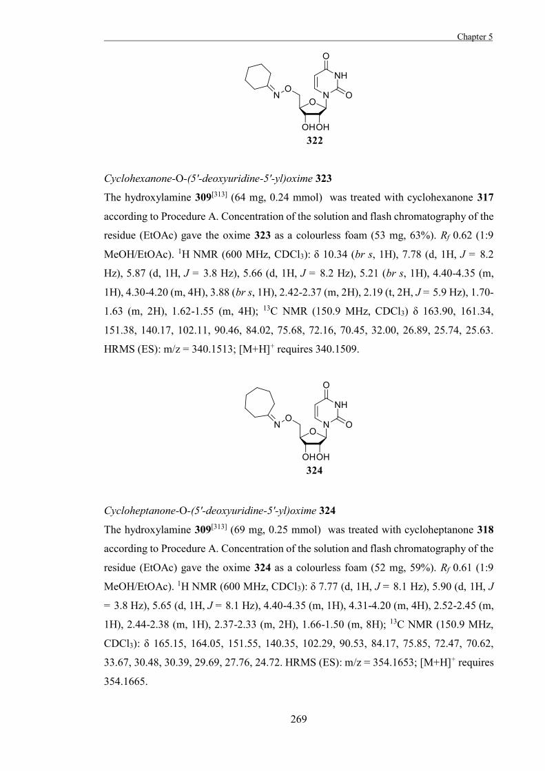
Chapter 5
269
Cyclohexanone-O-(5'-deoxyuridine-5'-yl)oxime 323
The hydroxylamine 309[313] (64 mg, 0.24 mmol) was treated with cyclohexanone 317
according to Procedure A. Concentration of the solution and flash chromatography of the
residue (EtOAc) gave the oxime 323 as a colourless foam (53 mg, 63%). Rf 0.62 (1:9
MeOH/EtOAc). 1H NMR (600 MHz, CDCl3): δ 10.34 (br s, 1H), 7.78 (d, 1H, J = 8.2
Hz), 5.87 (d, 1H, J = 3.8 Hz), 5.66 (d, 1H, J = 8.2 Hz), 5.21 (br s, 1H), 4.40-4.35 (m,
1H), 4.30-4.20 (m, 4H), 3.88 (br s, 1H), 2.42-2.37 (m, 2H), 2.19 (t, 2H, J = 5.9 Hz), 1.70-
1.63 (m, 2H), 1.62-1.55 (m, 4H); 13C NMR (150.9 MHz, CDCl3) δ 163.90, 161.34,
151.38, 140.17, 102.11, 90.46, 84.02, 75.68, 72.16, 70.45, 32.00, 26.89, 25.74, 25.63.
HRMS (ES): m/z = 340.1513; [M+H]+ requires 340.1509.
Cycloheptanone-O-(5'-deoxyuridine-5'-yl)oxime 324
The hydroxylamine 309[313] (69 mg, 0.25 mmol) was treated with cycloheptanone 318
according to Procedure A. Concentration of the solution and flash chromatography of the
residue (EtOAc) gave the oxime 324 as a colourless foam (52 mg, 59%). Rf 0.61 (1:9
MeOH/EtOAc). 1H NMR (600 MHz, CDCl3): δ 7.77 (d, 1H, J = 8.1 Hz), 5.90 (d, 1H, J
= 3.8 Hz), 5.65 (d, 1H, J = 8.1 Hz), 4.40-4.35 (m, 1H), 4.31-4.20 (m, 4H), 2.52-2.45 (m,
1H), 2.44-2.38 (m, 1H), 2.37-2.33 (m, 2H), 1.66-1.50 (m, 8H); 13C NMR (150.9 MHz,
CDCl3): δ 165.15, 164.05, 151.55, 140.35, 102.29, 90.53, 84.17, 75.85, 72.47, 70.62,
33.67, 30.48, 30.39, 29.69, 27.76, 24.72. HRMS (ES): m/z = 354.1653; [M+H]+ requires
354.1665.
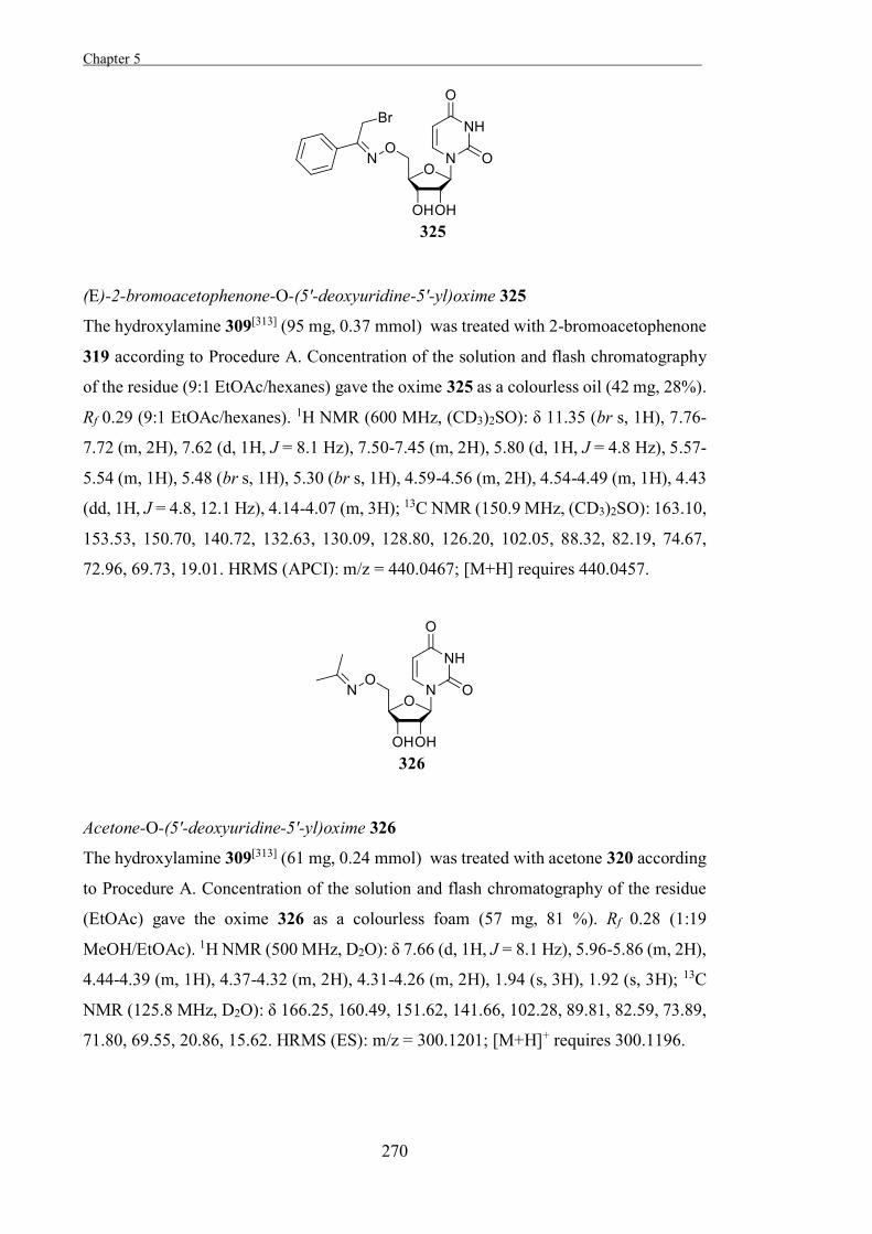
Chapter 5 .
270
(E)-2-bromoacetophenone-O-(5'-deoxyuridine-5'-yl)oxime 325
The hydroxylamine 309[313] (95 mg, 0.37 mmol) was treated with 2-bromoacetophenone
319 according to Procedure A. Concentration of the solution and flash chromatography
of the residue (9:1 EtOAc/hexanes) gave the oxime 325 as a colourless oil (42 mg, 28%).
Rf 0.29 (9:1 EtOAc/hexanes). 1H NMR (600 MHz, (CD3)2SO): δ 11.35 (br s, 1H), 7.76-
7.72 (m, 2H), 7.62 (d, 1H, J = 8.1 Hz), 7.50-7.45 (m, 2H), 5.80 (d, 1H, J = 4.8 Hz), 5.57-
5.54 (m, 1H), 5.48 (br s, 1H), 5.30 (br s, 1H), 4.59-4.56 (m, 2H), 4.54-4.49 (m, 1H), 4.43
(dd, 1H, J = 4.8, 12.1 Hz), 4.14-4.07 (m, 3H); 13C NMR (150.9 MHz, (CD3)2SO): 163.10,
153.53, 150.70, 140.72, 132.63, 130.09, 128.80, 126.20, 102.05, 88.32, 82.19, 74.67,
72.96, 69.73, 19.01. HRMS (APCI): m/z = 440.0467; [M+H] requires 440.0457.
Acetone-O-(5'-deoxyuridine-5'-yl)oxime 326
The hydroxylamine 309[313] (61 mg, 0.24 mmol) was treated with acetone 320 according
to Procedure A. Concentration of the solution and flash chromatography of the residue
(EtOAc) gave the oxime 326 as a colourless foam (57 mg, 81 %). Rf 0.28 (1:19
MeOH/EtOAc). 1H NMR (500 MHz, D2O): δ 7.66 (d, 1H, J = 8.1 Hz), 5.96-5.86 (m, 2H),
4.44-4.39 (m, 1H), 4.37-4.32 (m, 2H), 4.31-4.26 (m, 2H), 1.94 (s, 3H), 1.92 (s, 3H); 13C
NMR (125.8 MHz, D2O): δ 166.25, 160.49, 151.62, 141.66, 102.28, 89.81, 82.59, 73.89,
71.80, 69.55, 20.86, 15.62. HRMS (ES): m/z = 300.1201; [M+H]+ requires 300.1196.
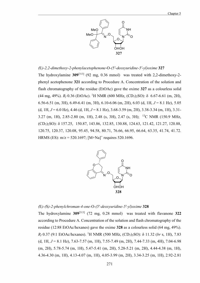
Chapter 5
271
(E)-2,2-dimethoxy-2-phenylacetophenone-O-(5'-deoxyuridine-5'-yl)oxime 327
The hydroxylamine 309[313] (92 mg, 0.36 mmol) was treated with 2,2-dimethoxy-2-
phenyl acetophenone 321 according to Procedure A. Concentration of the solution and
flash chromatography of the residue (EtOAc) gave the oxime 327 as a colourless solid
(44 mg, 49%). Rf 0.36 (EtOAc). 1H NMR (600 MHz, (CD3)2SO): δ 6.67-6.61 (m, 2H),
6.56-6.51 (m, 3H), 6.49-6.41 (m, 3H), 6.10-6.06 (m, 2H), 6.03 (d, 1H, J = 8.1 Hz), 5.05
(d, 1H, J = 6.0 Hz), 4.46 (d, 1H, J = 8.1 Hz), 3.68-3.59 (m, 2H), 3.38-3.34 (m, 1H), 3.31-
3.27 (m, 1H), 2.85-2.80 (m, 1H), 2.48 (s, 3H), 2.47 (s, 3H); 13C NMR (150.9 MHz,
(CD3)2SO): δ 157.25, 150.87, 143.86, 132.85, 130.88, 124.63, 121.42, 121.27, 120.88,
120.75, 120.37, 120.08, 95.45, 94.58, 80.71, 76.66, 66.95, 66.64, 63.35, 41.74, 41.72.
HRMS (ES): m/z = 520.1697; [M+Na]+ requires 520.1696.
(E)-(S)-2-phenylchroman-4-one-O-(5'-deoxyuridine-5'-yl)oxime 328
The hydroxylamine 309[313] (72 mg, 0.28 mmol) was treated with flavanone 322
according to Procedure A. Concentration of the solution and flash chromatography of the
residue (12:88 EtOAc/hexanes) gave the oxime 328 as a colourless solid (64 mg, 49%).
Rf 0.37 (9:1 EtOAc/hexanes). 1H NMR (500 MHz, (CD3)2SO): δ 11.32 (br s, 1H), 7.83
(d, 1H, J = 8.1 Hz), 7.63-7.57 (m, 1H), 7.55-7.49 (m, 2H), 7.44-7.33 (m, 4H), 7.04-6.98
(m, 2H), 5.78-5.74 (m, 1H), 5.47-5.41 (m, 2H), 5.28-5.21 (m, 2H), 4.44-4.38 (m, 1H),
4.36-4.30 (m, 1H), 4.13-4.07 (m, 1H), 4.05-3.99 (m, 2H), 3.34-3.25 (m, 1H), 2.92-2.81
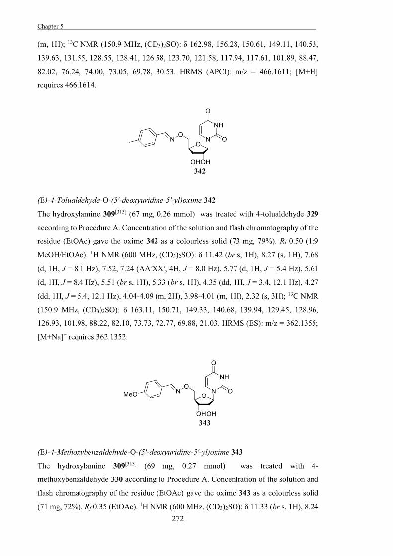
Chapter 5 .
272
(m, 1H); 13C NMR (150.9 MHz, (CD3)2SO): δ 162.98, 156.28, 150.61, 149.11, 140.53,
139.63, 131.55, 128.55, 128.41, 126.58, 123.70, 121.58, 117.94, 117.61, 101.89, 88.47,
82.02, 76.24, 74.00, 73.05, 69.78, 30.53. HRMS (APCI): m/z = 466.1611; [M+H]
requires 466.1614.
(E)-4-Tolualdehyde-O-(5'-deoxyuridine-5'-yl)oxime 342
The hydroxylamine 309[313] (67 mg, 0.26 mmol) was treated with 4-tolualdehyde 329
according to Procedure A. Concentration of the solution and flash chromatography of the
residue (EtOAc) gave the oxime 342 as a colourless solid (73 mg, 79%). Rf 0.50 (1:9
MeOH/EtOAc). 1H NMR (600 MHz, (CD3)2SO): δ 11.42 (br s, 1H), 8.27 (s, 1H), 7.68
(d, 1H, J = 8.1 Hz), 7.52, 7.24 (AA'XX', 4H, J = 8.0 Hz), 5.77 (d, 1H, J = 5.4 Hz), 5.61
(d, 1H, J = 8.4 Hz), 5.51 (br s, 1H), 5.33 (br s, 1H), 4.35 (dd, 1H, J = 3.4, 12.1 Hz), 4.27
(dd, 1H, J = 5.4, 12.1 Hz), 4.04-4.09 (m, 2H), 3.98-4.01 (m, 1H), 2.32 (s, 3H); 13C NMR
(150.9 MHz, (CD3)2SO): δ 163.11, 150.71, 149.33, 140.68, 139.94, 129.45, 128.96,
126.93, 101.98, 88.22, 82.10, 73.73, 72.77, 69.88, 21.03. HRMS (ES): m/z = 362.1355;
[M+Na]+ requires 362.1352.
(E)-4-Methoxybenzaldehyde-O-(5'-deoxyuridine-5'-yl)oxime 343
The hydroxylamine 309[313] (69 mg, 0.27 mmol) was treated with 4-
methoxybenzaldehyde 330 according to Procedure A. Concentration of the solution and
flash chromatography of the residue (EtOAc) gave the oxime 343 as a colourless solid
(71 mg, 72%). Rf 0.35 (EtOAc). 1H NMR (600 MHz, (CD3)2SO): δ 11.33 (br s, 1H), 8.24
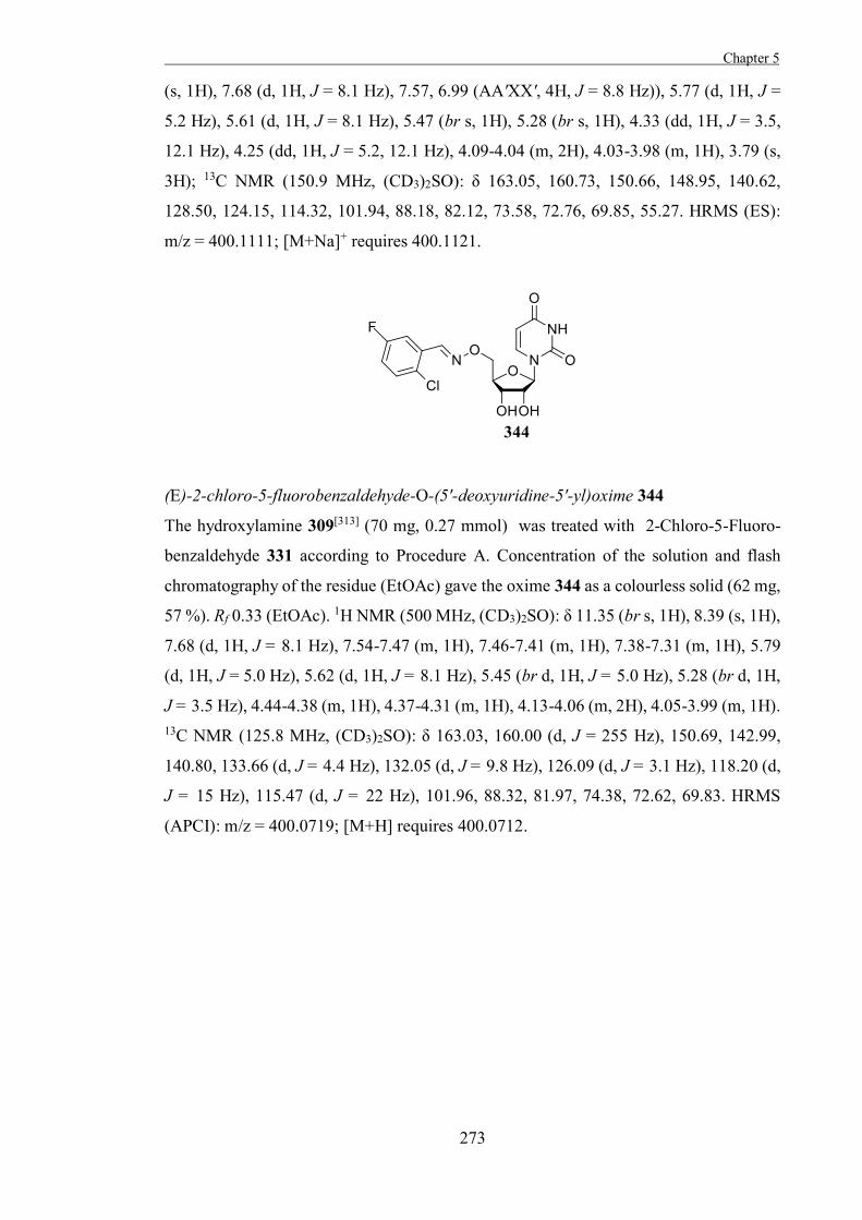
Chapter 5
273
(s, 1H), 7.68 (d, 1H, J = 8.1 Hz), 7.57, 6.99 (AA'XX', 4H, J = 8.8 Hz)), 5.77 (d, 1H, J =
5.2 Hz), 5.61 (d, 1H, J = 8.1 Hz), 5.47 (br s, 1H), 5.28 (br s, 1H), 4.33 (dd, 1H, J = 3.5,
12.1 Hz), 4.25 (dd, 1H, J = 5.2, 12.1 Hz), 4.09-4.04 (m, 2H), 4.03-3.98 (m, 1H), 3.79 (s,
3H); 13C NMR (150.9 MHz, (CD3)2SO): δ 163.05, 160.73, 150.66, 148.95, 140.62,
128.50, 124.15, 114.32, 101.94, 88.18, 82.12, 73.58, 72.76, 69.85, 55.27. HRMS (ES):
m/z = 400.1111; [M+Na]+ requires 400.1121.
(E)-2-chloro-5-fluorobenzaldehyde-O-(5'-deoxyuridine-5'-yl)oxime 344
The hydroxylamine 309[313] (70 mg, 0.27 mmol) was treated with 2-Chloro-5-Fluoro-
benzaldehyde 331 according to Procedure A. Concentration of the solution and flash
chromatography of the residue (EtOAc) gave the oxime 344 as a colourless solid (62 mg,
57 %). Rf 0.33 (EtOAc). 1H NMR (500 MHz, (CD3)2SO): δ 11.35 (br s, 1H), 8.39 (s, 1H),
7.68 (d, 1H, J = 8.1 Hz), 7.54-7.47 (m, 1H), 7.46-7.41 (m, 1H), 7.38-7.31 (m, 1H), 5.79
(d, 1H, J = 5.0 Hz), 5.62 (d, 1H, J = 8.1 Hz), 5.45 (br d, 1H, J = 5.0 Hz), 5.28 (br d, 1H,
J = 3.5 Hz), 4.44-4.38 (m, 1H), 4.37-4.31 (m, 1H), 4.13-4.06 (m, 2H), 4.05-3.99 (m, 1H). 13C NMR (125.8 MHz, (CD3)2SO): δ 163.03, 160.00 (d, J = 255 Hz), 150.69, 142.99,
140.80, 133.66 (d, J = 4.4 Hz), 132.05 (d, J = 9.8 Hz), 126.09 (d, J = 3.1 Hz), 118.20 (d,
J = 15 Hz), 115.47 (d, J = 22 Hz), 101.96, 88.32, 81.97, 74.38, 72.62, 69.83. HRMS
(APCI): m/z = 400.0719; [M+H] requires 400.0712.
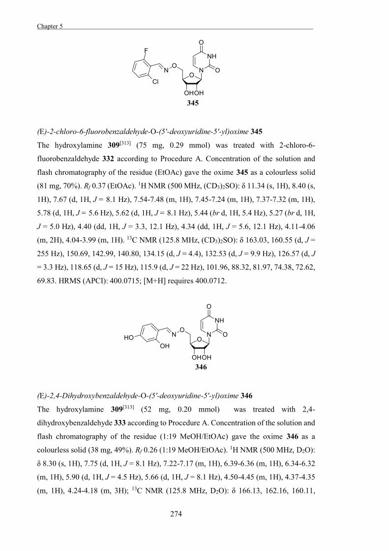
Chapter 5 .
274
(E)-2-chloro-6-fluorobenzaldehyde-O-(5'-deoxyuridine-5'-yl)oxime 345
The hydroxylamine 309[313] (75 mg, 0.29 mmol) was treated with 2-chloro-6-
fluorobenzaldehyde 332 according to Procedure A. Concentration of the solution and
flash chromatography of the residue (EtOAc) gave the oxime 345 as a colourless solid
(81 mg, 70%). Rf 0.37 (EtOAc). 1H NMR (500 MHz, (CD3)2SO): δ 11.34 (s, 1H), 8.40 (s,
1H), 7.67 (d, 1H, J = 8.1 Hz), 7.54-7.48 (m, 1H), 7.45-7.24 (m, 1H), 7.37-7.32 (m, 1H),
5.78 (d, 1H, J = 5.6 Hz), 5.62 (d, 1H, J = 8.1 Hz), 5.44 (br d, 1H, 5.4 Hz), 5.27 (br d, 1H,
J = 5.0 Hz), 4.40 (dd, 1H, J = 3.3, 12.1 Hz), 4.34 (dd, 1H, J = 5.6, 12.1 Hz), 4.11-4.06
(m, 2H), 4.04-3.99 (m, 1H). 13C NMR (125.8 MHz, (CD3)2SO): δ 163.03, 160.55 (d, J =
255 Hz), 150.69, 142.99, 140.80, 134.15 (d, J = 4.4), 132.53 (d, J = 9.9 Hz), 126.57 (d, J
= 3.3 Hz), 118.65 (d, J = 15 Hz), 115.9 (d, J = 22 Hz), 101.96, 88.32, 81.97, 74.38, 72.62,
69.83. HRMS (APCI): 400.0715; [M+H] requires 400.0712.
(E)-2,4-Dihydroxybenzaldehyde-O-(5'-deoxyuridine-5'-yl)oxime 346
The hydroxylamine 309[313] (52 mg, 0.20 mmol) was treated with 2,4-
dihydroxybenzaldehyde 333 according to Procedure A. Concentration of the solution and
flash chromatography of the residue (1:19 MeOH/EtOAc) gave the oxime 346 as a
colourless solid (38 mg, 49%). Rf 0.26 (1:19 MeOH/EtOAc). 1H NMR (500 MHz, D2O):
δ 8.30 (s, 1H), 7.75 (d, 1H, J = 8.1 Hz), 7.22-7.17 (m, 1H), 6.39-6.36 (m, 1H), 6.34-6.32
(m, 1H), 5.90 (d, 1H, J = 4.5 Hz), 5.66 (d, 1H, J = 8.1 Hz), 4.50-4.45 (m, 1H), 4.37-4.35
(m, 1H), 4.24-4.18 (m, 3H); 13C NMR (125.8 MHz, D2O): δ 166.13, 162.16, 160.11,
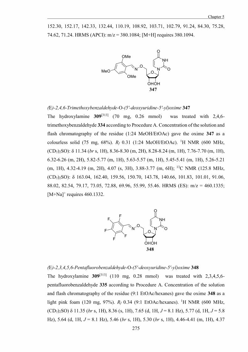
Chapter 5
275
152.30, 152.17, 142.33, 132.44, 110.19, 108.92, 103.71, 102.79, 91.24, 84.30, 75.28,
74.62, 71.24. HRMS (APCI): m/z = 380.1084; [M+H] requires 380.1094.
(E)-2,4,6-Trimethoxybenzaldehyde-O-(5'-deoxyuridine-5'-yl)oxime 347
The hydroxylamine 309[313] (70 mg, 0.26 mmol) was treated with 2,4,6-
trimethoxybenzaldehyde 334 according to Procedure A. Concentration of the solution and
flash chromatography of the residue (1:24 MeOH/EtOAc) gave the oxime 347 as a
colourless solid (75 mg, 68%). Rf 0.31 (1:24 MeOH/EtOAc). 1H NMR (600 MHz,
(CD3)2SO): δ 11.34 (br s, 1H), 8.36-8.30 (m, 2H), 8.28-8.24 (m, 1H), 7.76-7.70 (m, 1H),
6.32-6.26 (m, 2H), 5.82-5.77 (m, 1H), 5.63-5.57 (m, 1H), 5.45-5.41 (m, 1H), 5.26-5.21
(m, 1H), 4.32-4.19 (m, 2H), 4.07 (s, 3H), 3.88-3.77 (m, 6H); 13C NMR (125.8 MHz,
(CD3)2SO): δ 163.04, 162.40, 159.56, 150.70, 143.78, 140.66, 101.83, 101.01, 91.06,
88.02, 82.54, 79.17, 73.05, 72.88, 69.96, 55.99, 55.46. HRMS (ES): m/z = 460.1335;
[M+Na]+ requires 460.1332.
(E)-2,3,4,5,6-Pentafluorobenzaldehyde-O-(5'-deoxyuridine-5'-yl)oxime 348
The hydroxylamine 309[313] (110 mg, 0.28 mmol) was treated with 2,3,4,5,6-
pentafluorobenzaldehyde 335 according to Procedure A. Concentration of the solution
and flash chromatography of the residue (9:1 EtOAc/hexanes) gave the oxime 348 as a
light pink foam (120 mg, 97%). Rf 0.34 (9:1 EtOAc/hexanes). 1H NMR (600 MHz,
(CD3)2SO) δ 11.35 (br s, 1H), 8.36 (s, 1H), 7.65 (d, 1H, J = 8.1 Hz), 5.77 (d, 1H, J = 5.8
Hz), 5.64 (d, 1H, J = 8.1 Hz), 5.46 (br s, 1H), 5.30 (br s, 1H), 4.46-4.41 (m, 1H), 4.37
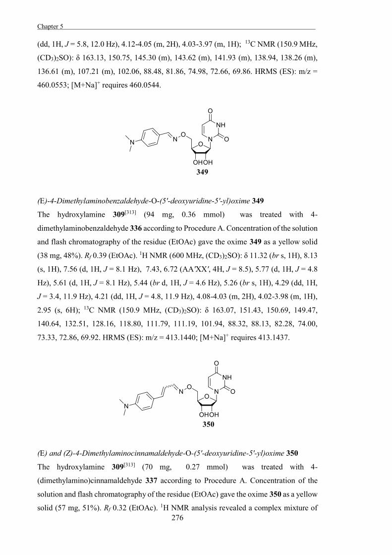
Chapter 5 .
276
(dd, 1H, J = 5.8, 12.0 Hz), 4.12-4.05 (m, 2H), 4.03-3.97 (m, 1H); 13C NMR (150.9 MHz,
(CD3)2SO): δ 163.13, 150.75, 145.30 (m), 143.62 (m), 141.93 (m), 138.94, 138.26 (m),
136.61 (m), 107.21 (m), 102.06, 88.48, 81.86, 74.98, 72.66, 69.86. HRMS (ES): m/z =
460.0553; [M+Na]+ requires 460.0544.
(E)-4-Dimethylaminobenzaldehyde-O-(5'-deoxyuridine-5'-yl)oxime 349
The hydroxylamine 309[313] (94 mg, 0.36 mmol) was treated with 4-
dimethylaminobenzaldehyde 336 according to Procedure A. Concentration of the solution
and flash chromatography of the residue (EtOAc) gave the oxime 349 as a yellow solid
(38 mg, 48%). Rf 0.39 (EtOAc). 1H NMR (600 MHz, (CD3)2SO): δ 11.32 (br s, 1H), 8.13
(s, 1H), 7.56 (d, 1H, J = 8.1 Hz), 7.43, 6.72 (AA'XX', 4H, J = 8.5), 5.77 (d, 1H, J = 4.8
Hz), 5.61 (d, 1H, J = 8.1 Hz), 5.44 (br d, 1H, J = 4.6 Hz), 5.26 (br s, 1H), 4.29 (dd, 1H,
J = 3.4, 11.9 Hz), 4.21 (dd, 1H, J = 4.8, 11.9 Hz), 4.08-4.03 (m, 2H), 4.02-3.98 (m, 1H),
2.95 (s, 6H); 13C NMR (150.9 MHz, (CD3)2SO): δ 163.07, 151.43, 150.69, 149.47,
140.64, 132.51, 128.16, 118.80, 111.79, 111.19, 101.94, 88.32, 88.13, 82.28, 74.00,
73.33, 72.86, 69.92. HRMS (ES): m/z = 413.1440; [M+Na]+ requires 413.1437.
(E) and (Z)-4-Dimethylaminocinnamaldehyde-O-(5'-deoxyuridine-5'-yl)oxime 350
The hydroxylamine 309[313] (70 mg, 0.27 mmol) was treated with 4-
(dimethylamino)cinnamaldehyde 337 according to Procedure A. Concentration of the
solution and flash chromatography of the residue (EtOAc) gave the oxime 350 as a yellow
solid (57 mg, 51%). Rf 0.32 (EtOAc). 1H NMR analysis revealed a complex mixture of
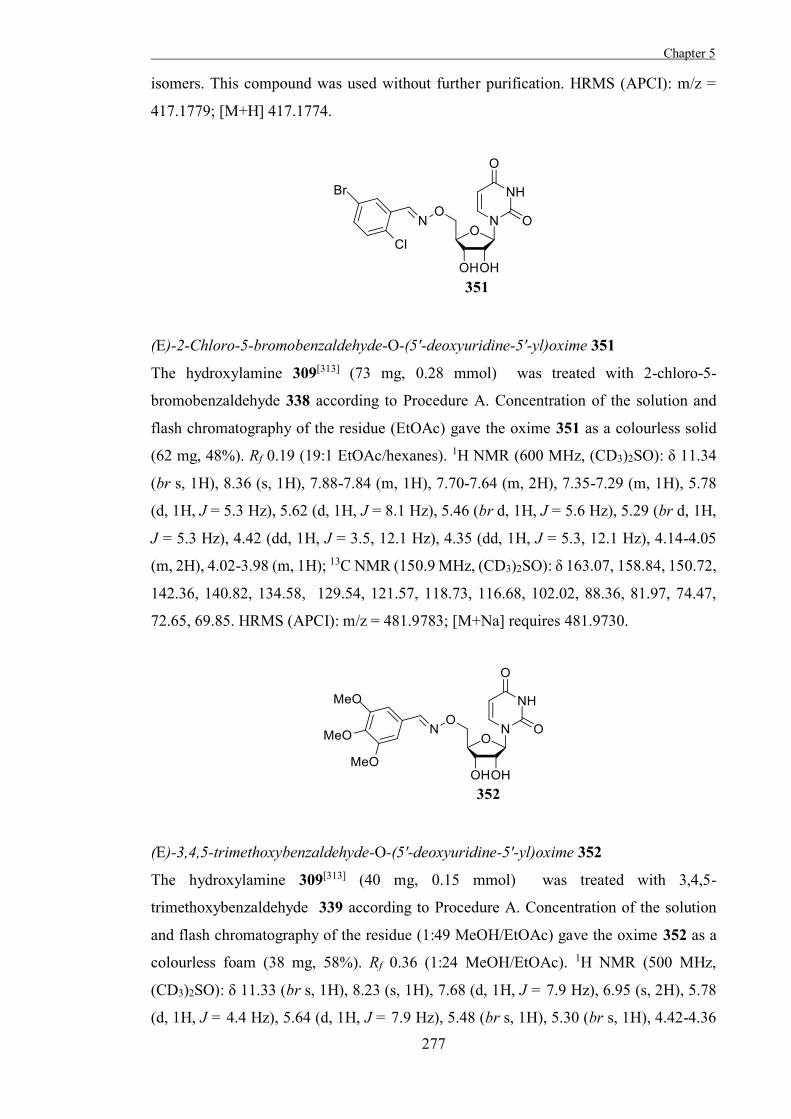
Chapter 5
277
isomers. This compound was used without further purification. HRMS (APCI): m/z =
417.1779; [M+H] 417.1774.
(E)-2-Chloro-5-bromobenzaldehyde-O-(5'-deoxyuridine-5'-yl)oxime 351
The hydroxylamine 309[313] (73 mg, 0.28 mmol) was treated with 2-chloro-5-
bromobenzaldehyde 338 according to Procedure A. Concentration of the solution and
flash chromatography of the residue (EtOAc) gave the oxime 351 as a colourless solid
(62 mg, 48%). Rf 0.19 (19:1 EtOAc/hexanes). 1H NMR (600 MHz, (CD3)2SO): δ 11.34
(br s, 1H), 8.36 (s, 1H), 7.88-7.84 (m, 1H), 7.70-7.64 (m, 2H), 7.35-7.29 (m, 1H), 5.78
(d, 1H, J = 5.3 Hz), 5.62 (d, 1H, J = 8.1 Hz), 5.46 (br d, 1H, J = 5.6 Hz), 5.29 (br d, 1H,
J = 5.3 Hz), 4.42 (dd, 1H, J = 3.5, 12.1 Hz), 4.35 (dd, 1H, J = 5.3, 12.1 Hz), 4.14-4.05
(m, 2H), 4.02-3.98 (m, 1H); 13C NMR (150.9 MHz, (CD3)2SO): δ 163.07, 158.84, 150.72,
142.36, 140.82, 134.58, 129.54, 121.57, 118.73, 116.68, 102.02, 88.36, 81.97, 74.47,
72.65, 69.85. HRMS (APCI): m/z = 481.9783; [M+Na] requires 481.9730.
(E)-3,4,5-trimethoxybenzaldehyde-O-(5'-deoxyuridine-5'-yl)oxime 352
The hydroxylamine 309[313] (40 mg, 0.15 mmol) was treated with 3,4,5-
trimethoxybenzaldehyde 339 according to Procedure A. Concentration of the solution
and flash chromatography of the residue (1:49 MeOH/EtOAc) gave the oxime 352 as a
colourless foam (38 mg, 58%). Rf 0.36 (1:24 MeOH/EtOAc). 1H NMR (500 MHz,
(CD3)2SO): δ 11.33 (br s, 1H), 8.23 (s, 1H), 7.68 (d, 1H, J = 7.9 Hz), 6.95 (s, 2H), 5.78
(d, 1H, J = 4.4 Hz), 5.64 (d, 1H, J = 7.9 Hz), 5.48 (br s, 1H), 5.30 (br s, 1H), 4.42-4.36
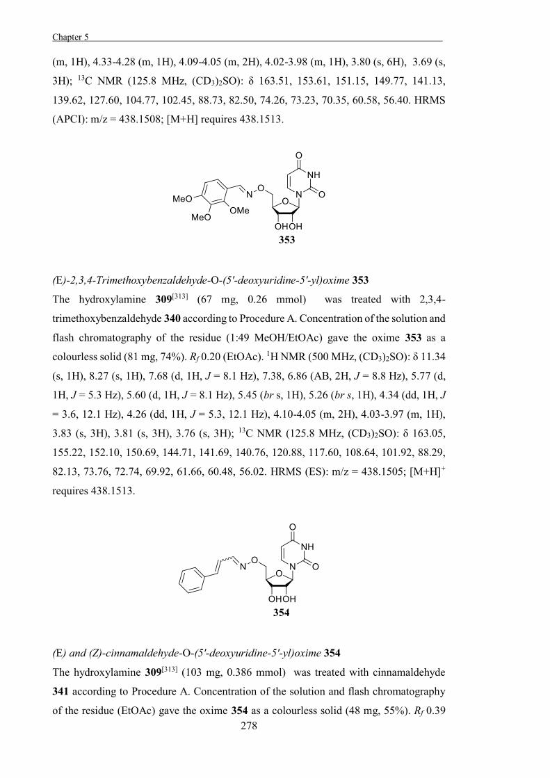
Chapter 5 .
278
(m, 1H), 4.33-4.28 (m, 1H), 4.09-4.05 (m, 2H), 4.02-3.98 (m, 1H), 3.80 (s, 6H), 3.69 (s,
3H); 13C NMR (125.8 MHz, (CD3)2SO): δ 163.51, 153.61, 151.15, 149.77, 141.13,
139.62, 127.60, 104.77, 102.45, 88.73, 82.50, 74.26, 73.23, 70.35, 60.58, 56.40. HRMS
(APCI): m/z = 438.1508; [M+H] requires 438.1513.
(E)-2,3,4-Trimethoxybenzaldehyde-O-(5'-deoxyuridine-5'-yl)oxime 353
The hydroxylamine 309[313] (67 mg, 0.26 mmol) was treated with 2,3,4-
trimethoxybenzaldehyde 340 according to Procedure A. Concentration of the solution and
flash chromatography of the residue (1:49 MeOH/EtOAc) gave the oxime 353 as a
colourless solid (81 mg, 74%). Rf 0.20 (EtOAc). 1H NMR (500 MHz, (CD3)2SO): δ 11.34
(s, 1H), 8.27 (s, 1H), 7.68 (d, 1H, J = 8.1 Hz), 7.38, 6.86 (AB, 2H, J = 8.8 Hz), 5.77 (d,
1H, J = 5.3 Hz), 5.60 (d, 1H, J = 8.1 Hz), 5.45 (br s, 1H), 5.26 (br s, 1H), 4.34 (dd, 1H, J
= 3.6, 12.1 Hz), 4.26 (dd, 1H, J = 5.3, 12.1 Hz), 4.10-4.05 (m, 2H), 4.03-3.97 (m, 1H),
3.83 (s, 3H), 3.81 (s, 3H), 3.76 (s, 3H); 13C NMR (125.8 MHz, (CD3)2SO): δ 163.05,
155.22, 152.10, 150.69, 144.71, 141.69, 140.76, 120.88, 117.60, 108.64, 101.92, 88.29,
82.13, 73.76, 72.74, 69.92, 61.66, 60.48, 56.02. HRMS (ES): m/z = 438.1505; [M+H]+
requires 438.1513.
(E) and (Z)-cinnamaldehyde-O-(5'-deoxyuridine-5'-yl)oxime 354
The hydroxylamine 309[313] (103 mg, 0.386 mmol) was treated with cinnamaldehyde
341 according to Procedure A. Concentration of the solution and flash chromatography
of the residue (EtOAc) gave the oxime 354 as a colourless solid (48 mg, 55%). Rf 0.39
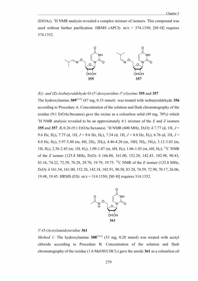
Chapter 5
279
(EtOAc). 1H NMR analysis revealed a complex mixture of isomers. This compound was
used without further purification. HRMS (APCI): m/z = 374.1350; [M+H] requires
374.1352.
(E)- and (Z)-Isobutyraldehyde-O-(5'-deoxyuridine-5'-yl)oxime 355 and 357
The hydroxylamine 309[313] (87 mg, 0.33 mmol) was treated with isobutyraldehyde 356
according to Procedure A. Concentration of the solution and flash chromatography of the
residue (9:1 EtOAc/hexanes) gave the oxime as a colourless solid (49 mg, 70%) which 1H NMR analysis revealed to be an approximately 4:1 mixture of the E and Z isomers
355 and 357. Rf 0.26 (9:1 EtOAc/hexanes). 1H NMR (600 MHz, D2O): δ 7.77 (d, 1H, J =
9.6 Hz, HZ), 7.75 (d, 1H, J = 9.6 Hz, HE), 7.54 (d, 1H, J = 8.0 Hz, HZ), 6.76 (d, 1H, J =
8.0 Hz, HE), 5.97-5.80 (m, 4H, 2HE, 2HZ), 4.46-4.20 (m, 10H, 5HE, 5HZ), 3.12-3.02 (m,
1H, HE), 2.56-2.45 (m, 1H, HZ), 1.09-1.07 (m, 6H, HZ), 1.06-1.03 (m, 6H, HE); 13C NMR
of the Z isomer (125.8 MHz, D2O): δ 166.88, 161.00, 152.28, 142.43, 102.98, 90.43,
83.16, 74.22, 72.59, 70.28, 29.70, 19.79, 19.75. 13C NMR of the E isomer (125.8 MHz,
D2O): δ 161.54, 161.00, 152.28, 142.18, 102.91, 90.50, 83.28, 74.59, 72.98, 70.17, 26.06,
19.48, 19.45. HRMS (ES): m/z = 314.1350; [M+H] requires 314.1352.
5'-O-(Acetylamido)uridine 361
Method 1: The hydroxylamine 308[313] (53 mg, 0.20 mmol) was treated with acetyl
chloride according to Procedure B. Concentration of the solution and flash
chromatography of the residue (1:6 MeOH/CHCl3) gave the amide 361 as a colourless oil
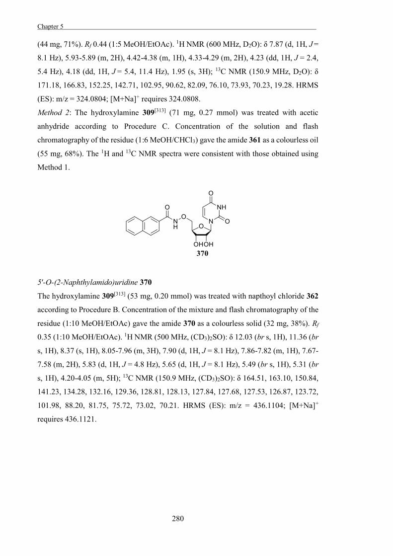
Chapter 5 .
280
(44 mg, 71%). Rf 0.44 (1:5 MeOH/EtOAc). 1H NMR (600 MHz, D2O): δ 7.87 (d, 1H, J =
8.1 Hz), 5.93-5.89 (m, 2H), 4.42-4.38 (m, 1H), 4.33-4.29 (m, 2H), 4.23 (dd, 1H, J = 2.4,
5.4 Hz), 4.18 (dd, 1H, J = 5.4, 11.4 Hz), 1.95 (s, 3H); 13C NMR (150.9 MHz, D2O): δ
171.18, 166.83, 152.25, 142.71, 102.95, 90.62, 82.09, 76.10, 73.93, 70.23, 19.28. HRMS
(ES): m/z = 324.0804; [M+Na]+ requires 324.0808.
Method 2: The hydroxylamine 309[313] (71 mg, 0.27 mmol) was treated with acetic
anhydride according to Procedure C. Concentration of the solution and flash
chromatography of the residue (1:6 MeOH/CHCl3) gave the amide 361 as a colourless oil
(55 mg, 68%). The 1H and 13C NMR spectra were consistent with those obtained using
Method 1.
5′-O-(2-Naphthylamido)uridine 370
The hydroxylamine 309[313] (53 mg, 0.20 mmol) was treated with napthoyl chloride 362
according to Procedure B. Concentration of the mixture and flash chromatography of the
residue (1:10 MeOH/EtOAc) gave the amide 370 as a colourless solid (32 mg, 38%). Rf
0.35 (1:10 MeOH/EtOAc). 1H NMR (500 MHz, (CD3)2SO): δ 12.03 (br s, 1H), 11.36 (br
s, 1H), 8.37 (s, 1H), 8.05-7.96 (m, 3H), 7.90 (d, 1H, J = 8.1 Hz), 7.86-7.82 (m, 1H), 7.67-
7.58 (m, 2H), 5.83 (d, 1H, J = 4.8 Hz), 5.65 (d, 1H, J = 8.1 Hz), 5.49 (br s, 1H), 5.31 (br
s, 1H), 4.20-4.05 (m, 5H); 13C NMR (150.9 MHz, (CD3)2SO): δ 164.51, 163.10, 150.84,
141.23, 134.28, 132.16, 129.36, 128.81, 128.13, 127.84, 127.68, 127.53, 126.87, 123.72,
101.98, 88.20, 81.75, 75.72, 73.02, 70.21. HRMS (ES): m/z = 436.1104; [M+Na]+
requires 436.1121.
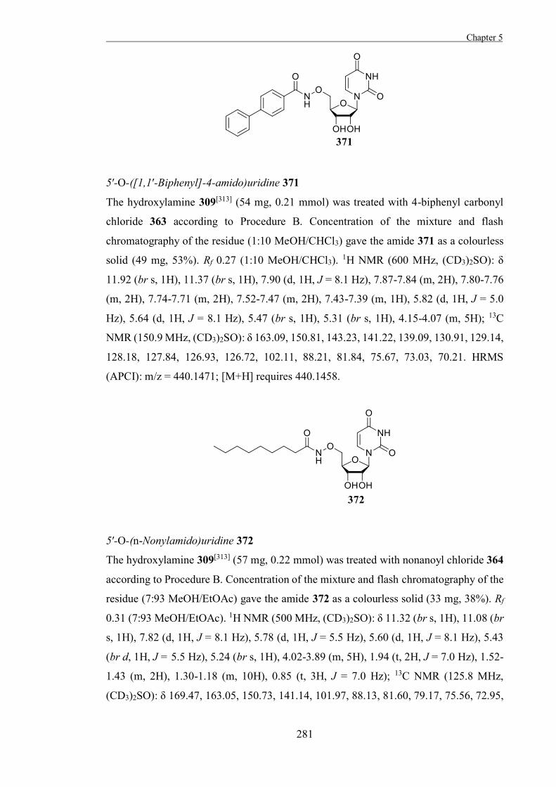
Chapter 5
281
5′-O-([1,1′-Biphenyl]-4-amido)uridine 371
The hydroxylamine 309[313] (54 mg, 0.21 mmol) was treated with 4-biphenyl carbonyl
chloride 363 according to Procedure B. Concentration of the mixture and flash
chromatography of the residue (1:10 MeOH/CHCl3) gave the amide 371 as a colourless
solid (49 mg, 53%). Rf 0.27 (1:10 MeOH/CHCl3). 1H NMR (600 MHz, (CD3)2SO): δ
11.92 (br s, 1H), 11.37 (br s, 1H), 7.90 (d, 1H, J = 8.1 Hz), 7.87-7.84 (m, 2H), 7.80-7.76
(m, 2H), 7.74-7.71 (m, 2H), 7.52-7.47 (m, 2H), 7.43-7.39 (m, 1H), 5.82 (d, 1H, J = 5.0
Hz), 5.64 (d, 1H, J = 8.1 Hz), 5.47 (br s, 1H), 5.31 (br s, 1H), 4.15-4.07 (m, 5H); 13C
NMR (150.9 MHz, (CD3)2SO): δ 163.09, 150.81, 143.23, 141.22, 139.09, 130.91, 129.14,
128.18, 127.84, 126.93, 126.72, 102.11, 88.21, 81.84, 75.67, 73.03, 70.21. HRMS
(APCI): m/z = 440.1471; [M+H] requires 440.1458.
5′-O-(n-Nonylamido)uridine 372
The hydroxylamine 309[313] (57 mg, 0.22 mmol) was treated with nonanoyl chloride 364
according to Procedure B. Concentration of the mixture and flash chromatography of the
residue (7:93 MeOH/EtOAc) gave the amide 372 as a colourless solid (33 mg, 38%). Rf
0.31 (7:93 MeOH/EtOAc). 1H NMR (500 MHz, (CD3)2SO): δ 11.32 (br s, 1H), 11.08 (br
s, 1H), 7.82 (d, 1H, J = 8.1 Hz), 5.78 (d, 1H, J = 5.5 Hz), 5.60 (d, 1H, J = 8.1 Hz), 5.43
(br d, 1H, J = 5.5 Hz), 5.24 (br s, 1H), 4.02-3.89 (m, 5H), 1.94 (t, 2H, J = 7.0 Hz), 1.52-
1.43 (m, 2H), 1.30-1.18 (m, 10H), 0.85 (t, 3H, J = 7.0 Hz); 13C NMR (125.8 MHz,
(CD3)2SO): δ 169.47, 163.05, 150.73, 141.14, 101.97, 88.13, 81.60, 79.17, 75.56, 72.95,
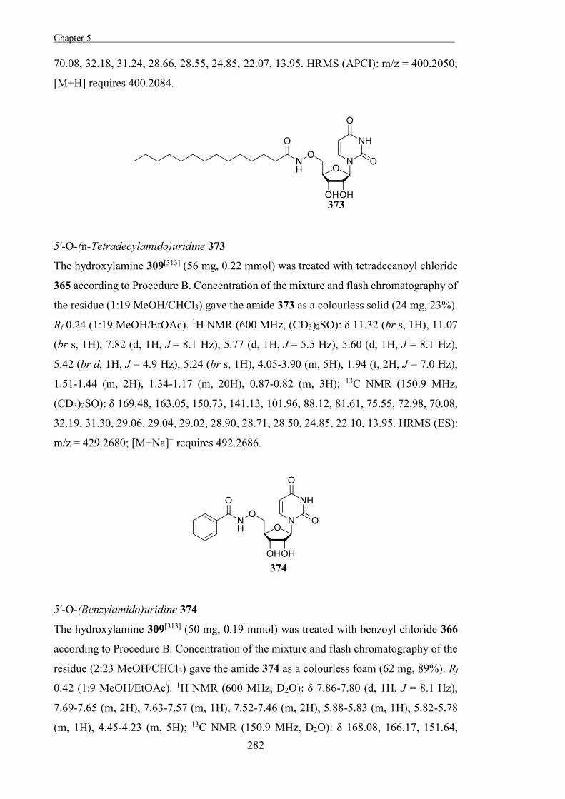
Chapter 5 .
282
70.08, 32.18, 31.24, 28.66, 28.55, 24.85, 22.07, 13.95. HRMS (APCI): m/z = 400.2050;
[M+H] requires 400.2084.
5′-O-(n-Tetradecylamido)uridine 373
The hydroxylamine 309[313] (56 mg, 0.22 mmol) was treated with tetradecanoyl chloride
365 according to Procedure B. Concentration of the mixture and flash chromatography of
the residue (1:19 MeOH/CHCl3) gave the amide 373 as a colourless solid (24 mg, 23%).
Rf 0.24 (1:19 MeOH/EtOAc). 1H NMR (600 MHz, (CD3)2SO): δ 11.32 (br s, 1H), 11.07
(br s, 1H), 7.82 (d, 1H, J = 8.1 Hz), 5.77 (d, 1H, J = 5.5 Hz), 5.60 (d, 1H, J = 8.1 Hz),
5.42 (br d, 1H, J = 4.9 Hz), 5.24 (br s, 1H), 4.05-3.90 (m, 5H), 1.94 (t, 2H, J = 7.0 Hz),
1.51-1.44 (m, 2H), 1.34-1.17 (m, 20H), 0.87-0.82 (m, 3H); 13C NMR (150.9 MHz,
(CD3)2SO): δ 169.48, 163.05, 150.73, 141.13, 101.96, 88.12, 81.61, 75.55, 72.98, 70.08,
32.19, 31.30, 29.06, 29.04, 29.02, 28.90, 28.71, 28.50, 24.85, 22.10, 13.95. HRMS (ES):
m/z = 429.2680; [M+Na]+ requires 492.2686.
5′-O-(Benzylamido)uridine 374
The hydroxylamine 309[313] (50 mg, 0.19 mmol) was treated with benzoyl chloride 366
according to Procedure B. Concentration of the mixture and flash chromatography of the
residue (2:23 MeOH/CHCl3) gave the amide 374 as a colourless foam (62 mg, 89%). Rf
0.42 (1:9 MeOH/EtOAc). 1H NMR (600 MHz, D2O): δ 7.86-7.80 (d, 1H, J = 8.1 Hz),
7.69-7.65 (m, 2H), 7.63-7.57 (m, 1H), 7.52-7.46 (m, 2H), 5.88-5.83 (m, 1H), 5.82-5.78
(m, 1H), 4.45-4.23 (m, 5H); 13C NMR (150.9 MHz, D2O): δ 168.08, 166.17, 151.64,
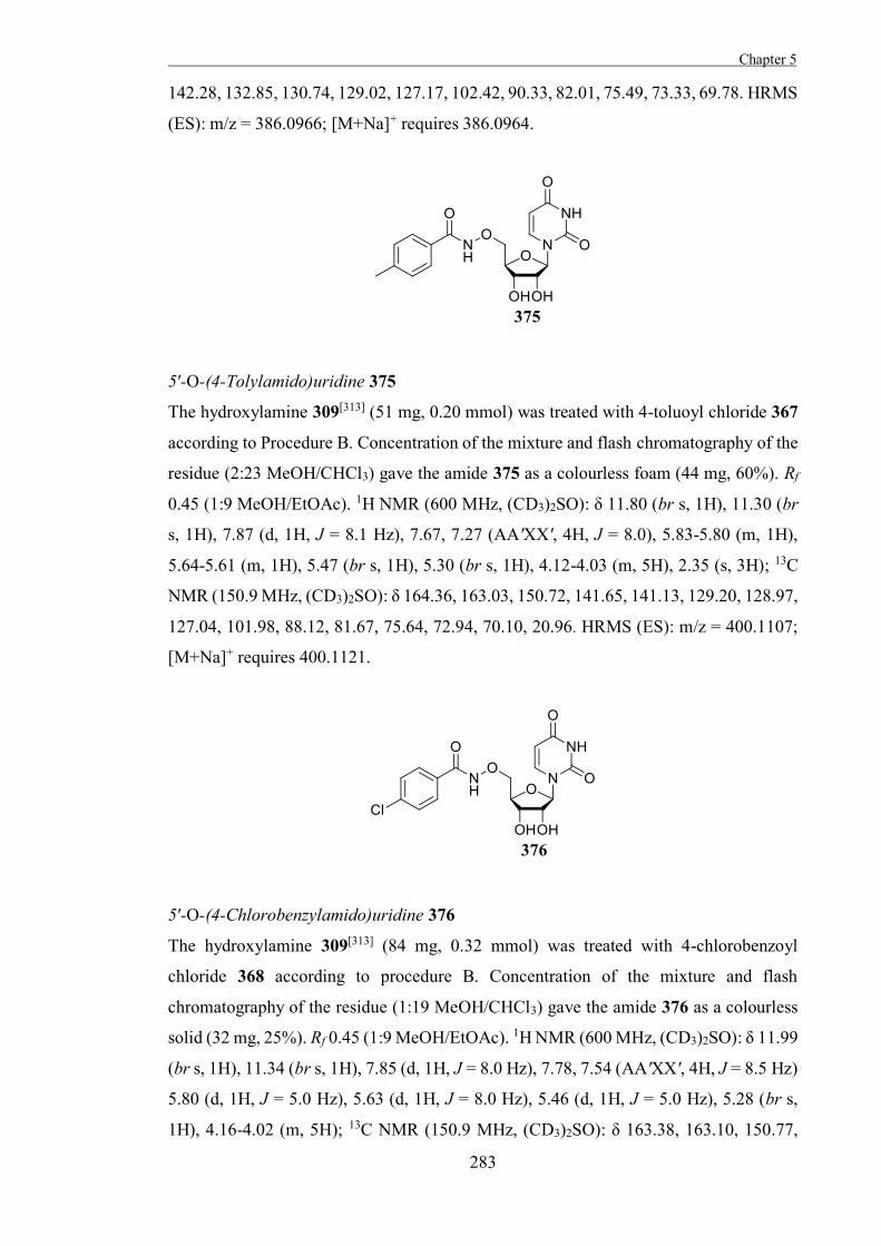
Chapter 5
283
142.28, 132.85, 130.74, 129.02, 127.17, 102.42, 90.33, 82.01, 75.49, 73.33, 69.78. HRMS
(ES): m/z = 386.0966; [M+Na]+ requires 386.0964.
5′-O-(4-Tolylamido)uridine 375
The hydroxylamine 309[313] (51 mg, 0.20 mmol) was treated with 4-toluoyl chloride 367
according to Procedure B. Concentration of the mixture and flash chromatography of the
residue (2:23 MeOH/CHCl3) gave the amide 375 as a colourless foam (44 mg, 60%). Rf
0.45 (1:9 MeOH/EtOAc). 1H NMR (600 MHz, (CD3)2SO): δ 11.80 (br s, 1H), 11.30 (br
s, 1H), 7.87 (d, 1H, J = 8.1 Hz), 7.67, 7.27 (AA'XX', 4H, J = 8.0), 5.83-5.80 (m, 1H),
5.64-5.61 (m, 1H), 5.47 (br s, 1H), 5.30 (br s, 1H), 4.12-4.03 (m, 5H), 2.35 (s, 3H); 13C
NMR (150.9 MHz, (CD3)2SO): δ 164.36, 163.03, 150.72, 141.65, 141.13, 129.20, 128.97,
127.04, 101.98, 88.12, 81.67, 75.64, 72.94, 70.10, 20.96. HRMS (ES): m/z = 400.1107;
[M+Na]+ requires 400.1121.
5′-O-(4-Chlorobenzylamido)uridine 376
The hydroxylamine 309[313] (84 mg, 0.32 mmol) was treated with 4-chlorobenzoyl
chloride 368 according to procedure B. Concentration of the mixture and flash
chromatography of the residue (1:19 MeOH/CHCl3) gave the amide 376 as a colourless
solid (32 mg, 25%). Rf 0.45 (1:9 MeOH/EtOAc). 1H NMR (600 MHz, (CD3)2SO): δ 11.99
(br s, 1H), 11.34 (br s, 1H), 7.85 (d, 1H, J = 8.0 Hz), 7.78, 7.54 (AA'XX', 4H, J = 8.5 Hz)
5.80 (d, 1H, J = 5.0 Hz), 5.63 (d, 1H, J = 8.0 Hz), 5.46 (d, 1H, J = 5.0 Hz), 5.28 (br s,
1H), 4.16-4.02 (m, 5H); 13C NMR (150.9 MHz, (CD3)2SO): δ 163.38, 163.10, 150.77,
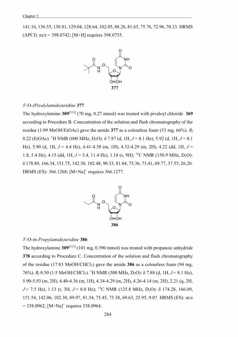
Chapter 5 .
284
141.16, 136.55, 130.81, 129.04, 128.64, 102.05, 88.26, 81.65, 75.76, 72.96, 70.13. HRMS
(APCI): m/z = 398.0742; [M+H] requires 398.0755.
5′-O-(Pivalylamido)uridine 377
The hydroxylamine 309[313] (70 mg, 0.27 mmol) was treated with pivaloyl chloride 369
according to Procedure B. Concentration of the solution and flash chromatography of the
residue (1:99 MeOH/EtOAc) gave the amide 377 as a colourless foam (53 mg, 66%). Rf
0.22 (EtOAc). 1H NMR (600 MHz, D2O): δ 7.87 (d, 1H, J = 8.1 Hz), 5.92 (d, 1H, J = 8.1
Hz), 5.90 (d, 1H, J = 4.4 Hz), 4.41-4.38 (m, 1H), 4.32-4.29 (m, 2H), 4.22 (dd, 1H, J =
1.8, 5.4 Hz), 4.15 (dd, 1H, J = 5.4, 11.4 Hz), 1.18 (s, 9H); 13C NMR (150.9 MHz, D2O):
δ 178.89, 166.34, 151.75, 142.30, 102.48, 90.33, 81.84, 75.36, 73.41, 69.77, 37.55, 26.20.
HRMS (ES): 366.1268; [M+Na]+ requires 366.1277.
5′-O-(n-Propylamido)uridine 386
The hydroxylamine 309[313] (101 mg, 0.390 mmol) was treated with propanoic anhydride
378 according to Procedure C. Concentration of the solution and flash chromatography
of the residue (17:83 MeOH/CHCl3) gave the amide 386 as a colourless foam (94 mg,
76%). Rf 0.50 (1:5 MeOH/CHCl3). 1H NMR (500 MHz, D2O): δ 7.88 (d, 1H, J = 8.1 Hz),
5.90-5.93 (m, 2H), 4.40-4.36 (m, 1H), 4.34-4.29 (m, 2H), 4.26-4.14 (m, 2H), 2.21 (q, 2H,
J = 7.5 Hz), 1.13 (t, 3H, J = 8.0 Hz); 13C NMR (125.8 MHz, D2O): δ 174.28, 166.09,
151.54, 142.06, 102.30, 89.97, 81.54, 75.45, 73.38, 69.63, 25.95, 9.07. HRMS (ES): m/z
= 338.0962; [M+Na]+ requires 338.0964.
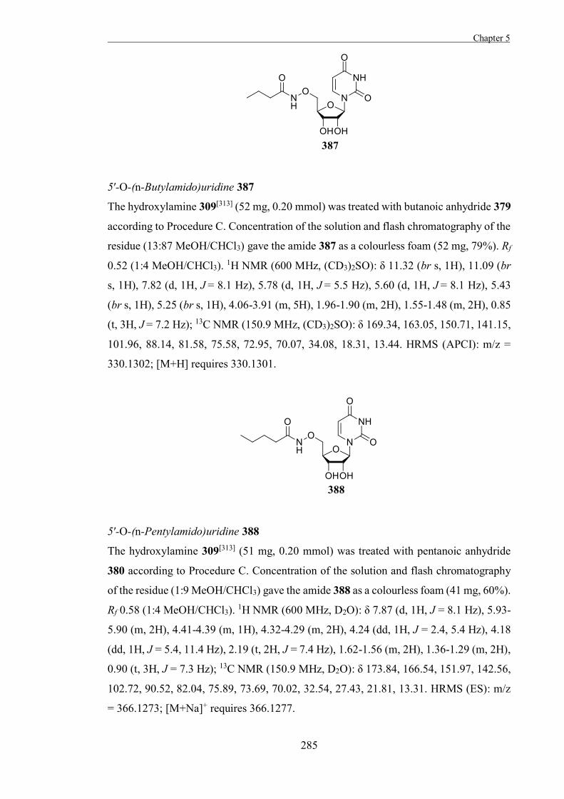
Chapter 5
285
5′-O-(n-Butylamido)uridine 387
The hydroxylamine 309[313] (52 mg, 0.20 mmol) was treated with butanoic anhydride 379
according to Procedure C. Concentration of the solution and flash chromatography of the
residue (13:87 MeOH/CHCl3) gave the amide 387 as a colourless foam (52 mg, 79%). Rf
0.52 (1:4 MeOH/CHCl3). 1H NMR (600 MHz, (CD3)2SO): δ 11.32 (br s, 1H), 11.09 (br
s, 1H), 7.82 (d, 1H, J = 8.1 Hz), 5.78 (d, 1H, J = 5.5 Hz), 5.60 (d, 1H, J = 8.1 Hz), 5.43
(br s, 1H), 5.25 (br s, 1H), 4.06-3.91 (m, 5H), 1.96-1.90 (m, 2H), 1.55-1.48 (m, 2H), 0.85
(t, 3H, J = 7.2 Hz); 13C NMR (150.9 MHz, (CD3)2SO): δ 169.34, 163.05, 150.71, 141.15,
101.96, 88.14, 81.58, 75.58, 72.95, 70.07, 34.08, 18.31, 13.44. HRMS (APCI): m/z =
330.1302; [M+H] requires 330.1301.
5′-O-(n-Pentylamido)uridine 388
The hydroxylamine 309[313] (51 mg, 0.20 mmol) was treated with pentanoic anhydride
380 according to Procedure C. Concentration of the solution and flash chromatography
of the residue (1:9 MeOH/CHCl3) gave the amide 388 as a colourless foam (41 mg, 60%).
Rf 0.58 (1:4 MeOH/CHCl3). 1H NMR (600 MHz, D2O): δ 7.87 (d, 1H, J = 8.1 Hz), 5.93-
5.90 (m, 2H), 4.41-4.39 (m, 1H), 4.32-4.29 (m, 2H), 4.24 (dd, 1H, J = 2.4, 5.4 Hz), 4.18
(dd, 1H, J = 5.4, 11.4 Hz), 2.19 (t, 2H, J = 7.4 Hz), 1.62-1.56 (m, 2H), 1.36-1.29 (m, 2H),
0.90 (t, 3H, J = 7.3 Hz); 13C NMR (150.9 MHz, D2O): δ 173.84, 166.54, 151.97, 142.56,
102.72, 90.52, 82.04, 75.89, 73.69, 70.02, 32.54, 27.43, 21.81, 13.31. HRMS (ES): m/z
= 366.1273; [M+Na]+ requires 366.1277.
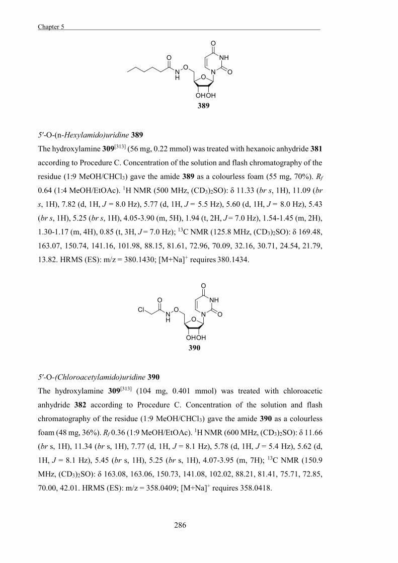
Chapter 5 .
286
5′-O-(n-Hexylamido)uridine 389
The hydroxylamine 309[313] (56 mg, 0.22 mmol) was treated with hexanoic anhydride 381
according to Procedure C. Concentration of the solution and flash chromatography of the
residue (1:9 MeOH/CHCl3) gave the amide 389 as a colourless foam (55 mg, 70%). Rf
0.64 (1:4 MeOH/EtOAc). 1H NMR (500 MHz, (CD3)2SO): δ 11.33 (br s, 1H), 11.09 (br
s, 1H), 7.82 (d, 1H, J = 8.0 Hz), 5.77 (d, 1H, J = 5.5 Hz), 5.60 (d, 1H, J = 8.0 Hz), 5.43
(br s, 1H), 5.25 (br s, 1H), 4.05-3.90 (m, 5H), 1.94 (t, 2H, J = 7.0 Hz), 1.54-1.45 (m, 2H),
1.30-1.17 (m, 4H), 0.85 (t, 3H, J = 7.0 Hz); 13C NMR (125.8 MHz, (CD3)2SO): δ 169.48,
163.07, 150.74, 141.16, 101.98, 88.15, 81.61, 72.96, 70.09, 32.16, 30.71, 24.54, 21.79,
13.82. HRMS (ES): m/z = 380.1430; [M+Na]+ requires 380.1434.
5′-O-(Chloroacetylamido)uridine 390
The hydroxylamine 309[313] (104 mg, 0.401 mmol) was treated with chloroacetic
anhydride 382 according to Procedure C. Concentration of the solution and flash
chromatography of the residue (1:9 MeOH/CHCl3) gave the amide 390 as a colourless
foam (48 mg, 36%). Rf 0.36 (1:9 MeOH/EtOAc). 1H NMR (600 MHz, (CD3)2SO): δ 11.66
(br s, 1H), 11.34 (br s, 1H), 7.77 (d, 1H, J = 8.1 Hz), 5.78 (d, 1H, J = 5.4 Hz), 5.62 (d,
1H, J = 8.1 Hz), 5.45 (br s, 1H), 5.25 (br s, 1H), 4.07-3.95 (m, 7H); 13C NMR (150.9
MHz, (CD3)2SO): δ 163.08, 163.06, 150.73, 141.08, 102.02, 88.21, 81.41, 75.71, 72.85,
70.00, 42.01. HRMS (ES): m/z = 358.0409; [M+Na]+ requires 358.0418.
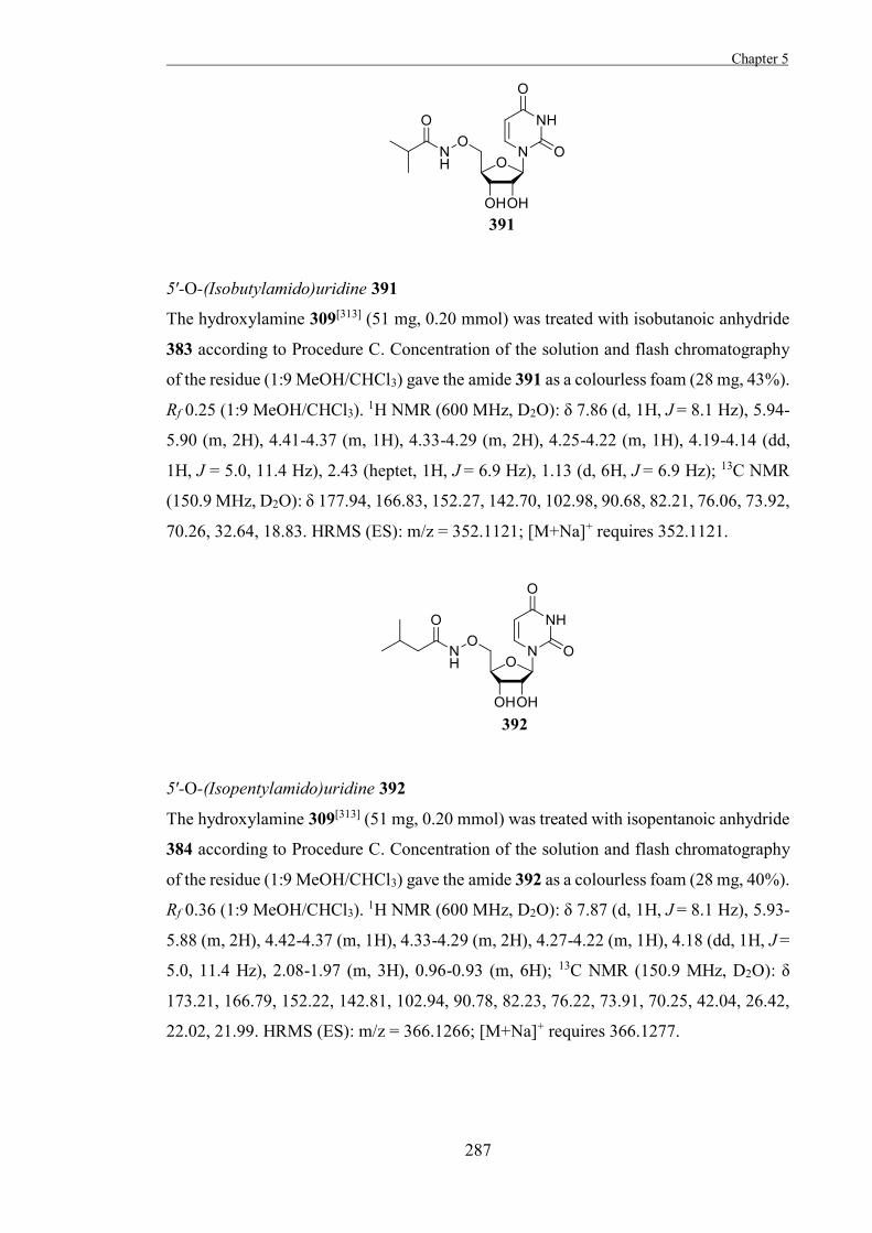
Chapter 5
287
5′-O-(Isobutylamido)uridine 391
The hydroxylamine 309[313] (51 mg, 0.20 mmol) was treated with isobutanoic anhydride
383 according to Procedure C. Concentration of the solution and flash chromatography
of the residue (1:9 MeOH/CHCl3) gave the amide 391 as a colourless foam (28 mg, 43%).
Rf 0.25 (1:9 MeOH/CHCl3). 1H NMR (600 MHz, D2O): δ 7.86 (d, 1H, J = 8.1 Hz), 5.94-
5.90 (m, 2H), 4.41-4.37 (m, 1H), 4.33-4.29 (m, 2H), 4.25-4.22 (m, 1H), 4.19-4.14 (dd,
1H, J = 5.0, 11.4 Hz), 2.43 (heptet, 1H, J = 6.9 Hz), 1.13 (d, 6H, J = 6.9 Hz); 13C NMR
(150.9 MHz, D2O): δ 177.94, 166.83, 152.27, 142.70, 102.98, 90.68, 82.21, 76.06, 73.92,
70.26, 32.64, 18.83. HRMS (ES): m/z = 352.1121; [M+Na]+ requires 352.1121.
5′-O-(Isopentylamido)uridine 392
The hydroxylamine 309[313] (51 mg, 0.20 mmol) was treated with isopentanoic anhydride
384 according to Procedure C. Concentration of the solution and flash chromatography
of the residue (1:9 MeOH/CHCl3) gave the amide 392 as a colourless foam (28 mg, 40%).
Rf 0.36 (1:9 MeOH/CHCl3). 1H NMR (600 MHz, D2O): δ 7.87 (d, 1H, J = 8.1 Hz), 5.93-
5.88 (m, 2H), 4.42-4.37 (m, 1H), 4.33-4.29 (m, 2H), 4.27-4.22 (m, 1H), 4.18 (dd, 1H, J =
5.0, 11.4 Hz), 2.08-1.97 (m, 3H), 0.96-0.93 (m, 6H); 13C NMR (150.9 MHz, D2O): δ
173.21, 166.79, 152.22, 142.81, 102.94, 90.78, 82.23, 76.22, 73.91, 70.25, 42.04, 26.42,
22.02, 21.99. HRMS (ES): m/z = 366.1266; [M+Na]+ requires 366.1277.
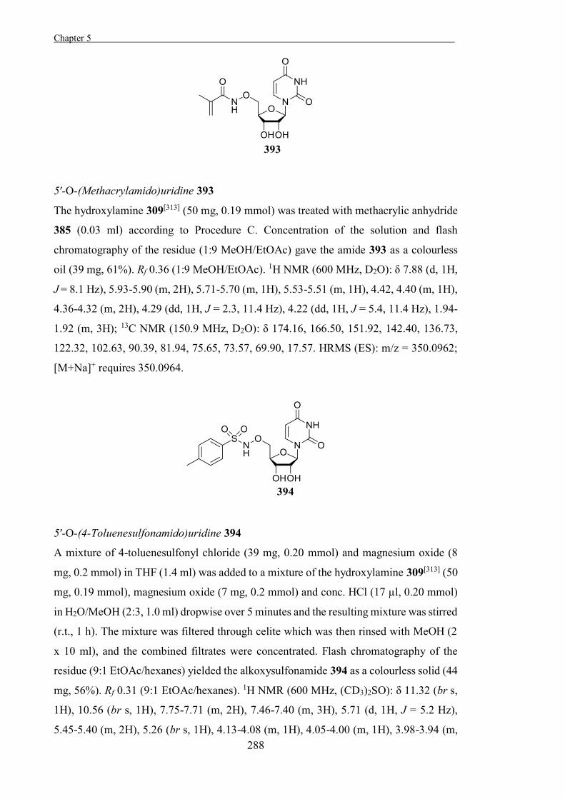
Chapter 5 .
288
5′-O-(Methacrylamido)uridine 393
The hydroxylamine 309[313] (50 mg, 0.19 mmol) was treated with methacrylic anhydride
385 (0.03 ml) according to Procedure C. Concentration of the solution and flash
chromatography of the residue (1:9 MeOH/EtOAc) gave the amide 393 as a colourless
oil (39 mg, 61%). Rf 0.36 (1:9 MeOH/EtOAc). 1H NMR (600 MHz, D2O): δ 7.88 (d, 1H,
J = 8.1 Hz), 5.93-5.90 (m, 2H), 5.71-5.70 (m, 1H), 5.53-5.51 (m, 1H), 4.42, 4.40 (m, 1H),
4.36-4.32 (m, 2H), 4.29 (dd, 1H, J = 2.3, 11.4 Hz), 4.22 (dd, 1H, J = 5.4, 11.4 Hz), 1.94-
1.92 (m, 3H); 13C NMR (150.9 MHz, D2O): δ 174.16, 166.50, 151.92, 142.40, 136.73,
122.32, 102.63, 90.39, 81.94, 75.65, 73.57, 69.90, 17.57. HRMS (ES): m/z = 350.0962;
[M+Na]+ requires 350.0964.
5′-O-(4-Toluenesulfonamido)uridine 394
A mixture of 4-toluenesulfonyl chloride (39 mg, 0.20 mmol) and magnesium oxide (8
mg, 0.2 mmol) in THF (1.4 ml) was added to a mixture of the hydroxylamine 309[313] (50
mg, 0.19 mmol), magnesium oxide (7 mg, 0.2 mmol) and conc. HCl (17 µl, 0.20 mmol)
in H2O/MeOH (2:3, 1.0 ml) dropwise over 5 minutes and the resulting mixture was stirred
(r.t., 1 h). The mixture was filtered through celite which was then rinsed with MeOH (2
x 10 ml), and the combined filtrates were concentrated. Flash chromatography of the
residue (9:1 EtOAc/hexanes) yielded the alkoxysulfonamide 394 as a colourless solid (44
mg, 56%). Rf 0.31 (9:1 EtOAc/hexanes). 1H NMR (600 MHz, (CD3)2SO): δ 11.32 (br s,
1H), 10.56 (br s, 1H), 7.75-7.71 (m, 2H), 7.46-7.40 (m, 3H), 5.71 (d, 1H, J = 5.2 Hz),
5.45-5.40 (m, 2H), 5.26 (br s, 1H), 4.13-4.08 (m, 1H), 4.05-4.00 (m, 1H), 3.98-3.94 (m,
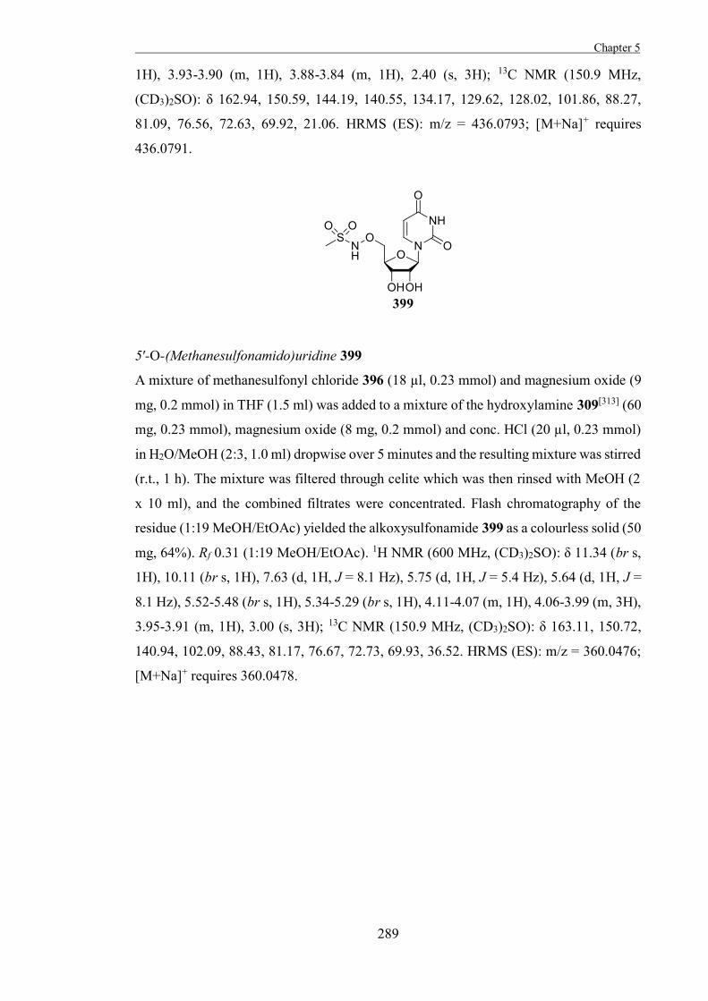
Chapter 5
289
1H), 3.93-3.90 (m, 1H), 3.88-3.84 (m, 1H), 2.40 (s, 3H); 13C NMR (150.9 MHz,
(CD3)2SO): δ 162.94, 150.59, 144.19, 140.55, 134.17, 129.62, 128.02, 101.86, 88.27,
81.09, 76.56, 72.63, 69.92, 21.06. HRMS (ES): m/z = 436.0793; [M+Na]+ requires
436.0791.
5′-O-(Methanesulfonamido)uridine 399
A mixture of methanesulfonyl chloride 396 (18 µl, 0.23 mmol) and magnesium oxide (9
mg, 0.2 mmol) in THF (1.5 ml) was added to a mixture of the hydroxylamine 309[313] (60
mg, 0.23 mmol), magnesium oxide (8 mg, 0.2 mmol) and conc. HCl (20 µl, 0.23 mmol)
in H2O/MeOH (2:3, 1.0 ml) dropwise over 5 minutes and the resulting mixture was stirred
(r.t., 1 h). The mixture was filtered through celite which was then rinsed with MeOH (2
x 10 ml), and the combined filtrates were concentrated. Flash chromatography of the
residue (1:19 MeOH/EtOAc) yielded the alkoxysulfonamide 399 as a colourless solid (50
mg, 64%). Rf 0.31 (1:19 MeOH/EtOAc). 1H NMR (600 MHz, (CD3)2SO): δ 11.34 (br s,
1H), 10.11 (br s, 1H), 7.63 (d, 1H, J = 8.1 Hz), 5.75 (d, 1H, J = 5.4 Hz), 5.64 (d, 1H, J =
8.1 Hz), 5.52-5.48 (br s, 1H), 5.34-5.29 (br s, 1H), 4.11-4.07 (m, 1H), 4.06-3.99 (m, 3H),
3.95-3.91 (m, 1H), 3.00 (s, 3H); 13C NMR (150.9 MHz, (CD3)2SO): δ 163.11, 150.72,
140.94, 102.09, 88.43, 81.17, 76.67, 72.73, 69.93, 36.52. HRMS (ES): m/z = 360.0476;
[M+Na]+ requires 360.0478.
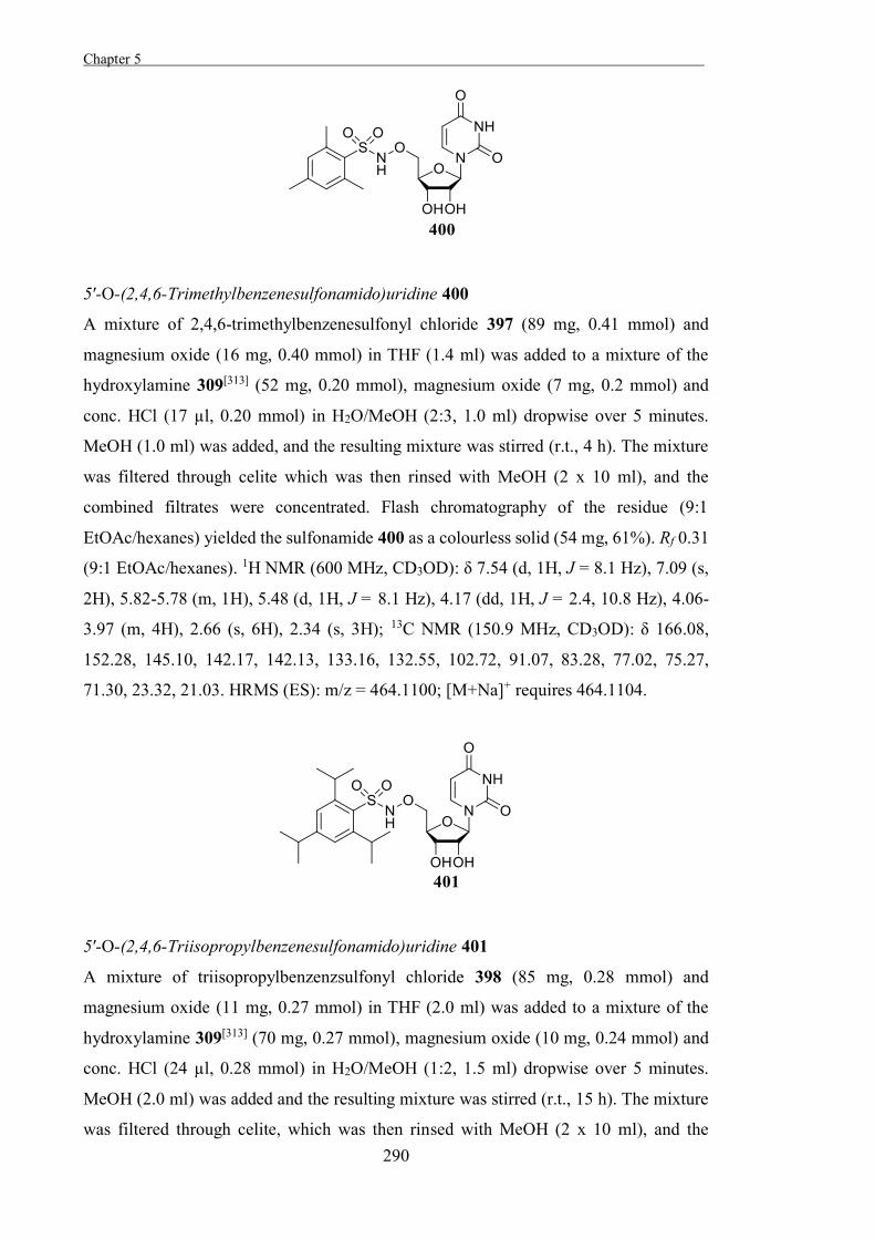
Chapter 5 .
290
5′-O-(2,4,6-Trimethylbenzenesulfonamido)uridine 400
A mixture of 2,4,6-trimethylbenzenesulfonyl chloride 397 (89 mg, 0.41 mmol) and
magnesium oxide (16 mg, 0.40 mmol) in THF (1.4 ml) was added to a mixture of the
hydroxylamine 309[313] (52 mg, 0.20 mmol), magnesium oxide (7 mg, 0.2 mmol) and
conc. HCl (17 µl, 0.20 mmol) in H2O/MeOH (2:3, 1.0 ml) dropwise over 5 minutes.
MeOH (1.0 ml) was added, and the resulting mixture was stirred (r.t., 4 h). The mixture
was filtered through celite which was then rinsed with MeOH (2 x 10 ml), and the
combined filtrates were concentrated. Flash chromatography of the residue (9:1
EtOAc/hexanes) yielded the sulfonamide 400 as a colourless solid (54 mg, 61%). Rf 0.31
(9:1 EtOAc/hexanes). 1H NMR (600 MHz, CD3OD): δ 7.54 (d, 1H, J = 8.1 Hz), 7.09 (s,
2H), 5.82-5.78 (m, 1H), 5.48 (d, 1H, J = 8.1 Hz), 4.17 (dd, 1H, J = 2.4, 10.8 Hz), 4.06-
3.97 (m, 4H), 2.66 (s, 6H), 2.34 (s, 3H); 13C NMR (150.9 MHz, CD3OD): δ 166.08,
152.28, 145.10, 142.17, 142.13, 133.16, 132.55, 102.72, 91.07, 83.28, 77.02, 75.27,
71.30, 23.32, 21.03. HRMS (ES): m/z = 464.1100; [M+Na]+ requires 464.1104.
5′-O-(2,4,6-Triisopropylbenzenesulfonamido)uridine 401
A mixture of triisopropylbenzenzsulfonyl chloride 398 (85 mg, 0.28 mmol) and
magnesium oxide (11 mg, 0.27 mmol) in THF (2.0 ml) was added to a mixture of the
hydroxylamine 309[313] (70 mg, 0.27 mmol), magnesium oxide (10 mg, 0.24 mmol) and
conc. HCl (24 µl, 0.28 mmol) in H2O/MeOH (1:2, 1.5 ml) dropwise over 5 minutes.
MeOH (2.0 ml) was added and the resulting mixture was stirred (r.t., 15 h). The mixture
was filtered through celite, which was then rinsed with MeOH (2 x 10 ml), and the
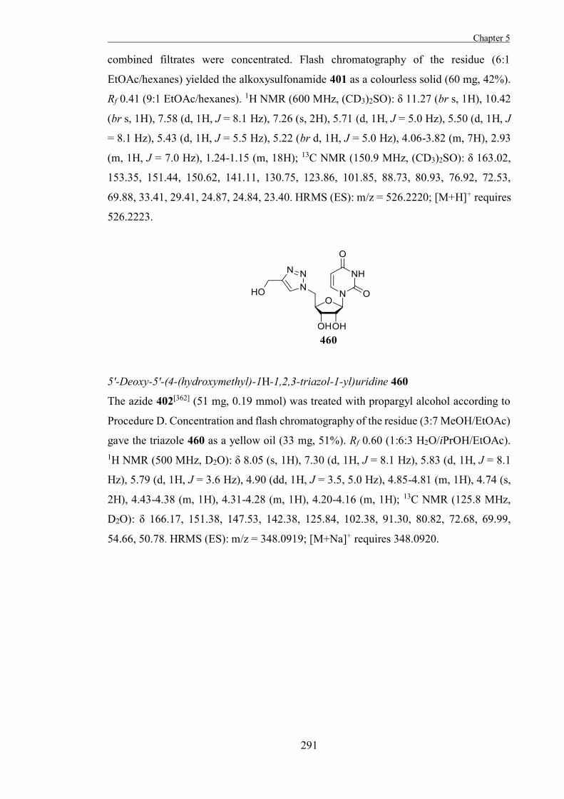
Chapter 5
291
combined filtrates were concentrated. Flash chromatography of the residue (6:1
EtOAc/hexanes) yielded the alkoxysulfonamide 401 as a colourless solid (60 mg, 42%).
Rf 0.41 (9:1 EtOAc/hexanes). 1H NMR (600 MHz, (CD3)2SO): δ 11.27 (br s, 1H), 10.42
(br s, 1H), 7.58 (d, 1H, J = 8.1 Hz), 7.26 (s, 2H), 5.71 (d, 1H, J = 5.0 Hz), 5.50 (d, 1H, J
= 8.1 Hz), 5.43 (d, 1H, J = 5.5 Hz), 5.22 (br d, 1H, J = 5.0 Hz), 4.06-3.82 (m, 7H), 2.93
(m, 1H, J = 7.0 Hz), 1.24-1.15 (m, 18H); 13C NMR (150.9 MHz, (CD3)2SO): δ 163.02,
153.35, 151.44, 150.62, 141.11, 130.75, 123.86, 101.85, 88.73, 80.93, 76.92, 72.53,
69.88, 33.41, 29.41, 24.87, 24.84, 23.40. HRMS (ES): m/z = 526.2220; [M+H]+ requires
526.2223.
5'-Deoxy-5'-(4-(hydroxymethyl)-1H-1,2,3-triazol-1-yl)uridine 460
The azide 402[362] (51 mg, 0.19 mmol) was treated with propargyl alcohol according to
Procedure D. Concentration and flash chromatography of the residue (3:7 MeOH/EtOAc)
gave the triazole 460 as a yellow oil (33 mg, 51%). Rf 0.60 (1:6:3 H2O/iPrOH/EtOAc). 1H NMR (500 MHz, D2O): δ 8.05 (s, 1H), 7.30 (d, 1H, J = 8.1 Hz), 5.83 (d, 1H, J = 8.1
Hz), 5.79 (d, 1H, J = 3.6 Hz), 4.90 (dd, 1H, J = 3.5, 5.0 Hz), 4.85-4.81 (m, 1H), 4.74 (s,
2H), 4.43-4.38 (m, 1H), 4.31-4.28 (m, 1H), 4.20-4.16 (m, 1H); 13C NMR (125.8 MHz,
D2O): δ 166.17, 151.38, 147.53, 142.38, 125.84, 102.38, 91.30, 80.82, 72.68, 69.99,
54.66, 50.78. HRMS (ES): m/z = 348.0919; [M+Na]+ requires 348.0920.
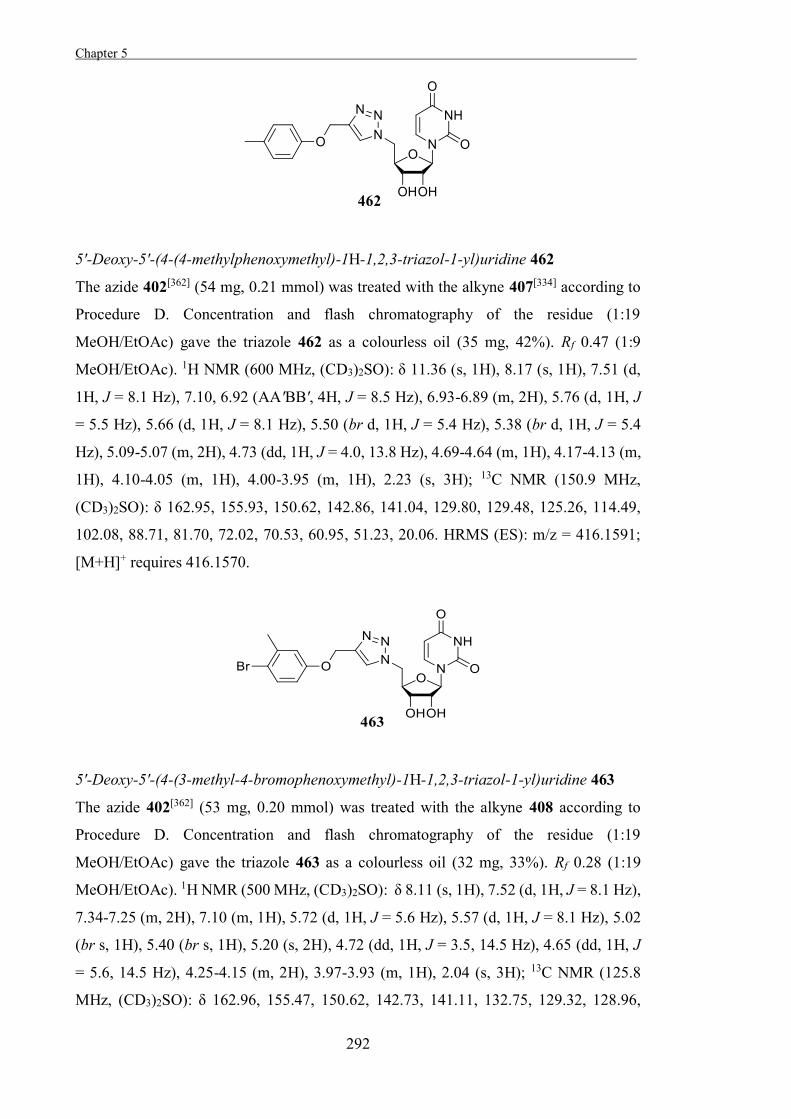
Chapter 5 .
292
5'-Deoxy-5'-(4-(4-methylphenoxymethyl)-1H-1,2,3-triazol-1-yl)uridine 462
The azide 402[362] (54 mg, 0.21 mmol) was treated with the alkyne 407[334] according to
Procedure D. Concentration and flash chromatography of the residue (1:19
MeOH/EtOAc) gave the triazole 462 as a colourless oil (35 mg, 42%). Rf 0.47 (1:9
MeOH/EtOAc). 1H NMR (600 MHz, (CD3)2SO): δ 11.36 (s, 1H), 8.17 (s, 1H), 7.51 (d,
1H, J = 8.1 Hz), 7.10, 6.92 (AA'BB', 4H, J = 8.5 Hz), 6.93-6.89 (m, 2H), 5.76 (d, 1H, J
= 5.5 Hz), 5.66 (d, 1H, J = 8.1 Hz), 5.50 (br d, 1H, J = 5.4 Hz), 5.38 (br d, 1H, J = 5.4
Hz), 5.09-5.07 (m, 2H), 4.73 (dd, 1H, J = 4.0, 13.8 Hz), 4.69-4.64 (m, 1H), 4.17-4.13 (m,
1H), 4.10-4.05 (m, 1H), 4.00-3.95 (m, 1H), 2.23 (s, 3H); 13C NMR (150.9 MHz,
(CD3)2SO): δ 162.95, 155.93, 150.62, 142.86, 141.04, 129.80, 129.48, 125.26, 114.49,
102.08, 88.71, 81.70, 72.02, 70.53, 60.95, 51.23, 20.06. HRMS (ES): m/z = 416.1591;
[M+H]+ requires 416.1570.
5'-Deoxy-5'-(4-(3-methyl-4-bromophenoxymethyl)-1H-1,2,3-triazol-1-yl)uridine 463
The azide 402[362] (53 mg, 0.20 mmol) was treated with the alkyne 408 according to
Procedure D. Concentration and flash chromatography of the residue (1:19
MeOH/EtOAc) gave the triazole 463 as a colourless oil (32 mg, 33%). Rf 0.28 (1:19
MeOH/EtOAc). 1H NMR (500 MHz, (CD3)2SO): δ 8.11 (s, 1H), 7.52 (d, 1H, J = 8.1 Hz),
7.34-7.25 (m, 2H), 7.10 (m, 1H), 5.72 (d, 1H, J = 5.6 Hz), 5.57 (d, 1H, J = 8.1 Hz), 5.02
(br s, 1H), 5.40 (br s, 1H), 5.20 (s, 2H), 4.72 (dd, 1H, J = 3.5, 14.5 Hz), 4.65 (dd, 1H, J
= 5.6, 14.5 Hz), 4.25-4.15 (m, 2H), 3.97-3.93 (m, 1H), 2.04 (s, 3H); 13C NMR (125.8
MHz, (CD3)2SO): δ 162.96, 155.47, 150.62, 142.73, 141.11, 132.75, 129.32, 128.96,
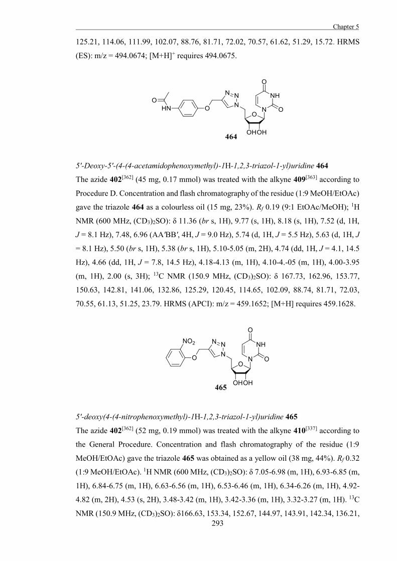
Chapter 5
293
125.21, 114.06, 111.99, 102.07, 88.76, 81.71, 72.02, 70.57, 61.62, 51.29, 15.72. HRMS
(ES): m/z = 494.0674; [M+H]+ requires 494.0675.
5'-Deoxy-5'-(4-(4-acetamidophenoxymethyl)-1H-1,2,3-triazol-1-yl)uridine 464
The azide 402[362] (45 mg, 0.17 mmol) was treated with the alkyne 409[363] according to
Procedure D. Concentration and flash chromatography of the residue (1:9 MeOH/EtOAc)
gave the triazole 464 as a colourless oil (15 mg, 23%). Rf 0.19 (9:1 EtOAc/MeOH); 1H
NMR (600 MHz, (CD3)2SO): δ 11.36 (br s, 1H), 9.77 (s, 1H), 8.18 (s, 1H), 7.52 (d, 1H,
J = 8.1 Hz), 7.48, 6.96 (AA'BB', 4H, J = 9.0 Hz), 5.74 (d, 1H, J = 5.5 Hz), 5.63 (d, 1H, J
= 8.1 Hz), 5.50 (br s, 1H), 5.38 (br s, 1H), 5.10-5.05 (m, 2H), 4.74 (dd, 1H, J = 4.1, 14.5
Hz), 4.66 (dd, 1H, J = 7.8, 14.5 Hz), 4.18-4.13 (m, 1H), 4.10-4.-05 (m, 1H), 4.00-3.95
(m, 1H), 2.00 (s, 3H); 13C NMR (150.9 MHz, (CD3)2SO): δ 167.73, 162.96, 153.77,
150.63, 142.81, 141.06, 132.86, 125.29, 120.45, 114.65, 102.09, 88.74, 81.71, 72.03,
70.55, 61.13, 51.25, 23.79. HRMS (APCI): m/z = 459.1652; [M+H] requires 459.1628.
5'-deoxy(4-(4-nitrophenoxymethyl)-1H-1,2,3-triazol-1-yl)uridine 465
The azide 402[362] (52 mg, 0.19 mmol) was treated with the alkyne 410[337] according to
the General Procedure. Concentration and flash chromatography of the residue (1:9
MeOH/EtOAc) gave the triazole 465 was obtained as a yellow oil (38 mg, 44%). Rf 0.32
(1:9 MeOH/EtOAc). 1H NMR (600 MHz, (CD3)2SO): δ 7.05-6.98 (m, 1H), 6.93-6.85 (m,
1H), 6.84-6.75 (m, 1H), 6.63-6.56 (m, 1H), 6.53-6.46 (m, 1H), 6.34-6.26 (m, 1H), 4.92-
4.82 (m, 2H), 4.53 (s, 2H), 3.48-3.42 (m, 1H), 3.42-3.36 (m, 1H), 3.32-3.27 (m, 1H). 13C
NMR (150.9 MHz, (CD3)2SO): δ166.63, 153.34, 152.67, 144.97, 143.91, 142.34, 136.21,
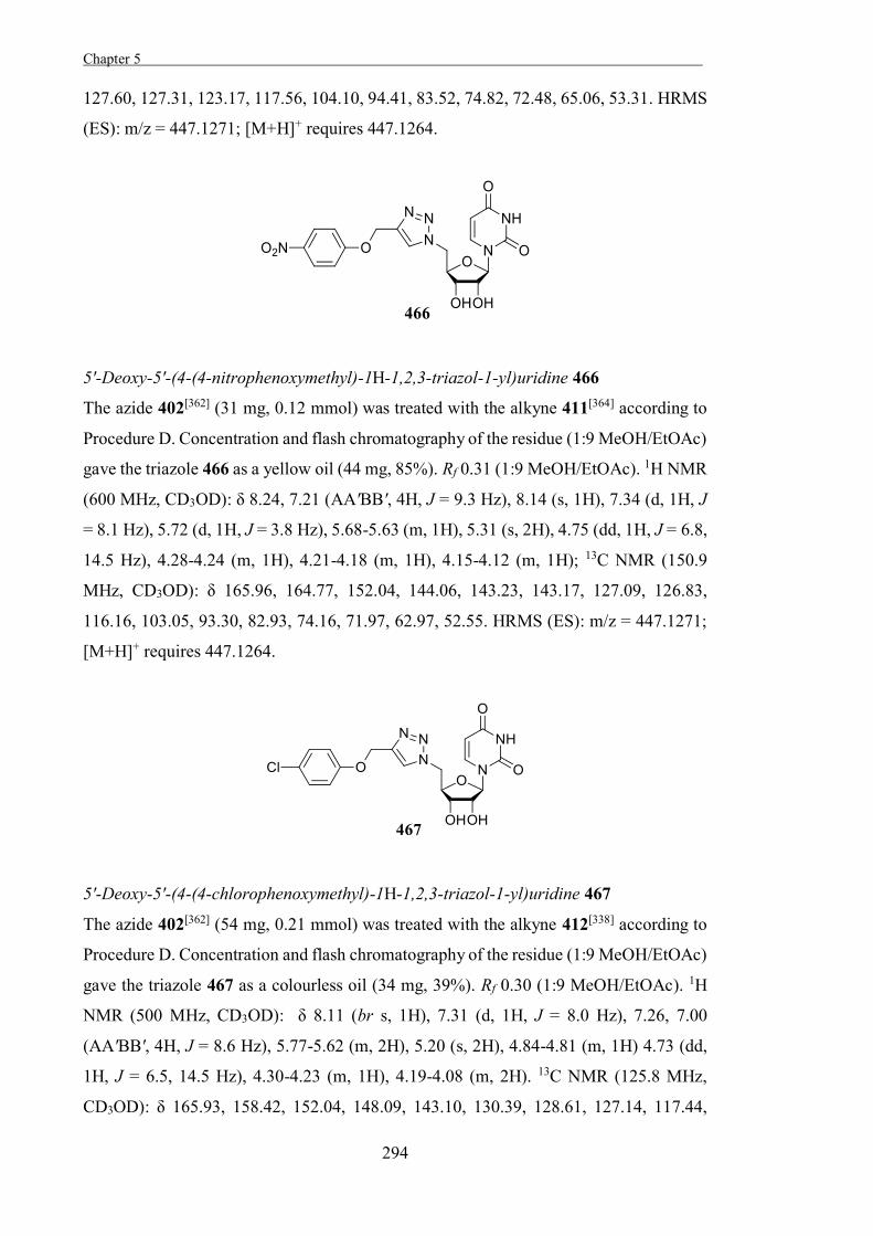
Chapter 5 .
294
127.60, 127.31, 123.17, 117.56, 104.10, 94.41, 83.52, 74.82, 72.48, 65.06, 53.31. HRMS
(ES): m/z = 447.1271; [M+H]+ requires 447.1264.
5'-Deoxy-5'-(4-(4-nitrophenoxymethyl)-1H-1,2,3-triazol-1-yl)uridine 466
The azide 402[362] (31 mg, 0.12 mmol) was treated with the alkyne 411[364] according to
Procedure D. Concentration and flash chromatography of the residue (1:9 MeOH/EtOAc)
gave the triazole 466 as a yellow oil (44 mg, 85%). Rf 0.31 (1:9 MeOH/EtOAc). 1H NMR
(600 MHz, CD3OD): δ 8.24, 7.21 (AA'BB', 4H, J = 9.3 Hz), 8.14 (s, 1H), 7.34 (d, 1H, J
= 8.1 Hz), 5.72 (d, 1H, J = 3.8 Hz), 5.68-5.63 (m, 1H), 5.31 (s, 2H), 4.75 (dd, 1H, J = 6.8,
14.5 Hz), 4.28-4.24 (m, 1H), 4.21-4.18 (m, 1H), 4.15-4.12 (m, 1H); 13C NMR (150.9
MHz, CD3OD): δ 165.96, 164.77, 152.04, 144.06, 143.23, 143.17, 127.09, 126.83,
116.16, 103.05, 93.30, 82.93, 74.16, 71.97, 62.97, 52.55. HRMS (ES): m/z = 447.1271;
[M+H]+ requires 447.1264.
5'-Deoxy-5'-(4-(4-chlorophenoxymethyl)-1H-1,2,3-triazol-1-yl)uridine 467
The azide 402[362] (54 mg, 0.21 mmol) was treated with the alkyne 412[338] according to
Procedure D. Concentration and flash chromatography of the residue (1:9 MeOH/EtOAc)
gave the triazole 467 as a colourless oil (34 mg, 39%). Rf 0.30 (1:9 MeOH/EtOAc). 1H
NMR (500 MHz, CD3OD): δ 8.11 (br s, 1H), 7.31 (d, 1H, J = 8.0 Hz), 7.26, 7.00
(AA'BB', 4H, J = 8.6 Hz), 5.77-5.62 (m, 2H), 5.20 (s, 2H), 4.84-4.81 (m, 1H) 4.73 (dd,
1H, J = 6.5, 14.5 Hz), 4.30-4.23 (m, 1H), 4.19-4.08 (m, 2H). 13C NMR (125.8 MHz,
CD3OD): δ 165.93, 158.42, 152.04, 148.09, 143.10, 130.39, 128.61, 127.14, 117.44,
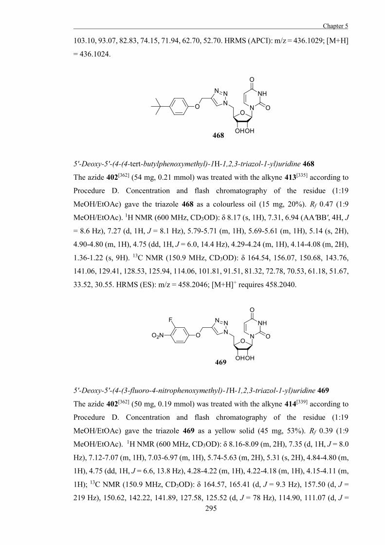
Chapter 5
295
103.10, 93.07, 82.83, 74.15, 71.94, 62.70, 52.70. HRMS (APCI): m/z = 436.1029; [M+H]
= 436.1024.
5'-Deoxy-5'-(4-(4-tert-butylphenoxymethyl)-1H-1,2,3-triazol-1-yl)uridine 468
The azide 402[362] (54 mg, 0.21 mmol) was treated with the alkyne 413[335] according to
Procedure D. Concentration and flash chromatography of the residue (1:19
MeOH/EtOAc) gave the triazole 468 as a colourless oil (15 mg, 20%). Rf 0.47 (1:9
MeOH/EtOAc). 1H NMR (600 MHz, CD3OD): δ 8.17 (s, 1H), 7.31, 6.94 (AA'BB', 4H, J
= 8.6 Hz), 7.27 (d, 1H, J = 8.1 Hz), 5.79-5.71 (m, 1H), 5.69-5.61 (m, 1H), 5.14 (s, 2H),
4.90-4.80 (m, 1H), 4.75 (dd, 1H, J = 6.0, 14.4 Hz), 4.29-4.24 (m, 1H), 4.14-4.08 (m, 2H),
1.36-1.22 (s, 9H). 13C NMR (150.9 MHz, CD3OD): δ 164.54, 156.07, 150.68, 143.76,
141.06, 129.41, 128.53, 125.94, 114.06, 101.81, 91.51, 81.32, 72.78, 70.53, 61.18, 51.67,
33.52, 30.55. HRMS (ES): m/z = 458.2046; [M+H]+ requires 458.2040.
5'-Deoxy-5'-(4-(3-fluoro-4-nitrophenoxymethyl)-1H-1,2,3-triazol-1-yl)uridine 469
The azide 402[362] (50 mg, 0.19 mmol) was treated with the alkyne 414[339] according to
Procedure D. Concentration and flash chromatography of the residue (1:19
MeOH/EtOAc) gave the triazole 469 as a yellow solid (45 mg, 53%). Rf 0.39 (1:9
MeOH/EtOAc). 1H NMR (600 MHz, CD3OD): δ 8.16-8.09 (m, 2H), 7.35 (d, 1H, J = 8.0
Hz), 7.12-7.07 (m, 1H), 7.03-6.97 (m, 1H), 5.74-5.63 (m, 2H), 5.31 (s, 2H), 4.84-4.80 (m,
1H), 4.75 (dd, 1H, J = 6.6, 13.8 Hz), 4.28-4.22 (m, 1H), 4.22-4.18 (m, 1H), 4.15-4.11 (m,
1H); 13C NMR (150.9 MHz, CD3OD): δ 164.57, 165.41 (d, J = 9.3 Hz), 157.50 (d, J =
219 Hz), 150.62, 142.22, 141.89, 127.58, 125.52 (d, J = 78 Hz), 114.90, 111.07 (d, J =
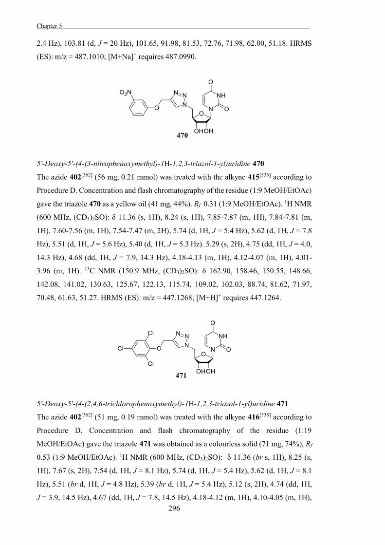
Chapter 5 .
296
2.4 Hz), 103.81 (d, J = 20 Hz), 101.65, 91.98, 81.53, 72.76, 71.98, 62.00, 51.18. HRMS
(ES): m/z = 487.1010; [M+Na]+ requires 487.0990.
5'-Deoxy-5'-(4-(3-nitrophenoxymethyl)-1H-1,2,3-triazol-1-yl)uridine 470
The azide 402[362] (56 mg, 0.21 mmol) was treated with the alkyne 415[336] according to
Procedure D. Concentration and flash chromatography of the residue (1:9 MeOH/EtOAc)
gave the triazole 470 as a yellow oil (41 mg, 44%). Rf 0.31 (1:9 MeOH/EtOAc). 1H NMR
(600 MHz, (CD3)2SO): δ 11.36 (s, 1H), 8.24 (s, 1H), 7.85-7.87 (m, 1H), 7.84-7.81 (m,
1H), 7.60-7.56 (m, 1H), 7.54-7.47 (m, 2H), 5.74 (d, 1H, J = 5.4 Hz), 5.62 (d, 1H, J = 7.8
Hz), 5.51 (d, 1H, J = 5.6 Hz), 5.40 (d, 1H, J = 5.3 Hz). 5.29 (s, 2H), 4.75 (dd, 1H, J = 4.0,
14.3 Hz), 4.68 (dd, 1H, J = 7.9, 14.3 Hz), 4.18-4.13 (m, 1H), 4.12-4.07 (m, 1H), 4.01-
3.96 (m, 1H). 13C NMR (150.9 MHz, (CD3)2SO): δ 162.90, 158.46, 150.55, 148.66,
142.08, 141.02, 130.63, 125.67, 122.13, 115.74, 109.02, 102.03, 88.74, 81.62, 71.97,
70.48, 61.63, 51.27. HRMS (ES): m/z = 447.1268; [M+H]+ requires 447.1264.
5'-Deoxy-5'-(4-(2,4,6-trichlorophenoxymethyl)-1H-1,2,3-triazol-1-yl)uridine 471
The azide 402[362] (51 mg, 0.19 mmol) was treated with the alkyne 416[338] according to
Procedure D. Concentration and flash chromatography of the residue (1:19
MeOH/EtOAc) gave the triazole 471 was obtained as a colourless solid (71 mg, 74%), Rf
0.53 (1:9 MeOH/EtOAc). 1H NMR (600 MHz, (CD3)2SO): δ 11.36 (br s, 1H), 8.25 (s,
1H), 7.67 (s, 2H), 7.54 (d, 1H, J = 8.1 Hz), 5.74 (d, 1H, J = 5.4 Hz), 5.62 (d, 1H, J = 8.1
Hz), 5.51 (br d, 1H, J = 4.8 Hz), 5.39 (br d, 1H, J = 5.4 Hz), 5.12 (s, 2H), 4.74 (dd, 1H,
J = 3.9, 14.5 Hz), 4.67 (dd, 1H, J = 7.8, 14.5 Hz), 4.18-4.12 (m, 1H), 4.10-4.05 (m, 1H),
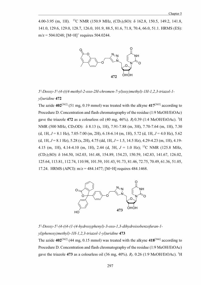
Chapter 5
297
4.00-3.95 (m, 1H). 13C NMR (150.9 MHz, (CD3)2SO): δ 162.8, 150.5, 149.2, 141.8,
141.0, 129.6, 129.0, 128.7, 126.0, 101.9, 88.5, 81.6, 71.8, 70.4, 66.0, 51.1. HRMS (ES):
m/z = 504.0248; [M+H]+ requires 504.0244.
5'-Deoxy-5'-(4-(((4-methyl-2-oxo-2H-chromen-7-yl)oxy)methyl)-1H-1,2,3-triazol-1-
yl)uridine 472
The azide 402[362] (51 mg, 0.19 mmol) was treated with the alkyne 417[365] according to
Procedure D. Concentration and flash chromatography of the residue (1:9 MeOH/EtOAc)
gave the triazole 472 as a colourless oil (40 mg, 46%). Rf 0.39 (1:4 MeOH/EtOAc). 1H
NMR (500 MHz, CD3OD): δ 8.13 (s, 1H), 7.91-7.88 (m, 3H), 7.70-7.64 (m, 1H), 7.30
(d, 1H, J = 8.1 Hz), 7.05-7.00 (m, 2H), 6.18-6.14 (m, 1H), 5.72 (d, 1H, J = 4.0 Hz), 5.62
(d, 1H, J = 8.1 Hz), 5.28 (s, 2H), 4.75 (dd, 1H, J = 1.5, 14.5 Hz), 4.29-4.23 (m, 1H), 4.19-
4.15 (m, 1H), 4.14-4.10 (m, 1H), 2.44 (d, 3H, J = 1.0 Hz); 13C NMR (125.8 MHz,
(CD3)2SO): δ 164.50, 162.03, 161.48, 154.89, 154.23, 150.59, 142.83, 141.67, 126.02,
125.64, 113.81, 112.74, 110.98, 101.59, 101.43, 91.73, 81.46, 72.75, 70.49, 61.36, 51.05,
17.24. HRMS (APCI): m/z = 484.1477; [M+H] requires 484.1468.
5'-Deoxy-5'-(4-((4-(1-(4-hydroxyphenyl)-3-oxo-1,3-dihydroisobenzofuran-1-
yl)phenoxy)methyl)-1H-1,2,3-triazol-1-yl)uridine 473
The azide 402[362] (44 mg, 0.15 mmol) was treated with the alkyne 418[366] according to
Procedure D. Concentration and flash chromatography of the residue (1:9 MeOH/EtOAc)
gave the triazole 473 as a colourless oil (36 mg, 40%). Rf. 0.26 (1:9 MeOH/EtOAc). 1H
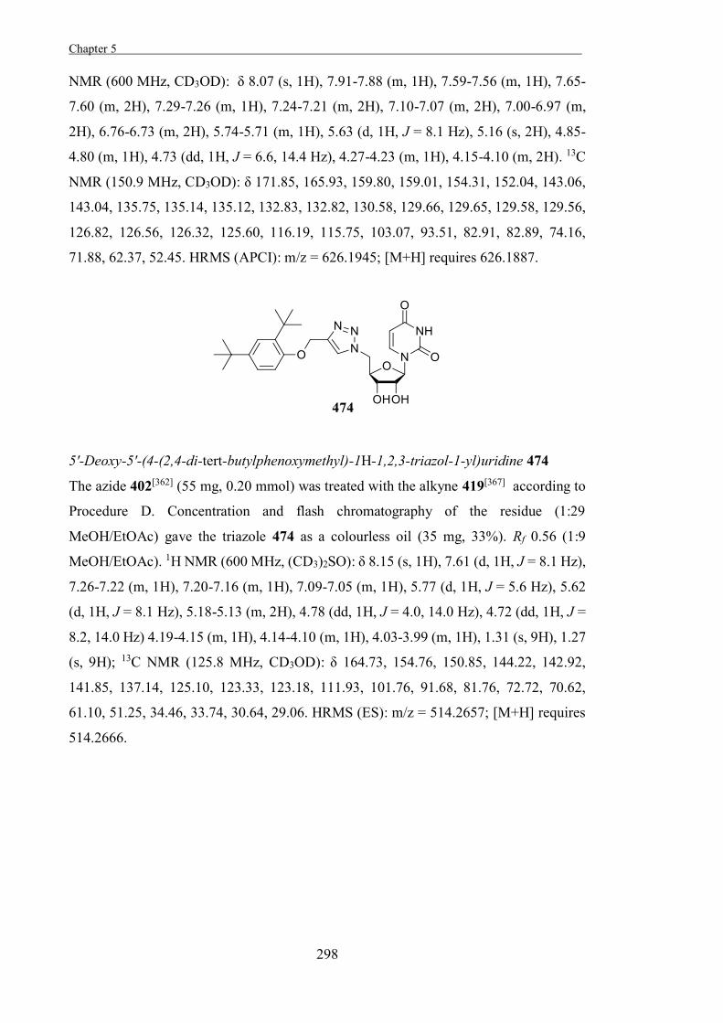
Chapter 5 .
298
NMR (600 MHz, CD3OD): δ 8.07 (s, 1H), 7.91-7.88 (m, 1H), 7.59-7.56 (m, 1H), 7.65-
7.60 (m, 2H), 7.29-7.26 (m, 1H), 7.24-7.21 (m, 2H), 7.10-7.07 (m, 2H), 7.00-6.97 (m,
2H), 6.76-6.73 (m, 2H), 5.74-5.71 (m, 1H), 5.63 (d, 1H, J = 8.1 Hz), 5.16 (s, 2H), 4.85-
4.80 (m, 1H), 4.73 (dd, 1H, J = 6.6, 14.4 Hz), 4.27-4.23 (m, 1H), 4.15-4.10 (m, 2H). 13C
NMR (150.9 MHz, CD3OD): δ 171.85, 165.93, 159.80, 159.01, 154.31, 152.04, 143.06,
143.04, 135.75, 135.14, 135.12, 132.83, 132.82, 130.58, 129.66, 129.65, 129.58, 129.56,
126.82, 126.56, 126.32, 125.60, 116.19, 115.75, 103.07, 93.51, 82.91, 82.89, 74.16,
71.88, 62.37, 52.45. HRMS (APCI): m/z = 626.1945; [M+H] requires 626.1887.
5'-Deoxy-5'-(4-(2,4-di-tert-butylphenoxymethyl)-1H-1,2,3-triazol-1-yl)uridine 474
The azide 402[362] (55 mg, 0.20 mmol) was treated with the alkyne 419[367] according to
Procedure D. Concentration and flash chromatography of the residue (1:29
MeOH/EtOAc) gave the triazole 474 as a colourless oil (35 mg, 33%). Rf 0.56 (1:9
MeOH/EtOAc). 1H NMR (600 MHz, (CD3)2SO): δ 8.15 (s, 1H), 7.61 (d, 1H, J = 8.1 Hz),
7.26-7.22 (m, 1H), 7.20-7.16 (m, 1H), 7.09-7.05 (m, 1H), 5.77 (d, 1H, J = 5.6 Hz), 5.62
(d, 1H, J = 8.1 Hz), 5.18-5.13 (m, 2H), 4.78 (dd, 1H, J = 4.0, 14.0 Hz), 4.72 (dd, 1H, J =
8.2, 14.0 Hz) 4.19-4.15 (m, 1H), 4.14-4.10 (m, 1H), 4.03-3.99 (m, 1H), 1.31 (s, 9H), 1.27
(s, 9H); 13C NMR (125.8 MHz, CD3OD): δ 164.73, 154.76, 150.85, 144.22, 142.92,
141.85, 137.14, 125.10, 123.33, 123.18, 111.93, 101.76, 91.68, 81.76, 72.72, 70.62,
61.10, 51.25, 34.46, 33.74, 30.64, 29.06. HRMS (ES): m/z = 514.2657; [M+H] requires
514.2666.
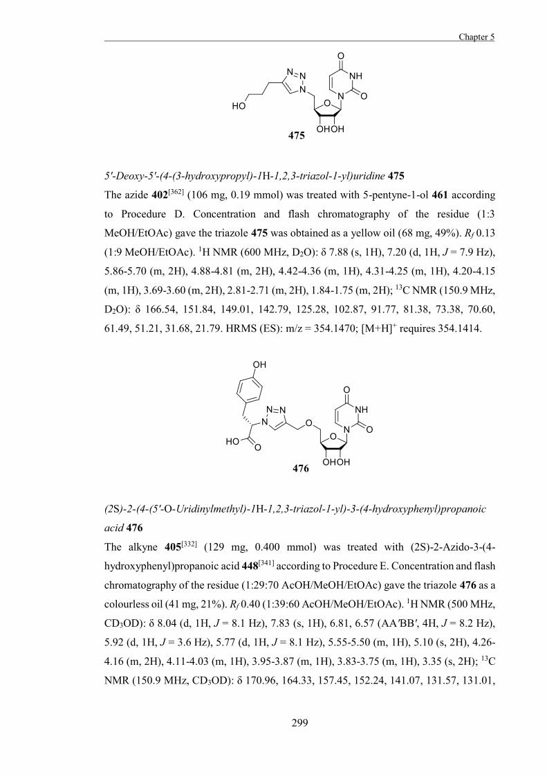
Chapter 5
299
5'-Deoxy-5'-(4-(3-hydroxypropyl)-1H-1,2,3-triazol-1-yl)uridine 475
The azide 402[362] (106 mg, 0.19 mmol) was treated with 5-pentyne-1-ol 461 according
to Procedure D. Concentration and flash chromatography of the residue (1:3
MeOH/EtOAc) gave the triazole 475 was obtained as a yellow oil (68 mg, 49%). Rf 0.13
(1:9 MeOH/EtOAc). 1H NMR (600 MHz, D2O): δ 7.88 (s, 1H), 7.20 (d, 1H, J = 7.9 Hz),
5.86-5.70 (m, 2H), 4.88-4.81 (m, 2H), 4.42-4.36 (m, 1H), 4.31-4.25 (m, 1H), 4.20-4.15
(m, 1H), 3.69-3.60 (m, 2H), 2.81-2.71 (m, 2H), 1.84-1.75 (m, 2H); 13C NMR (150.9 MHz,
D2O): δ 166.54, 151.84, 149.01, 142.79, 125.28, 102.87, 91.77, 81.38, 73.38, 70.60,
61.49, 51.21, 31.68, 21.79. HRMS (ES): m/z = 354.1470; [M+H]+ requires 354.1414.
(2S)-2-(4-(5'-O-Uridinylmethyl)-1H-1,2,3-triazol-1-yl)-3-(4-hydroxyphenyl)propanoic
acid 476
The alkyne 405[332] (129 mg, 0.400 mmol) was treated with (2S)-2-Azido-3-(4-
hydroxyphenyl)propanoic acid 448[341] according to Procedure E. Concentration and flash
chromatography of the residue (1:29:70 AcOH/MeOH/EtOAc) gave the triazole 476 as a
colourless oil (41 mg, 21%). Rf 0.40 (1:39:60 AcOH/MeOH/EtOAc). 1H NMR (500 MHz,
CD3OD): δ 8.04 (d, 1H, J = 8.1 Hz), 7.83 (s, 1H), 6.81, 6.57 (AA'BB', 4H, J = 8.2 Hz),
5.92 (d, 1H, J = 3.6 Hz), 5.77 (d, 1H, J = 8.1 Hz), 5.55-5.50 (m, 1H), 5.10 (s, 2H), 4.26-
4.16 (m, 2H), 4.11-4.03 (m, 1H), 3.95-3.87 (m, 1H), 3.83-3.75 (m, 1H), 3.35 (s, 2H); 13C
NMR (150.9 MHz, CD3OD): δ 170.96, 164.33, 157.45, 152.24, 141.07, 131.57, 131.01,
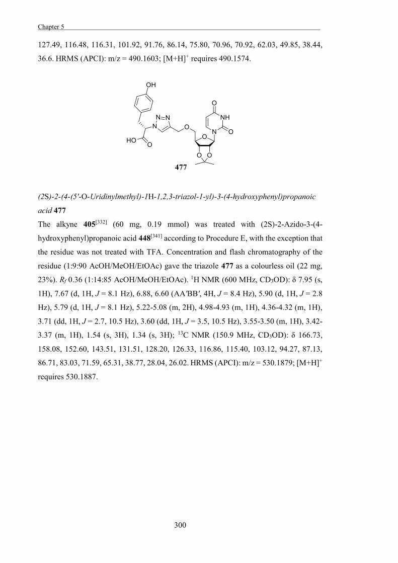
Chapter 5 .
300
127.49, 116.48, 116.31, 101.92, 91.76, 86.14, 75.80, 70.96, 70.92, 62.03, 49.85, 38.44,
36.6. HRMS (APCI): m/z = 490.1603; [M+H]+ requires 490.1574.
(2S)-2-(4-(5'-O-Uridinylmethyl)-1H-1,2,3-triazol-1-yl)-3-(4-hydroxyphenyl)propanoic
acid 477
The alkyne 405[332] (60 mg, 0.19 mmol) was treated with (2S)-2-Azido-3-(4-
hydroxyphenyl)propanoic acid 448[341] according to Procedure E, with the exception that
the residue was not treated with TFA. Concentration and flash chromatography of the
residue (1:9:90 AcOH/MeOH/EtOAc) gave the triazole 477 as a colourless oil (22 mg,
23%). Rf 0.36 (1:14:85 AcOH/MeOH/EtOAc). 1H NMR (600 MHz, CD3OD): δ 7.95 (s,
1H), 7.67 (d, 1H, J = 8.1 Hz), 6.88, 6.60 (AA'BB', 4H, J = 8.4 Hz), 5.90 (d, 1H, J = 2.8
Hz), 5.79 (d, 1H, J = 8.1 Hz), 5.22-5.08 (m, 2H), 4.98-4.93 (m, 1H), 4.36-4.32 (m, 1H),
3.71 (dd, 1H, J = 2.7, 10.5 Hz), 3.60 (dd, 1H, J = 3.5, 10.5 Hz), 3.55-3.50 (m, 1H), 3.42-
3.37 (m, 1H), 1.54 (s, 3H), 1.34 (s, 3H); 13C NMR (150.9 MHz, CD3OD): δ 166.73,
158.08, 152.60, 143.51, 131.51, 128.20, 126.33, 116.86, 115.40, 103.12, 94.27, 87.13,
86.71, 83.03, 71.59, 65.31, 38.77, 28.04, 26.02. HRMS (APCI): m/z = 530.1879; [M+H]+
requires 530.1887.
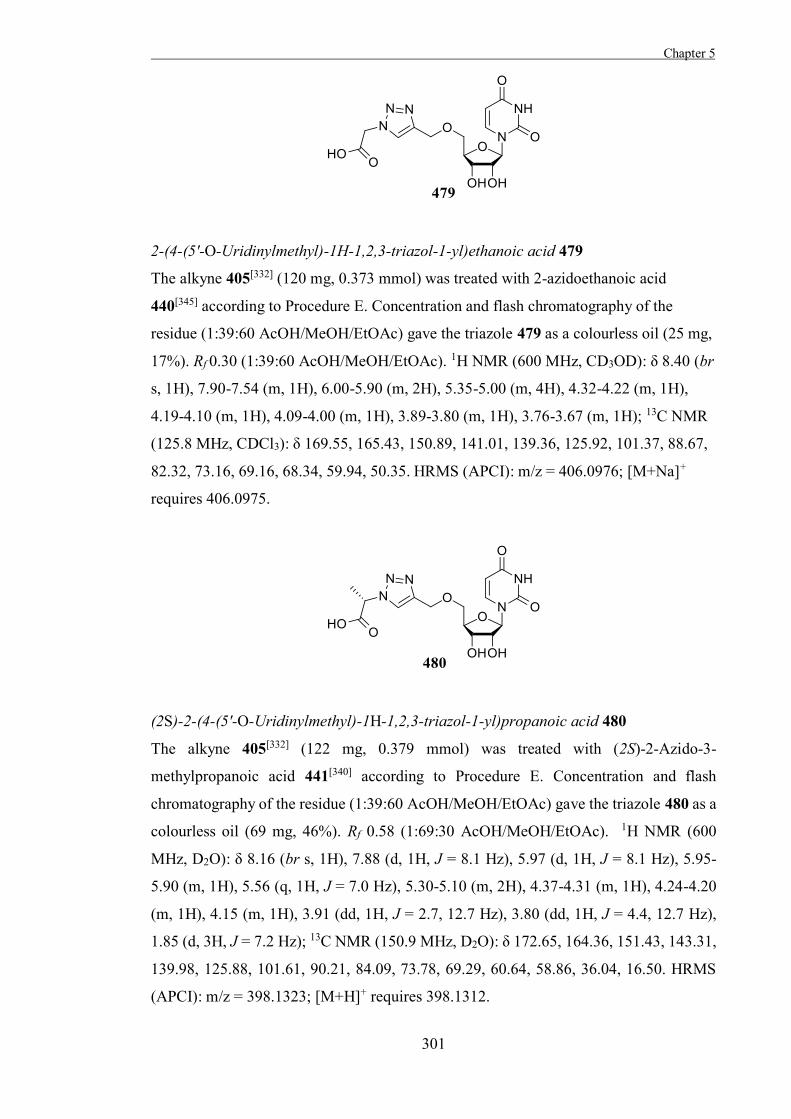
Chapter 5
301
2-(4-(5'-O-Uridinylmethyl)-1H-1,2,3-triazol-1-yl)ethanoic acid 479
The alkyne 405[332] (120 mg, 0.373 mmol) was treated with 2-azidoethanoic acid
440[345] according to Procedure E. Concentration and flash chromatography of the
residue (1:39:60 AcOH/MeOH/EtOAc) gave the triazole 479 as a colourless oil (25 mg,
17%). Rf 0.30 (1:39:60 AcOH/MeOH/EtOAc). 1H NMR (600 MHz, CD3OD): δ 8.40 (br
s, 1H), 7.90-7.54 (m, 1H), 6.00-5.90 (m, 2H), 5.35-5.00 (m, 4H), 4.32-4.22 (m, 1H),
4.19-4.10 (m, 1H), 4.09-4.00 (m, 1H), 3.89-3.80 (m, 1H), 3.76-3.67 (m, 1H); 13C NMR
(125.8 MHz, CDCl3): δ 169.55, 165.43, 150.89, 141.01, 139.36, 125.92, 101.37, 88.67,
82.32, 73.16, 69.16, 68.34, 59.94, 50.35. HRMS (APCI): m/z = 406.0976; [M+Na]+
requires 406.0975.
(2S)-2-(4-(5'-O-Uridinylmethyl)-1H-1,2,3-triazol-1-yl)propanoic acid 480
The alkyne 405[332] (122 mg, 0.379 mmol) was treated with (2S)-2-Azido-3-
methylpropanoic acid 441[340] according to Procedure E. Concentration and flash
chromatography of the residue (1:39:60 AcOH/MeOH/EtOAc) gave the triazole 480 as a
colourless oil (69 mg, 46%). Rf 0.58 (1:69:30 AcOH/MeOH/EtOAc). 1H NMR (600
MHz, D2O): δ 8.16 (br s, 1H), 7.88 (d, 1H, J = 8.1 Hz), 5.97 (d, 1H, J = 8.1 Hz), 5.95-
5.90 (m, 1H), 5.56 (q, 1H, J = 7.0 Hz), 5.30-5.10 (m, 2H), 4.37-4.31 (m, 1H), 4.24-4.20
(m, 1H), 4.15 (m, 1H), 3.91 (dd, 1H, J = 2.7, 12.7 Hz), 3.80 (dd, 1H, J = 4.4, 12.7 Hz),
1.85 (d, 3H, J = 7.2 Hz); 13C NMR (150.9 MHz, D2O): δ 172.65, 164.36, 151.43, 143.31,
139.98, 125.88, 101.61, 90.21, 84.09, 73.78, 69.29, 60.64, 58.86, 36.04, 16.50. HRMS
(APCI): m/z = 398.1323; [M+H]+ requires 398.1312.
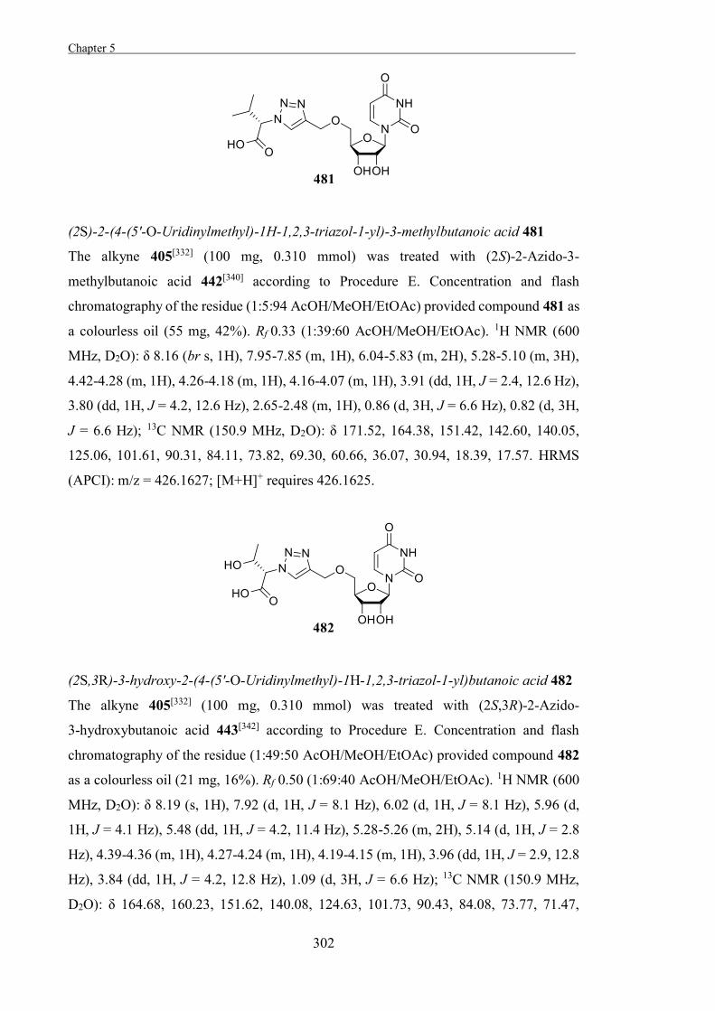
Chapter 5 .
302
(2S)-2-(4-(5'-O-Uridinylmethyl)-1H-1,2,3-triazol-1-yl)-3-methylbutanoic acid 481
The alkyne 405[332] (100 mg, 0.310 mmol) was treated with (2S)-2-Azido-3-
methylbutanoic acid 442[340] according to Procedure E. Concentration and flash
chromatography of the residue (1:5:94 AcOH/MeOH/EtOAc) provided compound 481 as
a colourless oil (55 mg, 42%). Rf 0.33 (1:39:60 AcOH/MeOH/EtOAc). 1H NMR (600
MHz, D2O): δ 8.16 (br s, 1H), 7.95-7.85 (m, 1H), 6.04-5.83 (m, 2H), 5.28-5.10 (m, 3H),
4.42-4.28 (m, 1H), 4.26-4.18 (m, 1H), 4.16-4.07 (m, 1H), 3.91 (dd, 1H, J = 2.4, 12.6 Hz),
3.80 (dd, 1H, J = 4.2, 12.6 Hz), 2.65-2.48 (m, 1H), 0.86 (d, 3H, J = 6.6 Hz), 0.82 (d, 3H,
J = 6.6 Hz); 13C NMR (150.9 MHz, D2O): δ 171.52, 164.38, 151.42, 142.60, 140.05,
125.06, 101.61, 90.31, 84.11, 73.82, 69.30, 60.66, 36.07, 30.94, 18.39, 17.57. HRMS
(APCI): m/z = 426.1627; [M+H]+ requires 426.1625.
(2S,3R)-3-hydroxy-2-(4-(5'-O-Uridinylmethyl)-1H-1,2,3-triazol-1-yl)butanoic acid 482
The alkyne 405[332] (100 mg, 0.310 mmol) was treated with (2S,3R)-2-Azido-
3-hydroxybutanoic acid 443[342] according to Procedure E. Concentration and flash
chromatography of the residue (1:49:50 AcOH/MeOH/EtOAc) provided compound 482
as a colourless oil (21 mg, 16%). Rf 0.50 (1:69:40 AcOH/MeOH/EtOAc). 1H NMR (600
MHz, D2O): δ 8.19 (s, 1H), 7.92 (d, 1H, J = 8.1 Hz), 6.02 (d, 1H, J = 8.1 Hz), 5.96 (d,
1H, J = 4.1 Hz), 5.48 (dd, 1H, J = 4.2, 11.4 Hz), 5.28-5.26 (m, 2H), 5.14 (d, 1H, J = 2.8
Hz), 4.39-4.36 (m, 1H), 4.27-4.24 (m, 1H), 4.19-4.15 (m, 1H), 3.96 (dd, 1H, J = 2.9, 12.8
Hz), 3.84 (dd, 1H, J = 4.2, 12.8 Hz), 1.09 (d, 3H, J = 6.6 Hz); 13C NMR (150.9 MHz,
D2O): δ 164.68, 160.23, 151.62, 140.08, 124.63, 101.73, 90.43, 84.08, 73.77, 71.47,
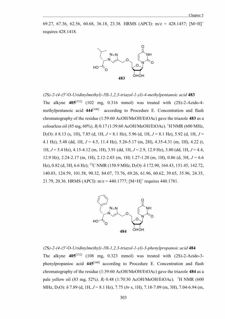
Chapter 5
303
69.27, 67.36, 62.56, 60.68, 36.18, 23.38. HRMS (APCI): m/z = 428.1457; [M+H]+
requires 428.1418.
(2S)-2-(4-(5'-O-Uridinylmethyl)-1H-1,2,3-triazol-1-yl)-4-methylpentanoic acid 483
The alkyne 405[332] (102 mg, 0.316 mmol) was treated with (2S)-2-Azido-4-
methylpentanoic acid 444[340] according to Procedure E. Concentration and flash
chromatography of the residue (1:59:60 AcOH/MeOH/EtOAc) gave the triazole 483 as a
colourless oil (85 mg, 60%). Rf 0.17 (1:39:60 AcOH/MeOH/EtOAc). 1H NMR (600 MHz,
D2O): δ 8.13 (s, 1H), 7.85 (d, 1H, J = 8.1 Hz), 5.96 (d, 1H, J = 8.1 Hz), 5.92 (d, 1H, J =
4.1 Hz), 5.48 (dd, 1H, J = 4.5, 11.4 Hz), 5.26-5.17 (m, 2H), 4.35-4.31 (m, 1H), 4.22 (t,
1H, J = 5.4 Hz), 4.15-4.12 (m, 1H), 3.91 (dd, 1H, J = 2.9, 12.9 Hz), 3.80 (dd, 1H, J = 4.4,
12.9 Hz), 2.24-2.17 (m, 1H), 2.12-2.03 (m, 1H) 1.27-1.20 (m, 1H), 0.86 (d, 3H, J = 6.6
Hz), 0.82 (d, 3H, 6.6 Hz); 13C NMR (150.9 MHz, D2O): δ 172.90, 164.43, 151.45, 142.72,
140.03, 124.59, 101.58, 90.32, 84.07, 73.76, 69.26, 61.96, 60.62, 39.65, 35.96, 24.35,
21.79, 20.36. HRMS (APCI): m/z = 440.1777; [M+H]+ requires 440.1781.
(2S)-2-(4-(5'-O-Uridinylmethyl)-1H-1,2,3-triazol-1-yl)-3-phenylpropanoic acid 484
The alkyne 405[332] (108 mg, 0.323 mmol) was treated with (2S)-2-Azido-3-
phenylpropanioc acid 445[340] according to Procedure E. Concentration and flash
chromatography of the residue (1:39:60 AcOH/MeOH/EtOAc) gave the triazole 484 as a
pale yellow oil (83 mg, 52%). Rf 0.48 (1:70:30 AcOH/MeOH/EtOAc). 1H NMR (600
MHz, D2O): δ 7.89 (d, 1H, J = 8.1 Hz), 7.75 (br s, 1H), 7.18-7.09 (m, 3H), 7.04-6.94 (m,
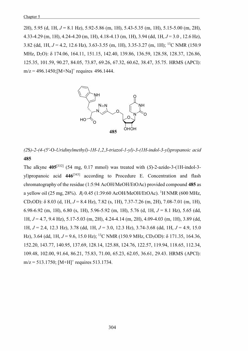
Chapter 5 .
304
2H), 5.95 (d, 1H, J = 8.1 Hz), 5.92-5.86 (m, 1H), 5.43-5.35 (m, 1H), 5.15-5.00 (m, 2H),
4.33-4.29 (m, 1H), 4.24-4.20 (m, 1H), 4.18-4.13 (m, 1H), 3.94 (dd, 1H, J = 3.0 , 12.6 Hz),
3.82 (dd, 1H, J = 4.2, 12.6 Hz), 3.63-3.55 (m, 1H), 3.35-3.27 (m, 1H); 13C NMR (150.9
MHz, D2O): δ 174.06, 164.11, 151.15, 142.40, 139.86, 136.59, 128.58, 128.37, 126.86,
125.35, 101.59, 90.27, 84.05, 73.87, 69.26, 67.32, 60.62, 38.47, 35.75. HRMS (APCI):
m/z = 496.1450;[M+Na]+ requires 496.1444.
(2S)-2-(4-(5'-O-Uridinylmethyl)-1H-1,2,3-triazol-1-yl)-3-(1H-indol-3-yl)propanoic acid
485
The alkyne 405[332] (54 mg, 0.17 mmol) was treated with (S)-2-azido-3-(1H-indol-3-
yl)propanoic acid 446[343] according to Procedure E. Concentration and flash
chromatography of the residue (1:5:94 AcOH/MeOH/EtOAc) provided compound 485 as
a yellow oil (25 mg, 28%). Rf 0.45 (1:39:60 AcOH/MeOH/EtOAc). 1H NMR (600 MHz,
CD3OD): δ 8.03 (d, 1H, J = 8.4 Hz), 7.82 (s, 1H), 7.37-7.26 (m, 2H), 7.08-7.01 (m, 1H),
6.98-6.92 (m, 1H), 6.80 (s, 1H), 5.96-5.92 (m, 1H), 5.76 (d, 1H, J = 8.1 Hz), 5.65 (dd,
1H, J = 4.7, 9.4 Hz), 5.17-5.03 (m, 2H), 4.24-4.14 (m, 2H), 4.09-4.03 (m, 1H), 3.89 (dd,
1H, J = 2.4, 12.3 Hz), 3.78 (dd, 1H, J = 3.0, 12.3 Hz), 3.74-3.68 (dd, 1H, J = 4.9, 15.0
Hz), 3.64 (dd, 1H, J = 9.6, 15.0 Hz); 13C NMR (150.9 MHz, CD3OD): δ 171.35, 164.36,
152.20, 143.77, 140.95, 137.69, 128.14, 125.88, 124.76, 122.57, 119.94, 118.65, 112.34,
109.48, 102.00, 91.64, 86.21, 75.83, 71.00, 65.23, 62.05, 36.61, 29.43. HRMS (APCI):
m/z = 513.1750; [M+H]+ requires 513.1734.
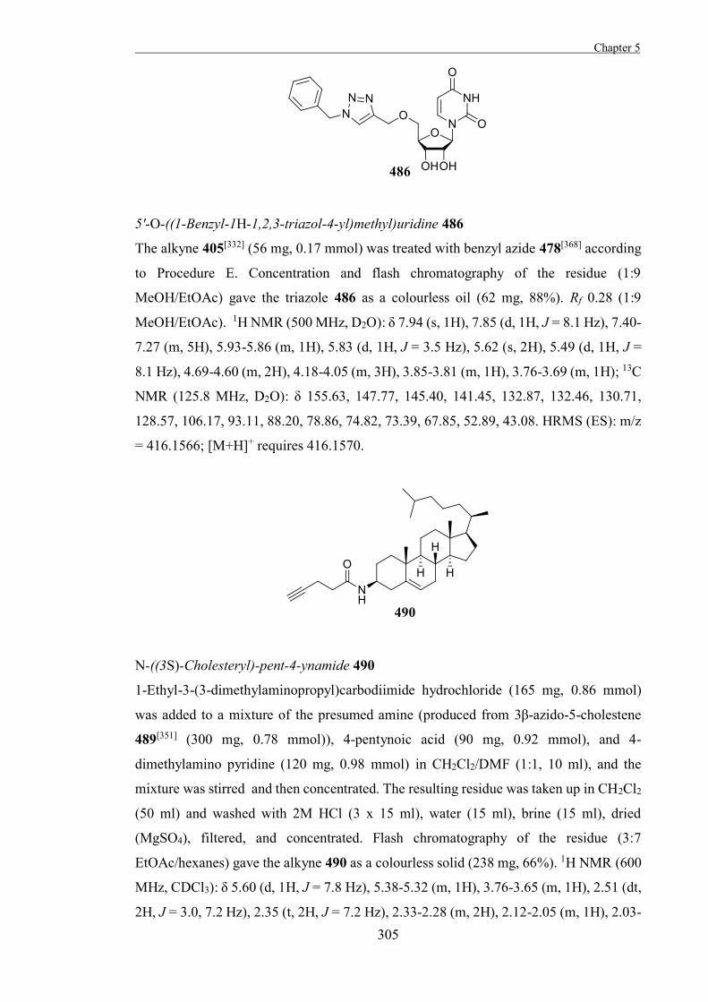
Chapter 5
305
5'-O-((1-Benzyl-1H-1,2,3-triazol-4-yl)methyl)uridine 486
The alkyne 405[332] (56 mg, 0.17 mmol) was treated with benzyl azide 478[368] according
to Procedure E. Concentration and flash chromatography of the residue (1:9
MeOH/EtOAc) gave the triazole 486 as a colourless oil (62 mg, 88%). Rf 0.28 (1:9
MeOH/EtOAc). 1H NMR (500 MHz, D2O): δ 7.94 (s, 1H), 7.85 (d, 1H, J = 8.1 Hz), 7.40-
7.27 (m, 5H), 5.93-5.86 (m, 1H), 5.83 (d, 1H, J = 3.5 Hz), 5.62 (s, 2H), 5.49 (d, 1H, J =
8.1 Hz), 4.69-4.60 (m, 2H), 4.18-4.05 (m, 3H), 3.85-3.81 (m, 1H), 3.76-3.69 (m, 1H); 13C
NMR (125.8 MHz, D2O): δ 155.63, 147.77, 145.40, 141.45, 132.87, 132.46, 130.71,
128.57, 106.17, 93.11, 88.20, 78.86, 74.82, 73.39, 67.85, 52.89, 43.08. HRMS (ES): m/z
= 416.1566; [M+H]+ requires 416.1570.
N-((3S)-Cholesteryl)-pent-4-ynamide 490
1-Ethyl-3-(3-dimethylaminopropyl)carbodiimide hydrochloride (165 mg, 0.86 mmol)
was added to a mixture of the presumed amine (produced from 3β-azido-5-cholestene
489[351] (300 mg, 0.78 mmol)), 4-pentynoic acid (90 mg, 0.92 mmol), and 4-
dimethylamino pyridine (120 mg, 0.98 mmol) in CH2Cl2/DMF (1:1, 10 ml), and the
mixture was stirred and then concentrated. The resulting residue was taken up in CH2Cl2
(50 ml) and washed with 2M HCl (3 x 15 ml), water (15 ml), brine (15 ml), dried
(MgSO4), filtered, and concentrated. Flash chromatography of the residue (3:7
EtOAc/hexanes) gave the alkyne 490 as a colourless solid (238 mg, 66%). 1H NMR (600
MHz, CDCl3): δ 5.60 (d, 1H, J = 7.8 Hz), 5.38-5.32 (m, 1H), 3.76-3.65 (m, 1H), 2.51 (dt,
2H, J = 3.0, 7.2 Hz), 2.35 (t, 2H, J = 7.2 Hz), 2.33-2.28 (m, 2H), 2.12-2.05 (m, 1H), 2.03-
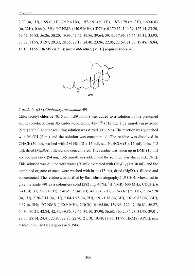
Chapter 5 .
306
2.00 (m, 1H), 1.99 (t, 1H, J = 2.4 Hz), 1.97-1.91 (m, 1H), 1.87-1.79 (m, 3H), 1.60-0.83
(m, 32H), 0.66 (s, 3H); 13C NMR (150.9 MHz, CDCl3): δ 170.15, 140.29, 122.14, 83.20,
69.42, 56.82, 56.26, 50.20, 49.93, 42.42, 39.86, 39.64, 39.43, 37.96, 36.66, 36.31, 35.93,
35.68, 31.98, 31.97, 29.32, 28.35, 28.13, 24.40, 23.96, 22.95, 22.69, 21.09, 19.48, 18.84,
15.13, 11.99. HRMS (APCI): m/z = 466.4042; [M+H] requires 466.4049.
2-azido-N-((3S)-Cholesteryl)acetamide 491
Chloroacetyl chloride (0.15 ml, 1.89 mmol) was added to a solution of the presumed
amine (produced from 3β-azido-5-cholestene 489[351] (512 mg, 1.32 mmol)) in pyridine
(5 ml) at 0° C, and the resulting solution was stirred (r.t., 15 h). The reaction was quenched
with MeOH (5 ml) and the solution was concentrated. The residue was dissolved in
CH2Cl2 (50 ml), washed with 2M HCl (3 x 15 ml), sat. NaHCO3 (3 x 15 ml), brine (15
ml), dried (MgSO4), filtered and concentrated. The residue was taken up in DMF (10 ml)
and sodium azide (94 mg, 1.45 mmol) was added, and the solution was stirred (r.t., 24 h).
This solution was diluted with water (20 ml), extracted with CH2Cl2 (3 x 30 ml), and the
combined organic extracts were washed with brine (15 ml), dried (MgSO4), filtered and
concentrated. The residue was purified by flash chromatography (1:9 CH2Cl2/hexanes) to
give the azide 491 as a colourless solid (282 mg, 46%). 1H NMR (600 MHz, CDCl3): δ
6.41 (d, 1H, J = 2.0 Hz), 5.40-5.35 (m, 1H), 4.02 (s, 2H), 3.76-3.67 (m, 1H), 2.36-2.28
(m, 1H), 2.20-2.11 (m, 1H), 2.04-1.93 (m, 2H), 1.91-1.78 (m, 3H), 1.61-0.82 (m, 33H),
0.67 (s, 3H); 13C NMR (150.9 MHz, CDCl3): δ 165.06, 139.96, 122.47, 56.81, 56.27,
50.30, 50.21, 42.84, 42.44, 39.86, 39.65, 39.10, 37.90, 36.68, 36.32, 35.93, 31.98, 29.01,
28.36, 28.14, 24.41, 23.97, 22.95, 22.70, 21.10, 19.48, 18.85, 11.99. HRMS (APCI): m/z
= 469.3897; [M+H] requires 469.3906.
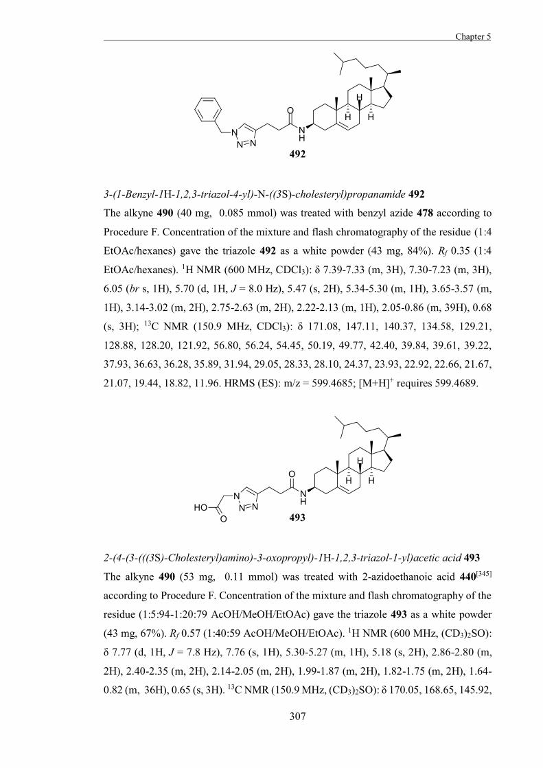
Chapter 5
307
3-(1-Benzyl-1H-1,2,3-triazol-4-yl)-N-((3S)-cholesteryl)propanamide 492
The alkyne 490 (40 mg, 0.085 mmol) was treated with benzyl azide 478 according to
Procedure F. Concentration of the mixture and flash chromatography of the residue (1:4
EtOAc/hexanes) gave the triazole 492 as a white powder (43 mg, 84%). Rf 0.35 (1:4
EtOAc/hexanes). 1H NMR (600 MHz, CDCl3): δ 7.39-7.33 (m, 3H), 7.30-7.23 (m, 3H),
6.05 (br s, 1H), 5.70 (d, 1H, J = 8.0 Hz), 5.47 (s, 2H), 5.34-5.30 (m, 1H), 3.65-3.57 (m,
1H), 3.14-3.02 (m, 2H), 2.75-2.63 (m, 2H), 2.22-2.13 (m, 1H), 2.05-0.86 (m, 39H), 0.68
(s, 3H); 13C NMR (150.9 MHz, CDCl3): δ 171.08, 147.11, 140.37, 134.58, 129.21,
128.88, 128.20, 121.92, 56.80, 56.24, 54.45, 50.19, 49.77, 42.40, 39.84, 39.61, 39.22,
37.93, 36.63, 36.28, 35.89, 31.94, 29.05, 28.33, 28.10, 24.37, 23.93, 22.92, 22.66, 21.67,
21.07, 19.44, 18.82, 11.96. HRMS (ES): m/z = 599.4685; [M+H]+ requires 599.4689.
2-(4-(3-(((3S)-Cholesteryl)amino)-3-oxopropyl)-1H-1,2,3-triazol-1-yl)acetic acid 493
The alkyne 490 (53 mg, 0.11 mmol) was treated with 2-azidoethanoic acid 440[345]
according to Procedure F. Concentration of the mixture and flash chromatography of the
residue (1:5:94-1:20:79 AcOH/MeOH/EtOAc) gave the triazole 493 as a white powder
(43 mg, 67%). Rf 0.57 (1:40:59 AcOH/MeOH/EtOAc). 1H NMR (600 MHz, (CD3)2SO):
δ 7.77 (d, 1H, J = 7.8 Hz), 7.76 (s, 1H), 5.30-5.27 (m, 1H), 5.18 (s, 2H), 2.86-2.80 (m,
2H), 2.40-2.35 (m, 2H), 2.14-2.05 (m, 2H), 1.99-1.87 (m, 2H), 1.82-1.75 (m, 2H), 1.64-
0.82 (m, 36H), 0.65 (s, 3H). 13C NMR (150.9 MHz, (CD3)2SO): δ 170.05, 168.65, 145.92,
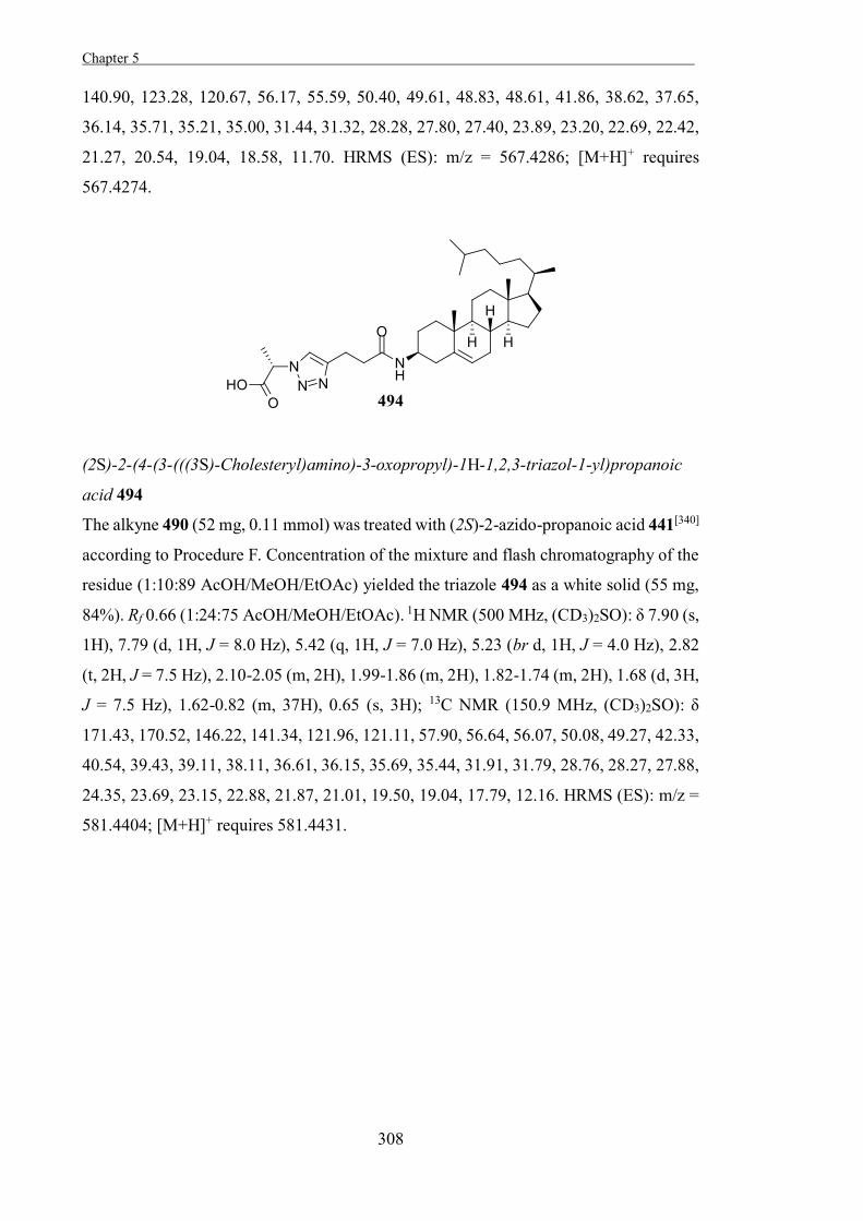
Chapter 5 .
308
140.90, 123.28, 120.67, 56.17, 55.59, 50.40, 49.61, 48.83, 48.61, 41.86, 38.62, 37.65,
36.14, 35.71, 35.21, 35.00, 31.44, 31.32, 28.28, 27.80, 27.40, 23.89, 23.20, 22.69, 22.42,
21.27, 20.54, 19.04, 18.58, 11.70. HRMS (ES): m/z = 567.4286; [M+H]+ requires
567.4274.
(2S)-2-(4-(3-(((3S)-Cholesteryl)amino)-3-oxopropyl)-1H-1,2,3-triazol-1-yl)propanoic
acid 494
The alkyne 490 (52 mg, 0.11 mmol) was treated with (2S)-2-azido-propanoic acid 441[340]
according to Procedure F. Concentration of the mixture and flash chromatography of the
residue (1:10:89 AcOH/MeOH/EtOAc) yielded the triazole 494 as a white solid (55 mg,
84%). Rf 0.66 (1:24:75 AcOH/MeOH/EtOAc). 1H NMR (500 MHz, (CD3)2SO): δ 7.90 (s,
1H), 7.79 (d, 1H, J = 8.0 Hz), 5.42 (q, 1H, J = 7.0 Hz), 5.23 (br d, 1H, J = 4.0 Hz), 2.82
(t, 2H, J = 7.5 Hz), 2.10-2.05 (m, 2H), 1.99-1.86 (m, 2H), 1.82-1.74 (m, 2H), 1.68 (d, 3H,
J = 7.5 Hz), 1.62-0.82 (m, 37H), 0.65 (s, 3H); 13C NMR (150.9 MHz, (CD3)2SO): δ
171.43, 170.52, 146.22, 141.34, 121.96, 121.11, 57.90, 56.64, 56.07, 50.08, 49.27, 42.33,
40.54, 39.43, 39.11, 38.11, 36.61, 36.15, 35.69, 35.44, 31.91, 31.79, 28.76, 28.27, 27.88,
24.35, 23.69, 23.15, 22.88, 21.87, 21.01, 19.50, 19.04, 17.79, 12.16. HRMS (ES): m/z =
581.4404; [M+H]+ requires 581.4431.
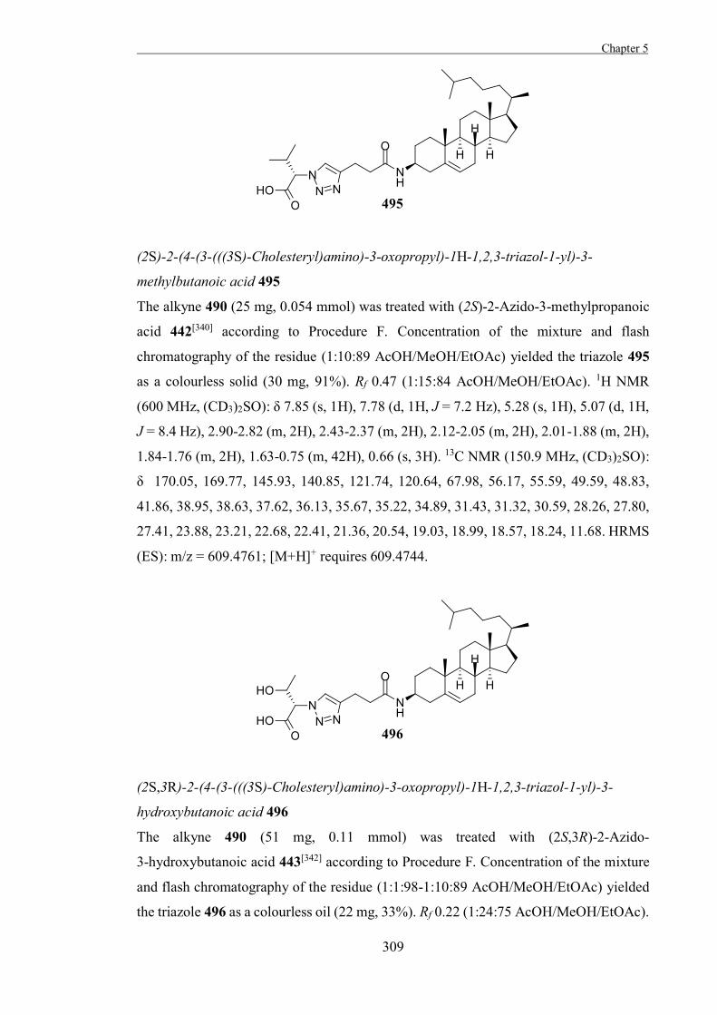
Chapter 5
309
(2S)-2-(4-(3-(((3S)-Cholesteryl)amino)-3-oxopropyl)-1H-1,2,3-triazol-1-yl)-3-
methylbutanoic acid 495
The alkyne 490 (25 mg, 0.054 mmol) was treated with (2S)-2-Azido-3-methylpropanoic
acid 442[340] according to Procedure F. Concentration of the mixture and flash
chromatography of the residue (1:10:89 AcOH/MeOH/EtOAc) yielded the triazole 495
as a colourless solid (30 mg, 91%). Rf 0.47 (1:15:84 AcOH/MeOH/EtOAc). 1H NMR
(600 MHz, (CD3)2SO): δ 7.85 (s, 1H), 7.78 (d, 1H, J = 7.2 Hz), 5.28 (s, 1H), 5.07 (d, 1H,
J = 8.4 Hz), 2.90-2.82 (m, 2H), 2.43-2.37 (m, 2H), 2.12-2.05 (m, 2H), 2.01-1.88 (m, 2H),
1.84-1.76 (m, 2H), 1.63-0.75 (m, 42H), 0.66 (s, 3H). 13C NMR (150.9 MHz, (CD3)2SO):
δ 170.05, 169.77, 145.93, 140.85, 121.74, 120.64, 67.98, 56.17, 55.59, 49.59, 48.83,
41.86, 38.95, 38.63, 37.62, 36.13, 35.67, 35.22, 34.89, 31.43, 31.32, 30.59, 28.26, 27.80,
27.41, 23.88, 23.21, 22.68, 22.41, 21.36, 20.54, 19.03, 18.99, 18.57, 18.24, 11.68. HRMS
(ES): m/z = 609.4761; [M+H]+ requires 609.4744.
(2S,3R)-2-(4-(3-(((3S)-Cholesteryl)amino)-3-oxopropyl)-1H-1,2,3-triazol-1-yl)-3-
hydroxybutanoic acid 496
The alkyne 490 (51 mg, 0.11 mmol) was treated with (2S,3R)-2-Azido-
3-hydroxybutanoic acid 443[342] according to Procedure F. Concentration of the mixture
and flash chromatography of the residue (1:1:98-1:10:89 AcOH/MeOH/EtOAc) yielded
the triazole 496 as a colourless oil (22 mg, 33%). Rf 0.22 (1:24:75 AcOH/MeOH/EtOAc).
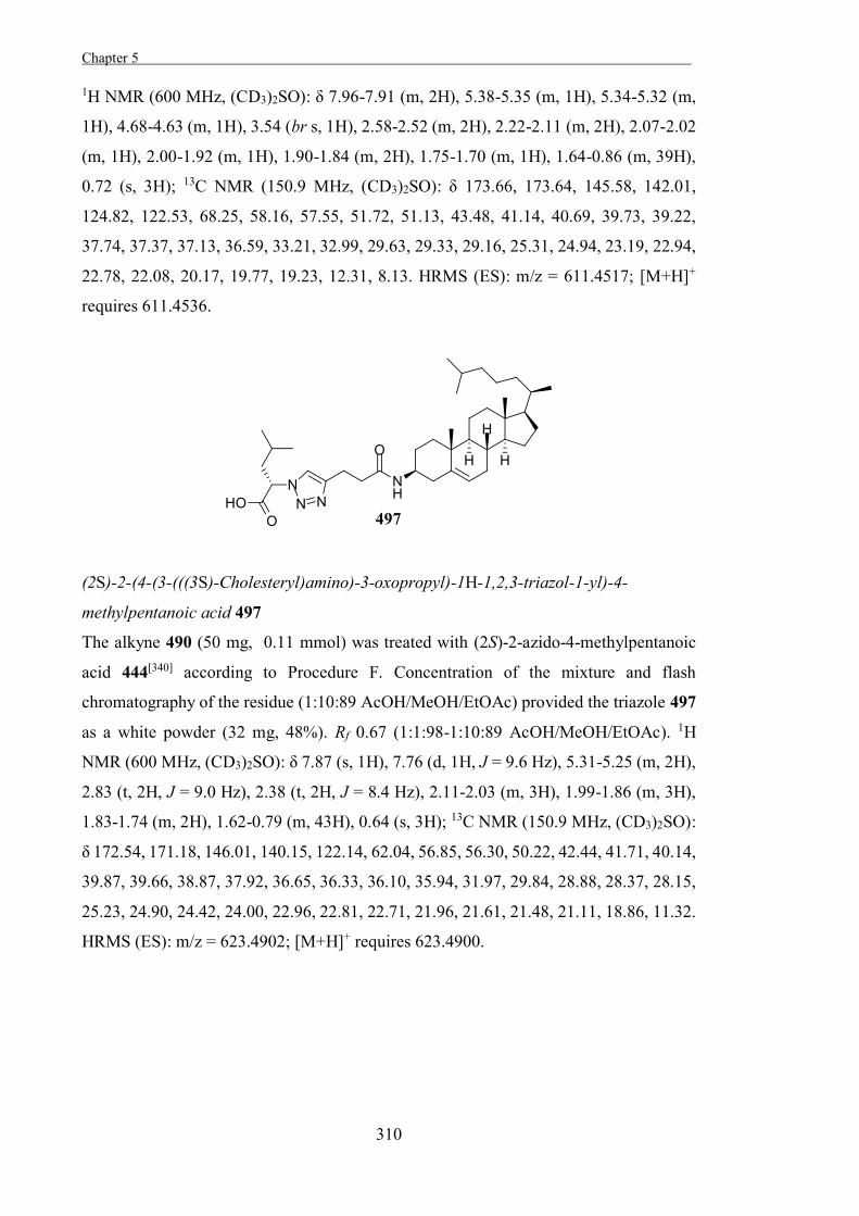
Chapter 5 .
310
1H NMR (600 MHz, (CD3)2SO): δ 7.96-7.91 (m, 2H), 5.38-5.35 (m, 1H), 5.34-5.32 (m,
1H), 4.68-4.63 (m, 1H), 3.54 (br s, 1H), 2.58-2.52 (m, 2H), 2.22-2.11 (m, 2H), 2.07-2.02
(m, 1H), 2.00-1.92 (m, 1H), 1.90-1.84 (m, 2H), 1.75-1.70 (m, 1H), 1.64-0.86 (m, 39H),
0.72 (s, 3H); 13C NMR (150.9 MHz, (CD3)2SO): δ 173.66, 173.64, 145.58, 142.01,
124.82, 122.53, 68.25, 58.16, 57.55, 51.72, 51.13, 43.48, 41.14, 40.69, 39.73, 39.22,
37.74, 37.37, 37.13, 36.59, 33.21, 32.99, 29.63, 29.33, 29.16, 25.31, 24.94, 23.19, 22.94,
22.78, 22.08, 20.17, 19.77, 19.23, 12.31, 8.13. HRMS (ES): m/z = 611.4517; [M+H]+
requires 611.4536.
(2S)-2-(4-(3-(((3S)-Cholesteryl)amino)-3-oxopropyl)-1H-1,2,3-triazol-1-yl)-4-
methylpentanoic acid 497
The alkyne 490 (50 mg, 0.11 mmol) was treated with (2S)-2-azido-4-methylpentanoic
acid 444[340] according to Procedure F. Concentration of the mixture and flash
chromatography of the residue (1:10:89 AcOH/MeOH/EtOAc) provided the triazole 497
as a white powder (32 mg, 48%). Rf 0.67 (1:1:98-1:10:89 AcOH/MeOH/EtOAc). 1H
NMR (600 MHz, (CD3)2SO): δ 7.87 (s, 1H), 7.76 (d, 1H, J = 9.6 Hz), 5.31-5.25 (m, 2H),
2.83 (t, 2H, J = 9.0 Hz), 2.38 (t, 2H, J = 8.4 Hz), 2.11-2.03 (m, 3H), 1.99-1.86 (m, 3H),
1.83-1.74 (m, 2H), 1.62-0.79 (m, 43H), 0.64 (s, 3H); 13C NMR (150.9 MHz, (CD3)2SO):
δ 172.54, 171.18, 146.01, 140.15, 122.14, 62.04, 56.85, 56.30, 50.22, 42.44, 41.71, 40.14,
39.87, 39.66, 38.87, 37.92, 36.65, 36.33, 36.10, 35.94, 31.97, 29.84, 28.88, 28.37, 28.15,
25.23, 24.90, 24.42, 24.00, 22.96, 22.81, 22.71, 21.96, 21.61, 21.48, 21.11, 18.86, 11.32.
HRMS (ES): m/z = 623.4902; [M+H]+ requires 623.4900.
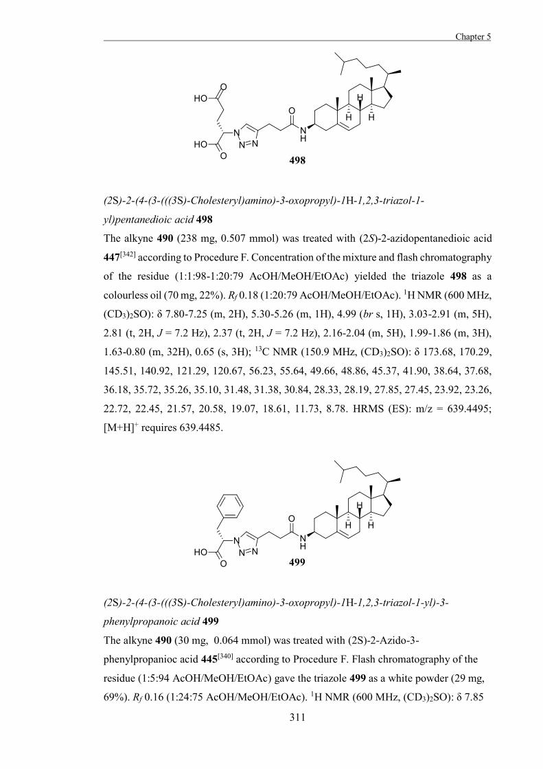
Chapter 5
311
(2S)-2-(4-(3-(((3S)-Cholesteryl)amino)-3-oxopropyl)-1H-1,2,3-triazol-1-
yl)pentanedioic acid 498
The alkyne 490 (238 mg, 0.507 mmol) was treated with (2S)-2-azidopentanedioic acid
447[342] according to Procedure F. Concentration of the mixture and flash chromatography
of the residue (1:1:98-1:20:79 AcOH/MeOH/EtOAc) yielded the triazole 498 as a
colourless oil (70 mg, 22%). Rf 0.18 (1:20:79 AcOH/MeOH/EtOAc). 1H NMR (600 MHz,
(CD3)2SO): δ 7.80-7.25 (m, 2H), 5.30-5.26 (m, 1H), 4.99 (br s, 1H), 3.03-2.91 (m, 5H),
2.81 (t, 2H, J = 7.2 Hz), 2.37 (t, 2H, J = 7.2 Hz), 2.16-2.04 (m, 5H), 1.99-1.86 (m, 3H),
1.63-0.80 (m, 32H), 0.65 (s, 3H); 13C NMR (150.9 MHz, (CD3)2SO): δ 173.68, 170.29,
145.51, 140.92, 121.29, 120.67, 56.23, 55.64, 49.66, 48.86, 45.37, 41.90, 38.64, 37.68,
36.18, 35.72, 35.26, 35.10, 31.48, 31.38, 30.84, 28.33, 28.19, 27.85, 27.45, 23.92, 23.26,
22.72, 22.45, 21.57, 20.58, 19.07, 18.61, 11.73, 8.78. HRMS (ES): m/z = 639.4495;
[M+H]+ requires 639.4485.
(2S)-2-(4-(3-(((3S)-Cholesteryl)amino)-3-oxopropyl)-1H-1,2,3-triazol-1-yl)-3-
phenylpropanoic acid 499
The alkyne 490 (30 mg, 0.064 mmol) was treated with (2S)-2-Azido-3-
phenylpropanioc acid 445[340] according to Procedure F. Flash chromatography of the
residue (1:5:94 AcOH/MeOH/EtOAc) gave the triazole 499 as a white powder (29 mg,
69%). Rf 0.16 (1:24:75 AcOH/MeOH/EtOAc). 1H NMR (600 MHz, (CD3)2SO): δ 7.85
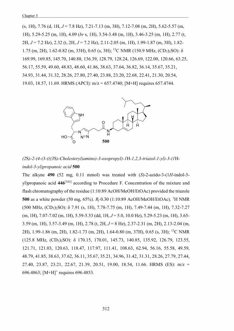
Chapter 5 .
312
(s, 1H), 7.76 (d, 1H, J = 7.8 Hz), 7.21-7.13 (m, 3H), 7.12-7.08 (m, 2H), 5.62-5.57 (m,
1H), 5.29-5.25 (m, 1H), 4.09 (br s, 1H), 3.54-3.48 (m, 1H), 3.46-3.25 (m, 1H), 2.77 (t,
2H, J = 7.2 Hz), 2.32 (t, 2H, J = 7.2 Hz), 2.11-2.05 (m, 1H), 1.99-1.87 (m, 3H), 1.82-
1.75 (m, 2H), 1.62-0.82 (m, 33H), 0.65 (s, 3H); 13C NMR (150.9 MHz, (CD3)2SO): δ
169.99, 169.85, 145.70, 140.88, 136.39, 128.79, 128.24, 126.69, 122.00, 120.66, 63.25,
56.17, 55.59, 49.60, 48.83, 48.60, 41.86, 38.63, 37.64, 36.82, 36.14, 35.67, 35.21,
34.93, 31.44, 31.32, 28.26, 27.80, 27.40, 23.88, 23.20, 22.68, 22.41, 21.30, 20.54,
19.03, 18.57, 11.69. HRMS (APCI): m/z = 657.4740; [M+H] requires 657.4744.
(2S)-2-(4-(3-(((3S)-Cholesteryl)amino)-3-oxopropyl)-1H-1,2,3-triazol-1-yl)-3-(1H-
indol-3-yl)propanoic acid 500
The alkyne 490 (52 mg, 0.11 mmol) was treated with (S)-2-azido-3-(1H-indol-3-
yl)propanoic acid 446[343] according to Procedure F. Concentration of the mixture and
flash chromatography of the residue (1:10:89 AcOH/MeOH/EtOAc) provided the triazole
500 as a white powder (50 mg, 65%). Rf 0.30 (1:10:89 AcOH/MeOH/EtOAc). 1H NMR
(500 MHz, (CD3)2SO): δ 7.91 (s, 1H), 7.78-7.75 (m, 1H), 7.49-7.44 (m, 1H), 7.32-7.27
(m, 1H), 7.07-7.02 (m, 1H), 5.59-5.53 (dd, 1H, J = 5.0, 10.0 Hz), 5.29-5.23 (m, 1H), 3.65-
3.59 (m, 1H), 3.57-3.49 (m, 1H), 2.78 (t, 2H, J = 8 Hz), 2.37-2.31 (m, 2H), 2.13-2.04 (m,
2H), 1.99-1.86 (m, 2H), 1.82-1.73 (m, 2H), 1.64-0.80 (m, 37H), 0.65 (s, 3H); 13C NMR
(125.8 MHz, (CD3)2SO): δ 170.15, 170.01, 145.73, 140.85, 135.92, 126.79, 123.55,
121.71, 121.03, 120.63, 118.47, 117.97, 111.41, 108.63, 62.94, 56.16, 55.58, 49.59,
48.79, 41.85, 38.63, 37.62, 36.11, 35.67, 35.21, 34.96, 31.42, 31.31, 28.26, 27.79, 27.44,
27.40, 23.87, 23.21, 22.67, 21.39, 20.51, 19.00, 18.54, 11.66. HRMS (ES): m/z =
696.4863; [M+H]+ requires 696.4853.
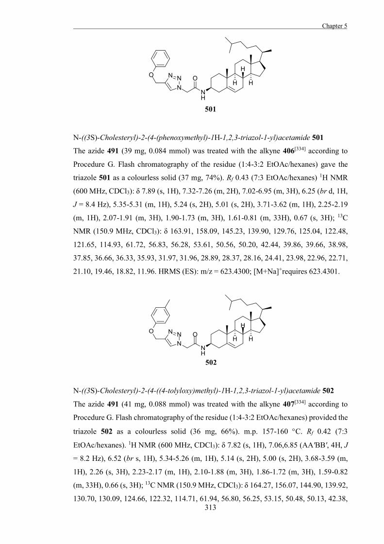
Chapter 5
313
N-((3S)-Cholesteryl)-2-(4-(phenoxymethyl)-1H-1,2,3-triazol-1-yl)acetamide 501
The azide 491 (39 mg, 0.084 mmol) was treated with the alkyne 406[334] according to
Procedure G. Flash chromatography of the residue (1:4-3:2 EtOAc/hexanes) gave the
triazole 501 as a colourless solid (37 mg, 74%). Rf 0.43 (7:3 EtOAc/hexanes) 1H NMR
(600 MHz, CDCl3): δ 7.89 (s, 1H), 7.32-7.26 (m, 2H), 7.02-6.95 (m, 3H), 6.25 (br d, 1H,
J = 8.4 Hz), 5.35-5.31 (m, 1H), 5.24 (s, 2H), 5.01 (s, 2H), 3.71-3.62 (m, 1H), 2.25-2.19
(m, 1H), 2.07-1.91 (m, 3H), 1.90-1.73 (m, 3H), 1.61-0.81 (m, 33H), 0.67 (s, 3H); 13C
NMR (150.9 MHz, CDCl3): δ 163.91, 158.09, 145.23, 139.90, 129.76, 125.04, 122.48,
121.65, 114.93, 61.72, 56.83, 56.28, 53.61, 50.56, 50.20, 42.44, 39.86, 39.66, 38.98,
37.85, 36.66, 36.33, 35.93, 31.97, 31.96, 28.89, 28.37, 28.16, 24.41, 23.98, 22.96, 22.71,
21.10, 19.46, 18.82, 11.96. HRMS (ES): m/z = 623.4300; [M+Na]+requires 623.4301.
N-((3S)-Cholesteryl)-2-(4-((4-tolyloxy)methyl)-1H-1,2,3-triazol-1-yl)acetamide 502
The azide 491 (41 mg, 0.088 mmol) was treated with the alkyne 407[334] according to
Procedure G. Flash chromatography of the residue (1:4-3:2 EtOAc/hexanes) provided the
triazole 502 as a colourless solid (36 mg, 66%). m.p. 157-160 C. Rf 0.42 (7:3
EtOAc/hexanes). 1H NMR (600 MHz, CDCl3): δ 7.82 (s, 1H), 7.06,6.85 (AA'BB', 4H, J
= 8.2 Hz), 6.52 (br s, 1H), 5.34-5.26 (m, 1H), 5.14 (s, 2H), 5.00 (s, 2H), 3.68-3.59 (m,
1H), 2.26 (s, 3H), 2.23-2.17 (m, 1H), 2.10-1.88 (m, 3H), 1.86-1.72 (m, 3H), 1.59-0.82
(m, 33H), 0.66 (s, 3H); 13C NMR (150.9 MHz, CDCl3): δ 164.27, 156.07, 144.90, 139.92,
130.70, 130.09, 124.66, 122.32, 114.71, 61.94, 56.80, 56.25, 53.15, 50.48, 50.13, 42.38,
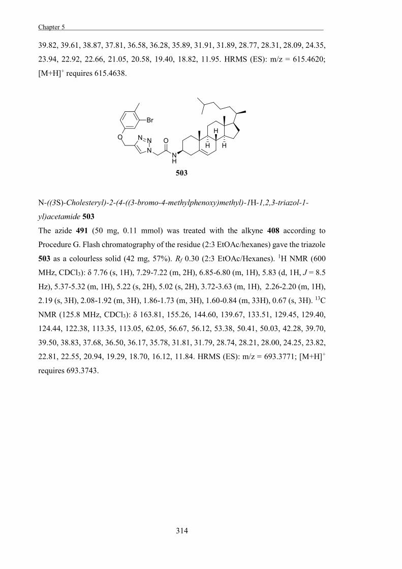
Chapter 5 .
314
39.82, 39.61, 38.87, 37.81, 36.58, 36.28, 35.89, 31.91, 31.89, 28.77, 28.31, 28.09, 24.35,
23.94, 22.92, 22.66, 21.05, 20.58, 19.40, 18.82, 11.95. HRMS (ES): m/z = 615.4620;
[M+H]+ requires 615.4638.
N-((3S)-Cholesteryl)-2-(4-((3-bromo-4-methylphenoxy)methyl)-1H-1,2,3-triazol-1-
yl)acetamide 503
The azide 491 (50 mg, 0.11 mmol) was treated with the alkyne 408 according to
Procedure G. Flash chromatography of the residue (2:3 EtOAc/hexanes) gave the triazole
503 as a colourless solid (42 mg, 57%). Rf 0.30 (2:3 EtOAc/Hexanes). 1H NMR (600
MHz, CDCl3): δ 7.76 (s, 1H), 7.29-7.22 (m, 2H), 6.85-6.80 (m, 1H), 5.83 (d, 1H, J = 8.5
Hz), 5.37-5.32 (m, 1H), 5.22 (s, 2H), 5.02 (s, 2H), 3.72-3.63 (m, 1H), 2.26-2.20 (m, 1H),
2.19 (s, 3H), 2.08-1.92 (m, 3H), 1.86-1.73 (m, 3H), 1.60-0.84 (m, 33H), 0.67 (s, 3H). 13C
NMR (125.8 MHz, CDCl3): δ 163.81, 155.26, 144.60, 139.67, 133.51, 129.45, 129.40,
124.44, 122.38, 113.35, 113.05, 62.05, 56.67, 56.12, 53.38, 50.41, 50.03, 42.28, 39.70,
39.50, 38.83, 37.68, 36.50, 36.17, 35.78, 31.81, 31.79, 28.74, 28.21, 28.00, 24.25, 23.82,
22.81, 22.55, 20.94, 19.29, 18.70, 16.12, 11.84. HRMS (ES): m/z = 693.3771; [M+H]+
requires 693.3743.

Chapter 5
315
N-((3S)-Cholesteryl)-2-(4-((4-acetamidophenoxy)methyl)-1H-1,2,3-triazol-1-
yl)acetamide 504
The azide 491 (50 mg, 0.11 mmol) was treated with the alkyne 409[363] according to
Procedure G. Flash chromatography of the residue (0:1-1:9 MeOH/EtOAc) provided the
triazole 504 as a yellow solid (18 mg, 26%). Rf 0.16 (EtOAc). 1H NMR (500 MHz,
CD3OD/CDCl3): δ 9.78 (s, 1H), 8.32 (d, 1H, J = 7.5 Hz), 8.14 (s, 1H), 7.45, 6.95 (AA'XX',
4H, J = 8.2 Hz), 5.33-5.28 (m, 1H), 5.09 (s, 2H), 5.05 (s, 2H), 2.16-2.10 (m, 2H), 2.00 (s,
3H), 2.19-0.82 (m, 39H), 0.66 (s, 3H). 13C NMR (125.8 MHz, CD3OD/CDCl3): δ 169.77,
164.27, 154.64, 144.80, 139.95, 131.97, 126.01, 121.99, 121.80, 114.92, 61.67, 56.60,
56.03, 52.66, 50.18, 50.00, 42.18, 39.62, 39.39, 38.45, 37.67, 36.44, 36.06, 35.68, 31.73,
29.55, 28.36, 28.09, 27.87, 24.13, 23.69, 23.29, 22.59, 22.33, 20.84, 19.07, 18.52, 11.67.
HRMS (ES): m/z = 658.4726; [M+H]+ requires 658.4696.
N-((3S)-Cholesteryl)-2-(4-((2-nitrophenoxy)methyl)-1H-1,2,3-triazol-1-yl)acetamide
505
The azide 491 (41 mg, 0.088 mmol) was treated with the alkyne 410[337] according to
Procedure G. Flash chromatography of the residue (3:2 EtOAc/hexanes) provided the
triazole 505 as a colourless solid (27 mg, 48%). Rf 0.41 (7:3 EtOAc/Hexanes). 1H NMR
(600 MHz, CDCl3): δ 7.88 (s, 1H), 7.87-7.82 (m, 1H), 7.59-7.52 (m, 1H), 7.33-7.27 (m,
1H), 7.11-7.05 (m, 1H), 5.87-5.77 (m, 1H), 5.39 (s, 2H), 5.36-5.32 (m, 1H), 5.03 (s, 2H),
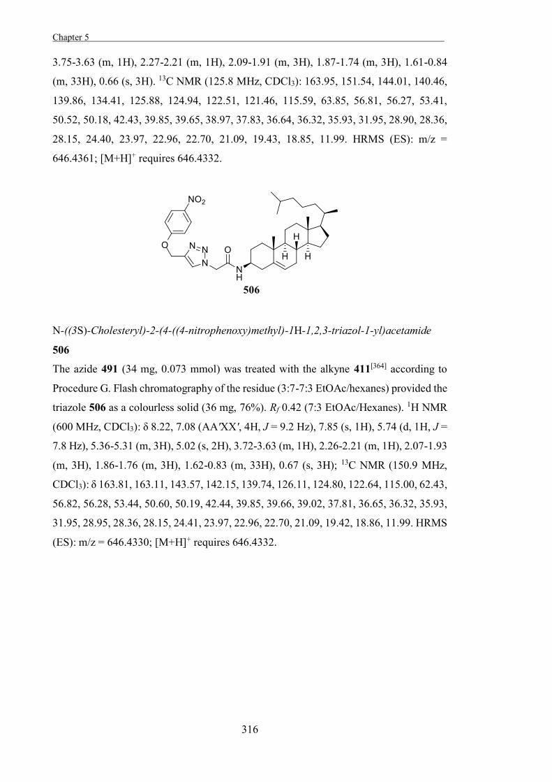
Chapter 5 .
316
3.75-3.63 (m, 1H), 2.27-2.21 (m, 1H), 2.09-1.91 (m, 3H), 1.87-1.74 (m, 3H), 1.61-0.84
(m, 33H), 0.66 (s, 3H). 13C NMR (125.8 MHz, CDCl3): 163.95, 151.54, 144.01, 140.46,
139.86, 134.41, 125.88, 124.94, 122.51, 121.46, 115.59, 63.85, 56.81, 56.27, 53.41,
50.52, 50.18, 42.43, 39.85, 39.65, 38.97, 37.83, 36.64, 36.32, 35.93, 31.95, 28.90, 28.36,
28.15, 24.40, 23.97, 22.96, 22.70, 21.09, 19.43, 18.85, 11.99. HRMS (ES): m/z =
646.4361; [M+H]+ requires 646.4332.
N-((3S)-Cholesteryl)-2-(4-((4-nitrophenoxy)methyl)-1H-1,2,3-triazol-1-yl)acetamide
506
The azide 491 (34 mg, 0.073 mmol) was treated with the alkyne 411[364] according to
Procedure G. Flash chromatography of the residue (3:7-7:3 EtOAc/hexanes) provided the
triazole 506 as a colourless solid (36 mg, 76%). Rf 0.42 (7:3 EtOAc/Hexanes). 1H NMR
(600 MHz, CDCl3): δ 8.22, 7.08 (AA'XX', 4H, J = 9.2 Hz), 7.85 (s, 1H), 5.74 (d, 1H, J =
7.8 Hz), 5.36-5.31 (m, 3H), 5.02 (s, 2H), 3.72-3.63 (m, 1H), 2.26-2.21 (m, 1H), 2.07-1.93
(m, 3H), 1.86-1.76 (m, 3H), 1.62-0.83 (m, 33H), 0.67 (s, 3H); 13C NMR (150.9 MHz,
CDCl3): δ 163.81, 163.11, 143.57, 142.15, 139.74, 126.11, 124.80, 122.64, 115.00, 62.43,
56.82, 56.28, 53.44, 50.60, 50.19, 42.44, 39.85, 39.66, 39.02, 37.81, 36.65, 36.32, 35.93,
31.95, 28.95, 28.36, 28.15, 24.41, 23.97, 22.96, 22.70, 21.09, 19.42, 18.86, 11.99. HRMS
(ES): m/z = 646.4330; [M+H]+ requires 646.4332.
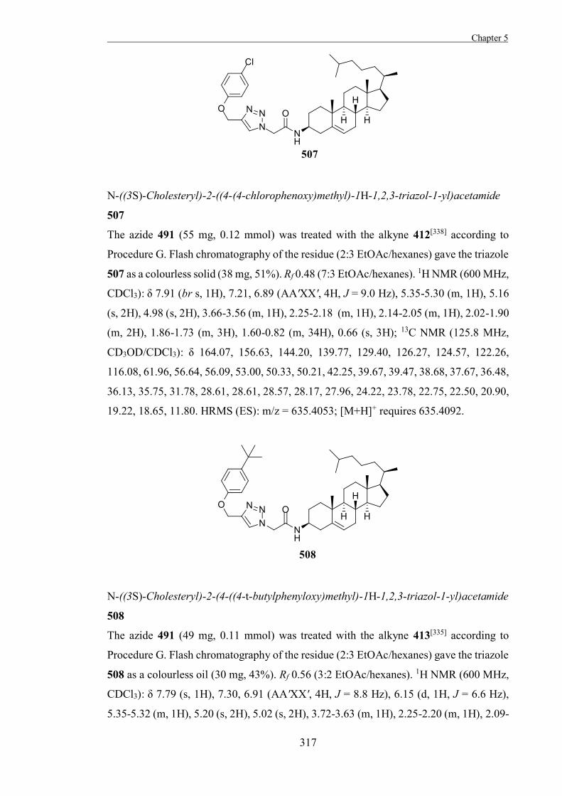
Chapter 5
317
N-((3S)-Cholesteryl)-2-((4-(4-chlorophenoxy)methyl)-1H-1,2,3-triazol-1-yl)acetamide
507
The azide 491 (55 mg, 0.12 mmol) was treated with the alkyne 412[338] according to
Procedure G. Flash chromatography of the residue (2:3 EtOAc/hexanes) gave the triazole
507 as a colourless solid (38 mg, 51%). Rf 0.48 (7:3 EtOAc/hexanes). 1H NMR (600 MHz,
CDCl3): δ 7.91 (br s, 1H), 7.21, 6.89 (AA'XX', 4H, J = 9.0 Hz), 5.35-5.30 (m, 1H), 5.16
(s, 2H), 4.98 (s, 2H), 3.66-3.56 (m, 1H), 2.25-2.18 (m, 1H), 2.14-2.05 (m, 1H), 2.02-1.90
(m, 2H), 1.86-1.73 (m, 3H), 1.60-0.82 (m, 34H), 0.66 (s, 3H); 13C NMR (125.8 MHz,
CD3OD/CDCl3): δ 164.07, 156.63, 144.20, 139.77, 129.40, 126.27, 124.57, 122.26,
116.08, 61.96, 56.64, 56.09, 53.00, 50.33, 50.21, 42.25, 39.67, 39.47, 38.68, 37.67, 36.48,
36.13, 35.75, 31.78, 28.61, 28.61, 28.57, 28.17, 27.96, 24.22, 23.78, 22.75, 22.50, 20.90,
19.22, 18.65, 11.80. HRMS (ES): m/z = 635.4053; [M+H]+ requires 635.4092.
N-((3S)-Cholesteryl)-2-(4-((4-t-butylphenyloxy)methyl)-1H-1,2,3-triazol-1-yl)acetamide
508
The azide 491 (49 mg, 0.11 mmol) was treated with the alkyne 413[335] according to
Procedure G. Flash chromatography of the residue (2:3 EtOAc/hexanes) gave the triazole
508 as a colourless oil (30 mg, 43%). Rf 0.56 (3:2 EtOAc/hexanes). 1H NMR (600 MHz,
CDCl3): δ 7.79 (s, 1H), 7.30, 6.91 (AA'XX', 4H, J = 8.8 Hz), 6.15 (d, 1H, J = 6.6 Hz),
5.35-5.32 (m, 1H), 5.20 (s, 2H), 5.02 (s, 2H), 3.72-3.63 (m, 1H), 2.25-2.20 (m, 1H), 2.09-
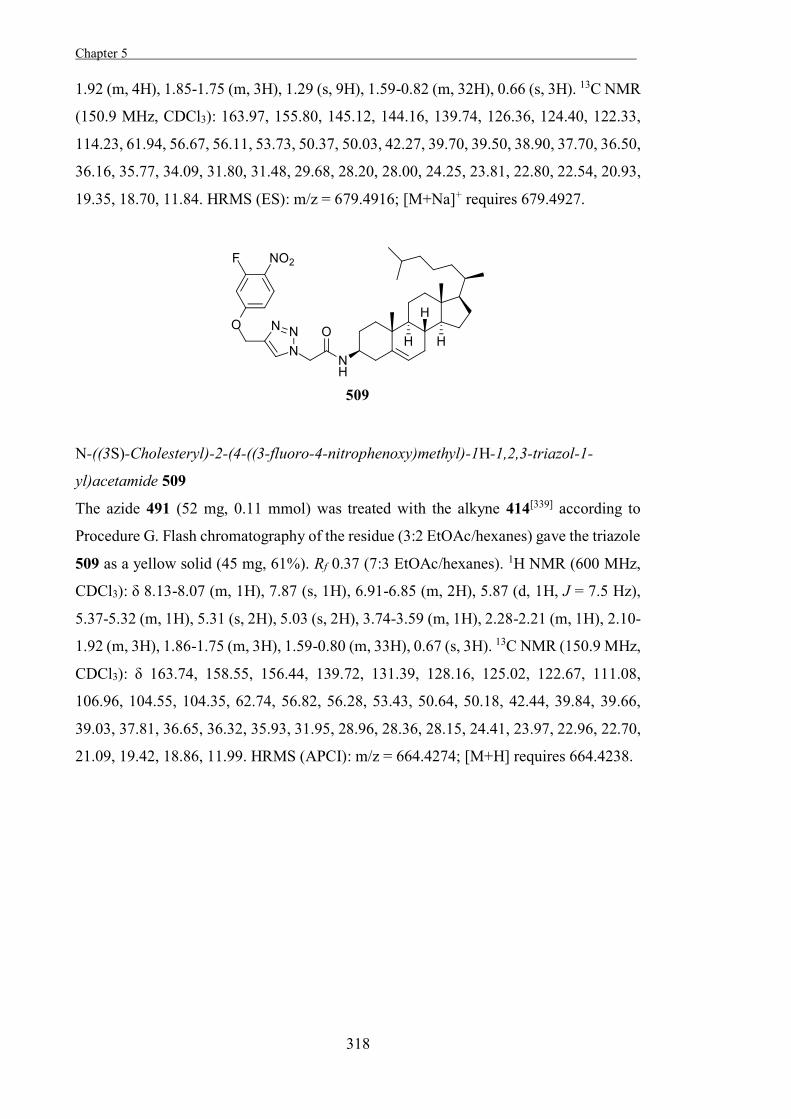
Chapter 5 .
318
1.92 (m, 4H), 1.85-1.75 (m, 3H), 1.29 (s, 9H), 1.59-0.82 (m, 32H), 0.66 (s, 3H). 13C NMR
(150.9 MHz, CDCl3): 163.97, 155.80, 145.12, 144.16, 139.74, 126.36, 124.40, 122.33,
114.23, 61.94, 56.67, 56.11, 53.73, 50.37, 50.03, 42.27, 39.70, 39.50, 38.90, 37.70, 36.50,
36.16, 35.77, 34.09, 31.80, 31.48, 29.68, 28.20, 28.00, 24.25, 23.81, 22.80, 22.54, 20.93,
19.35, 18.70, 11.84. HRMS (ES): m/z = 679.4916; [M+Na]+ requires 679.4927.
N-((3S)-Cholesteryl)-2-(4-((3-fluoro-4-nitrophenoxy)methyl)-1H-1,2,3-triazol-1-
yl)acetamide 509
The azide 491 (52 mg, 0.11 mmol) was treated with the alkyne 414[339] according to
Procedure G. Flash chromatography of the residue (3:2 EtOAc/hexanes) gave the triazole
509 as a yellow solid (45 mg, 61%). Rf 0.37 (7:3 EtOAc/hexanes). 1H NMR (600 MHz,
CDCl3): δ 8.13-8.07 (m, 1H), 7.87 (s, 1H), 6.91-6.85 (m, 2H), 5.87 (d, 1H, J = 7.5 Hz),
5.37-5.32 (m, 1H), 5.31 (s, 2H), 5.03 (s, 2H), 3.74-3.59 (m, 1H), 2.28-2.21 (m, 1H), 2.10-
1.92 (m, 3H), 1.86-1.75 (m, 3H), 1.59-0.80 (m, 33H), 0.67 (s, 3H). 13C NMR (150.9 MHz,
CDCl3): δ 163.74, 158.55, 156.44, 139.72, 131.39, 128.16, 125.02, 122.67, 111.08,
106.96, 104.55, 104.35, 62.74, 56.82, 56.28, 53.43, 50.64, 50.18, 42.44, 39.84, 39.66,
39.03, 37.81, 36.65, 36.32, 35.93, 31.95, 28.96, 28.36, 28.15, 24.41, 23.97, 22.96, 22.70,
21.09, 19.42, 18.86, 11.99. HRMS (APCI): m/z = 664.4274; [M+H] requires 664.4238.
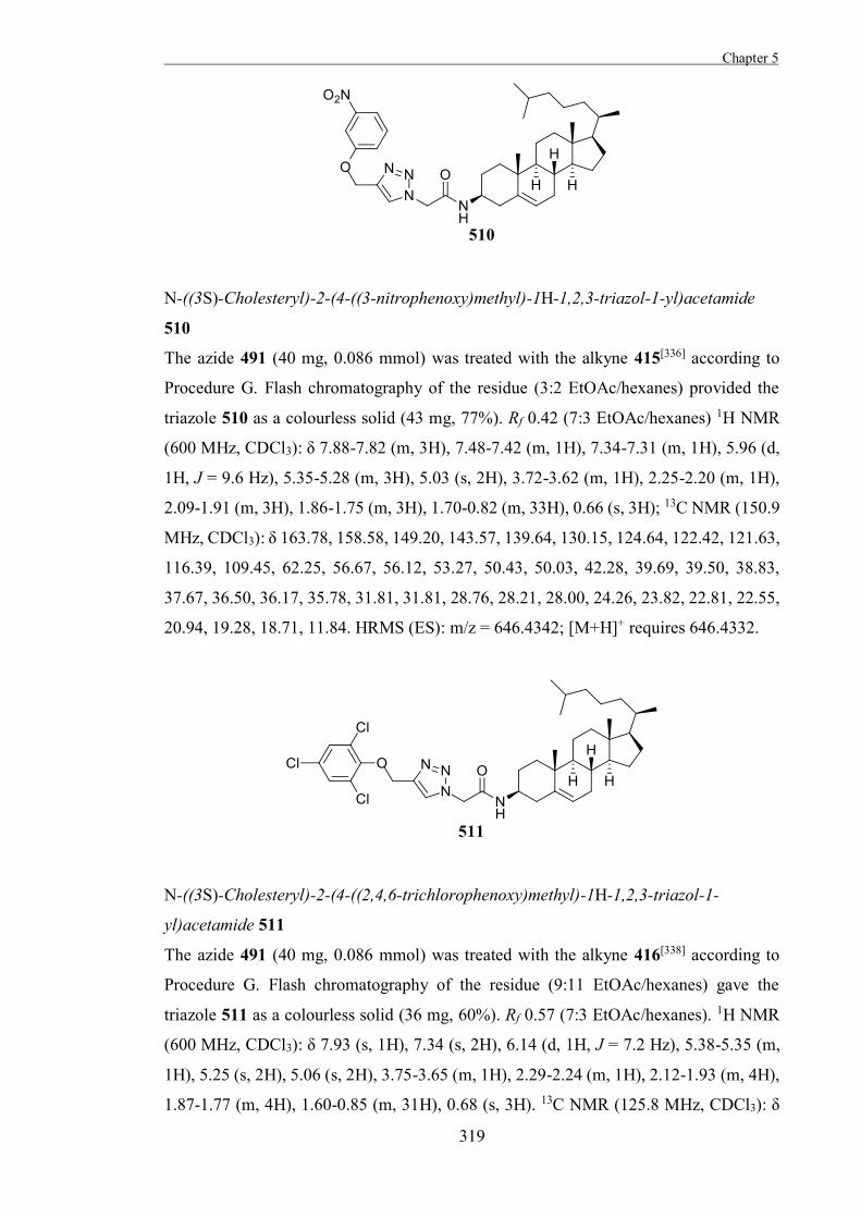
Chapter 5
319
N-((3S)-Cholesteryl)-2-(4-((3-nitrophenoxy)methyl)-1H-1,2,3-triazol-1-yl)acetamide
510
The azide 491 (40 mg, 0.086 mmol) was treated with the alkyne 415[336] according to
Procedure G. Flash chromatography of the residue (3:2 EtOAc/hexanes) provided the
triazole 510 as a colourless solid (43 mg, 77%). Rf 0.42 (7:3 EtOAc/hexanes) 1H NMR
(600 MHz, CDCl3): δ 7.88-7.82 (m, 3H), 7.48-7.42 (m, 1H), 7.34-7.31 (m, 1H), 5.96 (d,
1H, J = 9.6 Hz), 5.35-5.28 (m, 3H), 5.03 (s, 2H), 3.72-3.62 (m, 1H), 2.25-2.20 (m, 1H),
2.09-1.91 (m, 3H), 1.86-1.75 (m, 3H), 1.70-0.82 (m, 33H), 0.66 (s, 3H); 13C NMR (150.9
MHz, CDCl3): δ 163.78, 158.58, 149.20, 143.57, 139.64, 130.15, 124.64, 122.42, 121.63,
116.39, 109.45, 62.25, 56.67, 56.12, 53.27, 50.43, 50.03, 42.28, 39.69, 39.50, 38.83,
37.67, 36.50, 36.17, 35.78, 31.81, 31.81, 28.76, 28.21, 28.00, 24.26, 23.82, 22.81, 22.55,
20.94, 19.28, 18.71, 11.84. HRMS (ES): m/z = 646.4342; [M+H]+ requires 646.4332.
N-((3S)-Cholesteryl)-2-(4-((2,4,6-trichlorophenoxy)methyl)-1H-1,2,3-triazol-1-
yl)acetamide 511
The azide 491 (40 mg, 0.086 mmol) was treated with the alkyne 416[338] according to
Procedure G. Flash chromatography of the residue (9:11 EtOAc/hexanes) gave the
triazole 511 as a colourless solid (36 mg, 60%). Rf 0.57 (7:3 EtOAc/hexanes). 1H NMR
(600 MHz, CDCl3): δ 7.93 (s, 1H), 7.34 (s, 2H), 6.14 (d, 1H, J = 7.2 Hz), 5.38-5.35 (m,
1H), 5.25 (s, 2H), 5.06 (s, 2H), 3.75-3.65 (m, 1H), 2.29-2.24 (m, 1H), 2.12-1.93 (m, 4H),
1.87-1.77 (m, 4H), 1.60-0.85 (m, 31H), 0.68 (s, 3H). 13C NMR (125.8 MHz, CDCl3): δ
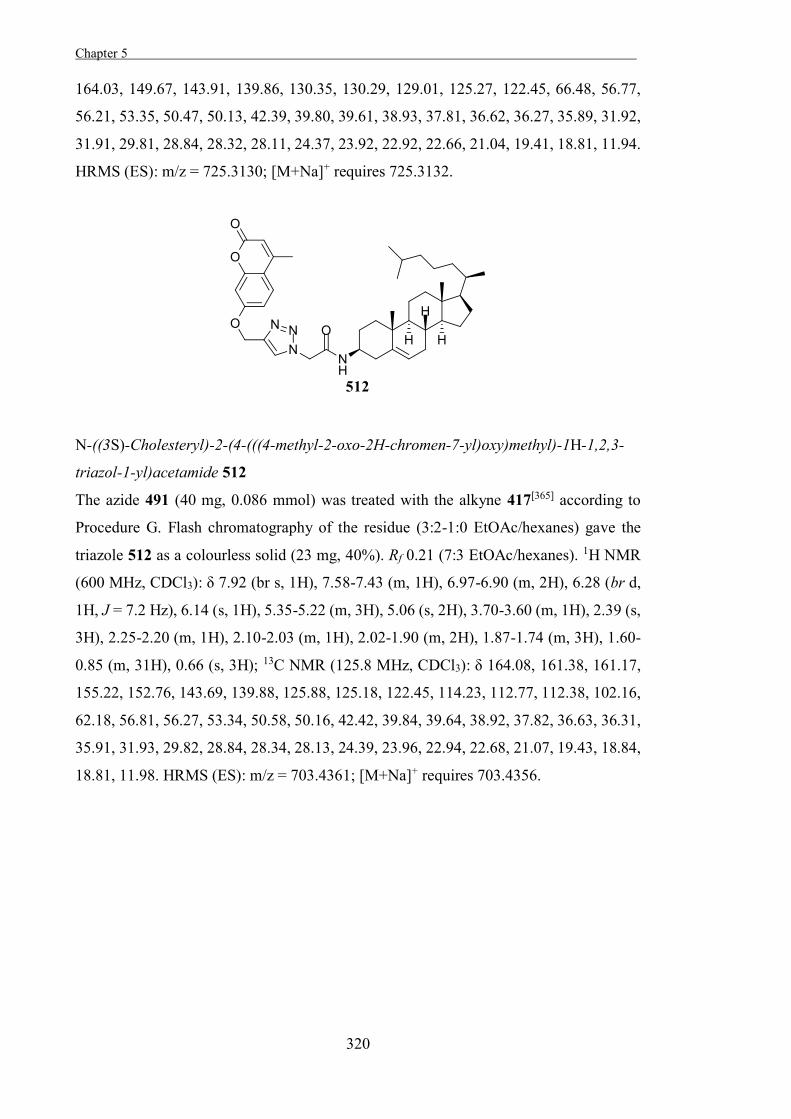
Chapter 5 .
320
164.03, 149.67, 143.91, 139.86, 130.35, 130.29, 129.01, 125.27, 122.45, 66.48, 56.77,
56.21, 53.35, 50.47, 50.13, 42.39, 39.80, 39.61, 38.93, 37.81, 36.62, 36.27, 35.89, 31.92,
31.91, 29.81, 28.84, 28.32, 28.11, 24.37, 23.92, 22.92, 22.66, 21.04, 19.41, 18.81, 11.94.
HRMS (ES): m/z = 725.3130; [M+Na]+ requires 725.3132.
N-((3S)-Cholesteryl)-2-(4-(((4-methyl-2-oxo-2H-chromen-7-yl)oxy)methyl)-1H-1,2,3-
triazol-1-yl)acetamide 512
The azide 491 (40 mg, 0.086 mmol) was treated with the alkyne 417[365] according to
Procedure G. Flash chromatography of the residue (3:2-1:0 EtOAc/hexanes) gave the
triazole 512 as a colourless solid (23 mg, 40%). Rf 0.21 (7:3 EtOAc/hexanes). 1H NMR
(600 MHz, CDCl3): δ 7.92 (br s, 1H), 7.58-7.43 (m, 1H), 6.97-6.90 (m, 2H), 6.28 (br d,
1H, J = 7.2 Hz), 6.14 (s, 1H), 5.35-5.22 (m, 3H), 5.06 (s, 2H), 3.70-3.60 (m, 1H), 2.39 (s,
3H), 2.25-2.20 (m, 1H), 2.10-2.03 (m, 1H), 2.02-1.90 (m, 2H), 1.87-1.74 (m, 3H), 1.60-
0.85 (m, 31H), 0.66 (s, 3H); 13C NMR (125.8 MHz, CDCl3): δ 164.08, 161.38, 161.17,
155.22, 152.76, 143.69, 139.88, 125.88, 125.18, 122.45, 114.23, 112.77, 112.38, 102.16,
62.18, 56.81, 56.27, 53.34, 50.58, 50.16, 42.42, 39.84, 39.64, 38.92, 37.82, 36.63, 36.31,
35.91, 31.93, 29.82, 28.84, 28.34, 28.13, 24.39, 23.96, 22.94, 22.68, 21.07, 19.43, 18.84,
18.81, 11.98. HRMS (ES): m/z = 703.4361; [M+Na]+ requires 703.4356.
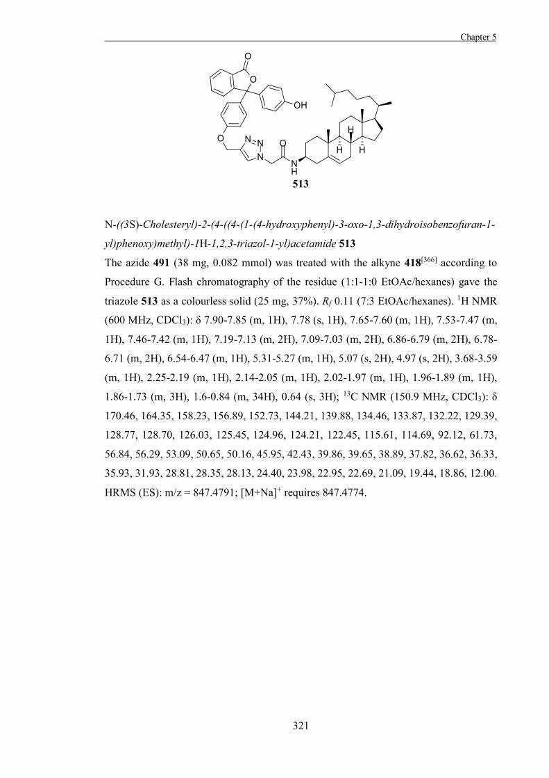
Chapter 5
321
N-((3S)-Cholesteryl)-2-(4-((4-(1-(4-hydroxyphenyl)-3-oxo-1,3-dihydroisobenzofuran-1-
yl)phenoxy)methyl)-1H-1,2,3-triazol-1-yl)acetamide 513
The azide 491 (38 mg, 0.082 mmol) was treated with the alkyne 418[366] according to
Procedure G. Flash chromatography of the residue (1:1-1:0 EtOAc/hexanes) gave the
triazole 513 as a colourless solid (25 mg, 37%). Rf 0.11 (7:3 EtOAc/hexanes). 1H NMR
(600 MHz, CDCl3): δ 7.90-7.85 (m, 1H), 7.78 (s, 1H), 7.65-7.60 (m, 1H), 7.53-7.47 (m,
1H), 7.46-7.42 (m, 1H), 7.19-7.13 (m, 2H), 7.09-7.03 (m, 2H), 6.86-6.79 (m, 2H), 6.78-
6.71 (m, 2H), 6.54-6.47 (m, 1H), 5.31-5.27 (m, 1H), 5.07 (s, 2H), 4.97 (s, 2H), 3.68-3.59
(m, 1H), 2.25-2.19 (m, 1H), 2.14-2.05 (m, 1H), 2.02-1.97 (m, 1H), 1.96-1.89 (m, 1H),
1.86-1.73 (m, 3H), 1.6-0.84 (m, 34H), 0.64 (s, 3H); 13C NMR (150.9 MHz, CDCl3): δ
170.46, 164.35, 158.23, 156.89, 152.73, 144.21, 139.88, 134.46, 133.87, 132.22, 129.39,
128.77, 128.70, 126.03, 125.45, 124.96, 124.21, 122.45, 115.61, 114.69, 92.12, 61.73,
56.84, 56.29, 53.09, 50.65, 50.16, 45.95, 42.43, 39.86, 39.65, 38.89, 37.82, 36.62, 36.33,
35.93, 31.93, 28.81, 28.35, 28.13, 24.40, 23.98, 22.95, 22.69, 21.09, 19.44, 18.86, 12.00.
HRMS (ES): m/z = 847.4791; [M+Na]+ requires 847.4774.
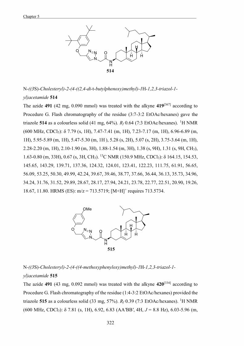
Chapter 5 .
322
N-((3S)-Cholesteryl)-2-(4-((2,4-di-t-butylphenoxy)methyl)-1H-1,2,3-triazol-1-
yl)acetamide 514
The azide 491 (42 mg, 0.090 mmol) was treated with the alkyne 419[367] according to
Procedure G. Flash chromatography of the residue (3:7-3:2 EtOAc/hexanes) gave the
triazole 514 as a colourless solid (41 mg, 64%). Rf 0.64 (7:3 EtOAc/hexanes). 1H NMR
(600 MHz, CDCl3): δ 7.79 (s, 1H), 7.47-7.41 (m, 1H), 7.23-7.17 (m, 1H), 6.96-6.89 (m,
1H), 5.95-5.89 (m, 1H), 5.47-5.30 (m, 1H ), 5.28 (s, 2H), 5.07 (s, 2H), 3.75-3.64 (m, 1H),
2.28-2.20 (m, 1H), 2.10-1.90 (m, 3H), 1.88-1.54 (m, 3H), 1.38 (s, 9H), 1.31 (s, 9H, CH3),
1.63-0.80 (m, 33H), 0.67 (s, 3H, CH3). 13C NMR (150.9 MHz, CDCl3): δ 164.15, 154.53,
145.65, 143.29, 139.71, 137.36, 124.32, 124.01, 123.41, 122.23, 111.75, 61.91, 56.65,
56.09, 53.25, 50.30, 49.99, 42.24, 39.67, 39.46, 38.77, 37.66, 36.44, 36.13, 35.73, 34.96,
34.24, 31.76, 31.52, 29.89, 28.67, 28.17, 27.94, 24.21, 23.78, 22.77, 22.51, 20.90, 19.26,
18.67, 11.80. HRMS (ES): m/z = 713.5719; [M+H]+ requires 713.5734.
N-((3S)-Cholesteryl)-2-(4-((4-methoxyphenyloxy)methyl)-1H-1,2,3-triazol-1-
yl)acetamide 515
The azide 491 (43 mg, 0.092 mmol) was treated with the alkyne 420[334] according to
Procedure G. Flash chromatography of the residue (1:4-3:2 EtOAc/hexanes) provided the
triazole 515 as a colourless solid (33 mg, 57%). Rf 0.39 (7:3 EtOAc/hexanes). 1H NMR
(600 MHz, CDCl3): δ 7.81 (s, 1H), 6.92, 6.83 (AA'BB', 4H, J = 8.8 Hz), 6.03-5.96 (m,
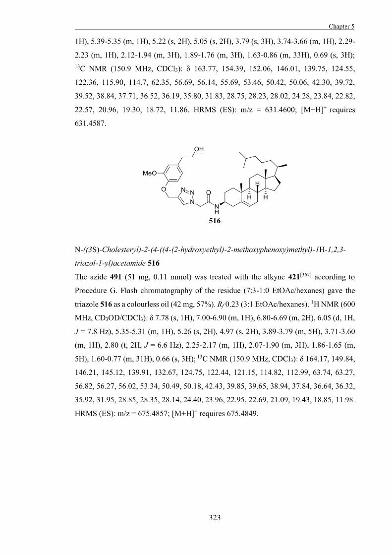
Chapter 5
323
1H), 5.39-5.35 (m, 1H), 5.22 (s, 2H), 5.05 (s, 2H), 3.79 (s, 3H), 3.74-3.66 (m, 1H), 2.29-
2.23 (m, 1H), 2.12-1.94 (m, 3H), 1.89-1.76 (m, 3H), 1.63-0.86 (m, 33H), 0.69 (s, 3H); 13C NMR (150.9 MHz, CDCl3): δ 163.77, 154.39, 152.06, 146.01, 139.75, 124.55,
122.36, 115.90, 114.7, 62.35, 56.69, 56.14, 55.69, 53.46, 50.42, 50.06, 42.30, 39.72,
39.52, 38.84, 37.71, 36.52, 36.19, 35.80, 31.83, 28.75, 28.23, 28.02, 24.28, 23.84, 22.82,
22.57, 20.96, 19.30, 18.72, 11.86. HRMS (ES): m/z = 631.4600; [M+H]+ requires
631.4587.
N-((3S)-Cholesteryl)-2-(4-((4-(2-hydroxyethyl)-2-methoxyphenoxy)methyl)-1H-1,2,3-
triazol-1-yl)acetamide 516
The azide 491 (51 mg, 0.11 mmol) was treated with the alkyne 421[367] according to
Procedure G. Flash chromatography of the residue (7:3-1:0 EtOAc/hexanes) gave the
triazole 516 as a colourless oil (42 mg, 57%). Rf 0.23 (3:1 EtOAc/hexanes). 1H NMR (600
MHz, CD3OD/CDCl3): δ 7.78 (s, 1H), 7.00-6.90 (m, 1H), 6.80-6.69 (m, 2H), 6.05 (d, 1H,
J = 7.8 Hz), 5.35-5.31 (m, 1H), 5.26 (s, 2H), 4.97 (s, 2H), 3.89-3.79 (m, 5H), 3.71-3.60
(m, 1H), 2.80 (t, 2H, J = 6.6 Hz), 2.25-2.17 (m, 1H), 2.07-1.90 (m, 3H), 1.86-1.65 (m,
5H), 1.60-0.77 (m, 31H), 0.66 (s, 3H); 13C NMR (150.9 MHz, CDCl3): δ 164.17, 149.84,
146.21, 145.12, 139.91, 132.67, 124.75, 122.44, 121.15, 114.82, 112.99, 63.74, 63.27,
56.82, 56.27, 56.02, 53.34, 50.49, 50.18, 42.43, 39.85, 39.65, 38.94, 37.84, 36.64, 36.32,
35.92, 31.95, 28.85, 28.35, 28.14, 24.40, 23.96, 22.95, 22.69, 21.09, 19.43, 18.85, 11.98.
HRMS (ES): m/z = 675.4857; [M+H]+ requires 675.4849.
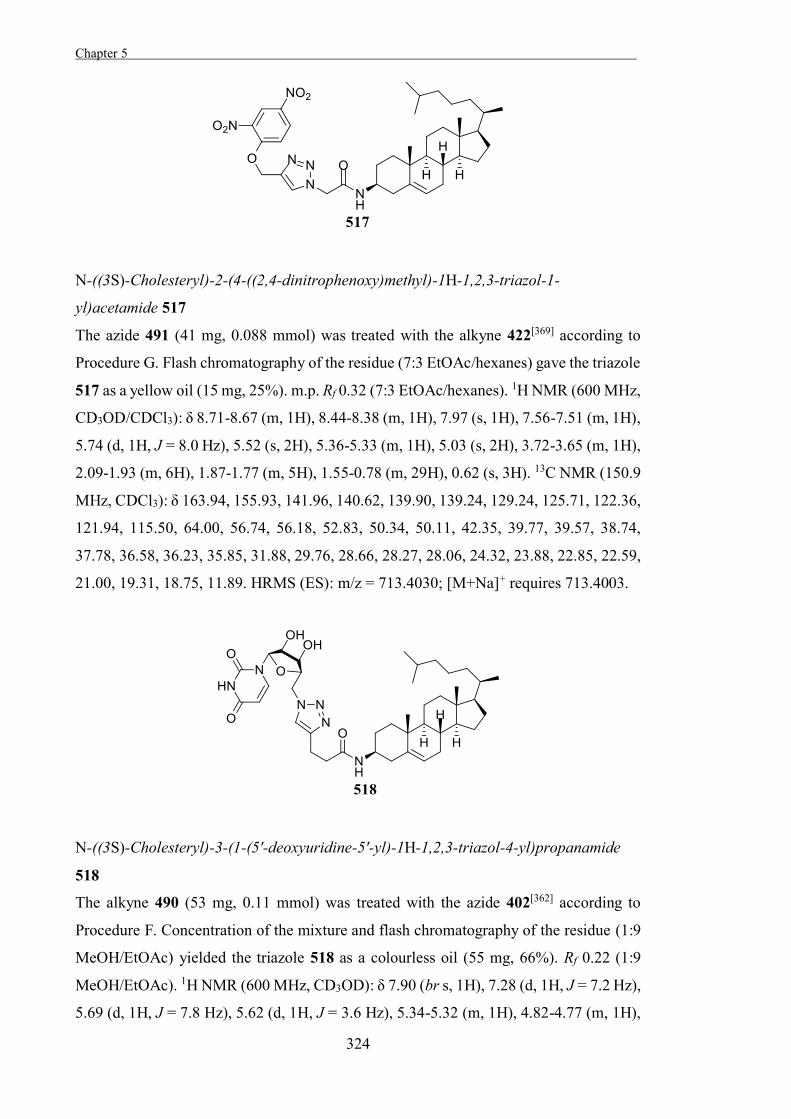
Chapter 5 .
324
N-((3S)-Cholesteryl)-2-(4-((2,4-dinitrophenoxy)methyl)-1H-1,2,3-triazol-1-
yl)acetamide 517
The azide 491 (41 mg, 0.088 mmol) was treated with the alkyne 422[369] according to
Procedure G. Flash chromatography of the residue (7:3 EtOAc/hexanes) gave the triazole
517 as a yellow oil (15 mg, 25%). m.p. Rf 0.32 (7:3 EtOAc/hexanes). 1H NMR (600 MHz,
CD3OD/CDCl3): δ 8.71-8.67 (m, 1H), 8.44-8.38 (m, 1H), 7.97 (s, 1H), 7.56-7.51 (m, 1H),
5.74 (d, 1H, J = 8.0 Hz), 5.52 (s, 2H), 5.36-5.33 (m, 1H), 5.03 (s, 2H), 3.72-3.65 (m, 1H),
2.09-1.93 (m, 6H), 1.87-1.77 (m, 5H), 1.55-0.78 (m, 29H), 0.62 (s, 3H). 13C NMR (150.9
MHz, CDCl3): δ 163.94, 155.93, 141.96, 140.62, 139.90, 139.24, 129.24, 125.71, 122.36,
121.94, 115.50, 64.00, 56.74, 56.18, 52.83, 50.34, 50.11, 42.35, 39.77, 39.57, 38.74,
37.78, 36.58, 36.23, 35.85, 31.88, 29.76, 28.66, 28.27, 28.06, 24.32, 23.88, 22.85, 22.59,
21.00, 19.31, 18.75, 11.89. HRMS (ES): m/z = 713.4030; [M+Na]+ requires 713.4003.
N-((3S)-Cholesteryl)-3-(1-(5'-deoxyuridine-5'-yl)-1H-1,2,3-triazol-4-yl)propanamide
518
The alkyne 490 (53 mg, 0.11 mmol) was treated with the azide 402[362] according to
Procedure F. Concentration of the mixture and flash chromatography of the residue (1:9
MeOH/EtOAc) yielded the triazole 518 as a colourless oil (55 mg, 66%). Rf 0.22 (1:9
MeOH/EtOAc). 1H NMR (600 MHz, CD3OD): δ 7.90 (br s, 1H), 7.28 (d, 1H, J = 7.2 Hz),
5.69 (d, 1H, J = 7.8 Hz), 5.62 (d, 1H, J = 3.6 Hz), 5.34-5.32 (m, 1H), 4.82-4.77 (m, 1H),
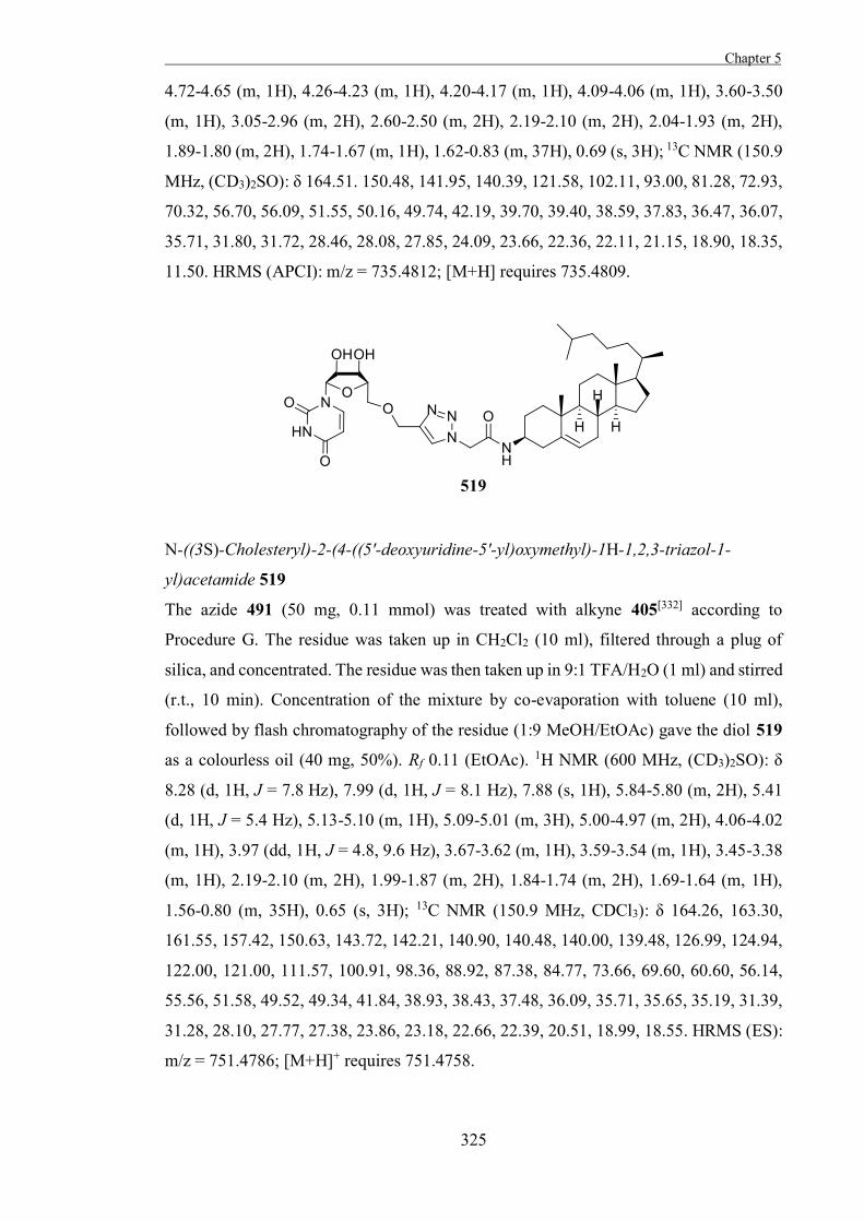
Chapter 5
325
4.72-4.65 (m, 1H), 4.26-4.23 (m, 1H), 4.20-4.17 (m, 1H), 4.09-4.06 (m, 1H), 3.60-3.50
(m, 1H), 3.05-2.96 (m, 2H), 2.60-2.50 (m, 2H), 2.19-2.10 (m, 2H), 2.04-1.93 (m, 2H),
1.89-1.80 (m, 2H), 1.74-1.67 (m, 1H), 1.62-0.83 (m, 37H), 0.69 (s, 3H); 13C NMR (150.9
MHz, (CD3)2SO): δ 164.51. 150.48, 141.95, 140.39, 121.58, 102.11, 93.00, 81.28, 72.93,
70.32, 56.70, 56.09, 51.55, 50.16, 49.74, 42.19, 39.70, 39.40, 38.59, 37.83, 36.47, 36.07,
35.71, 31.80, 31.72, 28.46, 28.08, 27.85, 24.09, 23.66, 22.36, 22.11, 21.15, 18.90, 18.35,
11.50. HRMS (APCI): m/z = 735.4812; [M+H] requires 735.4809.
N-((3S)-Cholesteryl)-2-(4-((5'-deoxyuridine-5'-yl)oxymethyl)-1H-1,2,3-triazol-1-
yl)acetamide 519
The azide 491 (50 mg, 0.11 mmol) was treated with alkyne 405[332] according to
Procedure G. The residue was taken up in CH2Cl2 (10 ml), filtered through a plug of
silica, and concentrated. The residue was then taken up in 9:1 TFA/H2O (1 ml) and stirred
(r.t., 10 min). Concentration of the mixture by co-evaporation with toluene (10 ml),
followed by flash chromatography of the residue (1:9 MeOH/EtOAc) gave the diol 519
as a colourless oil (40 mg, 50%). Rf 0.11 (EtOAc). 1H NMR (600 MHz, (CD3)2SO): δ
8.28 (d, 1H, J = 7.8 Hz), 7.99 (d, 1H, J = 8.1 Hz), 7.88 (s, 1H), 5.84-5.80 (m, 2H), 5.41
(d, 1H, J = 5.4 Hz), 5.13-5.10 (m, 1H), 5.09-5.01 (m, 3H), 5.00-4.97 (m, 2H), 4.06-4.02
(m, 1H), 3.97 (dd, 1H, J = 4.8, 9.6 Hz), 3.67-3.62 (m, 1H), 3.59-3.54 (m, 1H), 3.45-3.38
(m, 1H), 2.19-2.10 (m, 2H), 1.99-1.87 (m, 2H), 1.84-1.74 (m, 2H), 1.69-1.64 (m, 1H),
1.56-0.80 (m, 35H), 0.65 (s, 3H); 13C NMR (150.9 MHz, CDCl3): δ 164.26, 163.30,
161.55, 157.42, 150.63, 143.72, 142.21, 140.90, 140.48, 140.00, 139.48, 126.99, 124.94,
122.00, 121.00, 111.57, 100.91, 98.36, 88.92, 87.38, 84.77, 73.66, 69.60, 60.60, 56.14,
55.56, 51.58, 49.52, 49.34, 41.84, 38.93, 38.43, 37.48, 36.09, 35.71, 35.65, 35.19, 31.39,
31.28, 28.10, 27.77, 27.38, 23.86, 23.18, 22.66, 22.39, 20.51, 18.99, 18.55. HRMS (ES):
m/z = 751.4786; [M+H]+ requires 751.4758.
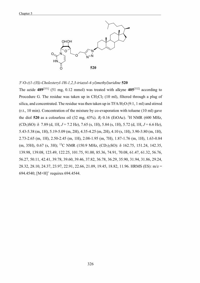
Chapter 5 .
326
5'-O-((1-(3S)-Cholesteryl-1H-1,2,3-triazol-4-yl)methyl)uridine 520
The azide 489[351] (51 mg, 0.12 mmol) was treated with alkyne 405[332] according to
Procedure G. The residue was taken up in CH2Cl2 (10 ml), filtered through a plug of
silica, and concentrated. The residue was then taken up in TFA/H2O (9:1, 1 ml) and stirred
(r.t., 10 min). Concentration of the mixture by co-evaporation with toluene (10 ml) gave
the diol 520 as a colourless oil (32 mg, 43%). Rf 0.16 (EtOAc). 1H NMR (600 MHz,
(CD3)SO): δ 7.89 (d, 1H, J = 7.2 Hz), 7.65 (s, 1H), 5.84 (s, 1H), 5.72 (d, 1H, J = 6.6 Hz),
5.43-5.38 (m, 1H), 5.19-5.09 (m, 2H), 4.35-4.25 (m, 2H), 4.10 (s, 1H), 3.90-3.80 (m, 1H),
2.73-2.65 (m, 1H), 2.50-2.45 (m, 1H), 2.08-1.95 (m, 7H), 1.87-1.76 (m, 1H), 1.63-0.84
(m, 35H), 0.67 (s, 3H); 13C NMR (150.9 MHz, (CD3)2SO): δ 162.75, 151.24, 142.35,
139.98, 139.08, 123.49, 122.25, 101.75, 91.00, 85.36, 74.91, 70.08, 61.47, 61.32, 56.76,
56.27, 50.11, 42.41, 39.78, 39.60, 39.46, 37.82, 36.78, 36.29, 35.90, 31.94, 31.86, 29.24,
28.32, 28.10, 24.37, 23.97, 22.91, 22.66, 21.09, 19.45, 18.82, 11.96. HRMS (ES): m/z =
694.4540; [M+H]+ requires 694.4544.
Chapter 5
327
Evaluating Biological Activity
Inhibitors
All assays were carried out in triplicate using the procedure of Fukuda et al.,[355] except
that the concentration of UDP-glucose used was 100 M and cholesterol 5 M. Assays
were initiated by the careful addition of CGT to the reaction and the overall DMSO
concentration in all assays was 5%. The range of inhibitor concentrations used was from
100 nM to 10 mM (with the specific range used dependent on the measured IC50 for each
inhibitor). The reaction mixtures were monitored at 340 nm for 60 min at 37 °C. Time-
dependent analysis of the enzyme used revealed that the enzyme was stable over the
period of the assay.
Bacterial growth assays
The 26695 wild-type and isogenic 26695 Δcgt mutant strains used in this study were
kindly provided by Dr. Aleksandra Debowski. H. pylori was routinely grown under
microaerobic conditions on Columbia blood agar (CBA) plates containing Columbia
blood agar base (Oxoid) with 5% (v/v) horse blood and 5% (v/v) new-born calf serum.
Plates were incubated at 37 °C for 24-48 hours within sealed jars using the Anoxomat™
MarkII system (Mart Microbiology B.V., The Netherlands) after one atmosphere
replacement with the following gas composition N2:H2:CO2, 85:5:10. For the growth
inhibition studies bacteria were grown in Brain Heart Infusion (BHI) medium
supplemented with 10% Newborn Calf Serum (NCS). Cultures were inoculated with
bacteria suspended in PBS to give a starting OD600 = 0.05, and were then grown under
microaerobic conditions at 37 °C and 120 rpm. The optical density of the cultures were
measured every 12 h for up to 60 h. Growth studies were performed without any prior
adaptation of H. pylori strains to liquid media and each experiment was performed in
triplicate.

328
329
References [1] B. Ganem, Acc. Chem. Res. 1996, 29, 340-347. [2] L. L. Lairson, B. Henrissat, G. J. Davies, S. G. Withers, Annu. Rev. Biochem
2008, 77, 521-555. [3] M. L. Sinnott, Chem. Rev. 1990, 90, 1171-1202. [4] C. S. Rye, S. G. Withers, Curr. Opin. Chem. Biol. 2000, 4, 573-580. [5] R. Bernacki, M. Niedbala, W. Korytnyk, Cancer Metast Rev 1985, 4, 81-101. [6] K. Wang, W. Guo, N. Li, J. Shi, C. Zhang, W. Y. Lau, M. Wu, S. Cheng, Br. J.
Cancer 2014, 110, 1811-1819. [7] T. F. Scanlin, M. C. Glick, BBA-Mol. Basis Dis. 1999, 1455, 241-253. [8] N. Asano, Cell. Mol. Life Sci. 2009, 66, 1479-1492. [9] O. Abian, P. Alfonso, A. Velazquez-Campoy, P. Giraldo, M. Pocovi, J. Sancho,
Mol. Pharm. 2011, 8, 2390-2397. [10] G. W. J. Fleet, A. Karpas, R. A. Dwek, L. E. Fellows, A. S. Tyms, S. Petursson,
S. K. Namgoong, N. G. Ramsden, P. W. Smith, J. C. Son, F. Wilson, D. R. Witty, G. S. Jacob, T. W. Rademacher, FEBS Lett. 1988, 237, 128-132.
[11] T. Feizi, Trends Biochem. Sci 1991, 16, 84-86. [12] B. Henrissat, Biochem. J. 1991, 280, 309-316. [13] V. Lombard, H. Golaconda Ramulu, E. Drula, P. M. Coutinho, B. Henrissat,
Nucleic Acids Res. 2014, 42, 490-495. [14] T. M. Gloster, J. P. Turkenburg, J. R. Potts, B. Henrissat, G. J. Davies, Chem.
Biol. 2008, 15, 1058-1067. [15] D. E. Koshland, Biol. Rev. 1953, 28, 416-436. [16] M. Okuyama, A. Okuno, N. Shimizu, H. Mori, A. Kimura, S. Chiba, Eur. J.
Biochem. 2001, 268, 2270-2280. [17] A. Lammerts van Bueren, A. Ardèvol, J. Fayers-Kerr, B. Luo, Y. Zhang, M.
Sollogoub, Y. Blériot, C. Rovira, G. J. Davies, J. Am. Chem. Soc. 2010, 132, 1804-1806.
[18] V. L. Y. Yip, A. Varrot, G. J. Davies, S. S. Rajan, X. Yang, J. Thompson, W. F. Anderson, S. G. Withers, J. Am. Chem. Soc. 2004, 126, 8354-8355.
[19] S. S. Rajan, X. Yang, F. Collart, V. L. Y. Yip, S. G. Withers, A. Varrot, J. Thompson, G. J. Davies, W. F. Anderson, Structure 2004, 12, 1619-1629.
[20] S. J. Angyal, Angew. Chem. Int. Ed. Engl. 1969, 8, 157-166. [21] S. G. Sunderwirth, G. G. Olson, J. Chem. Educ. 1962, 39, 410. [22] G. J. Davies, A. Planas, C. Rovira, Acc. Chem. Res. 2012, 45, 308-316. [23] E. Westhof, M. Sundaralingam, J. Am. Chem. Soc. 1980, 102, 1493-1500. [24] G. Speciale, A. J. Thompson, G. J. Davies, S. J. Williams, Curr. Opin. Struc.
Biol. 2014, 28, 1-13. [25] V. Notenboom, S. J. Williams, R. Hoos, S. G. Withers, D. R. Rose,
Biochemistry 2000, 39, 11553-11563. [26] T. M. Gloster, D. J. Vocadlo, Nat. Chem. Biol. 2012, 8, 683-694. [27] M. D. Witte, G. A. van der Marel, J. M. F. G. Aerts, H. S. Overkleeft, Org.
Biomol. Chem. 2011, 9, 5908-5926. [28] B. P. Rempel, S. G. Withers, Glycobiology 2008, 18, 570-586. [29] P. J. Marshall, M. L. Sinnott, P. J. Smith, D. Widdows, J. Chem. Soc., Perkin
Trans. 1 1981, 366-376. [30] C.-S. Kuhn, J. Lehmann, G. Jung, S. Stevanović, Carbohydr. Res. 1992, 232,
227-233. [31] C.-S. Kuhn, J. Lehmann, Carbohydr. Res. 1987, 160, C6-C8.
330
[32] S. Halazy, C. Danzin, A. Ehrhard, F. Gerhart, J. Am. Chem. Soc. 1989, 111, 3484-3485.
[33] G. Sulzenbacher, M. Schülein, G. J. Davies, Biochemistry 1997, 36, 5902-5911. [34] G. Caron, S. G. Withers, Biochem. Biophys. Res. Commun. 1989, 163, 495-499. [35] S. G. Withers, R. Aebersold, Protein Sci. 1995, 4, 361-372. [36] S. G. Withers, I. P. Street, P. Bird, D. H. Dolphin, J. Am. Chem. Soc. 1987, 109,
7530-7531. [37] S. G. Withers, K. Rupitz, I. P. Street, J. Biol. Chem. 1988, 263, 7929-7932. [38] S. G. Withers, I. P. Street, J. Am. Chem. Soc. 1988, 110, 8551-8553. [39] S. G. Withers, R. A. J. Warren, I. P. Street, K. Rupitz, J. B. Kempton, R.
Aebersold, J. Am. Chem. Soc. 1990, 112, 5887-5889. [40] D. J. Vocadlo, G. J. Davies, R. Laine, S. G. Withers, Nature 2001, 412, 835-838. [41] J. Wicki, D. R. Rose, S. G. Withers, in Methods Enzymol., Vol. Volume 354
(Ed.: L. P. Daniel), Academic Press, 2002, pp. 84-105. [42] C. Yu, M. Crispin, A. F.-P. Sonnen, D. J. Harvey, V. T. Chang, E. J. Evans, C.
N. Scanlan, D. I. Stuart, R. J. C. Gilbert, S. J. Davis, Acta Crystallogr. Sect. F Struct. Biol. Cryst. Commun. 2011, 67, 785-789.
[43] S. A. Yuzwa, M. S. Macauley, J. E. Heinonen, X. Shan, R. J. Dennis, Y. He, G. E. Whitworth, K. A. Stubbs, E. J. McEachern, G. J. Davies, D. J. Vocadlo, Nat. Chem. Biol. 2008, 4, 483-490.
[44] T. M. Wrodnigg, A. J. Steiner, B. J. Ueberbacher, Anticancer Agents Med. Chem. 2008, 8, 77-85.
[45] B. L. Stegelmeier, R. J. Molyneux, N. Asano, A. A. Watson, R. J. Nash, Toxicol. Pathol. 2008, 36, 651-659.
[46] E. B. Arias, J. Kim, G. D. Cartee, Diabetes 2004, 53, 921-930. [47] P. H. Joubert, W. J. Bam, N. Manyane, Eur J Clin Pharmacol 1986, 30, 253-
255. [48] F. G. Hayden, J. J. Treanor, R. Fritz, et al., JAMA 1999, 282, 1240-1246. [49] Y. Lee, S. Kim, J. Y. Kim, M. Arooj, S. Kim, S. Hwang, B.-W. Kim, K. H.
Park, K. W. Lee, PLoS ONE 2014, 9, e85827. [50] T. D. Heightman, A. T. Vasella, Angew. Chem. Int. Ed. 1999, 38, 750-770. [51] D. C. Koester, A. Holkenbrink, D. B. Werz, Synthesis 2010, 19, 3217-3242. [52] V. Vinader, M. H. Haji-Abdullahi, L. H. Patterson, K. Afarinkia, PLoS ONE
2013, 8, e82111. [53] G. Sulzenbacher, H. Driguez, B. Henrissat, M. Schülein, G. J. Davies,
Biochemistry 1996, 35, 15280-15287. [54] J.-F. Espinosa, F. J. Cañada, J. L. Asensio, M. Martín-Pastor, H. Dietrich, M.
Martín-Lomas, R. R. Schmidt, J. Jiménez-Barbero, J. Am. Chem. Soc. 1996, 118, 10862-10871.
[55] M. H. D. Postema, J. L. Piper, L. Liu, J. Shen, M. Faust, P. Andreana, J. Org. Chem. 2003, 68, 4748-4754.
[56] M. Hayashida, N. Sakairi, H. Kuzuhara, Carbohydr. Res. 1986, 158, C5-C8. [57] K. Seri, K. Sanai, N. Matsuo, K. Kawakubo, C. Xue, S. Inoue, Metabolism
1996, 45, 1368-1374. [58] B. Stoffer, A. E. Aleshin, L. M. Firsov, B. Svensson, R. B. Honzatko, FEBS
Lett. 1995, 358, 57-61. [59] R. R. Holman, C. A. Cull, R. C. Turner, Diabetes Care 1999, 22, 960-964. [60] V. L. Schramm, J. Biol. Chem. 2007, 282, 28297-28300. [61] L. Pauling, Nature 1948, 161, 707-709. [62] T. M. Gloster, G. J. Davies, Org. Biomol. Chem. 2010, 8, 305-320. [63] R. Wolfenden, Annu. Rev. Biophys. Bioeng. 1976, 5, 271-306.
331
[64] G. E. Whitworth, M. S. Macauley, K. A. Stubbs, R. J. Dennis, E. J. Taylor, G. J. Davies, I. R. Greig, D. J. Vocadlo, J. Am. Chem. Soc. 2007, 129, 635-644.
[65] T. M. Gloster, P. Meloncelli, R. V. Stick, D. Zechel, A. Vasella, G. J. Davies, J. Am. Chem. Soc. 2007, 129, 2345-2354.
[66] E. Borges de Melo, A. da Silveira Gomes, I. Carvalho, Tetrahedron 2006, 62, 10277-10302.
[67] M. Bols, Acc. Chem. Res. 1998, 31, 1-8. [68] N. Asano, R. J. Nash, R. J. Molyneux, G. W. J. Fleet, Tetrahedron: Asymmetry
2000, 11, 1645-1680. [69] G. Hanozet, H. P. Pircher, P. Vanni, B. Oesch, G. Semenza, J. Biol. Chem. 1981,
256, 3703-3711. [70] T. M. Jespersen, W. Dong, M. R. Sierks, T. Skrydstrup, I. Lundt, M. Bols,
Angew. Chem. Int. Ed. Engl. 1994, 33, 1778-1779. [71] R. Khanna, E. R. Benjamin, L. Pellegrino, A. Schilling, B. A. Rigat, R. Soska,
H. Nafar, B. E. Ranes, J. Feng, Y. Lun, A. C. Powe, D. J. Palling, B. A. Wustman, R. Schiffmann, D. J. Mahuran, D. J. Lockhart, K. J. Valenzano, FEBS J. 2010, 277, 1618-1638.
[72] D. L. Zechel, A. B. Boraston, T. Gloster, C. M. Boraston, J. M. Macdonald, D. M. G. Tilbrook, R. V. Stick, G. J. Davies, J. Am. Chem. Soc. 2003, 125, 14313-14323.
[73] G. W. J. Fleet, A. N. Shaw, S. V. Evans, L. E. Fellows, J. Chem. Soc., Chem. Commun. 1985, 841-842.
[74] B. G. Davis, Tetrahedron: Asymmetry 2009, 20, 652-671. [75] U. Fuhrmann, E. Bause, G. Legler, H. Ploegh, Nature 1984, 307, 755-758. [76] A. Varrot, C. A. Tarling, J. M. Macdonald, R. V. Stick, D. L. Zechel, S. G.
Withers, G. J. Davies, J. Am. Chem. Soc. 2003, 125, 7496-7497. [77] A. Varrot, J. Macdonald, R. V. Stick, G. Pell, H. J. Gilbert, G. J. Davies, Chem.
Commun. 2003, 946-947. [78] N. Shah, D. A. Kuntz, D. R. Rose, Biochemistry 2003, 42, 13812-13816. [79] P. Compain, A. Bodlenner, ChemBioChem 2014, 15, 1239-1251. [80] J. Diot, M. I. Garcia-Moreno, S. G. Gouin, C. Ortiz Mellet, K. Haupt, J.
Kovensky, Org. Biomol. Chem. 2009, 7, 357-363. [81] R. Wolfenden, Biorg. Med. Chem. 1999, 7, 647-652. [82] S. Ezaki, J. Biochem. 1940, 32, 107-120. [83] J. Conchie, G. A. Levvy, Biochem. J. 1957, 65, 389-380. [84] D. H. Leaback, Biochem. Biophys. Res. Commun. 1968, 32, 1025-1030. [85] T. E. Couling, R. Goodey, Biochem. J. 1970, 119, 303-306. [86] E. T. Reese, F. W. Parrish, M. Ettlinger, Carbohydr. Res. 1971, 18, 381-388. [87] F. Vallée, K. Karaveg, A. Herscovics, K. W. Moremen, P. L. Howell, J. Biol.
Chem. 2000, 275, 41287-41298. [88] J. M. H. van den Elsen, D. A. Kuntz, D. R. Rose, EMBO J. 2001, 20, 3008-
3017. [89] D. Beer, A. Vasella, Helv. Chim. Acta. 1985, 68, 2254-2274. [90] M. D. Balcewich, K. A. Stubbs, Y. He, T. W. James, G. J. Davies, D. J.
Vocadlo, B. L. Mark, Protein Sci. 2009, 18, 1541-1551. [91] J. C. Gandy, A. E. Rountree, G. N. Bijur, FEBS Lett. 2006, 580, 3051-3058. [92] K. A. Stubbs, M. Balcewich, B. L. Mark, D. J. Vocadlo, J. Biol. Chem. 2007,
282, 21382-21391. [93] D. Beer, A. Vasella, Helv. Chim. Acta. 1986, 69, 267-270. [94] G. W. Hart, K. D. Greis, L.-Y. D. Dong, M. A. Blomberg, T.-Y. Chou, M.-S.
Jiang, E. P. Roquemore, D. M. Snow, L. K. Kreppel, R. C. C. and, B. K. Hayes, Pure & Appl. Chem. 1995, 67, 1637-1645.
332
[95] M. P. Dale, H. E. Ensley, K. Kern, K. A. R. Sastry, L. D. Byers, Biochemistry 1985, 24, 3530-3539.
[96] E. Moreno-Clavijo, A. T. Carmona, Y. Vera-Ayoso, A. J. Moreno-Vargas, C. Bello, P. Vogel, I. Robina, Organic & Biomolecular Chemistry 2009, 7, 1192-1202.
[97] K. A. Stubbs, N. Zhang, D. J. Vocadlo, Org. Biomol. Chem. 2006, 4, 839-845. [98] K. A. Stubbs, J.-P. Bacik, G. E. Perley-Robertson, G. E. Whitworth, T. M.
Gloster, D. J. Vocadlo, B. L. Mark, ChemBioChem 2013, 14, 1973-1981. [99] C. S. Arnold, G. V. W. Johnson, R. N. Cole, D. L.-Y. Dong, M. Lee, G. W.
Hart, J. Biol. Chem. 1996, 271, 28741-28744. [100] K. Vosseller, L. Wells, M. D. Lane, G. W. Hart, Proc. Natl. Acad. Sci. U. S. A.
2002, 99, 5313-5318. [101] N. A. N. d. Alencar, P. R. M. Sousa, J. R. A. Silva, J. Lameira, C. N. Alves, S.
Martí, V. Moliner, J. Chem. Inf. Model. 2012, 52, 2775-2783. [102] B. Ganem, G. Papandreou, J. Am. Chem. Soc. 1991, 113, 8984-8985. [103] A. Correa, S. Pacheco, A. E. Mechaly, G. Obal, G. Béhar, B. Mouratou, P.
Oppezzo, P. M. Alzari, F. Pecorari, PLoS ONE 2014, 9, e97438. [104] B. Mouratou, F. Schaeffer, I. Guilvout, D. Tello-Manigne, A. P. Pugsley, P. M.
Alzari, F. Pecorari, Proc. Natl. Acad. Sci. U. S. A. 2007, 104, 17983-17988. [105] S.-W. Liu, C.-S. Chen, S.-S. Chang, K.-K. T. Mong, C.-H. Lin, C.-W. Chang, C.
Y. Tang, Y.-K. Li, Biochemistry 2008, 48, 110-120. [106] K. Hiraishi, K. Suzuki, S.-I. Hakomori, M. Adachi, Glycobiology 1993, 3, 381-
390. [107] D. J. Becker, J. B. Lowe, BBA-Mol. Basis Dis. 1999, 1455, 193-204. [108] D. J. Becker, J. B. Lowe, Glycobiology 2003, 13, 41R-53R. [109] K. Phopin, W. Nimlamool, L. J. Lowe-Krentz, E. W. Douglass, J. N. Taroni, B.
S. Bean, Mol. Reprod. Dev 2013, 80, 273-285. [110] C. Green, FEMS Microbiol. Lett. 1989, 47, 321-330. [111] P. Durand, C. Borrone, G. D. Cella, J. Pediatr. 1969, 75, 665-674. [112] T. F. Scanlin, S. S. Matacic, M. C. Glick, Clin. Chim. Acta 1979, 91, 197-202. [113] D. J. Moloney, L. H. Shair, F. M. Lu, J. Xia, R. Locke, K. L. Matta, R. S.
Haltiwanger, J. Biol. Chem. 2000, 275, 9604-9611. [114] J. J. Listinsky, G. P. Siegal, C. M. Listinsky, Am. J. Transl. Res. 2011, 3, 292-
322. [115] K. A. Kretz, D. Cripe, G. S. Carson, H. Fukushima, J. S. O'Brien, Genomics
1992, 12, 276-280. [116] W. J. White Jr, K. J. Schray, G. Legler, J. A. Alhadeff, BBA-Protein Struct. Mol.
Enzymol. 1987, 912, 132-138. [117] T. Katayama, A. Sakuma, T. Kimura, Y. Makimura, J. Hiratake, K. Sakata, T.
Yamanoi, H. Kumagai, K. Yamamoto, J. Bacteriol. 2004, 186, 4885-4893. [118] G. Sulzenbacher, C. Bignon, T. Nishimura, C. A. Tarling, S. G. Withers, B.
Henrissat, Y. Bourne, J. Biol. Chem. 2004, 279, 13119-13128. [119] A. Lammerts van Bueren, S. D. Popat, C.-H. Lin, G. J. Davies, ChemBioChem
2010, 11, 1971-1974. [120] K. J. Gregg, R. Finn, D. W. Abbott, A. B. Boraston, J. Biol. Chem. 2008, 283,
12604-12613. [121] A. Hottin, D. W. Wright, A. Steenackers, P. Delannoy, F. Dubar, C. Biot, G. J.
Davies, J.-B. Behr, Chem. Eur. J. 2013, 19, 9526-9533. [122] S. Khunsook, B. S. Bean, S. R. McGowan, J. A. Alhadeff, Biol. Reprod. 2003,
68, 709-716. [123] H. Hashimoto, K. Shimada, S. Horito, Tetrahedron: Asymmetry 1994, 5, 2351-
2366.
333
[124] Z. J. Witczak, J. Sun, R. Mielguj, Bioorg. Med. Chem. Lett. 1995, 5, 2169-2174. [125] H. Hashimoto, T. Fujimori, H. Yuasa, J. Carbohydr. Chem. 1990, 9, 683-694. [126] H. Yuasa, M. Izumi, H. Hashimoto, Curr. Top. Med. Chem. 2009, 9, 76-86. [127] M. Izumi, O. Tsuruta, S. Harayama, H. Hashimoto, J. Org. Chem. 1997, 62,
992-998. [128] S. Ogawa, A. Maruyama, T. Odagiri, H. Yuasa, H. Hashimoto, Eur. J. Org.
Chem. 2001, 2001, 967-974. [129] S. Ogawa, M. Mori, G. Takeuchi, F. Doi, M. Watanabe, Y. Sakata, Bioorg. Med.
Chem. Lett. 2002, 12, 2811-2814. [130] E. Moreno-Clavijo, A. T. Carmona, A. J. Moreno-Vargas, L. Molina, I. Robina,
Current Organic Synthesis 2011, 8, 102-133. [131] B. Winchester, C. Barker, S. Baines, G. S. Jacob, S. K. Namgoong, G. Fleet,
Biochem. J. 1990, 265, 277-282. [132] M. Dubernet, A. Defoin, C. Tarnus, Bioorg. Med. Chem. Lett. 2006, 16, 1172-
1174. [133] C.-Y. Wu, C.-F. Chang, J. S.-Y. Chen, C.-H. Wong, C.-H. Lin, Angew. Chem.
Int. Ed. 2003, 42, 4661-4664. [134] C.-F. Chang, C.-W. Ho, C.-Y. Wu, T.-A. Chao, C.-H. Wong, C.-H. Lin, Chem.
Biol. 2004, 11, 1301-1306. [135] H.-J. Wu, C.-W. Ho, T.-P. Ko, S. D. Popat, C.-H. Lin, A. H. J. Wang, Angew.
Chem. Int. Ed. 2010, 49, 337-340. [136] C.-W. Ho, Y.-N. Lin, C.-F. Chang, S.-T. Li, Y.-T. Wu, C.-Y. Wu, C.-F. Chang,
S.-W. Liu, Y.-K. Li, C.-H. Lin, Biochemistry 2006, 45, 5695-5702. [137] A. Kato, T. Okaki, S. Ifuku, K. Sato, Y. Hirokami, R. Iwaki, A. Kamori, S.
Nakagawa, I. Adachi, P. G. Kiria, O. Onomura, D. Minato, K. Sugimoto, Y. Matsuya, N. Toyooka, Biorg. Med. Chem. 2013, 21, 6565-6573.
[138] D. W. Wright, A. J. Moreno-Vargas, A. T. Carmona, I. Robina, G. J. Davies, Biorg. Med. Chem. 2013, 21, 4751-4754.
[139] J. Calveras, M. Egido-Gabás, L. Gómez, J. Casas, T. Parella, J. Joglar, J. Bujons, P. Clapés, Chem. Eur. J. 2009, 15, 7310-7328.
[140] C.-H. Wong, L. Provencher, J. A. Porco, S.-H. Jung, Y.-F. Wang, L. Chen, R. Wang, D. H. Steensma, J. Org. Chem. 1995, 60, 1492-1501.
[141] P. Elias-Rodriguez, E. Moreno-Clavijo, A. T. Carmona, A. J. Moreno-Vargas, I. Robina, Org. Biomol. Chem. 2014, 12, 5898-5904.
[142] E. Moreno-Clavijo, A. T. Carmona, A. J. Moreno-Vargas, L. Molina, D. W. Wright, G. J. Davies, I. Robina, Eur. J. Org. Chem. 2013, 2013, 7328-7336.
[143] G. Y. Wiederschain, E. L. Rosenfeld, Bull. Soc. Chim. Biol. 1969, 51, 1075-1084.
[144] G. W. J. Fleet, N. G. Ramsden, R. A. Dwek, T. W. Rademacher, L. E. Fellows, R. J. Nash, D. S. C. Green, B. Winchester, J. Chem. Soc., Chem. Commun. 1988, 483-485.
[145] A. Peer, A. Vasella, Helv. Chim. Acta. 1999, 82, 1044-1065. [146] D. J. A. Schedler, B. R. Bowen, B. Ganem, Tetrahedron Lett. 1994, 35, 3845-
3848. [147] H. Mohan, A. Vasella, Helv. Chim. Acta. 2000, 83, 114-118. [148] V. Duléry, O. Renaudet, C. Philouze, P. Dumy, Carbohydr. Res. 2007, 342, 894-
900. [149] K. Yoshimoto, Y. Itatani, K. Shibata, Y. Tsuda, Chem. Pharm. Bull. 1980, 28,
208-219. [150] A. Regueiro-Ren, B. N. Naidu, X. Zheng, T. W. Hudyma, T. P. Connolly, J. D.
Matiskella, Y. Zhang, O. K. Kim, M. E. Sorenson, M. Pucci, J. Clark, J. J. Bronson, Y. Ueda, Bioorg. Med. Chem. Lett. 2004, 14, 171-175.
334
[151] S. Wood, Clin. Chim. Acta 1975, 58, 251-256. [152] D. J. Opheim, O. Touster, in Methods Enzymol., Vol. Volume 50 (Ed.: V.
Ginsburg), Academic Press, 1978, pp. 505-510. [153] S. F. Altschul, W. Gish, W. Miller, E. W. Meyers, D. J. Lipman, J. Mol. Biol.
1990, 215, 403-410. [154] R. Hoos, A. Vasella, K. Rupitz, S. G. Withers, Carbohydr. Res. 1997, 298, 291-
298. [155] D. R. Burfield, K.-H. Lee, R. H. Smithers, J. Org. Chem. 1977, 42, 3060-3065. [156] D. R. Burfield, R. H. Smithers, J. Org. Chem. 1978, 43, 3966-3968. [157] D. R. Burfield, R. H. Smithers, A. S. C. Tan, J. Org. Chem. 1981, 46, 629-631. [158] D. R. Burfield, R. H. Smithers, J. Org. Chem. 1983, 48, 2420-2422. [159] M. Dixon, Biochem. J. 1953, 55, 170-171. [160] W. G. T. Willats, C. G. Steele-King, S. E. Marcus, J. P. Knox, Plant Journal
1999, 20, 619-628. [161] L. Jones, J. L. Milne, D. Ashford, S. J. McQueen-Mason, Proc. Natl. Acad. Sci.
U. S. A. 2003, 100, 11783-11788. [162] D. Shallom, Y. Shoham, Curr. Opin. Microbiol. 2003, 6, 219-228. [163] J. S. Van Dyk, B. I. Pletschke, Biotechnol. Adv. 2012, 30, 1458-1480. [164] V. A. McKie, G. W. Black, S. J. Millward-Sadler, G. P. Hazlewood, J. I. Laurie,
H. J. Gilbert, Biochem. J. 1997, 323, 547-555. [165] M. T. Numan, N. Bhosle, Journal of Industrial Microbiology & Biotechnology
2006, 33, 247-260. [166] E. D. Goddard-Borger, R. Carapito, J.-M. Jeltsch, V. Phalip, R. V. Stick, A.
Varrot, Chemical Communications 2011, 47, 9684-9686. [167] C.-F. Wan, W.-H. Chen, C.-T. Chen, M. D.-T. Chang, L.-C. Lo, Y.-K. Li,
Biochem. J 2007, 401, 551-558. [168] D. Shallom, V. Belakhov, D. Solomon, G. Shoham, T. Baasov, Y. Shoham,
Journal of Biological Chemistry 2002, 277, 43667-43673. [169] D. Nurizzo, J. P. Turkenburg, S. J. Charnock, S. M. Roberts, E. J. Dodson, V. A.
McKie, E. J. Taylor, H. J. Gilbert, G. J. Davies, Nat Struct Mol Biol 2002, 9, 665-668.
[170] D. G. Naumoff, Mol Biol 2012, 46, 322-327. [171] Z. Fujimoto, H. Ichinose, T. Maehara, M. Honda, M. Kitaoka, S. Kaneko, J.
Biol. Chem. 2010, 285, 34134-34143. [172] A. Cartmell, L. S. McKee, M. J. Peña, J. Larsbrink, H. Brumer, S. Kaneko, H.
Ichinose, R. J. Lewis, A. Viksø-Nielsen, H. J. Gilbert, J. Marles-Wright, J. Biol. Chem. 2011, 286, 15483-15495.
[173] G. Lopez, R. Daniellou, M. O’Donohue, V. Ferrières, C. Nugier-Chauvin, Bioorg. Med. Chem. Lett. 2007, 17, 434-438.
[174] R. J. Williams, J. Iglesias-Fernández, J. Stepper, A. Jackson, A. J. Thompson, E. C. Lowe, J. M. White, H. J. Gilbert, C. Rovira, G. J. Davies, S. J. Williams, Angew. Chem. Int. Ed. 2014, 53, 1087-1091.
[175] M. T. Axamawaty, G. W. Fleet, K. A. Hannah, S. K. Namgoong, M. L. Sinnott, Biochem. J. 1990, 266, 245-249.
[176] E. D. Goddard-Borger, R. Carapito, J.-M. Jeltsch, V. Phalip, R. V. Stick, A. Varrot, Chem. Commun. 2011, 47, 9684-9686.
[177] K. Schwabe, A. Grossmann, B. Fehrmann, B. Tschiersch, Carbohydr. Res. 1978, 67, 541-544.
[178] T. F. Leal, I. de Sá-Nogueira, FEMS Microbiol. Lett. 2004, 241, 41-48. [179] G. L. Miller, Anal. Chem. 1959, 31, 426-428. [180] M. Somogyi, J. Biol. Chem. 1952, 195, 19-23. [181] N. Nelson, J. Biol. Chem. 1944, 153, 375-380.
335
[182] C. R. Santos, C. C. Polo, M. C. M. F. Costa, A. F. Z. Nascimento, A. N. Meza, J. Cota, Z. B. Hoffmam, R. V. Honorato, P. S. L. Oliveira, G. H. Goldman, H. J. Gilbert, R. A. Prade, R. Ruller, F. M. Squina, D. W. S. Wong, M. T. Murakami, J. Biol. Chem. 2014, 289, 7362-7373.
[183] A. Alhassid, A. Ben‑David, O. Tabachnikov, D. Libster, E. Naveh, G. Zolotnitsky, Y. Shoham, G. Shoham, Biochem. J 2009, 422, 73-82.
[184] A. Ferrari, Y. Gaber, M. Fraaije, Biotechnology for Biofuels 2014, 7, 37. [185] K. H. Aamlid, G. Lee, B. V. Smith, A. C. Richardson, R. G. Price, Carbohydr.
Res. 1990, 205, C5-C9. [186] A. H. Fielding, L. Hough, Carbohydr. Res. 1965, 1, 327-329. [187] M. A. Kelly, M. L. Sinnott, D. Widdows, Carbohydrate Research 1988, 181,
262-266. [188] R. K. Ness, H. G. Fletcher, Journal of the American Chemical Society 1958, 80,
2007-2010. [189] R. R. Schmidt, J. Michel, Angew. Chem. Int. Ed. Engl. 1980, 19, 731-732. [190] Y. Du, Q. Pan, F. Kong, Carbohydrate Research 2000, 329, 17-24. [191] K. Mizutani, R. Kasai, M. Nakamura, O. Tanaka, H. Matsuura, Carbohydr. Res.
1989, 185, 27-38. [192] D. Arndt, A. Graffi, Journal für Praktische Chemie 1975, 317, 752-756. [193] M. Almendros, D. Danalev, M. Francois-Heude, P. Loyer, L. Legentil, C.
Nugier-Chauvin, R. Daniellou, V. Ferrieres, Organic & Biomolecular Chemistry 2011, 9, 8371-8378.
[194] D. Arndt, A. Graffi, Carbohydr. Res. 1976, 48, 128-131. [195] J. Larsbrink, T. E. Rogers, G. R. Hemsworth, L. S. McKee, A. S. Tauzin, O.
Spadiut, S. Klinter, N. A. Pudlo, K. Urs, N. M. Koropatkin, A. L. Creagh, C. A. Haynes, A. G. Kelly, S. N. Cederholm, G. J. Davies, E. C. Martens, H. Brumer, Nature 2014, 506, 498-502.
[196] D. Shallom, V. Belakhov, D. Solomon, S. Gilead-Gropper, T. Baasov, G. Shoham, Y. Shoham, FEBS Lett. 2002, 514, 163-167.
[197] B. C. Saha, Biotechnol. Adv. 2000, 18, 403-423. [198] M. T. Numan, N. Bhosle, J. Ind. Microbiol. Biot. 2006, 33, 247-260. [199] D.-H. Im, K.-i. Kimura, F. Hayasaka, T. Tanaka, M. Noguchi, A. Kobayashi, S.-
i. Shoda, K. Miyazaki, T. Wakagi, S. Fushinobu, Bioscience, Biotechnology, and Biochemistry 2012, 76, 423-428.
[200] A. Miyanaga, T. Koseki, H. Matsuzawa, T. Wakagi, H. Shoun, S. Fushinobu, J. Biol. Chem. 2004, 279, 44907-44914.
[201] I. Chlubnova, D. Filipp, V. Spiwok, H. Dvorakova, R. Daniellou, C. Nugier-Chauvin, B. Kralova, V. Ferrieres, Organic & Biomolecular Chemistry 2010, 8, 2092-2102.
[202] R. Carapito, C. Carapito, J.-M. Jeltsch, V. Phalip, Bioresour. Technol. 2009, 100, 845-850.
[203] K. Takaya, N. Nagahori, M. Kurogochi, T. Furuike, N. Miura, K. Monde, Y. C. Lee, S.-I. Nishimura, J. Med. Chem. 2005, 48, 6054-6065.
[204] K. Descroix, T. Pesnot, Y. Yoshimura, S. S. Gehrke, W. Wakarchuk, M. M. Palcic, G. K. Wagner, J. Med. Chem. 2012, 55, 2015-2024.
[205] J. R. Brown, M. M. Fuster, R. Li, N. Varki, C. A. Glass, J. D. Esko, Clin. Cancer Res. 2006, 12, 2894-2901.
[206] G. D. Wright, Science 2007, 315, 1373-1374. [207] C. Breton, L. Snajdrova, C. Jeanneau, J. Koca, A. Imberty, Glycobiology 2006,
16, 29R-37R. [208] J. A. Campbell, G. J. Davies, V. Bulone, B. Henrissat, Biochem. J. 1997, 326,
929-939.
336
[209] T. M. Gloster, Curr. Opin. Struc. Biol. 2014, 28, 131-141. [210] J. P. Alexander, B. F. Cravatt, Chem. Biol., 12, 1179-1187. [211] H. D. Ly, S. Howard, K. Shum, S. He, A. Zhu, S. G. Withers, Carbohydr. Res.
2000, 329, 539-547. [212] H. D. Ly, B. Lougheed, W. W. Wakarchuk, S. G. Withers, Biochemistry 2002,
41, 5075-5085. [213] S. S. Lee, S. Y. Hong, J. C. Errey, A. Izumi, G. J. Davies, B. G. Davis, Nat.
Chem. Biol. 2011, 7, 631-638. [214] E. Lira-Navarrete, J. Iglesias-Fernández, W. F. Zandberg, I. Compañón, Y.
Kong, F. Corzana, B. M. Pinto, H. Clausen, J. M. Peregrina, D. J. Vocadlo, C. Rovira, R. Hurtado-Guerrero, Angew. Chem. Int. Ed. 2014, 53, 8206-8210.
[215] H. Hashimoto, T. Endo, Y. Kajihara, J. Org. Chem. 1997, 62, 1914-1915. [216] J. Jiang, M. B. Lazarus, L. Pasquina, P. Sliz, S. Walker, Nat. Chem. Biol. 2012,
8, 72-77. [217] L. V. Lee, M. L. Mitchell, S.-J. Huang, V. V. Fokin, K. B. Sharpless, C.-H.
Wong, J. Am. Chem. Soc. 2003, 125, 9588-9589. [218] C. Unverzagt, H. Kunz, J. C. Paulson, J. Am. Chem. Soc. 1990, 112, 9308-9309. [219] T. Kajimoto, M. Node, Synthesis 2009, 2009, 3179,3210. [220] S. Niewiadomski, Z. Beebeejaun, H. Denton, T. K. Smith, R. J. Morris, G. K.
Wagner, Org. Biomol. Chem. 2010, 8, 3488-3499. [221] L. Chen, D. J. Wilson, Y. Xu, C. C. Aldrich, K. Felczak, Y. Y. Sham, K. W.
Pankiewicz, J. Med. Chem. 2010, 4768-4778. [222] L. V. Lee, M. L. Mitchell, S.-J. Huang, V. V. Fokin, B. K. Sharpless, C.-H.
Wong, J. Am. Chem. Soc. 2003, 9588-9589. [223] H. Yuasa, M. M. Palcic, O. Hindsgaul, Can. J. Chem. 1995, 73, 2190-2195. [224] P. Compain, O. R. Martin, Biorg. Med. Chem. 2001, 9, 3077-3092. [225] S. Vidal, I. Bruyère, A. Malleron, C. Augé, J.-P. Praly, Biorg. Med. Chem. 2006,
14, 7293-7301. [226] S. Takayama, S. J. Chung, Y. Igarashi, Y. Ichikawa, A. Sepp, R. I. Lechler, J.
Wu, T. Hayashi, G. Siuzdak, C.-H. Wong, Biorg. Med. Chem. 1999, 7, 401-409. [227] H. C. Kolb, M. G. Finn, K. B. Sharpless, Angew. Chem. Int. Ed. 2001, 40, 2004-
2021. [228] O. Tsuruta, G. Shinohara, H. Yuasa, H. Hashimoto, Bioorg. Med. Chem. Lett.
1997, 7, 2523-2526. [229] T. M. Gloster, W. F. Zandberg, J. E. Heinonen, D. L. Shen, L. Deng, D. J.
Vocadlo, Nat. Chem. Biol. 2011, 7, 174-181. [230] D. J. Vocadlo, H. C. Hang, E.-J. Kim, J. A. Hanover, C. R. Bertozzi, Proc. Natl.
Acad. Sci. U. S. A. 2003, 100, 9116-9121. [231] K. A. Winans, C. R. Bertozzi, Chem. Biol. 2002, 113-129. [232] R. Wang, D. H. Steensma, Y. Takaoka, J. W. Yun, T. Kajimoto, C.-H. Wong,
Bioorg. Med. Chem. 1997, 661-667. [233] T. Pesnot, R. Jørgensen, M. M. Palcic, G. K. Wagner, Nat. Chem. Biol. 2010, 6,
321-323. [234] P. A. Frantom, J. K. Coward, J. S. Blanchard, J. Am. Chem. Soc. 2010, 132,
6626-6627. [235] B. L. Wilkinson, H. Long, E. Sim, A. J. Fairbanks, Bioorg. Med. Chem. 2008,
6265-6267. [236] G.-L. MetabolismZhang, L.-H. Zhang, X.-S. Ye, Org. Biomol. Chem. 2010, 8,
5062-5068. [237] R. Wang, D. H. Steensma, Y. Takaoka, J. W. Yun, T. Kajimoto, C.-H. Wong,
Biorg. Med. Chem. 1997, 5, 661-672.
337
[238] K. Vembaiyan, J. A. Pearcey, M. Bhasin, T. L. Lowary, W. Zou, Bioorg. Med. Chem. 2011, 19, 58-66.
[239] R. Kumar, R. Nasi, M. Bhasin, N. Huan Khieu, M. Hsieh, M. Gilbert, H. Jarrell, W. Zou, H. J. Jennings, Carbohydr. Res. 2013, 378, 45-55.
[240] M. M. Vaghefi, R. J. Bernacki, N. K. Dalley, B. E. Wilson, R. K. Robins, J. Med. Chem. 1987, 30, 1383-1391.
[241] J. P. Gaughran, M. H. Lai, D. R. Kirsch, S. J. Silverman, J. Bacteriol. 1994, 176, 5857-5860.
[242] B. L. Wilkinson, H. Long, E. Sim, A. J. Fairbanks, Bioorg. Med. Chem. Lett. 2008, 18, 6265-6267.
[243] Y. Ichikawa, Y. C. Lin, D. P. Dumas, G. J. Shen, E. Garcia-Junceda, M. A. Williams, R. Bayer, C. Ketcham, L. E. Walker, J. Am. Chem. Soc. 1992, 114, 9283-9298.
[244] C. H. Wong, Y. Ichikawa, T. Krach, C. Gautheron-Le Narvor, D. P. Dumas, G. C. Look, J. Am. Chem. Soc. 1991, 113, 8137-8145.
[245] S. K. Sinha, K. Brew, Carbohydr. Res. 1980, 81, 239-247. [246] O. Hindsgaul, K. J. Kaur, G. Srivastava, M. Blaszczyk-Thurin, S. C. Crawley, L.
D. Heerze, M. M. Palcic, J. Biol. Chem. 1991, 266, 17858-17862. [247] K. Persson, H. D. Ly, M. Dieckelmann, W. W. Wakarchuk, S. G. Withers, N. C.
J. Strynadka, Nature Struct. Biol. 2001, 8, 166. [248] S. H. Khan, S. C. Crawley, O. Kanie, O. Hindsgaul, J. Biol. Chem. 1993, 268,
2468-2473. [249] T. L. Lowary, O. Hindsgaul, Carbohydr. Res. 1993, 249, 163-195. [250] T. L. Lowary, O. Hindsgaul, Carbohydr. Res. 1994, 251, 33-67. [251] P. Compain, O. R. Martin, Curr. Top. Med. Chem. 2003, 3, 541-560. [252] A. Bastida, A. Fernández-Mayoralas, R. Gómez Arrayás, F. Iradier, J. C.
Carretero, E. García-Junceda, Chem. Eur. J. 2001, 7, 2390-2397. [253] E. T. A. Marques Jr., Y. Ichikawa, M. Strand, J. T. August, G. W. Hart, R. L.
Schnaar, Glycobiology 2001, 11, 249-259. [254] T. Jank, M. O. P. Ziegler, G. E. Schulz, K. Aktories, FEBS Lett. 2008, 582,
2277-2282. [255] L. Qiao, B. W. Murray, M. Shimazaki, J. Schultz, C.-H. Wong, J. Am. Chem.
Soc. 1996, 118, 7653-7662. [256] M. Izumi, H. Yuasa, H. Hashimoto, Curr. Top. Med. Chem. 2009, 9, 87-105. [257] M. M. Palcic, L. D. Heerze, O. P. Srivastava, O. Hindsgaul, J. Biol. Chem. 1989,
264, 17174-17181. [258] J. C. Errey, S. S. Lee, R. P. Gibson, C. Martinez Fleites, C. S. Barry, P. M. J.
Jung, A. C. O'Sullivan, B. G. Davis, G. J. Davies, Angew. Chem. Int. Ed. 2010, 49, 1234-1237.
[259] M. Durka, K. Buffet, J. Iehl, M. Holler, J.-F. Nierengarten, S. P. Vincent, Chem. Eur. J. 2012, 18, 641-651.
[260] K. A. Winans, C. R. Bertozzi, Chem. Biol. 2002, 9, 113-129. [261] C. L. Sears, Anaerobe 2005, 11, 247-251. [262] S. T. Cole, R. Brosch, J. Parkhill, T. Garnier, C. Churcher, D. Harris, S. V.
Gordon, K. Eiglmeier, S. Gas, C. E. Barry, F. Tekaia, K. Badcock, D. Basham, D. Brown, T. Chillingworth, R. Connor, R. Davies, K. Devlin, T. Feltwell, S. Gentles, N. Hamlin, S. Holroyd, T. Hornsby, K. Jagels, A. Krogh, J. McLean, S. Moule, L. Murphy, K. Oliver, J. Osborne, M. A. Quail, M. A. Rajandream, J. Rogers, S. Rutter, K. Seeger, J. Skelton, R. Squares, S. Squares, J. E. Sulston, K. Taylor, S. Whitehead, B. G. Barrell, Nature 1998, 393, 537-544.
[263] R. G. Lewis, Nature 1971, 230, 304-305. [264] N. Howard-Jones, BMJ 1984, 288, 379-381.
338
[265] K. Suzuki, T. Akama, A. Kawashima, A. Yoshihara, R. R. Yotsu, N. Ishii, J. Dermatol. 2012, 39, 121-129.
[266] G. M. Antal, S. A. Lukehart, A. Z. Meheus, Microbes Infect. 2002, 4, 83-94. [267] R. Austrian, Bacteriol. Rev. 1960, 24, 261-265. [268] J. Davies, D. Davies, MMBR 2010, 74, 417-433. [269] H. C. Neu, Science 1992, 1064-1073. [270] K. M. Overbye, J. F. Barrett, Drug Discovery Today 2005, 45-52. [271] J. Davies, D. Davies, MMBR 2010, 74, 417-433. [272] F. Megraud, Gut 2004, 374-1384. [273] X. Z. Li, D. Ma, D. M. Livermore, H. Nikaido, Antimicrob. Agents Chemother.
1994, 38, 1742-1752. [274] M. M. Gerrits, A. H. M. van Vliet, E. J. Kuipers, J. G. Kusters, Lancet Infect.
Dis. 2006, 6, 699-709. [275] M. A. Fischbach, C. T. Walsh, Science 2009, 325, 1089-1093. [276] F. Peláez, Biochem. Pharmacol. 2006, 71, 981-990. [277] C. G. Pierce, J. L. Lopez-Ribot, Expert Opin. Drug Discov. 2013, 8, 1117-1126. [278] M. Benghezal, E. Adam, A. Lucas, C. Burn, M. G. Orchard, C. Deuschel, E.
Valentino, S. Braillard, J.-P. Paccaud, P. Cosson, Cell. Microbiol. 2007, 1336-1342.
[279] P. Correa, M. B. Piazualo, Gut Liver 2012, 6, 21-28. [280] Y. Hirai, M. Haque, T. Yoshida, K. Yokota, T. Yasuda, K. Oguma, J. Bacteriol.
1995, 5327-5333. [281] M. BJ, Med J Aust 1985, 30, 319. [282] B. Marshall, J. R. Warren, The Lancet 1984, 323, 1311-1315. [283] D. Y. Graham, Helicobacter 1996, 1, 1-3. [284] M. Rowland, L. Daly, M. Vaughan, A. Higgins, B. Bourke, B. Drumm,
Gastroenterology 2006, 130, 65-72. [285] J. G. Kusters, A. H. M. van Vliet, E. J. Kuipers, Clin. Microbiol. Rev. 2006, 19,
449-490. [286] P. B. Ernst, B. D. Gold, Annu. Rev. Microbiol. 2000, 54, 615-640. [287] S. L. Hazell, A. Lee, L. Brady, W. Hennessy, J. Infect. Dis. 1986, 153, 658-663. [288] D. R. Scott, E. A. Marcus, D. L. Weeks, G. Sachs, Gastroenterology 2002, 123,
187-195. [289] K. A. Eaton, S. Suerbaum, C. Josenhans, S. Krakowka, Infect. Immun. 1996, 64,
2445-2448. [290] S. Schreiber, M. Konradt, C. Groll, P. Scheid, G. Hanauer, H.-O. Werling, C.
Josenhans, S. Suerbaum, Proceedings of the National Academy of Sciences of the United States of America 2004, 101, 5024-5029.
[291] F. Megraud, T. pascale, H. Lamouliatte, L. Boyanova, Antimicrob. Agents Chemother. 1991, 35, 869-872.
[292] C. Prinz, M. Schöniger, R. Rad, I. Becker, E. Keiditsch, S. Wagenpfeil, M. Classen, T. Rösch, W. Schepp, M. Gerhard, Cancer Res. 2001, 61, 1903-1909.
[293] D. J. Evans, D. G. Evans, Helicobacter 2000, 5, 183-195. [294] B. J. Appelmelk, R. Negrini, A. P. Moran, E. J. Kuipers, Trends Microbiol.
1997, 5, 70-73. [295] D. E. Taylor, D. A. Rasko, R. Sherburne, C. Ho, L. D. Jewell, Gastroenterology,
115, 1113-1122. [296] C. Wunder, Y. Churin, F. Winau, D. Warnecke, M. Vieth, B. Lindner, U.
Zahringer, H.-J. Mollenkopf, E. Heinz, T. F. Meyer, Nat Med 2006, 12, 1030-1038.
[297] C. S. Goodwin, J. A. Armstrong, B. J. Marshall, J. Clin. Pathol. 1986, 39, 353-365.
339
[298] R. Rad, A. Dossumbekova, B. Neu, R. Lang, S. Bauer, D. Saur, M. Gerhard, C. Prinz, Gut 2004, 53, 1082-1089.
[299] E. A. J. Rauws, G. N. J. Tytgat, The Lancet 1990, 335, 1233-1235. [300] M. M. Gerrits, A. H. M. van Vliet, E. J. Kuipers, J. G. Kusters, Lancet Infect.
Dis. 2006, 699-709. [301] A. Pope, N. Toseland, B. Rushant, S. Richardson, M. McVey, J. Hills, Dig Dis
Sci 1998, 43, 109-119. [302] C. Wunder, Y. Churin, F. Winau, D. Warnecke, M. Veith, B. Lindner, U.
Zahringer, H.-J. Mollenkopf, E. Heinz, T. Meyer, Nature Medicine 2006, 1030-1038.
[303] S. Gunasekara, A. Vrielink, K. A. Stubbs, Carbohydr. Res. 2010, 960-964. [304] S. Grille, A. Zaslawski, S. Thiele, J. Plat, D. Warnecke, Prog. Lipid Res. 2010,
49, 262-288. [305] H. Hoshino, A. Tsuchida, K. Kametani, M. Mori, T. Nishizawa, T. Suzuki, H.
Nakamura, H. Lee, Y. Ito, M. Kobayashi, J. Masumoto, M. Fujita, M. Fukuda, J. Nakayama, Journal of Histochemistry & Cytochemistry 2011, 59, 98-105.
[306] S. J. Lee, B. I. Lee, S. W. Suh, Proteins: Struct., Funct., Bioinf. 2011, 79, 2321-2326.
[307] A.-H. Lebrun, C. Wunder, J. Hildebrand, Y. Churin, U. Zahringer, B. Lindner, T. F. Meyer, E. Heinz, D. Warnecke, J. Biol. Chem. 2006, 281, 27765-27772.
[308] D. J. McGee, A. E. George, E. A. Trainor, K. E. Horton, E. Hildebrandt, T. L. Testerman, Antimicrob. Agents Chemother. 2011, 55, 2897-2904.
[309] M. Kawakubo, Y. Ito, Y. Okimura, M. Kobayashi, K. Sakura, S. Kasama, M. N. Fukuda, M. Fukuda, T. Katsuyama, J. Nakayama, Science 2004, 305, 1003-1006.
[310] H. Lee, P. Wang, H. Hoshino, Y. Ito, M. Kobayashi, J. Nakayama, P. H. Seeberger, M. Fukuda, Glycobiology 2008, 18, 549-558.
[311] S. Gunasekara, A. Vrielink, K. A. Stubbs, Carbohydr. Res. 2010, 345, 960-964. [312] M. C. Reed, A. Lieb, H. F. Nijhout, Bioessays 2010, 32, 422-429. [313] K. A. Winans, C. R. Bertozzi, Chem. Biol., 9, 113-129. [314] K. Vembaiyan, J. A. Pearcey, M. Bhasin, T. L. Lowary, W. Zou, Bioorganic
& Medicinal Chemistry 2011, 19, 58-66. [315] G.-L. Zhang, L.-H. Zhang, X.-S. Ye, Org. Biomol. Chem. 2010, 8, 5062-5068. [316] R. Huisgen, Angew. Chem. Int. Ed. Engl. 1963, 2, 565-598. [317] J. E. Hein, V. V. Fokin, Chem. Soc. Rev. 2010, 39, 1302-1315. [318] V. V. Rostovtsev, L. G. Green, V. V. Fokin, K. B. Sharpless, Angew. Chem. Int.
Ed. 2002, 41, 2596-2599. [319] C. W. Tornøe, C. Christensen, M. Meldal, J. Org. Chem. 2002, 67, 3057-3064. [320] S. Diez-Gonzalez, Catal. Sci. Tech. 2011, 1, 166-178. [321] G. J. Karabatsos, N. Hsi, Tetrahedron 1967, 23, 1079-1095. [322] R. M. Myers, S. P. Langston, S. P. Conway, C. Abell, Org. Lett. 2000, 2, 1349-
1352. [323] G. J. Atwell, S. Yang, F. B. Pruijn, S. M. Pullen, A. Hogg, A. V. Patterson, W.
R. Wilson, W. A. Denny, J. Med. Chem. 2007, 50, 1197-1212. [324] P. Wessig, H.-G. Henning, Liebigs Ann. Chem. 1991, 1991, 983-986. [325] J. Cieślak, A. Grajkowski, C. Ausín, A. Gapeev, S. L. Beaucage, Nucleic Acids
Res. 2012, 40, 2312-2329. [326] J. F. DuMond, S. B. King, Antioxid. Redox Signaling 2011, 14, 1637-1648. [327] O. Piloty, Ber. Dtsch. Chem. Ges. 1896, 29, 1559-1567. [328] R. Banerjee, S. B. King, Org. Lett. 2009, 11, 4580-4583. [329] J. F. DuMond, S. B. King, Antioxid. Redox Signaling 2011, 14, 1637-1648. [330] A. Porcheddu, D. L. L, G. G, Synlett 2009, 13, 2149-2153.
340
[331] A. Bozzoli, W. Kazmierski, G. Kennedy, A. Pasquarello, A. Pecunioso, Bioorg. Med. Chem. Lett. 2000, 10, 2759-2763.
[332] W. Tjarks, A. K. M. Anisuzzaman, L. Liu, A. H. Soloway, R. F. Barth, D. J. Perkins, D. M. Adams, J. Med. Chem. 1992, 35, 1628-1633.
[333] E. Moreno-Clavijo, A. T. Carmona, Y. Vera-Ayoso, A. J. Moreno-Vargas, C. Bello, P. Vogel, I. Robina, Org. Biomol. Chem. 2009, 7, 1192-1202.
[334] A. H. Banday, S. A. Shameem, B. D. Gupta, H. M. S. Kumar, Steroids 2010, 75, 801-804.
[335] J. M. Schulman, A. A. Friedman, J. Panteleev, M. Lautens, Chem. Commun. 2012, 48, 55-57.
[336] P. Bach, K. Nilsson, A. Wållberg, U. Bauer, L. G. Hammerland, A. Peterson, T. Svensson, K. Österlund, D. Karis, M. Boije, D. Wensbo, Bioorg. Med. Chem. Lett. 2006, 16, 4792-4795.
[337] N. G. Kundu, G. Chaudhuri, A. Upadhyay, J. Org. Chem. 2000, 66, 20-29. [338] S. Boerresen, J. K. Crandall, J. Org. Chem. 1976, 41, 678-681. [339] S. Raeppel, F. Raeppel, J. Suffert, Synlett 1998, 1998, 794-796. [340] E. D. Goddard-Borger, R. V. Stick, Org. Lett. 2007, 9, 3797-3800. [341] V. D. Bock, D. Speijer, H. Hiemstra, J. H. van Maarseveen, Org. Biomol. Chem.
2007, 5, 971-975. [342] K. M. Halkes, P. M. St. Hilaire, A. M. Jansson, C. H. Gotfredsen, M. Meldal, J.
Chem. Soc., Perkin Trans. 1 2000, 2127-2133. [343] J. Zaloom, D. C. Roberts, J. Org. Chem. 1981, 46, 5173-5176. [344] D. T. S. Rijkers, G. W. van Esse, R. Merkx, A. J. Brouwer, H. J. F. Jacobs, R. J.
Pieters, R. M. J. Liskamp, Chem. Commun. 2005, 4581-4583. [345] P. S. Ghosh, A. D. Hamilton, Chem. Eur. J. 2012, 18, 2361-2365. [346] E. Lieber, C. N. R. Rao, T. S. Chao, C. W. W. Hoffman, Anal. Chem. 1957, 29,
916-918. [347] A. Listkowski, P. Ing, R. Cheaib, S. Chambert, A. Doutheau, Y. Queneau,
Tetrahedron: Asymmetry 2007, 18, 2201-2210. [348] K. K. Yeoh, T. D. Butters, B. L. Wilkinson, A. J. Fairbanks, Carbohydr. Res.
2009, 344, 586-591. [349] X. Creary, A. Anderson, C. Brophy, F. Crowell, Z. Funk, J. Org. Chem. 2012,
77, 8756-8761. [350] K. Cheng, J. Liu, H. Sun, J. Xie, Synthesis 2010, 6, 1046-1052. [351] Q. Sun, S. Cai, B. R. Peterson, Org. Lett. 2008, 11, 567-570. [352] S. Chang, M. Lee, D. Y. Jung, E. J. Yoo, S. H. Cho, S. K. Han, J. Am. Chem.
Soc. 2006, 128, 12366-12367. [353] V. Haridas, Y. K. Sharma, S. Sahu, R. P. Verma, S. Sadanandan, B. G.
Kacheshwar, Tetrahedron 2011, 67, 1873-1884. [354] E. S. Guzman Barron, R. H. DeMeio, F. Klemperer, J. Biol. Chem. 1936, 625-
640. [355] M. Fukuda, M. Kawakubo, Y. Ito, M. Kobayashi, H. Lee, J. Nakayama, in
Methods Enzymol., Vol. 415 (Ed.: F. Minoru), Academic Press, 2006, pp. 164-179.
[356] G. K. Wagner, T. Pesnot, ChemBioChem 2010, 1939-1949. [357] H. Lee, P. Wang, H. Hoshino, Y. Ito, M. Kobayashi, J. Nakayama, P. H.
Seeberger, M. Fukuda, Glycobiology 2008, 549-558. [358] H. Akiyama, N. Sasaki, S. Hanazawa, M. Gotoh, S. Kobayashi, Y. Hirabayashi,
K. Murakami-Murofushi, Biochim. Biophys. Acta 2011, 314-322. [359] A. S. Rowan, N. I. Nicely, N. Cochrane, W. A. Wlassoff, A. Claiborne, C. J.
Hamilton, Org. Biomol. Chem. 2009, 7, 4029-4036.
341
[360] J. E. Donald, D. W. Kulp, W. F. DeGrado, Proteins: Struct., Funct., Bioinf. 2011, 79, 898-915.
[361] J.-F. W. O. Tomb, Nature 1997, 388, 539. [362] W. Zhang, I. Ntai, M. L. Bolla, S. J. Malcolmson, D. Kahne, N. L. Kelleher, C.
T. Walsh, J. Am. Chem. Soc. 2011, 133, 5240-5243. [363] Y. Wang, K. Ji, S. Lan, L. Zhang, Angew. Chem. Int. Ed. 2012, 51, 1915-1918. [364] I. T. Ho, J.-H. Chu, W.-S. Chung, Eur. J. Org. Chem. 2011, 2011, 1472-1481. [365] R. J. Naik, M. V. Kulkarni, K. Sreedhara Ranganath Pai, P. G. Nayak, Chem.
Biol. Drug Des. 2012, 80, 516-523. [366] R. Berscheid, M. Nieger, F. Vögtle, Chem. Ber. 1992, 125, 2539-2552. [367] S. A. Worlikar, T. Kesharwani, T. Yao, R. C. Larock, J. Org. Chem. 2007, 72,
1347-1353. [368] L. S. Campbell-Verduyn, L. Mirfeizi, R. A. Dierckx, P. H. Elsinga, B. L.
Feringa, Chem. Commun. 2009, 2139-2141. [369] T. Achard, A. Lepronier, Y. Gimbert, H. Clavier, L. Giordano, A. Tenaglia, G.
Buono, Angew. Chem. Int. Ed. 2011, 50, 3552-3556.