Embed Size (px)
Citation preview
THE MICROSCOPIC APPEARANCE OF ECTHYMA CONTAGIOSUM(ORF) IN SHEEP, RABBITS, AND MAN *
CLAYTON E. WHxELmi, M.D., and EDWARD P. CAWLEY, M.D.From the Department of Dermatology, University of Virginia School of Medicine,
Charlottesvale, Va.
Ecthyma contagiosum, otherwise known as orf and contagious pustu-lar dermatitis, is a virus disease of sheep which is occasionally trans-mitted to man. The microscopic appearance of the lesions of ecthymacontagiosum presents several unusual features which merit detaileddescription. The lesions will be described as they appear in sheep,rabbits, and man.
ECTHYMA CONTAGIOSUM IN SHEEPThe microscopic appearance of the lesions of ecthyma contagiosum
in sheep has been reported by Aynaud,7 Lloyd et al.,2 Pask et al.,3Asakawa et al.,4 and Glover.5 The descriptions of these authors areessentially in agreement with our own except they are not as detailedand do not stress the granulomatous and papillomatous features of thedisease as much as would seem indicated.The description of the disease that follows is based upon the inocu-
lation of the virus of ecthyma contagiosum into the scarified skin of 6sheep. Tissue was removed for biopsy on 8o occasions, ranging fromthe 4th to the 42nd day after inoculation of the virus.On the fourth day after inoculation there was essentially no change
in the epidermis and the dermis showed a small amount of vasculardilatation. By the fifth and sixth days the epidermis was mildlyacanthotic and the uppermost four or five layers of cells in the pricklecell layer were undergoing ballooning degeneration. The dermal in-filtrate was prominent and was composed largely of round cells andreticulo-endothelial cells. There were many newly formed blood ves-sels and their walls showed well developed, proliferative endothelialchanges. The vascular changes remain a prominent feature of the dis-ease throughout its course and they will not be mentioned again.On the seventh and eighth days the ballooning degeneration of the
uppermost portion of the prickle cell layer resulted in almost completeloss of nuclei and cytoplasm of the cells, and the remaining cell wallsproduced a "basket-weave" appearance. A few tiny, superficial vesi-cles were produced by the process of reticular degeneration. The
* Received for publication, August 3i, 1955.
535
WHEELER AND CAWLEY
underlying prickle cell layer presented mild pseudo-epitheliomatoushyperplasia and there was considerable dissolution of the basal layer.The dermal infiltrate was increased and there was edema of the upperportion of the dermis. At times there was enough fluid in the dermisto produce small subepidermal vesicles. By the eleventh day the sur-face of the lesion was composed of a superficially placed, multilocularpustule, whose roof was a thin horny layer, and whose base was theproliferating prickle cell layer. At the base of the pustule there re-mained a few scattered balloon cells. The underlying epidermis wasmoderately pseudo-epitheliomatous and the basal layer presentedmarked dissolution. The dermal infiltrate was very dense and com-posed largely of round cells and reticulo-endothelial cells or youngfibroblasts. A few polymorphonuclear cells could be seen in the upperportion of the dermis.
Between the iith and I 7th days the cells of the pustule disintegratedand the surface of the lesion became covered with a heavy layer ofhyperkeratotic and parakeratotic material. The epidermis showedmarked pseudo-epitheliomatous hyperplasia and dissolution of thebasal layer. The dense dermal infiltrate persisted. Between the 17thand 22nd days the lesion was covered with a dense crust which becameprogressively less parakeratotic and more hyperkeratotic. The retepegs grew downward and the dermal papillae grew upward to producea finger-like papillomatous appearance. The dermal infiltrate wascomposed of the same cell types but it was decreased slightly inamount. Less edema was evident and there was very little dissolutionof the basal layer of the epidermis.
Between the 22nd and 4oth days the lesion gradually involuted.The papillomatous character gradually disappeared and the dermalinfiltrate gradually resolved. At the time the crust separated, the epi-dermis showed mild acanthosis and the dermis contained a minimalcellular infiltrate which was largely perivascular and periappendageal.Slight increase in connective tissue was present but this was not suffi-cient to be evident grossly as scar.
In summary, the early phases of the disease are characterized byballooning degeneration of the uppermost portion of the prickle celllayer, which results in multiloculated, superficially placed vesicles andpustules which are replaced late in the disease by a heavy crust. Themiddle phase of the disease is characterized by pseudo-epitheliomatoushyperplasia and granuloma formation which gives way near the endof the disease to a papillomatous phase. The corresponding phases ofthe disease present grossly as red macules which develop successively
S36
ECTHYMA CONTAGIOSUM5
into red papules, gray pustules, and brown crusts which overlie warty,papillomatous bases.
Re-inoculation of the virus of ecthyma contagiosum into sheep (par-tially immune animals) produces essentially the same lesions grosslyand microscopically. The re-inoculation disease lasts for a muchshorter period. The pustular, crusted, and granulomatous phases ap-pear much earlier and in most animals the papillomatous phase is notseen. When it does occur it is inconspicuous and of short duration.
ECTHYMA CONTAGIOSUM IN RABBITSMost authors1'4'5 have failed to observe evidence of disease in rabbits
after the inoculation of the virus of ecthyma contagiosum, but a few" 7have reported the production of a mild disorder. We have observedthe appearance of tiny (i to 2 mm.), erythematous maculopapules inrabbit skin after intradermal inoculation.8 The lesions appeared afteran incubation period of 7 to 9 days and the duration of the disease was3 to 5 days. We performed three serial passages in rabbit skin andproduced typical ecthyma contagiosum in a sheep inoculated withthird passage rabbit material. The dilutions in these serial passageswere great enough to make it seem quite certain that the virus wasable to proliferate in the skin of the rabbit.
There is only one microscopic description of ecthyma contagiosumin rabbits to be found in the literature, to the best of our knowledge,and there is some doubt whether the disease described in that instanceis ecthyma contagiosum.9Our description of the microscopic appearance of ecthyma con-
tagiosum in rabbits is based upon the intradermal inoculation of thevirus in 8 animals. During three serial passages of the virus the micro-scopic appearance of the lesions did not change. Under low magnifi-cation the lesions usually appeared flat, but an occasional sectionshowed very mild papillomatosis. Higher magnification showed nospecific change in the epidermis (occasional insignificant crusting andslight spotty decrease in thickness). The dermis contained a round-cell infiltrate which varied in amount from moderately dense to verydense. Some edema and vascular dilatation were evident and therewas a moderate amount of new blood vessel formation and prolifera-tion of the endothelial lining of these vessels.The lesions in rabbits are characterized microscopically by mild
chronic inflammatory changes in the dermis and lack of ballooningdegeneration and the prominent granulomatous and proliferative char-acter of the disease in sheep. One might expect such findings from the
537
WHEELER AND CAWLEY
gross appearance of the lesions which are tiny, evanescent, erythema-tous maculopapules.
ECTHYMA CONTAGIOSUM IN MAN
Descriptions of the microscopic appearance of ecthyma contagiosumin man can be found in the writings of Robert and Orbaneja,'0 Kingeryand Dah,"l Percival et al.,12 and Pask et al.3 It is probable, too, thatStark et al.13 have described the disease in some detail under the termmilkers' nodules. The limited number of microscopic examinations ofhuman lesions of ecthyma contagiosum make a complete descriptionof the disease impossible for any one observer, but a good idea of thehistologic picture can be gained from a compilation of the findings ofthe several authors, including an analysis of the material availableto us.'4The epidermis became thickened, largely through proliferation of
the cells of the prickle layer. The cells of this layer underwent bal-looning degeneration and, by an extension of the process (reticular andcolliquative degeneration), vesicles were formed in the epidermis. Aconsiderable degree of parakeratosis often developed and at timespseudo-epitheliomatous hyperplasia was found. Inclusion bodiesmight or might not be seen in smears prepared from the vesicles.2'3'6The dermis presented edema and dilatation of the blood vessels and
lymphatics. New blood vessel formation was prominent and the endo-thelial cells of many of the vessels showed active proliferation. Vary-ing amounts of dermal infiltrate were seen, but usually there was adense collection of lymphocytes and reticulo-endothelial cells amongwhich could be found a few polymorphonuclear leukocytes and plasmacells.The microscopic picture, therefore, resembles lesions produced by
the pock viruses (ballooning degeneration leading to vesiculation)with added findings that usually are associated with a chronic granu-loma. The microscopic picture is consistent with the appearance ofthe gross lesions which are often multiloculated vesicles and at othertimes resemble pyogenic granulomas.
SUMMARY
The inoculation of the virus of ecthyma contagiosum into the skinof sheep, rabbits, and man produces lesions which are different inmany respects. In sheep, ballooning degeneration and the formationof superficial, multiloculated vesicles and pustules are characteristic ofthe early phases of the disease and granulomatous and papillomatous
538
ECTHYMA CONTAGIOSUM 539
features predominate in the later phases. The disease in rabbits doesnot show ballooning degeneration and it lacks the pronounced granulo-matous or papillomatous character of the lesions in sheep. Lesions inman usually are characterized by vesiculation, which is a result ofballooning degeneration of the cells of the prickle layer, but a granulo-matous character occasionally dominates the microscopic appearance.These differences in the microscopic features in the three species con-stitute an excellent example of the important part the reaction of thehost plays in the disease picture produced by an infectious agent.The virus of ecthyma contagiosum usually is classified with the
pock-producing dermatropic viruses (herpes simplex, herpes zoster,vaccinia, variola, varicella). There is some justification for this classi-fication because ballooning degeneration is a prominent feature of thelesions in sheep and man. The consistent granulomatous and papillo-matous features of the lesions in sheep, the lack of a pock-like picturein rabbits, and the tendency of the lesions to become granulomatous inman, however, set ecthyma contagiosum apart from the usual pockdisorders.
REFERENCESi. Aynaud, M. La stomatite pustuleuse contagieuse des ovins (chancre du mouton).
Ann. Inst. Pasteur, I923, 37, 498-527.2. Lloyd, G. M.; MacDonald, A., and Glover, R. E. Human infection with the
virus of contagious pustular dermatitis. Lancet, Ig5I, i, 720-72I.3. Pask, V. M.; Mackerras, I. M.; Sutherland, A. K., and Simmons, G. C. Trans-
mission of contagious ecthyma from sheep to man. M. J. Australia, I95I, 2,628-632.
4. Asakawa, Y.; Imaizumi, K.; Tajima, Y., and Endo, M. Studies on a contagiousecthyma-like diseape observed among the sheep. Jap. J. M. Sc. & Biol., 1952,5, 475-486.
5. Glover, R. E. Contagious pustular dermatitis of the sheep. J. Comp. Path. &Therap., I928, 41, 3I8-340.
6. Blakemore, F.; Abdussalam, M., and Goldsmith, W. N. A case of orf (con-tagious pustular dermatitis): identification of the virus. Brit. J. Dermat.,I948, 60, 404-409.
7. Blanc, G., and Martin, L. A. Sensibilite du lapin et de l'homme au virus de lastomatite des ovins. Compt. rend. Acad. d. sc., I933, 197, 586-587.
8. Wheeler, C. E.; Potter, M., and Cawley, E. P. Experimental ecthyma con-tagiosum. J. Invest. Dermat. In press.
9. Selbie, F. R. Shope papilloma and sheep dermatitis in the rabbit: mutual inter-ference, superinfection and effects of chemical pre-treatment of the skin. Brit.J. Exper. Path., I946, 27, I43-I54.
IO. Robert, P., and Orbaneja, G. Trois cas de granulomes angiopapillomateauxeruptifs infectieux. Ann. de dermat. et syph., I937, 8, 45-55.
ii. Kingery, L. B., and Dahl, J. Ecthyma contagiosum in man. Data concerning itsincidence in several Western states; report of a case. Arch. Dermat. & Syph.,I945, 51, 359-364.
540 WHE:LER AND CAWLEY
I2. Percival, G. H.; Drennan, A. M., and Dodds, T. C. Atlas of Histopathology ofthe Skin. E. & S. Livingstone, Ltd., Edinburgh, I947, PP. i87 and I97.
I3. Stark, A. M.; Tresenhausen, M. M.; Gozanskaja, N. M.; Skrozky, D.; Schtastny,D. S., and Zuk, W. A. tUber die Pockenatiologie der sog. Melkerknoten. Arch.f. Dermat. u. Syph., I934, 170, 38-60.
14. Wheeler, C. E.; Cawley, E. P., and Johnson, H. A. Ecthyma contagiosum (orf).A. M. A. Arch. Dermat., 1955, 73, 48I-485.
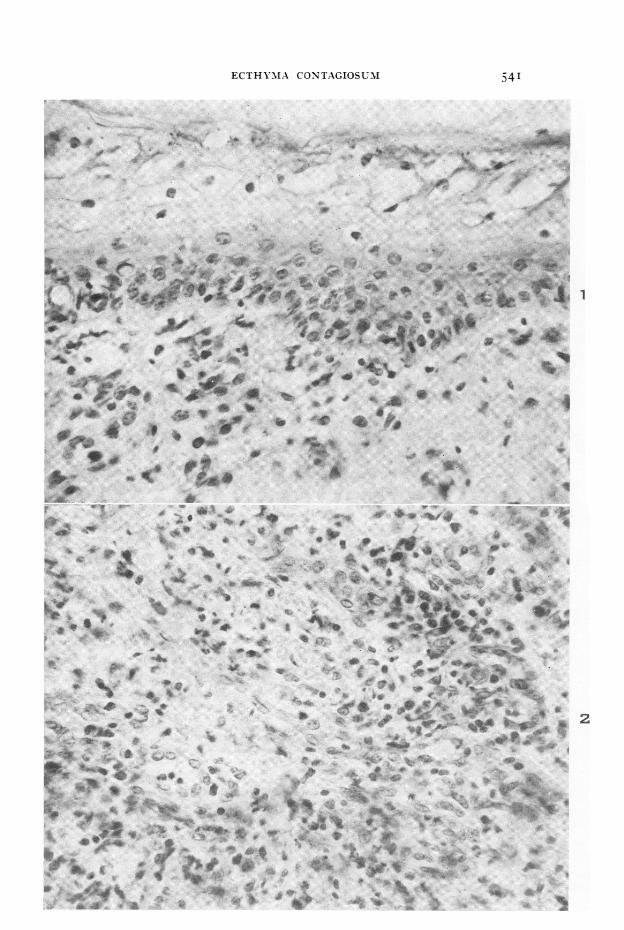
LEGENDS FOR FIGURESFIG. I. Sheep V. Five days after inoculation of the virus of ecthyma contagiosum.
Ballooning degeneration is present in the uppermost cells of the prickle layer andvascular changes and a sparse round-cell infiltrate can be seen in the dermis.X 445.
FIG. 2. Sheep II. Ten days after inoculation of the virus. The granulomatous changesin the dermis are well developed. X 445.
ECTHYMNIA CONTAGIOSUM4
9
1
! r ..
w
M ..'. |. ".t0.si
... ..qmA:'
2
541
WHEELER AND CAWLEY
N ,
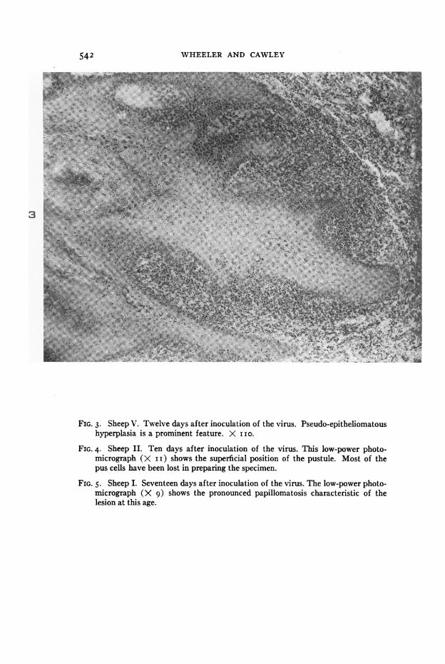
FIG. 3. Sheep V. Twelve days after inoculation of the virus. Pseudo-epitheliomatoushyperplasia is a prominent feature. X I IO.
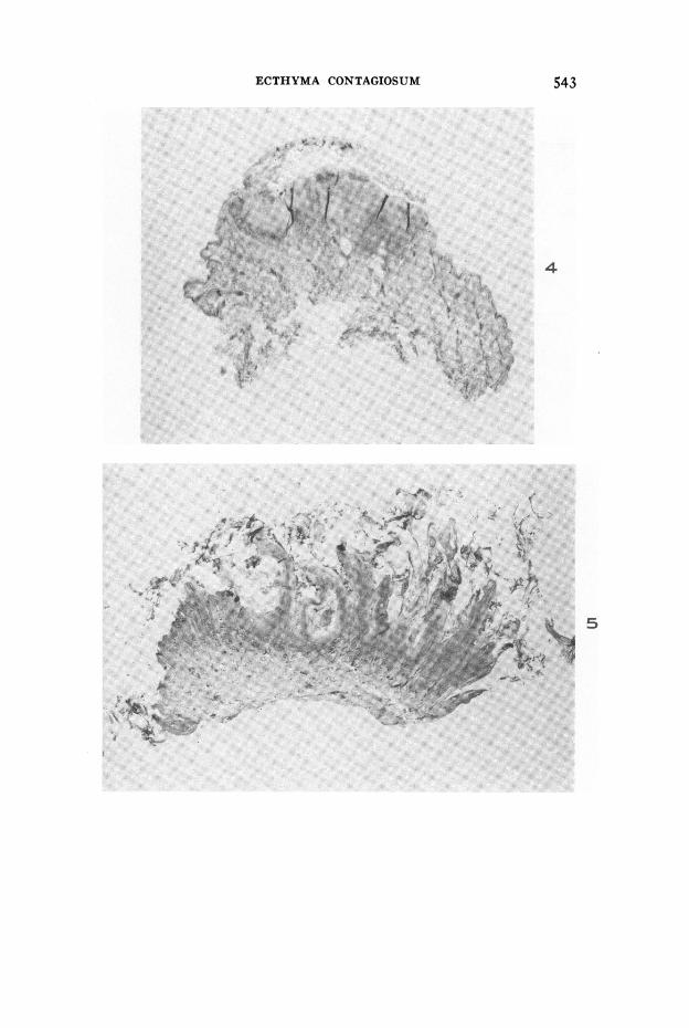
FIG. 4. Sheep II Ten days after inoculation of the virus. This low-power photo-micrograph (X i i) shows the superficial position of the pustule. Most of thepus cells have been lost in preparing the specimen.
FIG. 5. Sheep I. Seventeen days after inoculation of the virus. The low-power photo-micrograph (X 9) shows the pronounced papillomatosis characteristic of thelesion at this age.
542
ECTHYMA CONTAGIOSUM 543
....
-~~~~~~~~~~~~~~~~~~~~~-
5
WHEELER AND CAWLEY
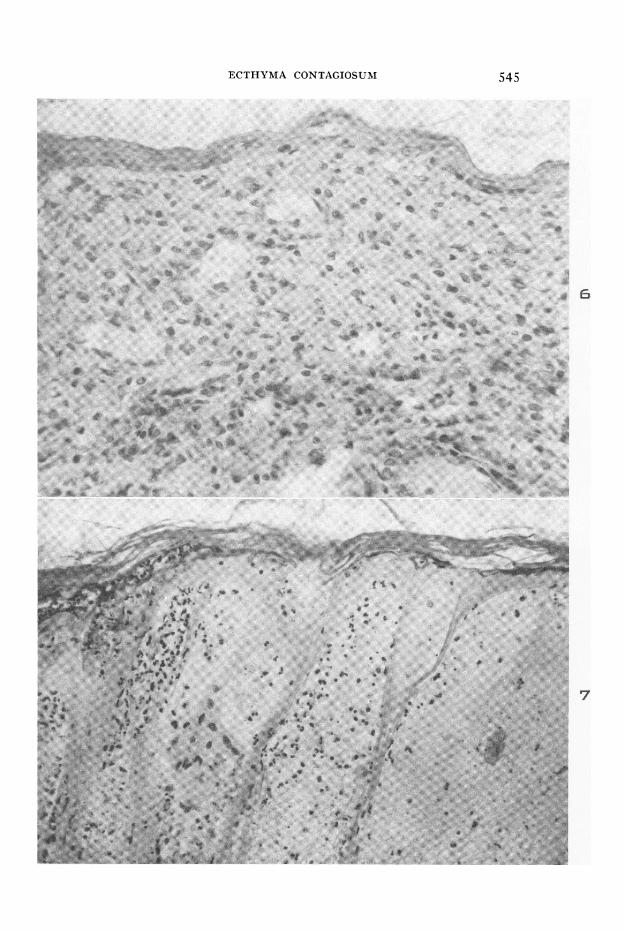
FIG. 6. Rabbit V. Third-passage lesion 9 days after inoculation of the virus. Thelesion is essentially a granuloma. X 445.
FIG. 7. Human lesion approximately 3 weeks after inoculation of the virus. Balloon-ing and reticular degeneration have resulted in a multilocular vesicle. X 270.
544
ECTHYMA CONTAGIOSUNI
6
7
545