Embed Size (px)
Citation preview
LUND UNIVERSITY
PO Box 117221 00 Lund+46 46-222 00 00
The coordination chemistry of the structural zinc ion in alcohol dehydrogenase studiedby ab initio quantum chemical calculations
Ryde, Ulf
Published in:European Biophysics Journal
DOI:10.1007/BF00205102
1996
Document Version:Peer reviewed version (aka post-print)
Link to publication
Citation for published version (APA):Ryde, U. (1996). The coordination chemistry of the structural zinc ion in alcohol dehydrogenase studied by abinitio quantum chemical calculations. European Biophysics Journal, 24(4), 213-221.https://doi.org/10.1007/BF00205102
Total number of authors:1
General rightsUnless other specific re-use rights are stated the following general rights apply:Copyright and moral rights for the publications made accessible in the public portal are retained by the authorsand/or other copyright owners and it is a condition of accessing publications that users recognise and abide by thelegal requirements associated with these rights. • Users may download and print one copy of any publication from the public portal for the purpose of private studyor research. • You may not further distribute the material or use it for any profit-making activity or commercial gain • You may freely distribute the URL identifying the publication in the public portal
Read more about Creative commons licenses: https://creativecommons.org/licenses/Take down policyIf you believe that this document breaches copyright please contact us providing details, and we will removeaccess to the work immediately and investigate your claim.
The coordination chemistry of the structural zinc ion
in alcohol dehydrogenase studied by ab initio quantum
chemical calculations
Ulf Ryde
Department of Theoretical Chemistry
University of Lund
Chemical Centre
P.O.B 124
S-221 00 Lund
Sweden
Tel: 46-46-2224502
Fax: 46-46-2224543
Email: [email protected]
Running title: The structural zinc ion in alcohol dehydrogenase
2
Abstract
The coordination chemistry of the structural zinc ion in horse liver alcohol
dehydrogenase has been examined by quantum chemical geometry optimisations. It is
shown that all the four cysteine ligands are deprotonated in the enzyme, not only two of
them as has been suggested. The Zn-S bond lengths are very sensitive to the theoretical
treatment; in vacuum they are predicted to be 15 pm longer than in the crystal structure.
Half of this discrepancy is due to electronic correlation, the rest can be attributed to
screening of the negative sulphide charges by the enzyme, in particular by N-H…S
hydrogen bonds. The potential surface is rather flat, so the large difference in geometry
between the crystal and the vacuum structure corresponds to an energy change of less than
35 kJ/mole. The crystal bond lengths can be reproduced only with methods that accounts
explicitly for the enzyme. A dielectric continuum model gives too long bond lengths,
indicating that the enzyme solvates the coordination sphere better than water. Thus, the
structural zinc ion can be used as a sensitive test of methods trying to model the
surrounding medium in quantum chemical computations.
3
Introduction
Alcohol dehydrogenase (EC 1.1.1.1) catalyses the reversible oxidation of primary
and secondary alcohols using NAD+ as coenzyme (Pettersson, 1987). Each subunit of the
dimeric enzyme contains two zinc ions, one that is essential in catalysis, and one that has
been suggested to play a structural role (Cedergren-Zeppezauer et al., 1985).
Crystallographic studies of the horse liver enzyme (Eklund et al., 1981, Eklund & Brändén,
1983, Al-Karadaghi et al., 1994, 1995) have shown that the latter zinc ion is bound to the
enzyme by four cysteine residues. As can be seen in Table 1, the coordination geometry
around this zinc ion is almost tetrahedral, with small but reproducible differences between
the ligands. A characteristic feature of the structure is the extensive hydrogen bonding to
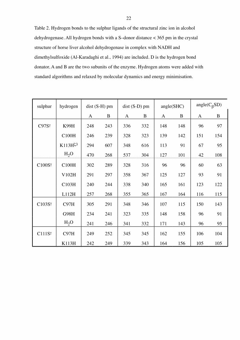
the cysteine sulphur atoms, illustrated in Fig. 1 and Table 2. Each sulphur atom interacts
with at least three polar hydrogens in the surrounding enzyme, mostly NH-amide groups of
the backbone. Similar hydrogen bonds have been observed in several other proteins
containing a metal bound to sulphur atoms, e.g. ferredoxins (Adman et al., 1975).
It is usually assumed (Eklund & Brändén, 1983, Pettersson, 1987) that all the four
cysteine zinc ligands are deprotonated, i.e. that the total charge of the coordination sphere
is –2 e0. Yet, Garmer and Krauss (1993) recently suggested that only two of the cysteine
residues are deprotonated on the basis of the interpretation of the electronic spectra of the
Co-substituted enzyme. From a theoretical point of view, such a proposal may seem
legitimate. The pKa of a free cysteine ligand is about 8.3. It is easy to conceive that for the
first two cysteines coordinating to the zinc ion, this pKa decreases to well below 7 through
the electrostatic interaction with the positively charged zinc ion. However, for the third and
forth ligands, the total charge of the zinc-complex is 0 and –1 e0, respectively, and it is then
less clear why the pKa should be significantly lowered. Furthermore, the lengths of the Zn-
S bonds are significantly different (221-246 pm), indicating that the bonds are not
equivalent, and they are also substantially longer than the average zinc-cysteine bonds in
protein crystal structures (212 pm according to Chakrabarti, 1989).
The structural zinc ion in alcohol dehydrogenase is well suited for theoretical
calculations. The enzyme has been thoroughly studied and the crystal structure is known
4
for several complexes (Eklund et al., 1981, Eklund & Brändén, 1983, Al-Karadaghi et al.,
1994, 1995). Furthermore, if all the cysteines are deprotonated, there are eight equivalent
Zn-S bonds in the enzyme, which gives a very accurate estimate of the average Zn-S bond
length. We therefore decided to estimate the number of deprotonated cysteine residues by
quantum chemical geometry optimisations. During the course of the calculations, it turned
out that the system is very sensitive to the surrounding medium and that it therefore can be
used as test of different methods to model the surrounding enzyme.
5
Methods
Quantum chemical vacuum computations
[Zn(HS)4-n(HS2)n,]–2+n where n=0-2, was chosen as a model of the structural zinc
ion and its ligands in alcohol dehydrogenase. The full geometry of the models was
optimised until the change in energy and the internal coordinates were below 2.6 J/mole
and 0.053 pm or 0.057°, respectively, using analytical gradient methods at the Hartree-
Fock self-consistent field (SCF) level. No symmetry restrictions were imposed and several
starting structures were tested to reduce the risk of being trapped in local minima. If not
otherwise stated, the results refer to optimisations with the double-z zinc basis
(62111111/33111/311) of Schäfer et al. (1992), enhanced with a p function with exponent
0.162 (called DZP), and the 6-31G* basis sets on all other atoms (Hehre et al., 1986). The
contaminant d and f functions were always removed. Electronic correlation effects were
included through second order perturbation theory (MP2), keeping the core orbitals frozen.
The calculations were performed using the quantum chemical program packages
TURBOMOLE 2.0b (Ahlrichs et al., 1989) and Mulliken 2.18d (MP2 geometry
optimisations, Rice, et al., 1995) on IBM RISC RS/6000 workstations.
Geometry optimisations in a dielectric cavity
Reaction field calculations (Tapia & Goscinski, 1975, Mikkelsen et al., 1988) were
performed by placing [Zn(HS)4]2– in a spherical cavity (radius r0), surrounded by a
dielectric medium with a dielectric constant, e. The charge distribution of the ligand sphere
introduces an electric field acting on the dielectric medium. This reaction field interacts
with the charge distribution of the ligand sphere and perturbs the one-electron Hamiltonian.
This perturbation is calculated by a multipole expansion of the electron distribution,
truncated after the fifth term. The Pauli repulsion due to the medium is treated by use of
spherical well integrals added to the one-electron Hamiltonian (a sum of four spherical
gaussian shell functions with exponents 5.0, 3.5, 2.0 and 1.4 at the radius r0 + 106, 159,
6
265, and 370 pm (r0 + 2, 3, 5, and 7 atomic units), respectively (Andersson, et al., 1994).
The origin of the cavity was at the centre of mass of the ligand sphere, which almost
coincide with the zinc ion. The radius of the cavity was optimised by minimising the total
energy of the system at a fixed geometry and e=80.0 and was found to be 133 pm plus the
average Zn-H distance. The optimal Zn-S bond lengths were then determined by varying
the mean Zn-S distance (and r0) stepwise at six different dielectric constants between 1.0
and 80.0, keeping the rest of the geometry fixed. The calculations were performed both at
the SCF and MP2 level using the program package MOLCAS-3 (Andersson et al, 1994). At
the MP2 level, the reaction field from SCF calculations at the same geometry was used.
This treatment is valid as long as the electron structure does not change much from SCF to
MP2, an approximation that is acceptable in these rather crude computations.
Combined quantum chemical and classical geometry optimisations
Integrated quantum chemical and molecular mechanical geometry optimisations
were performed using the program COMQUM (Ryde, 1996). In this program, the enzyme is
divided into four subsystems. The central system 1 is optimised using the sum of the
quantum chemical gradients within the system and molecular mechanical gradients from
system 2. All electrostatic interactions are included in the quantum chemical calculations;
system 2 is represented by partial charges, one for each atom, and system 3 and 4 by
integer charges, i.e. one charge for each charged amino acid, located at the position of the
Nz, Cz, Cg, Cd, Sg, Ce1, and both P atoms of Lys, Arg, Asp, Glu, Cys-, His+, and NADH,
respectively. The integer charges are damped by a dielectric constant e=4.0, while in
systems 1-3, e=1.0. In each step of the optimisation, system 2 is relaxed by molecular
mechanics (keeping the other systems fixed), representing system 1-3 with all atoms (using
charges obtained from a quantum chemical Mulliken analysis for system 1 and partial
charges for system 2 and 3), and system 4 by damped integer point charges. Special action
is taken at the junction between the classical and quantum chemical systems (Ryde, 1996).
7
The full geometry of system 1 and 2 was optimised until the change in energy and
the coordinates were below 0.26 kJ/mole and 0.53 pm, respectively. Then, system 2 was
fixed and the optimisation was continued until the changes were below 2.6 J/mole and
0.053 pm. In some cases, system 2 was kept fixed all the time and the tighter thresholds
were used. The quantum chemical computations were performed at the Hartree-Fock level
with the DZP/6-31G* basis sets. The effect of correlation was simulated by the method of
offset forces (Fogarasi et al., 1992). A factor of +0.010 atomic units was added to each Zn-
S bond (SCF–) gradient before the geometry relaxation. This factor is the average negative
gradient of the Zn-S bond with the same basis set at the MP2 optimised geometry in
vacuum.
The calculations were performed using the quantum chemical program TURBOMOLE
(Ahlrichs et al., 1989) combined with the molecular dynamics simulation package MUMOD
(Teleman & Jönsson, 1986, Ryde, 1995a). The potential function of the latter program
contains a harmonic potential for bond stretches and angle bending, a truncated
trigonometric series (n=1-3) for the dihedral angles, a Coulombic term for the electrostatic
interactions and a 6-12 Lennard-Jones potential for the van der Waals interactions. The
force field does not contain any specific terms for hydrogen bonds or improper dihedral
angles. The interactions between the zinc ion and its ligands were treated purely quantum
mechanically; in the molecular mechanical gradients the zinc terms cancel out and in the
classical optimisation of system 2 the zinc ion interacts only by a non-bonded potential.
The enzyme
Throughout, the coordinates of horse liver alcohol dehydrogenase in complex with
NADH and dimethylsulfoxide at 0.18 nm resolution were used (R-factor=0.172; Al-
Karadaghi et al., 1994). This is at present the most accurate structure of alcohol
dehydrogenase and it represents the catalytically active closed conformation of the
enzyme. Charge assignment was performed as described by Ryde (1995a). To determine
the positions of the hydrogen atoms and the solvent water structure around the structural
8
zinc ion, a series of classical simulations was performed on all amino acids within 0.3 nm
from any atom in system 3 (see below) plus a spherical cap of water molecules within 1.0
pm from the zinc ion: three 6 ps molecular dynamics simulations at 300, 100, and 0 K,
followed by a molecular mechanics optimisation until the change in energy and the norm
of the gradient were below 4.2.10-4 kJ/mole and 4.2.10-6 kJ/mole/pm, respectively. All
heavy atoms were kept fixed except the oxygen atoms of the solvent water molecules. The
non-bonded cut-off radius was 1.0 nm and the program package AMBER 4.0 (Pearlman et
al., 1991) was used.
In the integrated geometry optimisations, system 1 consisted of [Zn(SH)4]2–, as a
model of Zn, Cys-97, Cys-100, Cys-103, and Cys-111 (from subunit A of the enzyme). In
system 2, all amino acids within 0.6 nm from any atom in system 1 were included, viz.
Thr-94, Pro-95, Gln-96, Gly-98, Lys-99, Arg-101, Val-102, Lys-104, His-105, Asn-109,
Phe-110, Leu-112, Lys-113, Asn-114, Ile-155, Lys-323, 8 crystal water molecules and the
rest of four cysteine zinc ligands (totally 339 atoms). System 3 was composed of all atoms
of residues within 0.3 nm of any atom in system 2, viz. amino acids number 92-93, 106-
108, 115-117, 124, 153-154, 156-157, 318-319, 321-322, 324-327, amino acids number
258-259, 261, 283-286, 310 from the other subunit of the enzyme, and 28 crystal water
molecules, in total, 500 atoms. Finally, system 4 comprised 171 integer charges.
9
Results and Discussion
The protonation status of the structural zinc ion
In order to determine the number of deprotonated cysteine ligands of the structural
zinc ion in alcohol dehydrogenase, [Zn(HS)4-n(HS2)n]–2+n, with n=0-2, was optimised
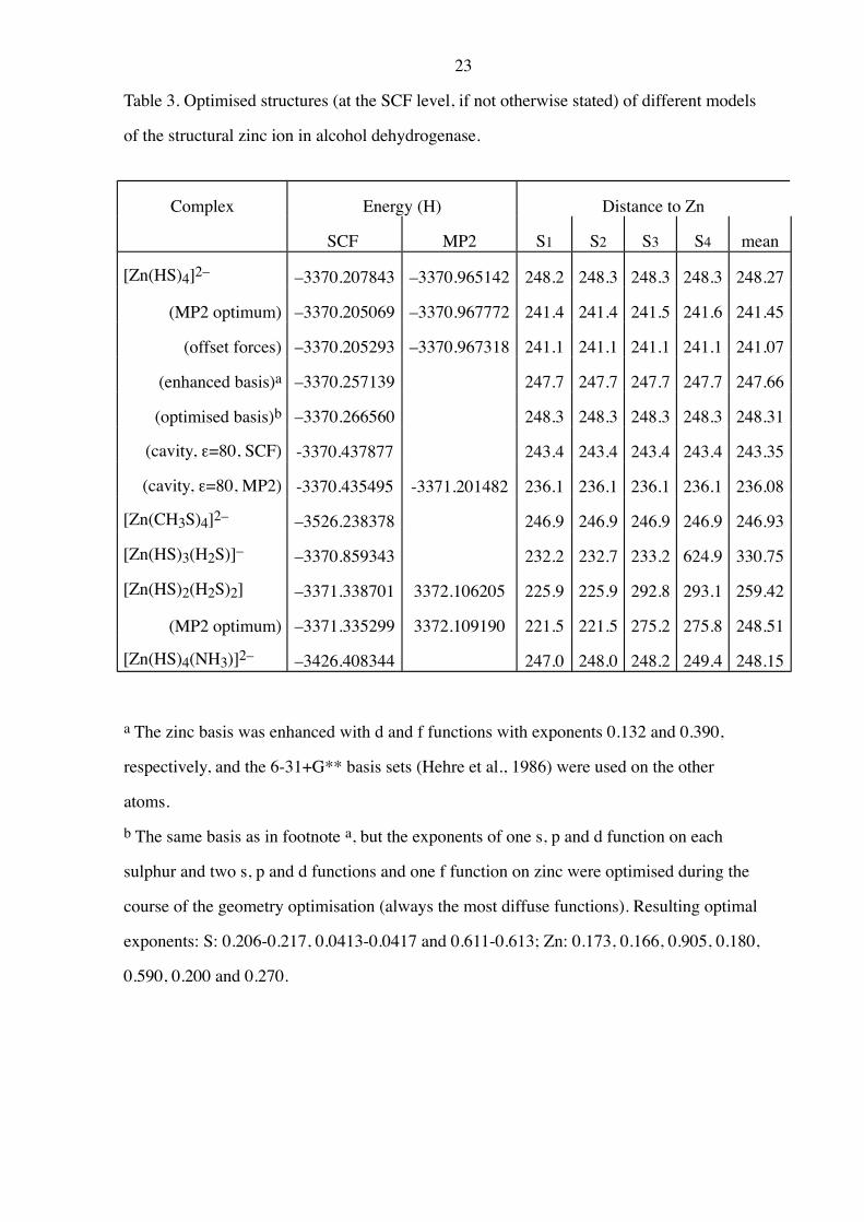
quantum chemically. As shown by the results in Table 3 and in Figure 2, [Zn(HS)4]2– is
four-coordinate with 248.3 pm Zn-S distances. All four hydrogen atoms form weak
hydrogen bonds to a sulphide ion (373 pm), which make the S-Zn-S bond angles different:
either 106° (the four SH…S pairs) or 117° (the two non-hydrogen-bonded pairs).
Zn(HS)2(H2S)2 in Figure 3 is also four-coordinate but with very dissimilar Zn-S
bond lengths; 226 pm for HS–, and 293 pm for H2S. This reflects that the interaction
between the zinc ion and HS– is much stronger than with H2S, due to the charge-charge
attraction. Analogous results were obtained with OH– and H2O at the catalytic zinc ion of
alcohol dehydrogenase (211 and 187 pm bonding distance, respectively, according to Ryde,
1994). The structure is a distorted tetrahedron with a large (148°) HS–-Zn-SH– angle.
[Zn(HS)3(H2S)]–, on the other hand, is three-coordinate with the three HS– ligands at
233 pm Zn-S distance and the H2S ligand in the second coordination sphere of the zinc ion
(625 pm from Zn), hydrogen bonded to a sulphur ion. This is due to the negative charges
on the HS– ligands, which make the total charge of the complex negative, thereby
destabilising the Zn-SH2 bond so much that the HSH…S hydrogen bond becomes
energetically more favourable. Similar lowering of the coordination number of Zn2+-
complexes when the total charge becomes negative has been observed in both experiments
(Irish et al., 1963, Cotton & Wilkinson, 1988), and theoretical calculations (Tossell, 1991,
Ryde, 1994). It was impossible to find any four-coordinate structure of [Zn(HS)3(H2S)]–;
no such local minimum exists in vacuum.
These results clearly show that all the cysteine ligands of the structural zinc ion in
alcohol dehydrogenase must be deprotonated. If only two of the cysteine ligands were
deprotonated as suggested by Garmer & Krauss (1993) the bond lengths would have been
10
very different (226-290 pm) and the angles would have been more distorted (92-148°). If
only one ligand was protonated, the differences would have been even larger.
Influence of the ligand choice, basis set, and correlation on the Zn-S bond lengths
Even if it is clear that all the cysteine ligands are deprotonated, there still remains
one problem: the calculated average Zn-S bond length in [Zn(HS)4]2– is 15 pm longer than
in the crystal. Therefore, more detailed calculations were performed on this complex. First,
the effect of improving the cysteine models was tested by replacing HS– with the more
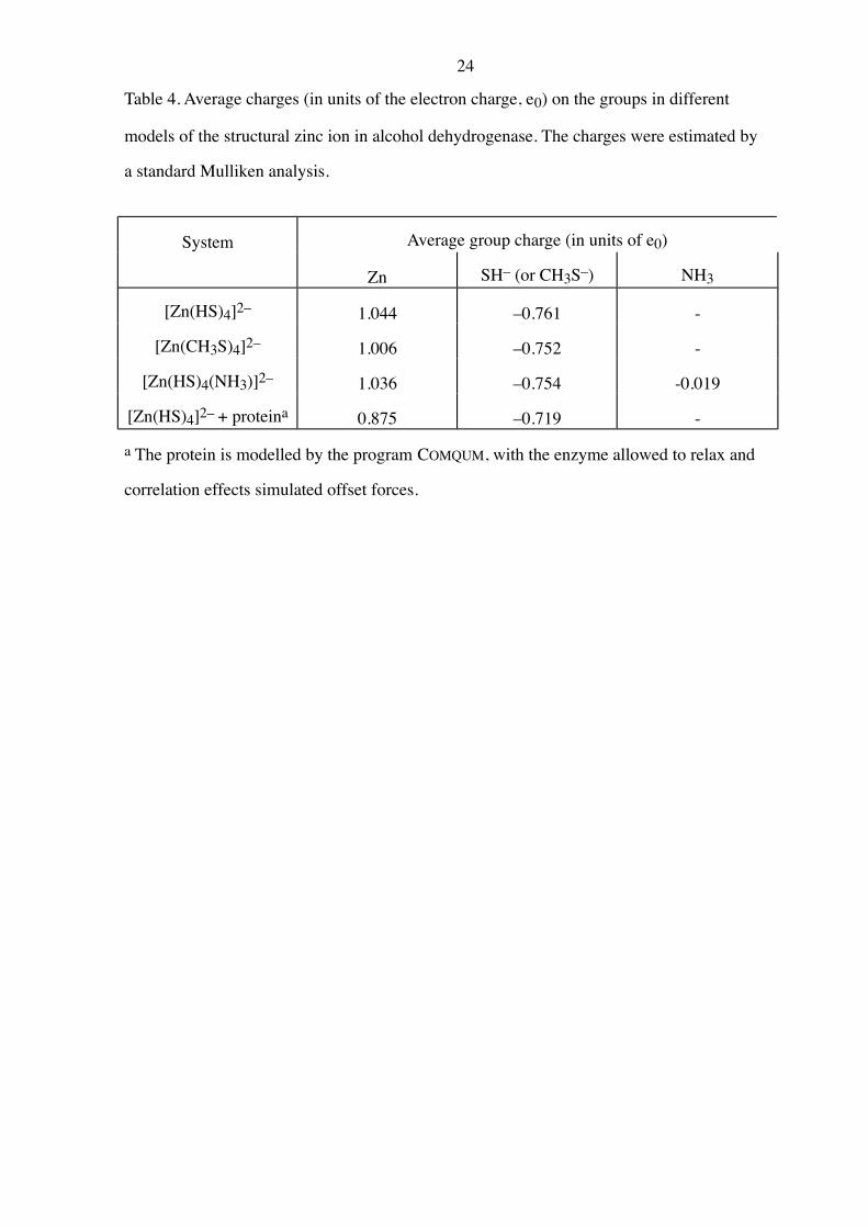
realistic, but also appreciably more expensive CH3S– ion. This ligand gave 1.4 pm shorter
Zn-S bond lengths (246.9 pm), probably due to a decreased charge on the CH3S– groups (–
0.752 e0 compared to –0.761 e0, see Table 4). In this structure there are twelve C-H…S
hydrogen bonds (243-245 pm), which are weaker than the HS…S bonds and therefore give
a difference in the S-Zn-S bond angles of only 1.8° (four 108.9° angles and two 110.7°).
Thus, the ligand choice has a small but significant influence on the Zn-S bonds, larger than
the one observed for the catalytic zinc ion in alcohol dehydrogenase (0.8 pm change
according to Ryde, 1994). This can probably be ascribed to the larger net charge of the
coordination sphere at the structural zinc ion.
Secondly, the basis sets were improved. It turned out that the basis sets do not affect
the geometry very much; the bond lengths obtained with basis sets enhanced with
optimised s, p, and d functions on sulphur and s, p, d, and f functions on zinc, differed by
less than 0.1 pm from the calculations with the normal basis set (Table 3).
Finally, the effect of electronic correlation was tested. A correct treatment of the
correlation has been shown to be crucial for the geometry of first-row open-shell transition
metal complexes (Langhoff & Bauschlicher, 1988, Lüthi et al, 1985), and it turned out to
be equally important also for zinc complexes. When the Zn-S bond lengths were optimised
at the MP2 level, the Zn-S bond lengths decreased by 6.8 pm to 241.5 pm. Thus,
correlation accounts for approximately half of the discrepancy between the calculated
structure and the crystal.
11
These calculations indicate that in vacuum, the Zn-S bond lengths of the structural
zinc coordination sphere should be about 240 pm, still 6 pm longer than in the enzyme.
Thus, the surrounding enzyme seems to shorten the Zn-S bonds by about 6 pm. A
prominent characteristic of the enzyme around the structural zinc ion in alcohol
dehydrogenase is the large number of hydrogen bonds to the cysteine sulphur ions in the
enzyme. We therefore tested the effect of adding a hydrogen bond donator, NH3, to the
second coordination sphere of the zinc complex. In the optimised structure (Table 3), the
NH3 molecule forms three hydrogen bonds to three sulphur ions (309-327 pm) and the
average Zn-S bond length decreases by 0.4 pm. This decrease is probably due to that the
electrons on the SH– groups are slightly shifted against the NH3 molecule, which leads to a
lowered effective charge on the SH– groups. Therefore, the effect is largest on the sulphur
ion that does not receive any NH-hydrogen bond. As can be seen in Table 4, the average
SH– charge decreases by 0.007 e0, which is compensated by a 0.010 e0 lower charge on the
zinc ion and -0.019 e0 net charge on the ammonia.
Geometry optimisations in a dielectric cavity
Most probably, the major factor decreasing the Zn-S bond lengths in the enzyme is
screening of the negative charges on the sulphur ions. In vacuum, these charges strongly
repel each other and push the sulphur ions away from the zinc ion. If the dielectric constant
was larger than 1.0, this repulsion would be smaller, and the Zn-S bond lengths would
decrease. A popular method to account in an average way for an increased dielectric
constant is the cavity reaction field approach (Tapia & Goscinski, 1975, Mikkelsen et al.,
1988). This technique is well suited for the present model system, since [Zn(HS)4]2– fits
well into a spherical cavity with little uncertainty in the location of the origin.
Reaction field calculations were performed on Zn(HS)4]2– by first determining the
optimal radius of the cavity (by energy minimisation) and then optimising the Zn-S bond
lengths at different dielectric constants. The method behaves well; the monopole term
accounts for 99.4 % of the total reaction field energy, while the dipole and octopole terms
12
account for 0.2 and 0.3 % respectively, indicating that the multipole expansion is well
converged. As expected, the Zn-S bond lengths decrease appreciably, about 5 pm, showing
that screening of the sulphide charges is important in the enzyme. Thus, the structural zinc
site is very polar, with an effective dielectric constant much larger than 3.0-4.0 as is usually
assumed for the enzyme interior (Sharp & Honig, 1990).
In fact, it turned out that even with an infinite dielectric constant of the surrounding
medium, the average Zn-S bond length could not become smaller than 236.0 pm, see
Figure 4 (243.3 pm at the SCF-level), which is still 2 pm longer than in the crystal. Thus,
the enzyme solvates the coordination sphere of the structural zinc ion better than water.
This is most probably due to detailed directed atomic interactions in the enzyme, and that
atoms can come in between the sulphur ions and screen the charges (i.e. that the model
system is not perfectly spherical).
Combined quantum chemical and molecular mechanic geometry optimisations
Apparently, a continuum model can in an average way model the screening of
charges around the structural zinc ion and alcohol dehydrogenase, yet it does not manage
to account for the effect fully, and obviously it can not reproduce the differences between
the ligands either. Therefore an approach that explicitly accounts for all the detailed
interactions in the enzyme was tested. In the program COMQUM by Ryde (1996), the course
of a quantum chemical geometry optimisation is influenced by classical forces exerted by
the enzymic environment. In this way, it is possible to quantify changes in geometry
induced by an enzyme onto a bounded subsystem. The effect of electronic correlation was
simulated by the offset force method, which has been successfully applied to many small
and medium sized molecules (Fogarasi et al., 1992). From Table 3, it can be seen that it is a
very effective technique to obtain a quasi MP2-quality geometry at the SCF level; the
average Zn-S bond length in the calculation using offset forces differs from the optimal
MP2 bond lengths by only 0.4 pm and the energy difference is 1 kJ/mole.
13
The average Zn-S bond length of the structural zinc ion optimised with COMQUM is
235 pm irrespective if the enzyme is allowed to relax or not (Table 5), i.e. very near the
bond length in the crystal. Again, this shortening of the Zn-S bond length correlate with a
lowered average charge on the SH– groups (–0.719 e0 compared to –0.761 e0, see Table 4),
and the SH– group with the shortest zinc distance also has the lowest charge. The S-Zn-S
angles are also well reproduced: 105, 121, 102, 103, 119, 104° compared to 106, 115, 102,
107, 119, 108° in the crystal (same order as in Table 1) and the calculated structure (Figure
5) is quite similar to the one found in the crystal. The S-Zn bond length of Cys-97 is
correctly predicted to be the longest one, and the calculations show that this can be
ascribed to the interaction of this group with the positively charged amine group of Lys113
(hydrogen bond distance 260 pm). This depicts the importance of hydrogen bonds from the
enzyme backbone amide groups to the cysteine sulphur ions. Hydrogen bonds from
dipoles, such as amide groups and water molecules shorten the Zn-S bonds, while
hydrogen bonds from positively charged groups lengthen them.
The simulations, however, fail to reproduce the very short Zn-S bond length of Cys-
103, and instead predict that Cys-111 should have the shortest bond length. The
explanation of this problem, and also of why the average Zn-S bond length still is about 0.8
pm too long is most probably the neglect of the polarisation of the classical system by the
quantum system. The energy of this polarisation can be approximately calculated after the
geometry optimisations (Epol in Table 5) and it turns out that it largely decreases with the
Zn-S bond length; the polarisation energy of the crystal structure is about 10 kJ/mole lower
than in any of the optimised systems. Thus, if this energy had been allowed to influence the
optimisation of the geometry, the Zn-S bonds had most probably become shorter.
Since the effect of correlation and the surrounding enzyme was so large on the
[Zn(HS)4]2– system, similar calculations were performed on Zn(HS)2(H2S)2 also. The
optimal Zn-S bond lengths decreased at the MP2-level to 221 and 275 pm. In the COMQUM
calculations, where Cys-97 and Cys-111 (which have the longest Zn-S bonds in the crystal)
were assumed to be protonated, the Zn-S bond lengths decreased further to 217 and 249-
266 pm (the offset forces were 0.0133 and 0.0056 au for the SH– and the SH2 groups,
14
respectively). The average bond length, 238 pm is rather similar to the one found in the
crystal, but the span of the bond lengths is all too large and the structure is more strained
than the structures with all cysteines deprotonated (88 kJ/mole compared to 35-55
kJ/mole). Consequently, correlation and the enzyme do not change the conclusion that all
the four cysteine ligands to the structural zinc ion in alcohol dehydrogenase are
deprotonated.
The strain induced by the enzyme onto the zinc coordination sphere
The importance of strain in enzyme structure and function has been much discussed
in the literature (e.g. Fraústo da Silvia & Williams, 1991, Warshel, 1991). The comparison
of a structure optimised quantum chemically in vacuum and with COMQUM provides an
estimate of the change in geometry and energy when it is inserted into the enzyme. Thus, ?
EQC1 in Table 5 is a measure of the strain forced by the enzyme onto the zinc coordination
sphere in alcohol dehydrogenase. This strain amounts to 35-55 kJ/mole for the structural
zinc ion. This is equal to the energy of one or two hydrogen bonds, and it is not more than
what was observed for the catalytic zinc ion in alcohol dehydrogenase (Ryde 1995b, 1996),
i.e. in energy terms the enzyme does not influence the structural zinc ion very much.
Nevertheless, the change in the geometry is rather large. Thus, the potential surface is
rather flat; shortening the Zn-S bond lengths the first 3 pm, for example, costs only 0.8
kJ/mole.
Warshel (1991) has strongly argued that strain plays a very modest role in enzyme
catalysis. By strain he then understands only contributions of bonds, bond angles, and
torsional deformations plus the repulsive van der Waals interaction (i.e. forces that vary
fast with small molecular deformation), as opposed to the electrostatic interactions (that
change slowly with distance). In order to quantify how much of the distortion of the zinc
geometry is due purely to electrostatics, another simulation with COMQUM was run with
the Ca, Cb, and Hb atoms of the four cysteine zinc ligands removed (thereby eliminating
the covalent strain effects; the charges were the same in both calculations). As can be seen
15
in Table 5, this change increases the average Zn-S bond length by only 1.5 pm, keeping the
trends between the different ligands. Yet, the strain energy ?EQC1 is only 9 kJ/mole. Thus,
strain in the restricted meaning of Warshel, dominates ?EQC1, while the electrostatic
interactions are responsible for most of the change in the geometry of the zinc coordination
sphere. This is an effect of the flatness of the potential surface; the strong and steep
covalent interactions are needed to force the Zn-S bond lengths the last 2 pm that costs
about 30 kJ/mole.
Concluding remarks
The present calculations undoubtedly show that all the cysteine ligands of the
structural zinc ion in alcohol dehydrogenase are negatively charged; [Zn(HS)4]2–, with an
appropriate treatment of electronic correlation and the enzymic environment, reproduces
the crystal geometry satisfactorily, while any other system with a total charge less than –2
e0 assume a structure very different from that found in crystals. The calculations also show
that the divergences among the Zn-S bond lengths in the crystal are due to differences in
the immediate neighbourhood of the ligand, i.e. the number and character of hydrogen
bonds to the ligand. The long average Zn-S bond length of the structural zinc ion,
compared to the average zinc-cysteine bond lengths in other proteins (Chakrabarti, 1989),
can be ascribed to the repulsion between the negative charges of the sulphur ions. In
analogy, it was found for the catalytic zinc ion in alcohol dehydrogenase that if the zinc-
bound water molecule was deprotonated, (giving a total charge of –1 e0), the distance to
zinc of the other ligands increased by 6-10 pm (Ryde, 1994). Consequently, it must be
concluded that the improvements on the electronic spectra observed by Garmer and Krauss
with the Co(SH)2(SH2)2 model of the structural zinc site in alcohol dehydrogenase were
only fortuitous.
Naturally, the fact that all the cysteine ligands of the structural zinc ion in alcohol
dehydrogenase are deprotonated strongly influences the physical properties of the enzyme.
16
The isoelectric point of the protein is a function of the number and acid constants of
ionisable residues in the protein (the total charge of the dimer at normal pH becomes about
+8 e0, instead of +12 e0 if only two of the ligands were deprotonated). Similarly, the
geometrical and spectral properties of native or substituted structural metal ion is directly
affected by the charge of the metal ligands. Several other properties may be indirectly
modulated through the electrostatic potential, e.g. the pKa values of nearby residues and
the stability and structure of the whole protein. Furthermore, a correct charge assignment
and a proper description of the electrostatic potential are absolutely essential for an
accurate description of the enzyme in theoretical simulations.
The results also have implications on other zinc proteins. Since the surrounding of
the structural zinc ion in alcohol dehydrogenase is not very special, it seems safe to assume
that all cysteine ligands are deprotonated also in other proteins with a four-coordinate zinc
ion. Four-coordinate zinc ions with several cysteine ligands are present in many enzymes,
e.g. aspartate carbamoyltransferase, methionine-tRNA synthetase, metallothionein (all
ZnCys4), and the zinc finger proteins (ZnCys2His2, ZnCys3His, and ZnCys4) (Fraústo da
Silvia & Williams, 1991, Brändén & Tooze, 1991). Such zinc ions are usually assumed to
play an important stabilising role as cross links in intracellular proteins (Fraústo da Silvia
& Williams, 1991).
The calculations clearly illustrate that danger of studying an isolated subsystem of a
larger structure in vacuum. In general, if the net charge of the system is high, the vacuum
geometry may be very different from the actual structure. An even more obvious example
of this is [ZnS4]–6 which has a Zn-S bond length of 235 pm in zincblende but dissociates in
vacuum calculations.
Our investigations have also shown that the coordination geometry of the structural
zinc ion is very sensitive to the theoretical method. A balanced treatment of correlation,
electrostatics and polarisation is needed to obtain a correct structure. Clearly, dielectric
continuum models are inappropriate to mimic the protein matrix. In fact, the probably most
important factor for the protein function is the detailed placement of charged groups and
dipoles in well-defined positions. Therefore, only methods that incorporate the details of
17
the enzyme structure during the geometry optimisation, such as QUEST (Singh & Kollman,
1986) and COMQUM, can be expected to give geometries that are relevant to the protein
environment.
Acknowledgements
This investigation has been supported by a grant from the Swedish Natural Science
Research Council (NFR) and by IBM Sweden under a joint study contract. The protein
coordinates were courteously provided by Eila Cedergren-Zeppezauer. The author is
thankful to Gunnar Karlström and Björn O. Roos for fruitful discussions.
18
References
Adman, E T, Watenpaugh, K D & Jensen, L H (1975) NH…S hydrogen bonds in
Peptococcus aerogenes ferredoxin, Clostridium pasteurianum rubredoxin, and
Chromatium high potential iron protein. Proc Nat Acad Sci USA 72:4854-4858,
Ahlrichs, R, Bär, M, Häser, M, Horn, H & Kölmel, C (1989) Electronic structure
calculations on workstation computers: The program system Turbomole. Chem
Phys Lett 162:165-169
Al-Karadaghi, S, Cedergren-Zeppezauer, E S, Petrantos, K, Hovmöller, S, Terry, H, Dauter,
Z & Wilson, K S (1994) Refined crystal structure of liver alcohol dehydrogenase-
NADH complex at 1.8 Å resolution. Acta Cryst D50:793-807
Al-Karadaghi, S, Cedergren-Zeppezauer, E S, Dauter, Z & Wilson, K S (1995) Refined
crystal structure of Cu-substituted alcohol dehydrogenase at 2.1 Å resolution. Acta
Cryst D51:805-813
Andersson, K, Blomberg, M R A, Fülscher, M P, Kellö, V, Lindh, R, Malmqvist, P-Å,
Noga, J, Olsen, J, Roos, B O, Sadlej, A J, Siegbahn, P E M, Urban, M, Widmark, P-
O (1994) MOLCAS version 3. University of Lund, Sweden
Brändén, C I, Tooze, J. (1991) Introduction To Protein Structure, Garland Inc., New York,
pp. 113-285.
Cedergren-Zeppezauer, E S, Andersson, I, Ottonello, S & Bignetti, E (1985) X-ray analysis
of structural changes induced by reduced nicotinamide adenine dinucleotide when
bound to cysteine-46-carboxymetylated liver alcohol dehydrogenase. Biochemistry
24:4000-4010
Chakrabarti, P (1989) Geometry of interaction of metal ions with sulfur-containing ligands
in protein structures. Biochemistry 28:6081-6085
Cotton, F A & Wilkinson, G (1988) Advanced inorganic chemistry, Wiley, New York
Eklund, H & Brändén, C-I (1983) The role of zinc in alcohol dehydrogenase. In: Spiro, T
G (ed) Zinc enzymes, John Wiley & Sons, New York, pp 124-153
19
Eklund, H, Samama, J-P, Wallén, L & Brändén, C I (1981) Structure of a triclinic ternary
complex of horse-liver alcohol dehydrogenase at 2.9 Å resolution. J Mol Biol
146:561-587
Fogarasi, G, Zhou, X, Taylor, P W & Pulay, P (1992) The calculation of ab initio molecular
geometries: efficient optimization by natural internal coordinates and empirical
correction by offset forces. J Am Chem Soc 114:8191-8201
Fraústo da Silvia, J J R & Williams, R J P (1991) The Biological Chemistry of the
Elements, Clarendon, Oxford, pp. 182-184
Garmer, D R & Krauss, M (1993) Ab initio quantum chemical study of the cobalt d-d
spectroscopy of several substituted zinc enzymes. J Am Chem Soc 115:10247-
10257
Hehre, W J, Radom, L, Schleyer, P v R & Pople, J A (1986) Ab initio molecular orbital
theory, Wiley-Interscience, New York, pp 251-260
Irish, D E, McCarroll, B & Young, T F (1963) Raman study of zinc chloride solutions. J
Chem Phys 39:3436-3444
Langhoff, S R & Bauschlicher, C W (1988) Ab initio studies of transition metal systems.
Annu Rev Phys Chem 39:181-212
Lüthi, H P, Siegbahn, P E M & Almlöf, J (1985) The effect of electron correlation on the
metal-ligand interaction in Fe(CO)5. J Phys Chem 89:2156-2161
Mikkelsen, K V, Ågren, H, Jensen, H J A & Helgaker, T (1988) A multiconfigurational self-
consistent reaction-field method. J Chem Phys 89:3086-3095
Pearlman, D A, Case, D A, Caldwell, J C, Seibel, G L, Singh, U C, Weiner, P & Kollman, P
A (1991) AMBER 4.0, University of California, San Francisco, U S A
Pettersson, G (1987) Liver alcohol dehydrogenase. CRC Crit Rev Biochem 21:349-389
Rice, J E, Horn, H, Lengsfiels, B H, McLean, A D, Carter, J T, Replogle, E S, Barnes, L A,
Maluendes, S A, Lie, GC, Gutwski, M, Rude, W E, Sauer, S P A, Lindh, R,
Andersson, K, Chevalier, T S, Widmark, P-O, Bouzida, D, PPacansky, G, Singh, K,
Gillan, C J, Carnevali, P, Swope, W C § Liu, B (1995) Mulliken™ Version 2.18d,
internal release, IBM Corporation, Almaden, USA.
20
Ryde, U (1994) The coordination chemistry of the catalytic zinc ion in alcohol
dehydrogenase studied by ab initio quantum chemical calculations. Int J Quant
Chem 52:1229-1243
Ryde, U (1995a) Molecular dynamics simulations of alcohol dehydrogenase with a varying
coordination number of the catalytic zinc ion. Proteins: Struct Funct Genet, 21:40-
56.
Ryde, U (1995b) On the role of Glu-68 in alcohol dehydrogenase. Protein Science, 4:1124-
1132
Ryde, U (1996) The coordination of the catalytic zinc ion in alcohol dehydrogenase studied
by combined quantum chemical and molecular mechanical calculation. J Comput-
Aided Molec Design, submitted
Schäfer, A, Horn, H & Ahlrichs, R (1992) Fully optimized contracted Gaussian basis sets
for atoms Li to Kr. J Chem Phys 97:2571-2577
Singh, U C & Kollman, P A (1986) A combined ab initio quantum mechanical and
molecular mechanical method for carrying out simulations on complex molecular
systems. J Comp Chem 7:718-730
Sharp, K A & Honig, B (1990) Electrostatic interactions in macromolecules: theory and
applications. Annu Rev Biophys Biophys Chem 19:301-332
Tapia, O & Goscinski, O (1975) The self-consistent reaction field theory of solvent effects.
Mol Phys 29:1653-1661
Teleman, O & Jönsson, B (1986) Vectorizing a general purpose molecular dynamics
simulation program. J Comp Chem 7:58-66
Tossell, J A (1991) Calculations of the structures, stabilities, and Raman and Zn NMR
spectra of ZnCln(OH2)a2-n species in aqueous solution. J Phys Chem 95:366-371
Warshel, A (1991) Computer modeling of chemical reactions in enzymes and solutions, J
Wiley & Sons, New York, pp 209-211
21
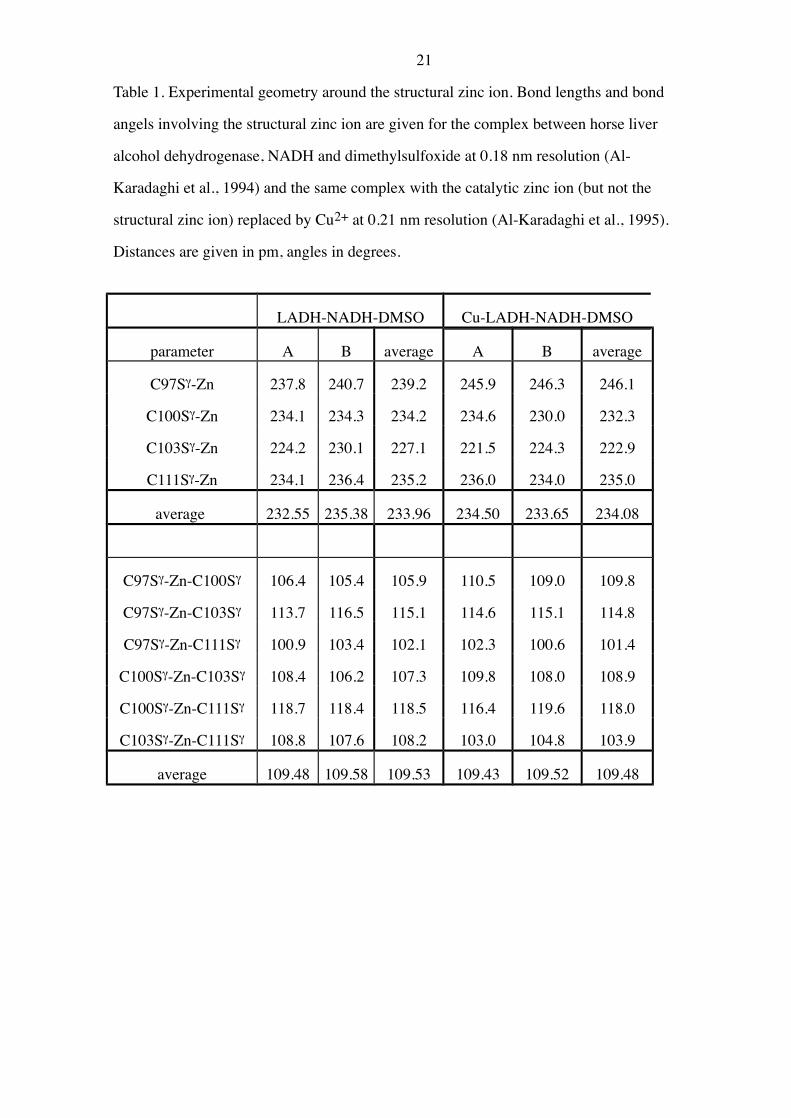
Table 1. Experimental geometry around the structural zinc ion. Bond lengths and bond
angels involving the structural zinc ion are given for the complex between horse liver
alcohol dehydrogenase, NADH and dimethylsulfoxide at 0.18 nm resolution (Al-
Karadaghi et al., 1994) and the same complex with the catalytic zinc ion (but not the
structural zinc ion) replaced by Cu2+ at 0.21 nm resolution (Al-Karadaghi et al., 1995).
Distances are given in pm, angles in degrees.
LADH-NADH-DMSO Cu-LADH-NADH-DMSO
parameter A B average A B average
C97Sg-Zn 237.8 240.7 239.2 245.9 246.3 246.1
C100Sg-Zn 234.1 234.3 234.2 234.6 230.0 232.3
C103Sg-Zn 224.2 230.1 227.1 221.5 224.3 222.9
C111Sg-Zn 234.1 236.4 235.2 236.0 234.0 235.0
average 232.55 235.38 233.96 234.50 233.65 234.08
C97Sg-Zn-C100Sg 106.4 105.4 105.9 110.5 109.0 109.8
C97Sg-Zn-C103Sg 113.7 116.5 115.1 114.6 115.1 114.8
C97Sg-Zn-C111Sg 100.9 103.4 102.1 102.3 100.6 101.4
C100Sg-Zn-C103Sg 108.4 106.2 107.3 109.8 108.0 108.9
C100Sg-Zn-C111Sg 118.7 118.4 118.5 116.4 119.6 118.0
C103Sg-Zn-C111Sg 108.8 107.6 108.2 103.0 104.8 103.9
average 109.48 109.58 109.53 109.43 109.52 109.48
22
Table 2. Hydrogen bonds to the sulphur ligands of the structural zinc ion in alcohol
dehydrogenase. All hydrogen bonds with a S–donor distance < 365 pm in the crystal
structure of horse liver alcohol dehydrogenase in complex with NADH and
dimethylsulfoxide (Al-Karadaghi et al., 1994) are included. D is the hydrogen bond
donator, A and B are the two subunits of the enzyme. Hydrogen atoms were added with
standard algorithms and relaxed by molecular dynamics and energy minimisation.
sulphur hydrogen dist (S-H) pm dist (S-D) pm angle(SHC) angle(CbSD)
A B A B A B A B
C97Sg K99H 248 243 336 332 148 148 96 97
C100H 246 239 328 323 139 142 151 154
K113Hz3 294 607 348 616 113 91 67 95
H2O 470 268 537 304 127 101 42 108
C100Sg C100H 302 289 328 316 96 96 60 63
V102H 291 297 358 367 125 127 93 91
C103H 240 244 338 340 165 161 123 122
L112H 257 268 355 365 167 164 116 115
C103Sg C97H 305 291 348 346 107 115 150 143
G98H 234 241 323 335 148 158 96 91
H2O 241 246 341 332 171 143 96 95
C111Sg C97H 249 252 345 345 162 155 106 104
K113H 242 249 339 343 164 156 105 105
23
Table 3. Optimised structures (at the SCF level, if not otherwise stated) of different models
of the structural zinc ion in alcohol dehydrogenase.
Complex Energy (H) Distance to Zn
SCF MP2 S1 S2 S3 S4 mean
[Zn(HS)4]2– –3370.207843 –3370.965142 248.2 248.3 248.3 248.3 248.27
(MP2 optimum) –3370.205069 –3370.967772 241.4 241.4 241.5 241.6 241.45
(offset forces) –3370.205293 –3370.967318 241.1 241.1 241.1 241.1 241.07
(enhanced basis)a –3370.257139 247.7 247.7 247.7 247.7 247.66
(optimised basis)b –3370.266560 248.3 248.3 248.3 248.3 248.31
(cavity, e=80, SCF) -3370.437877 243.4 243.4 243.4 243.4 243.35
(cavity, e=80, MP2) -3370.435495 -3371.201482 236.1 236.1 236.1 236.1 236.08
[Zn(CH3S)4]2– –3526.238378 246.9 246.9 246.9 246.9 246.93
[Zn(HS)3(H2S)]– –3370.859343 232.2 232.7 233.2 624.9 330.75
[Zn(HS)2(H2S)2] –3371.338701 3372.106205 225.9 225.9 292.8 293.1 259.42
(MP2 optimum) –3371.335299 3372.109190 221.5 221.5 275.2 275.8 248.51
[Zn(HS)4(NH3)]2– –3426.408344 247.0 248.0 248.2 249.4 248.15
a The zinc basis was enhanced with d and f functions with exponents 0.132 and 0.390,
respectively, and the 6-31+G** basis sets (Hehre et al., 1986) were used on the other
atoms.
b The same basis as in footnote a, but the exponents of one s, p and d function on each
sulphur and two s, p and d functions and one f function on zinc were optimised during the
course of the geometry optimisation (always the most diffuse functions). Resulting optimal
exponents: S: 0.206-0.217, 0.0413-0.0417 and 0.611-0.613; Zn: 0.173, 0.166, 0.905, 0.180,
0.590, 0.200 and 0.270.
24
Table 4. Average charges (in units of the electron charge, e0) on the groups in different
models of the structural zinc ion in alcohol dehydrogenase. The charges were estimated by
a standard Mulliken analysis.
System Average group charge (in units of e0)
Zn SH– (or CH3S–) NH3
[Zn(HS)4]2– 1.044 –0.761 -
[Zn(CH3S)4]2– 1.006 –0.752 -
[Zn(HS)4(NH3)]2– 1.036 –0.754 -0.019
[Zn(HS)4]2– + proteina 0.875 –0.719 -
a The protein is modelled by the program COMQUM, with the enzyme allowed to relax and
correlation effects simulated offset forces.
25
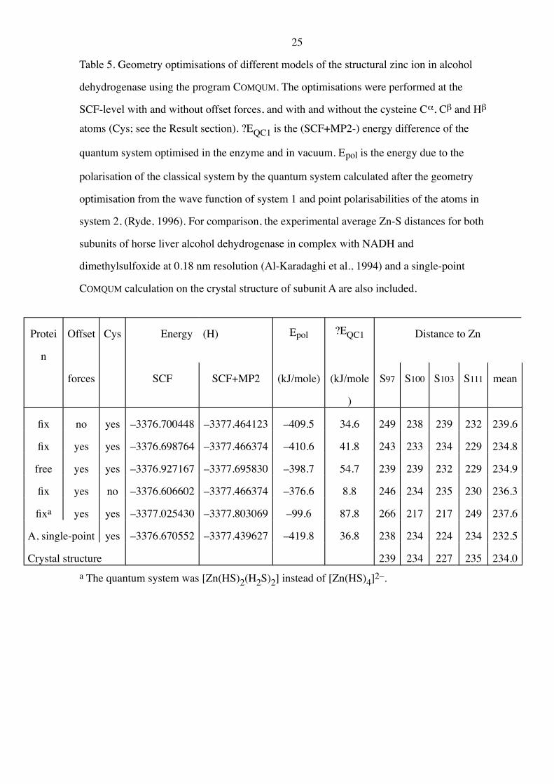
Table 5. Geometry optimisations of different models of the structural zinc ion in alcohol
dehydrogenase using the program COMQUM. The optimisations were performed at the
SCF-level with and without offset forces, and with and without the cysteine Ca, Cb and Hb
atoms (Cys; see the Result section). ?EQC1 is the (SCF+MP2-) energy difference of the
quantum system optimised in the enzyme and in vacuum. Epol is the energy due to the
polarisation of the classical system by the quantum system calculated after the geometry
optimisation from the wave function of system 1 and point polarisabilities of the atoms in
system 2, (Ryde, 1996). For comparison, the experimental average Zn-S distances for both
subunits of horse liver alcohol dehydrogenase in complex with NADH and
dimethylsulfoxide at 0.18 nm resolution (Al-Karadaghi et al., 1994) and a single-point
COMQUM calculation on the crystal structure of subunit A are also included.
Protei
n
Offset Cys Energy (H) Epol ?EQC1 Distance to Zn
forces SCF SCF+MP2 (kJ/mole) (kJ/mole
)
S97 S100 S103 S111 mean
fix no yes –3376.700448 –3377.464123 –409.5 34.6 249 238 239 232 239.6
fix yes yes –3376.698764 –3377.466374 –410.6 41.8 243 233 234 229 234.8
free yes yes –3376.927167 –3377.695830 –398.7 54.7 239 239 232 229 234.9
fix yes no –3376.606602 –3377.466374 –376.6 8.8 246 234 235 230 236.3
fixa yes yes –3377.025430 –3377.803069 –99.6 87.8 266 217 217 249 237.6
A, single-point yes –3376.670552 –3377.439627 –419.8 36.8 238 234 224 234 232.5
Crystal structure 239 234 227 235 234.0
a The quantum system was [Zn(HS)2(H2S)2] instead of [Zn(HS)4]2–.
26
Legends to the figures
Figure 1. Stereo view of horse liver alcohol dehydrogenase around the structural zinc ion,
showing the extensive hydrogen bonding to the cysteine sulphur ions. Data from the
crystal structure of the complex between horse liver alcohol dehydrogenase, NADH
and dimethylsulfoxide at 0.18 nm resolution (Al-Karadaghi et al., 1994) with
hydrogen atoms and water molecules inserted as described in the Methods.
Figure 2. Stereo view of the optimal structure of [Zn(HS)4]2– at the MP2 level in vacuum.
Figure 3. Stereo view of the optimal structure of Zn(H2S)2(HS)2 at the MP2 level in
vacuum.
Figure 4. The optimal average Zn-S bond length as a function of the dielectric constant e of
the dielectric medium surrounding a cavity enclosing [Zn(HS)4]2– in the reaction
field calculations. The upper curve is calculated at the SCF level, the lower curve at
the MP2 level. The fitted curves are (243.3 + 4.83/e) pm and (236.0 + 4.35/e) pm.
Figure 5. Stereo view of the structural zinc ion, optimised by COMQUM (free enzyme with
offset forces). All amino acids in System 1-2 are shown and the structure is
compared to the crystal structure of the complex between horse liver alcohol
dehydrogenase, NADH and dimethylsulfoxide at 0.18 nm resolution (no hydrogen
atoms; Al-Karadaghi et al., 1994).