Embed Size (px)
Citation preview

LUND UNIVERSITY
PO Box 117221 00 Lund+46 46-222 00 00
The coordination chemistry of the catalytic zinc ion in alcohol dehydrogenase studiedby ab initio quantum chemical calculations
Ryde, Ulf
Published in:International Journal of Quantum Chemistry
DOI:10.1002/qua.560520508
1994
Document Version:Peer reviewed version (aka post-print)
Link to publication
Citation for published version (APA):Ryde, U. (1994). The coordination chemistry of the catalytic zinc ion in alcohol dehydrogenase studied by abinitio quantum chemical calculations. International Journal of Quantum Chemistry, 52(5), 1229-1243.https://doi.org/10.1002/qua.560520508
Total number of authors:1
General rightsUnless other specific re-use rights are stated the following general rights apply:Copyright and moral rights for the publications made accessible in the public portal are retained by the authorsand/or other copyright owners and it is a condition of accessing publications that users recognise and abide by thelegal requirements associated with these rights. • Users may download and print one copy of any publication from the public portal for the purpose of private studyor research. • You may not further distribute the material or use it for any profit-making activity or commercial gain • You may freely distribute the URL identifying the publication in the public portal
Read more about Creative commons licenses: https://creativecommons.org/licenses/Take down policyIf you believe that this document breaches copyright please contact us providing details, and we will removeaccess to the work immediately and investigate your claim.

87
The coordination chemistry of the catalytic zinc ion in
alcohol dehydrogenase studied by ab initio quantum
chemical calculations
by
Ulf Ryde
Department of Theoretical Chemistry
University of Lund
Chemical Centre
P.O.B 124
S-221 00 Lund
Sweden
Tel: 46-46-104502
Fax: 46-46-104543
Email: [email protected]

Keywords: geometry optimisation, reaction mechanism, ligand exchange, five-coordination,
zinc complexes

Abstract
The coordination chemistry of the zinc ion in the active site of alcohol dehydrogenase
has been studied by the ab initio Hartree-Fock method. Geometry optimisations were
performed using analytical gradients and basis sets of double zeta quality. Correlation effects
were included at the MP2 level. The active site was modelled by Zn(HS)2XL(H2O)0-2, where
X denotes ammonia or imidazole and L denotes water, methanol, ethanol or the
corresponding aldehydes or anions. It is shown that with uncharged L-ligands the four-
coordinate complexes are about 20, 17 and 40 kJ/mole more stable than the corresponding
three-, five- and six-coordinate complexes, respectively. If the L-ligand is negatively charged
only the four-coordinate complexes are stable. These results suggest that the active-site zinc
ion in alcohol dehydrogenase prefers a coordination number of four during the catalytic
reaction, especially when the non-protein ligand is negatively charged. Ligand exchange at
the zinc ion is likely to proceed by an associative mechanism with intermittent formation of a
five-coordinate complex. The results lend no support to mechanistic proposals attributing an
important catalytic role to a negatively charged five-coordinate hydroxide or alkoxide ligand.

Introduction
Alcohol dehydrogenase (EC 1.1.1.1) catalyses the reversible oxidation of primary and
secondary alcohols using NAD+ as coenzyme [1-3]. The active site of the enzyme contains a
zinc ion that is essential for catalysis. Crystallographic studies [4-6] have shown that this zinc
ion is bound by the enzyme through one histidine and two cysteine residues. In free enzyme,
the catalytic zinc ion appears to be tetrahedrally coordinated with a water molecule (or
hydroxide ion, depending on pH) as the fourth first-sphere ligand.
Kinetic pH-dependence studies of individual reaction steps in the mechanism of action
of alcohol dehydrogenase led Kvassman and Pettersson [7] to propose that substrates are
bound to catalytic zinc in the enzyme-coenzyme complexes with displacement of zinc-bound
water. According to their mechanistic proposal, deprotonation of the enzyme-bound alcohol
represents a catalytically crucial step facilitating subsequent hydride transfer from the
substrate to NAD+. Alternative proposals have been put forward, however, according to
which five-coordinate intermediates may play an essential role during catalysis [8-14]. In
particular, a five-coordinate hydroxide ion has been suggested to facilitate hydride transfer
from zinc-bound alcohols to NAD+.
Crystallographic studies of the enzyme and its binary or ternary complexes with
coenzyme and different substrates have shown that the catalytic zinc ion as a rule exhibits
four-coordination [4-6] and several spectroscopic investigations have provided evidence in
the same direction [15-17]. On the other hand, there is also strong crystallographic and
spectroscopic evidence showing that binding to zinc of certain bidentate inhibitors is five-
coordinate [18-20]. Furthermore, spectroscopic studies of metal-substituted alcohol
dehydrogenase have indicated that several binary and ternary complexes may be five-
coordinate [11-13,20-22]. The kinetic evidence is also scattered and has been taken to favour
four-coordination [3,23], as well as five-coordination [8-10], of zinc in the catalytically
productive ternary-complexes.
The present investigation was undertaken to obtain mechanistically relevant
information on the coordination number of the catalytic zinc ion in alcohol dehydrogenase by

a quantum chemical approach. Geometry optimisations have been performed on a model of
the active site with a varied number of different non-protein ligands of biological interest. The
results would seem to significantly advance our understanding of the factors governing the
coordination structure of the catalytic metal site in free enzyme, as well as during catalysis,
and hence provide several inferences of interest regarding the mechanism of action of alcohol
dehydrogenase.
Methods
Zn(HS)2XL(H2O)0-2, where X denotes NH3 or imidazole and L denotes water,
methanol, ethanol or the corresponding aldehydes or anions, was chosen as a model of the
active site of alcohol dehydrogenase. The full geometry of these models were optimized until
the change in energy and the coordinates were below 10-6 Hartree and 10-3 Bohr,
respectively, using analytical gradient methods in the self-consistent field (SCF) Hartree-Fock
approximation. Electron correlation effects were included through second order perturbation
theory (Møller-Plesset, MP2), by single point calculations on the optimized structure.
Thermodynamic corrections, i.e. the translational, rotational and vibrational (including zero-
point energy) contributions to the Helmholtz free energy at 300 K and 101.3 kPa pressure,
were calculated from the coordinates, atomic masses and vibrational frequencies of the
systems, the latter uniformly scaled by a factor 0.9 [24]. All reported energies are total free
energy (SCF + MP2 + thermodynamic corrections) unless otherwise stated. Several starting
structures were tested to reduce the risk of being trapped in local minima and no symmetry
restrictions were imposed. The Hessian matrix of each structure was examined to ensure that
it represents a true potential minimum. The basis sets were of double zeta quality for all
atoms (H: (31); C, N, O: (5111/31); S: (521111/4111); Zn: (62111111/51111/311)) [25]. All
calculations were performed using the semi-direct program package TURBOMOLE [26] on an
IBM RISC RS/6000 (530H) workstation.

Results and Discussion
Choice of the model system
The optimized geometry of Zn(imidazole)(SH)2(H2O) is shown in Figure 1a. The
geometry of this complex is similar to those previously reported for other model systems of
the active site of alcohol dehydrogenase examined with smaller basis sets and ammonia
instead of imidazole [27-28]. The geometry is also very similar to the crystallographic
structure of the active site of alcohol dehydrogenase [5], shown in Figure 1b, with a root-
mean-square deviation between the two structures of 27 pm. The model system may,
therefore, be expected to provide reliable inferences as to the coordination around the zinc
ion. The similarity in geometry further indicates that the enzyme does not impose any major
geometric constraints on the zinc ligands, which justifies the use of full geometry
optimisations and indicates that to a first approximation no more enzyme residues need to be
included in the model.
Fig 1c shows the optimized geometry of Zn(NH3)(SH)2(H2O), where imidazole in the
model above has been exchanged for ammonia. The charge distribution of these two
complexes (according to a Mulliken population analysis) is similar, as is the geometry; the
root-mean-square deviation is only 8.4 pm. To reduce computational costs, therefore, most
calculations were performed using ammonia as a model for the histidinyl zinc ligand. Critical
structures were examined also with imidazole.
The energy, coordination number and zinc-ligand distances of all examined complexes
are recorded in Table I.
The coordination number of the model system
In order to obtain information on the relative stability of four- and five-coordinate
complexes, a fifth ligand was added to the system in Fig. 1c. This ligand may be placed in the
first- or second coordination sphere of the zinc ion. With uncharged ligands, there are several

stable structures (local minima) of each kind. Fig 2a shows the optimized geometry of the
most stable five-coordinate structure of Zn(NH3)(SH)2(H2O)2, a quasi-trigonal bipyramidal
structure with the two water molecules as axial ligands. Fig. 2b shows the most stable four-
coordinate structure of the same complex, with one water ligand in the second coordination
sphere of the zinc ion, hydrogen bonded to a sulphur ion and a hydrogen of the first-sphere
water. Analogous structures were obtained with all combinations of uncharged water, alcohol
and aldehyde ligands, as well as when ammonia was exchanged for imidazole.
In Table II, the energy of these four- and five-coordinate structures with different
ligands are compared. It can be seen that the four-coordinate structures are 16-22 kJ/mole
more stable than the corresponding five-coordinate structures (for Zn(imidazole)(SH)2(H2O)2,
the four-coordinate structure is 22.7 kJ/mole more stable than the five-coordinate one at the
SCF level). With two different ligands (e.g. water and methanol) there are two possible four-
coordinate complexes, depending on which ligand is in the first coordination sphere and
which is in the second. It can be seen from Table II that with one alcohol and one water
ligand, the structure with the alcohol in the first coordination sphere is most stable, while with
aldehydes the structure with the aldehyde in the second sphere is much more stable. A stable
planar three-coordinate structure of the complex in Fig. 2 with the two water ligands in the
second coordination sphere of the zinc ion, was also examined. This structure, however, was
appreciably less stable than the corresponding four- and five-coordinate complexes (43.0 and
27.1 kJ/mole).
For Zn(NH3)(SH)2(H2O)3, three different kinds of complexes were compared: five-
and four- coordinate structures with one or two water molecules in the second coordination
sphere and a six-coordinate octahedral structure. The most stable structure of each kind is
shown in Fig. 3 and the relative stability of these complexes and their methanol or
formaldehyde analogues are given in Table III. Again, the four-coordinate complexes, with
both second-sphere ligands hydrogen bonded to a sulphur and a water or alcohol hydrogen,
are about 40 and 25 kJ/mole more stable than the corresponding six- and five-coordinate

structures. No stable complexes with more than six first-sphere zinc ligands could be
obtained.
When one of the water molecules in the five-coordinate complex in Fig. 2a was
deprotonated, the second molecule was pushed into the second coordination sphere (Fig 4).
As shown by the potential curve for the binding of a water molecule to Zn(NH3)(SH)2(OH)-
in Fig 5, there is no local energy minimum representing a negatively charged five-coordinate
complex. Similar results were obtained for every tested combination of five and six ligands of
which one was a hydroxyl or alkoxide ion, as well as when ammonia was exchanged for
imidazole. The only exception from this behaviour was observed for Zn(NH3)(SH)2(OH)
(CH3OH)- and its ethanol analogue. For these two species apparently stable, distorted (all
zinc-ligand distances are longer than typical for a five-coordinate complex, especially one Zn-
S distance) five-coordinate complexes were obtained (c.f. Table I). Yet, they were 77.1
kJ/mole less stable than the corresponding four-coordinate complexes with the alcohol in the
second coordination sphere, and are therefore most certainly of minor significance.
Binding energy of the ligands
Tables IV and V give the binding energy of ligands in the first and second coordination
sphere to the model of the active site at different coordination numbers. It can be seen that the
binding energy for four-coordinate water and alcohol ligands in the first coordination sphere
is about 45 kJ/mole, about 400 kJ/mole for hydroxyl ion and about 300 kJ/mole for alkoxide
ions. The binding energy of uncharged ligands decreases with about 15 and 15 kJ/mole when
the coordination number increases from four to five and from five to six. When ammonia is
exchanged for imidazole the SCF binding energy is lowered by about 10 and 50 kJ/mole for
uncharged and charged ligands, respectively (about 10%). Alcohols bind slightly stronger
than water at the SCF level (5 kJ/mole) but the difference is cancelled by the thermodynamic
corrections. The hydroxyl ion binds appreciably (about 100 kJ/mole) stronger than the
alkoxide ions.

The binding energy of aldehydes is much lower than for the corresponding alcohols and
also more strongly dependent on the size of the hydrocarbon side chain (acetaldehyde binds
much stronger than formaldehyde, while ethanol binds with about the same strength as
methanol). This is in accord with experimental observations that aldehydes bind much weaker
to the zinc ion than the corresponding alcohols and that the binding is more dependent on the
size of the side chain [1,3,29]. The binding energy of formaldehyde is only 10 kJ/mole, which
indicates that formaldehyde hardly binds at all to alcohol dehydrogenase in water solution
(the equilibrium binding constant in water should be about 10-8 M-1). These observations
provides a theoretical explanation for the experimental observation [1] that formaldehyde is a
very poor substrate for alcohol dehydrogenase.
The binding energy of ligands in the second coordination sphere (Table V) is strongly
dependent on the type and geometry of the hydrogen bonds formed. Hydrogen bonds to
hydroxide or alkoxide ions are strongest and yield binding energies (50-75 kJ/mole) higher
than those of four-coordinate first-sphere zinc ligands. The binding energy of second-sphere
ligands involved in hydrogen bonds to a sulphide ion and a water hydrogen (35-45 kJ/mole)
is similar to that of first-sphere zinc ligands. Second-sphere ligands with hydrogen bonds to
water oxygen and ammonia hydrogen have binding energies (about 25 kJ/mole) similar to
those of five-coordinate zinc ligands. Water and alcohols in the second coordination sphere
bind with about the same strength, while aldehydes, which lack oxygen-bond hydrogens and
therefore have to interact by the carbonyl oxygen and aliphatic hydrogens, have about 20-30
kJ/mole lower binding energies.
Zinc-ligand distances
The differences in binding energy of different ligands is paralleled by differences in the
corresponding zinc-ligand distances (Table I). The Zn-O distance for alcohols are slightly (1
pm) shorter than for water, and appreciably shorter than for aldehydes (about 7 and 12 pm for
formaldehyde and acetaldehyde, respectively). The hydroxide ion has shorter Zn-O distance

than the methoxide and ethoxide ion, but the difference less than 1 pm. For second sphere
ligands, again the alcohols and water have almost the same Zn-O distance, while the bond
length of aldehydes are about 40 pm longer. The Zn-O distance for anions increases 3 pm for
each additional second-sphere zinc ligand, while for uncharged ligands, there are no clear
correlation of the Zn-O distance and the coordination number of the zinc ion. The Zn-S
distance increases 4, 7 and 12 pm when the number of first-sphere ligands are increased from
three to six, while the Zn-N distance is almost constant at about 212-217 pm. Both the Zn-N
and the Zn-S distance are strongly dependent on the charge and the type (water, alcohol or
aldehyde) of the non-protein ligands. Model systems with imidazole have 4-8 pm shorter Zn-
N distances, 2-6 pm longer Zn-S distances and 0-6 pm shorter Zn-O distances than the
corresponding systems containing ammonia.
Comparison with experiments
The present results show that the model system prefers (by at least 16 kJ/mole) four-
coordination for all tested combinations of up to six ligands. This is somewhat unexpected,
considering that the zinc ion forms six-coordinate complexes with most ligands in aqueous
solution [30]. The lower coordination number of the metal in the model system might to some
extent reflect the bulkiness of the two sulphide ligands, but can primarily be attributed to the
negative charges of these ligands which render the model system uncharged as a whole. This
suggestion is in accord with previous reported experimental [30,31] and theoretical [32]
evidence showing that the coordination number of zinc in hydroxide or halogenide solutions
decreases from six to four as the net charge of the complex decreases from +2 to –2, as well
as with the present observation that no stable five- or six-coordinate complexes exist when
one of the non-protein ligands in the model system is negatively charged (yielding a net
charge of -1).
Crystallographic studies of native alcohol dehydrogenase invariably have shown that
the catalytic zinc ion is four-coordinate in free enzyme and in enzymic complexes with

monodentate substrates or inhibitors [4-6]. According to the present results, this preference
for four-coordination does not result from steric restrictions imposed by the folding of the
enzyme, but is primarily a consequence of the chemical properties of the protein ligands.
Spectroscopic studies of cadmium, copper, or cobalt substituted alcohol dehydrogenase
in several instances have been taken to provide evidence for the presence of a five-
coordinated catalytic metal site [18-20]. The present results indicate that, to the extent that
five-coordinate complexes do form with metal-substituted enzyme, this is likely to reflect the
disparity in coordination preferences of different metal ions and cannot be taken to suggest
that the catalytic metal site in native enzyme is five-coordinate. A similar conclusion was
recently drawn from a comparison between crystallographic and spectroscopic results on the
coordination chemistry of the catalytic metal ion in carbonic anhydrase [33].
The present results were obtained from studies of small model systems in vacuum. It is
likely that the surrounding enzyme and solvent have some influence on the coordination of
the active-site zinc ion. Yet, the similarity of the four-coordinate model systems to the crystal
structure of the active site indicates that the enzyme favours four-coordination, if anything. In
future publications, the impact of the enzyme on the coordination of the zinc-ion will be more
thoroughly studied by combined classical and quantum chemical geometry optimisations, and
with molecular dynamics simulations using a force-field parametrization of the zinc ion and
its ligands obtained from the present calculations.
Mechanistic implications
Several mechanistic proposals have been put forward according to which the catalytic
action of alcohol dehydrogenase involves intermediate formation of negatively charged five-
coordinate zinc complexes. Eklund & Brändén [4] and Merz et al. [14] suggested that the
alcohol substrate is deprotonated by an internal proton transfer within a five-coordinate
complex of the alcohol and a hydroxyl ion. Other authors have proposed that this complex

[8,10] or the resulting five-coordinate alkoxide-water complex [9], is the intermediate
undergoing the catalytic hydride transfer. The ideas have also been advance that five-
coordinate zinc complexes with an alkoxide ion and hydroxyl [9] or hydronium ion [13] are
involved in the catalytic reaction mechanism.
The stability of the five-coordinate complexes postulated to form according to the
above mechanistic proposals was examined by geometry optimisation using methanol as the
alcohol ligand. The results (Table I) invariably showed that four-coordination is the only
stable (in the first case, the most stable by about 77 kJ/mole) system configuration. This
argues strongly against all of the above mechanistic proposals.
The present results are fully consistent, however, with the proposal of Kvassman and
Pettersson [7] that alcohol dehydrogenase catalysis involves the formation of a four-
coordinate active-site zinc complex where the alcohol substrate has displaced a zinc-bond
water molecule. Such a ligand displacement can occur via intermediate three-coordination
(dissociative mechanism) or five-coordination (associative mechanism) [34]. It is commonly
assumed that the corresponding transitions states should resemble the three- and the five-
coordinate species for the dissociative and the associative mechanisms, respectively [34].
More exactly, the energy of these two species should be a lower limit to the corresponding
transition state energies. With this approximation, the calculated transition state energy for the
exchange of a zinc-bond water molecule with another water molecule, or a methanol or
formaldehyde molecule, initially present in the second coordination sphere of the metal ion, is
15.9, 17.5 or 21.9 kJ/mole, respectively, for the associative mechanism and 43.0, 43.2 or 35.2
kJ/mole, respectively, for the dissociative mechanism. These results strongly indicate that first
sphere ligands are exchanged via a five-coordinate intermediate. Such a five-coordinate
intermediate should be rather short-lived, however; at equilibrium, only about one active site
out of 6000 is predicted to be five-coordinate. This conclusion is supported by kinetic
measurements [7]

Acknowledgements
This investigation has been supported by a grant from the Swedish Natural Science
Research Council (NFR) and by IBM Sweden under a joint study contract. The author is
indebted to Björn O. Roos, Gunnar Karlström and Gösta Pettersson for fruitful discussions.

Bibliography
[1] H. Sund and H. Teorell, in The Enzymes, 2. edition, P. D. Boyer, H. Lardy and K.
Myrbäck, eds. vol. 7, p. 25 (1963).
[2] J. P. Klinman, CRC Crit. Rev. Biochem. 10, 39 (1981).
[3] G. Pettersson CRC Crit. Rev. Biochem. 21, 349 (1987).
[4] Eklund, H and C. I. Brändén Zinc enzymes, Spiro, T. G. ed. (John Wiley & Sons, New
York) p. 124 (1983).
[5] H. Eklund, J.-P. Samama, L. Wallén and C. I. Brändén, J. Mol. Biol. 146, 561 (1981).
[6] (a) C. I. Brändén, H. Jörnvall, H. Eklund and B. Furugren, The Enzymes, P. D. Boyer
ed (Academic Press, London) vol.11A, p. 103 (1975). (b) E. S. Cedergren-Zeppezauer,
in Zinc Enzymes, I. Bertini, C. Luchinat, W. Maret and M. Zeppezauer eds.
(Birkhauser, Stuttgart) p. 393 (1986). (c) B. V. Plapp, H. Eklund and C. I. Brändén J.
Mol. Biol. 122, 23 (1978). (d) H. Eklund, B. V. Plapp, J.- P. Samama and C. I.
Brändén, J. Biol. Chem. 257, 14349 (1982). (e) H. Eklund, A. Jones and G.Schneider,
in Zinc Enzymes, I. Bertini, C. Luchinat, W. Maret and M. Zeppezauer eds.
(Birkhauser, Stuttgart) p. 377 (1986). (f) H. Eklund, J.-P. Samama and L. Wallén,
Biochemistry 21, 4858 (1982).
[7] J. Kvassman and G. Pettersson, Eur. J, Biochem. 103, 565 (1974).
[8] R. T. Dworschack and B. V. Plapp, Biochemistry 16, 2716 (1977).
[9] J. Schmidt, J. Chen, M. DeTraglia, D. Minkel and J. T. McFarland, J. Am. Chem. Soc.
101, 3634 (1979).
[10] (a) H. Dutler, A. Ambar and J. Donatsch, in Zinc Enzymes, I. Bertini, C. Luchinat, W.
Maret and M. Zeppezauer eds. (Birkhauser, Stuttgart) p. 471 (1986). (b) H. Dutler and
A. Ambar, in The Coordination Chemistry of Metalloenzymes, I. Bertini, Drago, R. S.
and C. Luchinat eds. (Reidel, London) p. 135 (1983).
[11] M. F. Dunn, H. Dietrich, A. K. H. Macgibbon, S. C. Koerber and M. Zeppezauer
Biochemistry 21, 354 (1982).
[12] (a) M. W. Makinen and M. B. Yim, Proc. Nat. Acad. Sci. USA 78, 6221 (1981). (b)M.
W. Makinen, W. Maret and M. B. Yim, Proc. Natl. Acad. Sci. USA, 80, 2584 (1983).
[13] M. W. Makinen and G. B. Wells, Met Ions Biol. Syst. 22, 129 (1987).
[14] K. M. Merz, R. Hoffmann and M. J. S. Dewar, J. Am. Chem. Soc. 111, 5636 (1989).
[15] D. T. Corwin, R. Fikar and S. A. Koch, Inorg. Chem. 26, 3079 (1987).
[16] (a) I. Bertini, M. Gerber, G. Lanini, W. Maret, S. Rawer and M. Zeppezauer J. Am.
Chem. Soc. 106, 1826 (1984). (b) I. Bertini and C. Luchinat, Adv. Inorg. Biochem. 6,
71 (1984).
[17] (a) W. Maret, M. Zeppezauer, J. Sanders-Loehr and T. M. Loehr, Biochemistry 22,
3202 (1983). (b) W. Maret, A. K. Shiemke, W. D. Wheeler, T. M. Loehr and J.
Sanders-Loehr, J. Am. Chem. Soc. 108, 6351 (1986). (c) W. Maret and M. Zeppezauer,
Biochemistry 25, 1584 (1986).
[18] T. Boiwe and C. I. Brändén, Eur. J. Biochem. 77, 173 (1977).
[19] C. Sartorius, M. F. Dunn and M. Zeppezauer, Eur. J. Biochem. 177, 493 (1988).
[20] R. Bauer, H. W. Adolph, I. Andersson, E. Danielsen, G. Formicka and M. Zeppezauer,
Eur. Biophys. J. 20, 215 (1991).
[21] (a) W. Maret, M. Zeppezauer, A. Desideri, L. Morpurgo and G. Rotilio, FEBS Lett.
136, 72 (1981). (b) I. Andersson, W. Maret, M. Zeppezauer, R. D. Brown and S. H.
Koenig, Biochemistry 20, 3424 (1981). (c) I. Andersson, R. Bauer and I. Demeter,
Inorg. Chim. Acta 67, 53 (1982).
[22] I. Bertini, C. Luchinat, M. Rosi, A. Sgamellotti and F. Tarantelli, Inorg. Chem. 29,
1460 (1990).
[23] Y. Pocker, K. W. Raymond and W. H. Thompson, in Zinc Enzymes, I. Bertini, C.
Luchinat, W. Maret and M. Zeppezauer eds. (Birkhauser, Stuttgart) p. 435 (1986).
[24] (a) B. J. McClelland, Statistical Thermodynamics, (Chapman and Hall, London)
(1973). (b) W. J. Hehre, L. Radom, P. v. R. Schleyer and J. A. Pople, Ab initio
molecular orbital theory, (Wiley-Interscience, New York) p. 251 (1986).
[25] (a) A. J. H. Wachters, J. Chem. Phys. 52, 1033 (1970). (b) S. J. Huzinaga, Chem. Phys.
42, 1293 (1965).
[26] R. Ahlrichs, M. Bär, M. Häser, H. Horn and C. Kölmel, Chem. Phys. Lett.162, 165
(1989).
[27] I. Bertini and C. Luchinat, Met Ions Biol. Syst. 15, 101 (1983).
[28] O. Tapia, R. Cardenas, J. Andres, J. Krechl, M. Campillo and F. Colonna-Cesari, Int. J.
Quant. Chem. 39, 767 (1991).
[29] P. Andersson, J. Kvassman, A. Lindström, B. Oldén and G. Pettersson, Eur. J.
Biochem. 113, 425 (1981).
[30] F. A. Cotton and G. Wilkinson, Advanced inorganic chemistry, Interscience, London,
(1967).
[31] (a) D. H. Powell, P. M. N. Gullidge, G. W. Neilson and M. C. Bellissent-Funel, Mol.
Phys. 71, 1107 (1990). (b) D. E. Irish, B. McCarroll and T. F. Young, J. Chem. Phys.
(1963) 39, 3436.
[32] J. A. Tossell, J. Phys. Chem. 95, 366 (1991).
[33] A. Lijas, M. Carlsson, K. Håkansson, M. Lindahl, L. A. Svensson and A. Wehnert,
Phil. Trans. R. Soc. Lond. A 340, 301 (1992).
[34] (a) J. Burgess, Metal Ions in Solution, p. 310 (1978). (b) F. Basolo and R. G. Pearson,
Mechanism of Inorganic Reactions, (Wiley, New York) p. 130 (1967).

Table I. Energies (Self Consistent Field and MP2), thermodynamic corrections (TDC), coordination
numbers and the zinc-ligand distances of all optimized structures. A and B denote Zn(H2S)2(NH3)
and Zn(H2S)2(imidazole), respectively. A “+” in the formula indicates second sphere coordination.
The Zn-O distances are ordered after the size. When ambiguity may arise, an a marks out the
non-water ligands.
Complex Energy (H) TDC Coord Distance to Zn (pm)
SCF SCF+MP2 (kJ/mole) no. N S1 S2 O1 O2 O3
H2O –75.965370 -76.105120 8.16
OH- –75.290344 -75.427460 -27.50
CH3OH –114.967478 -115.203467 66.27
CH3O- –114.321033 -114.563914 31.34
CH3CH2OH –153.986222 -154.323220 131.83
CH3CH2O- –153.345265 -153.691555 95.40
CH2O –113.793902 -114.033405 11.17
CH3CH2O –152.819554 -153.160202 73.04
A -2629.982041 -2631.055505 40.10 3 212 228 228
B -2798.534621 -2800.015914 124.20 3 202 230 230
A(H2O) -2705.984879 -2707.197040 93.75 4 212 233 233 211
A+(H2O) -2705.972771 -2707.189253 92.45 3 207 229 231 369
B(H2O) -2874.532927 -2876.153390 172.50 4 204 235 237 211
A(OH)- -2705.441774 -2706.650303 62.36 4 218 243 243 187
B(OH)- -2873.976467 4 210 246 247 187
A(CH3OH) -2744.989145 -2746.297380 161.35 4 213 233 233 208
A+(CH3OH) -2744.974700 -2746.287712 157.51 3 207 229 231 368

A(CH3O)- -2744.445435 -2745.751907 121.11 4 217 243 243 187
A(CH3CH2OH) -2784.007543 -2785.417111 226.24 4 213 235 235 208
A(CH3CH2O)- -2783.466060 -2784.875172 187.51 4 217 243 243 188
A(CH2O) -2743.804699 -2745.112300 102.32 4 211 232 235 216
A+(CH2O) -2743.795703 -2745.109129 97.42 3 207 229 230 407
A(CH3CH2O) -2782.835988 -2784.242939 165.91 4 211 234 234 212
A(H2O)2 -2781.977110 -2783.330823 141.55 5 212 241 241 216 217
A(H2O)+(H2O) -2781.984127 -2783.339457 148.34 4 212 234 238 204 369
A+(H2O)2 -2781.961057 -2783.320461 141.43 3 203 232 232 362 363
B(H2O)2 -2950.521465 5 207 247 247 210 211
B(H2O)+(H2O) -2950.530125 4 206 237 240 204 374
A(OH)-+(H2O) -2781.449373 -2782.804665 117.50 4 214 243 244 190 358
B(OH)-+(H2O) -2949.981630 4 210 243 247 190 384
A(CH3OH)(OH)- -2820.422826 -2821.873553 175.96 5 223 242 266 189 230
A(OH)-+(CH3OH) -2820.451240 -2821.904741 180.77 4 214 242 244 191 359
A(CH3CH2OH)(OH)- -2859.441758 5 222 242 265 189 231
A(OH)-+(CH3CH2OH) -2859.470030 4 215 242 244 191 360
A(H2O)(CH3OH) -2820.980628 -2822.431294 213.22 5 212 240 244 212a 218
A(H2O)+(CH3OH) -2820.986327 -2822.437838 212.91 4 212 234 238 204 370
A(CH3OH)+(H2O) -2820.986343 -2822.438199 242.62 4 213 234 239 203 369
A+(H2O)(CH3OH) -2820.962895 -2822.418863 206.34 3 203 232 232 362 364a
A(CH3OH)2 -2859.983235 5 213 243 243 216 216
A(CH3O)-+(H2O) -2820.449651 -2821.903215 178.85 4 214 242 243 191 355
A(OH)(MeO)2- -2819.741307 4 348 257 263 190 191a
A(H2O)(CH3CH2OH) -2859.998709 -2861.550902 278.59 5 212 240 244 215a 219
A(H2O)+(CH3CH2OH) -2860.004974 -2861.557618 278.54 4 213 234 238 204 369
A(CH3CH2OH)+(H2O) -2860.004521 -2861.558008 277.59 4 213 234 239 203 369
A(CH3CH2O)-+(H2O) -2859.469062 -2861.025387 246.21 4 214 242 243 191 355

A(H2O)(CH2O) -2819.796592 -2821.246793 152.40 5 211 236 240 219 233a
A(H2O)+(CH2O) -2819.804571 -2821.255803 154.16 4 213 234 236 205 411
A(CH2O)+(H2O) -2819.795728 -2821.246837 154.43 5 208 235 236 216 369
A+(CH2O)(H2O) -2819.783785 -2821.239537 146.63 3 204 231 232 363 378a
A(H2O)(CH3CH2O) -2858.826232 -2860.375323 215.44 5 211 236 241 221 227a
A(H2O)+(CH3CH2O) -2858.833702 -2860.384107 217.31 4 213 234 236 204 410
A(CH3CH2O)+(H2O) -2859.826726 -2860.377110 218.71 4 207 234 239 211 368
A(H2O)3 -2857.965023 -2859.460733 197.59 6 217 253 257 213 217 217
A(H2O)2+(H2O) -2857.973358 -2859.470847 197.84 5 212 243 247 209 217 382
A(H2O)+(H2O)2 -2857.976787 -2859.475117 195.60 4 214 237 238 199 364 372
A(OH)-+(H2O)2 -2857.444986 -2858.947625 177.55 4 212 242 242 193 330 331
A(H2O)2(CH3OH) -2896.968277 -2898.560498 264.63 6 216 253 259 213a 214 217
A(H2O)2+(CH3OH) -2896.975412 -2898.569250 262.58 6 212 243 247 208 219 383
A(H2O)(CH3OH)+
(H2O)
-2896.975664 -2898.569994 265.80 6 213 241 251 211 217a 385
A(H2O)+(CH3OH)
(H2O)
-2896.981096 -2898.575755 265.04 4 212 238 238 199 366 366a
A(CH3OH)+(H2O)2 -2896.976263 -2898.572805 267.26 4 209 238 238 203 367 370
A(CH3O)-+(H2O)2 -2896.443956 -2898.045254 238.62 4 212 242 242 194 329 329
A(H2O)2(CH3CH2OH) -2935.986697 -2937.680843 330.29 6 216 253 258 213a 215 217
A(CH3CH2O)-+(H2O)2 -2935.462476 -2937.166917 306.48 4 212 242 242 193 329 329
A(H2O)2(CH2O) -2895.785783 -2897.378081 208.34 6 215 253 255 213 214 225a
A(H2O)2+(CH2O) -2895.793369 -2897.387284 203.56 5 212 243 244 210 221 429
A(H2O)(CH2O)+(H2O) -2895.792885 -2897.387491 204.87 5 211 240 240 212 240a 388
A(H2O)+(CH2O)(H2O) -2895.800369 -2897.395335 206.19 4 213 236 239 199 364 405a
A(CH2O)+(H2O)2 -2895.783677 -2897.377908 206.01 4 204 236 239 214 364 369
A(H2O)2(CH3CH2O) -2934.815438 -2936.506883 271.68 6 215 254 257 214 214 220a

Table II. The relative stability (kJ/mole) of different complexes with five ligands. The
optimized energy of five-coordinate complexes are compared to the energy of the four-
coordinate complexes with the ligand (L) or water (W) in the second coordination sphere of
the zinc ion, i.e. E(Zn(NH3)(SH)2L(H2O)) – E(Zn(NH3)(SH)2(H2O)+L) (L) or E(Zn(NH3)
(SH)2L(H2O)) – E(Zn(NH3)(SH)2L+H2O) (W), where “+” indicates second sphere
coordination and L is the ligand. The SCF energy is given in parenthesis.
Type Ligand
H2O CH3OH CH3CH2OH CH2O CH3CH2O
W 15.9(18.4) 18.7(15.0) 19.7(15.3) -1.9(-2.3) 1.4(1.3)
L 15.9(18.4) 17.5(15.0) 17.7(16.4) 21.9(20.9) 21.1(19.6)

Table III. The relative stability (kJ/mole) of different complexes with six ligands. The
optimized energy of six-coordinate complexes are compared to the corresponding five- or
four-coordinate systems with one or two ligands in the second coordination sphere. The
energy differences (kJ/mole) are calculated as in Table 2. The SCF energy is given in
parenthesis.
Coordination Type Ligand
number H2O CH3OH CH2O
4 W 39.8(30.9) 29.7(21.0) 4.9(-5.5)
4 L 39.8(30.9) 37.2(33.7) 47.5(38.3)
5 W 26.3(21.9) 23.8(19.4) 28.2(18.6)
5 L 26.3(21.9) 25.0(18.7) 28.9(19.9)
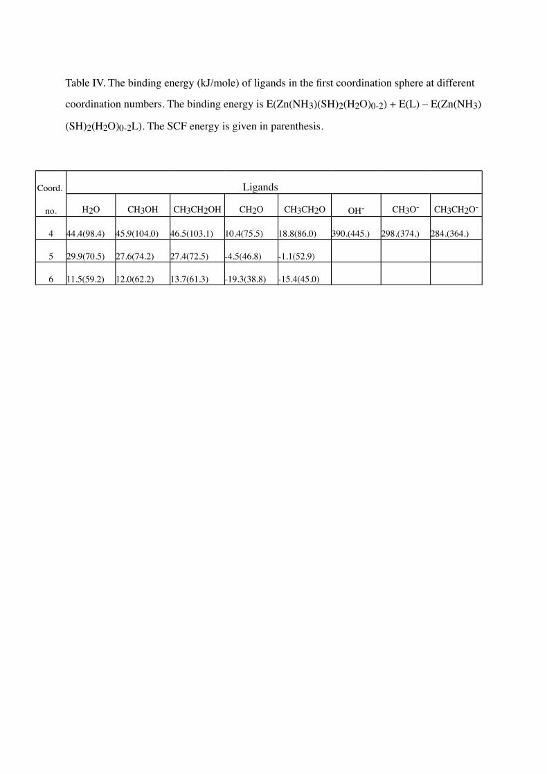
Table IV. The binding energy (kJ/mole) of ligands in the first coordination sphere at different
coordination numbers. The binding energy is E(Zn(NH3)(SH)2(H2O)0-2) + E(L) – E(Zn(NH3)
(SH)2(H2O)0-2L). The SCF energy is given in parenthesis.
Coord. Ligands
no. H2O CH3OH CH3CH2OH CH2O CH3CH2O OH- CH3O- CH3CH2O-
4 44.4(98.4) 45.9(104.0) 46.5(103.1) 10.4(75.5) 18.8(86.0) 390.(445.) 298.(374.) 284.(364.)
5 29.9(70.5) 27.6(74.2) 27.4(72.5) -4.5(46.8) -1.1(52.9)
6 11.5(59.2) 12.0(62.2) 13.7(61.3) -19.3(38.8) -15.4(45.0)

Table V. The binding energy (kJ/mole) of ligands in the second coordination sphere at
different coordination numbers. The binding energy is E(L) + E(Zn(NH3)(SH)2(H2O)0-2) –
E(Zn(NH3)(SH)2(H2O)0-2+L). The SCF energy is given in parenthesis.
Coord. Ligands
no. H2O CH3OH CH3CH2OH CH2O CH3CH2O
3 25.3(66.6) 24.3(66.1) 6.9(51.9)
4 45.8(88.9) 45.1(89.2) 45.1(88.9) 17.3(67.7) 20.1(72.5)
5 37.8(81.1) 37.0(80.9) 9.7(58.7)

Legends to the Figures
Figure 1. Optimized geometry of a. Zn(imidazole)(SH)2(H2O) and c. Zn(NH3)(SH)2(H2O).
1b. shows the zinc ligands (including the CB atom) in the x-ray crystal structure of alcohol
dehydrogenase in complex with NADH and dimethylsulfoxide [5].
Figure 2. Two optimized structures of the most stable five- (A) and four-coordinate (B)
structures of Zn(NH3)(SH)2(H2O)2.
Figure 3. Optimized geometry of the most stable six- (A), five (B) and four-coordinate (C)
structures of Zn(NH3)(SH)2(H2O)3.
Figure 4. Optimized geometry of Zn(NH3)(SH)2(OH)(H2O)_.
Figure 5. The binding energy of a water molecule to Zn(NH3)(SH)2(OH)_ as a function of
the Zn-OH2 distance. At each fixed Zn-OH2 distance, all other internal coordinates were
optimized.


0
20
40
60
80
100
120
200 250 300 350 400
Zn-OH2 distance (pm)
Binding energy
(kJ/mole)