Embed Size (px)
Citation preview
The Contribution of Nitric Oxide Detoxification and
Nitrous Oxide Production to Salmonella Pathogenesis
Isabel Johnston
Thesis for the degree of Doctor of Philosophy
School of Biological Sciences, University of East Anglia
September 2017
© This copy of the thesis has been supplied on condition that anyone who consults it
is understood to recognise that its copyright rests with the author and that no
quotation from the thesis, nor any information derived therefrom, may be published
without the author’s prior, written consent.

2
Abstract
Salmonella is responsible for millions of infections worldwide, with varied disease
outcomes, ranging from localised gastroenteritis to a fatal systemic infection. As a
facultative anaerobe, Salmonella is able to respire using nitrate as an alternative
electron acceptor. This study highlights the role of two gaseous intermediates
produced during nitrate respiration, nitric oxide (NO) and nitrous oxide (N2O), during
Salmonella pathogenesis.
Building from previous studies, a link is drawn between the high level of production of
N2O by Salmonella, and the metabolically expensive, and seemingly unimportant
synthesis of vitamin B12. We demonstrate the importance of the vitamin B12
independent methionine synthase, MetE, both during nitrate respiration and for
survival within macrophages. We hypothesise that when respiring on nitrate in the
intestine Salmonella releases N2O which is detrimental to vitamin B12 availability and
commensal survival in the intestine. Salmonella however is protected two-fold from
the toxicity of N2O, by the presence of MetE, and by the ability to synthesise vitamin
B12 de novo. This phenomenon is further investigated across multiple Salmonella
serovars, with the indication that enteric strains produce both higher levels of vitamin
B12 and N2O.
As well as during nitrate respiration, Salmonella also encounters NO exogenously
when challenged by the host defense inside macrophages. Since NO is a highly toxic
compound effective detoxification systems are essential for survival. A relationship
between NO and tellurite resistance in Salmonella is shown. Three putative tellurite
resistance proteins, TehB, STM1808 and YeaR were confirmed to provide resistance
against tellurite in Salmonella and share functional redundancy in the anaerobic
detoxification of NO and infection of macrophages. Using a suite of NO sensitive
mutants, we also demonstrate a clear correlation between tellurite and NO sensitivity.
Collectively this study highlights two important aspects of the nitrogen cycle which
contribute to pathogenesis in Salmonella.
3
Table of Contents
Abstract ...................................................................................................................... 2
Table of Contents ....................................................................................................... 3
List of Figures ............................................................................................................. 9
List of Tables ............................................................................................................ 12
Abbreviations ........................................................................................................... 13
Acknowledgments .................................................................................................... 16
1 Introduction ........................................................................................................... 17
1.1 General ........................................................................................................... 18
1.2 Sources of contamination ............................................................................... 19
1.3 Route of infection ............................................................................................ 19
1.4 Disease Types ................................................................................................ 20
1.4.1 Gastroenteritis .......................................................................................... 20
1.4.2 Enteric fever ............................................................................................. 20
1.4.3 Chronic carrier stage ................................................................................ 21
1.4.4 Invasive Non-typhoidal Salmonella .......................................................... 21
1.5 Current Treatment .......................................................................................... 22
1.5.1 Antibiotic resistance ................................................................................. 22
1.5.2 Vaccines................................................................................................... 23
1.6 Immune defenses and responses, and infection methods .............................. 25
1.6.1 Salmonella Pathogenicity Islands ............................................................. 25
1.7 Global microbial N-cycle ................................................................................. 28
1.8 Denitrification in Salmonella ........................................................................... 30
1.9 Toxicity of Nitric Oxide .................................................................................... 31
1.10 NO detoxification .......................................................................................... 33
4
1.10.1 Mechanisms for NO detoxification .......................................................... 34
1.11 Nitrous Oxide ................................................................................................ 36
1.12 Vitamin B12 and Salmonella .......................................................................... 37
1.12.1 Vitamin B12 synthesis ............................................................................. 38
1.12.2 Vitamin B12 requirements ....................................................................... 39
1.13 Methionine synthesis .................................................................................... 40
1.13.1 Regulation of methionine synthesis ........................................................ 42
1.13.2 Methionine synthases and nitrous oxide ................................................ 42
1.4 Research Gap: ................................................................................................ 44
2 Materials and Methods .......................................................................................... 45
2.1 Strains & plasmids .......................................................................................... 46
2.2 Media .............................................................................................................. 48
2.2.1 Rich media ............................................................................................... 48
2.2.2 Minimal media .......................................................................................... 48
2.2.3 Overnight cultures .................................................................................... 49
2.4 Aerobic growth ................................................................................................ 49
2.5 Anaerobic growth ............................................................................................ 49
2.6 Mutant Construction ........................................................................................ 50
2.7 Gel electrophoresis ......................................................................................... 54
2.8 B12 bioassay .................................................................................................... 54
2.9 Measurement of nitrogen cycle intermediates ................................................ 54
2.10 Western Blot ................................................................................................. 55
2.11 Gentamicin protection assay......................................................................... 56
2.12 MIC assay ..................................................................................................... 56
2.13 Statistics ....................................................................................................... 56

5
3. A link between tellurite, nitric oxide and virulence in Salmonella Typhimurium: the
role of TehB, STM1808 and YeaR. .......................................................................... 57
3.1 Introduction ..................................................................................................... 58
3.1.1 Aims ............................................................................................................. 61
3.2 Results ............................................................................................................ 62
3.2.2 Mutant construction .................................................................................. 62
3.2.3 There is no general growth defect for the TeR mutants cultured aerobically
in LB .................................................................................................................. 65
3.2.4 TehB, STM1808 and YeaR all contribute to tellurite resistance in
Salmonella ........................................................................................................ 66
3.2.5 Functional overlap between genes required for tellurite resistance in
anaerobic protection against nitric oxide ........................................................... 68
3.2.6 TehB H32 and H82 are not important for tellurite protection but are
important for nitric oxide protection ................................................................... 70
3.2.7 Nitric oxide accumulates during nitrate respiration in ΔtehB ΔSTM1808
ΔyeaR triple mutant ........................................................................................... 76
3.2.8 Proteins involved in protection against reactive nitrogen species also
protect against potassium tellurite ..................................................................... 79
3.2.9 MIC of tellurite, selenite and hydrogen peroxide for WT and ΔtehB
ΔSTM1808 ΔyeaR ............................................................................................ 81
3.2.10 The ΔtehB ΔSTM1808 ΔyeaR triple mutant is severely attenuated in IFN-
γ activated macrophages .................................................................................. 82
3.3 Discussion: ..................................................................................................... 84
3.3.1 Future Work ............................................................................................. 90
4. The role of CstA, Hcr and STM1273 in nitric oxide and tellurite resistance .......... 91
4.1 Introduction: .................................................................................................... 92
4.1.1 CstA ......................................................................................................... 93
4.1.2 Hcr ........................................................................................................... 93

6
4.1.3 STM1273.................................................................................................. 94
4.2 Aims ................................................................................................................ 95
4.3 Results ............................................................................................................ 96
4.3.1 Δhcr ΔSTM1273 and ΔcstA grow comparably to WT in LB both aerobically
and anaerobically .............................................................................................. 96
4.3.2 STM1273, CstA and Hcr are important for nitric oxide protection both
aerobically and anaerobically ............................................................................ 97
4.3.3 Nitric oxide accumulates in Δhcr in comparison to WT when grown under
denitrifying conditions ........................................................................................ 99
4.3.4 Δhcr and ΔcstA are significantly attenuated in activated macrophages . 100
4.3.5 STM1273, Hcr and CstA all offer Salmonella protection against potassium
tellurite in the presence and absence of oxygen. ............................................ 101
4.4 Discussion .................................................................................................... 104
5 Contribution of Vitamin B12 dependent and independent methionine synthases to
anaerobic nitrate respiration ................................................................................... 107
5.1 Introduction ................................................................................................... 108
5.2 Aims: ............................................................................................................. 110
5.3 Results .......................................................................................................... 111
5.3.1 Construction of metE and metR truncated mutants. ............................... 111
5.3.2 All mutants grow like WT in LB aerobically............................................. 114
5.3.3 ΔmetE and ΔmetR are attenuated in aerobic minimal media without the
addition of vitamin B12 or methionine ............................................................... 115
5.3.4 Vitamin B12 is toxic to Salmonella, but not due to cobalt toxicity ............ 117
5.3.5 MetE is important for the survival of denitrifying Salmonella .................. 119
5.3.6 Construction of a PocR mutant .............................................................. 123
5.3.7 PocR is not required for growth in LB or M9 aerobically ........................ 124
5.3.8 PocR is not essential for the regulation of metE..................................... 125
7
5.3.9 The ΔmetE mutant does not produce the MetE protein ......................... 125
5.3.10 A ΔmetEΔcobS mutant is unable to grow in M9 media in anaerobic
conditions ........................................................................................................ 127
5.3.11 MetE is important for survival of Salmonella in macrophages .............. 128
5.4 Discussion .................................................................................................... 130
5.4.1 Importance of MetE for denitrifying Salmonella ...................................... 130
5.4.2 Vitamin B12 toxicity ................................................................................. 133
5.4.3 Regulation of the cob operon by PocR ................................................... 135
5.4.4 The pathogenic effect of the production of nitrous oxide by Salmonella in
the intestine ..................................................................................................... 138
5.4.5 Future work ............................................................................................ 144
6 A correlation between nitrous oxide emissions and vitamin B12 synthesis in
Salmonella serovars ............................................................................................... 145
6.1 Introduction ................................................................................................... 146
6.1.1 Salmonella Typhimurium SL1344/474 ................................................... 146
6.1.2 Salmonella Enteriditis ............................................................................. 146
6.1.3 Salmonella Dublin .................................................................................. 147
6.1.4 Salmonella Choleraesuis........................................................................ 147
6.1.5 Salmonella Gallinarum and Pullorum ..................................................... 147
6.1.6 Citrobacter rodentium ............................................................................. 148
6.1.7 Differences in vitamin B12 synthesis between Salmonella serovars ....... 148
6.1.8 Measurement of vitamin B12 ................................................................... 150
6.2 Aims .............................................................................................................. 151
6.3 Results: ......................................................................................................... 152
6.3.1 Systemic Salmonella serovars have mutations in the cob operon ......... 152
6.3.2 Development and optimisation of a Vitamin B12 quantification assay ..... 156

8
6.3.4 Levels of Vitamin B12 produced by Salmonella serovars and Citrobacter
rodentium ........................................................................................................ 157
6.3.5 Comparison of NO and N2O released by different Salmonella serovars
during anaerobic nitrate growth .......................................................................... 160
6.4 Discussion .................................................................................................... 164
7 General Discussion ............................................................................................. 169
7.1 Context ...................................................................................................... 170
7.2 Nitric oxide ................................................................................................ 170
7.3 Nitrous oxide ............................................................................................. 171
7.4 Vitamin B12 ................................................................................................ 172
7.5 Characterisation of NO detoxification enzymes ......................................... 172
7.6 A link between nitric oxide and tellurite resistance in Salmonella .............. 174
7.7 Nitrous oxide ............................................................................................. 175
7.8 Survival within macrophages..................................................................... 178
7.9 Final conclusions ....................................................................................... 179
8 References .......................................................................................................... 180

9
List of Figures
Figure 1 The microbial Nitrogen Cycle. .................................................................... 28
Figure 2 Nitrogen cycle in Salmonella. .................................................................... 33
Figure 3 Levels of N2O produced by S. Typhimurium SL1344 and E. coli MG1655..
................................................................................................................................. 37
Figure 4 Genomic location of metH and metE, metR.. ............................................. 40
Figure 5 Methionine synthesis in Salmonella. .......................................................... 41
Figure 6 Outline of the inhibition of N2O on the production of methionine. ............... 43
Figure 7 Tellurite resistance genes are up-regulated by exogenous nitric oxide. ..... 60
Figure 8 PCR Verification of STM1808 mutant. ....................................................... 62
Figure 9 PCR Verification of yeaR mutant. ............................................................... 63
Figure 10 PCR Verification of tehB mutant ............................................................... 64
Figure 11: PCR Verification of yeaR tehB STM1808 triple mutant. .......................... 64
Figure 12 The four single TeR mutants and triple TeR mutant have no general growth
defect in LB aerobically. ........................................................................................... 65
Figure 13 In Salmonella resistance against potassium tellurite requires TehB,
STM1808 and TehB, both aerobically and anaerobically. ........................................ 67
Figure 14 Functional overlap between TehB, STM1808 and TehB in protection
against nitric oxide only during anaerobic growth. .................................................... 69
Figure 15 Alignment of STM1808 and yeaR. ........................................................... 71
Figure 16 Alignment of tehB, STM1808 and yeaR. .................................................. 71
Figure 17 Complementation strains all grow comparably both aerobically and
anaerobically. ........................................................................................................... 73
Figure 18 Complementation with yeaR or yeaRH32AH82A provides a low level of
growth restoration of ΔtehB ΔSTM1808 ΔyeaR when grown in the presence of
potassium tellurite.. .................................................................................................. 74
Figure 19 H32 and H82 are important for yeaR mediated nitric oxide protection
anaerobically.. .......................................................................................................... 75

10
Figure 20 NO levels peak at 40 hours for WT Salmonella. ....................................... 77
Figure 21 Relative levels of nitric oxide produced by Salmonella mutants. .............. 78
Figure 22 Genes involved in RNS defense are also involved in resistance against
potassium tellurite. ................................................................................................... 80
Figure 23 The ΔtehB ΔSTM1808 ΔyeaR triple mutant is severely attenuated in IFN-γ
activated macrophages.. .......................................................................................... 83
Figure 24 Tellurite reduction model .......................................................................... 89
Figure 25 The three mutant strains, Δhcr, ΔSTM1273 and ΔcstA grow comparably to
WT in LB.. ................................................................................................................ 96
Figure 26 STM1273, CstA and Hcr are important for nitric oxide protection both
aerobically and anaerobically.. ................................................................................. 98
Figure 27 Nitric oxide accumulates in Δhcr in comparison to WT when grown under
denitrifying conditions. .............................................................................................. 99
Figure 28 Hcr and CstA are important for survival within activated macrophages. 101
Figure 29 STM1273, CstA and Hcr all play a role in tellurite protection, both
aerobically and anaerobically. ................................................................................ 103
Figure 30 The organisation of the cob operon of vitamin B12 synthesis with respect to
the pocR gene and pdu operon.. ............................................................................ 109
Figure 31 Position of metE and metR on the Salmonella SL1344 genome ............ 111
Figure 32 MetE and MetR promoter site mapping in E. coli ................................... 111
Figure 33 PCR Verification of truncated metE and metR mutants.. ....................... 112
Figure 34 Confirmation of metH mutant construction. ............................................ 113
Figure 35 All mutant strains grow at similar rates to WT aerobically in LB broth.. . 114
Figure 36 ΔmetE and ΔmetR are attenuated in aerobic minimal media without the
addition of vitamin B12 or methionine. ..................................................................... 116
Figure 37 Vitamin B12 exhibits a toxic effect on WT Salmonella. ............................ 118
Figure 38 Cobalt is not toxic for WT Salmonella. ................................................... 118
Figure 39 MetE is important for the survival of denitrifying Salmonella. ................. 121

11
Figure 40 MetE is important for the survival of Salmonella grown with exogenous
N2O. ....................................................................................................................... 122
Figure 41 Confirmation of correct mutate construction of ΔpocR and ΔmetEΔpocR .
............................................................................................................................... 123
Figure 42 PocR is not required for growth in LB or M9 aerobically. ....................... 124
Figure 43 The growth of ΔmetE is partially restored by the deletion of pocR when
growing under denitrifying conditions. .................................................................... 125
Figure 44 The MetE protein is not produced by ΔmetE or ΔmetEΔpocR during
anaerobic growth .................................................................................................... 126
Figure 45 The deletion of cobS and metE results in an inability of S. Typhimurium to
grow.. ..................................................................................................................... 127
Figure 46 MetE is required for intracellular survival in murine macrophages in the
absence of methionine or vitamin B12.. ................................................................... 129
Figure 47 Working model – regulation of the cob operon. ...................................... 137
Figure 48 Working model ....................................................................................... 143
Figure 49 Alignment of the cob operon between Salmonella serovars. ................. 153
Figure 50 Vitamin B12 bioassay. ............................................................................. 157
Figure 51 Vitamin B12 bioassay. ............................................................................. 159
Figure 52 All tested Salmonella serovars and Citrobacter rodentium grow
comparably. ............................................................................................................ 161
Figure 53 Nitrous oxide levels of Salmonella serovars and Citrobacter rodentium. 162
Figure 54 Nitric oxide levels of Salmonella serovars and Citrobacter rodentium.163

12
List of Tables
Table 1 A list of all strains used in this study, their genotypes and origins. .... 46
Table 2 A list of all plasmids used in this study, their genotypes and origins. 48
Table 3 Primers used for mutant construction .................................................... 51
Table 4 Primers used for mutant confirmation .................................................... 52
Table 5 PCR components. ..................................................................................... 53
Table 6 PCR program ............................................................................................. 53
Table 7 MIC assay.. ................................................................................................. 81
Table 8: Summary of growth abilites of Salmonella mutants. .......................... 131
Table 9 Presence of metE, nosZ and ability to synthesise B12 of eight common
commensal bacteria and human epithelial cells ............................................... 141
Table 10 Genetic consensus of the cob operon across Salmonella serovars 154

13
Abbreviations
Amp Ampillicin
Bp Base Pair
B12 Vitamin B12
Cm Chloramphenicol
Cfu Colony forming units
ºC Degrees Celsius
dH2O Distilled water
DMEM Dulbecco’s modified Eagle medium
DUF Domain of unknown function
DNA Deoxyribonucleic acid
FLP Flip recombinase
g Grams
IFN-γ Interferon gamma
iNOS Inducible nitric oxide synthase
Kb Kilobase-pair
Kan Kanamycin
LB Luria-Bertani
L-NAME N-Nitroarginine methyl ester
µg Micrograms
µM Micromolar

14
M Molar
MDR Multi-drug resistant
mg Milligrams
ml Millileters
mM Millimolar
min Minutes
MOI Multiplicity of infection
N2 Dinitrogen
N2O Nitrous oxide
NH4+ Ammonium
NO Nitric oxide
NO2 Nitrite
NO3 Nitrate
OD Optical density
ONOO Peroxynitrite
PBS Phosphate buffered saline
PCR Polymerase chain reaction
Ppb Parts per billion
Ppm Parts per million
RNS Reactive nitrogen species
ROS Reactive oxygen species

15
SD Standard deviation
SE Standard error
TeO3 Tellurite

16
Acknowledgments
Firstly, thanks go to my supervisory team, Gary Rowley, Andy Gates and David
Richardson. Special thanks to Gary for all your help and support through the years.
Thanks go to all the students I supervised during my PhD, Angela Kalair, Maria Dik,
and Emily Addington. Special thanks to Claire Hews who assisted with a large
amount of the work on PocR, and to Alistair Middlemiss for his work on the
Salmonella serovars.
Many thanks to all members of the Rowley lab past and present. Special thanks to
Hannah Wells who helped me enormously in my first year and taught me many of
the basic skills and techniques required for completion of this work. Enormous
thanks to my brother, friend, and colleague Hannah Gaimster, you were a huge
support both scientifically and otherwise. Maybe one day we will be able to stop and
go on that cruise.
I owe my interest in microbiology to Lab 1.29 especially to Andy Johnston and Jon
Todd and my thanks go to Emily Fowler and Simone Payne for your continued
support and friendship.
Many thanks to my examiners, Richard Bowater and Mark Roberts.

17
1 Introduction

18
1.1 General
Salmonella is a genus of Gram negative bacteria which contains just two species:
Salmonella bongori and Salmonella enterica. Within enterica there are over 2500
different Salmonella serovars, a sub-species categorisation system based on outer
membrane antigens, and includes all of those which cause disease in humans
(Ochman and Groisman 1994).
Only about 50 of these serovars have been isolated as known pathogens (Coburn,
Grassl and Finlay 2007). Different serovars of Salmonella can infect different hosts,
some are host specific, and restricted to a single host, whilst others are generalists
and have the ability to infect a wide range of mammalian and avian hosts. There are
two main disease types associated with Salmonella. A systemic typhoidal infection
caused by S. Typhi in humans and more common non-typhoidal strains which cause
localised gastrointestinal/enteric infections (enteritis), such as S. Typhimurium in
humans and S. Dublin in calves (Coburn et al. 2007)
Salmonella Typhi and Paratyphi (see 1.4.2 Enteric fever) almost exclusively cause a
systemic infection in humans and therefore are referred to as host restricted strains.
Similarly, Salmonella Gallinarum is only known to cause a systemic infection in
chickens/fowl. Salmonella Cholerasuis and Salmonella Dublin are most commonly
known to cause a systemic infection in pigs and cattle respectively, but also have the
ability to infect other hosts, including humans, these serovars are referred to as host
adapted strains, since they most commonly infect a single host but it is not a strict
restriction. There are then further strains such as Salmonella Typhimurium and
Salmonella Enteritidis which cause an enteric infection in a wide range of hosts, and
as such are un-restricted serovars (Uzzau et al. 2000). The most common non-
typhoidal serovar used as a model laboratory strain is Salmonella enterica subs.
enterica serovar Typhimurium (S. Typhimurium), so called because as well as causing
a self-limiting gastroenteritis in humans resulting in fever and diarrhoea, it causes, as
the name suggests Typhoid like symptoms in mice. S. Typhimurium SL1344 is used
throughout this thesis and referred to as Salmonella, unless otherwise stated.

19
1.2 Sources of contamination
A natural Salmonella infection progresses post oral infection. The origin of infection
can be from numerous sources. Approximately 95% of infections are associated with
the eating of contaminated foodstuffs, this can include raw or undercooked meat either
from an infected animal or one that has become contaminated post slaughter, eggs
from infected chickens, and unpasteurised milk (Mead et al. 1999). There are also
non-animal foods which can be sources of contamination, often due to the plants
having been fertilized with contaminated water. Salad crops and other foods which are
eaten raw are therefore common sources, this is due to the fact that they are poorly
washed and not cooked before eating, so any bacteria which have adhered to the
leaves will remain (L Plym and Wierup 2006, Lynch, Tauxe and Hedberg 2009, Islam
et al. 2004, Henao-Herreño et al. 2016, Ibenyassine et al. 2007).
Salmonella is frequently found in environmental water samples, soil, and sediment. A
review on water-borne Salmonella infections highlighted that the majority of papers
looking at infections from this source were from Asia, and very few were from the other
endemic regions of Africa and Central/Southern America (Threlfall 2002). These
studies have highlighted the effect that lack of chlorination, equipment failures and
back-siphonage in the water obtained from well-water, unboiled spring water and
piped water of these areas have on infection numbers.
1.3 Route of infection
Once an individual has consumed the bacteria, since it has the ability to withstand high
pH (Foster and Hall 1991, Garcia-del Portillo, Foster and Finlay 1993), it travels
through the stomach and to the small intestine. Here it preferentially invades the
epithelial cells of the small intestine (terminal ileum) or enters into the immune system
by passing through M-cells which filter the gut lumen and transport foreign bodies to
the Peyer’s patches where they can be taken up by macrophages (Jones, Ghori and
Falkow 1994, Galán and Curtiss 1989). Internalisation into macrophages occurs via
the process of phagocytosis, dependent on the recognition of specific molecules by
receptors on the surface of the macrophage. Inside the macrophage, Salmonella can
replicate and in the case of typhodal strains disseminate and cause systemic infection

20
of other organs such as the spleen and liver (Alvarez-Ordóñez et al. 2011, Dougan et
al. 2011, Mastroeni and Grant 2013, Carter and Collins 1974).
1.4 Disease Types
1.4.1 Gastroenteritis
The most common Salmonella infection in humans is Salmonellosis, which causes a
gastrointestinal infection. A number of different S. enterica subspecies can lead to
Salmonellosis in humans with around 20 serovars causing approximately 70% of
cases, with the top three, S. enterica Typhimurium, Enteritidis and Newport causing
40% of infections. Symptoms of the infection usually occur 8-72 hours after ingestion
of contaminated material and include inflammation of the intestine, abdominal
cramping, vomiting and, diarrhea or constipation. In the large majority of cases the
infection will be self-limiting and will be eradicated by the host immune system in 2-5
days with no requirement for treatment (Hohmann 2001). However, those under 5 or
over 65 and immunocompromised patients may require treatment, which could either
include hospitalisation in order to replace lost fluids or, antibiotics (Gordon 2008, Wen,
Best and Nourse 2017, Musil et al. 2016). While Salmonellosis is a worldwide disease,
the rates of infections vary between countries based on levels of contaminated food
and water. A 2010 study estimates that there are 93.8 million cases of Salmonella
induced gastroenteritis globally each year, with 155,000 (0.17%) of these resulting in
death (Majowicz et al. 2010). While the cost to life of Salmonellosis is therefore quite
low there is a large economic cost due to loss of productivity.
1.4.2 Enteric fever
In humans a systemic Salmonella infection is known as enteric fever and includes both
typhoid fever and paratyphoid fever caused by S. Typhi and S. Paratyphi serovars.
Symptoms of typhoid fever start similarly to Salmonellosis, with abdominal cramps,
vomiting and diarrhea/constipation but combined with a high fever. Progression of the
infection includes dissemination and subsequent inflammation of the liver and spleen,
coupled with weight loss and severe confusion. If left untreated the infection can cause

21
either internal bleeding in the digestive tract or peroration of the digestive tract, this
can result in sepsis and lead to multi organ failure which can be fatal.
Accurate data for the numbers of typhoid fever cases are unavailable, however
estimates put the global total instances of typhoid between 11.9-20 million with about
1% of these, 129-200,000, resulting in death. In addition to this there are an estimated
5 million paratyphoid cases annually. (Crump, Luby and Mintz 2004, Crump 2014,
Mogasale et al. 2014)
Typhoid fever must be treated with antibiotics in order to stop the infection spreading
and becoming fatal. Therefore, in addition to the large number of deaths caused by
the infection there is a large economic cost. Current vaccines and antibiotics and the
issues faced in these areas are discussed in 1.5 Current Treatment.
1.4.3 Chronic carrier stage
Chronic carriers of typhoid fever are those who are infected with S. Typhi, appear to
recover and are then symptomless and appear healthy. In these patients, the bacteria
colonise the gall bladder, where they are protected from the host immune defense.
From here the bacteria can be periodically released into the bile and excreted and
hence a chronic carrier can go on to infect other patients without being aware they are
doing so or that they are even infected. This is of particular concern in regions where
there is poor sanitation and drinking water can become contaminated with human
waste (Gonzalez-Escobedo, Marshall and Gunn 2011).
1.4.4 Invasive Non-typhoidal Salmonella
Invasive non-typhoidal Salmonella (iNTS) are non typhoidal strains, commonly the two
serovars primarily responsible for gastroenteritis, S. Typhimurium or S. Enteritidis,
which can cause a more typhoid-like infection. These strains are more resistant to the
host-mediated killing by macrophages and hence are able to enter the bloodstream
causing a systemic spread of the bacteria (Feasey et al. 2012). This disease has
emerged in Africa, commonly developing into invasive diseases in individuals already
suffering from malaria or HIV (Okoro et al. 2012). Between 22-25% of African patients
succumb to a iNTS infection, much higher percentages than the global deaths from

22
typhoid fever, approximately 1% (Gordon et al. 2008). The link between HIV positive
patients and those suffering from a iNTS disease is extremely high, with 95% of adults
presenting with iNTS being HIV positive – this is not a link seen for Typhoid fever
(Kankwatira, Mwafulirwa and Gordon 2004). The development of this disease in HIV
positive individuals is due to abnormalities in the host not mutations of the bacteria.
Three key defects have been described: a weakened gut mucosal layer which leads
to increased dissemination from the gut; dysregulation of cytokines within immune
cells; and impaired serum killing of the bacteria (Feasey, 2012).
1.5 Current Treatment
1.5.1 Antibiotic use and resistance
Typhoid fever is seen more in the developing world, partly due to poorer sanitation in
these areas leading to acquisition of the infection through contaminated food or water,
and partly due to the emergence of multi-drug resistant (MDR) strains in these areas.
To avoid fatality, patients with typhoid fever require treatment with antibiotics. The
most common, ‘first-line’ antibiotics used to treat typhoid fever are chloramphenicol,
amplicillin and trimethoprim; however, since 1989, strains of both Salmonella Typhi
and Paratyphi are becoming increasingly resistant to these drugs, and these multi-
drug-resistant (MDR) strains have been the cause of large outbreaks (Glynn et al.
1998, Rahman et al. 2014, Wong et al. 2015, Wirth 2015).
It is thought that in the Global North these MDR strains developed in the animal
agriculture sector, where antibiotics are frequently and inappropriately used, before
spreading to humans through contaminated foodstuffs (Marshall and Levy 2011,
Holmberg et al. 1984, Helke et al. 2017).
The current antibiotic of choice is ciprofloxacin, however strains have been isolated
which also show resistance to this drug (Threlfall 2002, Rowe, Ward and Threlfall
1997). This is becoming a rapidly increasing problem in the commonly endemic
regions of the Global South but also due to individuals travelling more frequently to
these regions, there are increasing reports of patients in the Global North with MDR
Salmonella infections.
23
Along with methods aimed at reducing contamination and therefore cases of typhoid
fever, new antibiotics are required to target the pressing issue of severe MDR
Salmonella.
1.5.2 Vaccines
Due to the severity and increasingly hard to treat strains of Salmonella there is an
increase in pressure on the availability of a long lasting vaccine.
A vaccine for typhoid fever was first developed in 1916 and relied on heat-killed
Salmonella Typhi cells to produce a parenteral whole cell vaccine. This vaccine was
reasonably effective, protecting 51 to 88% of those treated for up to 7 years. However,
there were side effects that came with vaccination, including fever in up to 30% of
cases, and headaches and severe localised pain in up to 35% (Guzman et al. 2006).
A modern vaccine was generated using purified, non-denatured, Vi capsular
polysaccharide of S. Typhi. The Vi capsular antigen is encoded on the Salmonella
pathogenicity island 7 (SPI-7), which is absent in the non-typhoid Salmonella
typhimurium, and is known to be acutely important for virulence (Tran et al. 2010). The
Vi vaccine is produced in multiple forms, Typhim Vi® (Sanofi Pasteur),
Typherix® (GSK) and Typbar Vi® (Bharat Biotech) and has a reported efficiency of
between 64 and 72% (Klugman et al. 1987, Acharya et al. 1987) with minimal side
effects, the vaccine must be boosted every 2 years. In addition, there is a conjugated
form of the vaccine which provides vaccination against tetanus in addition to typhoid,
Typbar TCV®.
The other current vaccine is a live attenuated vaccine; this offers multiple benefits. It
can be orally administered which both means no hazardous materials such as needles
are produced and it can be easily administered not just by medical professionals.
Importantly they can induce mucosal, cellular and humoral immune responses. Since
live attenuated vaccines have the theoretical ability to revert to a fully virulent strain it
is important that the attenuation is produced via multiple well defined mutations.
The Ty21a vaccine is an attenuated strain of S. Typhi Ty2 and contains a mutation in
GalE and lacks the Vi antigen (Formal et al. 1981). The GalE mutation results in the
strain being unable to produce the enzyme responsible for the reversible conversion
of UDP-glucose to UDP-galactose, the accumulation of UDP-galactose in the

24
cytoplasm results in cell lysis (Germanier and Füer 1975). This strain also has
numerous other mutations including in the gene encoding RpoS which is important for
stress responses. This vaccine is currently in use as Vivotif® (PaxVax Corporation),
which is administered as three or four capsule pills on alternate days, with protection
being achieved 7 days after the final dose. High rates of efficiency and tolerability are
seen with this vaccine, with up to 87% efficiency for up to 7 years and suggested re-
immunisation after 5 years (Jin et al. 2017, Levine et al. 2007). Additionally, Ty21a has
been shown to provide a level of protection against S. Paratyphoid B, which doesn’t
possess the Vi-antigen and therefore Vi polysaccharide based vaccines are
ineffective, however use of both vaccines together has been heralded as a way to
increase the immune response (Pakkanen et al. 2015).
One potential target for vaccine development is the recently discovered Salmonella
Typhi toxin. This toxin is only found in S. Typhi and S. Paratyphi – two human host-
restricted strains which cause enteric fever. Since the toxin is only present in these
strains human studies will need to be carried out in order to know exactly what role the
toxin plays in virulence and whether it can therefore be targeted as a preventative
strategy; however the presence of the toxin has been linked to the progression of the
disease and therefore it is expected that an antitoxin immune response should be able
to be utlised to protect against Typhoid fever (Galán 2016).
There are many aspects to an ideal typhoid vaccine and each presents its own
problems. Ideally a vaccine would protect against the range of Salmonella serovars
which are responsible for invasive infections worldwide, these have been highlighted
as Salmonella Typhi, Salmonella Paratyphi A, Salmonella Paratyphi B, Salmonella
Typhimurium, Salmonella Enteritidis and Salmonella Choleraesuis (Tennant and
Levine 2015). In addition, the current vaccines cannot be administered to the very
young and this would be highly beneficial as the infection is more dangerous in these
individuals (Crump and Mintz 2010). The live attenuated vaccines offer the best
immune response but come with a risk of reversion to a virulent state, while this can
be avoided with careful combinations of mutations, this requires extensive research
(Spreng, Dietrich and Weidinger 2006).

25
1.6 Immune defenses and responses, and infection methods
Initial Salmonella infection results in the inflammation of local tissue via the presence
of cell membrane components including lipopolysaccharide (LPS). This inflammation
results in the recruitment of cytokines such as interferon-γ (IFN-γ) or tumor necrosis
factor-α (TNF-α) which are required to activate macrophages (Arango Duque and
Descoteaux 2014, Hu and Ivashkiv 2009, Duluc et al. 2009). As highlighted in the 1.3
Routes of infection section, it is important for the progression of Salmonella disease
that the bacteria can survive within phagocytic cells including macrophages, as these
phagocytes are central to the control of infection, being critical for survival in mice
infected with Salmonella. Macrophages employ multiple antibacterial mechanisms in
order to attempt to kill any bacteria that they have engulfs. This includes the release
of reactive oxygen species (ROS) and the release of nitric oxide (NO) (Lowenstein et
al. 1993). The inducible nitric oxide synthase, iNOS, is specific to macrophages and
is responsible for their nitric oxide production (Aktan 2004).
1.6.1 Salmonella Pathogenicity Islands
The pathogenicity of Salmonella is largely down to regions of the genome referred to
as Salmonella pathogenicity islands (Lostroh and Lee 2001). These islands contain
the proteins required, and regulatory elements, for a specific virulence phenotype.
These islands are highly conserved between the numerous Salmonella serovars and
due to their lower GC content than the rest of the genome are thought to have been
acquired via horizontal gene transfer (Li et al. 1995).
Salmonella pathogenicity islands 1 and 2 (SPI-1 and SPI-2) are found in all serovars
in the Salmonella enterica species (Ochman and Groisman 1996). These SPIs encode
type III secretion systems (TTSS), which allow for the transfer of bacterial proteins into
the host cell. TTSSs are needle-like structures which span the bacterial envelope
(Waterman and Holden 2003). The proteins injected by SPI-1 interfere with
macrophage biochemistry and physiology and therefore vital processes, having
effects including initiating the uptake of the bacteria. SPI-2 then promotes survival
within the cell and causes apoptosis of macrophages.
26
1.6.1.1 Salmonella Pathogenicity Island 1
SPI-1 is 40kb in length, contains 29 genes, and is primarily known to be vital for the
uptake by intestinal epithelial cells, which are not usually phagocytic (Zhou and Galán
2001). SPI-1 is important for the survival of all Salmonella within the intestine whether
the particular serovar causes an enteric or a systemic infection (Lostroh and Lee
2001). Indeed while SPI-1 has been shown to be required for an orally-progressed
infection it is non-essential for a systemic infection (Marcus et al. 2000, Jones et al.
1994). There are at least 13 proteins delivered by the SPI-1 machinery. Three
important members of this group are SopB, SipA and SipC which are required for the
rearrangement of actin in the host cell which results in the uptake of the bacteria
(Raffatellu et al. 2005). The main regulatory trigger for SPI-1 is a change in pH that
occur between the stomach and small intestine. HilA, HilC, HilD and InvF are the main
regulators of SPI-1 (Eichelberg and Galán 1999). HilA is a member of the OmpR family
of regulators and is known to be of particular importance for the regulation of SPI-1
(Bajaj, Hwang and Lee 1995).
1.6.1.2 Salmonella Pathogenicity Island 2
Once engulfed within a cell the large phagosome quickly reduces, within hours, to the
Salmonella containing vacuole (SCV). SPI-2 behaves similarly to SPI-1 and inserts
effector proteins across the SCV membrane into the cell cytosol via the translocon
pore (Guignot and Tran Van Nhieu 2016). SPI-2 has four main functions, the
acquisition of proteins from the host cell, the movement towards the nucleus and golgi
apparatus, bacterial replication within the SCV and prevention of the binding of iNOS
to the SCV reducing RNS exposure (Haraga, Ohlson and Miller 2008, Figueira and
Holden 2012).
The positioning of the SCV near to the Golgi apparatus has been shown to be
associated with an increase in bacterial growth. The effectors SseG, SseF and SifA
are particularly important for this association of the SCV with the golgi (Ramsden et
al. 2007). Myosin II, an actin-based motor, has also been shown to be important for
this movement (Wasylnka et al. 2008, Steele-Mortimer 2008). A large number of
effectors function to maintain the SCV, including SifA, PipB2, SseJ and SopD2, this

27
maintainance is vital for the ability of Salmonella to replicate within the host cell
(Haraga et al. 2008, Waterman and Holden 2003, Cirillo et al. 1998).
Alteration of iNOS locality by the SCV is considered to be another SPI-2 dependent
process, which in turn reduces the amount of RNS which target the bacteria; with
peroxynitirite being excluded from the SCV via SPI-2 effectors. The significance of this
role of SPI-2 has been demonstrated with the reduction in a virulence defect in iNOS-
/- mice (Chakravortty, Hansen-Wester and Hensel 2002, Waterman and Holden 2003).
NRAMP1 is a macrophage transmembrane protein which is key to the killing of
Salmonella by the macrophage and controls bacterial growth in the reticuloendothelial
system. This has been well studied in the mouse model but a role for NRAMPp1 in
human infections has been harder to confirm (Dunstan et al. 2001, Mastroeni and
Sheppard 2004).

28
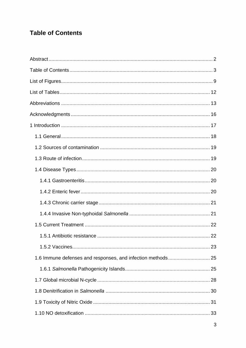
1.7 Global microbial N-cycle
The nitrogen cycle is one of the key nutrient cycles in the environment; and microbes
play a large role in this. All organisms rely on nitrogen for the synthesis of nucleic acids
and proteins as well as many other essential compounds. The nitrogen cycle exists in
6 main parts, demonstrated in Figure 1.
In the atmosphere nitrogen exists as dinitrogen gas (N2), an inert form of nitrogen. This
therefore cannot be taken up by plants and so the nitrogen must be ‘fixed’ into
ammonia (NH4+). This process is primarily undertaken by bacteria along with archaea.
Nitrogen can also be fixed by lightning, but at an insignificant level (Ferguson and
Libby 1971).
Figure 1 The microbial Nitrogen Cycle. Nitrogen Fixation (1), Aerobic Ammonia
Oxidation/Nitrification (2), Nitrite Oxidation (3), Denitrification (4), Annamox (5),
Dissimilatory Nitrate Reduction Pathway (6)

29
Nitrogen fixation is carried out by Diazotrophs, bacteria and archaea which are able to
grow without the presence of fixed nitrogen. Some plants have symbiotic relationships
with nitrogen fixing bacteria, commonly in the root nodules of legumes where Rhizobia
are the most abundant. Some Cyanobacteria also form symbiotic relationships with
plants such as ferns and fungi including lichens, these bacteria produce oxygen as a
by-product of the process (Berman-Frank, Lundgren and Falkowski 2003). In
traditional farming, fields would be cycled through rotations of food crops and legumes,
often clover, these legumes then are left to rot on the fields and the captured nitrogen
is released into the soil (Wang et al. 2014, Zhang et al. 2015). However, in modern
farming practices this takes too long and is therefore not an efficient way to ensure the
food crops are getting enough nitrogen and therefore nitrogen fertilisers are added.
The Haber Bosch process requires very high pressures and temperatures to fix
nitrogen from air mixed with natural gas (Sutton et al. 2011).
The next stage of the cycle is nitrification, where ammonium is oxidized via
hydroxylamine, NH2OH, to nitrite (NO2-), and subsequently to nitrate (NO3
-). The first
step is performed by ammonia-oxidising bacteria (AOB), primarily Nitrosomonas and
Nitrosococcus and the ammonia-oxidising archaea (AOA), Nitrosopumilus
maritimus and Nitrososphaera viennensis (Martens-Habbena et al. 2009). Nitrification
can lead to issues with nitrogen run off and associated eutrophication, as nitrite is
more soluble than ammonium (Conley et al. 2009).
Nitrite can then be returned to ammonia, via the dissimilatory nitrate reduction to
ammonium (DNRA) pathway, to N2 via N2H4 by the anammox pathway, or to N2 via
denitrification (Jetten 2008, Jetten et al. 2009). Traditionally little consideration has
been given to the impact of the dissimilatory nitrate reduction to ammonium pathway
to global nitrogen cycling; however, many soil bacteria have the ability to perform
DNRA and is it thought to be the dominant pathway for nitrite cycling in some
environments (Giblin et al, 2013).
Denitrification is the anaerobic reduction of nitrate (NO3-) via nitrite (NO2
-) nitric oxide
(NO) and nitrous oxide (N2O) to dinitrogen (N2). This process has been well studied in
numerous bacteria, with the best understanding in the soil dwelling bacteria
Paracoccus denitrificans. Research in this field has focused on the truncation of the
pathway and subsequent release of the potent greenhouse gas N2O into the
atmosphere, and ways this could be reduced (Sullivan et al. 2013).
30
The nitrous oxide reductase, NosZ, is responsible for the conversion of nitrous oxide
to dinitrogen gas and the completion of the nitrogen cycle. There are multiple factors
known to affect the function of NosZ in agricultural soil dwelling bacteria, and hence
the levels of N2O which are successfully reduced. These include the amount of
nitrogen fertilizer added to soils, water content, oxygen levels, nutrient availability,
temperature and pH (Sahrawat, 1986(Sullivan et al. 2013).
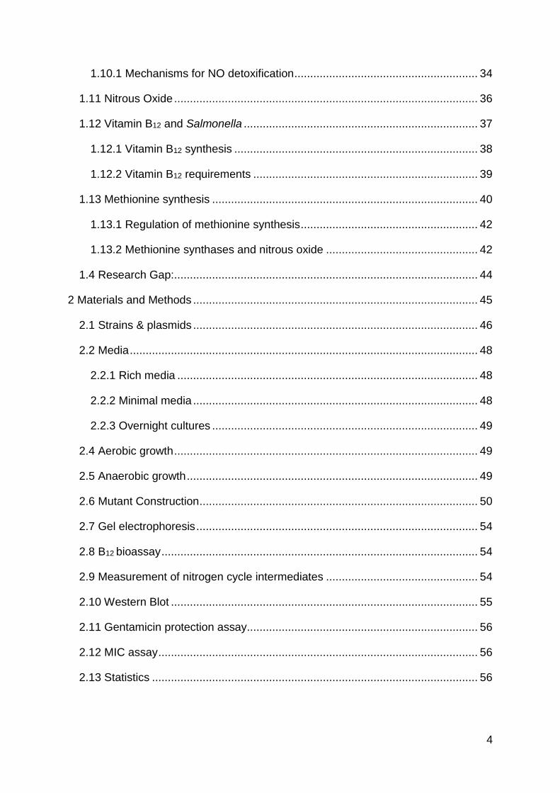
1.8 Denitrification in Salmonella
Salmonella, along with other Enterobacteriaceae also undertake denitrification.
However, unlike classical denitrifiers, the final step in the pathway, the reduction of
N2O to N2 is missing, we therefore refer to denitrification in Salmonella as truncated
denitrification (Rowley et al. 2012). This process is important for Salmonella in
anaerobic or microoxic conditions because as facultative anaerobes they can use
alternative electron acceptors, including nitrate, in order to respire.
The first step in this process, the reduction of NO3 to NO2 either uses the membrane-
bound dissimilatory nitrate reducatase, Nar in the cytoplasm, or the periplasmic
dissimilatory nitrate reductase, Nap. There are two membrane bound systems, that
encoded by narGHJI, which functions anaerobically, and that by narZYWV, which is
active aerobically, and a single periplasmic system, encoded by napFDAGHBC.
(Rowley et al. 2012) NarG accounts for about 98% of nitrate reduction. In addition to
nitrate reduction, NarG is able to catalyse the reduction of nitrite if no nitrate is present.
The other enzyme responsible for this step is Nir, either NirK, the cytochrome cd1, cu-
containing nitrite reductase (Gilberthorpe and Poole 2008) or a siroheme-containing
nitrite reductase (NirB), Salmonella only possesses NirB.Ideally, to prevent NO
accumulation, the NO-generating and NO-consuming processes should function in a
concerted fashion to form N2O, which until recently was considered to be less cytotoxic
(discussed in 1.11 Nitrous Oxide).
The reduction of nitric oxide will be further discussed in 1.10 NO detoxification, and
can take three forms, oxidation to nitrate, or reduction to ammonium or N2O. There is
no enzyme present in the entire Enterobacteriaceae family which reduces N2O and
hence in Salmonella denitrification is truncated at this stage.

31
Exposure to nitrate and nitrite in low oxygen conditions may occur outside the host but
also in the mammalian intestine, as part of the host-generated inflammatory response
(Lopez et al, 2012). Inflammation is generally thought to be a way that the host fights
a pathogen, however this host generated nitrate has been shown to provide a growth
advantage to members of the Enterobacteriaceae family including E. coli and
Salmonella. The gut microbiota, which play many important roles including providing
pathogen colonization resistance, is primarily made up of fermenting microbes which
would be unable to use the nitrate as an electron source for respiration (Guarner and
Malagelada 2003, Guarner 2006, Tremaroli and Bäckhed 2012). This phenomenon
has been demonstrated multiple times, suggesting the ability of Salmonella to respire
on nitrate and to trigger the host to produce this nitrate, is of key importance to its
survival and virulence (Winter et al. 2013).
SopE, a type III effector protein, has been shown to increase the severity of
inflammation in the intestine via Rho GTPase mediated caspase-1 activation and
consequent release of the cytokine IL-1β (Müller et al. 2009). SipA is also delivered
by a type III secretion system and plays a role in pathogen-mediated inflammation
(Higashide et al. 2002, Stecher et al. 2007)
1.9 Toxicity of Nitric Oxide
Nitric oxide plays many roles in the human body including as a neuronal signaling
molecule and as a regulator of vasodilation of blood vessels and hence blood pressure
(Irshad and Chaudhuri 2002). Indeed, the latter of these functions has led to the
increased use of nitric oxide as an athletic supplement, although studies on the
benefits of these are inconclusive (Bescós et al. 2012). Conversely at the same time
there is growing industry based around antioxidants designed to reduce RNS and ROS
(Devasagayam et al. 2004).
As a free radical, nitric oxide (NO) has cytostatic and cytotoxic antibacterial activity in
both aerobic and anaerobic environments, causing widespread damage to numerous
cellular targets. Targets of this damage include DNA, proteins (both fully formed
proteins and via amino acid targeting) and transcriptional regulators (Fang 2004). NO

32
is highly reactive, and can form other toxic reactive nitrogen species (RNS), such as
peroxynitrite (Brunelli, Crow and Beckman 1995).
Salmonella is exposed to RNS at different locations during its infectious cycle in
vertebrate hosts (Prior et al. 2009). Nitrogen species are found distributed throughout
the length of the gastrointestinal tract, representing a serious problem for enteric
pathogens. The acidity of the stomach converts dietary and salivary nitrite to nitrous
acid, and subsequently by disproportionation to other nitrogen species, including NO.
The endogenous production of nitric oxide via the denitrification pathway has already
been mentioned in 1.8 Denitrification in Salmonella.
Numerous cell types including leukocytes and hepatocytes can produce NO via the
conversion of L-arginine to L-citrulline. Macrophages form a crucial part of the early
response of the immune system to attack by pathogens, and production of NO is a
vital part of the macrophage armory. Activated macrophages produce micromolar
concentrations of NO through expression of inducible nitrous oxide synthase (iNOS),
in response to inflammatory cytokines (IL-1, TNF-α, IFN-γ) and bacterial
lipopolysaccharide (LPS) (De Groote and Fang 1995). The release of nitric oxide by
macrophages has been seen to occur within two hours of phagocytosis and been
shown to clear 99% of S. Typhimurium within six hours (Vazquez-Torres et al. 2000).
The iNOS mediated production of NO is an important defense mechanism against S.
Typhimurium infection, with mice lacking functional alleles of iNOS significantly more
susceptible to Salmonella infection (Alam et al. 2002, Mastroeni et al. 2000).
Paradoxically, host derived NO and the ensuing pro-inflammatory cascade during
enteritis has also been shown to be of benefit to Salmonella, in outcompeting the
commensal flora of the intestine (Stecher et al. 2007).

33
1.10 NO detoxification
Enzymes
Hmp flavohaemoglobin
Nar membrane-bound dissimilatory nitrate reductase
Nap periplasmic dissimilatory nitrate reductase
NirB siroheme containing nitrite reductase
NrfA cytochrome c nitrite reductase
Nif nitrogenase
NorVW anaerobic nitric oxide reductase
flavorubredoxin
Regulators
Fur ferric uptake regulator
FNR fumarate and nitrate reductase
regulator
MetR methionine repressor
NsrR nitric oxide sensitive repressor
NorR nitric oxide reductase regulator
Figure 2 Nitrogen cycle in Salmonella. Enzymes and regulators involved are shown in red, positive
regulators are indicated by arrows and repressors by perpendicular lines. Adapted from (Arkenberg et
al. 2011)
34
Two key enzymes in detoxification of NO are flavohemoglobin (HmpA) and
flavorubredoxin (NorVW) (Mills et al, 2008), which convert nitric oxide to nitrous oxide
under anoxic conditions. This represents the conversion of a potent cytotoxin to a
potent greenhouse gas. In order to better understand the underlying metabolic driving
force for intracellular nitric oxide production under nitrate-sufficient / glycerol limited
and nitrate-limited / glycerol sufficient conditions in continuous cultures of Salmonella
Typhimurium have been studied (Rowley et al 2013) and transcription of genes of the
nitric oxide responsive NsrR regulon were used as a qualitative reporter of intracellular
NO generation. Since nitric oxide is detoxified by conversion to nitrous oxide in the
cytoplasm, direct measurement of nitric oxide released by bacteria will grossly
underestimate the actual level produced intracellularly by nitrate metabolism. S.
Typhimurium cannot reduce nitrous oxide and so nitrous oxide production has been
measured as a more quantitative measure of the fraction of nitrate metabolized that
forms nitric oxide. Under nitrate-sufficient / nitrite-sufficient / glycerol-limited conditions
this can be a remarkable ~15% and 100% ~of the total nitrate and nitrite consumed in
the bioreactor, respectively. This decreases to <0.1% under nitrate-limited / nitrite-
limited / glycerol-sufficient conditions.
1.10.1 Mechanisms for NO detoxification
As a critical component of intracellular survival much more focus has been given to
the detoxification of exogenous, rather than endogenous NO, with several systems
implicated in the enzymatic detoxification of NO. Well-characterized enzymes with the
ability to metabolize NO include the soluble flavohaemoglobin HmpA, the di-iron
centred flavorubredoxin NorV with its NADH- dependant oxidoreductase NorW
(NorVW) and the cytochrome c nitrite reductase NrfA. NorVW and NrfA are only active
under anaerobic or micro-oxic conditions. NorVW reduces NO to N2O, whereas NrfA
uses either NO or nitrite to form ammonia. Flavorubredoxin (NorV) is an oxygen-
sensitive nitric oxide reductase which reduces NO to N2O (Hutchings, Mandhana and
Spiro 2002). Neither, NorV or NrfA are required for Salmonella survival in mice (Bang
et al., 2006).
HmpA has only a minor role in NO detoxification under anoxic conditions, where it
converts NO to N2O, but is the crucial enzyme when oxygen is present, reducing NO

35
to NO3-. Expression of hmpA in Salmonella has been shown to be induced by NO, and
repressed by intracellular iron, an important mechanism by which detoxification of NO
is accomplished without causing oxidative stress (Bang et al. 2006). Such NO
detoxification is vital for survival in the presence of both oxidative and nitrosative
stresses. hmpA is highly induced in S. Typhimurium inside macrophages (Eriksson et
al. 2003), suggesting that HmpA is involved in the bacterial defence against the
nitrosative burst. However, this is not the case in HeLa epithelial cells where
Salmonella is not exposed to either oxidative or nitrosative stress (Hautefort et al.
2007). hmpA mutants are moderately attenuated in a C3H/HeN mouse virulence
model (ityR), but not in C57/BL6 mice (ityS) (Bang et al. 2006). C57/BL6 mice succumb
to S. Typhimurium infection before they produce a nitrosative burst. A key regulator of
HmpA expression is the nitric oxide-sensitive repressor NsrR. NsrR belongs to the
Rrf2 family of transcriptional repressors and senses NO specifically by a [2Fe-2S]
cluster. This assumption results from great similarities between NsrR and other [2Fe-
2S] cluster containing members of the Rrf2 family like IscR or RirA (Schwartz et al.,
2001). The presence of the [Fe-S] clusters makes the protein structure and binding
prone to damage by NO so that genes repressed by NsrR, are de-repressed after
exposure to NO. The NsrR regulon of Salmonella consists of at least hmpA, ytfE, ygbA,
hcp-hcr, yeaR-yoaG, nrfA, tehB and STM1808 (Karlinsey et al. 2012). ytfE (nipC), is
highly induced in E. coli under conditions of nitrosative stress, contains a di-iron centre
of the histidine-carboxylate family and is involved in the repair of damaged [Fe-S]
clusters. Mutation of ytfE in E. coli results in a strain that grows poorly under anaerobic
respiratory conditions and that has an increased sensitivity to iron starvation (Justino
et al., 2006). In S. Typhimurium, yftE expression is induced by nitrite and during
RAW264.7 macrophage infection, although the ytfE mutant constructed did not display
a growth defect under these and other inducing conditions. Most surprisingly given the
NO sensitivity, a ytfE mutant has a lower LD50 post low dose oral infection of C57BL/6J
mice than the isogenic parent. STM1808 is annotated on the S. Typhimurium genome
as a putative cytoplasmic protein, but the conserved domain database identifies a
DUF1971 superfamily domain within the protein sequence, possibly involved in
tellurite resistance. During a bioinformatic screen for NsrR binding sites, an NsrR
promoter box (ggtgtatattaaatacatc) was first predicted upstream of STM1808
(Rhodionov et al 2005). TehB and YeaR are other putative tellurite resistance proteins
identified as NsrR regulated in E. coli, indicating a role for tellurite resistance proteins,
36
either directly or indirectly, in NO detoxification. In S. Typhimurium STM1808 was the
most up-regulated gene during RNS stress at pH4.4. More recently deletion mutants
of NsrR regulated genes were exposed to the NO-releasing compound Spermidine-
NONOate in LB cultures in the presence of oxygen (Karlinsey et al 2012). Confirmation
of hmpA sensitivity was demonstrated along with impaired growth of ∆STM1808. No
sensitivity of other NsrR regulated genes was observed. In M9 minimal medium the
sensitivity of strains increases. A contribution of HmpA, Hcp, YgbA and STM1808 is
proposed to help with the aerobic resistance of S. Typhimurium against nitrosative
stress (Karlinsey et al. 2012).
1.11 Nitrous Oxide
The end protect of Salmonella’s truncated denitrification is nitrous oxide (N2O). N2O is
most commonly thought of a potent greenhouse gas, with atmospheric concentrations
rising and a radiation potential over 300 fold higher than carbon dioxide and a high
stability meaning it can last for 120 years (Ravishankara, Daniel and Portmann 2009,
Canfield, Glazer and Falkowski 2010). A large amount, over 60%, of the nitrous oxide
released into the atmosphere comes from agricultural soils where nitrogen fertilisers
are added to increase crop yield but simultaneously triggers denitrification in microbes,
some of which under certain conditions do not undergo complete denitrification and
release nitrous oxide.
There is therefore a focus on the part microbes have to play in the production of N2O
and potential mitigation strategies which could be employed to halt the increase of the
gas in our atmosphere (Richardson et al. 2009). In some bactria, nitrous oxide
reductase (NosZ) is responsible for the conversion of N2O to inert N2 gas and therefore
the correct functioning of this enzyme is vital to the reduction of this gas in the
atmosphere. One study unveiled the importance of extracellular copper concentration
for the expression of nosZ as well as being important as a cofactor (Sullivan et al.
2013). A link was also made between N2O release and vitamin B12-dependent
pathways, showing a cytotoxic effect of N2O in the important denitrifier Paracoccus
denitrificans, this will be further expanded upon in this thesis and discussed in 1.13.2
Methionine synthases and nitrous oxide.

37
A combination of studies by (Stremińska et al. 2012, Rowley et al. 2012) and Runkel
et al (personal communication) has identified that nitrous oxide production by
Salmonella Typhimurium cultured in nitrate sufficient/glycerol limited chemostats is an
order of magnitude higher than that produced by related non-pathogenic E. coli strains
(Figure 3). E. coli possesses the same denitrification mechanisms as Salmonella, or
more importantly, lacks the same gene, nitrous oxide reductase and therefore similarly
has a net release of nitrous oxide.
Based on these studies, in a recent review on the biological role of nitrite (Maia and
Moura 2014) the authors queried why Salmonella produce a large level of both NO
and N2O. The question was raised that given the cytotoxic effects of NO why is it the
case that is it being produced – is it to be reduced in order to produce N2O and if so
for what reason? This is discussed further in 1.13.2 Methionine synthases and nitrous
oxide.
Figure 3 Levels of N2O produced by S. Typhimurium SL1344 and E. coli
MG1655. Strains grown in nitrate sufficient, glycerol limited minimal media in
chemostats. (Runkel, personal communication).

38
1.12 Vitamin B12 and Salmonella
1.12.1 Vitamin B12 synthesis
Vitamin B12, or cobalamin, is one of the most complex molecules in the natural world,
it consists of a central corrin ring containing a cobalt ion. The synthesis of this vitamin
is limited to bacteria and archaea; even though it is also required by animals. Due to
the molecules complexity, over 30 genes are required for its complete synthesis, in
Salmonella these genes are clustered together in the cob operon, which accounts for
1% of the genome (Martens et al. 2002, Jeter and Roth 1987). It is important to note
that it has been reported that the cob operon is only activated under anaerobic
conditions, and therefore the synthesis of vitamin B12 by Salmonella only occurs
anaerobically (Jeter, 1984).
The synthesis of vitamin B12 is split into three parts. Part I is the conversion of
uroporphyrinogen III (Uro III) to cobinamide. This involves the methylation of the
porphyrin ring, amidation of carboxyl groups, removal of ring carbon, insertion and
reduction of the cobalt and addition of the edenosyll moiety and aminopropanol side
chain. Part II produces the lower axial ligand of the cobalt atom, by synthesizing
dimethylbenzimisdazole (DMB). Part III covalently links the products of parts I and II,
the combinamide and DMB with a phosphoribosyl moiety to complete the production
of cobalamin (Roth et al. 1993).
Vitamin B12, produced by bacteria, including Lactobacillus reuteri is present in the
human gut, but bacterial uptake is often limited due to the presence of an efficient host
uptake system (LeBlanc et al. 2013). The absorption of Vitamin B12 in the human
intestine has been studied since the 1950’s (Booth and Mollin 1959, Cooper and
Castle 1960). Absorption of B12 is reliant on a glycoprotein called intrinsic factor which
is produced by the parietal cells of the stomach (Fedosov 2012, Fyfe et al. 2004,
Moestrup 2006).

39
1.12.2 Vitamin B12 requirements
Vitamin B12 is required for a wide range of processes in both bacteria and animals.
One common process is in the production of acetyl-CoA, an important molecule in the
Krebs cycle; another is in ribonucleotide reduction which is important for DNA
synthesis in bacteria; most enteric bacteria also rely on vitamin B12 in order to ferment
1,2-propandiol, ethanolamine and glycerol, which is important in anaerobic
environments (Martens et al. 2002).
In humans there are two processes which rely on vitamin B12, the folate dependent
methylation of homocysteine to methionine and the production of succinyl-coenzyme
A from methylmalonyl-coenzyme A (Fenech 2001). B12 deficiency in humans leads to
an increase in homocysteine levels which can have serious implications in the risk of
stroke and heart disease (Martens et al. 2002).
In Salmonella there are four vitamin B12 dependent enzymes: vitamin B12-dependent
methionine synthase, MetH; ethanolamine ammonia lyase, which degrades
ethanolamine (Roof and Roth 1989); propanediol dehydratase which allows
Salmonella to use propanediol as a carbon source; and queuosine synthase, which
produces queuosine, a nucleoside found in four tRNAs which is not essential for
growth under laboratory conditions (Frey et al. 1988).
Until 2001 (Price-Carter et al. 2001) it was thought that these processes were all non-
essential, that is, that the deletion of vitamin B12 synthesis from Salmonella had no
detrimental effect on Salmonella under any conditions tested. This therefore resulted
in a B12 paradox – why would Salmonella have retained this large and energetically
expensive operon if there is no physiological requirement for the end product? This
paradox has been researched in recent years and some insights have been reported.
Tetrathionate is an alternative electron acceptor which is produced in the inflamed
intestine via the reaction between reactive oxygen species and luminal sulphur
compounds. Salmonella has the ability to respire on tetrathionate, an ability that
enables it to outcompete the host microbiota (Winter et al. 2010). Tetrathionate allows
Salmonella to grow on both ethanolamine and propanediol, both of which are vitamin
B12 dependent processes (Price-Carter et al. 2001, Thiennimitr et al. 2011). Of note,
Price Carter et al state that the utilisation of ethanolamine in order for Salmonella to

40
grow using tetrathionate are the only conditions they know of where Salmonella
requires vitamin B12.
1.13 Methionine synthesis
Methionine is an essential amino acid that can be produced by Salmonella via two
enzymes, a vitamin B12 dependent (MetH) or vitamin B12 independent (MetE)
methionine synthase. This is the only process in Salmonella that has both a vitamin
B12 dependent and independent system. Both enzymes catalyse the transfer of a
methyl group from methyltetrahydrofolate to homocysteine which leads to the
production of H4Folate and methionine (Banerjee and Matthews 1990, Drennan,
Matthews and Ludwig 1994, Kräutler 2005). MetH uses methylcobalamin as the
intermediate by which this methyl transfer occurs, whilst MetE requires folate (Schulte,
Stauffer and Stauffer 1984). It has been reported that the vitamin B12 dependent
enzyme, MetH, is 60 times more active than the alternative vitamin B12 independent
enzyme, MetE (Smith and Neidhardt 1983). MetE is divergently transcribed to the
MetR transitional regulator, whilst MetH is located in a completely different genomic
loci (Figure 4).
It should therefore be the case that since Salmonella can only produce vitamin B12
under anaerobic conditions that metE mutants are methionine auxotrophs in the
presence of oxygen and in the absence of exogenous vitamin B12 (Escalante-
Semerena, Suh and Roth 1990). Jeter et al., showed that while metE mutants cannot
Figure 4 Genomic location of metH and metE, metR. Numbers indicate
nucleotide position on the S. Typhimurium LT2 genome. Constructed in
colibase.

41
grow aerobically they can grow anaerobically even without exogenous vitamin B12 or
methionine, due to their ability to synthesis the required vitamin B12 (Jeter, Olivera and
Roth 1984). A double metE cob mutant, which relies on the vitamin B12 dependent
MetH to synthesis methionine, and is lacking the proteins required to produce vitamin
B12, cannot grow aerobically or anaerobically.
In a study by Smith and Neidhardt in 1983 which looked at proteins in E. coli that are
induced by oxygen, metE was identified as oxygen inducible and also stated that metH
is inhibited by the presence of oxygen. This confirms the findings by Jeter et al., that
a metE mutant cannot grow aerobically, since the remaining methionine synthase,
metH, would be repressed (Jeter et al. 1984). Expression data produced by (Kröger
et al. 2012), supports this report, with the caveat that this is nutrient rich media, which
contains methionine.
Figure 5 Methionine synthesis in Salmonella. The final stage converts
homocysteine to methionine and can either be vitamin B12 dependent
(metH) or independent (metE).
42
1.13.1 Regulation of methionine synthesis
Both metE and metH are regulated by MetR (Urbanowski et al. 1987, Urbanowski and
Stauffer 1989). MetR belongs to the LysR family of helix-turn-helix transcriptional
regulators, and binds as a dimer to a dyad sequence. Homocysteine, an intermediate
in the biosynthesis of methionine, binds to MetR and enhances the activity of some
MetR-activated promoters (metE and glyA (a serine hydroxymethyltransferase). The
exception is metH, which is activated by MetR in the absence of homocysteine. There
is some inconsistency in the literature as to how well MetR induces expression of
metH, however in E. coli, (Cai et al. 1989) showed that MetR stimulated metE
expression ~16-fold and metH expression ~8-fold. MetR also acts as a negative
regulator of its own expression.
Expression of metE, as well as the other methionine biosynthetic genes, with the
exception of metH, are repressed by the addition of high levels of methionine to the
growth medium. This repression is mediated by a repressor protein, MetJ, and a co-
repressor, S-adenosylmethionine (SAM or AdoMet). Vitamin B12 can repress the
synthesis of MetE, mediated by the MetH holoenzyme, which contains a cobamide
prosthetic group.
There have been reported differences between the regulation of methionine
biosynthesis in Salmonella and E. coli. For example there is no MetJ binding site
upstream of metH in Salmonella, and we should consider this when relying on the E.
coli literature (Cai et al. 1989).
1.13.2 Methionine synthases and nitrous oxide
It was first reported in 1968 that nitrous oxide (N2O) can bind to, and inactivate vitamin
B12. Since then this has been confirmed many times over due to the use of N2O as an
anaesthetic (Nunn 1987, Sanders, Weimann and Maze 2008, LASSEN et al. 1954).
Vitamin B12 deficiency (hyperhomocysteinemia) is most commonly seen due to a lack
of the vitamin in the diet (Stabler and Allen 2004). However, there are also many
reports of the deficiency due to prolonged exposure to nitrous oxide, for instance in
anesthesiologists or those that recreationally abuse the gas (Chiang et al. 2013).

43
In mice, rats and in humans (where there is only a MetH type vitamin B12 dependent
methionine synthase) it has been shown that methionine levels drop after exposure to
N2O (Nunn 1987). This is due to N2O binding to and depleting the vitamin B12 pool and
thus MetH cannot function (Deacon et al. 1980).
In a similar scenario to that reported in mammalian cells, when nitrous oxide is present
in high levels in the bacteria Paracoccus denitrificans, the production of methionine by
MetH is inhibited due its requirement for vitamin B12. However, due to the presence of
a secondary methionine synthase, which does not require vitamin B12, production of
methionine can continue. It has been shown that under these conditions metE is
upregulated in order to compensate for the functional loss of metH (Sullivan et al.
2013). It has also been previously shown in Paracoccus denitrificans that in conditions
when N2O accumulate (a nosZ mutant), a metE deletion strain (where MetH is the only
enzyme available to produce methionine) has hampered growth.
Figure 6 Outline of the inhibition of N2O on the production of methionine by MetH
in Paracoccus denitrificans A) In the absence of N2O methionine is produced by MetH.
B) In the presence of N2O, vitamin B12 is targeted by N2O and so methionine is produced
by MetE not MetH.

44
There has been controversy in the past regarding whether or not nitric oxide as well
as nitrous oxide can react with vitamin B12 but there are some studies, which
demonstrate that nitric oxide has a similar inhibitory effect. It has been shown that NO
can inhibit the synthesis of methionine by MetH, in rat hepatocytes (Nicolaou et al.
1996) and demonstrated spectroscopically that NO is able to interact directly with
vitamin B12 and stop the methionine synthase activity in cultured rat muscle cells
(Danishpajooh et al. 2001). Biochemical analysis has revealed that nitric oxide has a
high affinity for cobalamin II, and interacts with cobalamin III at low pHs (Sharma et al.
2003).
1.4 Research Gap:
Salmonella is a pathogen which causes disease worldwide, with both a huge
economical burden and cost to human life. The ever growing concerns of MDR strains
are putting pressure on the discovery of new antibiotics to treat those infected and
new vaccines to prevent disease. A new potential vaccine strain has many desired
characteristics but to start with must have a range of mutations which allow it to survive
within the host without being virulent. It is therefore required to build on the
fundamental knowledge of processes Salmonella employs to contribute to its
pathogenesis. One area where there are gaps in the research is in the nitrogen cycle.
Nitric oxide is a highly toxic compound produced both by macrophages as part of the
host response and by Salmonella’s own metabolism when growing on nitrate. Since
Salmonella must be able to survive within macrophages in order for disease to
progress effective detoxification of nitric oxide is therefore vital. While this topic is fairly
well understood, particularly aerobically, there are potentially as yet undiscovered
proteins which are involved in the anaerobic detoxification of nitric oxide.
One route for detoxification of nitric oxide is to nitrous oxide. It has been observed that
Salmonella produces high levels of this gas, much higher levels than its close relative
E. coli; it is however not clear why this is the case. Understanding why Salmonella
releases this toxic gas in such large quantities is important to the overall understanding
of the nitrogen cycle in Salmonella.

45
2 Materials and Methods

46
2.1 Strains & plasmids
The isogenic parent strain from which all other mutants used in this study are derived
is Salmonella enterica serovar Typhimurium SL1344 (Hoiseth & Stocker, 1981). A list
of all strains and plasmids used in this study can be found in Table 1 and 2
respectively.
Table 1 A list of all strains used in this study, their genotypes and origins.
Strain Genotype Source
SL1344 S. enterica serovar Typhimurium
4/74 hisG rpsL
(Hoiseth and Stocker
1981)
S. Cholerasuis Salmonella enterica serovar
Choleraesuis
Mark Roberts, personal
communication
S. Dublin Salmonella enterica serovar Dublin Mark Roberts, personal
communication
S. Gallinarum Salmonella enterica serovar
Gallinarum
Mark Roberts, personal
communication
S. Pullorum Salmonella enterica serovar Pullorum Mark Roberts, personal
communication
S. Enteritidis Salmonella enterica serovar
Enteritidis
Mark Roberts, personal
communication
C. rodentium Citrobacter rodentium Gad Frankel, personal
communication
GR0301 SL1344 ΔcstA::cmR This Study
GR0303 SL1344 ΔytfE::kanR This Study
SL1344 ΔnorV::kanR Mills et al, 2008

47
SL1344 ΔnrfA::kanR Mills et al, 2008
GR0306 SL1344 ΔnsrR This Study
GR0308 SL1344 ΔtehB This Study
GR0309 SL1344 ΔhmpA::cmR This Study
GR0310 SL1344 ΔSTM1808 This Study
GR0311 SL1344 ΔyeaR This Study
GR0312 SL1344 ΔSTM1808 ΔtehB::cmR
ΔyeaR::kanR
This Study
GR0320 SL1344 ΔmetE::cmR This Study
GR0321 SL1344 ΔmetH::kanR This Study
GR0322 SL1344 ΔmetR::kanR This Study
GR0323 SL1344 ΔmetE::cmR ΔmetH::kanR This Study
GR0324 SL1344 ΔpocR::kanR This Study
GR0325 SL1344 ΔmetE::cmR ΔpocR This Study
GR0326 SL1344 ΔmetE::cmR ΔcobS This Study

48
Table 2 A list of all plasmids used in this study, their genotypes and origins.
Plasmid Genotype Source
pCP20 Temperature sensitive replication and
thermal induction of FLP synthesis,
AmpR, CmR
(Cherepanov and
Wackernagel 1995)
pkD3 AmpR, pANT-Sγ derivative containing a
FRT-flanked CmR cassette
(Datsenko and Wanner
2000)
pkD4 AmpR, pANT-Sγ derivative containing a
FRT-flanked KanR cassette
(Datsenko and Wanner
2000)
pkD46 AmpR, pINT-ts derivative containing a
araC-ParaB and γ, β, exo genes
(Datsenko and Wanner
2000)
2.2 Media
2.2.1 Rich media
The following rich media are used in this study:
• Luria-Bertani (LB) broth:
10 g Tryptone, 5 g Yeast Extract, 10 g NaCl per 1 L distilled H2O (dH2O)
• Green plates for selection of non-lysogenic P22 transductants:
8 g Tryptone, 1g Yeast Extract, 5 g NaCl, 1.5% (w/v) agar per 1 L dH2O; addition of
21 mL of 40% (w/v) glucose (F/S), 25 mL of 2.5% (w/v) alizarin yellow G and 3.3 mL
of 2% (w/v) aniline blue (F/S) after autoclaving.
2.2.2 Minimal media
M9 minimal media was used for all minimal media experiments composing of
Na2HPO4 6 g/l, KH2PO4 3 g/l, NaCl 0.5 g/l, NH4Cl 0.1 g/l to be autoclaved, and then
additionally MgSO4 1mM, CaCl2 0.1mM. Either glucose, 55mM, or NaNO3 20mM and
glycerol 5mM were added as indicated. When indicated methionine (30 µg/ml) or
vitamin B12 (concentrations as indicated) were added.

49
Sterilization of media is achieved by autoclaving at 121°C for 15 min. Media was
supplemented with Kanamycin (75 µg/mL), Chloramphenicol (10 µg/mL), Ampicillin
(100 µg/mL), when appropriate.
2.2.3 Overnight cultures
Aerobic overnight cultures are prepared using 10 ml of the same media as to be used
in the main experiment in a 25 ml universal, supplemented with appropriate antibiotics.
The bacterial cultures are incubated overnight at 37°C with shaking at 200 rpm.
Anaerobic overnight cultures are prepared using the same media as to be used in
main experiment, supplemented with appropriate antibiotics. A 50 ml falcon was filled
to the top with media and sealed with parafilm. The bacterial cultures are incubated
overnight at 37°C statically. When strains were grown in M9 minimal media,
methionine (30 mg/ml) was added to ensure equal growth of all strains (including
methionine auxotrophs); cultures were then washed twice with PBS prior to use.
2.4 Aerobic growth
Aerobic growth was conducted in 50 ml media in 250 ml conical flasks at 37°C with
agitation or in 24 well plates in 1 ml media at 37°C with 10 seconds agitation every 15
minutes as indicated.
2.5 Anaerobic growth
Anaerobic growth was primarily conducted in 200 ml media in 250 ml Durans. After
inoculation media was sparged with N2 gas for 10 minutes to create anaerobic
conditions. Cultures were incubated statically at 37°C. When the cultures were
saturated with N2O, first they underwent N2 saturation and then 5 minutes of sparging
with 99% N2O.
Where indicated growth was conducted in 1 ml media in 24 well plates, 400 µl of
mineral oil was gently pipetted on top of the media and the plates were sealed with a
silicon sealant and parafilm. Cultures were incubated statically at 37°C in a
SpectraMax Plus 384 Microplate Reader.
50
2.6 Mutant Construction
Deletion of genes was carried out using lambda red recombination as previously
described (Datsenko and Wanner 2000), briefly this relies on amplifying an antibiotic
gene cassette, either from plasmid pKD3 (CmR) or pKD4 (KanR), with homologous
flanking regions to the gene of interest and transforming this into S. Typhimurium
containing pKD46. This plasmid encodes the lambda red recombinase system which
recognises the homologous region and exchanges the gene of interest with the
antibiotic resistance cassette. Mutations were then moved to a clean SL1344
background via P22 bacteriophage transductions, and Green plates used to select for
non-lysogenic colonies.
Primers used for construction and verification of mutants are listed in Table 3 and 4,
PCR components and protocols are listed in Table 5 and 6. The four reactions carried
out to confirm correct mutant construction were, forward and reverse externals for the
gene of interest both on the ‘mutant’ and the WT, and forward external for the gene of
interest and reverse internal for the relevant cassette, both on the ‘mutant’ and the
WT. The former creates different size bands based on the difference between the
length of the gene and the size of the cassette, and the later cannot form a product for
the WT and confirms the cassette is inserted in the correct location for the mutant.
Expected sizes for each reaction are given throughout this thesis for each gel image.
DNA for PCR was either obtained via QIAprep Spin Miniprep Kit (QIAGEN) according
to manufacturer’s instructions and eluted in 50 µl dH2O, or alternatively via a colony
boilate whereby a colony was suspended in 100 µl dH2O, placed at 100 °C for 10
minutes, centrifuged at 13,000 rpm for 1 min and the supernatant used.

51
Table 3 Primers used for mutant construction
Forward Primer Reverse Primer
cobS ATGAGTAAGCTGTTTTGGGCCATGCTCGCTTTTATTAGCCGTGTAGGCTGGAGCTGCTTC
TCATAACAGAGCCAGCAGAAAGATCAATTCACCAAGTTCGCATATGAATATCCTCCTAG
cstA AATGTAACATCTCTCTGGAACACCCAAACGGACAACAACTGTGTAGGCTGGAGCTGCTTC
CCCTCTCCTTATTCTGGAGAGGGCTATTGATGTAAAAAGACATATGAATATCCTCCTTAG
hcr AAGAAGACATGAAGCAATTG CTGAGCGCGTAAGGAGGTCA GTGTAGGCTGGAGCTGCTTC
GCGCTGACGCTTACCGGGCC TACGATGGAACGTTTACCGA CATATGAATATCCTCCTTAG
hmpA ATGCTTGACGCACAAACCATCGCTACAGTAAAGGCCACCAGTGTAGGCTGGAGCTGCTTC
TTACAGCACTTTATGCGGGCCGAAGCATTCGTAATGAATGCATATGAATATCCTCCTTAG
metE TCGAAACGCGTCTGGATACGGAAGTTAAAAGCTGGTTTGCGTGTAGGCTGGAGCTGCTTC
TATTTCGCCTGACGCAGATTGTGCGCCGCTTTGACCATGCATATGAATATCCTCCTTAG
metH GTGAGCAGCAAAGTTGAACAACTGCGTGCGCAGTTAAATGGTGTAGGCTGGAGCTGCTTC
TCAGTCCGCATCGTAACCCAGGTTCGGCGCGAGCCACCGCCATATGAATATCCTCCTTAG
metR ATGATCGAAATTAAACATCTGAAAACACTCCAGGCATTGCGTGTAGGCTGGAGCTGCTTC
TTACAGGCGCGGCTGTGATCCTGGCTTCGCTGTGGGTGCACATATGAATATCCTCCTTAG
nsrR GTGCAGTTAACGAGTTTCACCGATTACGGCTTACGTGCGCGTGTAGGCTGGAGCTGCTTC
TCACTCCACCAGTAATAATTTATAAAGCGGTTGATTCTCTCATATGAATATCCTCCTTAG
pocR AATATCTGAGCTATTCAATTTCACGCCGTTTTGTCAGTTGGTGTAGGCTGGAGCTGCTTC
AAAAGACTATCAAAAATCGGCAATAGCAAAATATTGCTATCATATGAATATCCTCCTTAG
STM1273 CTTATGATTGGGTATTAAAAA CCAGAAGAGTCTTATAATCGT GTAGGCTGGAGCTGCTTC
ATCTATCCCATGTCGAATTCA TGGTCTTTTTTCAGCCGCGCA TATGAATATCCTCCTTAG
STM1808 TTAAATACATCTTTTAATCACCACATCAGGGAGATGTCTTGTGTAGGCTGGAGCTGCTTC
CTGGGCGGGACGCCGCCCAGTGGTGACTGGTTTACCGATGCATATGAATATCCTCCTTAG
yeaR TGAGATTATCGCTGAGTAACCTGCGTGAAGAGGGAAGCAAGTGTAGGCTGGAGCTGCTTC
TTATTCGTGACCGTGACGGTATAAGTAGCTTTTCCCATTACATATGAATATCCTCCTTAG

52
Table 4 Primers used for mutant confirmation
Forward Primer Reverse Primer
pkD3 -
TTATACGCAAGGCGACAAGG
pkD4 - CAGTCATAGCCGAATAGCCT
cobS ACTGGTGACAAATGAGGTGG CGTGCGCAGCAACATATTGA
cstA CACCCAAACGGACAACAACT GGGCTATTGATGTAAAAAGA
hcr CACCGGGCCGACCGCGCCGG AAGTTTAGTTGAAGATGACG
hmpA TTAATGCTATCGCGGCCTAC ATACTGCCCGGGACGGTATT
metE CGGAACTGCATCAACGTCTG CCCGTCAAAGGGTAGCGCCG
metH GAAAGAGATGGAGCGTCAGG GGACCGCTGTAGTTCTGCTC
metR ACTCTAAATAGTTCGGCTT TCATGAATAACATGCATGTT
nsrR GTGTCGGGTAAGCCATTACG
TTTCTGCTTCGCGTTCCTGG
pocR GTCAATATTCATGGTGATGA ACTATCAAAAATCGGCAATA
STM1273 GCGCTGTTTATTTACATCAG
CGAGTGGTTGAGTTTATAACC
STM1808 GATACACACGCTCCTTCGGG
AACGATGAAAGGGCTTGAGG
yeaR AAAATACTGTTTTTGGAGCG GGTACCAGCAACGTCATCGG

53
Table 5 PCR components.
Volume for 25 µl total
reaction (µl)
Volume for 50 µl total
reaction
BIOMIX (Bioline) 2x 12.5 25
Forward Primer (1 µM) 1 1
Reverse Primer (1 µM) 1 1
DNA 5 5
dH2O (Sigma) 5.5 18
Table 6 PCR program
Knockout construction Mutant confirmation
Temperature (°C) Time (Minutes) Temperature (°C) Time (Minutes)
1: Initial denaturation 95 2 95 2
2: Denaturation 95 2 95 2
3: Annealing 45-60 1 50 1
4: Elongation 72 2 72 2
5: Repeats (Steps 2-
4)
35x 25x
6: Final elongation 72 10 72 10
54
2.7 Gel electrophoresis
Gel electrophoresis was used both to check quantity and quality of DNA during cloning
and to confirm correct mutant construction. Either 5 µL of PCR product or 5 µL purified
DNA product or plasmid mixed with 1 µL loading dye was used. Gels were 1% (w/v)
agarose gel containing 0.004% (v/v) Ethidium Bromide, made and run in TBE buffer
(Thermo Fisher). The gel is run at 100 V for 45 minutes. For size comparison, a 1
kilobase (kb) Hyperladder (Bioline) is run as a marker. Imaging of the gel is done under
exposure to UV light.
2.8 B12 bioassay
The method for measuring vitamin B12 was optimised from (Lawrence and Roth 1996).
Strains were grown anaerobically in M9 minimal media for 24 hours, without
methionine and with the addition of nicotinic acid (2 ng/ml), aspartic acid (50 ng/ml),
thiamine (100 µM), leucine (5 µg/ml), cysteine (50 ng/ml), cobalt (II) chloride (5 µM)
and 0.2% (v/v) 1,2-propanediol. Cells were harvested by centrifugation for 10 mins
(4000 rpm) and pellet was resuspended in lysis buffer (45 mM Tris, 90 mM NaCl, 1%
(v/v) SDS) and boiled for 15 minutes. A ΔmetE LB O/N was washed 3X in PBS.
Minimal M9 agar (3g agar, 160 mL dH2O, 40 mL 5xM9 minimal media) with a ΔmetE
top agar was prepared. Discs were placed on-top of agar with 10 µL of each strain to
be tested. Plates were incubated O/N at 37 °C and the size of bacterial growth
measured the following day. A standard curve was generated using samples of known
concentration of vitamin B12.
2.9 Measurement of nitrogen cycle intermediates
Headspace gas samples (3 mL) were taken using a 5 mL gas-tight syringe (Hamilton)
and stored in 3 mL pre-evacuated screw cap EXETAINER® vials (Labco). N2O gas
samples were analysed by GC through injection of a 50 μL sample into a Clarus 500
gas chromatographer (PerkinElmer) with an electron capture detector and Elite-PLOT
Q [DVB Plot column, 30 m × 0.53 mm ID, carrier: N2, make-up: 95% (v/V) argon/5%
(v/v) methane]. Standards of N2O [5, 100, 1,000, 5,000, and 10,000 ppm (Scientific

55
and Technical Gases)] were used to quantify N2O levels. Total N2O amounts were
calculated by applying Henry’s Law constant for N2O at 30°C, KH cc of 0.5392.
NO levels in whole Duran bottles (200ml) were measured using the Nitric Oxide
Analyser (NOA 280i, General Electric). This method relies on calibration of the NOA
with 90 ppm NO gas, and provides a read-out in ppm. For NO measurements, the
entire headspace of the bottle was measured, this is required for the analyser to reach
a steady level for accurate measurement of the NO concentration. Separate bottles
were therefore set up and sacrificed at each timepoint.
1 mL liquid aliquots were sampled for each culture using a needle and syringe. The
optical density (λ600 nm) was measured using a SpectraMax M5 spectrophotometer.
The sample was centrifuged at 16,000 g for 5 minutes to pellet residual cells and the
supernatant stored at -20°C. NO3- and NO2- concentration was measured using NOA,
with 20 µL of thawed liquid sample being injected into a purge vessel. The purge
vessel contained 3 mL of either (NaI, 1% w/v in acetic acid) for analysis of NO2- or
(VCl3, 1% HCl) for NO3-, and was connected to a chemiluminescence detector. The
reducing agent continuously experienced agitation by N2 in order to transport NO
through the NO analyser and to maintain an anaerobic environment.
2.10 Western Blot
WT, ΔmetE and ΔmetEΔpocR bacteria were grown as anaerobic cultures in 200 mL
1X M9 as described above for 17h and 48h and an aerobic WT culture was grown for
24h in 50 mL 1X M9. A 6 mL aliquot was taken from each culture and centrifuged for
10 mins (4000 rpm). Pellets were resuspended in a mixture of 50 µL B-
mercaptoethanol and 950 µL Laemmli sample buffer. Solution was boiled for 5 mins
and centrifuged (20,000 x g) for 2 mins. Proteins were separated on 12% (v/v)
denaturing gels (SDS-PAGE) at 180 V for 90 mins. One gel was stained with instant
blue protein stain (Expedeon), incubated for 1h and de-stained in H2O O/N.
For MetE detection, a nitrocellulose membrane was soaked in methanol for 1 min and
then soaked with blotting paper in transfer buffer (48 mM Tris, 39 mM glycine, 0.04%
(v/v) SDS) for 5 mins. Electro-transfer occurred for 1 h at 10 V onto membrane.
Membrane was blocked with 5% (w/v) milk powder in 1X Tris-buffered saline with
Tween 20 (TBST) buffer (20 mM Tris, 150 mM NaCl and 0.1% (v/v) Tween 20) and

56
incubated O/N at 4 °C. The membrane was incubated for 1h in 1:10,000 anti-MetE
rabbit polyclonal antibody (44,45) diluted in 1% (w/v) milk powder in 1X TBST. The
membrane was washed twice with 1X TBST for 1 min with agitation then incubated for
1h in 1:10,000 horseradish peroxidase-conjugated anti-rabbit IgG (Pierce, Rockford,
IL) diluted in 1% (w/v) milk powder in 1X TBST. The membrane was washed 3X in 1X
TBST and then incubated in 1X TBST for 2h both with agitation. Blot was visualised
and protein detected using a lumino-based chemiluminescent detection system
(Qiagen).
2.11 Gentamicin protection assay
RAW 264.7 cells were maintained in DMEM with 10% foetal bovine serum (FBS) and
2mM L-glutamine in a 37°C, 5% CO2 incubator. Bacterial strains were freshly cultured
on agar plates and the dose added at a multiplicity of infection of 10 to 1 cells which
were seeded 48hrs prior to infection. IFN-λ (1000U) was added 24hrs prior to
infection, L-NAME (1 mM) was added with the dose and at the same concentration in
maintenance media. Strains were left to infect for 1 hr prior to treatment with
gentamycin (100 μg ml-1) for a further hour and either lysed or incubated for a further
22hrs with 10 μg ml-1 gentamycin. Prior to lysis with 1% Triton X-100 in H2O, cells were
washed twice with PBS and then plated on LB agar and cfu ml-1 counts taken.
2.12 MIC assay
Growth was conducted in 96 well plates in SpectraMax M5 plate reader, with culture
volumes of 100 uL. Starter cultures were grown overnight in relevant media/conditions,
the inoculum was added at a final concentration of 1/300 McFarland standard (Moore
and Kaplan 1992). Starting concentrations were as follows, potassium tellurite (20
mM), potassium selenite (4.87 mM) and hydrogen peroxide (5 μM); subsequent
concentrations were achieved by serial dilution.
2.13 Statistics
Results for multiple groups were analysed using GraphPad prism v.6 software by
ANOVA or student t-test as stated. Significance for all tests taken as p≤0.05.

57
3. A link between tellurite, nitric oxide and virulence in
Salmonella Typhimurium: the role of TehB, STM1808 and
YeaR.

58
3.1 Introduction
As described in the introduction, nitric oxide detoxification is of key importance to the
survival of Salmonella inside macrophages. This chapter will focus on the
characterisation of three proteins, TehB, STM1808 and YeaR, all annotated as tellurite
resistance proteins, namely in their roles in both tellurite and nitric oxide protection.
In a prior study in the Rowley group, a microarray was conducted in order to identify
proteins involved in anaerobic nitric oxide protection using RNA extracted from S.
Typhimurium strain SL1344 cultured anaerobically on glucose medium and exposed
to 40 μm NO (Anke Arkenberg, personal communication). Three genes, STM1808,
yeaR and tehB, all annotated as tellurite resistance genes, were amongst the genes
significantly up-regulated, (37, 11 and 15 fold respectively) post NO addition (Figure
7). The fact that all three genes were up-regulated and share the same predicted
function warranted further inquiry into their individual and combined role in NO
detoxification and forms the basis of this study.
The three proteins, STM1808, TehB and yeaR have numerous similarities which will
be discussed, but most importantly, they are all regulated by the nitric oxide-sensitive
repressor NsrR (Karlinsey et al. 2012). NsrR belongs to the Rrf2 family of
transcriptional repressors and senses NO specifically at a [2Fe-2S] site (Tucker et al.
2008). The presence of the [Fe-S] clusters makes NsrR highly sensitive to damage by
NO so that genes repressed by NsrR, are de-repressed following NO exposure. In
addition to the three proteins studied here, the NsrR regulon of Salmonella consists of
other proteins known to be involved in NO protection, including at least hmpA, ytfE,
ygbA and hcp-hcr (Gilberthorpe et al. 2007, Bang et al. 2006, Karlinsey et al. 2012).
Tellurite is an oxyanion of tellurium which is highly toxic to a large range of bacteria
(Taylor 1999). The cause of tellurite toxicity is not fully understood, however it is
thought that tellurite interacts with both cellular thiols and glutathione which leads to
its reduction to elemental tellurium (Te0) and the release of superoxide, the elimination
of which results in oxidation of cellular thiols and in turn the shutdown of multiple
essential processes (Taylor 1999, Turner et al. 2001, Turner, Weiner and Taylor
1995b)
59
Across bacteria there are at least five known tellurite resistance (TeR) determinants,
located both on the chromosome and on plasmids, all of which are unrelated at the
level of DNA/protein structure and includes tehAB, klaABC, terZABCDEF, tmp, trgAB
and cysK (Taylor 1999). Studies looking at these different systems suggest they work
in very different ways. Rhodobacter sphaeroides, which is resistant to approximately
4 mM tellurite (1000 fold higher than Salmonella) contains two of the known tellurite
resistance determinants, trgAB and immediately downstream, cysK (the latter also
found in E. coli and Salmonella). TrgAB encodes a membrane-associated protein
while CysK is required for cysteine synthesis (Moore and Kaplan 1992, O'Gara,
Gomelsky and Kaplan 1997). The KlaABC system does not require a cysteine
metabolic pathway or glutathione whereas TehAB does (Turner et al. 1995b). The TeR
determinant, Tmp, identified in the pea blight pathogen Pseudomonas syringae,
appears to be a thiapurine methytransferase. Similarly, TehAB, which is also found in
E. coli and Salmonella, contains motifs associated with SAM-dependent N-
methyltransferases (Turner, Weiner and Taylor 1995a). Additionally, TehA has been
shown to provide resistance against disinfectants and antiseptics as well as protecting
the cells from uncoupling (Turner, Taylor and Weiner 1997, Lohmeier-Vogel, Ung and
Turner 2004). Furthermore it is known that TehAB requires a functional electron
transport chain and a functional quinone pool in order to provide resistance against
tellurite (Turner et al. 1995b).
The fact that a number of these systems require either strong up-regulation or
mutations in the genes before TeR is observed, combined with the knowledge that
most bacteria will not encounter tellurite at significant levels, suggests that TeR may
not be the primary function of these proteins (Taylor 1999). However, the existence of
several separately evolved proteins with overlapping functions suggests a level of
physiological importance. This is even more noticeable in Salmonella. Of the three
tellurite resistance proteins studied here, TehB, STM1808 and YeaR, there is 46%
amino acid identity between STM1808 and YeaR but no clear homology between
TehB. One of these genes, STM1808, is not present in the close relative E. coli
suggesting an increased requirement for the function of TeR systems in Salmonella.
Of the three TeR proteins in this study, TehB has been the best characterised in E. coli
studies. It has, however, been reported that TehB in Salmonella, which is homologous
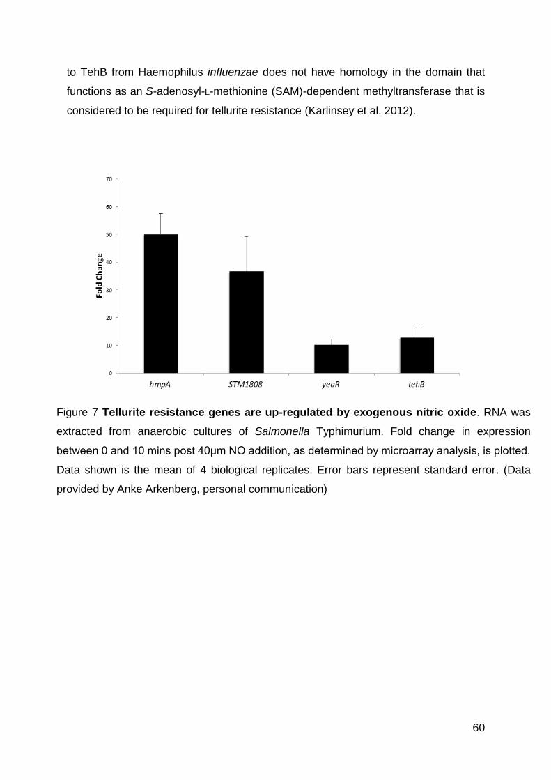
60
to TehB from Haemophilus influenzae does not have homology in the domain that
functions as an S-adenosyl-L-methionine (SAM)-dependent methyltransferase that is
considered to be required for tellurite resistance (Karlinsey et al. 2012).
Figure 7 Tellurite resistance genes are up-regulated by exogenous nitric oxide. RNA was
extracted from anaerobic cultures of Salmonella Typhimurium. Fold change in expression
between 0 and 10 mins post 40μm NO addition, as determined by microarray analysis, is plotted.
Data shown is the mean of 4 biological replicates. Error bars represent standard error. (Data
provided by Anke Arkenberg, personal communication)

61
3.1.1 Aims
This chapter aims to characterise TehB, TehB and STM1808 in relation to their roles
in nitric oxide protection by:
• Creating knockout mutants of the three genes, including a triple knockout strain
• Monitoring the survival of the strain in the presence of tellurite, aerobically and
anaerobically
• Determining the role the proteins play in NO protection and detoxification, both
by monitoring growth and measuring NO levels produced by denitrifying strains
• Investigate the tellurite resistance properties of known nitric oxide detoxification
systems
• Assess the survival of the mutant strains in macrophages

62
3.2 Results
3.2.2 Mutant construction
Knockout mutant constructs have been created for STM1808, yeaR and tehB using
the λ-red method in SL1344 (Datsenko and Wanner 2000) and described in detail in
the materials and methods. The single mutants have the pkD3 or pkD4 antibiotic
resistance cassette remaining whereas the ΔtehB ΔSTM1808 ΔyeaR triple mutant has
the cassette flipped out for the tehB and yeaR mutations but remaining for STM1808.
The cassettes were removed using pCP20. Constructs were confirmed using colony
PCR using both external binding primers and those that bind to the cassette combined
with an external primer.
Figure 8 PCR Verification of STM1808 mutant. PCR run as detailed in Methods with
an annealing temperature of 50ºC and resulting products run on an electrophoresis
gel. Lane 1: Hyperladder . Lanes 2-4: External STM1808 primers. Lane 2: WT,
expected size 660; Lane 3:STM1808 Kan 1, expected size 2078; Lane 4:STM1808
Kan 1, expected size 2078. Lanes 5-7: External STM1808 forward primer, internal
pkD4 reverse primer. Lane 5: WT, expected no band; Lane 6: STM1808 Kan 1,
expected size 789 Lane 7:STM1808 Kan 1, expected size 789.
1 2 3 4 5 6 7
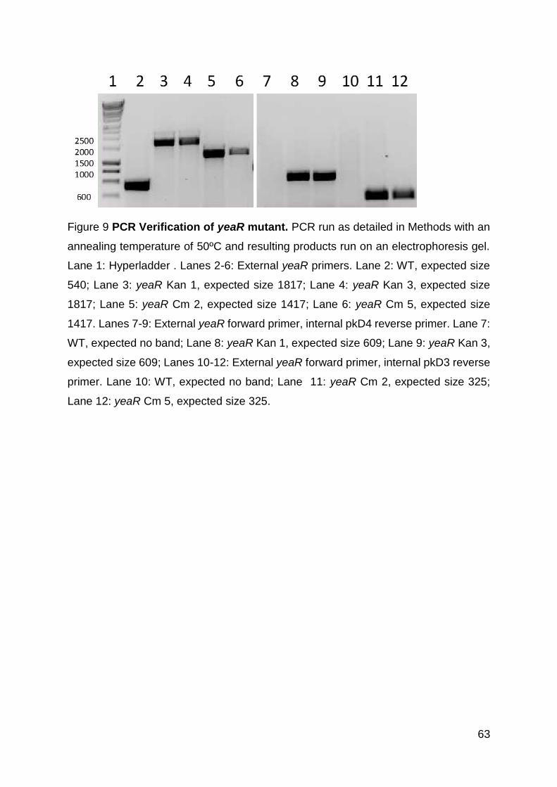
63
Figure 9 PCR Verification of yeaR mutant. PCR run as detailed in Methods with an
annealing temperature of 50ºC and resulting products run on an electrophoresis gel.
Lane 1: Hyperladder . Lanes 2-6: External yeaR primers. Lane 2: WT, expected size
540; Lane 3: yeaR Kan 1, expected size 1817; Lane 4: yeaR Kan 3, expected size
1817; Lane 5: yeaR Cm 2, expected size 1417; Lane 6: yeaR Cm 5, expected size
1417. Lanes 7-9: External yeaR forward primer, internal pkD4 reverse primer. Lane 7:
WT, expected no band; Lane 8: yeaR Kan 1, expected size 609; Lane 9: yeaR Kan 3,
expected size 609; Lanes 10-12: External yeaR forward primer, internal pkD3 reverse
primer. Lane 10: WT, expected no band; Lane 11: yeaR Cm 2, expected size 325;
Lane 12: yeaR Cm 5, expected size 325.

64
Figure 10 PCR Verification of tehB mutant. PCR run as detailed in Methods with an
annealing temperature of 50ºC and resulting products run on an electrophoresis gel.
Lanes 1 and 8: Hyperladder . Lanes 2-6: External tehB primers. Lane 2: WT, expected
size 800; Lane 3: tehB Kan 2, expected size 1903; Lane 4: tehB Kan 4, expected size
1903; Lanes 5-7: External year forward primer, internal pkD4 reverse primer. Lane 5:
WT, expected no band; Lane 6: tehB Kan 2, expected size 629; Lane 7: tehB Kan 4,
expected size 629
Figure 11: PCR Verification of yeaR tehB STM1808 triple mutant. PCR run as
detailed in Methods with an annealing temperature of 50ºC and resulting products run
on an electrophoresis gel. A: Lane 1: Hyperladder. Lanes 2-4: External yeaR primers.
Lane 2: WT, expected size 540. Lane 3: yeaR tehB STM1808 1, expected size 177;
Lane 4: yeaR tehB STM1808 6, expected size 177. Lanes 5-7: External tehB primers:
Lane 5: WT, expected size 800. Lane 6: yeaR tehB STM1808 1, expected size 403;
Lane 7: yeaR tehB STM1808 6, expected size 403. B: Lane 1: Hyperladder. Lanes 2-
4: External STM1808 primers. Lane 2: WT, expected size 720. Lane 3: yeaR tehB
STM1808 1, expected size 1817; Lane 4: yeaR tehB STM1808 6, expected size 1817.
1 2 3 4 5 6 7 1 2 3 4
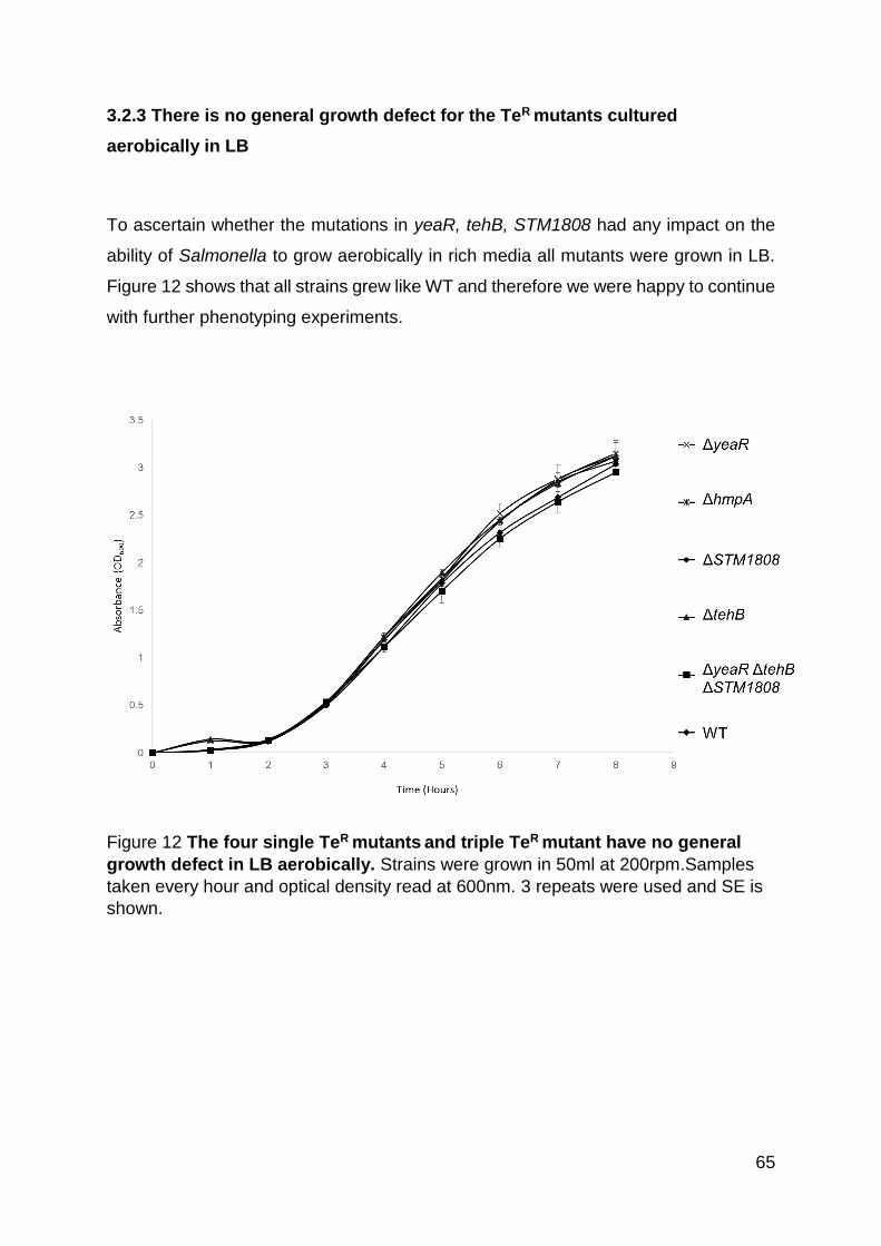
65
3.2.3 There is no general growth defect for the TeR mutants cultured
aerobically in LB
To ascertain whether the mutations in yeaR, tehB, STM1808 had any impact on the
ability of Salmonella to grow aerobically in rich media all mutants were grown in LB.
Figure 12 shows that all strains grew like WT and therefore we were happy to continue
with further phenotyping experiments.
Figure 12 The four single TeR mutants and triple TeR mutant have no general
growth defect in LB aerobically. Strains were grown in 50ml at 200rpm.Samples
taken every hour and optical density read at 600nm. 3 repeats were used and SE is
shown.

66
3.2.4 TehB, STM1808 and TehB all contribute to tellurite resistance in
Salmonella
Although referred to as putative tellurite resistance proteins on the basis of their
genome annotation (and contain a conserved domain, DUF1971, linked with tellurite
resistance) (NCBI.com, 2017) there is currently no experimental evidence, confirming
STM1808 and TehB as tellurite resistance proteins in Salmonella Typhimurium,
indeed it has been proposed that there is no role of these proteins in tellurite resistance
(Karlinsey et al. 2012). To determine whether they have any role in tellurite resistance
in Salmonella Typhimurium, ΔyeaR, ΔSTM1808, ΔtehB and ΔtehB ΔSTM1808 ΔyeaR
were tested for their ability to grow, aerobically and anaerobically, in the presence of
5 μM potassium tellurite (Figure 13).
This concentration was chosen due to the fact that the minimum inhibitory
concentration (MIC) of tellurite for E. coli is between 3.94-7.88 μM (Taylor 1999). In
both conditions the addition of tellurite had a clear toxic effect on WT Salmonella, with
a ~70% reduction in OD at 24 hours aerobically, and ~50% reduction under anaerobic
conditions. We, however, focused on how the mutant strains grew in comparison to
the WT with the addition of tellurite. Aerobically there is a clear lag in growth for both
ΔSTM1808 and ΔyeaR, which is less pronounced anaerobically, especially for ΔyeaR.
The growth defect seen for ΔtehB both aerobically and anaerobically is much more
pronounced (with a similar growth pattern seen for ΔtehB ΔSTM1808 ΔyeaR) with
almost no growth observed. The data demonstrates an important role for TehB in
protecting Salmonella against tellurite, with STM1808 and TehB also providing a
degree of tellurite resistance (TeR).

67
Figure 13 In Salmonella resistance against potassium tellurite requires TehB, STM1808 and YeaR, both aerobically and anaerobically. Strains were grown aerobically in 50ml LB (A) or anaerobically in 1ml LB (B) with the addition of 5 μM potassium tellurite

68
3.2.5 Functional overlap between genes required for tellurite resistance in
anaerobic protection against nitric oxide
Since expression of the three TeR genes was induced in the presence of NO the
mutant strains were then grown both aerobically and anaerobically in LB in the
presence of deta NONOate, an NO donor (Figure 14).
The addition of 5mM deta NONOate to LB has been shown to result in a maximum of
8 μM of NO being released, reducing to 4 μM after 24 hours, we can therefore estimate
that the addition of 10mM deta NONOate would result in a maximum of ≈16 μM of NO
(Henard and Vázquez-Torres 2012). We used ΔhmpA as a negative control and ΔnsrR
as a positive control. Consistent with previous studies (Gilberthorpe et al. 2007) there
is a growth defect for ΔhmpA both aerobically and anaerobically when exposed to an
NO donor and an increased rate of growth for ΔnsrR aerobically. The increase in
growth rate for ΔnsrR is depressed anaerobically, presumably due to the anaerobic
importance of NrfA and NorV which are controlled by additional regulators. Aerobically,
in the presence of NO, there is no growth defect for the single mutants, ΔyeaR, ΔtehB,
ΔSTM1808, mutants nor for the triple TeR mutant strain ΔtehB ΔSTM1808 ΔyeaR.
Anaerobically there is no difference in growth between the WT and the single TeR
mutants in the presence of the NO donor, however, in the triple mutant strain ΔtehB
ΔSTM1808 ΔyeaR, a clear reduction in growth rate and final OD can be seen.
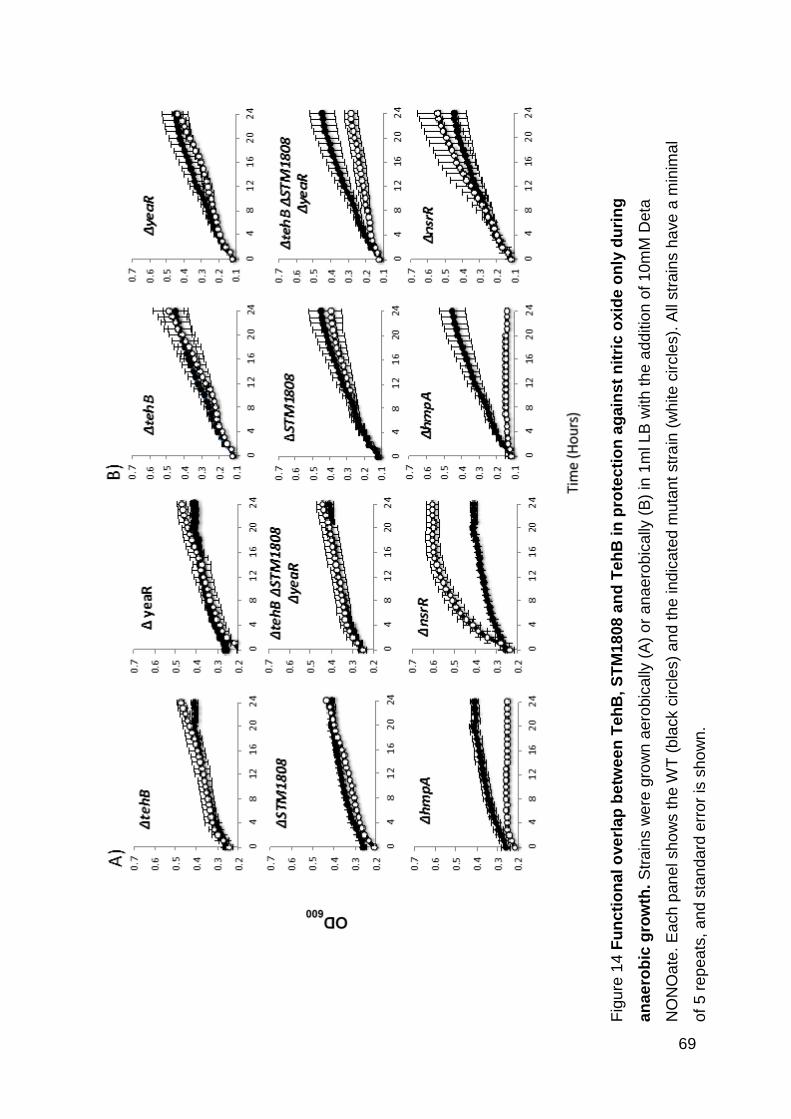
69
Fig
ure
14
Fu
ncti
on
al o
ve
rla
p b
etw
ee
n T
eh
B,
ST
M1
808
an
d T
eh
B i
n p
rote
cti
on
ag
ain
st
nit
ric
oxid
e o
nly
du
rin
g
an
ae
rob
ic g
row
th.
Str
ain
s w
ere
gro
wn
ae
robic
ally
(A
) o
r a
na
ero
bic
ally
(B
) in
1m
l L
B w
ith
the
add
itio
n o
f 1
0m
M D
eta
NO
NO
ate
. E
ach p
an
el sh
ow
s t
he
WT
(b
lack c
ircle
s)
an
d t
he
ind
icate
d m
uta
nt
str
ain
(w
hite
circle
s).
All
str
ain
s h
ave
a m
inim
al
of
5 r
ep
ea
ts, a
nd s
tan
da
rd e
rro
r is
sh
ow
n.

70
3.2.6 TehB H32 and H82 are not important for tellurite protection but are
important for nitric oxide protection
It has been previously shown in Salmonella that two histidine residues which are
conserved in the DUF1971 domain are important for the nitric oxide resistance of
STM1808 (Karlinsey et al. 2012). Numerous histidine residues were individually
mutated to alanines and both single mutants behaved as the total STM1808 knockout
strain. Further work then concluded that H82 is an important residue for zinc binding
(Karlinsey et al. 2012).
Since YeaR also possesses the DUF1971 domain with these same two conserved
histidine residues we hypothesized that these would be equally as important for nitric
oxide resistance, and possibly tellurite resistance, as in STM1808 (Figure 15). This
conserved domain is not seen in TehB (Figure 16). While both the Karlinsey study and
this work saw no growth phenotype for ΔyeaR in the presence of nitric oxide, we do
see a growth defect in the presence of tellurite. Additionally, the ΔtehB ΔSTM1808
ΔyeaR triple mutant strain does have a growth defect in the presence of nitric oxide
where a complementation effect might be able to be seen.
Six strains were constructed, the ΔtehB ΔSTM1808 ΔyeaR triple mutant was
complemented with either WT yeaR, yeaR with H32 and H82 mutated to alanines, or
the empty plasmid, from here referred to as ΔtehB ΔSTM1808 ΔyeaR PyeaR, ΔtehB
ΔSTM1808 ΔyeaR PyeaRH32AH82A or, ΔtehB ΔSTM1808 ΔyeaR Pempty,
respectively. The same plasmids were also transformed into the WT strain, with the
resulting strains being WT PyeaR, WT PyeaRH32AH82A and, WT Pempty.

71
Figure 15 Alignment of STM1808 and yeaR. Performed using Clustal W alignment.
H32 and H82 are highlighted in red.
Figure 16 Alignment of tehB, STM1808 and yeaR. Performed using Clustal W
alignment. H32 and H82 are highlighted in red.

72
All six strains were grown aerobically and anaerobically in LB to confirm no general
growth defect or advantages were present (Figure 17). The six strains were then
tested for their ability to grow aerobically in LB in the presence of 5 µM potassium
tellurite. This was not carried out anaerobically as no growth defect is seen for ΔyeaR
anaerobically (Figure 13).
The TehB complemented strain, as ΔtehB ΔSTM1808 ΔyeaR PyeaR, showed a mild
restoration in growth in comparison to the control strain, ΔtehB ΔSTM1808 ΔyeaR
Pempty (Figure 18). Both ΔtehB ΔSTM1808 ΔyeaR PyeaR and the histidine altered
complementation strain, ΔtehB ΔSTM1808 ΔyeaR PyeaRH32AH82A showed very
similar levels of growth; therefore suggesting H32 and H82 do not play an important
role in the tellurite resistance functions of TehB.
When grown in the presence of deta NONOate, ΔtehB ΔSTM1808 ΔyeaR PyeaR
shows a clear growth restoration in comparison to the control strain, ΔtehB ΔSTM1808
ΔyeaR Pempty (Figure 19). However, ΔtehB ΔSTM1808 ΔyeaR PyeaRH32AH82A
grows comparably to the triple mutant with the empty plasmid, therefore suggesting
that these two histidine residues are important for anaerobic protection against nitric
oxide.
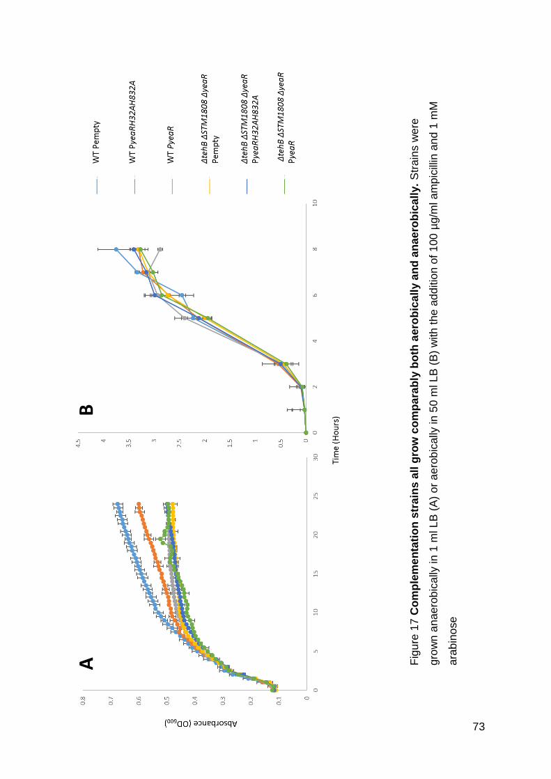
73
Fig
ure
17
Co
mp
lem
en
tati
on
str
ain
s a
ll g
row
co
mp
ara
bly
bo
th a
ero
bic
all
y a
nd
an
ae
rob
ica
lly.
Str
ain
s w
ere
gro
wn
an
ae
rob
ically
in
1 m
l L
B (
A)
or
ae
rob
ically
in
50
ml L
B (
B)
with
the
ad
ditio
n o
f 1
00
µg/m
l a
mp
icill
in a
nd 1
mM
ara
bin
ose
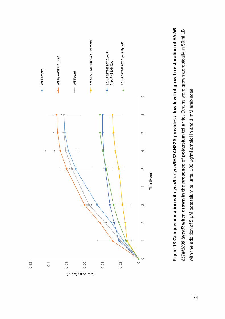
74
Fig
ure
18
Co
mp
lem
en
tati
on
wit
h y
ea
R o
r ye
aR
H32
AH
82
A p
rovid
es
a lo
w le
ve
l o
f g
row
th r
es
tora
tio
n o
f Δ
teh
B
ΔSTM1808 Δ
yea
R w
hen
gro
wn
in
th
e p
res
en
ce
of
po
tas
siu
m t
ellu
rite
. S
tra
ins w
ere
gro
wn
ae
rob
ically
in
50
ml L
B
with
th
e a
dd
itio
n o
f 5
μM
po
tassiu
m te
llurite
, 1
00
µg/m
l am
pic
illin
and
1 m
M a
rab
ino
se
.
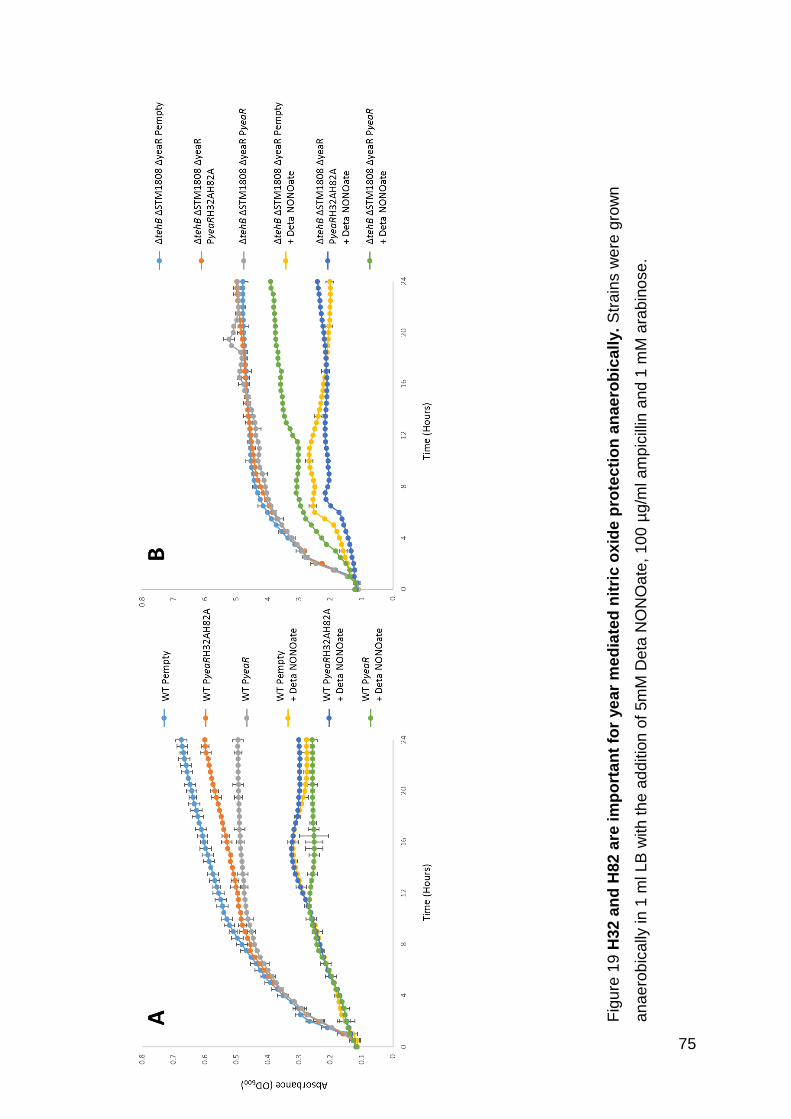
75
Fig
ure
19
H32
an
d H
82
are
im
po
rta
nt
for
ye
ar
me
dia
ted
nit
ric o
xid
e p
rote
cti
on
an
ae
rob
ica
lly.
Str
ain
s w
ere
gro
wn
an
ae
rob
ica
lly in
1 m
l LB
with
th
e a
dd
itio
n o
f 5m
M D
eta
NO
NO
ate
, 10
0 µ
g/m
l am
pic
illin
and 1
mM
ara
bin
ose
.

76
3.2.7 Nitric oxide accumulates during nitrate respiration in ΔtehB ΔSTM1808
ΔyeaR triple mutant
In order to ascertain if the TeR proteins have a direct role in detoxifying NO we grew
the strains anaerobically with nitrate as the alternative electron acceptor and
measured production of endogenous NO. Firstly the WT strain was grown and 3 ml
samples of headspace gas taken at 4 timepoints over 48 hours (Figure 20). NO levels
increased until 40 hours and then were reduced at 48 hours. We therefore determined
to efficiently compare levels of NO between the mutant strains measurements should
be taken at 40 hours. In order to obtain more accurate NO levels the entire headspace
(≈ 50 ml) was used for future results, the experiment was therefore sacrificed at this
timepoint.
Under these conditions (M9 minimal media, anaerobic, 20 mM nitrate) all the strains
grew comparably to WT. To establish the validity of our experimental set up, NO
released by ΔhmpA and ΔnsrR was measured and had higher and lower levels of
headspace NO respectively, with 1.59 ±0.16 μM and 0.31 ±0.15 μM NO compared to
0.72 ±0.14 μM from the WT Salmonella strain. We observed no significant (p < 0.05)
difference in levels of NO released from any of the single TeR mutants, although a
slight increase can be seen for ΔtehB (1.07 ±0.04 μM NO). However, the triple TeR
mutant strain, ΔtehB ΔSTM1808 ΔyeaR, releases significantly (p < 0.05) higher levels
of NO than the WT, with 1.33 ±0.11 μM NO detected, further supporting the functional
redundancy between the TeR proteins in directly detoxifying NO (Figure 21).

77
Fig
ure
20
NO
le
ve
ls p
ea
k a
t 4
0 h
ou
rs f
or
WT
Sa
lmo
nell
a. W
T w
as g
row
n in
M9
min
ima
l m
ed
ia
su
pp
lem
en
ted
with
40m
M n
itra
te a
nd
th
e N
O m
ea
su
red
fro
m 3
ml sam
ple
s ta
ken
fro
m t
he
he
ad
sp
ace
. 4
rep
ea
ts w
ere
use
d a
nd S
E is s
ho
wn
.
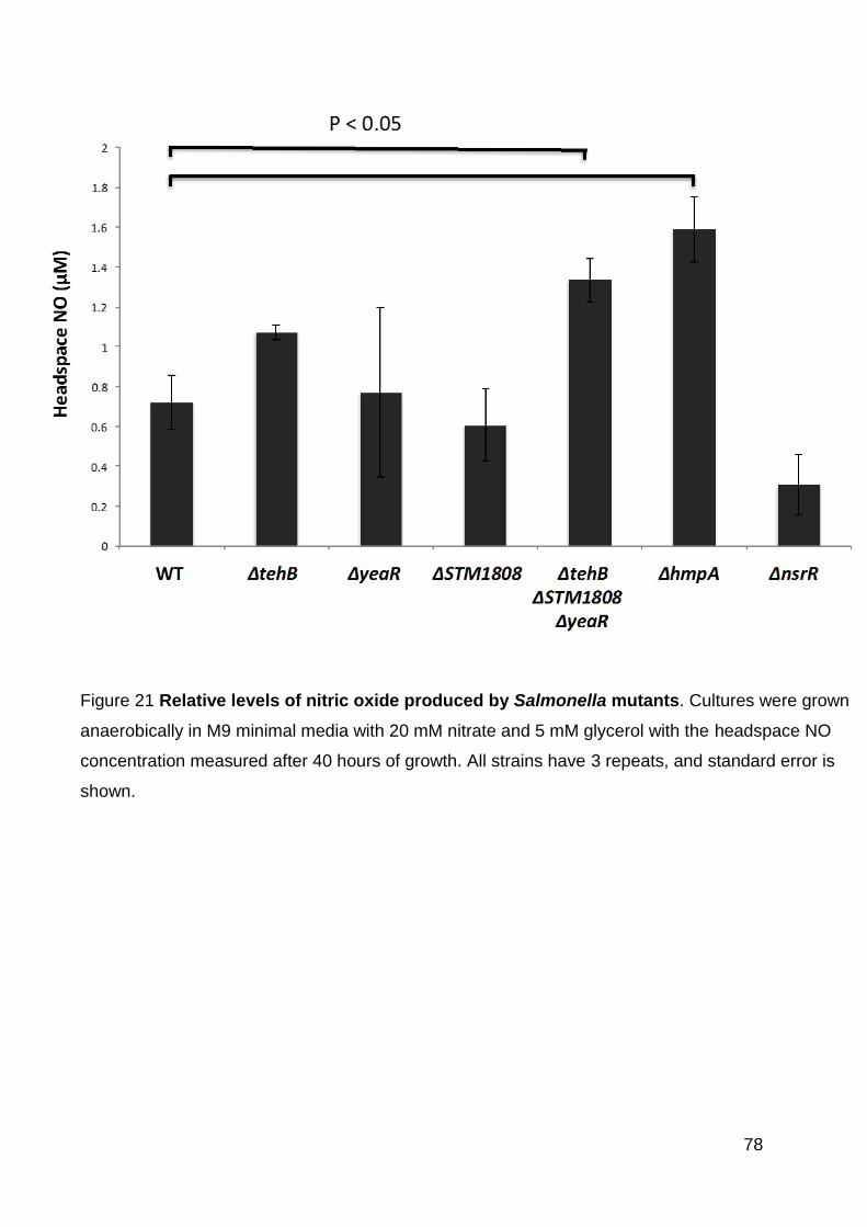
78
Figure 21 Relative levels of nitric oxide produced by Salmonella mutants. Cultures were grown
anaerobically in M9 minimal media with 20 mM nitrate and 5 mM glycerol with the headspace NO
concentration measured after 40 hours of growth. All strains have 3 repeats, and standard error is
shown.

79
3.2.8 Proteins involved in protection against reactive nitrogen species also
protect against potassium tellurite
Having demonstrated a role for the TeR proteins in both NO and tellurite resistance we
sought to ascertain whether there is a correlation between tellurite and nitric oxide
resistance. To do this four strains, each carrying a mutation in a system implicated in
NO protection were exposed to tellurite (Figure 22). Aerobically there was no growth
defect for the ΔytfE, ΔnrfA or ΔnorV mutants in the presence of tellurite. However,
there was a distinct growth defect in the absence of the key NO detoxification protein
HmpA. Anaerobically there is a clearer correlation between NO and tellurite resistance
with YtfE, NrfA and NorV all exhibiting a degree of protection against tellurite toxicity.
HmpA, however, does not appear to have a role in anaerobic tellurite protection. As
NsrR is the master regulator of NO detoxification in Salmonella the most significant
case for support of a correlation between NO and tellurite resistance can be seen with
the phenotype of the NsrR mutant. Aerobically the disruption of NsrR, allowing for the
up-regulation of the entire NsrR regulon, has a clear benefit to growth in the presence
of tellurite in comparison to the WT. After 8 hours the OD600 for the ΔnsrR mutant had
reached ≈0.50 as opposed to ≈0.08 for WT. This enhanced growth phenotype is not
apparent in the absence of oxygen, with similar maximal growth for WT and ΔnsrR.
However, the lag phase is shorter for the ΔnsrR mutant (at ~9 hrs) as opposed to ~14
hours for the WT. Additionally, this correlates with a two-fold increase in the aerobic
MIC of tellurite for the ΔnsrR mutant, 10 μM compared to 5 μM for the WT.

80
Fig
ure
22
Ge
ne
s i
nvo
lve
d i
n R
NS
de
fen
se
are
als
o i
nvo
lve
d i
n r
es
ista
nce
ag
ain
st
po
tas
siu
m t
ellu
rite
. S
tra
ins
we
re g
row
n a
ero
bic
ally
(A
) o
r a
na
ero
bic
ally
(B
) in
1m
l L
B w
ith
the
add
itio
n o
f 5 μ
M p
ota
ssiu
m te
llurite
. E
ach
pa
ne
l
sh
ow
s t
he
WT
(b
lack c
ircle
s)
an
d t
he
ind
icate
d m
uta
nt
str
ain
(w
hite
circle
s).
All
str
ain
s h
ave
a m
inim
al of
5 r
ep
ea
ts,
and
sta
nda
rd e
rro
r is
sho
wn
.

81
3.2.9 MIC of tellurite, selenite and hydrogen peroxide for WT and ΔtehB
ΔSTM1808 ΔyeaR
The toxicity of tellurite is frequently attributed to the production of ROS (Pérez et al.
2007, Borsetti et al. 2005) and it is also known that at comparable levels selenite
produces 4 fold higher levels of ROS than tellurite (Vrionis et al. 2015). One would
therefore assume that if the toxicity of tellurite was attributable to the release of ROS
that the MIC of selenite would be approximately 4 times lower than that of tellurite.
However, we have shown that the MIC of selenite in Salmonella is ≈1000 times higher
than tellurite (>4.9 mM vs 5.0 μM) suggesting that the toxicity observed with tellurite is
not due to production of ROS (Table 7). Importantly, the MICs of H2O2 and selenite for
the WT and the tellurite hypersensitive strain, ΔtehB ΔSTM1808 ΔyeaR are identical
at 1.25 μM and >4.87 mM respectively. This suggests that the hypersensitivity to
tellurite observed in ΔtehB ΔSTM1808 ΔyeaR is therefore not solely due to ROS.
Table 7 MIC assay. WT and ΔtehB ΔSTM1808 ΔyeaR grown aerobically in 100uL LB
to calculate MIC of potassium tellurite (K2O3Te), potassium selenite (K2O3Se) and
hydrogen peroxide (H2O2).
Strain
Compound WT ΔtehB ΔSTM1808
ΔyeaR
Tellurite 5.00 μM 2.50 μM
Selenite > 4.87 mM > 4.87 mM
Hydrogen Peroxide 1.25 μM 1.25 μM

82
3.2.10 The ΔtehB ΔSTM1808 ΔyeaR triple mutant is severely attenuated in IFN-
γ activated macrophages
One of the key host defense mechanisms that challenges Salmonella survival in host
macrophages is the production of NO via iNOS. In macrophages, NO levels can be
stimulated by interferon gamma (IFN-γ) or inhibited by addition of NG-nitro-L-arginine
methyl ester (L-NAME) upon hydrolysis to NG-nitro-L-arginine (L-NOARG) (Pfeiffer et
al. 1996). When L-NAME treated macrophages were infected with either WT or ΔtehB
ΔSTM1808 ΔyeaR there was no significant difference between the two strains in fold
change in bacterial numbers between 2 and 24 hours post infection (pre and post the
NO burst). When IFN-γ activated macrophages were infected with either WT or ΔtehB
ΔSTM1808 ΔyeaR there was significantly (p< 0.01) lower survival of ΔtehB
ΔSTM1808 ΔyeaR in comparison to WT with a 40.8 ±3.87 fold reduction for the triple
TeR strain compared to 10.8 ±3.93 fold for WT (Figure 23).

83
Figure 23 The ΔtehB ΔSTM1808 ΔyeaR triple mutant is severely attenuated in IFN-γ
activated macrophages. Fold change in cfu/ml between 2 and 24 hours post infection is
shown for L-NAME treated (filled) and IFN-y activated (shaded) RAW 264.7 macrophages.
6 repeats were used and SE is shown. A student’s t-test was used.

84
3.3 Discussion:
In this chapter we set out to identify any uncharacterized genes involved in anaerobic
NO detoxification by Salmonella Typhimurium and in doing so highlighted a connection
between NO and tellurite detoxification. We have shown that TehB, YeaR and
STM1808 are all involved in tellurite resistance in Salmonella, particularly aerobically;
and that they provide resistance against NO, which our data suggests they play a role
in detoxifying. A ΔtehB ΔSTM1808 ΔyeaR triple mutant has also been shown to be
severely attenuated in activated macrophages.
Of the three TeR proteins in this study, TehB has been the best characterised in E. coli
studies. It has, however, been reported that TehB in Salmonella, which is homologous
to TehB from Haemophilus influenzae, does not have homology in the domain that
functions as an S-adenosyl-L-methionine (SAM)-dependent methyltransferase that is
considered to be required for tellurite resistance (Karlinsey et al. 2012). We however
found TehB to be the most important of the three proteins for TeR in Salmonella both
aerobically and anaerobically (Figure 14). We have also shown a distinct phenotype
for both ΔyeaR and ΔSTM1808 when grown aerobically in the presence of 5 μM
potassium tellurite. Anaerobically this phenotype is reduced but is still visible, certainly
at least for ΔSTM1808. This is in contrast to the suggestion that the methyltransferase
domain that is lacking from TehB in Salmonella is vital for its role in protecting against
tellurite (Karlinsey et al. 2012); and confirms the putative function of all three proteins.
Although we have confirmed here the role of all three proteins, TehB, STM1808 and
TehB in TeR, it remains the case that tellurite is probably not physiologically relevant,
at least not at this stage of Salmonella’s evolution. Therefore, the fact that Salmonella
has retained three independent systems for this function does imply that they have
another important function and, from the results shown here, we propose this is in NO
detoxification. Aerobically there is no growth defect for the mutant strains, including
the triple mutant strain in the presence of 10 mM deta NONOate; but anaerobically
there is a clear phenotype when all three systems are missing. Previous studies have
shown a low level of protection provided by STM1808 against NO added to aerobic
cultures, but not a requirement for TehB (Karlinsey et al. 2012). Therefore, we
85
conclude that there is functional overlap between the three systems for anaerobic NO
protection.
In order to ascertain whether this protection is as a direct result of NO detoxification
as opposed to responding to the large amount of damage caused by NO in the cell,
we grew the mutant strains under denitrifying conditions and measured the levels of
NO released (Figure 21). While ΔtehB and ΔyeaR both accumulated higher levels of
NO than the WT this was not significant, however, the triple TeR showed significantly
higher levels of NO release. This confirmed the functional overlap between the three
TeR genes and gives strong indication that they are directly involved in NO
detoxification/reduction. It is not clear at this stage the mechanism by which this
detoxification occurs and the protein biochemistry of this enzymatic activity warrants
further work.
It was noted by Karlinsey et al., (2012) that two histidine residues, H32 and H82, in
STM1808 are important for the nitric oxide protection exhibited by the protein. These
residues are part of the DUF1971 which is conserved within proteins thought to be
involved in tellurite resistance. It was highlighted that a mutation of either of these
histidine residues in STM1808 was equivalent to knocking out the entire gene when
the strain was challenged with nitric oxide. Since STM1808 and YeaR are highly
homologous, as shown in Figure 15, with H32 and H82 being conserved, we
hypothesized that similarly to in STM1808 these residues would be vital for the role
YeaR plays in nitric oxide detoxification. The residues are not conserved within TehB
(Figure 16). Since, due to the functional redundancy of TehB, STM1808 and YeaR,
we only see a mild phenotype for the single YeaR knockout mutant we therefore
complemented the triple TeR mutant with either wildtype YeaR or YeaR with H32 and
H82 mutated to alanines.
These complementation strains were first tested for their ability to grow in the presence
of 5 µM potassium tellurite (Figure 18) where there was a 1.5 fold increase in final OD
for ΔtehB ΔSTM1808 ΔyeaR PyeaR compared to ΔtehB ΔSTM1808 ΔyeaR Pempty
(0.043 v. 0.029) taking it from 32% of the final OD for WT Pempty to 47%. However,
the ΔtehB ΔSTM1808 ΔyeaR PyeaRH32AH82A strain grew comparably, with a similar
final OD of 0.04. In combination these results show that while TehB does play a role
in the protection against tellurite in Salmonella, these two histidine residues are not
important for this function. The role of the histidine residues in tellurite resistance by

86
STM1808 has not been looked at previously or in this study; this could be something
of interest (Karlinsey et al. 2012).
In contrast, there is a role for H32 and H82 in TehB-mediated nitric oxide protection
(Figure 19). Complementation of the triple TeR mutant strain with WT TehB resulted in
a restoration of growth, with the final OD reaching 0.390 in comparison to 0.201 for
the control strain ΔtehB ΔSTM1808 ΔyeaR Pempty in the presence of deta NONOate
and 0.497 in plain LB. Under these conditions however the same was not seen for
ΔtehB ΔSTM1808 ΔyeaR PyeaRH32AH82A with the final OD only slightly higher than
the control strain at 0.241. We therefore conclude that H32 and H82 of YeaR are
important for nitric oxide, but not tellurite, protection in Salmonella. This result has two-
fold implications, the first being that there is a clear similarity between the functionality
of STM1808 and YeaR in nitric oxide detoxification. In STM1808 these residues have
been shown to be important for zinc binding – this could be looked at in YeaR.
Secondly, the fact that the two histidine residues are important for one function of
YeaR, nitric oxide protection, but not another, tellurite resistance, suggests that the
protein is not working in precisely the same way for these two separate functions.
Due to the role of the TeR proteins in NO detoxification we then tested whether the
converse is true, that in Salmonella any strain that is sensitive to NO is also sensitive
to tellurite; this could give an indication of the cause of toxicity. We demonstrated that
the well characterised NO oxygenase/reductase, HmpA, is very important for tellurite
resistance aerobically, but not anaerobically. When the NO sensitive repressor, NsrR,
is deleted the entire NsrR regulon (consisting of hcp-hcr, ytfE, ygbA, hmp, yeaR-yoaG
and STM1808) is upregulated (Karlinsey et al. 2012). In the presence of tellurite ΔnsrR
grows much better than WT aerobically and, although the phenotype is less
pronounced anaerobically, there is still a clear role for the NsrR regulon in tellurite
resistance; suggesting that the genes regulated by NsrR play a key role in tellurite
resistance as well as in NO resistance. We also tested three further proteins that have
been implicated in NO resistance, for their involvement in tellurite resistance. One of
these, YtfE (Gilberthorpe et al. 2007) is NsrR regulated, but the other two, NrfA and
NorV (Mills et al. 2005) are not. NorV and NrfA are known to be involved in NO
resistance anaerobically and we have shown involvement in resistance against
tellurite anaerobically. From our data, we conclude that there is a clear correlation
between tellurite resistance and NO resistance in Salmonella. There is the possibility

87
that in LB, TeO3-2 leads to the release of NO and therefore the tellurite phenotype, and
hence the correlation we are seeing is actually due to NO release post tellurite
addition. The mechanism by which this may occur is unknown however there would
be a varied mix of nitrogen and oxygen containing molecules present so it is feasible.
We however do not believe this to be the case – the phenotypes we see for tellurite
and NO are not directly comparable; and moreover we attempted to measure the NO
levels from aerobic and anaerobic LB media containing different combinations of
bacteria and tellurite and saw no NO release (data not shown). The results from our
study on the importance of two histidine residues in YeaR also would not correlate
with this hypothesis; the two residues are only seen to be important for protection
against NO, if tellurite was simply triggering the release of NO then the residues would
be important under both nitric oxide and tellurite stress.
There are indications that tellurite toxicity is as a result, at least in part, of reactive
oxygen species (ROS), which are released, when TeO32- is reduced to Te. Superoxide
dismutase was upregulated in E. coli and Rhodobacter capsulatus in response to
tellurite and, ROS protection mutant strains had increased tellurite sensitivities (Pérez
et al. 2007, Borsetti et al. 2005). However, it has been shown that treatment of E. coli
with selenite results in at least four times as much ROS than tellurite (Vrionis et al.
2015). In our study the MIC of selenite (>4.875 mM) is much higher than that of tellurite
(5 μM) (Table 1). This suggests that it is not solely the induction of ROS production
that causes the toxic effects of tellurite. We also exposed the tellurite hypersensitive
ΔtehB ΔSTM1808 ΔyeaR to H2O2 and selenite and saw no difference in sensitivity
than the WT – indicating that the tellurite sensitivity of this strain is not due to ROS. It
may be worth testing the sensitivity of the triple mutant to other sources of ROS, such
as paraquat, as there is the potential that different results would be seen.
STM1808 has previously been shown to be important for virulence in the murine
model, with the authors suggesting a role in protection against nitrosative stress in
macrophages (Karlinsey et al. 2012). In this study, we showed the tellurite
hypersensitive ΔtehB ΔSTM1808 ΔyeaR strain to be attenuated in IFN-γ activated
macrophages. The fact that we see this attenuation only in IFN-γ activated
macrophages and not in L-NAME treated macrophages, which will fail to produce the
nitrosative burst, further implicates these proteins in NO defense. It would be of merit
to repeat these experiments treating the macrophages both with IFN-γ and L-NAME

88
in order to confirm the differences we see between the IFN-γ and L-NAME samples
are solely due to NO release and not other defensive mechanisms controlled by
IFN-γ.
This work has raised a new area of research for the enigma of tellurite toxicity – the
link between tellurite sensitivity and NO sensitivity; and here there are still many
unanswered questions. Originally, was the primary role of these three TeR proteins in
protection against tellurite, but when over time the environmental conditions
Salmonella encounters changed, these proteins were retained due to their secondary
role in NO resistance? Or conversely are these proteins simply involved in NO
detoxification and by chance have the ability to protect Salmonella against tellurite? If
the latter is the case we perhaps would expect other tellurite resistance systems,
(KlaABC, TerZABCDEF, Tmp, TrgAB and CysK) to contribute to NO detoxification
and/or tellurite resistant bacteria such as Corynebacterium diphtheriae, Streptococcus
faecalis, and some drug resistant Staphylococcus aureus strains (Summers and Silver
1978) to have increased resistance to NO.
There is one previous study linking nitrogen metabolism to tellurite, this links NarG,
the nitrate reductase, to tellurite detoxification (Avazéri et al. 1997). They also
identified an additional, unidentified, soluble protein which showed tellurite reductase
activity. There was no tellurite sensitivity of the NarG mutant under anaerobic
conditions, and the authors suggest this is due to the additional protein they identified;
there is the potential that this is one of the proteins studied in this chapter. Avazéri et
al., did not indentify the mechanism of tellurite reduction by NarG and could not
conclude whether this single protein is responsible for the total reduction of Te4 to
elemental tellurium, Te0. The pathways of tellurite and nitrate reduction are somewhat
similar and as such we propose the potential model shown in Figure 24.
This model suggests that Salmonella treats tellurite very similarly to nitrate, reducing
it via the same pathway.

89
Fig
ure
24
Te
llu
rite
re
du
cti
on
mo
del

90
While this chapter has raised numerous further questions it has also achieved the
original intention of this work, which was to characterize novel anaerobic nitric oxide
detoxifiers. It is well known that the ability of Salmonella to survive within macrophages
is vital to the progression of Typhoid fever (Page 25) and therefore that a strain which
is unable to survive within this environment will not be able to progress into the
systemic, fatal, stage of the disease. While STM1808 and TehB have been studied
before in relation to nitric oxide detoxification (Karlinsey et al. 2012), their full effect,
and therefore potential importance in this area, was not seen due to the functional
overlap exhibited between these two proteins and TehB.
3.3.1 Future Work
The first thing that must now be done is protein biochemistry to confirm the role of
YeaR, TehB and STM1808 in direct nitric oxide detoxification. This could be in the
form of protein film voltammetry, a technique that has previously been used to quantify
the NO reductase activity of NrfA from E. coli (van Wonderen et al. 2008). This
technique works by immobilizing the enzyme onto an electrode, placing this in the
potential substrate and monitoring any charge produced due to the flow of electrons
(Angove et al. 2002).
Further work should be done to assess the potential for these three proteins in
combination with others as a potential vaccine strain. The first steps would be to
combine these three mutations with other proteins such as HmpA, NrfA and NorV and
assess the strain’s survival in macrophages. We would expect such a strain to have a
very severe attenuation.

91
4. The role of CstA, Hcr and STM1273 in nitric oxide and
tellurite resistance

92
4.1 Introduction:
Chapter 3 looked at the proteins transcribed by three genes which were upregulated
in response to NO and importantly all had the same annotated function in tellurite
resistance. This chapter looks at three further genes which were upregulated under
the same conditions but at the time had not been confirmed to have a role in nitric
oxide protection or detoxification. Background information on these three proteins,
CstA, Hcr and STM1273 is given below.
Two microarray analyses by Arkenberg et al., (personal communication) revealed
upregulation of these three genes both when subjected to an exogenous bolus of nitric
oxide (40 μM) or grown under denitrifying conditions which will cause Salmonella to
release both NO and N2O. cstA exhibited similar levels of upregulation in both of these
analyses with a 4 fold increase from exogenous exposure and 7 fold during
endogenous production. hcr was very highly upregulated under denitrifying conditions
with a 77 fold increase in expression, but was also 7 fold upregulated by an exogenous
NO bolus. STM1273 was upregulated 2.4 fold post exogenous NO exposure and not
significantly changed when respiring on nitrate.

93
4.1.1 CstA
CstA has been described as a peptide transporter, contains eighteen
transmembrane domains and is annotated as a carbon starvation protein, being
cAMP-CRP (cyclic adenosine monophosphate-catabolite regulation protein)
dependent in E. coli; that is when carbon levels drop cAMP levels rise and activate
expression of the gene (Blum et al. 1990, Garai et al. 2015). Glucose starvation of
E.coli results in a 6 fold increase in expression levels (Blum et al. 1990). CstA acts
as a virulence factor in Salmonella; as a cstA mutant had a 67% reduction in C.
elegans killing compared to WT. A cstA mutant was also 39% reduced in its ability to
initiate polymorphonuclear leukocytes migration and 54% reduced in epithelial cell
invasion (Tenor et al. 2004). There has been no study looking at this virulence
further, for example in the mouse model. Most studies on cstA have looked at its
regulation. CsrA, a global regulator, has been shown to translationally regulate CstA
in E. coli, the main targets of CsrA regulation are proteins involved in central carbon
metabolism, motility, and biofilm development (Dubey et al. 2003).
Another regulator is YjiY, in both S. Typhi and S. Typhimurium cstA has been shown
to be the predominant gene regulated by this protein. In E. coli YjiY is regulated by the
two-component system YehUT, which is conserved throughout the majority of enteric
bacteria. Similarly, to YjiY transcriptome analysis identified cstA as the predominantly
yehUT regulated gene in both S. Typhi and S. Typhimurium (Wong et al. 2013). The
YehUT operon of S. Typhi has been found to contain a higher than average level of
mutations, suggesting adaptive evolution is occurring; potentially indicating an
importance for this operon (Wong et al. 2013).
4.1.2 Hcr
Hcr is transcribed in an operon with Hcp, a hybrid cluster protein. Hcr catalyses the
reduction of the Hcp with NADH as the electron donor. In E. coli the two proteins are
only produced when the bacteria is grown in the presence of nitrate and nitrite –
suggesting a role in nitrogen metabolism (van den Berg, Hagen and van Dongen 2000,
Overton et al. 2006). There has been suggestion that hcr functions as a hydoxylamine
reductase and has peroxidase activity (Almeida et al. 2006, Wolfe et al. 2002).

94
A 2007 study tested the importance of Hcr for aerobic survival in LB in the presence
of 3 mM GSNO (a NO donor) and saw no growth defect for a S. Typhimurium hcr
mutant (Gilberthorpe et al. 2007). However, a role for Hcr in protection against NO-
mediated respiration inhibition has been shown in Salmonella. Since HmpA is said to
be the most important protein for NO detoxification, Karlinsey et al., constructed their
mutants in a ΔhmpA background to avoid this masking any functions, additionally a
nsrR deletion was used to avoid any interactions of NsrR related genes. The growth
profile of this ΔnsrR ΔhmpA Δhcr strain was monitored in LB containing 3mM
Spermine-NONOate and a growth defect was seen (Karlinsey et al. 2012). In E. coli,
Hcr has been confirmed to be a high affinity NO reductase during anaerobic growth
(Wang et al. 2016).
NsrR has been confirmed to regulate hcp-hcr in both E. coli and Salmonella (Karlinsey
et al. 2012, Filenko et al. 2007). In E. coli, hcp-hcr, hmpA, and ytfE are the three
transcripts most tightly regulated by NsrR (Filenko et al. 2007).
The hcp-hcr operon is induced in activated RAW 264.7 macrophages but not by
untreated macrophages or those where iNOS activity has been inhibited. (Kim,
Monack and Falkow 2003). Infection studies using C57BL/6J mice surprisingly
revealed increased killing of the mice for low doses of the hcp and hcr mutants (termed
nipA and nipB respectively) (Kim et al. 2003). It was then noted by Karlinsey et al.,
that C57Bl/6J mice lack a functional Nramp1 locus and as such have reduced NO
production, and so a study was conducted with C3H/HeN mice which do not lack this
locus. This study inoculated mice with a 1:1 ratio of WT and the mutant and examined
surviving bacteria in numerous organs. However, hcp mutants outcompeted the WT
strain indicating the lack of hcp results in increased survival of Salmonella and
corroborating the results of Kim et al. (Karlinsey et al. 2012).
4.1.3 STM1273
STM1273 lies 410bp downstream of yeaR, the role of which in nitric oxide and tellurite
resistance was confirmed in Chapter 3. There is very little in the literature about this
protein, however it was originally annotated as a putative nitric oxide reductase
(McClelland et al. 2001). The RyhB sRNA have mainly been shown to affect translation
of proteins requiring iron in response to low iron availability. One of the RhyB

95
homologs, ryhB-2 overlaps with STM1273, leading to the suggestion that STM1273
may be a target of RyhB regulation (Kim and Kwon 2013a, Kim and Kwon 2013b, Kim
2016). Previous work from the Rowley lab found no role for STM1273 anaerobically in
the presence of 40 μM aqueous NO. NO sensitivity had not been looked at during
aerobic growth. The fact that STM1273 is only upregulated after a NO bolus and not
during nitrate respiration suggests that it may be more important for exogenous rather
than endogenous NO detoxification.
4.2 Aims
Based on the results of the previous chapter, which found a link between NO and
tellurite protection both aerobically and anaerobically in Salmonella, this chapter will
further assess the importance of three proteins in both NO and tellurite resistance.
• Determine the role of CstA, Hcr and STM1273 in NO protection, aerobically and
anaerobically.
• Assess the importance of CstA, Hcr and STM1273 in macrophage survival
• Challenge ΔcstA, Δhcr and ΔSTM1273 with potassium tellurite to assess if they
play a role in protection

96
4.3 Results
4.3.1 Δhcr ΔSTM1273 and ΔcstA grow comparably to WT in LB both aerobically
and anaerobically
Deletion mutants (Arkenberg, personal communication) were first tested for any
general growth defects on LB both aerobically and anaerobically. All three mutant
strains grew comparably to WT (Figure 25).
Figure 25 The three mutant strains, Δhcr, ΔSTM1273 and ΔcstA grow comparably
to WT in LB. Conducted both aerobically (A) and anaerobically (B). Strains were grown
for 24 hours in 1 ml LB in a SpectraMax plate reader. Data shows 3 repeats.

97
4.3.2 STM1273, CstA and Hcr are important for nitric oxide protection both
aerobically and anaerobically
In order to determine the contribution of STM1273, CstA and HmpA to the survival of
Salmonella when challenged with NO, deletion mutants of STM1273, cstA and hcr
were grown in the presence of 5 mM Deta NONOate both aerobically and
anaerobically (Figure 26). Aerobically the three strains, ΔSTM1273, ΔcstA and Δhcr
showed similar levels of growth, with the final ODs being 67%, 61% and 66% of WT’s,
respectively. Anaerobically, again, the three strains ΔSTM1273, ΔcstA and Δhcr
showed similar levels of growth to each other, with the final ODs being 72%, 71% and
69% of WT’s. This therefore confirms the role of all three proteins in both aerobic and
anaerobic exogenous nitric oxide protection.

98
Figure 26 STM1273, CstA and Hcr are important for nitric oxide protection both
aerobically and anaerobically. Strains were grown both aerobically (A) and
anaerobically (B) in 1ml LB in a SpectraMax plate reader for 24 hours, with the addition
of 5 mM Deta NONOate. Solid black circles indicate the WT and white circles the
indicated mutant strain. All strains have a minimum of 3 repeats and SE is shown.

99
4.3.3 Nitric oxide accumulates in Δhcr in comparison to WT when grown under
denitrifying conditions
When grown in minimal media with the addition of nitrate and glycerol, Salmonella will
use the provided nitrate as a terminal electron acceptor and undergo denitrification.
Nitric oxide is produced as an intermediate of denitrification. We would therefore
expect a strain lacking a gene which is important in the reduction of nitric oxide to
accumulate greater levels than WT. When cultured anaerobically on M9 with 20 mM
nitrate, both ΔcstA and ΔSTM1273 produced slightly lower levels of NO than WT at
40 hours, suggesting they play no direct role in NO detoxification (Figure 27). There
was however a near 2-fold significant increase in the level of NO which accumulated
in the Δhcr mutant suggesting that this protein plays a direct role in the reduction of
NO (Figure 27).
Figure 27 Nitric oxide accumulates in Δhcr in comparison to WT when grown under
denitrifying conditions. Strains were grown in M9 minimal media with the addition of 20mM
NaO3 and 10mM glycerol. Nitric oxide from the headspace was measured at 40 hours. All strains
have a minimum of 3 repeats, SE is shown and a student’s T Test was used.

100
4.3.4 Δhcr and ΔcstA are significantly attenuated in activated macrophages
Macrophages release NO as an integral part of the host response via iNOS, the
inducible nitric oxide synthase. iNOS can be upregulated by interferon gamma (IFN-
γ) or inhibited by addition of NG-nitro-L-arginine methyl ester (L-NAME) (Pfeiffer et al.
1996). Therefore, IFN-γ-activated macrophages release higher, more physiologically
relevant, levels of NO and L-NAME-treated macrophages will release low or no NO.
Bacterial counts were taken 2 and 24 hours post macrophage infection and the fold
reduction in numbers is shown (Figure 28). As would be expected, all strains had
increased survival when the macrophages were treated with L-NAME in comparison
to IFN-γ. There is a significantly greater reduction in bacterial numbers for ΔcstA in
comparison to WT, this would suggest that this protein plays a role in survival inside
macrophages which is not based on NO detoxification. All three strains, Δhcr, ΔcstA
and ΔSTM1273 show a virulence defect in IFN-γ treated macrophages. The WT strain
has a 13.8 fold reduction in cfu/ml of Salmonella between 2 and 24 hours, this
increases to 22.2 fold for ΔcstA, 22.3 fold for ΔSTM1273 and 26.8 fold for Δhcr.

101
Figure 28 Hcr and CstA are important for survival within activated macrophages. Fold
change in cfu/ml between 2 and 24 hours post infection is shown for L-NAME treated (filled) and
IFN-y activated (shaded) RAW 264.7 macrophages. 4 repeats were used and SE is shown. A
student’s t-test was used.

102
4.3.5 STM1273, Hcr and CstA all offer Salmonella protection against potassium
tellurite in the presence and absence of oxygen.
Following on from Chapter 3, once Hcr, CstA and STM1273 had been shown to be
important for nitric oxide protection we wanted to see if like other known nitric oxide
detoxification systems, YtfE, NorV, NrfA, HmpA, there was a role for these proteins in
potassium tellurite protection. All three mutants exhibited a slight growth defect both
aerobically and anaerobically in the presence of potassium tellurite (Figure 29). It
should be noted in contrast that YtfE, NorV and NrfA only appear to play a role in TeR
anaerobically, and HmpA only aerobically (Figure 22).

103
Fig
ure
29
ST
M1
27
3, C
stA
an
d H
cr
all
pla
y a
ro
le i
n t
ell
uri
te p
rote
cti
on
, b
oth
ae
rob
ica
lly a
nd
an
ae
rob
ica
lly.
Str
ain
s w
ere
gro
wn
ae
rob
ica
lly in
50
ml L
B (
A)
or
an
ae
rob
ica
lly in
1m
l L
B (
B)
with
th
e a
dd
itio
n o
f 5
μM
pota
ssiu
m
tellu
rite
.
104
4.4 Discussion
The importance for Salmonella to efficiently detoxify nitric oxide and the systems in
place to do this has been discussed in detail in the introduction (Page 33) and
expanded upon in Chapter 3.
Of the three proteins studied in this chapter, Hcr has been the most well characterised,
especially in regards to NO protection. It is known to be NsrR regulated in Salmonella
which infers a strong likelihood to being involved in NO protection. Additionally, Hcr
has been shown to play a role in NO-mediated respiration inhibition (Karlinsey et al.
2012) but not in direct NO protection aerobically (Gilberthorpe et al. 2007). In E. coli,
Hcr has been shown to encode a NADH oxidoreductase, and be transcribed in an
operon with Hcp, the hybrid cluster protein; additionally it was seen that both proteins
are needed in combination order to reduce NO in anaerobic conditions (Wang et al.
2016, van den Berg et al. 2000, Filenko et al. 2007).
In this chapter, we have shown a role for Hcr in both aerobic and anaerobic growth –
with growth defects compared to WT in both conditions in the presence of 5mM deta
NONOate. Additionally, an experiment looking at the levels of NO in the headspace of
an hcr mutant growing under denitrifying conditions showed a near 2-fold increase in
comparison to WT. This indicates a direct role of Hcr in nitric oxide detoxification.
Finally, Δhcr was significantly attenuated in activated macrophages, with a 26.8 fold
reduction in bacterial numbers during infection compared to 13.8 fold for the WT strain.
It has been previously shown that hcr is upregulated in RAW 264.7 macrophages and
hence it would be expected the protein would play a role in survival (Kim et al. 2003).
As noted in the introduction of this chapter very little is known about the STM1273
protein, but one thing of note is that is it situated just downstream of YeaR, separated
only by one gene, YoaG. As shown in Chapter 3, YeaR is important both for nitric
oxide and tellurite protection, and while being closely situated on the genome is not
evidence alone that two proteins would have similar function, it is often common. It is
clear from Figure 26 that STM1273 plays a role in protection against NO both
aerobically and anaerobically in Salmonella. In contrast to Hcr however this did not
correlate to an increase in the NO that accumulated under denitrifying conditions
(Figure 27), which would indicate that the role STM1273 plays in protection against

105
NO is not in directly detoxifying it. It could however be the case that the loss of
STM1273 results in the upregulation of one or more other NO detoxification systems
and hence causes a decrease in NO emissions; therefore, this result should not be
used to totally disregard the putative function of STM1273 as a nitric oxide reductase.
While the STM1273 mutant did not fare significantly worse than WT in activated
macrophages, it did show an increase in bacterial death. It is therefore possible that
STM1273 plays a minor role in the survival of Salmonella inside macrophages, which
could be further highlighted in combination mutant strains.
CstA, annotated as a carbon starvation protein, also appears to play a role in NO
protection, both aerobically and anaerobically (Figure 26). The ΔcstA strain
accumulated similar levels of NO in comparison to WT when grown in minimal media
supplemented with nitrate, as with STM1273, this does not necessarily mean that CstA
does not directly detoxify NO, however it may also be the case that the protein is
instead involved in protecting Salmonella from some of the toxic effects of NO.
The results from the macrophage infection experiment have two implications for CstA.
Firstly, the strain has significantly reduced survival in activated macrophages in
comparison to the WT strain, suggesting that the protein is important for the survival
in normal, activated macrophages. Secondly, in L-NAME treated macrophages, where
iNOS is inhibited and hence NO is not produced, the cstA mutant also had significantly
reduced survival. This suggests that CstA is important for survival in macrophages not
solely because of its ability to provide protection against NO.
The most likely reason for this is the general stress response function of CstA. The
stress response sigma factor, σS, is thought to upregulate CstA (Dubey et al. 2003).
There are five alternative sigma factors in Salmonella, σS is primarily stimulated by
nutrient starvation or by the bacteria entering stationary phase (Hengge-Aronis 2002).
As a general stress response regulator σS is known to be important for numerous
stresses, including a number which would be present while Salmonella resides within
macrophages and indeed has been shown to be important for survival within the
environment of the macrophage. Specifically, σS is important for starvation, including
nitrogen and carbon limitation (McCann, Kidwell and Matin 1991, Mulvey et al. 1990,
Spector and Cubitt 1992, Spector 1998), oxidative stress (Lange and Hengge-Aronis
1991) and osmotic shock (McMeechan et al. 2007). Carbon starvation proteins have
been shown to be induced during macrophage growth (Abshire and Neidhardt 1993)

106
and it is known that Glucose and Glycolysis are necessary for a successful
macrophage (and mouse) infection by Salmonella (Bowden et al. 2009, Bowden et al.
2010). In addition, σS, has specifically been shown to be important for the survival of
Salmonella within the macrophage (Fang et al. 1992).
Lastly, we tested the three mutants, Δhcr ΔcstA and ΔSTM1273, for their survival in
the presence of potassium tellurite both aerobically and anaerobically. All three strains
showed a mild growth defect in comparison to WT under both conditions. This
strengthens the argument made in Chapter 3 that there is a correlation between
tellurite resistance and NO resistance in Salmonella; however, it does not bring any
new information as to why or how this is the case.
In summary, this chapter has confirmed the role of the three proteins, CstA, Hcr and
STM1273 in nitric oxide protection of Salmonella both aerobically and anerobically.
The role of Hcr seems the most clear, with a distinct increase in the amount of NO
which accumulates, and reduced survival in activated macrophages. While the role
that STM1273 and CstA play is slightly less concrete, there is good evidence that the
proteins are involved in the NO response and additionally with macrophage survival.
As with the proteins in Chapter 3, protein biochemistry, including protein film
voltammetry could be used to ascertain whether these proteins directly reduce NO.
107
5 Contribution of Vitamin B12 dependent and independent
methionine synthases to anaerobic nitrate respiration
108
5.1 Introduction
This chapter examines a potential link between the production of two compounds
produced by Salmonella, vitamin B12 and nitrous oxide – both of which should
seemingly come at a cost to the bacteria.
The overarching aim of this chapter will be to determine if nitrous oxide production by
Salmonella contributes to pathogenesis and is based on two major observations. The
first is a result of a combination of studies by Rowley et al, 2012, Streminska et al,
2012 and Runkel et al (in prep) who identified that nitrous oxide production by
Salmonella Typhimurium cultured in nitrate sufficient/glycerol limited chemostats is an
order of magnitude higher than that produced by related non-pathogenic E. coli strains
(Figure 3). The second being the observation by Sullivan et al, that N2O accumulation
in Paracoccus denitrificans nosZ mutants (N2O genic) induces a switch from vitamin
B12-dependent to vitamin B12-independent biosynthetic pathways (Sullivan et al.
2013). Nitrate respiration and therefore possibly N2O emissions, are particularly
relevant to two infection scenarios; the first in the inflamed intestine where nitrate is
produced as part of the host inflammatory response (Winter et al, 2010), and, the
second in the intracellular environment of the macrophage.
As described in more detail in the introduction (1.8 Denitrification in Salmonella, Page
30), as a facultative anaerobe Salmonella has the ability to respire on nitrate, and in
doing so releases nitrous oxide as an end product. When grown under denitrifying
conditions in a chemostat Salmonella has been shown to produce N2O at measurable
amounts of ≈1600 µM, in comparison to E. coli which releases > 100 µM (Figure 3).
This is a surprising phenotype as due to its interaction with vitamin B12, nitrous oxide
is deemed toxic.
The synthesis and requirements of vitamin B12 in Salmonella, primarily in methionine
synthesis, are described in the introduction (Page 39). This chapter will look at the
interactions between vitamin B12 and nitrous oxide and additionally the regulation of
vitamin B12 and methionine synthesis.

109
The 17 kb cob operon from which vitamin B12 is synthesised is grouped into three
clusters: cobI, cobIII, cobII (Rondon and Escalante-Semerena 1996, Chen et al.
1995b). The cob operon is located adjacent to the pdu operon (Figure 30) which codes
for propanediol dehydratase both of which are induced by propanediol. Furthermore
propanediol induces transcription of pocR, a gene encoding the positive regulator of
the cob operon, PocR (Rondon and Escalante-Semerena 1996). The expression of
pocR is under the control of two regulators, Crp and ArcA, both classed as global
regulators of genes in Salmonella (Ailion, Bobik and Roth 1993).
Crp is the receptor protein for cyclic AMP (cAMP) and is the global regulator for genes
involved in carbon source utilisation in the absence of glucose. With over 200 targets
in E. coli Crp is thought to regulate a wide range of proteins involved in transport of
carbon sources as well as their utilisation (Shimada et al. 2011). Since it is known that
PocR is important for the regulation of the pdu operon required for the use of the
carbon source propandediol, it is logical that this operon is under the control of this
regulator. ArcA, Aerobic Respiratory Control, is important for sensing oxygen levels
within the cell. The regulon of ArcA in E. coli is well characterised but fewer studies
have been conducted in Salmonella (Salmon et al. 2005, Liu and De Wulf 2004). In
Salmonella ArcA either directly or indirectly targets nearly 400 genes, including those
required for propanediol utilisation and ethanolamine utilisation; which is not the case
in E. coli (Evans et al. 2011).
Figure 30 The organisation of the cob operon of vitamin B12 synthesis with
respect to the pocR gene and pdu operon. The cob (vitamin B12 synthesis) and
pdu (propanediol dehydratase) operons are both induced by PocR, the product of
the pocR gene. (Diagram not to scale).
Despite studies demonstrating that deletion of pocR results in a downregulation of the
cob and pdu operons (Bobik, Ailion and Roth 1992), the precise regulatory
mechanisms of the cob operon and the importance of PocR remain unclear.

110
It is known that the presence of oxygen (as little as 2kPa) is enough to inhibit the cob
genes but the exact mechanism is unclear. Additionally, it has been shown that
anaerobic respiration as opposed to anaerobic glucose fermentation results in higher
transcription of the operon; this is partially due to cAMP levels. The production of B12
however is not dependent on cAMP and the compound is possibly more important in
establishing anaerobic conditions than in direct regulation (Escalante-Semerena and
Roth 1987).
5.2 Aims:
This chapter aims to assess the impact of both exogenous and endogenous N2O on
Salmonella growth, with specific attention to the activity of methionine synthesis. In
doing so we aim to provide an explanation for the synthesis of both N2O and vitamin
B12 in Salmonella.
Specifically, this will be achieved by:
• Creation of mutants in MetE, MetH, MetR.
• Monitoring survival of mutants under denitrifying conditions and in the presence
of exogenous N2O.
• Determining the importance of PocR for vitamin B12 synthesis.
• Investigating the importance of these proteins for Salmonella survival in
macrophages.
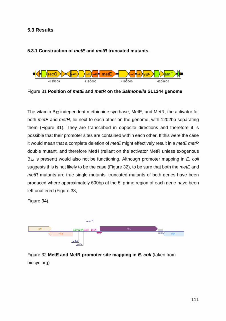
111
5.3 Results
5.3.1 Construction of metE and metR truncated mutants.
Figure 31 Position of metE and metR on the Salmonella SL1344 genome
The vitamin B12 independent methionine synthase, MetE, and MetR, the activator for
both metE and metH, lie next to each other on the genome, with 1202bp separating
them (Figure 31). They are transcribed in opposite directions and therefore it is
possible that their promoter sites are contained within each other. If this were the case
it would mean that a complete deletion of metE might effectively result in a metE metR
double mutant, and therefore MetH (reliant on the activator MetR unless exogenous
B12 is present) would also not be functioning. Although promoter mapping in E. coli
suggests this is not likely to be the case (Figure 32), to be sure that both the metE and
metR mutants are true single mutants, truncated mutants of both genes have been
produced where approximately 500bp at the 5’ prime region of each gene have been
left unaltered (Figure 33,
Figure 34).
Figure 32 MetE and MetR promoter site mapping in E. coli (taken from
biocyc.org)

112
Figure 33 PCR Verification of truncated metE and metR mutants. A: Lane 1:
Hyperladder. Lanes 2-6: External metE primers. Lane 2: WT, expected size 1500;
Lane 3: metE Kan 2, expected size 2130; Lane 4: metE Kan 6, expected size 2130;
Lane 5: metE Cm 2, expected size 1730; Lane 6: metE Cm 8, expected size 1730.
Lanes 7-11: External metR primers. Lane 7: WT, expected size 795; Lane 8: metR
Kan 1, expected size 2040; Lane 9: metR Kan 5, expected size 2040; Lane 10: metR
Cm 2, expected size 1640; Lane 11: metR Cm 4, expected size 1640. B: Lane 1:
Hyperladder. Lanes 2-4: External metE forward primer, internal pkD4 reverse primer.
Lane 2: WT, expected no band; Lane 3: metE Kan 2, expected size 748; Lane 4: metE
Kan 6, expected size 748. Lanes 5-7 External metE forward primer, internal pkD3
reverse primer. Lane 5: WT, expected no band. Lane 6: metE Cm 2, expected size
464; Lane 7: metE Cm 8, expected size 464. Lanes 8-10: External metR forward
primer, internal pkD4 reverse primer. Lane 8: WT, expected size, no band; Lane 9:
metR Kan 1, expected size 748; Lane 10: metR Kan 5, expected size 748; Lanes 11-
13: External metR forward primer, internal pkD3 reverse primer. Lane 11: WT,
expected size, no band; Lane 12: metR Cm 2, expected size 464; Lane 13: metR Cm
4, expected size 464.
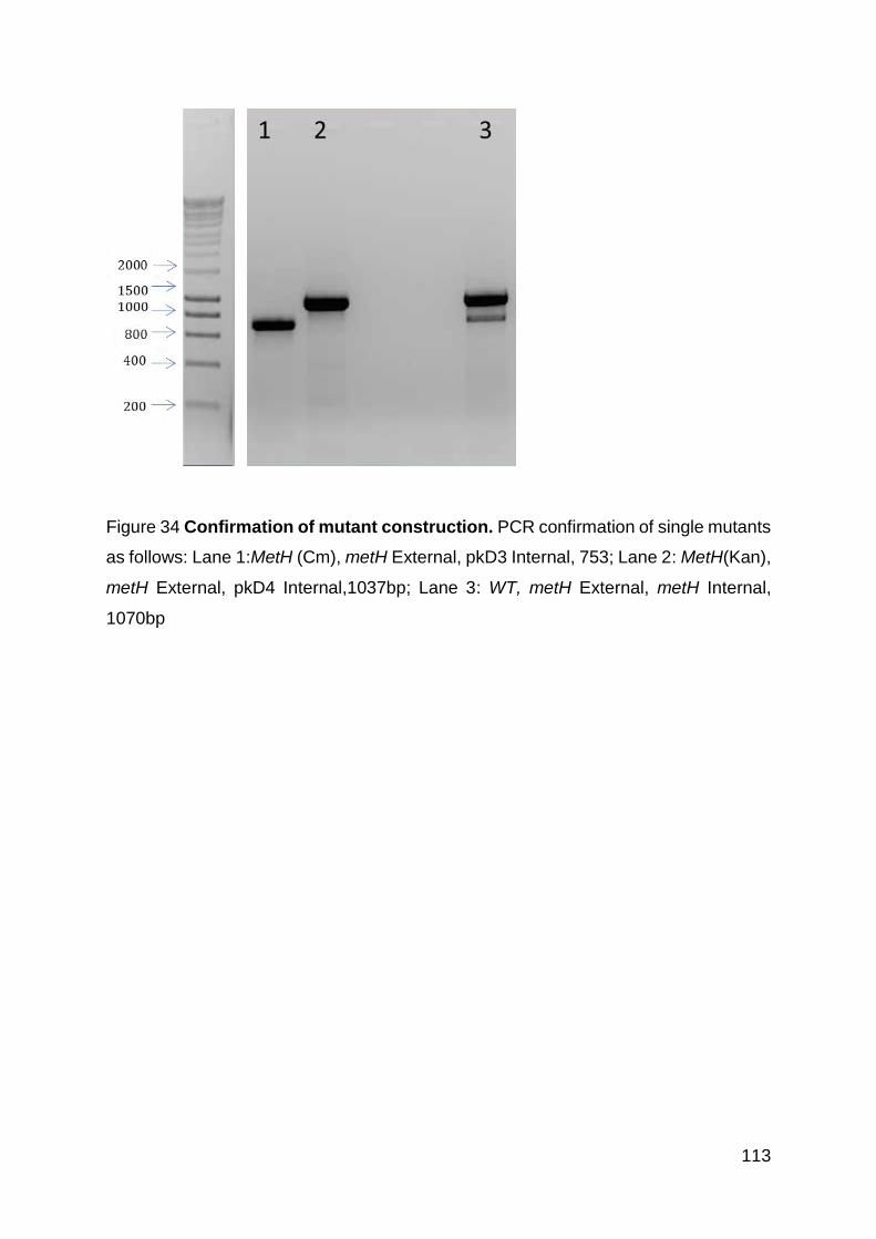
113
Figure 34 Confirmation of mutant construction. PCR confirmation of single mutants
as follows: Lane 1:MetH (Cm), metH External, pkD3 Internal, 753; Lane 2: MetH(Kan),
metH External, pkD4 Internal,1037bp; Lane 3: WT, metH External, metH Internal,
1070bp

114
5.3.2 All mutants grow like WT in LB aerobically
The methionine related mutants were grown alongside WT SL1344, aerobically, in rich
LB media, to confirm the absence of any general growth defect (Figure 35). The
concentration of methionine in LB broth has been shown to be between 2 and 6 mM
(0.3-0.9 mg/ml) (Sezonov, Joseleau-Petit and D'Ari 2007), at least two fold higher
than the amount required for supplementation of a known methionine autotroph, 150
µg/ml (Urbanowski et al. 1987).
Figure 35 All mutant strains grow at similar rates to WT aerobically in LB broth.
Strains were cultured in 50ml LB. Results are shown as means ± standard error of the
mean from two separate experiments performed in duplicate.
115
5.3.3 ΔmetE and ΔmetR are attenuated in aerobic minimal media without the
addition of vitamin B12 or methionine
Salmonella is only able to synthesis vitamin B12 anaerobically, therefore in aerobic
conditions where exogenous methionine is lacking, the vitamin B12 independent
methionine synthase, MetE, is required for survival (Figure 36). Three of the mutants,
ΔmetE, ΔmetE ΔmetH, and ΔmetR are therefore completely attenuated when grown
in M9 minimal media aerobically (Figure 36). When supplemented with either vitamin
B12 (100 µg/ml) or methionine (30 µg/ml) this growth defect is fully restored to WT
levels. The addition of methionine results in a growth benefit to all strains (in
comparison to the addition of B12 and for WT and ΔmetH which do not possess growth
defects under any conditions). This suggests there is a noticeable metabolic expense
of the synthesis of methionine by MetE.

116
Fig
ure
36
ΔmetE
an
d ΔmetR
are
att
en
uate
d in
ae
rob
ic m
inim
al m
ed
ia w
ith
ou
t th
e a
dd
itio
n o
f vit
am
in B
12 o
r m
eth
ion
ine
.
Str
ain
s w
ere
gro
wn
ae
rob
ically
in
50
ml M
9 m
ed
ia +
glu
co
se
with
the a
dd
itio
n o
f 1
00
µg/m
l vita
min
B12 a
nd
30
µg/m
l m
eth
ion
ine
wh
ere
in
dic
ate
d.
All
str
ain
s h
ave
3 r
epe
ats
, an
d s
tan
da
rd e
rro
r is
sho
wn
.
117
5.3.4 Vitamin B12 is toxic to Salmonella, but not due to cobalt toxicity
In order to chemically complement the B12 auxotrophic strain ΔmetE, an appropriate
concentration of vitamin B12 needed to be determined. Vitamin B12 was initially added
to anaerobic M9 cultures at 1 mg/ml as per (Sullivan et al. 2013) and while this resulted
in restoration of growth of ΔmetE it inhibited growth of the SL1344 WT strain (data not
shown). A range of concentrations of vitamin B12 were tested in order to determine a
concentration where the WT strain and ΔmetE mutant grew comparably, this was
found to be 0.05 mg/ml, 20 fold less than was used for the Paracoccus experiments
(Figure 37).
Since Vitamin B12 has a cobalt ion at its centre we thought the Vitamin B12 toxicity being
seen may be due to a cobalt toxicity. A previous study has put the MIC of cobalt at 18
µg/ml for S. enterica LT2 (Thorgersen and Downs 2009). Since cobalt makes up 4.3%
of vitamin B12 when B12 is added at 1 mg/ml this equates to 0.043 mg/ml cobalt –
however this is of course protected within the molecule. Salmonella was grown in the
presence of a range of concentrations of cobalt from 0.043 mg/ml up to 1 mg/ml (far
in exceeding that added with 1 mg/ml vitamin B12) with no defect seen (Figure 38). We
therefore conclude that the toxicity seen when Salmonella SL1344 WT is grown in the
presence of vitamin B12 is not due to cobalt toxicity.
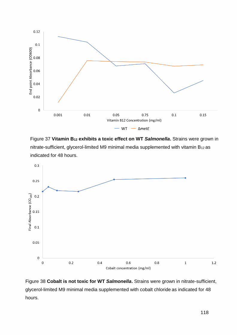
118
Figure 37 Vitamin B12 exhibits a toxic effect on WT Salmonella. Strains were grown in
nitrate-sufficient, glycerol-limited M9 minimal media supplemented with vitamin B12 as
indicated for 48 hours.
Figure 38 Cobalt is not toxic for WT Salmonella. Strains were grown in nitrate-sufficient,
glycerol-limited M9 minimal media supplemented with cobalt chloride as indicated for 48
hours.

119
5.3.5 MetE is important for the survival of denitrifying Salmonella
Salmonella has the ability to respire on nitrate when oxygen is unavailable, as
described in the introduction (Page 30) this results in the net production of N2O. To
create denitrifying conditions Salmonella was grown in anaerobic conditions, in M9
minimal media with the addition of 20mM nitrate.
The growth of WT Salmonella was compared to ΔmetE, ΔmetH, ΔmetE ΔmetH and
ΔmetR when grown in denitrifying conditions and with the addition of vitamin B12 or
methionine (Figure 39). WT Salmonella exhibits similar growth in the absence or
presence of methionine, with maximum growth of ≈0.2 OD600 reached by 24 hours.
The addition of vitamin B12 reduces the maximum growth to ≈0.15 OD600 which also
occurs by 24 hours.
In all three tested conditions the ΔmetH mutant grows as per WT. The ΔmetE mutant
exhibits very little growth until 40 hours at which point a maximum OD of ≈0.15 is
reached. If supplemented with either vitamin B12 or methionine the growth is
comparable to WT.
A double ΔmetE ΔmetH mutant has normal growth with the addition of methionine but
the addition of vitamin B12 does not allow for growth and very little growth is seen for
this, and the unsupplemented conditions over 48 hours. There is some growth seen
which in both cases may be as a result of cell death and methionine turnover.
When grown with no additional supplements ΔmetR exhibits a similar growth pattern
to ΔmetE, that is minimal growth seen until 40 hours at which point a maximum OD of
≈0.15 is reached. Supplementation with either Vitamin B12 or methionine results in a
restoration of this growth defect.
The same growth conditions were then carried out but with the additional step of the
bottles being saturated with N2O pre-inoculation. The addition of N2O to WT
Salmonella did not result in a growth defect except in the B12 supplemented bottles
where it further increased the mild growth defect seen. This resulted in a maximum
growth of 0.07 OD600 by 17 hours which didn’t increase by 48 hours. Without the
addition of N2O (but with vitamin B12) the WT strain exhibits similar growth until 17
hours but by 24 hours reaches maximum OD of ≈0.15.

120
For the ΔmetE mutant similar growth is seen to WT with the addition of methionine
either with or without exogenous N2O (Figure 40Error! Reference source not f
ound.). While in standard anaerobic conditions the addition of vitamin B12 provides a
partial restoration of the growth defect when exogenous N2O is added this affect is
greatly reduced and very little growth is seen. In the un-supplemented cultures ΔmetE
is able to reach an OD comparable to WT growth with methionine by 40 hours. When
exogenous N2O is added however this phenotype is lost and the strain exhibits almost
no growth until 48 hours.

121
Fig
ure
39
Me
tE is
im
po
rtan
t fo
r th
e s
urv
iva
l o
f d
en
itri
fyin
g S
alm
on
ell
a. S
tra
ins w
ere
gro
wn
an
ae
rob
ically
in
nitra
te-s
uff
icie
nt, g
lyce
rol-
limite
d M
9 m
inim
al m
ed
ia. W
he
re in
dic
ate
d v
ita
min
B12 (0.0
5 µ
g/m
l) o
r m
eth
ion
ine (
30
µg/m
l) w
ere
ad
ded
. A
ll str
ain
s h
ave
3 r
epe
ats
, a
nd
sta
nda
rd e
rro
r is
sh
ow
n.

122
Figure 40 MetE is important for the survival of Salmonella grown with
exogenous N2O. Strains were grown anaerobically in nitrate-sufficient, glycerol-
limited M9 minimal media which had been saturated with N2O. Where indicated
vitamin B12 (0.05 µg/ml) or methionine (30 µg/ml) were added. All strains have 3
repeats and SE is shown
Time (Hours)
0
0.05
0.1
0.15
0.2
0.25
0.3
0 5 10 15 20 25 30 35 40 45 50
WT
WTB12
WTMeth
WT
WT B
WTM
0
0.05
0.1
0.15
0.2
0.25
0.3
0 5 10 15 20 25 30 35 40 45 50
metE
metEB12
metEMeth
Ab
sorb
an
ce (
OD
600)
WT N2
WT N2 + B12
WT N2 + Methionine
WT N2O
WT N2O + B12
WT N2O + Methionine
ΔmetE N2
ΔmetE N2 + B12
ΔmetE N2 + Methionine
ΔmetE N2O
ΔmetE N2O + B12
ΔmetE N2O + Methionine
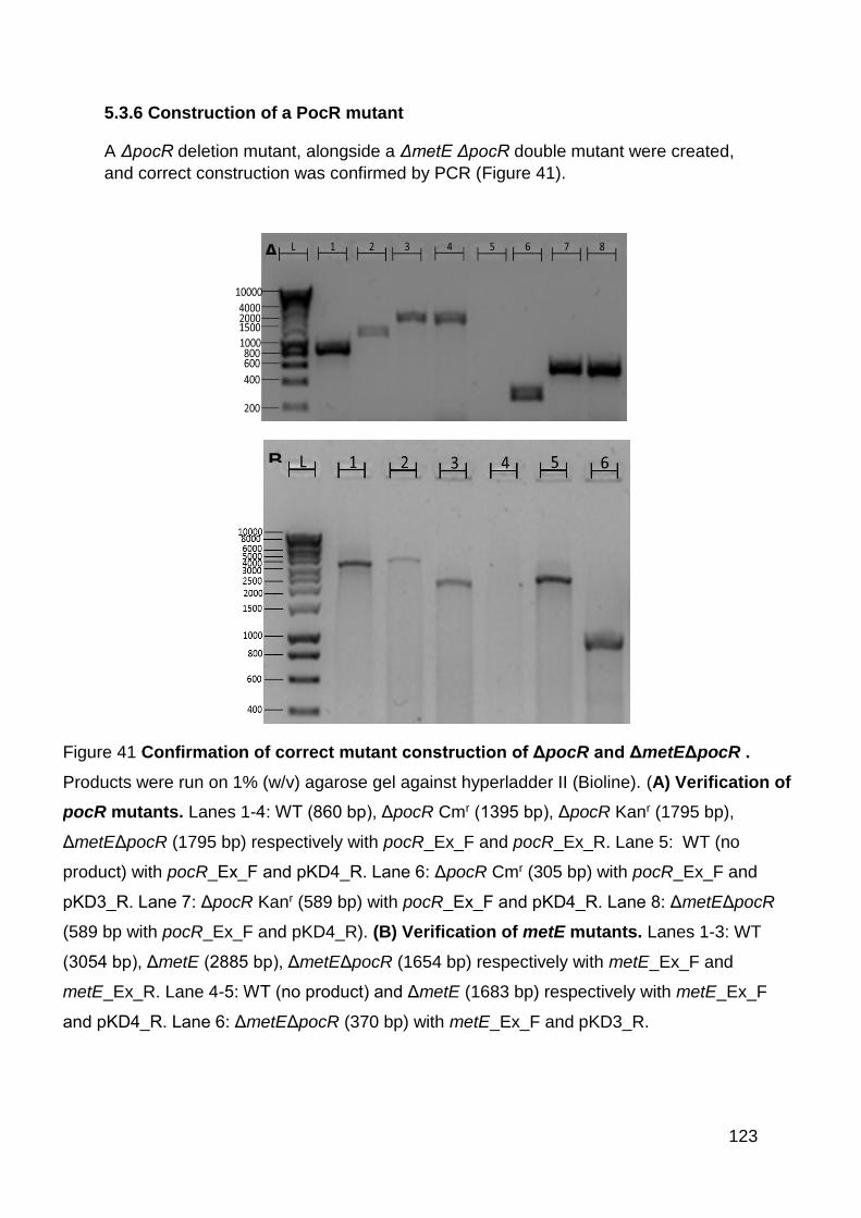
123
5.3.6 Construction of a PocR mutant
A ΔpocR deletion mutant, alongside a ΔmetE ΔpocR double mutant were created,
and correct construction was confirmed by PCR (Figure 41).
A
B
Figure 41 Confirmation of correct mutant construction of ΔpocR and ΔmetEΔpocR .
Products were run on 1% (w/v) agarose gel against hyperladder II (Bioline). (A) Verification of
pocR mutants. Lanes 1-4: WT (860 bp), ΔpocR Cmr (1395 bp), ΔpocR Kanr (1795 bp),
ΔmetEΔpocR (1795 bp) respectively with pocR_Ex_F and pocR_Ex_R. Lane 5: WT (no
product) with pocR_Ex_F and pKD4_R. Lane 6: ΔpocR Cmr (305 bp) with pocR_Ex_F and
pKD3_R. Lane 7: ΔpocR Kanr (589 bp) with pocR_Ex_F and pKD4_R. Lane 8: ΔmetEΔpocR
(589 bp with pocR_Ex_F and pKD4_R). (B) Verification of metE mutants. Lanes 1-3: WT
(3054 bp), ΔmetE (2885 bp), ΔmetEΔpocR (1654 bp) respectively with metE_Ex_F and
metE_Ex_R. Lane 4-5: WT (no product) and ΔmetE (1683 bp) respectively with metE_Ex_F
and pKD4_R. Lane 6: ΔmetEΔpocR (370 bp) with metE_Ex_F and pKD3_R.

124
5.3.7 PocR is not required for growth in LB or M9 aerobically
Both in LB, and M9 supplemented with glucose, ΔpocR exhibits comparable growth to
WT. In LB, the double mutant ΔmetEΔpocR also grows as WT, but in M9
supplemented with glucose, ΔmetEΔpocR grows in the same fashion as ΔmetE
exhibiting no growth over 24 hours (Figure 42).
Figure 42 PocR is not required for growth in LB or M9 aerobically. Strains were
grown in 50ml LB (A), or 50ml M9 + glucose (B).
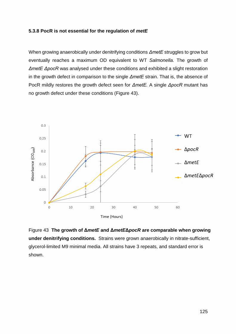
125
5.3.8 PocR is not essential for the regulation of metE
When growing anaerobically under denitrifying conditions ΔmetE struggles to grow but
eventually reaches a maximum OD equivalent to WT Salmonella. The growth of
ΔmetE ΔpocR was analysed under these conditions and exhibited a slight restoration
in the growth defect in comparison to the single ΔmetE strain. That is, the absence of
PocR mildly restores the growth defect seen for ΔmetE. A single ΔpocR mutant has
no growth defect under these conditions (Figure 43).
Figure 43 The growth of ΔmetE and ΔmetEΔpocR are comparable when growing
under denitrifying conditions. Strains were grown anaerobically in nitrate-sufficient,
glycerol-limited M9 minimal media. All strains have 3 repeats, and standard error is
shown.

126
5.3.9 The ΔmetE mutant does not produce the MetE protein
Since a low level of growth is seen for ΔmetEΔmetH under anaerobic conditions,
without the addition of vitamin B12 or methionine, a western blot was performed using
an anti-MetE antibody (Mogk et al. 1999, Mordukhova and Pan 2013) to confirm that
the protein is not being produced (potentially by a secondary copy of the gene).
Samples were taken from anaerobic cultures at various timepoints and at 48hrs for
aerobically growing WT. While protein can be seen on the coomassie stain for all
samples except ΔmetE at 17 hours (when no bacterial growth is seen), MetE is only
detectable in the WT sample growing aerobically (Figure 44). Thus, we confirm that
no MetE protein is being produced by the ΔmetE strain or subsequent double mutants.
Figure 44 The MetE protein is not produced by ΔmetE or ΔmetEΔpocR during
anaerobic growth. Gel and blot lanes: 1) WT 17h. 2) ΔmetE 17h. 3) ΔmetE 48h. 4)
ΔmetEΔpocR 17h. 5) ΔmetEΔpocR 48h. 6) WT 48h aerobic positive control. (A)
Whole cell lysates were run on 12% (v/v) SDS-PAGE. (B) Presence of MetE
detected with an anti-MetE rabbit polyclonal antibody.

127
5.3.10 A ΔmetEΔcobS mutant is unable to grow in M9 media in anaerobic
conditions
To confirm whether the ΔmetE mutant grows anaerobically due to the eventual
production of vitamin B12 and thus use of MetH, a further double mutant deficient in
metE and cobS, the final gene in the cob operon was generated.
It was found that after 48h, ΔmetEΔcobS grew significantly less than the WT. These
results suggest that this inability to grow is due to the inability to produce vitamin B12
(Figure 45).
Figure 45 The deletion of cobS and metE results in an inability of S.
Typhimurium to grow. ΔmetEΔcobS growth was significantly lower than the WT.
Bacteria were grown anaerobically for 48h in M9 minimal medium and OD600
measured to determine growth. Data plotted as mean values from one experiment
performed in duplicate and error bars representative of SE.
128
5.3.11 MetE is important for survival of Salmonella in macrophages
In order to assess the importance of MetE for the survival of Salmonella in
macrophages, a gentamycin protection assay was used. This was carried out both in
normal DMEM and in DMEM which lacks both methionine and vitamin B12. In normal
DMEM media there is no difference between the survival of the three strains tested,
WT, ΔmetE and ΔmetH. However, in the absence of methionine and vitamin B12 the
ΔmetE mutant is significantly (P > 0.05) attenuated (Figure 46).

129
Figure 46 MetE is required for intracellular survival in murine macrophages in
the absence of methionine or vitamin B12. Salmonella uptake into IFN-γ activated
macrophages was conducted for 2 hours and 24 hours before gentamicin treatment
killed all extracellular bacteria in normal tissue culture media in both the presence
and absence of methionine. Internalised bacteria were recovered by lysing the
macrophage and the numbers of bacteria enumerated by viable counts. (a) with
methionine (b) without methionine/B12. Data shown is an average of three
experiments. *Denotes significant difference P < 0.05 (independent t-test, n=3).

130
5.4 Discussion
At the beginning of this PhD there was a clear research gap consisting of two
phenomena in Salmonella. The first being the inexplicable production of high levels of
toxic nitrous oxide, and the second being the highly metabolically expensive synthesis
of the seemingly unnecessary compound, vitamin B12. This chapter has made the first
steps in explaining these two questions by showing a link between them, and in doing
so highlights a potential novel virulence strategy.
The synthesis of methionine by Salmonella is essential for growth unless the amino
acid is exogenously supplemented. Therefore, a MetH deletion mutant is fully reliant
on MetE for this process and a MetE deletion mutant is reliant on MetH. Since MetH
requires vitamin B12 to produce a functional holoenzyme, a MetE mutant is therefore
dependent on the endogenous production of vitamin B12 in order to grow. We can
conclude that when growth is seen for the MetE mutant strain, or any strains including
this deletion in combination with additional mutataions, that conditions are present
whereby Salmonella is able to synthesise vitamin B12 and produce a functioning MetH
protein.
The main growth phenotypes of the mutants described in this chapter are summarised
in (Table 8). The key mutant is ΔmetE, since this strain gives us the most information
about the requirements for vitamin B12.
5.4.1 Importance of MetE for denitrifying Salmonella
Growth in M9 minimal media with and without the addition of vitamin B12 and
methionine was used to assess the survival of Salmonella‘s requirements and
synthesis of vitamin B12 aerobically. WT Salmonella, as well as ΔmetH are able to use
the functional vitamin B12 independent MetE protein in order to synthesise methionine
and therefore exhibit the same levels of growth in the presence and absence of vitamin
B12 or methionine. The ΔmetE mutant is unable to grow in these conditions without
the addition of B12 or methionine – this confirms that Salmonella is unable to
aerobically synthesise vitamin B12. The same phenotype is also seen for ΔmetR, as
a positive regulator of both MetE and MetH this shows that aerobically

131
Table 8: Summary of growth abilities of Salmonella mutants. A tick (✔) indicated the strain grew as WT, a cross (✖) indicates
no growth was seen, both (✖/✔) indicates growth lower than that seen for WT.
LB M9 aerobic M9 anaerobic
+ B12 + Methionine + B12 + Methionine
WT ✔ ✔ ✔ ✔ ✔ ✔ ✔
ΔmetE ✔ ✖ ✔ ✔ ✖/✔ ✔ ✔
ΔmetH ✔ ✔ ✔ ✔ ✔ ✔ ✔
ΔmetE
ΔmetH
✔ ✖ ✖ ✔ ✖ ✖/✔ ✔
ΔmetR ✔ ✖ ✔ ✔ ✖/✔ ✔ ✔
ΔpocR ✔ ✔ ✖/✔
ΔmetE
ΔpocR
✔ ✖ ✖/✔

132
Salmonella is reliant on MetR in order to activate either the MetE or MetH protein. This
phenotype can also be recovered by the addition of vitamin B12, therefore the
conclusion can be drawn that vitamin B12 itself is able to regulate either the MetE or
MetH protein (most logically MetH). When both MetE and MetH are absent,
Salmonella is only able to grow aerobically on M9 with the addition of methionine.
Conditions where Salmonella will be required to use nitrate or nitrate for the terminal
electron acceptor in respiration can occur outside the host but also in the mammalian
intestine, as part of the host-generated inflammatory response (Lopez et al, 2012).
Growth of the various mutants was therefore analysed on M9 minimal media
anaerobically with nitrate present to assess the importance of these genes while
undergoing denitrification, which can occur when Salmonella is residing in the
intestine. WT Salmonella grows the same with and without the addition of methionine,
so while it only is able to reach a maximum OD600 of ≈0.2, this is probably due to the
minimal media and anaerobic conditions in general and not to do with vitamin B12-
/methionine metabolism. Similarly to aerobic growth on M9, ΔmetH is able to grow like
WT in all conditions, this confirms that the MetE protein is produced and functional, to
provide the bacteria with enough methionine for growth. While for the first 24 hours of
growth ΔmetE shows very little growth, by 40 hours the strain is able to reach an OD
comparable to WT. Since the ΔmetE strain relies on MetH for its methionine synthesis,
and MetH is dependent on vitamin B12, in these conditions, for this strain to grow,
Salmonella needs to be synthesising enough vitamin B12 in order for growth to be
seen. It therefore is likely to be the case that during the first 24 hours of growth the
bacteria is slowly attempting to build up a vitamin B12 pool, however since growth is
also reliant on denitrification there will be a net release of nitrous oxide which will be
interacting with the vitamin. Therefore, in order for visible growth to be seen for ΔmetE
a point must be reached where more vitamin B12 is being produced than is being
targeted by nitrous oxide.
A similar growth pattern is seen for ΔmetR as for ΔmetE;growth is slow until 40 hours
when growth can be seen, and the growth defect can be restored by the addition of
either vitamin B12 or methionine. The ΔmetR mutant has a functional MetH and MetE
– and while MetH will rely on vitamin B12, MetE should be able to produce methionine
from the beginning of growth such as occurs for ΔmetH. This phenotype therefore
133
suggests that the activation of MetE is dependent on either MetR or on vitamin B12,
since at later time points the vitamin B12 pool should have been established.
This hypothesis for the phenotype of ΔmetE and ΔmetR is corroborated by the growth
phenotype of ΔmetE ΔcobS under the same conditions. CobS is one of the final genes
in the cob operon and is its deletion is known to stop the synthesis of vitamin B12 in
Salmonella. This strain shows nearly no growth over 48 hours.
From the previous results it would be expected that a ΔmetE ΔmetH double mutant
would be unable to grow without the addition of methionine. However, growth, albeit
very low, is seen for both the unsupplemented culture and that supplemented with
vitamin B12. There are two potential explanations for this observation, the first being
that after 24 hours there is a low level of methionine turnover resulting from cell death
from the initial inoculation. The second possibility is that by 40/48 hours there is
contamination in the cultures, as described (Page 49), each timepoint requires four
needles, two one-use needles for liquid sampling, and two multi use needles for gas
sampling to be inserted through the silicon lid. While precautions were taken such as
cleaning the outside of the silicon lid with ethanol, and cleaning the reusable Hamilton
gas syringe between sampling, this is not a totally sterile process. Samples could be
taken throughout to determine if contamination is present.
5.4.2 Vitamin B12 toxicity
Vitamin B12 was initially added to aerobic M9 cultures at 100 µg/ml as per (Sullivan et
al. 2013) where it only had a slight negative effect on the growth of WT Salmonella.
However, when this same concentration was added to M9 anaerobically, a significant
growth defect was seen, reducing the OD of the WT to that similar to non-
supplemented Salmonella. A concentration of vitamin B12 was determined where
ΔmetE and WT Salmonella grew comparably, this was 0.05 µg/ml.
There is no information in the literature about a toxic effect of vitamin B12 on
Salmonella, or in fact for any bacteria. In humans no toxicity has been observed for
vitamin B12 and no safe upper limits have been defined (Flynn et al. 2003, Burgess,
Smid and van Sinderen 2009). There is a small body of work looking at the toxicity of
vitamin B12 on mammalian cells due to catalysing the oxidation of sulfhydryl groups in
134
sulfhydryl-dependent cells (Toohey 1975); this however should not have an effect on
Salmonella in anaerobic conditions, so is unlikely to be the explanation.
It was however considered that the toxicity could be due to the cobalt contained within
the compound. However even at levels far exceeding those which result in a toxic
effect from vitamin B12, no toxicity was seen and hence it is concluded that the toxicity
seen when Salmonella SL1344 WT is grown in the presence of vitamin B12 is not due
to cobalt toxicity.
Another possibility is that vitamin B12 has a regulatory effect on Salmonella. MetE is
known to be down-regulated by MetJ as well as the MetH-B12 holoenzyme (Wu,
Urbanowski and Stauffer 1992). Therefore, in conditions with high levels of vitamin B12
it could be expected that MetE is downregulated. However, this would be reliant on
the MetH-B12 holoenzyme and so growth should not be affected.
It should be noted that high levels of vitamin B12 are unlikely to occur in natural
conditions, since the synthesis of the vitamin is so metabolically expensive it is highly
improbable that any bacteria, even on lysis, would be releasing the compound in any
significant concentrations. The recommended daily intake for humans varies for
different age groups but is ≈2 µg, it can be assumed therefore that concentrations
would never reach 100 µg/ml in the intestines or anywhere that Salmonella might
reside. The toxic effect could therefore be simply a result of such an unusually high
level of a very large molecule.
The possibly was considered that even though a metE knockout was constructed the
bacteria was still producing the MetE protein via a potential secondary copy of the
gene, and this was allowing for the eventual growth of a ΔmetE ΔmetH mutant, or
indeed the single ΔmetE strain. To confirm this was not the case a western blot was
carried out using an anti-MetE antibody
A further interesting observation was made during the Western blot analysis. No MetE
protein was detected from the WT strain at the 17h time-point, suggesting it is only
relying on MetH for methionine synthesis. Potentially, the MetE protein is used at later
stages of growth, when N2O levels have begun to accumulate and inactivate vitamin
B12. However, this hypothesis highlights a difference in comparison to the anaerobic
growth curve data and does not explain why the growth of ΔmetE was reduced in
comparison to the WT at the 17h time-point if both strains are using the same enzyme

135
for synthesis of methionine, not that WT is using MetE as suspected. Potentially, MetE
may be present in the WT sample but at a lower concentration than the aerobic
sample. Exposure time may need to be increased during blot visualisation in order to
detect low levels of the protein. Further investigation should be undertaken in which
later time-points are sampled to quantify MetE production by the anaerobic WT strain.
5.4.3 Regulation of the cob operon by PocR
A PocR mutant, and subsequent ΔmetEΔpocR strain were constructed to determine
the importance of this gene for Salmonella, specifically in the regulation of the cob
operon. Since the cob operon is so large and the synthesis of vitamin B12 so
metabolically expensive it is logical to assume there is efficient regulation.
The cob operon is transcribed adjacently to the pdu operon, which encodes genes
required for the vitamin B12 dependent utilisation of propanediol as an energy source;
both operons are under the transcriptional control of PocR, encoded by pocR which is
situated between the two operons (Ailion et al. 1993). Aerobically it is known that
Crp/cAMP are important for induction of PocR and therefore the two operons, and
anaerobically ArcA/ArcB are also required. The importance of Crp suggests that
carbon utilisation is a primarly function of vitamin B12. Additionally it has been shown
that 1,2-propanediol enhances the binding of PocR and as such increases the
activation of the cob operon – presumably to provide the vitamin B12 required for the
utilisation of this energy source (Rondon and Escalante-Semerena 1996). A study has
also shown that PocR is induced by glycerol – the carbon source used for anaerobic
growth in this study (Bobik et al. 1992). However there lies a paradox in this co-
regulation. Aerobically Salmonella is unable to produce vitamin B12 (even if the cob
operon is fully transcribed) and therefore propanediol cannot be utilised.
Anaerobically, whilst the pdu operon is induced, oxygen is required for the utilisation
of the carbon source, alternative electron acceptors cannot be used and propanediol
cannot be fermented. Therefore, while it has been hypothesised in the literature that
the fact that PocR is the major regulator of the cob operon suggests propanediol
respiration is the primary use of vitamin B12 in Salmonella, this seems unlikely to be
the case (Bobik et al. 1992).

136
Another candidate regulator of the cob operon, CsrA (carbon storage regulator A), has
been proposed. First identified in E. coli, CsrA is classed as a global regulator, and
until recently in Salmonella was known only to upregulate genes of the pathogenicity
island SPI-1, necessary for intracellular invasion (Lawhon et al. 2003, Altier, Suyemoto
and Lawhon 2000). However, Lawhon et al. (2003) identified that genes of the cob
operon were significantly downregulated in a ΔcsrA mutant in comparison to the WT.
Furthermore a downregulation of genes located in the eut and pdu operons, both of
which are dependent on the presence of B12, were also observed, signifying that CsrA
has multiple roles aside from SPI-1 regulation (Lawhon et al. 2003).
There are many possibilities for future investigations into the regulation of vitamin B12
synthesis by Salmonella. Primarily it would be interesting to investigate the anaerobic
growth of a triple deletion mutant in metE, pocR and csrA. If this mutant was unable
to grow anaerobically, it would indicate that either pocR or csrA are essential to B12
synthesis and exhibit functional overlap. However, if growth was still observed, it would
show that there are other, unknown regulators yet to be identified.
One study looking at the transcription levels of the cob operon under different
conditions highlights the 40 fold increase in transcript levels between the anaerobic
fermentation of glucose and under conditions of anaerobic respiration. It is stated that
they believe there to be unidentified functions which exist that require B12 under
conditions of anaerobic respiration but that since cob mutants are able to grow
normally on glycerol fumarate they believe them to be inessential (Escalante-
Semerena and Roth 1987). In the context of this study it is not surprising that cob
mutants struggled to grow on glycerol fumarate – and it is more likely that the operon
is being upregulated in anaerobic conditions for growth of Salmonella on nitrate.
The results in this study suggest that the absence of PocR partially restores the growth
phenotype of ΔmetE when growing anaerobically on nitrate. This therefore indicates
that there is an alternative regulatory mechanism inducing the expression of the cob
operon for B12 production, enabling Salmonella to continue to use MetH to produce
methionine.
One potential explanation for this phenotype is that there are multiple regulators
inducing expression of the cob operon in the absence of PocR, resulting in a greater

137
level of transcription of cob genes. This would account for the increased growth rate
of ΔmetEΔpocR compared to ΔmetE.
Figure 47 outlines a possible model to explain the results observed in this study. The
binding of PocR to its recognition site potentially could block the binding sites of other,
currently unknown transcription factors of the cob operon. Whereas, in the absence of
PocR these sites would be available.
Figure 47 Working model – regulation of the cob operon (A) Proposed model for
induction of the cob operon by PocR. When transcribed, PocR binds to its binding
site (dark purple box) and induces cob expression. Access to other transcription factor
binding sites (grey boxes) is blocked by the presence of PocR. (B) The proposed
mechanism for the positive regulation of the cob operon in the absence of PocR.
Binding sites for other transcription factors are accessible and interaction may induce
expression of the cob operon.
A
B

138
5.4.4 The pathogenic effect of the production of nitrous oxide by Salmonella in
the intestine
Based on the results presented in this chapter the hypothesis was drawn that there is
a distinct link between the two phenomena highlighted in the introduction to this
chapter – the production and release of large quantities of nitrous oxide and the
synthesis of the metabolically expensive vitamin B12.
This study has shown that Salmonella is afforded a growth benefit by MetE when
growing anaerobically on nitrate, that is, that the absence of this gene results in a
distinct growth defect.
Nitrate is present in the human intestine from two primary routes, the first being dietry
products and the second endogenous synthesis. Different diets result in differing
amounts of nitrate in the gut, with highest concentrations coming from nitrate-rich
vegetables, particularly leafy greens (Lidder and Webb 2013). Vegetarians therefore
on average have higher daily intakes of nitrate, at ∼4.3 mM nitrate/day compared to
∼1.2 mM nitrate for a standard western diet, the Acceptable Daily Intake has been set
at 5 mM nitrate/day since 1962 (Katan 2009).
While most early studies looking at nitrate in the gastrointestinal tract are focused on
this from a human cell perspective, specifically in relation to cancer, these studies give
some insights into the nitrate environment of the gut.
A study looking at the nitrate and nitrite concentrations in human fecal and ileostomy
(a method of sampling directly from the end of the small intestine) samples concluded
that the lower gastrointestinal tract favours denitrification over nitritrication. Fecal
nitrate levels ranged from 0 to 14 µmol/kg (0 to 0.9 ppm) and ileostomy samples from
0 to 7 µmol/kg (0 to 0.4 ppm). Experiments were also carried out that showed that the
addition of 500 µmol/kg nitrate to a fecal sample resulted in this nitrate being reduced
10 fold within 15 minutes and fully depleted within 2 hours; thus there must be high
levels of denitryfing microorganisms present in faeces and additionally suggesting the
high turnover of nitrate in the gut may result in false underreporting of nitrate
concentrations from sampling (Saul et al. 1981).

139
It has been known that there is endogenous production of nitrate in the human
intestine since at least 1978 (Tannenbaum et al. 1978) but more modern studies have
looked at this from the perspective of gut bacteria.
Pathogenic bacteria, including Salmonella, can induce an inflammatory response in
the intestine. Whilst this was traditionally thought to be detrimental to the pathogen,
since it is part of the host inflammatory response, more recently this has be shown to
in fact be beneficial to pathogens, and that triggering the host immune’s defence can
disrupt the gut environment beneficially for pathogenic bacteria (Stecher et al. 2007).
This can result in the release of nitrate into the gut lumen, and additionally in the
decrease in the representation of obligate anaerobes and an increase in facultative
anaerobic members of the Enterobacteriaceae family (Winter et al. 2013, Lupp et al.
2007). In fact, it has been shown that in order to outcompete the microbiota and cause
an infection, Salmonella is reliant on its ability to trigger inflammation. The T3SS-1
effector protein, SopE, enhances intestinal inflammation during Salmonella infection,
via the activation of caspase 1 and the subsequent cleavage of interleukins into active
cytokines (Hoffmann et al. 2010, Lopez et al. 2012). These cytokines, IL-1β and IL-18
contribute to the production of IFN-γ which stimulates iNOS to produce NO. Nitric
oxide can then react with ROS and produce peroxynitrite, ONOO-, which isomerises to
nitrate, NO3 (Szabó, Ischiropoulos and Radi 2007). This means that SopE-dependent
iNOS expression generates host-derived nitrate and this nitrate has been shown to
increase the growth of Salmonella (Lopez et al. 2012). Interestingly, nitrate is shown
to be a negative regulator of tetrathionate respiration. Potentially this shows that nitrate
respiration is preferred to tetrathionate respiration in the intestine (Lopez et al. 2012).
Similarly to the pathogen mediated host release production of nitrate, Salmonella also
triggers the host to produce tetrathionate, which can then be used as an alternative
electron acceptor (Hensel et al. 1999). This occurs via the reaction of ROS generated
by inflammation with endogenous luminal thiosulfate. Indeed, data shows that the
ability to respire tetrathionate provides Salmonella a distinct advantage when growing
in the inflamed gut (Winter et al. 2010, Winter and Bäumler 2011). This therefore is a
secondary example of Salmonella exploiting the host immune response to provide an
altered ecological niche beneficial for its growth. Most interestingly however is the
carbon sources which are required to allow for tetrathionate respiration, either
ethanolamine or propanediol, both of which require vitamin B12. It is noted by the
140
authors that tetrathionate respiration using ethanolamine or propanediol are the only
conditions they know under which wild-type Salmonella requires B12 for growth (Price-
Carter et al. 2001).
Stecher et al., propose a working model for the three-way microbiota-pathogen-host
interaction during murine Salmonella colitis, which suggests that Salmonella infection
involves both triggering inflammation and secondarily surviving and thriving from the
change in the ecological niche.
The results of this chapter contribute additional knowledge to this story, by showing
that not only does the ability to respire on nitrate mean that Salmonella can survive
within the inflamed intestine but that in doing so will release toxic nitrous oxide to
further target the microbiota and potentially the host itself.
The importance of the gut microbiota is becoming ever more diverse, but one of the
well known functions of these bacteria is in colonisation resistance, via the barrier
effect. This happens in three main ways, the first being the commensal bacteria
providing a physical barrier to the pathogen, which aim to target attachment and
subsequent invasion into epithelial cells, the second involves a symbiotic relationship
with the host to control nutrient availability and the third in activity producing
antimicrobial compounds (Guarner and Malagelada 2003, Buffie and Pamer 2013).
It is therefore of interest to consider the impact of nitrous oxide on the commensals.
The four dominant phyla in the human gut are Firmicutes, Bacteroidetes,
Actinobacteria, and Proteobacteria, while each individual’s gut microbiota is different
there are some species which are common to most. These include, Bacteroides
fragilis, Bacteroides melaninogenicus, Bacteroides oralis, Bifidobacterium bifidum
Enterococcus faecalis, Escherichia coli, Enterobacter sp., Klebsiella sp. and
Lactobacillus. There are three processes to assess here, the vitamin B12 independent
synthesis of methionine, demonstrated by the presence of MetE, the ability to
synthesise vitamin B12 de novo and the ability to reduce nitrous oxide to N2 via the
enzyme NosZ (Table 9). Of all the commensal bacteria analysed, none have the ability
to reduce N2O – and therefore the presence of this gas in high volumes in the intestine
will impact on the vitamin B12 pool and any vitamin B12 they are able to synthesise.
Only one species however, Lactobacillus, is able to synthesise vitamin B12 de novo
and we can therefore conclude all other rely on vitamin B12 uptake from the

141
environment – of course as the vitamin B12 pool is depleted this will become a limited
resource. Lastly, the presence of the vitamin B12 independent methionine synthase
was determined, any bacteria lacking this strain will rely on the vitamin B12 dependent
MetH in order to synthesise methionine, only some members of the Klebsiella species
and some strains of Escherichia coli have metE.
Table 9 Presence of metE, nosZ and ability to synthesise B12 of eight common
commensal bacteria and human epithelial cells
metE B12 synthesis nosZ
Bacteroides fragilis ✖ ✖ ✖
Bacteroides melaninogenicus ✖ ✖ ✖
Bacteroides oralis ✖ ✖ ✖
Bifidobacterium bifidum ✖ ✖ ✖
Enterococcus faecalis ✖ ✖ ✖
Escherichia coli ✔ ✖ ✖
Klebsiella sp ✔ ✖ ✖
Lactobacillus sp ✖ ✔ ✖
Human epithelial cells ✖ ✖ ✖

142
Therefore, the overriding model drawn from this data is that when residing in the
intestine Salmonella triggers the release of nitrate into the gut lumen and uses this for
denitrification. This then results in previously inexplicably high levels of nitrous oxide.
The nitrous oxide released will target the vitamin B12 pool and as such the commensals
which rely on this cofactor for multiple processes, including methionine synthesis, will
not be able to survive. Additionally, Salmonella may be able to ‘anaesthetise’ the host
epithelium as similarly to the majority of commensal bacteria these cells are reliant on
vitamin B12 and lack the ability to produce it. The working model produced by Stecher
et al., has been adapted to include this hypothesis (Figure 48) (Stecher et al. 2007).
The ability of Salmonella to survival and reproduce inside macrophages is of key
importance to its pathogenesis in causing systemic infections. The importance of MetE
in macrophage survival was shown when media was lacking in methionine. This
therefore suggests that methionine is not present in macrophages, but additionally that
nitric oxide, which is produced by macrophages, is also involved in the targeting of the
vitamin B12 pool. As discussed at more length in Chapter 3, genes which are important
for intracellular survival of Salmonella inside macrophages have the potential to be
further studied in relation to making a new vaccine strain.

143
Figure 48 Working model A) Working model adapted from (Stecher et al. 2007) showing
the three way interactions between the host, microbiota and Salmonella in normal
conditions and in enterocolitis. B) Altered model of Salmonella enterocolitis with the
addition of N2O release by Salmonella targeting the vitamin B12 pool and thus the inhibition
of the microbiota by the pathogen.
A
B

144
5.4.5 Future work
Further work should now be done to show the importance of this system in competition
with the gut microbiota and in the survival and pathogenesis of Salmonella in vivo.
Initially simple cultured competition experiments could be conducted, mimicking the
previous experiments under denitrifying conditions, but with the addition of one or
multiple commensal bacteria. To create a more realistic environment, a microfluidic
gut model could be used. Microfluidic model organs are increasingly being used for
many different functions as they enable a much higher level of control over conditions
than in animal models (Esch, Bahinski and Huh 2015, Bhatia and Ingber 2014, Huh,
Hamilton and Ingber 2011). While new in the gut microbiota field there are a number
of systems being used, these allow a defined ‘microbiota’ to be stabilised before the
addition of the pathogen (Shah et al. 2016, Kim and Ingber 2013, Kim et al. 2016b).
Various mammalian cells can also be cultured in the system and so one could quantify
the ability of Salmonella to cross over the epithelium, and potentially invade
macrophages.
If these experiments, potentially in combination with mouse model studies, confirm the
importance of nitrous oxide production, vitamin B12 synthesis and the ability to produce
methionine independently of vitamin B12 for Salmonella in outcoming the gut
commensals, this is an exciting new window of research.
The most logical reaction would be to either attempt to reduce the nitrous oxide
produced by Salmonella, or reduce its detrimental effects on the gut commensals. This
could be done by introducing bacteria capable of complete denitrification to the gut.
Another option would be in the creation of ‘Smart’ probiotics, that is, genetically
modified probiotic bacteria. If our hypothesis is correct, Salmonella is able to exploit
the fact that the common gut microbiota are both unable to reduce nitrous oxide and
unable to synthesise essential methionine in its presence. Therefore, if these bacteria
were altered to either produce NosZ to reduce N2O or MetE, to enable them to
synthesise methionine independently of vitamin B12 this might lose Salmonella one of
its pieces of pathogenic machinery.

145
6 A correlation between nitrous oxide emissions and vitamin
B12 synthesis in Salmonella serovars
146
6.1 Introduction
Different Salmonella serovars are distinguished by the somatic (O), flagellar (H) and
capsular (Vi) antigens on the outer membrane (Shi et al. 2015). As introduced
previously (Page 18) different serovars infect different hosts and cause different
disease types, so naturally, in order to do so, there are differences in the genes they
have retained and in their nutritional requirements. One difference we have noticed is
the lack of conservation in the cob operon between different serovars, resulting in
differences in their ability to synthesise vitamin B12. As described in the introduction
(Page 38), the synthesis of vitamin B12 is split into three sections. Mutations, or loss of
function, can therefore occur in different parts of the operon resulting in different
effects, for example, a strain lacking a complete Part I can synthesise cobalamin if
provided with cobinamide and Part II of the cob operon is still able to synthesise
vitamin B12 if supplied with dimethylbenzimidazole (DMB) (Roth et al. 1993).
6.1.1 Salmonella Typhimurium SL1344/474
S. enterica serovar Typhimurium strains are split into sequence types (STs), both 4/74
and SL1344 belong to ST19 (Kingsley et al. 2009). Salmonella Typhimurium 4/74 was
orginally isolated from a calf exhibiting salmonellosis, and is the parent strain of
Salmonella Typhimurium SL1344 which is the WT strain which is used throughout this
thesis (Jones, Collins and Aitken 1988, Rankin and Taylor 1966, Hoiseth and Stocker
1981). S. enterica serovar Typhimurium strains are the most commonly used strain
for Typhoid fever research due to the fact that while it causes a gasteroenteric infection
in humans it causes typhoid like symptoms in mice.
6.1.2 Salmonella Enteriditis
S. Enteritidis is responsible for approximately 14% of all Salmonella gasteroenteritis
infections in the United States and is thought to be either the first or second most
common Salmonella serovar in most countries (Braden 2006). Infection from
Salmonella Enteritidis is highly associated with contamination from eggs, or more
specifically, the eggshell.
147
6.1.3 Salmonella Dublin
S. Dublin is most commonly associated with infection in cattle but can also less
commonly infect pigs and also humans. In cattle it causes an enteric fever and also
commonly induces abortion (Coburn et al. 2007). Cattle frequently enter a chronic
carrier stage, equivalent to that seen in humans post infection with S. Typhi, whereby
they can shed the bacteria in large numbers and spread the infection quickly within a
herd. The bacteria can also be passed through the milk and hence to calves or, if from
dairy farms, to humans (Nielsen et al. 2004).
6.1.4 Salmonella Choleraesuis
S. Choleraesuis is a host adapted serovar which causes a systemic infection in swine.
As well as the loss of life this disease causes, there is a large economic burden to the
meat industry. In 1958 Choleraesuis was responsible for 90% of isolated causes of
Salmonella in pigs, however by 1997 this had dropped to a single isolated case in the
UK (Sojka et al. 1977).
6.1.5 Salmonella Gallinarum and Pullorum
Salmonella Gallinarum and Pullorum were originally thought to be two separate
serovars but now are regarded as two biovars of the same serovar – that is they are
indistinguishable by standard serotyping. However, they differ physiologically and
biochemically (Barrow and Freitas Neto 2011).
S. Gallinarum causes typhoid in fowl, and can also rarely causes an infection in mice
and humans. The disease is acute or chronic septicemia which generally affects adult
birds. Interestingly the eradication of S. Gallinarum allowed for the increase of S.
Enteritidis, and subsequent increase in human infections, as this strain could then fill
the vacated ecological niche (Rabsch et al. 2000).
S. Pullorum is the causative agent of pullorum disease and contrary to Gallinarum
causes a systemic disease more commonly seen in young birds.
148
6.1.6 Citrobacter rodentium
In addition to studying a number of Salmonella serovars we also look at Citrobacter
rodentium. Since the Salmonella sytemic serovars Typhi and Paratyphi are Hazard
Group 3 human pathogens these were inpractical for use in this study. Citrobacter
rodentium provided us with a safe model of another systemic strain, as it a model for
the emerging pathogen enterohemorrhagic Escherichia coli (EHEC) (Mundy et al.
2005). It is worth noting that previous work in the Rowley lab (Figure 3) has shown a
signifciant difference between the amount of N2O released by Salmonella and a K12
E. coli strain, what is not clear from that work is whether the reduced N2O release from
E. coli is indicative of the entire species or due to the fact that this is a commensal
strain. This strain releases a toxin similar to the Shiga toxin produced by Shigella
dysenteriae (O'Brien and LaVeck 1983). EHEC causes severe and bloody diarrhoea,
haemorrhagic colitis (HC) and, haemorrhagic uraemic syndrome (HUS); the latter of
which causes acute renal failure,thrombocytopenia, and microangiopathic haemolytic
anaemia. The mortality from HUS is quite high at up to 17%, with a further 30%
suffering from permanaent disabilites (Carter et al. 1987, Griffin and Tauxe 1991,
Messens et al. 2015, Karmali 1989, Karmali, Gannon and Sargeant 2010, Welinder-
Olsson and Kaijser 2005).
6.1.7 Differences in vitamin B12 synthesis between Salmonella serovars
S. Gallinarum and S. Pullorum are two host-restricted strains which cause a systemic
infection in fowl. It has been noted that both serovars have developed pseudogenes
which result in the loss of ability to synthesise Vitamin B12, and hence this is a potential
marker of a switch from an enteric to a systemic disease (Langridge et al. 2015). One
of the functions vitamin B12 plays in Salmonella is in the degradation of 1,2-
propandediol required for the use of tetrathionate as an electron acceptor. It is known
that the ability to reduce tetrathionate was lost pre-divergence and the loss of B12
synthetic ability was lost at a later point (Langridge et al. 2015). We therefore believe
it may be the case that systemic strains have lost the ability to synthesise vitamin B12
due to the fact that they have moved out of the intestine where vitamin B12 dependent
tetrathionate respiration is of benefit (Nuccio and Bäumler 2014). This chapter aims to

149
build on this hypothesis and combine it with the possibility that the levels of N2O
produced by these strains may vary, due to its toxic effects on vitamin B12 (Page 36).
The vitamin B12 transporter, BtuB is present in EHEC and is a known virulence factor
in this strain, highlighting an importance of acquisition of vitamin B12 from the external
environment (Hochhut, Dobrindt and Hacker 2005), and there is evidence that EHEC
cannot synthesise B12 de novo (Cordonnier et al. 2016). Vitamin B12 is important
however for the induction of EutR which contributes to metabolism and virulence in
EHEC (Luzader et al. 2013).
Nuccio and Bäumler’s analysis revealed mutations in genes required for ethanolamine
degradation, propanediol utilisation and vitamin B12 synthesis and regulation, in the
systemic-disease causing strains: S. Choleraesuis, S. Gallinarum, S. Paratyphi A & C
and S. Typhi, whereas the strains which cause an enteric infection, S. Agona, S.
Enteritidis, S. Heideberg, S. Newport and S. Typhimurium, did not have mutations in
these locations (Nuccio and Bäumler 2014). In this chapter we aim to measure the
differing levels of vitamin B12 produced by different Salmonella serovars.
In Chapter 5 it was highlighted that since nitrate is present in the inflammed intestine,
Salmonella will be able to use this to respire and in turn release high levels of nitrous
oxide. As previously discussed it has been noted that Salmonella Typhimurium
produces an order of magnitude more nitrous oxide then the close relative E. coli. We
hypothesise that Salmonella produces this high level of nitrous oxide in order to
compete with other gut bacteria by targetting the vitamin B12 pool. The question is
therefore raised whether Salmonella serovars which cause systemic infections and
therefore do not remain in the microxic environment of the inflammed intestine where
nitrate is available, will produce similar levels of nitrous oxide.

150
6.1.8 Measurement of vitamin B12
The most common reason for wanting to quantify the level of B12 in a sample is for
medical purposes from a human blood. Here, two different measurements can be
taken, total cobalamin in the blood, or cobalamin bound to the transport protein
transcobalamin (termed holoTC). HoloTC is also referred to as active vitamin B12 as it
is only in this form that it can be taken up by the human cells for use (Nexo and
Hoffmann-Lücke 2011). The former is more common however the latter is more likely
to give relevant information for medical purposes such as the diagnosis of Vitamin B12
deficiency (Nexo et al. 2002, Refsum et al. 2006).
High throughput medical labs use immunoassay based techniques for the
quantitfication of Vitamin B12, there are several commercially available kits. Examples
include an electrochemiluminiscence immunoassay (ECLIA) measured on a Roche
Elecsys 2010 (Roche Diagnostics, Basel, Switzerland), Vitamin B12 AccuBind ELISA
Kits (Monobind, California, USA), Vitamin B12 ELISA (Eagle Biosciences, New
Hampshire, USA). These assays are based on the instrinsic factor, which is the
enzyme important for uptake of Vitamin B12 in humans. The main drawback of these
assays for use in this study is price, with most costing many hundreds of pounds; it is
also unclear whether these assays would be suitable for measuring the endogenously
produced vitamin B12 by Salmonella; that is there is no mention of this in the literature.
Microbiological assays have also been used to measure vitamin B12. One study used
the protozoan Ochromonas mahamensis which requires B12 for growth and responds
linearly to the vitamin B12 (FORD 1953). Mutant strains of E. coli have also been used
(BURKHOLDER 1951). In general, these microbiological assays are not as accurate
and take longer than immunoassays.
The measurement of vitamin B12 levels from bacterial cultures has not previously been
performed in our lab group. This chapter includes the optimisation of a vitamin B12
quantification assay for measurements of vitamin B12 synthesised by Salmonella,
based on a previously published protocol (Lawrence and Roth 1996). This assay is a
disc diffusion assay based on the B12 dependency of a Salmonella metE mutant.
151
6.2 Aims
Building on the results from Chapter 5 this chapter aims to uncover if the hypothesis
that Salmonella produces high levels of N2O and vitamin B12 in order to outcompete
the gut microbiota therefore means that systemic strains will not produce these
compounds to the same extent. Specifically we will:
• Bioinformatically analyse the cob operon across multiple Salmonella serovars.
• Measure the levels of vitamin B12 and N2O produced by a range of Salmonella
serovars.

152
6.3 Results:
6.3.1 Systemic Salmonella serovars have mutations in the cob operon
An alignment of the cob operon between the 8 Salmonella strains studied in this
chapter and also two further systemic strains, Salmonella Paratyphi A and Salmonella
Typhi, was constructed to visualise differences between the operon in these strains
(Figure 49). The percentage identities of each gene in comparison to SL1344 are
shown in Table 10. In addition to the core cob operon (cbiA – cobT), two upstream
genes, pocR and pduF, and one downstream, erfK, were included in the alignment.
RNA-seq analysis of the cob operon has revealed that a single transcript runs from
the beginning of the operon and finishes after the erfK gene (Jay Hinton, personal
communication), it is for this reason erfK was included in the alignment. In contrast, in
E. coli it is most likely the transcript is halted before erfK (Lawrence and Roth 1995).
There is very little in the literature about ErfK, except for the possible involvement of
the protein in protection against bile (Hernández et al. 2012). The gene product of
PocR is a positive transcriptional regulator of the cob operon, although is not essential
for its transcription. PocR also regulates the pdu operon, responsible for vitamin B12
dependent propanediol utilisation (Rondon and Escalante-Semerena 1992, Bobik et
al. 1999, Chen et al. 1995a, Escalante-Semerena and Roth 1987).
The entire cob operon was present in all the enteric strains, S. Typhimurium SL1344,
S. Typhimurium 4/74, S. Dublin and, S. Enteritidis, with 98% or higher consensus for
all genes. A complete cob operon is also present in the systemic swine strain, S.
Choleraesuis, with 97% or higher consensus for all genes. The closely related strains
S. Gallinarum and Pullorum both contain missing genes, the regulator pocR is absent
in Gallinarum, and the first gene of the operon cbiA is absent in Pullorum. The two
strains which cause systemic infections in humans, S. Typhi and Paratyphi both have
numerous mutated genes; in S. Typhi cbiB and cbiK, and in S. Paratyphi cbiA and
pduF (Figure 49, Table 10). In all the cases of ‘missing’ or ‘mutated’ genes there are
a large number of stop codons inserted into the DNA sequence, which has resulted in
the gene being visible in some cases on the alignment (Figure 49), such as in the case
of S. Typhi. However, it is very clear from viewing the DNA sequence that a functional
mRNA would not be transcribed and therefore no protein would be produced.

153
Fig
ure
49
Ali
gn
men
t o
f th
e c
ob
op
ero
n b
etw
ee
n S
alm
on
ella
se
rova
rs.
Gen
e n
am
es a
re s
ho
wn
by t
he t
op le
ge
nd
. A
ll
str
ain
s w
ere
co
mp
are
d t
o S
alm
on
ella
en
terica s
ub
sp.
en
terica
se
rova
ra T
yp
him
urium
SL
134
4.
Ge
ne
s w
hic
h a
re m
issed
or
hig
hly
mu
tate
d a
re h
igh
ligh
ted
by a
bla
ck b
ox.
Co
nstr
ucte
d u
sin
g M
ultiG
ene
Bla
st.
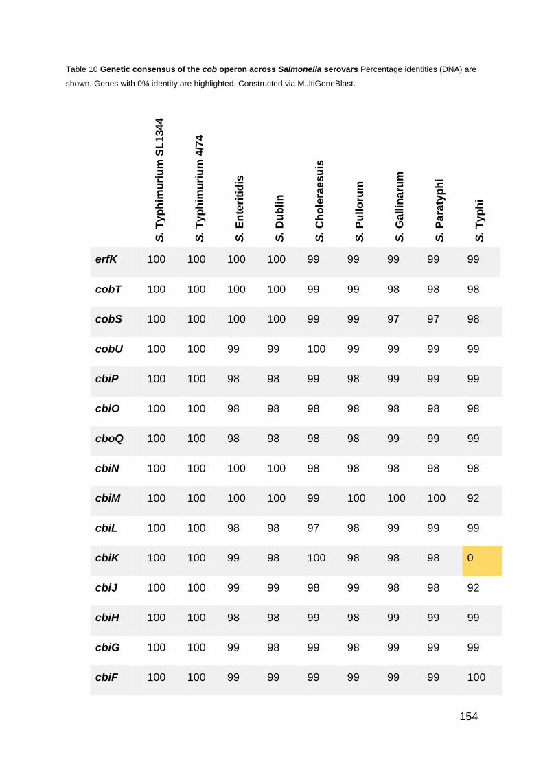
154
Table 10 Genetic consensus of the cob operon across Salmonella serovars Percentage identities (DNA) are
shown. Genes with 0% identity are highlighted. Constructed via MultiGeneBlast.
S
. T
yp
him
uri
um
SL
134
4
S.
Ty
ph
imu
riu
m 4
/74
S.
En
teri
tid
is
S.
Du
blin
S.
Ch
ole
rae
su
is
S.
Pu
llo
rum
S.
Gall
ina
rum
S.
Pa
raty
ph
i
S.
Ty
ph
i
erfK 100 100 100 100 99 99 99 99 99
cobT 100 100 100 100 99 99 98 98 98
cobS 100 100 100 100 99 99 97 97 98
cobU 100 100 99 99 100 99 99 99 99
cbiP 100 100 98 98 99 98 99 99 99
cbiO 100 100 98 98 98 98 98 98 98
cboQ 100 100 98 98 98 98 99 99 99
cbiN 100 100 100 100 98 98 98 98 98
cbiM 100 100 100 100 99 100 100 100 92
cbiL 100 100 98 98 97 98 99 99 99
cbiK 100 100 99 98 100 98 98 98 0
cbiJ 100 100 99 99 98 99 98 98 92
cbiH 100 100 98 98 99 98 99 99 99
cbiG 100 100 99 98 99 98 99 99 99
cbiF 100 100 99 99 99 99 99 99 100

155
cbiT 100 100 100 100 100 99 100 100 99
cbiE 100 100 98 98 98 97 100 100 100
cbiD 100 99 99 99 99 99 98 98 98
cbiC 100 100 100 100 99 99 98 98 98
cibB 100 100 98 99 98 98 97 97 0
cbiA 100 100 99 98 98 98 0 0 99
pocR 100 100 100 100 99 0 99 99 99
pduF 100 100 100 100 100 99 99 0 99
156
6.3.2 Development and optimisation of a Vitamin B12 quantification assay
As discussed in the Introduction to this chapter, the measurement of levels of vitamin
B12 produced by bacteria has not been done previously in our lab, and while there are
commercially available kits these come at a high cost. We therefore worked to develop
the protocol for a microbiological assay for B12 measurement which would be able to
measure the B12 produced by Salmonella, based on a previously published protocol
(Lawrence and Roth 1996). The assay is based on the vitamin B12 dependency of a
Salmonella Typhimurium SL1344 metE mutant. This strain is spread on a minimal
media plate which lacks vitamin B12 and a sample from the test strain placed on a filter
disc. The growth of the metE strain can then be measured which correlates with the
amount of vitamin B12 in the sample. The growth diameter of a range of standard
vitamin B12 concentrations (A) and the resulting standard curve (B) are shown in Figure
50.

157
Figure 50 Vitamin B12 bioassay. A SL1344 ΔmetE mutant was spread on an M9
minimal media plate. A disc is placed in the middle with a specific concentration
of vitamin B12 and the resulting growth measured. Diameter of growth is shown
in (A) and (B) for the construction of a calibration curve, error bars show SE.
Vitamin B12 concentration (µg/ml)
Vitamin B12 concentration (µg/ml)

158
6.3.4 Levels of Vitamin B12 produced by Salmonella serovars and Citrobacter
rodentium
In order to comparatively measure the amount of vitamin B12 produced by the various
strains, growth conditions had to be used under which all strains grew equally. There
was no external methionine or vitamin B12 and, components for the synthesis of
vitamin B12 were not limited (cobalt, 1,2-propanediol). M9 minimal medium was used,
supplemented with an amino acid mix containing all 19 amino acids except
methionine.
Under these conditions all strains grew comparably, that is there was no lag for any of
the serovars which had been seen previously for S. Pullorum and S. Gallinarum in the
absence of additional amino acids. All strains reached maximum OD by 24 hours so
the assay was conducted at this timepoint.
While this assay cannot give a clear or accurate measure of the exact amount or rate
of synthesis of vitamin B12 by each strain it is possible to compare relative levels
between the strains and to calculate the concentration of vitamin B12 in the 10 μl
sample using the calibration curve constructed (Figure 50).
The two S. Typhimurium strains, SL1344 and 4/74 produced the highest levels of B12,
at 0.1 µg/ml and 0.14 µg/ml respectively. This was then followed by the four further
‘enteric’ strains, S. Dublin (0.070 µg/ml), S. Enteritidis (0.045 µg/ml) and S.
Choleraesuis (0.025 µg/ml). Of the two ‘systemic’ strains, only S. Gallinarum showed
any level of B12 production (0.013 µg/ml), no production was seen for S. Pullorum.
Cirtrobacter rodentium produced the lowest measured level of B12 (0.011 µg/ml).

159
Figure 51 Vitamin B12 bioassay. A SL1344 ΔmetE mutant was spread on an M9 minimal media plate.
A disc is placed in the middle with a sample of lysed cells from the relevant strain. Protocol described
in detail in Materials and Methods (Page 54). The diameter of the growth is shown in (A), and the
concentration of the 10µl spot in (B), calculated using the calibration curve (Figure 50B). Data is
representative of 3 repeats and SE is shown.
A
B
S. T
yp
him
urium
S
L1
34
4
S. T
yp
him
urium
4/7
4
S.
Ente
ritid
is
S.
Dub
lin
S.
Ga
llina
rum
S.
Pu
lloru
m
S.
Cho
lera
esu
is
C.
rod
entiu
m
S. T
yp
him
urium
S
L1
34
4
S. T
yp
him
urium
4/7
4
S.
Ente
ritid
is
S.
Dub
lin
S.
Ga
llin
aru
m
S.
Pu
lloru
m
S.
Cho
lera
esu
is
C.
rod
entiu
m

160
6.3.5 Comparison of NO and N2O released by different Salmonella
serovars during anaerobic nitrate growth
Salmonella serovars and Citrobacter rodentium were grown under denitrifying
conditions to compare the levels of NO and N2O released by each strain. Strains were
grown in M9 minimal media supplemented with casamino acids to ensure comparable
growth of all strains irrespective of amino acid requirements, 20 mM nitrate and 10
mM glycerol. We attempted to conduct this experiment without the addition of
casamino acids to be more in line with other experiments conducted throughout this
thesis however the growth rate of the different serovars was highly variable and
therefore any attempt to draw comparisons would have been futile. With the addition
of casamino acids growth was comparable between all strains (Figure 52).
Nitric oxide and nitrous oxide measurements were taken at 17, 24, 40 and 48 hours of
growth, concentrations for each timepoint are shown as well as total NO/N2O for the
entire 48 hours (Figure 53 & Figure 54).
In general, a similar pattern is seen for the differences in NO and N2O released by
each strain, that is, the strains that release the most/least NO also release the
most/least N2O (Figure 53 & Figure 54). Additionally, all strains release greater levels
of N2O than NO, with an average difference of 7.7 fold. This pattern does not always
hold true however; the WT strain used throughout this thesis, S. Typhimurium SL1344,
produced the second highest level of N2O but the least amount of NO, a 13 fold
difference between the two gases. This suggests that there is, in comparison to the
other strains, a higher rate of conversion of NO to N2O. Surprisingly however, the
opposite is seen for the parent strain S. Typhimurium 4/74 with the lowest levels of
N2O but the second highest level of NO, a 3.7 fold difference (Figure 53 & Figure 54).
Salmonella Pullorum exhibited the highest levels of both NO and N2O release, totaling
108.7 µM and 1.0 mM respectively. The closely related strain, Salmonella Gallinarum
which also causes systemic fowl infections, showed much lower levels, 67.3 µM NO
and 0.47 mM N2O. Citrobacter rodentium released NO and N2O levels higher than all
but 2 of the Salmonella serovars, 83.2 µM and 0.67 mM respectively (Figure 53 &
Figure 54).
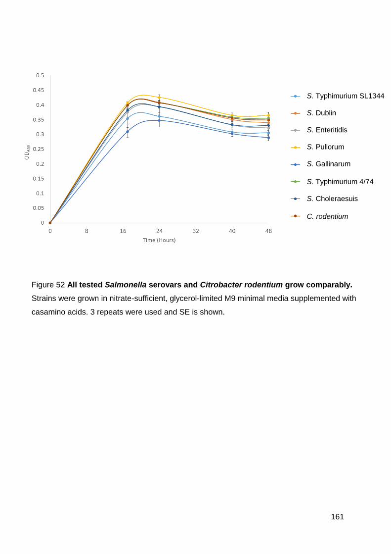
161
Figure 52 All tested Salmonella serovars and Citrobacter rodentium grow comparably.
Strains were grown in nitrate-sufficient, glycerol-limited M9 minimal media supplemented with
casamino acids. 3 repeats were used and SE is shown.
S. Typhimurium SL1344
S. Dublin
S. Enteritidis
S. Pullorum
S. Gallinarum
S. Typhimurium 4/74
S. Choleraesuis
C. rodentium

162
Fig
ure
53
Nit
rou
s o
xid
e l
eve
ls o
f S
alm
on
ella
se
rova
rs a
nd
Cit
rob
ac
ter
rod
en
tiu
m. S
tra
ins
we
re g
row
n in
nitra
te-s
uff
icie
nt,
gly
ce
rol-lim
ite
d M
9 m
inim
al m
ed
ia s
upp
lem
ente
d w
ith
ca
sam
ino
acid
s.
3 r
epe
ats
we
re u
se
d a
nd S
E is s
ho
wn
. A
) co
ncen
tra
tion
s o
f N
2O
ove
r 4
8 h
ou
rs, B
) to
tal N
2O
rele
ase
d o
ve
r 4
8 h
ou
rs.
S.
Typ
him
uri
um
S
L1
34
4
S.
Dub
lin
S.
En
teritid
is
S.
Pu
lloru
m
S.
Ga
llin
aru
m
S.
Typ
him
uri
um
4/7
4
S.
Cho
lera
esu
is
C.
rod
en
tiu
m

163
Fig
ure
54
Nit
ric
ox
ide
le
ve
ls o
f S
alm
on
ella
se
rova
rs a
nd
Cit
rob
ac
ter
rod
en
tiu
m. S
tra
ins w
ere
gro
wn
in n
itra
te-s
uff
icie
nt,
gly
ce
rol-lim
ite
d M
9 m
inim
al m
ed
ia s
upp
lem
ente
d w
ith
ca
sam
ino
acid
s.
3 r
epe
ats
we
re u
se
d a
nd S
E is s
ho
wn
. A
) co
ncen
tra
tion
s o
f
NO
ove
r 4
8 h
ou
rs, B
) to
tal N
O r
ele
ased
ove
r 4
8 h
ou
rs.
S.
Typ
him
uri
um
S
L1
34
4
S.
Dub
lin
S.
En
teritid
is
S.
Pu
lloru
m
S.
Ga
llin
aru
m
S.
Typ
him
uri
um
4
/74
S.
Cho
lera
esu
is
C.
rod
en
tiu
m

164
6.4 Discussion
This chapter asked the whether there is a link between vitamin B12 production and the
level of nitrous oxide released between different strains. This was partially based on
the hypothesis raised in Chapter 5 that Salmonella produces this high level of nitrous
oxide in order to compete with other gut bacteria by targetting the vitamin B12 pool.
The potential is therefore raised whether systemic pathogens, including some
Salmonella serovars which do not remain in the microxic environment of the inflammed
intestine where nitrate is available, will produce similar levels of nitrous oxide to those
that do not, and whether this is linked to their ability to produce vitamin B12.
The vitamin B12 sythesis ability of different Salmonella serovars was discussed in the
introduction of this chapter and built upon in the results. An alignment of the cob
operon (Figure 49, Table 10) confirmed that pseudogenes have formed and genes
been lost in 4 of the 5 systemic strains analysed, Salmonella Gallinarum, Pullorum,
Typhi and Paratyphi, but not in Salmonella Choleraesuis. There are however a large
number of mutations in the B12-dependent systems required for propanediol utilisation
and ethanolamine degradation in S. Choleraesuis (Nuccio and Bäumler 2014). It was
interesting that especially in Salmonella Typhi the genes were not completely missing
but instead have aquired large numbers of stop codons throughout the DNA sequence.
All the mutated systemic strains had a mutation in either cbiA or cbiB, the first two
genes of the operon, or in pocR, the positive regulator of the operon. A mutation at the
beginning of the cob operon would result in the synthesis of the vitamin being halted
in the very early stages, this would therefore stop the metabolically expensive process
before it progressed very far. A further mutation further down the operon would have
very little phenotypic effect at that point as the proteins work sequentially in respect to
the order of genes in the operon.
The mutation of pocR in Salmonella Pullorum is interesting, this strain contained a
complete cob operon except for this, and was not analysed in the Nuccio study. While
it is known that pocR is a positive regulator of the cob operon it is not clear from the
literature whether the translation of the operon is dependent on pocR or not (Rondon
and Escalante-Semerena 1992, Chen et al. 1995a). This study assessed the
importance of PocR for the synthesis of vitamin B12 in S. Typhimurium SL1344 and

165
concluded since the vitamin B12 addicted mutant ΔmetE when combined with a ΔpocR
mutation was still able to grow in anaerobic minimal media conditons under which a
ΔmetE ΔcobS mutant is not, that PocR is not an essential regulator of the operon and
that there are other unknown regulators. In the case of Salmonella Pullorum it
therefore may be the case that this mutation does not result in the total lack of ability
to trancribe the cob operon and produce vitamin B12, however we have not yet looked
into what an alternative regulator might be and whether this holds true across multiple
Salmonella serovars.
We then developed a bioassay which would enable us to compare vitamin B12
production between different Salmonella serovars (Figure 50) This assay, which is
based on a previously described protocol (Lawrence and Roth 1996), uses the vitamin
B12-dependency of a S. Typhimurium ΔmetE strain in order to assay the presence of
the vitamin in a sample. The main issue faced however was in growing the strains
equally and ensuring conditions where the production of vitamin B12 was required and
thereore should occur, and that all components of the synthesis (such a cobalt which
is required as a cofactor) were not limited. The major drawback however of this assay
is that it does no provide an exact amount of B12 produced by the strains or a
production rate. This is due to the fact that cells from 200 ml of culture must be lysed
to release the endogenously produced vitamin and this process involves boiling which
may damage a percentage of the vitamin – all strains were treated equally however
and so comparisions are warranted. From the final results of this (Figure 51) we can
conclude a few things. Our WT strain, S. Typhimurium SL1344 and the parent strain,
4/74 produced the highest levels of B12, this was followed by the other enteric strains,
S. Dublin and Enteriitdis, and then S. Choleraesuis. S. Gallinarum produced a very
low level of B12 and S. Pullorum showed no growth around the disc and so we conclude
no measurable B12 was released. Citrobacter rodentium, a model strain for the
systemic causing EHEC, produced the lowest measurable level of vitamin B12. There
is therefore a clear trend, that enteric strains which will spend long periods of time in
the intestine, produce high levels of vitamin B12 and systemic strains, which will quickly
disseminate into other parts of the body, produce much lower concentrations of B12.
The next stage of this study was to compare the levels of nitrous oxide produced by
the different strains while undergoing denitrification. The hypotheis was that since we
believe Salmonella Typhimurium releases such high levels of N2O in order to compete

166
with the gut microbiota by targetting the vitamin B12 pool of these B12-auxotrophs, that
non-enteric strains which do not remain in the intestine will not benefit from this survival
advantage of high N2O emmisons and hence will release lower N2O.
In general this hypothesis held true with the systemic strains producing less total N2O
than the enteric strains (Figure 53), however not in all cases. Our WT strain S.
Typhimurium SL1344, produced the second highest level of N2O whereas the parent
strain S. Typhimurium 4/74 released the lowest levels of N2O. The latter of this is quite
surprising and further study should be done to confirm the result. The two further
enteric strains, S. Dublin and S. Enteritidis then produced the next highest levels of
N2O, distinctly higher than S. Choleraesuis and S. Gallinarum, two systemic strains.
However, two systemic strains, S. Pullorum and Citrobacter rodentium released two
of the highest levels of N2O. This study needs to be repeated to draw any strong
conclusions, steady state culturing may also provide more useful insight into any
differences between the strains.
Taken in combination the results of this chapter potentially provide insight into a link
between vitamin B12 production and N2O release throughout systemic and enteric
strains. It is clear from previous studies, our alignment and the bioassay conducted
that enteric strains are capable of producing B12 and at higher levels than the systemic
strains. We cannot conclude from this data whether the reason for this phenomenon
is correlated to a change in the production of N2O by the strains or not. However, it
seems clear that since the systemic strains have lost the ability to synthesise vitamin
B12 this must not be of importance to their survival and pathogenesis.
Assessing the survival of the different strains when challenged with N2O could reveal
further insights in to their interactions with this gas, and moreover competition studies
with commensal gut bacteria would be of interest. Do the enteric strains outcompete
the gut microbiota, whereas the systemic strains do not – and would this difference
disappear in the absence of available nitrate?
A further question arises whether this link between N2O production and B12 synthesis
is present throughout a wider range of pathogenic bacteria, particularly enteric
pathogens. Most Campylobacter species are disease-causing, including C. jejuni, one
of the primary causes of foodborne disease in humans and C. coli which also causes

167
an enteric infection in humans (Skirrow 1977). Campylobacter species are unable to
produce both N2O and vitamin B12 (Payne et al. 1982, Rodionov et al. 2003).
The enteric pathogen Clostridium difficile causes similar symptoms including watery
diarrhea, fever and abdominal pain; it makes up about 20% of cases of antibiotic-
associated diarrhea (Kelly, Pothoulakis and LaMont 1994, Vogel 1995). C. difficile can
synthesise vitamin B12, however analysis of its genome shows that both NarG and
NapA are absent and it is therefore unlikely this strain can produce N2O (Rodionov et
al. 2003)
The enteric pathogen Vibrio cholera is the causative agent of cholera, which causes
vomiting and diarrhea, and has a fatality ratio of approximately 4% (WHO). V. cholera
can produce B12 (Rodionov et al. 2003) but only contains the periplasmic nitrate
reductase (Nap) enzyme (Braun and Thöny-Meyer 2005) and so, it could be expected
to produce lower levels of N2O. V. cholera can however produce NO and is able to
reduce N2O and use it as an electron acceptor (Janoff et al. 1997, Kaspar and Tiedje
1981, Yoshinari 1980).
From the three common enteric pathogens mentioned here it would appear that this
phenomenon does not translate to other strains. Another member of the
Enterobacteriaceae family, to which Salmonella and E. coli belong, Klebsiella
pneumonia, does produce both N2O and B12. When undergoing denitrification
Klebsiella converts ≈5% of the nitrate to N2O, a similar level to measured for
Salmonella in this study (Satoh, Hom and Shanmugam 1983). K. pneumonia is found
in our normal microbiota, in the mouth, skin and gut; however if inhaled it can result in
pneumonia which carries a fatality rate of abput 50% (Meatherall et al. 2009).
Klebsiella can also cause urinary tract infections, particularly in diabetics, and can lead
to bladder or kidney stones (Ronald 2003). This strain is therefore not an enteric
pathogen but contains the characteristics we see from enteric Salmonella strains.
There are many non-enteric, or systemic, pathogens which are unable to produce B12
including pathogenic Neisseria, Hemophilus influenza and Helicobacter pylori and
Bordetella pertussis. The latter three of these are unable to produce N2O, Neisseria
can reduce NO to N2O but cannot reduce nitrate, potentially resulting in low N2O
emissions (Rodionov et al. 2003, Stewart 2003, Barth, Isabella and Clark 2009, Marais
et al. 1999). In contrast, the non-enteric bacteria Pseudomonas aeruginosa, which

168
causes numerous infections in the urinary tract, skin wounds, blood and, airways,
particularly of cystic fibrosis patients, produces both B12 and N2O (Bodey et al. 1983,
Zumft 1997).
It therefore seems that while in Salmonella there is a link between B12 and N2O
production and being the causative agent of an enteric infection, throughout all
bacteria this may be a more general link to pathogenicity.
In Salmonella, vitamin B12 is required for the degradation of 1,2-propandediol required
for the use of tetrathionate as an electron acceptor, which is present in the inflamed
intestine. The loss of ability to reduce tetrathionate came at an earlier stage than the
loss of B12 synthetic ability (Langridge et al. 2015). With the conclusions drawn from
this chapter we therefore believe it may be the case that systemic strains have lost the
ability to synthesise vitamin B12 due to the fact that they have moved out of the intestine
where as well as vitamin B12 dependent tetrathionate respiration providing a growth
advantage, N2O is released targeting any exogenous B12 (Nuccio and Bäumler 2014).
If this is the case it could then be that enteric Salmonella strains which already require
B12 provide themselves with a pathogenic advantage by producing high levels of N2O
– something that provides no advantage, but a clear detriment to non-B12 producing,
systemic strains.

169
7 General Discussion

170
As each chapter has already been individually discussed this general discussion will
summarise the major results of the study, contextualise the results in the wider body
of Salmonella research, and provide suggestions for future studies.
7.1 Context
Salmonella is a global pathogen which causes a range of diseases in humans and
animals. In humans, there are two major disease types, gastroenteritis and an
systemic infection known as Typhoid fever. Enteric infections are generally self-
limiting, and apart from infections of the immunocompromised, elderly or young, do
not require any treatment. Typhoid fever however is a much more serious infection
which without treatment usually results in death, and as such there are ≈200,000
global deaths annually.
The development of antibiotic resistant strains is an increasingly severe problem
across a large number of pathogenic bacteria, primarily due to increased use and
misuse of antibiotics.
There are also multi-drug resistant strains of Salmonella which are resistant to the so
called ‘last resort’ antibiotics and therefore are untreatable. Current vaccines against
Typhoid fever have numerous drawbacks, including not providing life-long resistance
and hence being of limited use to those living in the endemic regions.
It is therefore of key global interest to better our understanding of how Salmonella
causes infection in order to develop new treatments, including, but not limited to
antibiotics, and vaccines.
7.2 Nitric oxide
Nitric oxide (NO) is a free radical which has both cytostatic and cytotoxic activities and
causes widespread damage to numerous cellular targets including DNA and proteins
(Fang 2004). Salmonella is exposed to NO both endogenously and exogenously
throughout its lifecycle. As a facultative anaerobe, Salmonella has the ability to use
alternative electron acceptors for respiration when oxygen is unavailable, one of these
is nitrate. The respiration of nitrate in Salmonella, referred to as truncated
denitrification, involves the sequential reduction of nitrate via nitrite and nitric oxide to
171
nitrous oxide. Hence the endogenous reduction of NO is required whenever
Salmonella is respiring using nitrate. Additionally, the release of NO is an essential
part of the host immune system particularly by macrophages. Salmonella have the
ability to reside, survive and replicate within host macrophages, and rely on this ability
in order to cause a systemic disease. NO release by macrophages is via the inducible
NO synthase, iNOS. iNOS has been shown to be of particular importance to the host
immune defence system in mice, with mice deficient in iNOS being significantly more
susceptible to Salmonella infection (Alam et al. 2002, Mastroeni et al. 2000).
There are three well characterised enzymes for NO detoxification in Salmonella,
HmpA, NorV and NrfA, the triple deletion of which does not render Salmonella
completely unable to withstand NO stress (Mills et al. 2008). This therefore suggests
there are further systems at play to enable Salmonella to detoxify NO.
Since NO detoxification is an essential step during Salmonella pathogenesis a better
understanding of this process could allow the development of antibacterial drugs or
vaccines that can target the enzymes involved in NO defense.
7.3 Nitrous oxide
Nitrous oxide is produced by Salmonella by the reduction of nitric oxide, primarily
carried out by Hmp and NorVW. In Salmonella, due to the lack of a nitrous oxide
reductase, denitrification is truncated and hence nitrous oxide is the end product.
Similarly to Salmonella, E. coli also lacks a nitrous oxide reductase and hence has a
net release of N2O; however a combination of studies by (Rowley et al. 2012)
(Stremińska et al. 2012) and Runkel et al (personal communication) has identified that
nitrous oxide production by Salmonella Typhimurium cultured in nitrate
sufficient/glycerol limited chemostats is an order of magnitude higher than that
produced by related non-pathogenic E. coli strains (Figure 3).
In fact it has been observed that Salmonella also produces high levels of NO, and has
been hypothesised that this is in order to enable the bacteria to produce such high
levels of nitrous oxide and if so, for what purpose (Maia and Moura 2014).
172
7.4 Vitamin B12
Salmonella is one of a reasonably small number of bacteria that is able to synthesise
vitamin B12 de novo. B12 synthesis is a highly metabolically expensive process, the
vitamin itself is very large, with a molecular weight of 1355.37 g/mol, and the process
involves 30 different genes, which together make up over 1% of the Salmonella
genome. Curiously however the requirements for vitamin B12 in Salmonella are very
low. There are only four B12 dependent processes - vitamin B12-dependent methionine
synthase, MetH, ethanolamine ammonia lyase, which degrades ethanolamine (Roof
and Roth 1989), propanediol dehydratase which allows Salmonella to use propanediol
as a carbon source, and queuosine synthase, which produces queuosine, a
nucleoside found in four tRNAs which is not essential for growth under laboratory
conditions (Frey et al. 1988).
It was not until 2001 that a deleterious effect of losing any of these processes was first
noted and therefore the beginning of an explanation as to why Salmonella produces
vitamin B12. Tetrathionate is an alternative electron acceptor which is produced in the
inflamed intestine, the ability of Salmonella to respire using tetrathionate is dependent
on either ethanolamine or propanediol the utilisation of which are both vitamin B12
dependent processes (Price-Carter et al. 2001, Thiennimitr et al. 2011).
However, the ability to respire on tetrathionate alone does not seem sufficient to
warrant the synthesis of vitamin B12. An observation was made in Paracoccus
denitrificans that N2O accumulation induces a switch from vitamin B12-dependent to
vitamin B12-independent biosynthetic methionine pathways (Sullivan et al. 2013); this
therefore suggested a possibility for a similar phenomenon in Salmonella, and a link
between nitrous oxide, vitamin B12 and methionine requirements.
7.5 Characterisation of NO detoxification enzymes
The basis of this work was a prior study done in the Rowley group where a microarray
was conducted in order to identify novel genes involved in the anaerobic protection
against nitric oxide. Six genes highlighted from this analysis were further investigated
during this study, cstA, hcr, STM1273, yeaR, tehB and STM1808, the latter three of
which have the same annotated function as tellurite resistance genes.

173
This study created knockout mutants of each of these six genes and a ΔtehB
ΔSTM1808 ΔyeaR triple mutant strain. Aerobically no growth defect was observed
when Salmonella was challenged with NO in the absence of these three genes, even
in combination. However, anaerobically, there is a clear defect for a triple ΔtehB
ΔSTM1808 ΔyeaR mutant growing in the presence of 10 mM of the NO donor deta
NONOate. Since there is no phenotype visible for the single mutants we conclude that
there is functional overlap between TehB, STM1808 and YeaR in anaerobic nitric
oxide protection. Further experiments then measured the amount of NO released by
the different strains when growing under denitrifying conditions and once more the
triple mutant strain showed significantly higher levels of release than the WT.
Three further genes which were upregulated by either exogenous or endogenous NO
were then investigated. We confirmed a mild role for all three, cstA, hcr and STM1273,
in both aerobic and anaerobic NO protection. The knockouts were only assessed as
single mutants, as opposed to in combination with either other well-known NO
detoxification systems or those identified in this study. It could therefore be the case
that more pronounced phenotypes would become visible when combining some of
these mutations, as seen for the TeR strains.
The clearest role for any of these three proteins in direct NO reduction was for Hcr.
When grown under denitrifying conditions the Δhcr mutant releases significantly higher
levels of NO than the WT strain, thus suggesting that Hcr directly detoxifies NO and
hence this strain has an impaired ability to do so. It is interesting that both the CstA
and STM1273 knockout strains in fact release slightly lower levels of NO than the WT,
indicating that these two proteins do not directly reduce endogenous NO.
STM1273 was only upregulated in the microarray conducted after exogenous NO
exposure, not during endogenous exposure during denitrification. If STM1273 is
present in the periplasm as opposed to cytoplasmic membrane this would further
strengthen this hypothesis, and the cellular localization of this protein needs to be
determined.
It is worth considering how the various NO protection systems across all bacteria
function. The most common proteins are globins, heme-containing globular proteins
which are important for transporting oxygen. The most well characterised and often
said to be key NO reduction enzyme in Salmonella is Hmp, a flavohaemoglobin.
174
Additional globin proteins with roles in NO tolerance include HbN from Mycobacterium
bovis and Vgb from Vitreoscilla sp (Ouellet et al. 2002) which similarly to Hmp convert
NO into nitrate (Frey et al. 2002). However, additionally there are globin proteins which
function in other ways such as Myoglobin and haemoglobin in animals which is thought
to have a role in NO scavenging (Brunori 2001) and, Cgb in Camplylobacter jejuni,
which confers resistance through an unknown mechanism (Elvers et al. 2004). A
second group of NO protection enzymes are the reductases, this include NorV and
NrfA from Salmonella and are capable of directly reducing the compound either to
nitrous oxide or ammonia.
The next step is to determine the exact mechanism of how the six proteins included in
this study provide Salmonella with protection against NO. Protein film voltammetry can
be used in order to determine and quantify the NO reductase activity, as has previously
been done for NrfA from E. coli (van Wonderen et al. 2008). This technique works by
immobilizing the enzyme onto an electrode, placing this in the potential substrate (NO)
and monitoring any charge produced due to the flow of electrons and thus the redox
potential (Angove et al. 2002). In addition, the effect of changing conditions such as
pH or other inhibitors can be assessed. Understanding the mechanism by which these
proteins provide protection against NO is important for building a complete picture of
NO resistance in Salmonella – especially if these different systems are acting in
different ways at different parts of the cell. It would be the case if designing vaccines
or drug targets based on any of these systems that one must be aware of how the
numerous proteins interact, especially if they are able to compensate for the loss of
others.
7.6 A link between nitric oxide and tellurite resistance in Salmonella
Three of the proteins shown in this study to be involved in NO protection are annotated
as tellurite resistance proteins. We therefore also assessed the impact a loss of these
proteins has on Salmonella growing in the presence of potassium tellurite. The results
of this study show a clear link between tellurite and nitric oxide resistance in
Salmonella.
We initially became interested in tellurite due to the fact that three of the proteins
upregulated in response to NO, and discussed above, are annotated as tellurite

175
resistance genes. Tellurite is highly toxic to a wide range of bacteria; the causation of
the toxicity is not currently clear but there are multiple resistance systems across the
bacterial kingdom. The results presented here confirm TehB, STM1808 and YeaR to
be important for tellurite protection in Salmonella, both aerobically and anaerobically.
Previous work has suggested that the version of TehB in Salmonella, as opposed to
the E. coli homologue, may not have the ability to protect against tellurite due to
differences in the SAM-methyltransferase domain (Karlinsey et al. 2012). Conversely,
we found TehB to afford Salmonella with the highest level of protection against
tellurite.
We then hypothesised that since tellurite resistance proteins also afford Salmonella
an advantage in the protection against NO that the reverse may also be true, that
proteins known to play a role in NO protection may also provide tellurite resistance.
The most striking result here was that of ΔnsrR. NsrR is the master negative regulator
of NO detoxification in Salmonella and is responsible for regulating hcp-hcr,
ytfE, ygbA, hmp, yeaR-yoaG and STM1808 (Karlinsey et al. 2012). When challenged
with tellurite ΔnsrR grows considerably better than WT. This therefore shows that the
upregulation of these genes results in Salmonella being able to survive in the presence
of this toxin. This was further investigated using numerous knockout strains which
showed that YtfE, NrfA and NorV all exhibit a small but measureable degree of
protection against tellurite anaerobically. The three proteins whose role in NO
protection was confirmed in this study, CstA, Hcr and STM1273, also were shown to
provide tellurite resistance.
7.7 Nitrous oxide
The second aspect of this thesis was looking at the role of nitrous oxide production in
Salmonella. Previous studies have highlighted that Salmonella produces and releases
nitrous oxide at high levels – but have been unable to provide an explanation as to
why this might be. From the results presented here we hypothesise that when residing
in the inflamed intestine, Salmonella releases N2O in order to outcompete the resident
gut microbiota.
The key protein linking nitrous oxide, vitamin B12 and the intestine together is MetE.
Since nitrous oxide binds to and inactivates vitamin B12, MetH, the vitamin B12
176
dependent methionine synthase is unable to function adequately in its presence,
however since Salmonella possesses a vitamin B12 independent methionine synthase,
MetE, this protein is able to perform this vital function. A key finding of this work was
the importance of MetE for denitrifying Salmonella, where a clear growth defect for
ΔmetE was seen, a defect which was further amplified by saturating the growth media
with N2O.
The regulation of vitamin B12 synthesis was also investigated. Previous studies have
highlighted PocR as being the primary regulator of the cob operon and hence of
vitamin B12 synthesis. However, results from this study suggest that there are
alternative regulators due to the fact that the absence of PocR is not detrimental for
the vitamin B12 addicted strain, ΔmetE, and in fact provides a slight restoration of the
growth defect. We provide a potential explanation that when PocR is present it binds
in such a location that it blocks the binding sites for other unknown regulatory
elements. The full understanding of the regulation of the cob operon may then provide
insights into when and why this operon is active.
Based on the hypothesis drawn we then queried whether the production of vitamin B12
and N2O is similar throughout a range of Salmonella serovars, in particular if systemic
strains produce these two compounds at the same rate as enteric strains. Systemic
strains of Salmonella, such as Salmonella Typhi the causative agent of Typhoid fever,
do not remain in the inflamed intestine where nitrate is available for extended periods
of time, and instead reside in areas of the body where oxygen is likely to be more
abundant.
By looking both bioinformatically and by the development of a vitamin B12 bioassay we
concluded that there is a general trend for enteric Salmonella serovars to produce
higher levels of vitamin B12 than the systemic strains. Both S. Typhi and S. Paratyphi
could not be assessed in our hands but bioinformatic analysis would suggest that they
are unable to produce any vitamin B12. There was a less strong correlation for the
amount of N2O produced by the different strains although a general trend could be
seen with the enteric strains releasing higher levels of N2O than the WT strain. What
is of most interest here is that the systemic strains must not require vitamin B12 and
hence have acquired pseudogenes or lost genes in the cob operon, this further
indicates the important of vitamin B12 for Salmonella residing in the intestine.
177
Initial future experiments should be based on competing Salmonella against members
of the gut microbiota under denitrifying conditions as would be found in the inflamed
intestine. Bioinformatic analysis of common gut microbiota revealed that almost no
members of the microbiota have the vitamin B12 independent enzyme, MetE (Table 9).
In addition, none of the microbiota we analysed possess NosZ, the enzyme required
for the reduction of nitrous oxide. It therefore is highly likely that when growing on
nitrate Salmonella will be able to outcompete the gut microbiota. Since it would be very
complex to assess this interaction in vivo an in vitro approach would be required. A
simple co-culture experiment could be used, whereby the same experimental setup
used in this study is used but with commensal gut bacteria and Salmonella cultured in
the same vessel. This would require thorough experimental design to ensure
conditions which allowed for both bacteria to grow, that is that when cultured
separately the commensal bacteria exhibits normal growth. One drawback of this set
up is that it does not closely mimic the natural conditions of the gut, where a complex
community of gut commensal bacteria are situated with a small number of Salmonella
infecting. A preferential system would be a model where a defined microbiota could
be allowed to become established and then a later inoculation of Salmonella be made,
and the effect of the pathogen on the microbiota numbers monitored. One system
which could allow for this would be a microfluidics gut model, where media is
constantly being pumped through the system and thus bacteria can survive for
extended periods of time. This is a new and rapidly developing field but a number of
groups are having successes with similar studies (Shah et al. 2016, Kim and Ingber
2013, Kim et al. 2016a).
Based on the general hypothesis drawn and also the significant reduction in survival
seen for a ΔmetE mutant in macrophages it would also be of interest to monitor this
strain in the mouse model. In particular this could be used not just to monitor the
survival of the mice, but to track the ability of the bacteria to disseminate from the
intestine. Analysis of the mouse stools to measure bacterial diversity and quantity
could also be used to give some information on how this strain, in comparison to the
WT, impacts on the mouse microbiota.
There are three potential ways to try and target Salmonella using this information. One,
by increasing the levels of vitamin B12 available, two, by removing the N2O from the

178
intestine or three, by providing the commensal bacteria with a way by to survive even
with high levels of N2O and low levels of vitamin B12.
Since we have shown that in vitro the effect of N2O can be overcome with the addition
of vitamin B12 there is the potential that this could be the case in the intestine. Vitamin
B12 could be delivered to the intestine in high levels, either orally in a prebiotic fashion,
or in combination with antibiotics, and this may be able to negate the advantage
Salmonella has over the non-vitamin B12 producing commensal bacteria.
Another possibility is reducing the amount of N2O present. There is a wide range of
work looking at the production and release of N2O into the atmosphere from
agricultural soils due to its function as a greenhouse gas; and therefore, research into
possible mitigation strategies (Ravishankara et al. 2009). The most feasible strategies
are those which rely on the natural bacteria N2O reductase system, NosZ, and
increasing the expression and function of this enzyme. It may be possible to engineer
a member of the gut microbiota to express nosZ. This would allow the gut bacteria to
reduce N2O to N2. This could then be used as a probiotic strain which would be able
to remove the N2O which Salmonella emits from the intestine and negate the impact
this has on the wider vitamin B12 pool.
Additionally, providing important members of the gut microbiota with MetE, the vitamin
B12 independent methionine synthase would allow them to survive even in the
presence of N2O. The creation of genetically modified, so called ‘smart’ probiotic
strains is controversial and not yet well studied; any work in this field would need to do
thorough testing to ensure that the modification to the bacteria does not result in a
conversion to a pathogenic phenotype, nor that the gene introduced cannot be
transferred to other bacteria. There are however increasing numbers of studies looking
to modify probiotic bacterial strains (Kullen and Klaenhammer 2000, Steidler and
Rottiers 2006, Durrer, Allen and Hunt von Herbing 2017, Hwang et al. 2017)
7.8 Survival within macrophages
Numerous strains constructed throughout this study were tested for their ability to
survive within murine macrophages. The ability for Salmonella to survive within the
host environment of the macrophage is essential to its virulence (Fields et al. 1986)
and therefore this is a key feature in the pathogenicity of the pathogen and potential
179
source of a vaccine stain. The most severely attenuated strain is the ΔtehB ΔSTM1808
ΔyeaR triple tellurite mutant with a four-fold reduction in bacterial survival compared
to the WT strain. Approximately a two-fold reduction was seen both for Δhcr and for
ΔmetE when methionine was lacking from the cell culture medium. These phenotypes
are all of worth pursuing to assess if the phenotypes translate to a reduced survival of
these strains in a mouse model. The combination of some of these mutations, or the
addition of knockout mutation of proteins such as HmpA may also further amplify the
phenotypes seen.
7.9 Final conclusions
In conclusion, this study highlights two aspects of denitrification in Salmonella which
may contribute to pathogenesis. We have characterised novel nitric oxide
detoxification proteins and shown a clear link between nitric oxide and tellurite
protection in Salmonella. We have also provided an explanation for two phenomena,
the production of vitamin B12 and the production of high levels of N2O. We hypothesise
that these two compounds afford Salmonella an advantage when residing in the
inflamed intestine.

180
8 References
181
Abshire, K. Z. & F. C. Neidhardt (1993) Analysis of proteins synthesized by
Salmonella typhimurium during growth within a host macrophage. J Bacteriol,
175, 3734-43.
Acharya, I. L., C. U. Lowe, R. Thapa, V. L. Gurubacharya, M. B. Shrestha, M. Cadoz,
D. Schulz, J. Armand, D. A. Bryla & B. Trollfors (1987) Prevention of typhoid
fever in Nepal with the Vi capsular polysaccharide of Salmonella typhi. A
preliminary report. N Engl J Med, 317, 1101-4.
Ailion, M., T. A. Bobik & J. R. Roth (1993) Two global regulatory systems (Crp and
Arc) control the cobalamin/propanediol regulon of Salmonella typhimurium. J
Bacteriol, 175, 7200-8.
Aktan, F. (2004) iNOS-mediated nitric oxide production and its regulation. Life Sci,
75, 639-53.
Alam, M. S., T. Akaike, S. Okamoto, T. Kubota, J. Yoshitake, T. Sawa, Y. Miyamoto,
F. Tamura & H. Maeda (2002) Role of nitric oxide in host defense in murine
salmonellosis as a function of its antibacterial and antiapoptotic activities.
Infect Immun, 70, 3130-42.
Almeida, C. C., C. V. Romão, P. F. Lindley, M. Teixeira & L. M. Saraiva (2006) The
role of the hybrid cluster protein in oxidative stress defense. J Biol Chem, 281,
32445-50.
Altier, C., M. Suyemoto & S. D. Lawhon (2000) Regulation of Salmonella enterica
serovar typhimurium invasion genes by csrA. Infect Immun, 68, 6790-7.
Alvarez-Ordóñez, A., M. Begley, M. Prieto, W. Messens, M. López, A. Bernardo & C.
Hill (2011) Salmonella spp. survival strategies within the host gastrointestinal
tract. Microbiology, 157, 3268-81.
Angove, H. C., J. A. Cole, D. J. Richardson & J. N. Butt (2002) Protein film
voltammetry reveals distinctive fingerprints of nitrite and hydroxylamine
reduction by a cytochrome C nitrite reductase. J Biol Chem, 277, 23374-81.
Arango Duque, G. & A. Descoteaux (2014) Macrophage cytokines: involvement in
immunity and infectious diseases. Front Immunol, 5, 491.

182
Arkenberg, A., Runkel, S., Richardson, D. J., & Rowley, G. (2011). The production
and detoxification of a potent cytotoxin, nitric oxide, by pathogenic enteric
bacteria. Biochemical Journal, 441 (2), 755-672
Avazéri, C., R. J. Turner, J. Pommier, J. H. Weiner, G. Giordano & A. Verméglio
(1997) Tellurite reductase activity of nitrate reductase is responsible for the
basal resistance of Escherichia coli to tellurite. Microbiology, 143 ( Pt 4),
1181-9.
Bajaj, V., C. Hwang & C. A. Lee (1995) hilA is a novel ompR/toxR family member
that activates the expression of Salmonella typhimurium invasion genes. Mol
Microbiol, 18, 715-27.
Banerjee, R. V. & R. G. Matthews (1990) Cobalamin-dependent methionine
synthase. FASEB J, 4, 1450-9.
Bang, I. S., L. Liu, A. Vazquez-Torres, M. L. Crouch, J. S. Stamler & F. C. Fang
(2006) Maintenance of nitric oxide and redox homeostasis by the salmonella
flavohemoglobin hmp. J Biol Chem, 281, 28039-47.
Barrow, P. A. & O. C. Freitas Neto (2011) Pullorum disease and fowl typhoid--new
thoughts on old diseases: a review. Avian Pathol, 40, 1-13.
Barth, K. R., V. M. Isabella & V. L. Clark (2009) Biochemical and genomic analysis of
the denitrification pathway within the genus Neisseria. Microbiology, 155,
4093-103.
Berman-Frank, I., P. Lundgren & P. Falkowski (2003) Nitrogen fixation and
photosynthetic oxygen evolution in cyanobacteria. Res Microbiol, 154, 157-64.
Bescós, R., A. Sureda, J. A. Tur & A. Pons (2012) The effect of nitric-oxide-related
supplements on human performance. Sports Med, 42, 99-117.
Bhatia, S. N. & D. E. Ingber (2014) Microfluidic organs-on-chips. Nat Biotechnol, 32,
760-72.
Blum, P. H., S. B. Jovanovich, M. P. McCann, J. E. Schultz, S. A. Lesley, R. R.
Burgess & A. Matin (1990) Cloning and in vivo and in vitro regulation of cyclic
AMP-dependent carbon starvation genes from Escherichia coli. J Bacteriol,
172, 3813-20.

183
Bobik, T. A., M. Ailion & J. R. Roth (1992) A single regulatory gene integrates control
of vitamin B12 synthesis and propanediol degradation. J Bacteriol, 174, 2253-
66.
Bobik, T. A., G. D. Havemann, R. J. Busch, D. S. Williams & H. C. Aldrich (1999)
The propanediol utilization (pdu) operon of Salmonella enterica serovar
Typhimurium LT2 includes genes necessary for formation of polyhedral
organelles involved in coenzyme B(12)-dependent 1, 2-propanediol
degradation. J Bacteriol, 181, 5967-75.
Bodey, G. P., R. Bolivar, V. Fainstein & L. Jadeja (1983) Infections caused by
Pseudomonas aeruginosa. Rev Infect Dis, 5, 279-313.
Booth, C. C. & D. L. Mollin (1959) The site of absorption of vitamin B12 in man.
Lancet, 1, 18-21.
Borsetti, F., V. Tremaroli, F. Michelacci, R. Borghese, C. Winterstein, F. Daldal & D.
Zannoni (2005) Tellurite effects on Rhodobacter capsulatus cell viability and
superoxide dismutase activity under oxidative stress conditions. Res
Microbiol, 156, 807-13.
Bowden, S. D., V. K. Ramachandran, G. M. Knudsen, J. C. Hinton & A. Thompson
(2010) An incomplete TCA cycle increases survival of Salmonella
Typhimurium during infection of resting and activated murine macrophages.
PLoS One, 5, e13871.
Bowden, S. D., G. Rowley, J. C. Hinton & A. Thompson (2009) Glucose and
glycolysis are required for the successful infection of macrophages and mice
by Salmonella enterica serovar typhimurium. Infect Immun, 77, 3117-26.
Braden, C. R. (2006) Salmonella enterica serotype Enteritidis and eggs: a national
epidemic in the United States. Clin Infect Dis, 43, 512-7.
Braun, M. & L. Thöny-Meyer (2005) Cytochrome c maturation and the physiological
role of c-type cytochromes in Vibrio cholerae. J Bacteriol, 187, 5996-6004.
Brunelli, L., J. P. Crow & J. S. Beckman (1995) The comparative toxicity of nitric
oxide and peroxynitrite to Escherichia coli. Arch Biochem Biophys, 316, 327-
34.
184
Brunori, M. (2001) Nitric oxide moves myoglobin centre stage. Trends Biochem Sci,
26, 209-10.
Buffie, C. G. & E. G. Pamer (2013) Microbiota-mediated colonization resistance
against intestinal pathogens. Nat Rev Immunol, 13, 790-801.
Burgess, C. M., E. J. Smid & D. van Sinderen (2009) Bacterial vitamin B2, B11 and
B12 overproduction: An overview. Int J Food Microbiol, 133, 1-7.
Burkholder, P. R. (1951) Determination of vitamin B12 with a mutant strain of
Escherichia coli. Science, 114, 459-60.
Cai, X. Y., M. E. Maxon, B. Redfield, R. Glass, N. Brot & H. Weissbach (1989)
Methionine synthesis in Escherichia coli: effect of the MetR protein on metE
and metH expression. Proc Natl Acad Sci U S A, 86, 4407-11.
Canfield, D. E., A. N. Glazer & P. G. Falkowski (2010) The evolution and future of
Earth's nitrogen cycle. Science, 330, 192-6.
Carter, A. O., A. A. Borczyk, J. A. Carlson, B. Harvey, J. C. Hockin, M. A. Karmali, C.
Krishnan, D. A. Korn & H. Lior (1987) A severe outbreak of Escherichia coli
O157:H7--associated hemorrhagic colitis in a nursing home. N Engl J Med,
317, 1496-500.
Carter, P. B. & F. M. Collins (1974) The route of enteric infection in normal mice. J
Exp Med, 139, 1189-203.
Chakravortty, D., I. Hansen-Wester & M. Hensel (2002) Salmonella pathogenicity
island 2 mediates protection of intracellular Salmonella from reactive nitrogen
intermediates. J Exp Med, 195, 1155-66.
Chen, P., M. Ailion, T. Bobik, G. Stormo & J. Roth (1995a) Five promoters integrate
control of the cob/pdu regulon in Salmonella typhimurium. J Bacteriol, 177,
5401-10.
Chen, P., M. Ailion, N. Weyand & J. Roth (1995b) The end of the cob operon:
evidence that the last gene (cobT) catalyzes synthesis of the lower ligand of
vitamin B12, dimethylbenzimidazole. J Bacteriol, 177, 1461-9.

185
Cherepanov, P. P. & W. Wackernagel (1995) Gene disruption in Escherichia coli:
TcR and KmR cassettes with the option of Flp-catalyzed excision of the
antibiotic-resistance determinant. Gene, 158, 9-14.
Chiang, T. T., C. T. Hung, W. M. Wang, J. T. Lee & F. C. Yang (2013) Recreational
nitrous oxide abuse-induced vitamin B12 deficiency in a patient presenting
with hyperpigmentation of the skin. Case Rep Dermatol, 5, 186-91.
Coburn, B., G. A. Grassl & B. B. Finlay (2007) Salmonella, the host and disease: a
brief review. Immunol Cell Biol, 85, 112-8.
Conley, D. J., H. W. Paerl, R. W. Howarth, D. F. Boesch, S. P. Seitzinger, K. E.
Havens, C. Lancelot & G. E. Likens (2009) Ecology. Controlling
eutrophication: nitrogen and phosphorus. Science, 323, 1014-5.
Cooper, B. A. & W. B. Castle (1960) Sequential mechanisms in the enhanced
absorption of vitamin B12 by intrinsic factor in the rat. J Clin Invest, 39, 199-
214.
Cordonnier, C., G. Le Bihan, J. G. Emond-Rheault, A. Garrivier, J. Harel & G. Jubelin
(2016) Vitamin B12 Uptake by the Gut Commensal Bacteria Bacteroides
thetaiotaomicron Limits the Production of Shiga Toxin by Enterohemorrhagic
Escherichia coli. Toxins (Basel), 8.
Crump, J. A. (2014) Updating and refining estimates of typhoid fever burden for
public health action. Lancet Glob Health, 2, e551-3.
Crump, J. A., S. P. Luby & E. D. Mintz (2004) The global burden of typhoid fever.
Bull World Health Organ, 82, 346-53.
Crump, J. A. & E. D. Mintz (2010) Global trends in typhoid and paratyphoid Fever.
Clin Infect Dis, 50, 241-6.
Danishpajooh, I. O., T. Gudi, Y. Chen, V. G. Kharitonov, V. S. Sharma & G. R. Boss
(2001) Nitric oxide inhibits methionine synthase activity in vivo and disrupts
carbon flow through the folate pathway. J Biol Chem, 276, 27296-303.
Datsenko, K. A. & B. L. Wanner (2000) One-step inactivation of chromosomal genes
in Escherichia coli K-12 using PCR products. Proc Natl Acad Sci U S A, 97,
6640-5.

186
De Groote, M. A. & F. C. Fang (1995) NO inhibitions: antimicrobial properties of nitric
oxide. Clin Infect Dis, 21 Suppl 2, S162-5.
Deacon, R., M. Lumb, J. Perry, I. Chanarin, B. Minty, M. Halsey & J. Nunn (1980)
Inactivation of methionine synthase by nitrous oxide. Eur J Biochem, 104,
419-23.
Devasagayam, T. P., J. C. Tilak, K. K. Boloor, K. S. Sane, S. S. Ghaskadbi & R. D.
Lele (2004) Free radicals and antioxidants in human health: current status
and future prospects. J Assoc Physicians India, 52, 794-804.
Dougan, G., V. John, S. Palmer & P. Mastroeni (2011) Immunity to salmonellosis.
Immunol Rev, 240, 196-210.
Drennan, C. L., R. G. Matthews & M. L. Ludwig (1994) Cobalamin-dependent
methionine synthase: the structure of a methylcobalamin-binding fragment
and implications for other B12-dependent enzymes. Curr Opin Struct Biol, 4,
919-29.
Dubey, A. K., C. S. Baker, K. Suzuki, A. D. Jones, P. Pandit, T. Romeo & P.
Babitzke (2003) CsrA regulates translation of the Escherichia coli carbon
starvation gene, cstA, by blocking ribosome access to the cstA transcript. J
Bacteriol, 185, 4450-60.
Duluc, D., M. Corvaisier, S. Blanchard, L. Catala, P. Descamps, E. Gamelin, S.
Ponsoda, Y. Delneste, M. Hebbar & P. Jeannin (2009) Interferon-gamma
reverses the immunosuppressive and protumoral properties and prevents the
generation of human tumor-associated macrophages. Int J Cancer, 125, 367-
73.
Dunstan, S. J., V. A. Ho, C. M. Duc, M. N. Lanh, C. X. Phuong, C. Luxemburger, J.
Wain, F. Dudbridge, C. S. Peacock, D. House, C. Parry, T. T. Hien, G.
Dougan, J. Farrar & J. M. Blackwell (2001) Typhoid fever and genetic
polymorphisms at the natural resistance-associated macrophage protein 1. J
Infect Dis, 183, 1156-60.
Durrer, K. E., M. S. Allen & I. Hunt von Herbing (2017) Genetically engineered
probiotic for the treatment of phenylketonuria (PKU); assessment of a novel

187
treatment in vitro and in the PAHenu2 mouse model of PKU. PLoS One, 12,
e0176286.
Eichelberg, K. & J. E. Galán (1999) Differential regulation of Salmonella typhimurium
type III secreted proteins by pathogenicity island 1 (SPI-1)-encoded
transcriptional activators InvF and hilA. Infect Immun, 67, 4099-105.
Elvers, K. T., G. Wu, N. J. Gilberthorpe, R. K. Poole & S. F. Park (2004) Role of an
inducible single-domain hemoglobin in mediating resistance to nitric oxide and
nitrosative stress in Campylobacter jejuni and Campylobacter coli. J Bacteriol,
186, 5332-41.
Eriksson, S., S. Lucchini, A. Thompson, M. Rhen & J. C. Hinton (2003) Unravelling
the biology of macrophage infection by gene expression profiling of
intracellular Salmonella enterica. Mol Microbiol, 47, 103-18.
Escalante-Semerena, J. C. & J. R. Roth (1987) Regulation of cobalamin biosynthetic
operons in Salmonella typhimurium. J Bacteriol, 169, 2251-8.
Escalante-Semerena, J. C., S. J. Suh & J. R. Roth (1990) cobA function is required
for both de novo cobalamin biosynthesis and assimilation of exogenous
corrinoids in Salmonella typhimurium. J Bacteriol, 172, 273-80.
Esch, E. W., A. Bahinski & D. Huh (2015) Organs-on-chips at the frontiers of drug
discovery. Nat Rev Drug Discov, 14, 248-60.
Evans, M. R., R. C. Fink, A. Vazquez-Torres, S. Porwollik, J. Jones-Carson, M.
McClelland & H. M. Hassan (2011) Analysis of the ArcA regulon in
anaerobically grown Salmonella enterica sv. Typhimurium. BMC Microbiol, 11,
58.
Fang, F. C. (2004) Antimicrobial reactive oxygen and nitrogen species: concepts and
controversies. Nat Rev Microbiol, 2, 820-32.
Fang, F. C., S. J. Libby, N. A. Buchmeier, P. C. Loewen, J. Switala, J. Harwood & D.
G. Guiney (1992) The alternative sigma factor katF (rpoS) regulates
Salmonella virulence. Proc Natl Acad Sci U S A, 89, 11978-82.
Feasey, N. A., G. Dougan, R. A. Kingsley, R. S. Heyderman & M. A. Gordon (2012)
Invasive non-typhoidal salmonella disease: an emerging and neglected
tropical disease in Africa. Lancet, 379, 2489-99.

188
Fedosov, S. N. (2012) Physiological and molecular aspects of cobalamin transport.
Subcell Biochem, 56, 347-67.
Fenech, M. (2001) The role of folic acid and Vitamin B12 in genomic stability of
human cells. Mutat Res, 475, 57-67.
Ferguson, E. E. & W. F. Libby (1971) Mechanism for the fixation of nitrogen by
lightning. Nature, 229, 37.
Fields, P. I., R. V. Swanson, C. G. Haidaris & F. Heffron (1986) Mutants of
Salmonella typhimurium that cannot survive within the macrophage are
avirulent. Proc Natl Acad Sci U S A, 83, 5189-93.
Figueira, R. & D. W. Holden (2012) Functions of the Salmonella pathogenicity island
2 (SPI-2) type III secretion system effectors. Microbiology, 158, 1147-61.
Filenko, N., S. Spiro, D. F. Browning, D. Squire, T. W. Overton, J. Cole & C.
Constantinidou (2007) The NsrR regulon of Escherichia coli K-12 includes
genes encoding the hybrid cluster protein and the periplasmic, respiratory
nitrite reductase. J Bacteriol, 189, 4410-7.
Flynn, A., O. Moreiras, P. Stehle, R. J. Fletcher, D. J. Müller & V. Rolland (2003)
Vitamins and minerals: a model for safe addition to foods. Eur J Nutr, 42, 118-
30.
Ford, J. E. (1953) The microbiological assay of vitamin B12; the specificity of the
requirement of Ochromonas malhamensis for cyanocobalamin. Br J Nutr, 7,
299-306.
Formal, S. B., L. S. Baron, D. J. Kopecko, O. Washington, C. Powell & C. A. Life
(1981) Construction of a potential bivalent vaccine strain: introduction of
Shigella sonnei form I antigen genes into the galE Salmonella typhi Ty21a
typhoid vaccine strain. Infect Immun, 34, 746-50.
Foster, J. W. & H. K. Hall (1991) Inducible pH homeostasis and the acid tolerance
response of Salmonella typhimurium. J Bacteriol, 173, 5129-35.
Frey, A. D., J. Farrés, C. J. Bollinger & P. T. Kallio (2002) Bacterial hemoglobins and
flavohemoglobins for alleviation of nitrosative stress in Escherichia coli. Appl
Environ Microbiol, 68, 4835-40.

189
Frey, B., J. McCloskey, W. Kersten & H. Kersten (1988) New function of vitamin B12:
cobamide-dependent reduction of epoxyqueuosine to queuosine in tRNAs of
Escherichia coli and Salmonella typhimurium. J Bacteriol, 170, 2078-82.
Fyfe, J. C., M. Madsen, P. Højrup, E. I. Christensen, S. M. Tanner, A. de la Chapelle,
Q. He & S. K. Moestrup (2004) The functional cobalamin (vitamin B12)-
intrinsic factor receptor is a novel complex of cubilin and amnionless. Blood,
103, 1573-9.
Galán, J. E. (2016) Typhoid toxin provides a window into typhoid fever and the
biology of Salmonella Typhi. Proc Natl Acad Sci U S A, 113, 6338-44.
Galán, J. E. & R. Curtiss (1989) Cloning and molecular characterization of genes
whose products allow Salmonella typhimurium to penetrate tissue culture
cells. Proc Natl Acad Sci U S A, 86, 6383-7.
Garai, P., A. Lahiri, D. Ghosh, J. Chatterjee & D. Chakravortty (2015) Peptide
utilizing carbon starvation gene yjiY is required for flagella mediated infection
caused by Salmonella. Microbiology.
Garcia-del Portillo, F., J. W. Foster & B. B. Finlay (1993) Role of acid tolerance
response genes in Salmonella typhimurium virulence. Infect Immun, 61, 4489-
92.
Germanier, R. & E. Füer (1975) Isolation and characterization of Gal E mutant Ty
21a of Salmonella typhi: a candidate strain for a live, oral typhoid vaccine. J
Infect Dis, 131, 553-8.
Gilberthorpe, N. J., M. E. Lee, T. M. Stevanin, R. C. Read & R. K. Poole (2007)
NsrR: a key regulator circumventing Salmonella enterica serovar
Typhimurium oxidative and nitrosative stress in vitro and in IFN-gamma-
stimulated J774.2 macrophages. Microbiology, 153, 1756-71.
Gilberthorpe, N. J. & R. K. Poole (2008) Nitric oxide homeostasis in Salmonella
typhimurium: roles of respiratory nitrate reductase and flavohemoglobin. J Biol
Chem, 283, 11146-54.
Glynn, M. K., C. Bopp, W. Dewitt, P. Dabney, M. Mokhtar & F. J. Angulo (1998)
Emergence of multidrug-resistant Salmonella enterica serotype typhimurium
DT104 infections in the United States. N Engl J Med, 338, 1333-8.

190
Gonzalez-Escobedo, G., J. M. Marshall & J. S. Gunn (2011) Chronic and acute
infection of the gall bladder by Salmonella Typhi: understanding the carrier
state. Nat Rev Microbiol, 9, 9-14.
Gordon, M. A. (2008) Salmonella infections in immunocompromised adults. J Infect,
56, 413-22.
Gordon, M. A., S. M. Graham, A. L. Walsh, L. Wilson, A. Phiri, E. Molyneux, E. E.
Zijlstra, R. S. Heyderman, C. A. Hart & M. E. Molyneux (2008) Epidemics of
invasive Salmonella enterica serovar enteritidis and S. enterica Serovar
typhimurium infection associated with multidrug resistance among adults and
children in Malawi. Clin Infect Dis, 46, 963-9.
Griffin, P. M. & R. V. Tauxe (1991) The epidemiology of infections caused by
Escherichia coli O157:H7, other enterohemorrhagic E. coli, and the
associated hemolytic uremic syndrome. Epidemiol Rev, 13, 60-98.
Guarner, F. (2006) Enteric flora in health and disease. Digestion, 73 Suppl 1, 5-12.
Guarner, F. & J. R. Malagelada (2003) Gut flora in health and disease. Lancet, 361,
512-9.
Guignot, J. & G. Tran Van Nhieu (2016) Bacterial Control of Pores Induced by the
Type III Secretion System: Mind the Gap. Front Immunol, 7, 84.
Guzman, C. A., S. Borsutzky, M. Griot-Wenk, I. C. Metcalfe, J. Pearman, A. Collioud,
D. Favre & G. Dietrich (2006) Vaccines against typhoid fever. Vaccine, 24,
3804-11.
Haraga, A., M. B. Ohlson & S. I. Miller (2008) Salmonellae interplay with host cells.
Nat Rev Microbiol, 6, 53-66.
Hautefort, I., A. Thompson, S. Eriksson-Ygberg, M. L. Parker, S. Lucchini, V. Danino,
R. J. Bongaerts, N. Ahmad, M. Rhen & J. C. Hinton (2007) During infection of
epithelial cells Salmonella enterica serovar Typhimurium undergoes a time-
dependent transcriptional adaptation that results in simultaneous expression
of three type 3 secretion systems. Cell Microbiol.
Helke, K. L., M. A. McCrackin, A. M. Galloway, A. Z. Poole, C. D. Salgado & B. P.
Marriott (2017) Effects of antimicrobial use in agricultural animals on drug-

191
resistant foodborne salmonellosis in humans: A systematic literature review.
Crit Rev Food Sci Nutr, 57, 472-488.
Henao-Herreño, L. X., A. M. López-Tamayo, J. P. Ramos-Bonilla, C. N. Haas & J.
Husserl (2016) Risk of Illness with Salmonella due to Consumption of Raw
Unwashed Vegetables Irrigated with Water from the Bogotá River. Risk Anal.
37(4), 733-743
Henard, C. A. & A. Vázquez-Torres (2012) DksA-dependent resistance of
Salmonella enterica serovar Typhimurium against the antimicrobial activity of
inducible nitric oxide synthase. Infect Immun, 80, 1373-80.
Hengge-Aronis, R. (2002) Signal transduction and regulatory mechanisms involved
in control of the sigma(S) (RpoS) subunit of RNA polymerase. Microbiol Mol
Biol Rev, 66, 373-95.
Hensel, M., A. P. Hinsley, T. Nikolaus, G. Sawers & B. C. Berks (1999) The genetic
basis of tetrathionate respiration in Salmonella typhimurium. Mol Microbiol,
32, 275-87.
Hernández, S. B., I. Cota, A. Ducret, L. Aussel & J. Casadesús (2012) Adaptation
and preadaptation of Salmonella enterica to Bile. PLoS Genet, 8, e1002459.
Higashide, W., S. Dai, V. P. Hombs & D. Zhou (2002) Involvement of SipA in
modulating actin dynamics during Salmonella invasion into cultured epithelial
cells. Cell Microbiol, 4, 357-65.
Hochhut, B., U. Dobrindt & J. Hacker (2005) Pathogenicity islands and their role in
bacterial virulence and survival. Contrib Microbiol, 12, 234-54.
Hoffmann, C., M. Galle, S. Dilling, R. Käppeli, A. J. Müller, P. Songhet, R. Beyaert &
W. D. Hardt (2010) In macrophages, caspase-1 activation by SopE and the
type III secretion system-1 of S. typhimurium can proceed in the absence of
flagellin. PLoS One, 5, e12477.
Hohmann, E. L. (2001) Nontyphoidal salmonellosis. Clin Infect Dis, 32, 263-9.
Hoiseth, S. K. & B. A. Stocker (1981) Aromatic-dependent Salmonella typhimurium
are non-virulent and effective as live vaccines. Nature, 291, 238-9.
192
Holmberg, S. D., M. T. Osterholm, K. A. Senger & M. L. Cohen (1984) Drug-resistant
Salmonella from animals fed antimicrobials. N Engl J Med, 311, 617-22.
Hu, X. & L. B. Ivashkiv (2009) Cross-regulation of signaling pathways by interferon-
gamma: implications for immune responses and autoimmune diseases.
Immunity, 31, 539-50.
Huh, D., G. A. Hamilton & D. E. Ingber (2011) From 3D cell culture to organs-on-
chips. Trends Cell Biol, 21, 745-54.
Hutchings, M. I., N. Mandhana & S. Spiro (2002) The NorR protein of Escherichia
coli activates expression of the flavorubredoxin gene norV in response to
reactive nitrogen species. J Bacteriol, 184, 4640-3.
Hwang, I. Y., E. Koh, A. Wong, J. C. March, W. E. Bentley, Y. S. Lee & M. W. Chang
(2017) Engineered probiotic Escherichia coli can eliminate and prevent
Pseudomonas aeruginosa gut infection in animal models. Nat Commun, 8,
15028.
Ibenyassine, K., R. A. Mhand, Y. Karamoko, B. Anajjar, M. M. Chouibani & M. Ennaji
(2007) Bacterial pathogens recovered from vegetables irrigated by
wastewater in Morocco. J Environ Health, 69, 47-51.
Irshad, M. & P. S. Chaudhuri (2002) Oxidant-antioxidant system: role and
significance in human body. Indian J Exp Biol, 40, 1233-9.
Islam, M., J. Morgan, M. P. Doyle, S. C. Phatak, P. Millner & X. Jiang (2004)
Persistence of Salmonella enterica serovar typhimurium on lettuce and
parsley and in soils on which they were grown in fields treated with
contaminated manure composts or irrigation water. Foodborne Pathog Dis, 1,
27-35.
Janoff, E. N., H. Hayakawa, D. N. Taylor, C. E. Fasching, J. R. Kenner, E. Jaimes &
L. Raij (1997) Nitric oxide production during Vibrio cholerae infection. Am J
Physiol, 273, G1160-7.
Jeter, R. M., B. M. Olivera & J. R. Roth (1984) Salmonella typhimurium synthesizes
cobalamin (vitamin B12) de novo under anaerobic growth conditions. J
Bacteriol, 159, 206-13.

193
Jeter, R. M. & J. R. Roth (1987) Cobalamin (vitamin B12) biosynthetic genes of
Salmonella typhimurium. J Bacteriol, 169, 3189-98.
Jetten, M. S. (2008) The microbial nitrogen cycle. Environ Microbiol, 10, 2903-9.
Jetten, M. S., L. Niftrik, M. Strous, B. Kartal, J. T. Keltjens & H. J. Op den Camp
(2009) Biochemistry and molecular biology of anammox bacteria. Crit Rev
Biochem Mol Biol, 44, 65-84.
Jin, C., M. M. Gibani, M. Moore, H. B. Juel, E. Jones, J. Meiring, V. Harris, J.
Gardner, A. Nebykova, S. A. Kerridge, J. Hill, H. Thomaides-Brears, C. J.
Blohmke, L. M. Yu, B. Angus & A. J. Pollard (2017) Efficacy and
immunogenicity of a Vi-tetanus toxoid conjugate vaccine in the prevention of
typhoid fever using a controlled human infection model of Salmonella Typhi: a
randomised controlled, phase 2b trial. Lancet.
Jones, B. D., N. Ghori & S. Falkow (1994) Salmonella typhimurium initiates murine
infection by penetrating and destroying the specialized epithelial M cells of the
Peyer's patches. J Exp Med, 180, 15-23.
Jones, P. W., P. Collins & M. M. Aitken (1988) Passive protection of calves against
experimental infection with Salmonella typhimurium. Vet Rec, 123, 536-41.
Kankwatira, A. M., G. A. Mwafulirwa & M. A. Gordon (2004) Non-typhoidal
salmonella bacteraemia--an under-recognized feature of AIDS in African
adults. Trop Doct, 34, 198-200.
Karlinsey, J. E., I. S. Bang, L. A. Becker, E. R. Frawley, S. Porwollik, H. F. Robbins,
V. C. Thomas, R. Urbano, M. McClelland & F. C. Fang (2012) The NsrR
regulon in nitrosative stress resistance of Salmonella enterica serovar
Typhimurium. Mol Microbiol, 85, 1179-93.
Karmali, M. A. (1989) Infection by verocytotoxin-producing Escherichia coli. Clin
Microbiol Rev, 2, 15-38.
Karmali, M. A., V. Gannon & J. M. Sargeant (2010) Verocytotoxin-producing
Escherichia coli (VTEC). Vet Microbiol, 140, 360-70.
Kaspar, H. F. & J. M. Tiedje (1981) Dissimilatory reduction of nitrate and nitrite in the
bovine rumen: nitrous oxide production and effect of acetylene. Appl Environ
Microbiol, 41, 705-9.

194
Katan, M. B. (2009) Nitrate in foods: harmful or healthy? Am J Clin Nutr, 90, 11-2.
Kelly, C. P., C. Pothoulakis & J. T. LaMont (1994) Clostridium difficile colitis. N Engl
J Med, 330, 257-62.
Kim, C. C., D. Monack & S. Falkow (2003) Modulation of virulence by two acidified
nitrite-responsive loci of Salmonella enterica serovar Typhimurium. Infect
Immun, 71, 3196-205.
Kim, H. J. & D. E. Ingber (2013) Gut-on-a-Chip microenvironment induces human
intestinal cells to undergo villus differentiation. Integr Biol (Camb), 5, 1130-40.
Kim, H. J., J. Lee, J. H. Choi, A. Bahinski & D. E. Ingber (2016a) Co-culture of Living
Microbiome with Microengineered Human Intestinal Villi in a Gut-on-a-Chip
Microfluidic Device. JoVE (Journal of Visualized Experiments), (114), e54344-
e54344.
Kim, H. J., H. Li, J. J. Collins & D. E. Ingber (2016b) Contributions of microbiome and
mechanical deformation to intestinal bacterial overgrowth and inflammation in
a human gut-on-a-chip. Proc Natl Acad Sci U S A, 113, E7-15.
Kim, J. N. (2016) Roles of two RyhB paralogs in the physiology of Salmonella
enterica. Microbiol Res, 186-187, 146-52.
Kim, J. N. & Y. M. Kwon (2013a) Genetic and phenotypic characterization of the
RyhB regulon in Salmonella Typhimurium. Microbiol Res, 168, 41-9.
--- (2013b) Identification of target transcripts regulated by small RNA RyhB homologs
in Salmonella: RyhB-2 regulates motility phenotype. Microbiol Res, 168, 621-
9.
Kingsley, R. A., C. L. Msefula, N. R. Thomson, S. Kariuki, K. E. Holt, M. A. Gordon,
D. Harris, L. Clarke, S. Whitehead, V. Sangal, K. Marsh, M. Achtman, M. E.
Molyneux, M. Cormican, J. Parkhill, C. A. MacLennan, R. S. Heyderman & G.
Dougan (2009) Epidemic multiple drug resistant Salmonella Typhimurium
causing invasive disease in sub-Saharan Africa have a distinct genotype.
Genome Res, 19, 2279-87.
Klugman, K. P., I. T. Gilbertson, H. J. Koornhof, J. B. Robbins, R. Schneerson, D.
Schulz, M. Cadoz & J. Armand (1987) Protective activity of Vi capsular
polysaccharide vaccine against typhoid fever. Lancet, 2, 1165-9.
195
Kräutler, B. (2005) Vitamin B12: chemistry and biochemistry. Biochem Soc Trans,
33, 806-10.
Kröger, C., S. C. Dillon, A. D. Cameron, K. Papenfort, S. K. Sivasankaran, K.
Hokamp, Y. Chao, A. Sittka, M. Hébrard, K. Händler, A. Colgan, P.
Leekitcharoenphon, G. C. Langridge, A. J. Lohan, B. Loftus, S. Lucchini, D.
W. Ussery, C. J. Dorman, N. R. Thomson, J. Vogel & J. C. Hinton (2012) The
transcriptional landscape and small RNAs of Salmonella enterica serovar
Typhimurium. Proc Natl Acad Sci U S A, 109, E1277-86.
Kullen, M. J. & T. R. Klaenhammer (2000) Genetic modification of intestinal
lactobacilli and bifidobacteria. Curr Issues Mol Biol, 2, 41-50.
L Plym, F. & M. Wierup (2006) Salmonella contamination: a significant challenge to
the global marketing of animal food products. Rev Sci Tech, 25, 541-54.
Lange, R. & R. Hengge-Aronis (1991) Identification of a central regulator of
stationary-phase gene expression in Escherichia coli. Mol Microbiol, 5, 49-59.
Langridge, G. C., M. Fookes, T. R. Connor, T. Feltwell, N. Feasey, B. N. Parsons, H.
M. Seth-Smith, L. Barquist, A. Stedman, T. Humphrey, P. Wigley, S. E.
Peters, D. J. Maskell, J. Corander, J. A. Chabalgoity, P. Barrow, J. Parkhill, G.
Dougan & N. R. Thomson (2015) Patterns of genome evolution that have
accompanied host adaptation in Salmonella. Proc Natl Acad Sci U S A, 112,
863-8.
Lassen, H. C., M. Bjørneboe, B. Ibsen & F. Neukirch (1954) Treatment of tetanus
with curarisation, general anaesthesia, and intratracheal positive-pressure
ventilation. Lancet, 267, 1040-4.
Lawhon, S. D., J. G. Frye, M. Suyemoto, S. Porwollik, M. McClelland & C. Altier
(2003) Global regulation by CsrA in Salmonella typhimurium. Mol Microbiol,
48, 1633-45.
Lawrence, J. G. & J. R. Roth (1995) The cobalamin (coenzyme B12) biosynthetic
genes of Escherichia coli. J Bacteriol, 177, 6371-80.
--- (1996) Evolution of coenzyme B12 synthesis among enteric bacteria: evidence for
loss and reacquisition of a multigene complex. Genetics, 142, 11-24.

196
LeBlanc, J. G., C. Milani, G. S. de Giori, F. Sesma, D. van Sinderen & M. Ventura
(2013) Bacteria as vitamin suppliers to their host: a gut microbiota
perspective. Curr Opin Biotechnol, 24, 160-8.
Levine, M. M., C. Ferreccio, R. E. Black, R. Lagos, O. San Martin & W. C.
Blackwelder (2007) Ty21a live oral typhoid vaccine and prevention of
paratyphoid fever caused by Salmonella enterica Serovar Paratyphi B. Clin
Infect Dis, 45 Suppl 1, S24-8.
Li, J., H. Ochman, E. A. Groisman, E. F. Boyd, F. Solomon, K. Nelson & R. K.
Selander (1995) Relationship between evolutionary rate and cellular location
among the Inv/Spa invasion proteins of Salmonella enterica. Proc Natl Acad
Sci U S A, 92, 7252-6.
Lidder, S. & A. J. Webb (2013) Vascular effects of dietary nitrate (as found in green
leafy vegetables and beetroot) via the nitrate-nitrite-nitric oxide pathway. Br J
Clin Pharmacol, 75, 677-96.
Liu, X. & P. De Wulf (2004) Probing the ArcA-P modulon of Escherichia coli by whole
genome transcriptional analysis and sequence recognition profiling. J Biol
Chem, 279, 12588-97.
Lohmeier-Vogel, E. M., S. Ung & R. J. Turner (2004) In vivo 31P nuclear magnetic
resonance investigation of tellurite toxicity in Escherichia coli. Appl Environ
Microbiol, 70, 7342-7.
Lopez, C. A., S. E. Winter, F. Rivera-Chávez, M. N. Xavier, V. Poon, S. P. Nuccio, R.
M. Tsolis & A. J. Bäumler (2012) Phage-mediated acquisition of a type III
secreted effector protein boosts growth of salmonella by nitrate respiration.
MBio, 3.
Lostroh, C. P. & C. A. Lee (2001) The Salmonella pathogenicity island-1 type III
secretion system. Microbes Infect, 3, 1281-91.
Lowenstein, C. J., E. W. Alley, P. Raval, A. M. Snowman, S. H. Snyder, S. W.
Russell & W. J. Murphy (1993) Macrophage nitric oxide synthase gene: two
upstream regions mediate induction by interferon gamma and
lipopolysaccharide. Proc Natl Acad Sci U S A, 90, 9730-4.

197
Lupp, C., M. L. Robertson, M. E. Wickham, I. Sekirov, O. L. Champion, E. C. Gaynor
& B. B. Finlay (2007) Host-mediated inflammation disrupts the intestinal
microbiota and promotes the overgrowth of Enterobacteriaceae. Cell Host
Microbe, 2, 204.
Luzader, D. H., D. E. Clark, L. A. Gonyar & M. M. Kendall (2013) EutR is a direct
regulator of genes that contribute to metabolism and virulence in
enterohemorrhagic Escherichia coli O157:H7. J Bacteriol, 195, 4947-53.
Lynch, M. F., R. V. Tauxe & C. W. Hedberg (2009) The growing burden of foodborne
outbreaks due to contaminated fresh produce: risks and opportunities.
Epidemiol Infect, 137, 307-15.
Maia, L. B. & J. J. Moura (2014) How biology handles nitrite. Chem Rev, 114, 5273-
357.
Majowicz, S. E., J. Musto, E. Scallan, F. J. Angulo, M. Kirk, S. J. O'Brien, T. F.
Jones, A. Fazil, R. M. Hoekstra & I. C. o. E. D. B. o. I. Studies (2010) The
global burden of nontyphoidal Salmonella gastroenteritis. Clin Infect Dis, 50,
882-9.
Marais, A., G. L. Mendz, S. L. Hazell & F. Mégraud (1999) Metabolism and genetics
of Helicobacter pylori: the genome era. Microbiol Mol Biol Rev, 63, 642-74.
Marcus, S. L., J. H. Brumell, C. G. Pfeifer & B. B. Finlay (2000) Salmonella
pathogenicity islands: big virulence in small packages. Microbes Infect, 2,
145-56.
Marshall, B. M. & S. B. Levy (2011) Food animals and antimicrobials: impacts on
human health. Clin Microbiol Rev, 24, 718-33.
Martens, J. H., H. Barg, M. J. Warren & D. Jahn (2002) Microbial production of
vitamin B12. Appl Microbiol Biotechnol, 58, 275-85.
Martens-Habbena, W., P. M. Berube, H. Urakawa, J. R. de la Torre & D. A. Stahl
(2009) Ammonia oxidation kinetics determine niche separation of nitrifying
Archaea and Bacteria. Nature, 461, 976-9.
Mastroeni, P. & A. Grant (2013) Dynamics of spread of Salmonella enterica in the
systemic compartment. Microbes Infect, 15, 849-57.
198
Mastroeni, P. & M. Sheppard (2004) Salmonella infections in the mouse model: host
resistance factors and in vivo dynamics of bacterial spread and distribution in
the tissues. Microbes Infect, 6, 398-405.
Mastroeni, P., A. Vazquez-Torres, F. C. Fang, Y. Xu, S. Khan, C. E. Hormaeche &
G. Dougan (2000) Antimicrobial actions of the NADPH phagocyte oxidase and
inducible nitric oxide synthase in experimental salmonellosis. II. Effects on
microbial proliferation and host survival in vivo. J Exp Med, 192, 237-48.
McCann, M. P., J. P. Kidwell & A. Matin (1991) The putative sigma factor KatF has a
central role in development of starvation-mediated general resistance in
Escherichia coli. J Bacteriol, 173, 4188-94.
McClelland, M., K. E. Sanderson, J. Spieth, S. W. Clifton, P. Latreille, L. Courtney, S.
Porwollik, J. Ali, M. Dante, F. Du, S. Hou, D. Layman, S. Leonard, C. Nguyen,
K. Scott, A. Holmes, N. Grewal, E. Mulvaney, E. Ryan, H. Sun, L. Florea, W.
Miller, T. Stoneking, M. Nhan, R. Waterston & R. K. Wilson (2001) Complete
genome sequence of Salmonella enterica serovar Typhimurium LT2. Nature,
413, 852-6.
McMeechan, A., M. Roberts, T. A. Cogan, F. Jørgensen, A. Stevenson, C. Lewis, G.
Rowley & T. J. Humphrey (2007) Role of the alternative sigma factors sigmaE
and sigmaS in survival of Salmonella enterica serovar Typhimurium during
starvation, refrigeration and osmotic shock. Microbiology, 153, 263-9.
Mead, P. S., L. Slutsker, V. Dietz, L. F. McCaig, J. S. Bresee, C. Shapiro, P. M.
Griffin & R. V. Tauxe (1999) Food-related illness and death in the United
States. Emerg Infect Dis, 5, 607-25.
Meatherall, B. L., D. Gregson, T. Ross, J. D. Pitout & K. B. Laupland (2009)
Incidence, risk factors, and outcomes of Klebsiella pneumoniae bacteremia.
Am J Med, 122, 866-73.
Messens, W., D. Bolton, G. Frankel, E. Liebana, J. McLAUCHLIN, S. Morabito, E.
Oswald & E. J. Threlfall (2015) Defining pathogenic verocytotoxin-producing
Escherichia coli (VTEC) from cases of human infection in the European
Union, 2007-2010. Epidemiol Infect, 143, 1652-61.

199
Mills, P. C., D. J. Richardson, J. C. Hinton & S. Spiro (2005) Detoxification of nitric
oxide by the flavorubredoxin of Salmonella enterica serovar Typhimurium.
Biochem Soc Trans, 33, 198-9.
Mills, P. C., G. Rowley, S. Spiro, J. C. Hinton & D. J. Richardson (2008) A
combination of cytochrome c nitrite reductase (NrfA) and flavorubredoxin
(NorV) protects Salmonella enterica serovar Typhimurium against killing by
NO in anoxic environments. Microbiology, 154, 1218-28.
Moestrup, S. K. (2006) New insights into carrier binding and epithelial uptake of the
erythropoietic nutrients cobalamin and folate. Curr Opin Hematol, 13, 119-23.
Mogasale, V., B. Maskery, R. L. Ochiai, J. S. Lee, V. V. Mogasale, E. Ramani, Y. E.
Kim, J. K. Park & T. F. Wierzba (2014) Burden of typhoid fever in low-income
and middle-income countries: a systematic, literature-based update with risk-
factor adjustment. Lancet Glob Health, 2, e570-80.
Mogk, A., T. Tomoyasu, P. Goloubinoff, S. Rüdiger, D. Röder, H. Langen & B. Bukau
(1999) Identification of thermolabile Escherichia coli proteins: prevention and
reversion of aggregation by DnaK and ClpB. EMBO J, 18, 6934-49.
Moore, M. D. & S. Kaplan (1992) Identification of intrinsic high-level resistance to
rare-earth oxides and oxyanions in members of the class Proteobacteria:
characterization of tellurite, selenite, and rhodium sesquioxide reduction in
Rhodobacter sphaeroides. J Bacteriol, 174, 1505-14.
Mordukhova, E. A. & J. G. Pan (2013) Evolved cobalamin-independent methionine
synthase (MetE) improves the acetate and thermal tolerance of Escherichia
coli. Appl Environ Microbiol, 79, 7905-15.
Mulvey, M. R., J. Switala, A. Borys & P. C. Loewen (1990) Regulation of transcription
of katE and katF in Escherichia coli. J Bacteriol, 172, 6713-20.
Mundy, R., T. T. MacDonald, G. Dougan, G. Frankel & S. Wiles (2005) Citrobacter
rodentium of mice and man. Cell Microbiol, 7, 1697-706.
Musil, V., L. Krbková, P. Mikolášek, L. Homola & A. Braunová (2016) [Salmonellosis
- a real threat for infants]. Klin Mikrobiol Infekc Lek, 22, 121-124.
Müller, A. J., C. Hoffmann, M. Galle, A. Van Den Broeke, M. Heikenwalder, L. Falter,
B. Misselwitz, M. Kremer, R. Beyaert & W. D. Hardt (2009) The S.
200
Typhimurium effector SopE induces caspase-1 activation in stromal cells to
initiate gut inflammation. Cell Host Microbe, 6, 125-36.
Nexo, E. & E. Hoffmann-Lücke (2011) Holotranscobalamin, a marker of vitamin B-12
status: analytical aspects and clinical utility. Am J Clin Nutr, 94, 359S-365S.
Nexo, E., A. M. Hvas, Ø. Bleie, H. Refsum, S. N. Fedosov, S. E. Vollset, J.
Schneede, J. E. Nordrehaug, P. M. Ueland & O. K. Nygard (2002) Holo-
transcobalamin is an early marker of changes in cobalamin homeostasis. A
randomized placebo-controlled study. Clin Chem, 48, 1768-71.
Nicolaou, A., S. H. Kenyon, J. M. Gibbons, T. Ast & W. A. Gibbons (1996) In vitro
inactivation of mammalian methionine synthase by nitric oxide. Eur J Clin
Invest, 26, 167-70.
Nielsen, L. R., Y. H. Schukken, Y. T. Gröhn & A. K. Ersbøll (2004) Salmonella Dublin
infection in dairy cattle: risk factors for becoming a carrier. Prev Vet Med, 65,
47-62.
Nuccio, S. P. & A. J. Bäumler (2014) Comparative analysis of Salmonella genomes
identifies a metabolic network for escalating growth in the inflamed gut. MBio,
5, e00929-14.
Nunn, J. F. (1987) Clinical aspects of the interaction between nitrous oxide and
vitamin B12. Br J Anaesth, 59, 3-13.
O'Brien, A. D. & G. D. LaVeck (1983) Purification and characterization of a Shigella
dysenteriae 1-like toxin produced by Escherichia coli. Infect Immun, 40, 675-
83.
O'Gara, J. P., M. Gomelsky & S. Kaplan (1997) Identification and molecular genetic
analysis of multiple loci contributing to high-level tellurite resistance in
Rhodobacter sphaeroides 2.4.1. Appl Environ Microbiol, 63, 4713-20.
Ochman, H. & E. A. Groisman (1994) The origin and evolution of species differences
in Escherichia coli and Salmonella typhimurium. EXS, 69, 479-93.
--- (1996) Distribution of pathogenicity islands in Salmonella spp. Infect Immun, 64,
5410-2.

201
Okoro, C. K., R. A. Kingsley, T. R. Connor, S. R. Harris, C. M. Parry, M. N. Al-
Mashhadani, S. Kariuki, C. L. Msefula, M. A. Gordon, E. de Pinna, J. Wain, R.
S. Heyderman, S. Obaro, P. L. Alonso, I. Mandomando, C. A. MacLennan, M.
D. Tapia, M. M. Levine, S. M. Tennant, J. Parkhill & G. Dougan (2012)
Intracontinental spread of human invasive Salmonella Typhimurium
pathovariants in sub-Saharan Africa. Nat Genet, 44, 1215-21.
Ouellet, H., Y. Ouellet, C. Richard, M. Labarre, B. Wittenberg, J. Wittenberg & M.
Guertin (2002) Truncated hemoglobin HbN protects Mycobacterium bovis
from nitric oxide. Proc Natl Acad Sci U S A, 99, 5902-7.
Overton, T. W., L. Griffiths, M. D. Patel, J. L. Hobman, C. W. Penn, J. A. Cole & C.
Constantinidou (2006) Microarray analysis of gene regulation by oxygen,
nitrate, nitrite, FNR, NarL and NarP during anaerobic growth of Escherichia
coli: new insights into microbial physiology. Biochem Soc Trans, 34, 104-7.
Pakkanen, S. H., J. M. Kantele, L. E. Savolainen, L. Rombo & A. Kantele (2015)
Specific and cross-reactive immune response to oral Salmonella Typhi Ty21a
and parenteral Vi capsular polysaccharide typhoid vaccines administered
concomitantly. Vaccine, 33, 451-8.
Payne, W. J., M. A. Grant, J. Shapleigh & P. Hoffman (1982) Nitrogen oxide
reduction in Wolinella succinogenes and Campylobacter species. J Bacteriol,
152, 915-8.
Pfeiffer, S., E. Leopold, K. Schmidt, F. Brunner & B. Mayer (1996) Inhibition of nitric
oxide synthesis by NG-nitro-L-arginine methyl ester (L-NAME): requirement
for bioactivation to the free acid, NG-nitro-L-arginine. Br J Pharmacol, 118,
1433-40.
Price-Carter, M., J. Tingey, T. A. Bobik & J. R. Roth (2001) The alternative electron
acceptor tetrathionate supports B12-dependent anaerobic growth of
Salmonella enterica serovar typhimurium on ethanolamine or 1,2-propanediol.
J Bacteriol, 183, 2463-75.
Prior, K., I. Hautefort, J. C. Hinton, D. J. Richardson & G. Rowley (2009) All stressed
out. Salmonella pathogenesis and reactive nitrogen species. Adv Microb
Physiol, 56, 1-28.

202
Pérez, J. M., I. L. Calderón, F. A. Arenas, D. E. Fuentes, G. A. Pradenas, E. L.
Fuentes, J. M. Sandoval, M. E. Castro, A. O. Elías & C. C. Vásquez (2007)
Bacterial toxicity of potassium tellurite: unveiling an ancient enigma. PLoS
One, 2, e211.
Rabsch, W., B. M. Hargis, R. M. Tsolis, R. A. Kingsley, K. H. Hinz, H. Tschäpe & A.
J. Bäumler (2000) Competitive exclusion of Salmonella enteritidis by
Salmonella gallinarum in poultry. Emerg Infect Dis, 6, 443-8.
Raffatellu, M., R. P. Wilson, D. Chessa, H. Andrews-Polymenis, Q. T. Tran, S.
Lawhon, S. Khare, L. G. Adams & A. J. Bäumler (2005) SipA, SopA, SopB,
SopD, and SopE2 contribute to Salmonella enterica serotype typhimurium
invasion of epithelial cells. Infect Immun, 73, 146-54.
Rahman, B. A., M. O. Wasfy, M. A. Maksoud, N. Hanna, E. Dueger & B. House
(2014) Multi-drug resistance and reduced susceptibility to ciprofloxacin among
Salmonella enterica serovar Typhi isolates from the Middle East and Central
Asia. New Microbes New Infect, 2, 88-92.
Rankin, J. D. & R. J. Taylor (1966) The estimation of doses of Salmonella
typhimurium suitable for the experimental production of disease in calves. Vet
Rec, 78, 706-7.
Ravishankara, A. R., J. S. Daniel & R. W. Portmann (2009) Nitrous oxide (N2O): the
dominant ozone-depleting substance emitted in the 21st century. Science,
326, 123-5.
Refsum, H., C. Johnston, A. B. Guttormsen & E. Nexo (2006) Holotranscobalamin
and total transcobalamin in human plasma: determination, determinants, and
reference values in healthy adults. Clin Chem, 52, 129-37.
Richardson, D., H. Felgate, N. Watmough, A. Thomson & E. Baggs (2009) Mitigating
release of the potent greenhouse gas N(2)O from the nitrogen cycle - could
enzymic regulation hold the key? Trends Biotechnol, 27, 388-97.
Rodionov, D. A., A. G. Vitreschak, A. A. Mironov & M. S. Gelfand (2003)
Comparative genomics of the vitamin B12 metabolism and regulation in
prokaryotes. J Biol Chem, 278, 41148-59.

203
Ronald, A. (2003) The etiology of urinary tract infection: traditional and emerging
pathogens. Dis Mon, 49, 71-82.
Rondon, M. R. & J. C. Escalante-Semerena (1992) The poc locus is required for 1,2-
propanediol-dependent transcription of the cobalamin biosynthetic (cob) and
propanediol utilization (pdu) genes of Salmonella typhimurium. J Bacteriol,
174, 2267-72.
--- (1996) In vitro analysis of the interactions between the PocR regulatory protein
and the promoter region of the cobalamin biosynthetic (cob) operon of
Salmonella typhimurium LT2. J Bacteriol, 178, 2196-203.
Roof, D. M. & J. R. Roth (1989) Functions required for vitamin B12-dependent
ethanolamine utilization in Salmonella typhimurium. J Bacteriol, 171, 3316-23.
Roth, J. R., J. G. Lawrence, M. Rubenfield, S. Kieffer-Higgins & G. M. Church (1993)
Characterization of the cobalamin (vitamin B12) biosynthetic genes of
Salmonella typhimurium. J Bacteriol, 175, 3303-16.
Rowe, B., L. R. Ward & E. J. Threlfall (1997) Multidrug-resistant Salmonella typhi: a
worldwide epidemic. Clin Infect Dis, 24 Suppl 1, S106-9.
Rowley, G., D. Hensen, H. Felgate, A. Arkenberg, C. Appia-Ayme, K. Prior, C.
Harrington, S. J. Field, J. N. Butt, E. Baggs & D. J. Richardson (2012)
Resolving the contributions of the membrane-bound and periplasmic nitrate
reductase systems to nitric oxide and nitrous oxide production in Salmonella
enterica serovar Typhimurium. Biochem J, 441, 755-62.
Salmon, K. A., S. P. Hung, N. R. Steffen, R. Krupp, P. Baldi, G. W. Hatfield & R. P.
Gunsalus (2005) Global gene expression profiling in Escherichia coli K12:
effects of oxygen availability and ArcA. J Biol Chem, 280, 15084-96.
Sanders, R. D., J. Weimann & M. Maze (2008) Biologic effects of nitrous oxide: a
mechanistic and toxicologic review. Anesthesiology, 109, 707-22.
Satoh, T., S. S. Hom & K. T. Shanmugam (1983) Production of nitrous oxide from
nitrite in Klebsiella pneumoniae: mutants altered in nitrogen metabolism. J
Bacteriol, 155, 454-8.
Saul, R. L., S. H. Kabir, Z. Cohen, W. R. Bruce & M. C. Archer (1981) Reevaluation
of nitrate and nitrite levels in the human intestine. Cancer Res, 41, 2280-3.
204
Schulte, L. L., L. T. Stauffer & G. V. Stauffer (1984) Cloning and characterization of
the Salmonella typhimurium metE gene. J Bacteriol, 158, 928-33.
Sezonov, G., D. Joseleau-Petit & R. D'Ari (2007) Escherichia coli physiology in Luria-
Bertani broth. J Bacteriol, 189, 8746-9.
Shah, P., J. V. Fritz, E. Glaab, M. S. Desai, K. Greenhalgh, A. Frachet, M.
Niegowska, M. Estes, C. Jäger, C. Seguin-Devaux, F. Zenhausern & P.
Wilmes (2016) A microfluidics-based in vitro model of the gastrointestinal
human-microbe interface. Nat Commun, 7, 11535.
Sharma, V. S., R. B. Pilz, G. R. Boss & D. Magde (2003) Reactions of nitric oxide
with vitamin B12 and its precursor, cobinamide. Biochemistry, 42, 8900-8.
Shi, C., P. Singh, M. L. Ranieri, M. Wiedmann & A. I. Moreno Switt (2015) Molecular
methods for serovar determination of Salmonella. Crit Rev Microbiol, 41, 309-
25.
Shimada, T., N. Fujita, K. Yamamoto & A. Ishihama (2011) Novel roles of cAMP
receptor protein (CRP) in regulation of transport and metabolism of carbon
sources. PLoS One, 6, e20081.
Skirrow, M. B. (1977) Campylobacter enteritis: a "new" disease. Br Med J, 2, 9-11.
Smith, M. W. & F. C. Neidhardt (1983) Proteins induced by aerobiosis in Escherichia
coli. J Bacteriol, 154, 344-50.
Sojka, W. J., C. Wray, J. Shreeve & A. J. Benson (1977) Incidence of salmonella
infection in animals in England and Wales 1968--1974. J Hyg (Lond), 78, 43-
56.
Spector, M. P. (1998) The starvation-stress response (SSR) of Salmonella. Adv
Microb Physiol, 40, 233-79.
Spector, M. P. & C. L. Cubitt (1992) Starvation-inducible loci of Salmonella
typhimurium: regulation and roles in starvation-survival. Mol Microbiol, 6,
1467-76.
Spreng, S., G. Dietrich & G. Weidinger (2006) Rational design of Salmonella-based
vaccination strategies. Methods, 38, 133-43.

205
Stabler, S. P. & R. H. Allen (2004) Vitamin B12 deficiency as a worldwide problem.
Annu Rev Nutr, 24, 299-326.
Stecher, B., R. Robbiani, A. W. Walker, A. M. Westendorf, M. Barthel, M. Kremer, S.
Chaffron, A. J. Macpherson, J. Buer, J. Parkhill, G. Dougan, C. von Mering &
W. D. Hardt (2007) Salmonella enterica serovar typhimurium exploits
inflammation to compete with the intestinal microbiota. PLoS Biol, 5, 2177-89.
Steidler, L. & P. Rottiers (2006) Therapeutic drug delivery by genetically modified
Lactococcus lactis. Ann N Y Acad Sci, 1072, 176-86.
Stewart, V. (2003) Biochemical Society Special Lecture. Nitrate- and nitrite-
responsive sensors NarX and NarQ of proteobacteria. Biochem Soc Trans,
31, 1-10.
Stremińska, M. A., H. Felgate, G. Rowley, D. J. Richardson & E. M. Baggs (2012)
Nitrous oxide production in soil isolates of nitrate-ammonifying bacteria.
Environ Microbiol Rep, 4, 66-71.
Sullivan, M. J., A. J. Gates, C. Appia-Ayme, G. Rowley & D. J. Richardson (2013)
Copper control of bacterial nitrous oxide emission and its impact on vitamin
B12-dependent metabolism. Proc Natl Acad Sci U S A, 110, 19926-31.
Summers, A. O. & S. Silver (1978) Microbial transformations of metals. Annu Rev
Microbiol, 32, 637-72.
Sutton, M. A., O. Oenema, J. W. Erisman, A. Leip, H. van Grinsven & W. Winiwarter
(2011) Too much of a good thing. Nature, 472, 159-61.
Szabó, C., H. Ischiropoulos & R. Radi (2007) Peroxynitrite: biochemistry,
pathophysiology and development of therapeutics. Nat Rev Drug Discov, 6,
662-80.
Tannenbaum, S. R., D. Fett, V. R. Young, P. D. Land & W. R. Bruce (1978) Nitrite
and nitrate are formed by endogenous synthesis in the human intestine.
Science, 200, 1487-9.
Taylor, D. E. (1999) Bacterial tellurite resistance. Trends Microbiol, 7, 111-5.
Tennant, S. M. & M. M. Levine (2015) Live attenuated vaccines for invasive
Salmonella infections. Vaccine, 33 Suppl 3, C36-41.

206
Tenor, J. L., B. A. McCormick, F. M. Ausubel & A. Aballay (2004) Caenorhabditis
elegans-based screen identifies Salmonella virulence factors required for
conserved host-pathogen interactions. Curr Biol, 14, 1018-24.
Thiennimitr, P., S. E. Winter, M. G. Winter, M. N. Xavier, V. Tolstikov, D. L. Huseby,
T. Sterzenbach, R. M. Tsolis, J. R. Roth & A. J. Bäumler (2011) Intestinal
inflammation allows Salmonella to use ethanolamine to compete with the
microbiota. Proc Natl Acad Sci U S A, 108, 17480-5.
Thorgersen, M. P. & D. M. Downs (2009) Oxidative stress and disruption of labile
iron generate specific auxotrophic requirements in Salmonella enterica.
Microbiology, 155, 295-304.
Threlfall, E. J. (2002) Antimicrobial drug resistance in Salmonella: problems and
perspectives in food- and water-borne infections. FEMS Microbiol Rev, 26,
141-8.
Toohey, J. I. (1975) Sulfhydryl dependence in primary explant hematopoietic cells.
Inhibition of growth in vitro with vitamin B12 compounds. Proc Natl Acad Sci U
S A, 72, 73-7.
Tran, Q. T., G. Gomez, S. Khare, S. D. Lawhon, M. Raffatellu, A. J. Bäumler, D.
Ajithdoss, S. Dhavala & L. G. Adams (2010) The Salmonella enterica
serotype Typhi Vi capsular antigen is expressed after the bacterium enters the
ileal mucosa. Infect Immun, 78, 527-35.
Tremaroli, V. & F. Bäckhed (2012) Functional interactions between the gut
microbiota and host metabolism. Nature, 489, 242-9.
Tucker, N. P., M. G. Hicks, T. A. Clarke, J. C. Crack, G. Chandra, N. E. Le Brun, R.
Dixon & M. I. Hutchings (2008) The transcriptional repressor protein NsrR
senses nitric oxide directly via a [2Fe-2S] cluster. PLoS One, 3, e3623.
Turner, R. J., Y. Aharonowitz, J. H. Weiner & D. E. Taylor (2001) Glutathione is a
target in tellurite toxicity and is protected by tellurite resistance determinants
in Escherichia coli. Can J Microbiol, 47, 33-40.
Turner, R. J., D. E. Taylor & J. H. Weiner (1997) Expression of Escherichia coli TehA
gives resistance to antiseptics and disinfectants similar to that conferred by
multidrug resistance efflux pumps. Antimicrob Agents Chemother, 41, 440-4.

207
Turner, R. J., J. H. Weiner & D. E. Taylor (1995a) Neither reduced uptake nor
increased efflux is encoded by tellurite resistance determinants expressed in
Escherichia coli. Can J Microbiol, 41, 92-8.
--- (1995b) The tellurite-resistance determinants tehAtehB and klaAklaBtelB have
different biochemical requirements. Microbiology, 141 ( Pt 12), 3133-40.
Urbanowski, M. L. & G. V. Stauffer (1989) Genetic and biochemical analysis of the
MetR activator-binding site in the metE metR control region of Salmonella
typhimurium. J Bacteriol, 171, 5620-9.
Urbanowski, M. L., L. T. Stauffer, L. S. Plamann & G. V. Stauffer (1987) A new
methionine locus, metR, that encodes a trans-acting protein required for
activation of metE and metH in Escherichia coli and Salmonella typhimurium.
J Bacteriol, 169, 1391-7.
Uzzau, S., D. J. Brown, T. Wallis, S. Rubino, G. Leori, S. Bernard, J. Casadesús, D.
J. Platt & J. E. Olsen (2000) Host adapted serotypes of Salmonella enterica.
Epidemiol Infect, 125, 229-55.
van den Berg, W. A., W. R. Hagen & W. M. van Dongen (2000) The hybrid-cluster
protein ('prismane protein') from Escherichia coli. Characterization of the
hybrid-cluster protein, redox properties of the [2Fe-2S] and [4Fe-2S-2O]
clusters and identification of an associated NADH oxidoreductase containing
FAD and [2Fe-2S]. Eur J Biochem, 267, 666-76.
van Wonderen, J. H., B. Burlat, D. J. Richardson, M. R. Cheesman & J. N. Butt
(2008) The nitric oxide reductase activity of cytochrome c nitrite reductase
from Escherichia coli. J Biol Chem, 283, 9587-94.
Vazquez-Torres, A., J. Jones-Carson, P. Mastroeni, H. Ischiropoulos & F. C. Fang
(2000) Antimicrobial actions of the NADPH phagocyte oxidase and inducible
nitric oxide synthase in experimental salmonellosis. I. Effects on microbial
killing by activated peritoneal macrophages in vitro. J Exp Med, 192, 227-36.
Vogel, L. C. (1995) Antibiotic-induced diarrhea. Orthop Nurs, 14, 38-41.
Vrionis, H. A., S. Wang, B. Haslam & R. J. Turner (2015) Selenite Protection of
Tellurite Toxicity Toward Escherichia coli. Front Mol Biosci, 2, 69.

208
Wang, J., C. E. Vine, B. K. Balasiny, J. Rizk, C. L. Bradley, M. Tinajero-Trejo, R. K.
Poole, L. L. Bergaust, L. R. Bakken & J. A. Cole (2016) The roles of the hybrid
cluster protein, Hcp and its reductase, Hcr, in high affinity nitric oxide
reduction that protects anaerobic cultures of Escherichia coli against
nitrosative stress. Mol Microbiol, 100, 877-92.
Wang, Z. G., X. Jin, X. G. Bao, X. F. Li, J. H. Zhao, J. H. Sun, P. Christie & L. Li
(2014) Intercropping enhances productivity and maintains the most soil fertility
properties relative to sole cropping. PLoS One, 9, e113984.
Waterman, S. R. & D. W. Holden (2003) Functions and effectors of the Salmonella
pathogenicity island 2 type III secretion system. Cell Microbiol, 5, 501-11.
Welinder-Olsson, C. & B. Kaijser (2005) Enterohemorrhagic Escherichia coli
(EHEC). Scand J Infect Dis, 37, 405-16.
Wen, S. C., E. Best & C. Nourse (2017) Non-typhoidal Salmonella infections in
children: Review of literature and recommendations for management. J
Paediatr Child Health.
Winter, S. E. & A. J. Bäumler (2011) A breathtaking feat: to compete with the gut
microbiota, Salmonella drives its host to provide a respiratory electron
acceptor. Gut Microbes, 2, 58-60.
Winter, S. E., P. Thiennimitr, M. G. Winter, B. P. Butler, D. L. Huseby, R. W.
Crawford, J. M. Russell, C. L. Bevins, L. G. Adams, R. M. Tsolis, J. R. Roth &
A. J. Bäumler (2010) Gut inflammation provides a respiratory electron
acceptor for Salmonella. Nature, 467, 426-9.
Winter, S. E., M. G. Winter, M. N. Xavier, P. Thiennimitr, V. Poon, A. M. Keestra, R.
C. Laughlin, G. Gomez, J. Wu, S. D. Lawhon, I. E. Popova, S. J. Parikh, L. G.
Adams, R. M. Tsolis, V. J. Stewart & A. J. Bäumler (2013) Host-derived nitrate
boosts growth of E. coli in the inflamed gut. Science, 339, 708-11.
Wirth, T. (2015) Massive lineage replacements and cryptic outbreaks of Salmonella
Typhi in eastern and southern Africa. Nat Genet, 47, 565-7.
Wolfe, M. T., J. Heo, J. S. Garavelli & P. W. Ludden (2002) Hydroxylamine
reductase activity of the hybrid cluster protein from Escherichia coli. J
Bacteriol, 184, 5898-902.

209
Wong, V. K., S. Baker, D. J. Pickard, J. Parkhill, A. J. Page, N. A. Feasey, R. A.
Kingsley, N. R. Thomson, J. A. Keane, F. X. Weill, D. J. Edwards, J. Hawkey,
S. R. Harris, A. E. Mather, A. K. Cain, J. Hadfield, P. J. Hart, N. T. Thieu, E. J.
Klemm, D. A. Glinos, R. F. Breiman, C. H. Watson, S. Kariuki, M. A. Gordon,
R. S. Heyderman, C. Okoro, J. Jacobs, O. Lunguya, W. J. Edmunds, C.
Msefula, J. A. Chabalgoity, M. Kama, K. Jenkins, S. Dutta, F. Marks, J.
Campos, C. Thompson, S. Obaro, C. A. MacLennan, C. Dolecek, K. H.
Keddy, A. M. Smith, C. M. Parry, A. Karkey, E. K. Mulholland, J. I. Campbell,
S. Dongol, B. Basnyat, M. Dufour, D. Bandaranayake, T. T. Naseri, S. P.
Singh, M. Hatta, P. Newton, R. S. Onsare, L. Isaia, D. Dance, V. Davong, G.
Thwaites, L. Wijedoru, J. A. Crump, E. De Pinna, S. Nair, E. J. Nilles, D. P.
Thanh, P. Turner, S. Soeng, M. Valcanis, J. Powling, K. Dimovski, G. Hogg, J.
Farrar, K. E. Holt & G. Dougan (2015) Phylogeographical analysis of the
dominant multidrug-resistant H58 clade of Salmonella Typhi identifies inter-
and intracontinental transmission events. Nat Genet, 47, 632-9.
Wong, V. K., D. J. Pickard, L. Barquist, K. Sivaraman, A. J. Page, P. J. Hart, M. J.
Arends, K. E. Holt, L. Kane, L. F. Mottram, L. Ellison, R. Bautista, C. J.
McGee, S. J. Kay, T. M. Wileman, L. J. Kenney, C. A. MacLennan, R. A.
Kingsley & G. Dougan (2013) Characterization of the yehUT two-component
regulatory system of Salmonella enterica Serovar Typhi and Typhimurium.
PLoS One, 8, e84567.
Wu, W. F., M. L. Urbanowski & G. V. Stauffer (1992) Role of the MetR regulatory
system in vitamin B12-mediated repression of the Salmonella typhimurium
metE gene. J Bacteriol, 174, 4833-7.
Yoshinari, T. (1980) N2O reduction by Vibrio succinogenes. Appl Environ Microbiol,
39, 81-4.
Zhang, J., B. Yin, Y. Xie, J. Li, Z. Yang & G. Zhang (2015) Legume-Cereal
Intercropping Improves Forage Yield, Quality and Degradability. PLoS One,
10, e0144813.
Zhou, D. & J. Galán (2001) Salmonella entry into host cells: the work in concert of
type III secreted effector proteins. Microbes Infect, 3, 1293-8.

210
Zumft, W. G. (1997) Cell biology and molecular basis of denitrification. Microbiol Mol
Biol Rev, 61, 533-616.

![General Principles of Detoxification - Image Awareness [Compatibility Mode].pdf · General Principles of Detoxification The Problem with Detoxification ... commercial fertilizers,](https://img.pdfslide.us/doc/110x75/5aaad8427f8b9a81188e8673/general-principles-of-detoxification-image-compatibility-modepdfgeneral-principles.jpg)