Embed Size (px)
Citation preview

RESEARCH ARTICLE Open Access
The complexity of Rhipicephalus (Boophilus)microplus genome characterised through detailedanalysis of two BAC clonesPaula M Moolhuijzen1,2, Ala E Lew-Tabor1,2,3, Jess A T Morgan2,3, Manuel Rodriguez Valle2,3, Daniel G Peterson5,Scot E Dowd6, Felix D Guerrero4, Matthew I Bellgard1* and Rudi Appels1
Abstract
Background: Rhipicephalus (Boophilus) microplus (Rmi) a major cattle ectoparasite and tick borne disease vector,impacts on animal welfare and industry productivity. In arthropod research there is an absence of a completeChelicerate genome, which includes ticks, mites, spiders, scorpions and crustaceans. Model arthropod genomessuch as Drosophila and Anopheles are too taxonomically distant for a reference in tick genomic sequence analysis.This study focuses on the de-novo assembly of two R. microplus BAC sequences from the understudied R microplusgenome. Based on available R. microplus sequenced resources and comparative analysis, tick genomic structureand functional predictions identify complex gene structures and genomic targets expressed during tick-cattleinteraction.
Results: In our BAC analyses we have assembled, using the correct positioning of BAC end sequences andtranscript sequences, two challenging genomic regions. Cot DNA fractions compared to the BAC sequencesconfirmed a highly repetitive BAC sequence BM-012-E08 and a low repetitive BAC sequence BM-005-G14 whichwas gene rich and contained short interspersed elements (SINEs). Based directly on the BAC and Cot datacomparisons, the genome wide frequency of the SINE Ruka element was estimated. Using a conservative approachto the assembly of the highly repetitive BM-012-E08, the sequence was de-convoluted into three repeat units, eachunit containing an 18S, 5.8S and 28S ribosomal RNA (rRNA) encoding gene sequence (rDNA), related internaltranscribed spacer and complex intergenic region.In the low repetitive BM-005-G14, a novel gene complex was found between to 2 genes on the same strand.Nested in the second intron of a large 9 Kb papilin gene was a helicase gene. This helicase overlapped in twoexonic regions with the papilin. Both these genes were shown expressed in different tick life stage important inectoparasite interaction with the host. Tick specific sequence differences were also determined for the papilin geneand the protein binding sites of the 18S subunit in a comparison to Bos taurus.
Conclusion: In the absence of a sequenced reference genome we have assembled two complex BAC sequences,characterised novel gene structure that was confirmed by gene expression and sequencing analyses. This is thefirst report to provide evidence for 2 eukaryotic genes with exon regions that overlap on the same strand, the firstto describe Rhipicephalinae papilin, and the first to report the complete ribosomal DNA repeated unit sequencestructure for ticks. The Cot data estimation of genome wide sequence frequency means this research will underpinfuture efforts for genome sequencing and assembly of the R. microplus genome.
* Correspondence: [email protected] for Comparative Genomics, Murdoch University, South St., Perth,Western Australia, 6150, AustraliaFull list of author information is available at the end of the article
Moolhuijzen et al. BMC Research Notes 2011, 4:254http://www.biomedcentral.com/1756-0500/4/254
© 2011 Bellgard et al; licensee BioMed Central Ltd. This is an open access article distributed under the terms of the Creative CommonsAttribution License (http://creativecommons.org/licenses/by/2.0), which permits unrestricted use, distribution, and reproduction inany medium, provided the original work is properly cited.
BackgroundThe cattle tick, Rhipicephalus (Boophilus) microplus(Rmi), is one of the most economically important ticksaffecting the global cattle population [1]. Currently, Rmiand its associated pathogens can be transmitted to cattleand lead to severe agricultural losses in milk and beefproduction and restrict the movement of livestock. Themost affected regions of the world are tropical and sub-tropical countries including northern Australia, Mexico,South America and South Africa, with threats to USAcattle populations at southern borders with Mexico [2].The genome sizes of three species of ixodid ticks,
Ambylomma americanum [3], Boophilus (Rhipicephalus)microplus and Ixodes scapularis (Isc) [4] have been esti-mated using Cot DNA reassociating kinetics, a procedurealso used to estimate repetitive DNA in genomes [4]. TheRmi genome has an estimated size of 7.1 Gb, three timesthe size of the Isc genome (2.3 Gb) [4,5]. The Rmi gen-ome is found to be composed of foldback (FB), highlyrepetitive (HR) and moderately repetitive (MR) elements,in the following proportion 0.82% FB, 31% HR, 38% MR,and 30% unique DNA, similar to Isc [4]. A short inter-spersed repetitive element (SINE) Ruka element, contain-ing RNA polymerase III promoters, is major componentof eukaryotic genomes that are particularly abundant inthe heterochromatic compartment of vertebrates andplants as reviewed Kidwell and Sunter [6,7]. SINE trans-posable elements have the ability to move to new loca-tions based on reverse transcription prior to genomicintegration. Most SINEs are derived from tRNA [8],although some, such as the Alu family which accountsfor approximately 10% of the human genome, arethought to originate from 7SL RNA sequences [9]. It hasbeen shown in R. appendiculatus that secondary struc-ture predictions indicate Ruka could adopt a tRNA struc-ture similar to a serine tRNA [6].The Isc Genome Project (IGP) [10,11], is the first tick
genome sequencing effort and currently a major resourcefor tick comparative genomic analyses. This project hasinfluenced the rapid rise in the number of sequences fortick DNA in NCBI [12]. The current Isc genome draft,represented by 369,492 supercontigs, (1.7 Gb) of lineargenomic sequence was used in this analysis to identifyconservation with available Rmi genomic DNA.To provide insights into the complexity of the tick
genome and that of specific BAC genes, the followingRmi sequence resources were available for analysis. TheBmiGI Version 2 gene index [13] containing 13,643non-redundant tentative consensus gene sequences. RmiCot reassociating kinetics genomic sequence, that hasbeen demonstrated as a useful tool to explore the genespace of large genome species [14]. A BAC end library,created with the view to probe the Rmi genome for
BAC sequencing [15]. A suppressive subtractive hybridi-zation (SSH) to identify transcripts associated with hostattachment and/or feeding, which identified both a largeincrease in rRNA transcripts thought to be associatedincrease protein production during tick feeding, and theproduction of a number of enzymes including serineprotease inhibitors (Serpins) [16]. The results for theseanalyses are described.
ResultsSelection of BAC clones for gene content: Serpin and rRNAIn order to select BAC clones for sequencing, BAC endsequences (BES) [17] were assessed against, the NCBICDD [18], the BmiGI [13,19,20], and the SSH transcripts[16] (Additional file 1). The BAC clone BM-005-G14(GenBank:HM748961) was identified in the BAC end ana-lysis with significant alignment to a serpin conserveddomain (CDD) [18] cd00172. The second BAC BM-012-E08 (GenBank:HM748964) was selected and sequencedbased on significant alignment to Rmi EST sequenceBEAE880F/R, a transcript highly expressed in tickresponding to cattle [16].The following result section describes the genomic;
gene and comparative analyses for the BAC sequencesBM-005-G14 and BM-012-E08.
Analyses for BAC BM-005-G14: low repetitive, gene richgenomic regionBAC assembly and analysisThe BAC clone BM-005-G14 was sequenced at 6.7× cov-erage (1,536 Sanger reads, insert size 135 Kb). The readswere de novo assembled with phred/phrap [21] into sixcontigs greater than 2 Kb and length 136,422 Kb. The BESpositioning in two contigs confirmed the correct contigassembly. The final contig set was ordered and oriented byread linkage results, BES positioning and gene annotations.The BAC sequence was finished with gap closure into a135 Kb genomic sequence (GenBank:HM748961). Geneprediction and comparative analysis identified regions ofsimilarity to seven genes displayed in Figure 1. The for-ward strand contained: a papilin with a CDS length of8,361 bp consisting of forty exons that span BAC sequenceposition 2,190 to 88,307 bp; a helicase with a CDS lengthof 4,800 bp consisting of four exons that span BACsequence position 6,015 to 14,766 bp; a hypothetical pro-tein (H1) with a CDS length of 2,394 bp consisting of ele-ven exons that span BAC sequence position 93,878 to10,9076 bp. On the complementary strand; a pogo trans-posable element with a CDS length of 615 bp consisting ofthree exons that span BAC sequence position 49,728 to50,977 bp; a hypothetical protein (H2) with a CDS lengthof 720 bp consisting of two exons that span BAC sequenceposition 110,728 to 111,698 bp; a hypothetical protein with
Moolhuijzen et al. BMC Research Notes 2011, 4:254http://www.biomedcentral.com/1756-0500/4/254
Page 2 of 16

a CDS length of 2,931 bp consisting of eleven exons thatspan BAC sequence position 112,452 to 122,035 bp. Thehypothetical protein was conserved to Isc and similar toan endonuclease reverse transcriptase (ERT) in Bos taurus,the predicted CDS also contained a serpin domain (seelater serine protease inhibitor result section). A final serpinwith a CDS length of 2,766 bp consisting of ten exons thatspan BAC sequence position 123,297 to 133,688 bp(Figure 1).Two genes of particular interest to the study were the
serine protease inhibitor (serpin) (cd00172), originallytargeted to select this BAC sequence, and the large mul-tiple domain papilin gene spanning approximately 90Kbp of the 135 Kb BM-005-G14 BAC sequence. Thepapilin an extracellular matrix glycoprotein that sharesa conserved protein domain order in orthologous geneswas then selected for further investigation.Papilin and Helicase cDNA: resolving nested genesA sequenced final papilin product of 8,761 bp, wasmerged from three cloned products, the 5’ Race to pri-mer AdamS_R1 product length of 867 bp, primerregions papilin57383F to PapilinR3 product length 7,723bp and pap12440F to pap13230R direct sequence pro-duct length 813 bp (primers can be found in Additionalfile 2). The conserved domains are as follows: a Throm-bospondin type 1 protein (TSP) domain (pfam00090)positioned 349-510 bp, an ADAM-TS Spacer 1 posi-tioned 823-1167 bp (pfam05986), a set of four TSPdomains in sequence positions 1204-1371,1387-1548,1561-1737,1724-1896 bp (pfam00090); ten BPT1/Kunitz family domains (KU) (cd00109) serine proteaseinhibitors can be found at positions 4654-4815,4831-4992, 5008-5169, 5185-5343, 5371-5532, 5545-5706,
5749-5907,5920-6081,6121-6279,6355-6510 bp; a wheyacidic protein-type four-disulphide core domain (WAP)(pfam00095) in position 6901-7065 bp; a set of threeimmunoglobulin family (IG) (pfam07679) domains inpositions 7198-7434, 7447-7680, 7864-8979; and a finalprotease and lacunin domain (PLAC) (pfam08686) posi-tioned in the 8110-8208 bp region. Nested in intron 2of the papilin, and on the same coding strand, is thehelicase. This helicase gene overlapped exon regionswith papilin exons 2 and 3 (Figure 1). Helicase exon 3position 9,987-10,727 bp and papilin exon2 position10,625-10,727 bp share 102 bp. The second shared exonregion of 86 bp length was located between helicaseexon4 position 14680-14766 bp and papilin exon3 posi-tion 14,680-14,813 bp. The shared overlap regions,circled in red in Figure 1, are shown in more detail inthe sequence alignment, Additional file 3.The expression of the papilin and helicase were deter-
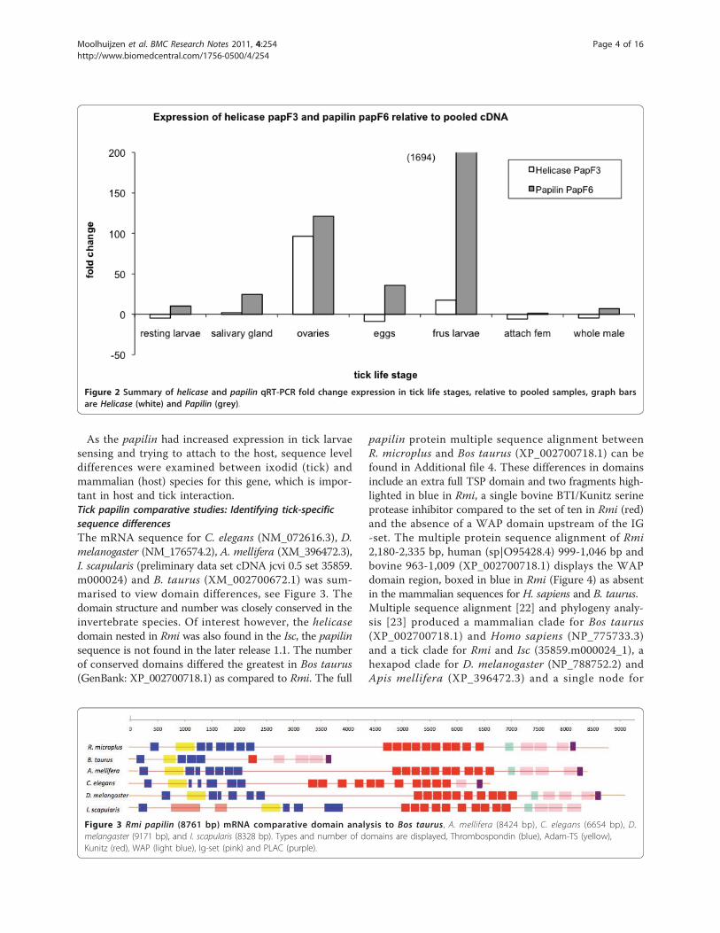
mined in a number of tick life stages.Papilin and Helicase qRT-PCR: gene expression in tick lifestagesThe gene expression fold change relative to pooledcDNA for a number of life stages were tested for bothpapilin and helicase genes. In quantitative real-timePCR (RT-PCR) analysis, it was demonstrated thatexpression of the papilin gene (white bar) was the stron-gest in tick larvae sensing and trying to attach to thehost (Figure 2). The helicase (white bar) shows greatestup regulation in the ovaries of female ticks semi-engorged (17 days old) attach to the host. The papilin(grey bar) also showed differential up regulation in theovaries. Confirming differential expression in at leasttwo tick life stages tested.
Figure 1 BAC BM-004-G14 (135 Kb) Gene Structure and Cot sequence frequency. Genes and Ruka elements (R1-R6) are displayed over aheatmap of Cot sequence frequency (log2), blue (low) = 0 and red (high) = 10, genes include a papilin, a nested helicase-like protein, a pogoelement protein, two hypothetical proteins (H1 and H2), an endonuclease reverse transcriptase protein (ERT) and a serpin. Low complexityregions are shown over the heatmap (green arrow). Exon overlaps are circled in read.
Moolhuijzen et al. BMC Research Notes 2011, 4:254http://www.biomedcentral.com/1756-0500/4/254
Page 3 of 16
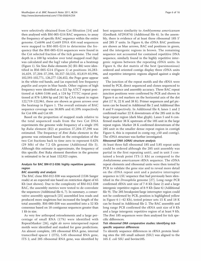
As the papilin had increased expression in tick larvaesensing and trying to attach to the host, sequence leveldifferences were examined between ixodid (tick) andmammalian (host) species for this gene, which is impor-tant in host and tick interaction.Tick papilin comparative studies: Identifying tick-specificsequence differencesThe mRNA sequence for C. elegans (NM_072616.3), D.melanogaster (NM_176574.2), A. mellifera (XM_396472.3),I. scapularis (preliminary data set cDNA jcvi 0.5 set 35859.m000024) and B. taurus (XM_002700672.1) was sum-marised to view domain differences, see Figure 3. Thedomain structure and number was closely conserved in theinvertebrate species. Of interest however, the helicasedomain nested in Rmi was also found in the Isc, the papilinsequence is not found in the later release 1.1. The numberof conserved domains differed the greatest in Bos taurus(GenBank: XP_002700718.1) as compared to Rmi. The full
papilin protein multiple sequence alignment betweenR. microplus and Bos taurus (XP_002700718.1) can befound in Additional file 4. These differences in domainsinclude an extra full TSP domain and two fragments high-lighted in blue in Rmi, a single bovine BTI/Kunitz serineprotease inhibitor compared to the set of ten in Rmi (red)and the absence of a WAP domain upstream of the IG-set. The multiple protein sequence alignment of Rmi2,180-2,335 bp, human (sp|O95428.4) 999-1,046 bp andbovine 963-1,009 (XP_002700718.1) displays the WAPdomain region, boxed in blue in Rmi (Figure 4) as absentin the mammalian sequences for H. sapiens and B. taurus.Multiple sequence alignment [22] and phylogeny analy-sis [23] produced a mammalian clade for Bos taurus(XP_002700718.1) and Homo sapiens (NP_775733.3)and a tick clade for Rmi and Isc (35859.m000024_1), ahexapod clade for D. melanogaster (NP_788752.2) andApis mellifera (XP_396472.3) and a single node for
Figure 2 Summary of helicase and papilin qRT-PCR fold change expression in tick life stages, relative to pooled samples, graph barsare Helicase (white) and Papilin (grey).
Figure 3 Rmi papilin (8761 bp) mRNA comparative domain analysis to Bos taurus, A. mellifera (8424 bp), C. elegans (6654 bp), D.melangaster (9171 bp), and I. scapularis (8328 bp). Types and number of domains are displayed, Thrombospondin (blue), Adam-TS (yellow),Kunitz (red), WAP (light blue), Ig-set (pink) and PLAC (purple).
Moolhuijzen et al. BMC Research Notes 2011, 4:254http://www.biomedcentral.com/1756-0500/4/254
Page 4 of 16

C. elegans (NP_505017.1) (Figure 5A). Evolutionary ana-lyses shows that mammalian (host) papilin diverge at anearlier time than the divergence of hexapoda papilinfrom tick papilin (Figure 5B).The serpin downstream of the papilin on the negative
strand was investigated for gene synteny in otherspecies.Serine Protease Inhibitor: Serpin pseudogenesDownstream of the papilin, a full CDS for serpin waspredicted. The predicted serpin domain structure, how-ever, was fragmented with the N-terminus and C-termi-nus rearranged, exon2 residues 266-364 and exon9 1-63residues. Attempts to sequence the serpin cDNA resultedin a 500 bp product. A single PCR product based on for-ward primer (SerpF3) in exon5 and reverse primer(SerpR2) in exon9 was sequenced (Additional file 2). Thesmall product sequenced matched only 148 bases of pre-dicted exon5 and 231 bases of predicted exon9 (exons
6-8 were not in alignment). Conserved serpin domainanalysis found also two fragments in predicted ERT geneexon2 residues 115-189 and exon4 residues 185-364. Todetermine whether the adjacent position of a serpin withpapilin is common, a search of mosquito, fly genomesand Isc found no evidence of a serpin downstream fromthe papilin indicating this arrangement as not conservedwithin arthropods.To gain better insight to genomic structures a Cot
DNA comparison to the BAC sequence was undertaken.BAC and Cot comparison: element genome wide frequencyestimationDNA reassociating kinetics based Cot filtration of geno-mic DNA was used to reduce the concentration of repeti-tive DNA sequences that dominate the Rmi genome, inorder to analyse the “gene-rich” single/low-copy and themoderate repetitive DNA fractions [14,24]. Two fractionsof moderate to low repetitive regions of Rmi’s genome
Figure 4 Protein sequence alignment of WAP domain position, Rmi (2,180-2,335 bp), Bos taurus (963-1,009) and Homo sapiens (999-1,046) papilin protein alignment. The Rmi WAP domain, position, 2,247-2,297 bp, (boxed light blue) in absent in mammalian sequences.
Figure 5 Phylogenetic analysis of papilin protein for Bos taurus (XP_002700718.1), Homo sapiens (NP_775733.3), Rmi , Isc (35859.m000024_1), D. melanogaster (NP_788752.2), Apis mellifera (XP_396472.3) and C. elegans (NP_505017.1). The trees are represented A)Neighbour-joining and B) Molecular clock.
Moolhuijzen et al. BMC Research Notes 2011, 4:254http://www.biomedcentral.com/1756-0500/4/254
Page 5 of 16
were selectively obtained from Cot filtration [14] andthen analysed with BM-005-G14 BAC sequence, to assaythe frequency of specific BAC sequence within the entiregenome. Cot696 and Cot69 DNA 454 read sequenceswere mapped to BM-005-G14 to determine the fre-quency that the BM-005-G14 sequences were found inthe Cot selected fraction of the tick genome. The readdepth in a 100 bp window over total mapped read (bp)was calculated and the log2 value plotted as a heatmap(Figure 1). Six Sine Ruka elements [6] (R1-R6) were iden-tified in BM-005-G14 (Figure 1) at positions 16,283-16,459, 37,204-37,398, 50,357-50,535, 83,819-83,996,102,593-102,771, 126,277-126,452, the Pogo gene appearin the white-red bands, and (as expected) low frequencypapilin and serpin in blue bands. Other regions of highfrequency were identified as a 321 bp ATCT repeat posi-tioned at 4,864-5186 and, a 124 bp TTTC repeat posi-tioned at 878-1,004 bp and 241 bp CAA repeat in region122,719-122,961, these are shown as green arrows overthe heatmap in Figure 1. The overall estimate of BACsequence coverage was 38.80% and 41.59% respectivelyfor Cot 696 and Cot 69.Based on the proportion of mapped reads relative to
the total sequenced reads from the two Cot DNAexperiments the genome wide frequency of a single 195bp Ruka element (R2) at position 37,204-37,398 wasestimated. The frequency of Rmi Ruka element in thegenome was estimated based on the extrapolation of thetwo Cot fractions back to time zero to represent 0.42%(29 Mb) of the 7.2 Gb genome (Additional file 5).Although this estimate is approximate, the frequency ofthis specific Rmi Ruka element therefore in the genomeis estimated to be at least 152,923 copies.
Analysis for BAC BM-012-E08: highly repetitive genomicregionBAC assembly and analysisThe BAC clone BM-012-E08 was sequenced (1536 Sangerreads) at an expected size based on restriction digest of 65Kb (not shown). Due to the complexity of BM-012-E08BAC, the assembly metrics were tested to de-convolutethe sequences (Additional file 6, 7). In summary, a conser-vative assembly approach [25] assembled less reads andproduced more singletons but increased the length of thetotal assembly. BM-000-E08 was assembled into a 52 Kbconsensus based on 18 contiguous sequences greater than1 kb in size.As very few arthropod retroelements and a large per-
centage of small RNA (17%) were identified withRepeatMasker [26], eight de novo interspersed repeatmotifs were identified and masked for gene predictions.An almost complete, 18S ribosomal RNA gene, internaltranscribed spacer 1 (ITS), 5.8S ribosomal RNA gene,ITS 2, and 28S ribosomal RNA gene, was identified by
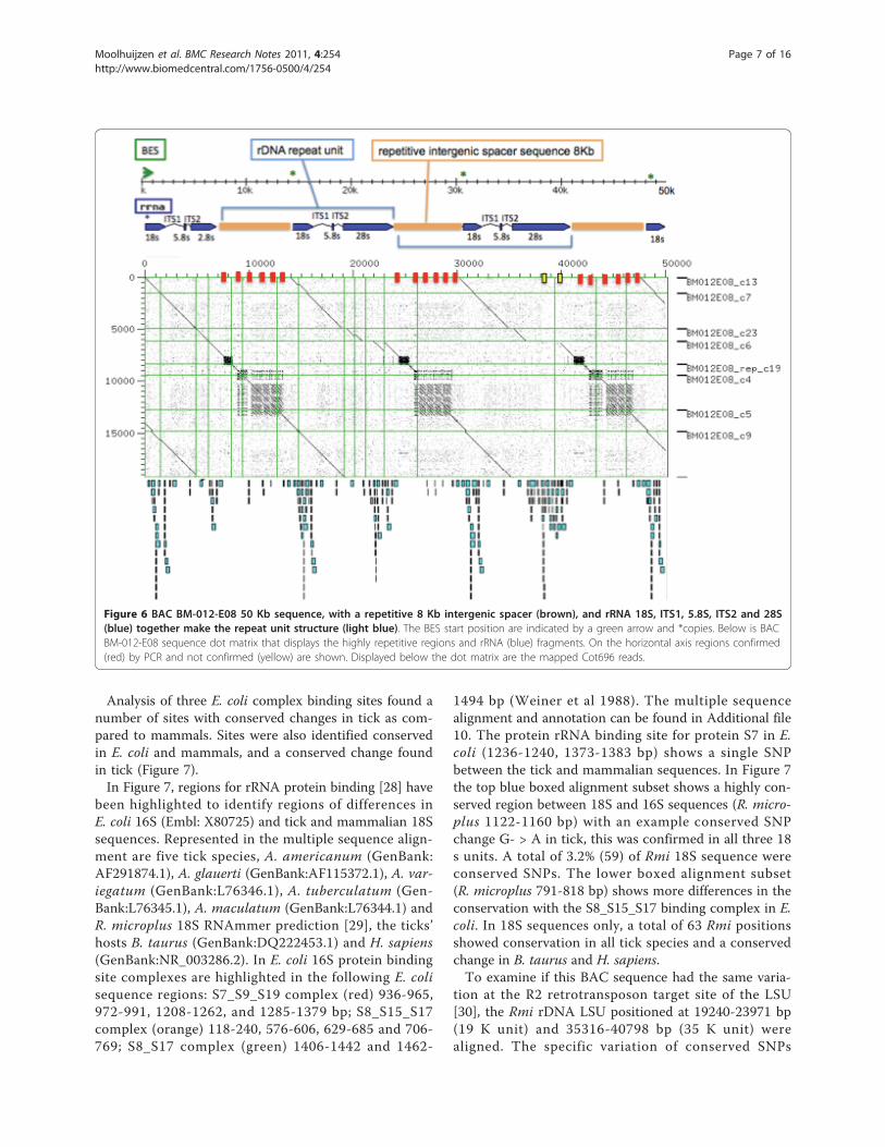
best sequence similarity to Amblyomma americanum(GenBank AF291874) (Additional file 6). In the assem-bly, there is evidence of at least three ribosomal 18S 5’and 28S 3’ units. In Figure 6, the rDNA BAC positionsare shown as blue arrows, BAC end positions in green,and the intergenic regions in brown. The remainingsequence not accounted for contained repetitive DNAsequence, similarly found in the highly repetitive inter-genic regions between the repeating rDNA units. InFigure 6, the dot matrix of the best (parsimonious)ordered and oriented contigs display three rDNA unitsand repetitive intergenic regions aligned against a singleunit.The junction of the repeat motifs and the rRNA were
tested by PCR, direct sequenced and clones sequenced toprove sequence and assembly accuracy. Three BAC repeatjunction positions were confirmed by PCR and shown inFigure 6 as red markers on the horizontal axis of the dotplot (17 K, 22 K and 38 K). Primer sequences and gel pic-tures can be found in Additional file 2 and Additional files8 and 9 respectively. In Additional file 8, lanes 1 and 2confirmed marker 22 K downstream of the 28S unit in thelarge repeat region (dark blue glyph). Lanes 5 and 6 con-firmed marker 38 K upstream of the 18S unit in the largerepeat region. Marker 28 K confirmed downstream of the28S unit in the smaller dense repeat region in contig6Figure 6, this is repeated in contg-rep_c50 and contig1.The rDNA structure was further investigated.Ribosomal DNA (rDNA) structure analysisAt least three full ribosomal 18S and 5.8S repeat unitscould be ordered although the 28S unit assembly waspartial in the first repeating unit1, and in unit 3 con-tained a break point ITS (1 Kb) as compared to theAmbylomma americanum rRNA sequence. The rDNArepeat elements and ribosomal units were then tested byPCR to validate the gene size and to reveal more detailon the rDNA repeat unit and a putative interruptersequence in LSU sequence that had previously been iden-tified in the Drosophila genome [27]. Long range PCRconfirmed rRNA unit size of 7-8 Kb (lane 2) and a largeintergenic repetitive region of 8-9 Kb (lane 6) (Additionalfile 9). The 28S breakpoint/large interrupter region couldnot be confirmed by PCR, position is highlighted yellowin Figure 6 (~42 Kb), tested primer sets 15 K and 18 Kcan be found in Additional file 2. The BAC assembly andlong range PCR confirmed the rRNA unit size of 8 Kband a large intergenic repetitive region of at least 9 Kb.The Rmi 18S sequences were then analysed for tick spe-cific differences.Tick ribosomal DNA comparative studies: Identifying tick-specific sequence differencesTo identify sequence differences in rRNA protein bind-ing sites the 18S small subunit (SSU) was aligned to the16S E. coli SSU and bovine18S.
Moolhuijzen et al. BMC Research Notes 2011, 4:254http://www.biomedcentral.com/1756-0500/4/254
Page 6 of 16
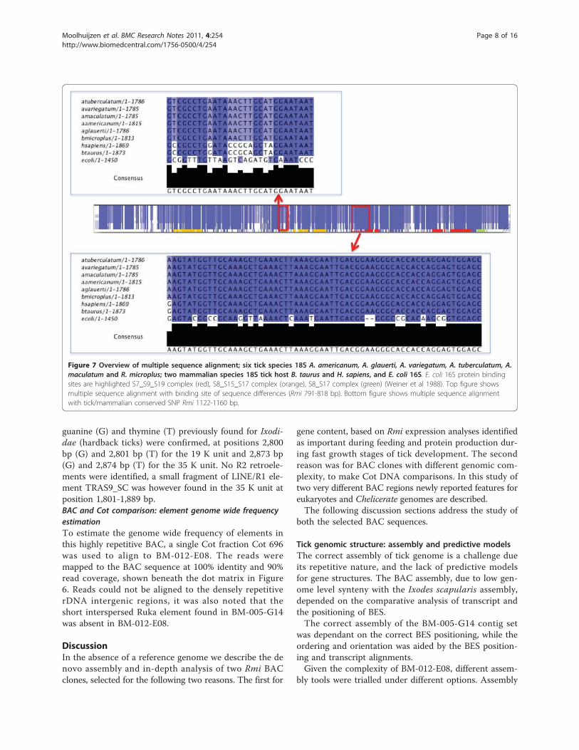
Analysis of three E. coli complex binding sites found anumber of sites with conserved changes in tick as com-pared to mammals. Sites were also identified conservedin E. coli and mammals, and a conserved change foundin tick (Figure 7).In Figure 7, regions for rRNA protein binding [28] have
been highlighted to identify regions of differences inE. coli 16S (Embl: X80725) and tick and mammalian 18Ssequences. Represented in the multiple sequence align-ment are five tick species, A. americanum (GenBank:AF291874.1), A. glauerti (GenBank:AF115372.1), A. var-iegatum (GenBank:L76346.1), A. tuberculatum (Gen-Bank:L76345.1), A. maculatum (GenBank:L76344.1) andR. microplus 18S RNAmmer prediction [29], the ticks’hosts B. taurus (GenBank:DQ222453.1) and H. sapiens(GenBank:NR_003286.2). In E. coli 16S protein bindingsite complexes are highlighted in the following E. colisequence regions: S7_S9_S19 complex (red) 936-965,972-991, 1208-1262, and 1285-1379 bp; S8_S15_S17complex (orange) 118-240, 576-606, 629-685 and 706-769; S8_S17 complex (green) 1406-1442 and 1462-
1494 bp (Weiner et al 1988). The multiple sequencealignment and annotation can be found in Additional file10. The protein rRNA binding site for protein S7 in E.coli (1236-1240, 1373-1383 bp) shows a single SNPbetween the tick and mammalian sequences. In Figure 7the top blue boxed alignment subset shows a highly con-served region between 18S and 16S sequences (R. micro-plus 1122-1160 bp) with an example conserved SNPchange G- > A in tick, this was confirmed in all three 18s units. A total of 3.2% (59) of Rmi 18S sequence wereconserved SNPs. The lower boxed alignment subset(R. microplus 791-818 bp) shows more differences in theconservation with the S8_S15_S17 binding complex in E.coli. In 18S sequences only, a total of 63 Rmi positionsshowed conservation in all tick species and a conservedchange in B. taurus and H. sapiens.To examine if this BAC sequence had the same varia-
tion at the R2 retrotransposon target site of the LSU[30], the Rmi rDNA LSU positioned at 19240-23971 bp(19 K unit) and 35316-40798 bp (35 K unit) werealigned. The specific variation of conserved SNPs
Figure 6 BAC BM-012-E08 50 Kb sequence, with a repetitive 8 Kb intergenic spacer (brown), and rRNA 18S, ITS1, 5.8S, ITS2 and 28S(blue) together make the repeat unit structure (light blue). The BES start position are indicated by a green arrow and *copies. Below is BACBM-012-E08 sequence dot matrix that displays the highly repetitive regions and rRNA (blue) fragments. On the horizontal axis regions confirmed(red) by PCR and not confirmed (yellow) are shown. Displayed below the dot matrix are the mapped Cot696 reads.
Moolhuijzen et al. BMC Research Notes 2011, 4:254http://www.biomedcentral.com/1756-0500/4/254
Page 7 of 16
guanine (G) and thymine (T) previously found for Ixodi-dae (hardback ticks) were confirmed, at positions 2,800bp (G) and 2,801 bp (T) for the 19 K unit and 2,873 bp(G) and 2,874 bp (T) for the 35 K unit. No R2 retroele-ments were identified, a small fragment of LINE/R1 ele-ment TRAS9_SC was however found in the 35 K unit atposition 1,801-1,889 bp.BAC and Cot comparison: element genome wide frequencyestimationTo estimate the genome wide frequency of elements inthis highly repetitive BAC, a single Cot fraction Cot 696was used to align to BM-012-E08. The reads weremapped to the BAC sequence at 100% identity and 90%read coverage, shown beneath the dot matrix in Figure6. Reads could not be aligned to the densely repetitiverDNA intergenic regions, it was also noted that theshort interspersed Ruka element found in BM-005-G14was absent in BM-012-E08.
DiscussionIn the absence of a reference genome we describe the denovo assembly and in-depth analysis of two Rmi BACclones, selected for the following two reasons. The first for
gene content, based on Rmi expression analyses identifiedas important during feeding and protein production dur-ing fast growth stages of tick development. The secondreason was for BAC clones with different genomic com-plexity, to make Cot DNA comparisons. In this study oftwo very different BAC regions newly reported features foreukaryotes and Chelicerate genomes are described.The following discussion sections address the study of
both the selected BAC sequences.
Tick genomic structure: assembly and predictive modelsThe correct assembly of tick genome is a challenge dueits repetitive nature, and the lack of predictive modelsfor gene structures. The BAC assembly, due to low gen-ome level synteny with the Ixodes scapularis assembly,depended on the comparative analysis of transcript andthe positioning of BES.The correct assembly of the BM-005-G14 contig set
was dependant on the correct BES positioning, while theordering and orientation was aided by the BES position-ing and transcript alignments.Given the complexity of BM-012-E08, different assem-
bly tools were trialled under different options. Assembly
Figure 7 Overview of multiple sequence alignment; six tick species 18S A. americanum, A. glauerti, A. variegatum, A. tuberculatum, A.maculatum and R. microplus; two mammalian species 18S tick host B. taurus and H. sapiens, and E. coli 16S. E. coli 16S protein bindingsites are highlighted S7_S9_S19 complex (red), S8_S15_S17 complex (orange), S8_S17 complex (green) (Weiner et al 1988). Top figure showsmultiple sequence alignment with binding site of sequence differences (Rmi 791-818 bp). Bottom figure shows multiple sequence alignmentwith tick/mammalian conserved SNP Rmi 1122-1160 bp.
Moolhuijzen et al. BMC Research Notes 2011, 4:254http://www.biomedcentral.com/1756-0500/4/254
Page 8 of 16
tools with uniform read distribution take a cautionaryapproach in contig building, and sometimes create twocontigs when it could have created one. This featurereduces over compression of repeats during the contigbuilding phase and ensures that, for example rRNAstretches which are present multiple times in a the gen-ome will also be present approximately the same num-ber of times in the result files [25]. The repeat motifs,intergenic regions, rRNA sequences and rDNA unit sizewere confirmed by PCR. However the assembly gapsand insertions show clear deviations from the perfectrepeat unit size. This is the first Rhipicephalinae assem-bly of rDNA and the first known attempt at assembly inArthopoda of three external intergenic repetitive unitsbetween the rDNA repeating subunits.
Tick gene structure: predictive modelsThe complexities of gene predictions included intronicregions of nested repeat elements, multiple short exonsand overlapping regions complicating the delineation ofexon coding regions. Overlapping genes have beenreported in Drosophila but these genes were on differentstands [31]. In eukaryotic research this is the firstdescription of same strand gene overlap between twogenes, the papilin and helicase.In Drosophila, papilin an extracellular matrix glycopro-
tein is found to be involved in, (1) thin matrix layers dur-ing gastrulation, (2) matrix associated with wandering,phagocytic hemocytes, (3) basement membranes and (4)space-filling matrix during Drosophila development [32].Essential also for normal embryonic development Caenor-habditis elegans [33]. This is also the first Chelicerate full-length papilin cDNA sequence produced. Our papilingene model (refer to methods) was confirmed by otherarthropod species. The papilin nested helicase was alsofound within the Isc genome supercontigs (version 1), thisinclusion of the helicase shows a level of gene synteny ispresent in this region between the two distant hard tickspecies.It has been reported that inhibiting papilin synthesis
in Drosophila or Caenorhabditis causes defective cellarrangements and embryonic death. Ectopic expressionof papilin in Drosophila causes lethal abnormalities inmuscle, Malpighian tubule and trachea formation. It hasbeen suggested that papilin influences cell rearrange-ments and may modulate metalloproteinases duringorganogenesis [32-35].These function/activities relate to the following specific
domains. An interesting gene domain complex the tickderived Kunitz type inhibitors act as antihemostatic factors[36]. Hematophagous organisms must overcome hosthemostasis in order to locate blood and maintain its flowduring ingestion [37]. Salivary components produce anti-hemostatic, anti-inflammatory, and immunosuppressive
effects that may facilitate feeding, as well as transmissionof tick-borne pathogens [37]. The number of Rmi KUdomains (x10) present compared to bovine (x1) indicates,based on this domains function, an important change inthis genes structure for tick survival.The whey acidic protein-type four-disulphide core
domain (WAP) has protein family members that includethe whey acidic protein, elafin (elastase-specific inhibi-tor) known to have anti-microbial acitivity [38], catrin-like protein (a calcium transport inhibitor and otherextracellular proteinase inhibitors. A significantsequence variance in bovine was the absence of theWAP domain (Figure 4).Isoforms of papilin have been found in a number of
Arthropoda species, six in Drosophila and two in Apis.Given the size and complexity of the Rmi papilin, iso-forms may exist that are yet to be investigated.The helicase was identified nested as a separate gene
between the first 5’ thrombospondin and the Adam-TSspacer of the Rmi papilin. RACE sequencing from theAdam-TS spacer domain exon in the 5’end directionproduced the complete papilin product minus the heli-case insertion, confirming our gene model.The discovery of shared exon regions for 2 eukaryote
genes, the papilin and helicase, is quite novel. Nestedgenes do occur in eukaryotes [31], nested genes inD. melanogaster and C. elegans have been found exclu-sively as embedded sequences in introns. Kumar 2009,reviewed that in D. melanogaster nested intronic genesconstitutes approximately 6% of the organism’s total genecomplement, and 85% of these nested genes are predictedto encode protein. For example the gart locus, the Pcpgene is nested in intron 1 of the ade3 gene on the com-plement strand. A nested ketoreductase was identified inan A. aegypti papilin - however not with exon overlap asshown for the helicase identified here. In the mouse gen-ome, 28 overlapping gene pairs had partial overlappingexons, and did not encompass the entire coding sequenceof either gene. In the human genome 51 exon overlapson opposite strands, again were partial. Neither thehuman nor the mouse genome contains any overlappinggenes that share coding sequences on the same strand.Further the majority of nested intronic genes are func-tionally unrelated and typically not co-expressed withtheir external host genes. Therefore further functionalanalysis of this gene’s novel arrangement warrants inves-tigation. No helicase element was found nested in thebovine intron region of papilin.The initial identification of the serpin domain led to
the adjacent papilin gene described above. No syntenicevidence was found for the down stream serpin regionin Isc. Full investigation of this gene family within Rmigenomic sequence and the I. scapularis genome remainsto be investigated.
Moolhuijzen et al. BMC Research Notes 2011, 4:254http://www.biomedcentral.com/1756-0500/4/254
Page 9 of 16
The genes for ribosomal DNA are tandem repeatedclusters in the heterochromatic regions of metazoangenomes [27,39], in Drosophila 77% of heterochromatinsequence is composed of fragmented and nested trans-posable elements and other repeated DNAs [39]. It hasbeen reported in vertebrate the splitting and apparentsplicing of ribosomal RNA occurs, and during proces-sing, in mammalian nuclear 28S pre-rRNA, tissue-speci-fic elimination of an ‘intron’ bearing a hidden break siteoccurs [40,41]. An almost complete, 18S ribosomal RNAgene, internal transcribed spacer (ITS) 1, 5.8S ribosomalRNA gene, ITS 2, and 28S ribosomal RNA gene, wasidentified by sequence similarity to Amblyomma ameri-canum (GenBank AF291874; [30]), which is the onlyother tick rDNA sequence analysed at this level of cov-erage. A similar unit was not identified in the Ixodesscapularis genome highlighting the difficulty in theassembly of this region. The Rmi rDNA units have beenidentified as novel due to the lack of the R2 retroele-ment previously identified in Amblyomma and now con-firmed in this study, even though the R2 retroelementbinding site hard tick sequence difference was con-served. The fragmented nature of the LSU makes it pos-sible that the BM-012-E08 BAC clones are derived fromthe end of an array of rDNA units in the genome whereincomplete and rearranged rDNA units may occur.
Tick DNA comparative studies: Identifying tick-specificsequence differencesA number of conserved changes within rRNA proteinbinding sites between the ticks as compared to mammalswere found. The hard tick specific sequence differences(SNPs) were also found in the LSU R2 retroelement tar-get site.Due to the uniqueness of tick rDNA sequences it is
feasible that tick rRNA could be a target for drug devel-opment analogous to the use of bacterial rRNA as anti-biotic targets. Consistent with this possibility, a numberof conserved changes within rRNA protein binding sitesbetween the tick as compared to mammal were found.The hard tick specific sequence differences (SNPs), werealso found in the LSU R2 retroelement target site.
Tick gene expression analysisThe qRT-PCR analysis of the papilin described con-firmed differential increased expression in two lifestages, most prominently in larvae trying to attach tothe host (Figure 2). It was also demonstrated that thehelicase is strongly expressed in ovaries of feedingfemales. Helicases are often utilized to separate strandsof a DNA double helix or a self-annealed RNA moleculeusing the energy from ATP hydrolysis, a process charac-terized by the breaking of hydrogen bonds betweenannealed nucleotide bases. The differential expression of
both the papilin and helicase in adult female ovariessuggests that is perhaps a conserved functionalarrangement.The abundance of RNA proteins as identified in the
subtraction library study was not surprising due toincreased protein production during feeding. Andersonet al. 2008 reported that the abundance ribosomal pro-tein coding genes is not unusual for a transcriptomeanalysis and illustrates the high degree of redundancyfound in such libraries, especially the occurrence ofnumerous sequences coding for proteins involved inprotein synthesis such as ribosomal RNA, e.g. 40S, 60Sand other ribosomal genes [42].
The analysis of genome sequence via back endsequencing and Cot DNAThe Rmi genomic DNA that was enriched for single/low-copy and moderately repetitive DNAs [14] alongwith BAC end sequencing have provided valuableinsights into Rmi genomic structure. Mapping the readsof the Cot DNA to the BAC sequencing identifiedregions of high repetitive content in BM-012-E08 com-plex intergenic region by the absence of mapped reads.Also moderately repetitive regions could be identifiedsuch as the RUKA element in BM-005-G14.In particular using the two Cot filtrations, we were
able to estimate the frequency of any specific genomicsequence within the entire genome. As an example pre-sented, major frequency peaks were identifiable and therelative frequency of the sine Ruka [6] element in thegenome was estimated for BM-005-G14. Althoughabsent from the euchromatic section of the relativelycompact 1.8 Gb genome of Drosophila melanogaster[43], several distinct families of SINEs with copy num-bers of up to 590 Kb per genome have been describedin Aedes aeygpti, the mosquito vector of the yellow fevervirus [44,45]The frequency of a single Rmi Ruka element in the
genome was estimated based on the extrapolation of thetwo Cot fractions to represent 0.42% of the genome, atleast 152,923 copies.A previous examination of 3 BAC sequences, and the
(DFCI) Gene Indices [46] for the four ixodid tick spe-cies, A. variegatum [47], R. appendiculatus (Rap) [48],B. (R.) microplus [19] and I. scapularis [49], estimatedthat the Ruka repeat sequences comprise approximately1.6% (4 kb) of the 250 Kb of Rap genome (BACsequence). Then on the following assumptions (1) thatthese Rap BAC contigs are representative, and (2) a gen-ome size for R. appendiculatus of 1 Gb, that a total of65,000 copies of Ruka could be predicted [6]. Since ourestimation in Rmi is based on a single element weexpect the number of Ruka families will occupy a muchlarger fraction of the genome than previously estimated.
Moolhuijzen et al. BMC Research Notes 2011, 4:254http://www.biomedcentral.com/1756-0500/4/254
Page 10 of 16
ConclusionThis analysis builds on the previous report by Guerreroet al 2010, to characterise genomic DNA in the tick Rhi-picelphalus microplus, The complete secreted extracellu-lar matrix protein gene papilin primarily found inbasement membranes and essential for embryonic devel-opment, was assembled and cDNA sequenced. This isthe first reporting in eukaryotes of same strand exonoverlap between the sequenced products of papilin andhelicase. Detection of these types of overlaps is a com-plication for current de-novo gene prediction tools. In asecond BAC clone, ribosomal DNA (rDNA) wasassembled into three repeat units, the first rRNA assem-bly in Rhipicephalinae, and the first attempt to assemblesequence of the rDNA repeat units and intergenicspacer in arthropods.In both papilin and rRNA, tick specific sites of
sequence variation were identified in tick R. microplusrelative to the host Bos taurus, in a detailed comparisonto identify targets for disrupting the pathogen-hostinteraction. In addition expression analysis of papilinand helicase demonstrated striking tissue specificexpression in response to sensing the host prior toattachment for feeding.Finally the two Cot-filtration resources provided a
means to estimate the frequency of an element in thecontext of the whole genome.In order to place the BAC sequences into a whole
genome context, the BAC sequences were probed with454 sequenced Cot 69.56 secs and 696.6 secs DNA. Thisanalysis allowed the representation of specific BACsequence to be estimated within the respective CotDNA sequences and thus estimate the frequency ofsequence occurrence in the whole R. microplus genome.The BAC, BAC end sequences (BES) and Cot DNA
have allowed an in-depth analysis of selected R. microplusgenomic DNA, and in terms of sequencing towards awhole genome provided a valuable insight into R. micro-plus genomic structure.
MethodsBAC end sequencesGlycerol plates with BAC clones (1,125 96-well plates)were submitted to Beckman Coulter Genomics (Beverley,MA, USA) to obtain approximately 12,000 reads using bi-directional sequencing of the clones. The Beckman Coul-ter Genomics protocol is described as follows: clones werepicked from the 96 well plates, cultured and DNA waspurified using SPRI®; following dye-terminator fluorescentsequencing the product was purified using CleanSEQ®
with sequencing fragments detected via ABI3730xl capil-lary electrophoresis. The total 10,582 BAC end sequences(BES) provided as trace files from Agencourt were clipped
of the vector (pECBAC1) with cross_match, Phrap pack-age version 0.990329 [21]. Sequences greater than 500 bphave been deposited GenBank GSS under HN108288-HN118367.
BAC Genomic DNA extraction, library construction, andBAC screening and sequencingTicks from the Deutsch strain of R. microplus werereared at the USDA-ARS Cattle Fever Tick ResearchLaboratory in Mission, TX [50]. Genomic DNA extrac-tion, library construction, and BAC screening are asdescribed by Guerrero et al., [14].
BAC sequencingBAC vector used was pECBAC1 and the cloning siteBamHI. BAC libraries were sequenced using 3-4 kb inserthigh copy shotgun library methods aiming for 8-fold cov-erage of 1,008,000 bases (high copy) using Sanger Sequen-cing ABI technology as described for the BAC endsequencing above (Beckman Coulter Genomics, MA,USA).phred 20 read lengths greater than 700 bp and pass
rates: > 90% and x6 coverage.
BAC assemblyBAC Sanger assemblies were conducted with phred/phrap [51], CAP3 [52] and Phusion [53] and MIRA [25].The BES were mapped to the assembly with BLAT [54]at 100% percent sequence identity. Dot plot matriceswere generated using Dotter64 [55]. Beckman CoulterGenomics (MA, USA) closed the sequence gaps based onpair end read linkage. The following BAC sequences havebeen deposited in GenBank, BM-005-G14 (HM748961)and BM-012-E08 (HM748964).The correct orientation and ordering of the contigs was
based on pair-end read assembly linkage results, back endsequencing positioning and gene annotation, as compara-tive analysis of Rmi papilin to a number of species showordered domain conservation. The finished BAC with gapclosure was 135 Kb, close to the estimated restrictiondigest size, with the papilins’ coding region of 8 Kbp span-ning a genomic region of 86 Kbp. The papilin gene wasfirst predicted with Genscan HumanIso model from the 2large coding sequences CDS8 and CDS6 (6,663 and 4,077bp respectively), which covered all the papilin proteindomains, except the initial 5’ end thombospondin domain.In addition, CDS8 contained a helicase domain (not pre-viously identified in papilin), an Adam-TS-spacer 1 andthe second expected thombospondin domain. CDS6 con-tained all the remaining papilin domains, KU domainsx10, WAP, IG x3, and the final 3’end PLAC domain.Direct cDNA sequencing confirmed the BAC data and 5’RACE assisted to confirm the presence of the missing
Moolhuijzen et al. BMC Research Notes 2011, 4:254http://www.biomedcentral.com/1756-0500/4/254
Page 11 of 16
thrombospondin domain and our model subsequentlyproposed the presence of 2 genes, the papilin and thehelicase.
BES analysesBlastN [56] nucleotide similarity searches were conductedon Dana Faber Cancer Institute (DFCI) Gene IndicesBmiGI [13,19], IscGI [49], subtraction library cDNA[16] and iscapularis.preliminary.TRANSCRIPTS_JCVI-IscaW1.0.5. and NCBI [57] datasets that included, (nr, est,genomic, refseq, GSS, WGS).BlastX [56] protein similarity searches were conducted
on NCBI [57] (nr, patent), and Ixodes scapularis peptidegene predictions 1.1 iscapularis. PEPTIDES-IscaW1.1protein datasets.Domain and protein family identification was con-
ducted with RPSBlast, on NCBI Conserved DomainDatabase (CDD) [18] database.BLAT [54] ixodes_scapularis_supercontigs.
Gene predictionGenscan [58] (model for human isoforms) was used toassemble the BAC contigs. Bioperl [59] scripts wereused to parse alignments and identify conserved regions.The In-silico workflow was designed based on opensource applications and CCG Grid computing [60].
Sequence alignment and phylogenyClustalW [22] was used for multiple sequence align-ments and multiple sequence alignments for manuscriptwere displayed in Jalview [61].Phylogeny analysis was conducted with Phylip version
3.6 [23] protein distance algorithm and Neighbor-Join-ing method [62] and bootstrap test of 100 replicates.The molecular clock test was performed by comparing
the ML value for the given topology with and withoutthe molecular clock constraints under Jones-Taylor-Thornton (1992) model [63,64]. Evolutionary analyseswere conducted in MEGA4 [64].The evolutionary history was inferred using the Neigh-
bor-Joining method [62]. The bootstrap consensus treeinferred from 500 replicates was taken to represent theevolutionary history of the taxa analyzed. The percentageof replicate trees in which the associated taxa clusteredtogether in the bootstrap test (500 replicates) are shownnext to the branches. The tree is drawn to scale, withbranch lengths in the same units as those of the evolution-ary distances used to infer the phylogenetic tree. The evo-lutionary distances were computed using the method [65]and are in the units of the number of amino acid differ-ences per site. The analysis involved 7 amino acidsequences. All positions containing gaps and missing datawere eliminated. There were a total of 1124 positions inthe final dataset.
Repeat identificationArthropod known repeats were identified with Repeat-Masker version 3.2.6 [26]. Repeatscout [66] was used forthe de-novo identification of repeat motifs. Perfect tan-dem repeats were identified with SSR finder Perl pro-gram. Sine RUKA elements were identified based onBlastN [56] homology to GenBank: EU018139.1 (9,947-10,084 bp), percentage identity greater than 84% andcoverage greater than 69%.
cDNA preparationTotal RNA was extracted from tick samples using Trizolreagent (Invitrogen Corporation, CA, USA). Tissue wasground to a fine powder using a mortar and pestle withliquid nitrogen and the powder transferred to a tube of Tri-zol with 1 mm glass beads. This mix was further homoge-nised for 45 seconds in a MiniBeadbeater-96 (BiospecProducts, Bartlesville, OK, USA) then the RNA wasextracted using chloroform and isopropanol. Doublestranded cDNA was created from 25 μg of total RNAusing a SuperScript™ Double-Stranded cDNA SynthesisKit following Kit protocols (Invitrogen Corporation, CA,USA).
Papilin PCR amplification and sequencingPrimers based on BAC sequences were designed withEMBOSS eprimer3 [67] and a minimum GC clamp of 2.Synthesis of primer sequences were by Sigma Aldrich(MO, USA) and sequences are presented in Additionalfile 2. The full papilin cDNA was PCR amplified fromcDNA extracted from frustrated larvew and cloned inthree steps. A 7723 bp product was amplified betweenprimers Papilin57383F and PapilinR3 (designed from pre-dicted coding sequence) using the Expand Long Tem-plate PCR System (Roche Applied Science, Mannheim,Germany) using Expand Long Template Buffer 2. Thisreaction was thermocycled in a DNA Engine (PTC-200)Peltier Thermal Cycler (Biorad Laboratories, CA, USA).The purified product was transformed into chemicallycompetent One Shot® TOP10 cells using a TOPO-XL®
PCR Cloning Kit (Invitrogen Corporation, CA, USA). Foreach transformation, DNA was prepared from six clonesusing a QIAprep Spin Miniprep Kit (Qiagen, CA, USA).Plasmid inserts were sequenced using Big Dye Vers 3.1technology (Applied Biosystems, CA, USA) and were runon an Applied Biosystems 3130xl Genetic Analyser (Grif-fith University DNA Sequencing Facility, School of Bio-molecular and Biomedical Science, Griffith University,Qld, Australia). Sequences were edited and aligned inSequencher (Vers 4.8 Gene Codes Corporation, AnnArbor, MI, USA). Additional sequencing primers weredesigned manually (Additional file 2).The start codon for the papilin gene was determined
following 5’ amplification of the cDNA ends from larval
Moolhuijzen et al. BMC Research Notes 2011, 4:254http://www.biomedcentral.com/1756-0500/4/254
Page 12 of 16

cDNA using the SMARTer™ RACE cDNA Amplifica-tion Kit as described by the kit manufacturer (ClontechLaboratories Inc., CA, USA). The 5’-RACE PCR used anAdvantage 2 PCR kit (Clontech Laboratories Inc., CA,USA) using the kit 5’ RACE UPM primer and the genespecific reverse primer AdamSR1 designed within theAdam spacer region of the papilin gene (Additionalfile 2). The gel-purified product was cloned into chemi-cally competent One Shot® TOP10 cells using aTOPO® TA Cloning Kit (Invitrogen Corporation, CA,USA). Clone inserts were sequenced as described above.The papilin stop codon was determined from the pre-
dicted coding sequence and a primer was designedanchored at the stop position PapStopR1 (Sup). A 611bp product was PCR amplified between primersPap12440F to PapStopR1. The product was cloned andsequenced as described above.
Papilin cloned productsFinal papilin product was 8,761 bp, from the 5’ Race toprimer AdamS_R1 product length 867 bp, and from pri-mer regions papilin57383F to PapilinR3 product length7,723 bp and pap12440F to pap13230R direct sequence.
Helicase PCR amplification and sequencingA second large PCR product 4886 bp in length wasamplified from the larval cDNA between primers Papili-nORFF2 and Papilin54900R. The 3’ end of the producthas a 229 bp overlap with the papilin gene (exon 2 and3, 5’ of the Adam spacer). The product was amplifiedusing the Expand Long Template PCR System (RocheApplied Science, Mannheim, Germany) and was clonedand sequenced as described above for the large papilinclone. Internal primers were designed to sequence thecomplete clone that was found to contain a helicasegene (Additional file 2).
BM-12-E08 PCRPCR 50 μl reaction contained Advantage 2 SA PCR buffer,10 mM of dNTP mix, 10 μM of each primer, 100 ng ofDNA template and Advantage 2 polymerase mix asrecommended by the manufacturer (Clontech Labora-tories Inc., CA, USA), Cycling Parameters (BIORAD DNAEngine Cycler): Initial denaturation for 2 mins at 94°C fol-lowed by 29 cycles of denaturation 1 min 94°C, annealing1 min 55°C, and extension 1 min 72°C, with a final exten-sion of 7 mins 72°C. The products were visualised follow-ing agarose gel electrophoresis (1.2%) containing Gel Red(Jomar Bioscience Pty Ltd, SA, Australia). PCR productsand purified plasmid DNA were sequenced using Big DyeVers 3.1 technology (Applied Biosystems, CA, USA) andwere run on an Applied Biosystems 3130xl Genetic Analy-ser at Griffith University DNA Sequencing Facility
(GUDSF). Sequences were edited and aligned inSequencher (Vers 4.7 Gene Codes Corporation).Amplified products were cloned using the pCR2.1 -
TOPO plasmid vector (Invitrogen Corporation, CA,USA). Transformed cells were plated on to LB agaroseplates containing 50 μg/ml kanamycin and grown over-night at 37°C. Colonies were picked and cultured in LBmedium broth containing 50 μg/ml kanamycin. PCRreactions were performed on 1 μl of the cultured brothsand analysed by agarose gel electrophoresis to confirminsertion. Plasmid DNA was purified as described above.
BM012-E08 Long range PCRTick genomic DNA was prepared following tissue grindingas described above for cDNA preparation and subse-quently purified using the QIAamp DNA mini-kit asdescribed by the manufacture (QIAGEN, CA, USA). TheExpand Long Template PCR system was used to amplifythe DNA under conditions recommended by the manufac-turer (Roche Applied Science, Mannheim, Germany) in aBioRad DNA Engine Peltier Thermal Cycler. Directsequencing was undertaken as described above. Allsequence alignment graphs were generated with Bioperl[59].
qRT-PCR analysisPrimers were designed manually within targeted exonregions for the helicase and papilin transcripts describedin this study (Additional file 2). Methods for qRT-PCRanalysis were described previously by Lew-Tabor et al.(2010), utilising tick extracts prepared from differenttick organs and stages, normalised against 2 house-keeper genes (Actin and rRNA 18S) and against apooled cDNA sample.
Cot selected genomic DNAThe Cot filtration on Rmi genomic DNA was performedas previously described [14], to enrich for single/low-copyand moderately repetitive DNAs. The two “conditions” arecalled Cot69 and Cot696. Starting DNA concentrationsfor both were 200 micrograms of sheared genomic DNA.Time for renaturation was 1 hr, 48 min, 6 sec for sampleCot696 (Cot of 695.6) and 10 min 49 sec for sample Cot69(Cot of 69.56). Renaturation was conducted at 70 degreesC, at 0.03 M NaPO4. Sequencing results form these twoCot-selected samples have been deposited in GenBankSRA, submission: SRA012677.4/SID00001.Cot DNA 454 read sequence was mapped to the BAC
sequence using the Newbler GS-FLX reference mapper,version 2.0.00.20 [68] and BLAT [54]. The total readnumber was calculated for each BAC window size 100bps (read number per 100 bp window). The percentage(read number per 100 bp window)/(Total BAC read
Moolhuijzen et al. BMC Research Notes 2011, 4:254http://www.biomedcentral.com/1756-0500/4/254
Page 13 of 16
count) was then plotted for each BAC. The gnuplotswere calculated by (Sum bp depth/window size)/(Sumtotal bp depth/BAC length).
Additional material
Additional file 1: BAC end sequence analysis. Results for BAC endsequence database searches.
Additional file 2: Rmi primer sequences and positions. Primersequence and sequence positions for BAC sequences BM-012-E08 andBM-004-G14 (papilin and serpin).
Additional file 3: Sequence alignment of exon overlap betweenpapilin and helicase. Sequence alignment of exon overlap betweenpapilin position 502-738 bp and helicase 3 positions 974-4802 bp. Theconsensus black bar indicates the region of overlap. Blue arrows indicateexon junctions, papilin T-G positions 604 and 605 and helicase G-Cpositions 4715, 4716.
Additional file 4: Rmi and Bos taurus papilin protein sequencealignment. Rmi and Bos taurus papilin protein sequence alignment, R.microplus and B. taurus. Domains are highlighted Kunitz BPTI (red), ADAMspacer1 (yellow), Ig-set (pink), PLAC (purple), WAP (orange) and TSP1(blue).
Additional file 5: BM-005-G14 Ruka frequency analysis. Table andgraph of BM-005-G14 Ruka frequency analysis using two Cot DNAexperiments.
Additional file 6: BAC BM-012-E08 sequence assembly statistics.Table of BAC sequence assembly statistics BM-012-E08.
Additional file 7: BM-012-E08 sequence dot matrix withAmbylomma rRNA alignment. Figure of full dot matrix BM-012-E08 withAmbylomma rRNA (blue) alignment.
Additional file 8: BM-012-E08 repetitive elements PCR results. PCRresults for BM-012-E08 repetitive elements, lanes: 1) 22900 (F1/R1) 2)22900 (F2/R2) 3) 17000 (F1/R1) 4) 17000 (F2/R2) 5) 38000 (F1/F2) 6)38000 (F2/R2) 7) negative control.
Additional file 9: BM-012-E08 long primer sets to amplify tickgenomic DNA. Long primer sets used to amplify tick genomic DNAusing Roche Expand Long Template PCR system, Lane 1 Fermentas Massruler 80 bp-10 kb (#SM0403), Lane 2 rDNA.1, Lane 3 rDNA.1 PCR negativecontrol, Lane 4 intergenic-region.1, Lane 5 intergenic-region.1 PCRnegative control, Lane 6 intergenic-region.2, Lane 7 intergenic-region.1PCR negative control, Lane 8 intergenic-region.2, Lane 9 intergenic-region.2 PCR negative control.
Additional file 10: Full multiple sequence alignment for 18S tickand fly species and 16S E. coli units. Full 18S unit multiple sequencealignment for: 5 tick species A. americanum, A. glauerti, A. variegatum, A.tuberculatum, A. maculatum and R. microplus; 2 fly species D. simulans, D.melanogaster; tick host B. taurus and E. coli 16S. In E. coli 16S proteinbinding sites are highlighted S7_S9_S19 complex (red), S8_S15_S17complex (green), S8_S17 complex (aqua) (Weiner et al 1988).
AcknowledgementsWe would like to acknowledge and thank: Mr Adam Hunter, Mr DavidSchibeci and Mr Mark O Shea from the Centre for Comparative Genomics(Murdoch University); and Ms Catherine Minchin and Ms Sandra Jarrett fromAgri-Science Queensland (Department of Employment, EconomicDevelopment and Innovation) - for all their expert technical assistance.
Author details1Centre for Comparative Genomics, Murdoch University, South St., Perth,Western Australia, 6150, Australia. 2Cooperative Research Centre for BeefGenetic Technologies, Armidale, NSW, Australia. 3Queensland Alliance forAgriculture & Food Innovation, The University of Queensland, (c/oDepartment of Employment, Economic Development and Innovation),Locked Mail Bag No. 4, Moorooka, QLD 4105, Australia. 4USDA-ARS, Knipling-
Bushland U.S. Livestock Insects Research Laboratory, 2700 Fredericksburg Rd.,Kerrville, TX 78028, USA. 5Department of Plant & Soil Sciences and LifeSciences & Biotechnology Institute, Mississippi State University, 117 DormanHall, Box 9555, Mississippi State, MS 39762, USA. 6Research and TestingLaboratory, 4321 Marsha Sharp Fwy, Lubbock, TX 79407, USA.
Authors’ contributionsPM carried out the bioinformatics analysis, and drafted the manuscript. RA,MB, ALT, FG, MRV, JATM participated in the interpretation of analysis andwriting/editing of this manuscript. FG (USDA) funded the Cot filtration andsequencing. FG performed BAC library filter hybridizations and the Cotselection protocols, and participated in the selection of BAC sequences. ALT(BeefCRC) funded the sequencing of the BES and BACs, and supervised PCRand cDNA sequencing. JATM cloned and sequenced papilin and helicasegenes and conducted qRT-PCR analyses. DP participated in and supervisedthe Cot-selection protocols. SD performed the 454 sequencing of Co-selected DNA. RA supervised the design and analysis for this manuscript. Allauthors have read and approved the final manuscript.
Competing interestsThe authors declare that they have no competing interests.
Received: 13 April 2011 Accepted: 22 July 2011 Published: 22 July 2011
References1. McCosker PJ: Global aspects of the management and control of ticks of
veterinary importance. Recent Adv Acarol 1979, 2:45-53.2. George JE, Davey RB, Pound JM: Introduced ticks and tick-borne diseases:
the threat and approaches to eradication. Vet Clin North Am Food AnimPract 2002, 18(3):401-416, vi.
3. Palmer MJ, Bantle JA, Guo X, Fargo WS: Genome size and organization inthe ixodid tick Amblyomma americanum (L.). Insect Mol Biol 1994,3(1):57-62.
4. Ullmann AJ, Lima CM, Guerrero FD, Piesman J, Black WCT: Genome sizeand organization in the blacklegged tick, Ixodes scapularis and theSouthern cattle tick, Boophilus microplus. Insect Mol Biol 2005,14(2):217-222.
5. Geraci NS, Spencer Johnston J, Paul Robinson J, Wikel SK, Hill CA: Variationin genome size of argasid and ixodid ticks. Insect Biochem Mol Biol 2007,37(5):399-408.
6. Sunter JD, Patel SP, Skilton RA, Githaka N, Knowles DP, Scoles GA, Nene V,de Villiers E, Bishop RP: A novel SINE family occurs frequently in bothgenomic DNA and transcribed sequences in ixodid ticks of thearthropod sub-phylum Chelicerata. Gene 2008, 415(1-2):13-22.
7. Kidwell MG: Transposable elements and the evolution of genome size ineukaryotes. Genetica 2002, 115(1):49-63.
8. Okada N: SINEs. Curr Opin Genet Dev 1991, 1(4):498-504.9. Ullu E, Tschudi C: Alu sequences are processed 7SL RNA genes. Nature
1984, 312(5990):171-172.10. Hill CA, Wikel SK: The Ixodes scapularis Genome Project: an opportunity
for advancing tick research. Trends Parasitol 2005, 21(4):151-153.11. Pagel Van Zee J, Geraci NS, Guerrero FD, Wikel SK, Stuart JJ, Nene VM,
Hill CA: Tick genomics: the Ixodes genome project and beyond. Int JParasitol 2007, 37(12):1297-1305.
12. Nene V: Tick genomics–coming of age. Front Biosci 2009, 14:2666-2673.13. Wang M, Guerrero FD, Pertea G, Nene VM: Global comparative analysis of
ESTs from the southern cattle tick, Rhipicephalus (Boophilus) microplus.BMC Genomics 2007, 8:368.
14. Guerrero FD, Moolhuijzen PM, Peterson DG, Bidwell S, Caler E, Appels R,Bellgard M, Nene VM, Djikeng A: Reassociation kinetics-based approachfor partial genome sequencing of the cattle tick, Rhipicephalus(Boophilus) microplus. BMC Genomics 2010, 11:374.
15. Sequencing of BAC ends from a Rhipicephalus microplus BAC library.[http://www.ars.usda.gov/research/projects/projects.htm?ACCN_NO=415116].
16. Lew-Tabor AE, Moolhuijzen PM, Vance ME, Kurscheid S, Valle MR, Jarrett S,Minchin CM, Jackson LA, Jonsson NN, Bellgard MI, et al: Suppressivesubtractive hybridization analysis of Rhipicephalus (Boophilus) micropluslarval and adult transcript expression during attachment and feeding.Vet Parasitol 2009, 167(2-4):304-320.
17. Bellgard MI, Guerrero FD, Moolhuijzen PM, Schibeci D, Hunter A, Rodriguez-Valle M, Barrero R, Gondro C, Lew-Tabor AE: Toward a genome sequence
Moolhuijzen et al. BMC Research Notes 2011, 4:254http://www.biomedcentral.com/1756-0500/4/254
Page 14 of 16
for Rhipicephalus (Boophilus) microplus: CattleTickBase available resourcesfor the research community..
18. Marchler-Bauer A, Anderson JB, Chitsaz F, Derbyshire MK, DeWeese-Scott C,Fong JH, Geer LY, Geer RC, Gonzales NR, Gwadz M, et al: CDD: specificfunctional annotation with the Conserved Domain Database. NucleicAcids Res 2009, 37:(Database issue):D205-210.
19. Guerrero FD, Miller RJ, Rousseau ME, Sunkara S, Quackenbush J, Lee Y,Nene V: BmiGI: a database of cDNAs expressed in Boophilus microplus,the tropical/southern cattle tick. Insect Biochem Mol Biol 2005,35(6):585-595.
20. Guerrero FD, Nene VM, George JE, Barker SC, Willadsen P: Sequencing anew target genome: the Boophilus microplus (Acari: Ixodidae) genomeproject. J Med Entomol 2006, 43(1):9-16.
21. Ewing B, Green P: Base-calling of automated sequencer traces usingphred. II. Error probabilities. Genome Res 1998, 8(3):186-194.
22. Thompson JD, Higgins DG, Gibson TJ: CLUSTAL W: improving thesensitivity of progressive multiple sequence alignment throughsequence weighting, position-specific gap penalties and weight matrixchoice. Nucleic Acids Res 1994, 22(22):4673-4680.
23. PHYLIP (the PHYLogeny Inference Package). [http://evolution.genetics.washington.edu/phylip.html].
24. Lamoureux D, Peterson DG, Li W, Fellers JP, Gill BS: The efficacy of Cot-based gene enrichment in wheat (Triticum aestivum L.). Genome 2005,48(6):1120-1126.
25. Chevreux B, Thomas Pfisterer T, Drescher B, Driesel AJ, Müller WEG,Wetter T, Suhai S: Using the miraEST Assembler for Reliable andAutomated mRNA Transcript Assembly and SNP Detection in SequencedESTs. Genome Research 2004, 14(6):1147-1159.
26. Smit AFA, Hubley R, Green P: RepeatMasker Open-3.0. 2004.27. Glover DM, Kidd SJ, Roiha HT, Jordan BR, Endow S, Appels R: Interrupter
Sequences that are widely distributed in the Drosophila genome.Biochemical society transcations 1978, 6:732-736.
28. Wiener L, Schuler D, Brimacombe R: Protein binding sites on Eschenichiacoli 16S ribosomal RNA; RNA regions that are protected by proteins S7,S9 and S19, and by proteins S8, S15 and S17. Nucleic Acids Research 1988,16(4):1233-1250.
29. Lagesen K, Hallin P, Rodland EA, Staerfeldt HH, Rognes T, Ussery DW:RNAmmer: consistent and rapid annotation of ribosomal RNA genes.Nucleic Acids Res 2007, 35(9):3100-3108.
30. Bunikis J, Barbour AG: Ticks have R2 retrotransposons but not theconsensus transposon target site of other arthropods. Insect Mol Biol2005, 14(5):465-474.
31. Kumar A: An Overview of Nested Genes in Eukaryotic Genomes.EUKARYOTIC CELL 2009, 8(9):1321-1329.
32. Campbell AG, Fessler LI, Salo T, Fessler JH: Papilin: a Drosophilaproteoglycan-like sulfated glycoprotein from basement membranes. JBiol Chem 1987, 262(36):17605-17612.
33. Fessler JH, Kramerova I, Kramerov A, Chen Y, Fessler LI: Papilin, a novelcomponent of basement membranes, in relation to ADAMTSmetalloproteases and ECM development. Int J Biochem Cell Biol 2004,36(6):1079-1084.
34. Kramerova IA, Kawaguchi N, Fessler LI, Nelson RE, Chen Y, Kramerov AA,Kusche-Gullberg M, Kramer JM, Ackley BD, Sieron AL, et al: Papilin indevelopment; a pericellular protein with a homology to the ADAMTSmetalloproteinases. Development 2000, 127(24):5475-5485.
35. Kramerova IA, Kramerov AA, Fessler JH: Alternative splicing of papilin andthe diversity of Drosophila extracellular matrix during embryonicmorphogenesis. Dev Dyn 2003, 226(4):634-642.
36. Corral-Rodriguez MA, Macedo-Ribeiro S, Barbosa Pereira PJ, Fuentes-Prior P:Tick-derived Kunitz-type inhibitors as antihemostatic factors. InsectBiochem Mol Biol 2009.
37. Ribeiro JM, Makoul GT, Levine J, Robinson DR, Spielman A: Antihemostatic,antiinflammatory, and immunosuppressive properties of the saliva of atick, Ixodes dammini. J Exp Med 1985, 161(2):332-344.
38. Simpson AJ, Maxwell AI, Govan JR, Haslett C, JM S: Elafin (elastase-specificinhibitor) has anti-microbial activity against gram-positive and gram-negative respiratory pathogens. FEBS Lett 1999, 452(3):309-313.
39. Smith CD, Shu S, Mungall CJ, Karpen GH: The Release 5.1 annotation ofDrosophila melanogaster heterochromatin. Science 2007,316(5831):1586-1591.
40. Matz MV: Amplification of representative cDNA samples frommicroscopic amounts of invertebrate tissue to search for new genes.Methods Mol Biol 2002, 183:3-18.
41. Melen GJ, Pesce CG, Rossi MS, Kornblihtt AR: Novel processing in amammalian nuclear 28S pre-rRNA: tissue-specific elimination of an‘intron’ bearing a hidden break site. EMBO J 1999, 18(11):3107-3118.
42. Anderson JM, Sonenshine DE, Valenzuela JG: Exploring the mialome ofticks: an annotated catalogue of midgut transcripts from the hard tick,Dermacentor variabilis (Acari: Ixodidae). BMC Genomics 2008, 9:552.
43. Kaminker JS, Bergman CM, Kronmiller B, Carlson J, Svirskas R, Patel S, Frise E,Wheeler DA, Lewis SE, Rubin GM, et al: The transposable elements of theDrosophila melanogaster euchromatin: a genomics perspective. GenomeBiol 2002, 3(12):RESEARCH0084.
44. Tu Z: Genomic and evolutionary analysis of Feilai, a diverse family ofhighly reiterated SINEs in the yellow fever mosquito, Aedes aegypti. MolBiol Evol 1999, 16(6):760-772.
45. Tu Z, Li S, Mao C: The changing tails of a novel short interspersedelement in Aedes aegypti: genomic evidence for slippageretrotransposition and the relationship between 3’ tandem repeats andthe poly(dA) tail. Genetics 2004, 168(4):2037-2047.
46. Quackenbush J, Cho J, Lee D, Liang F, Holt I, Karamycheva S, Parvizi B,Pertea G, Sultana R, White J: The TIGR Gene Indices: analysis of genetranscript sequences in highly sampled eukaryotic species. Nucleic AcidsRes 2001, 29(1):159-164.
47. Nene V, Lee D, Quackenbush J, Skilton R, Mwaura S, Gardner MJ, Bishop R:AvGI, an index of genes transcribed in the salivary glands of the ixodidtick Amblyomma variegatum. Int J Parasitol 2002, 32(12):1447-1456.
48. Nene V, Lee D, Kang’a S, Skilton R, Shah T, de Villiers E, Mwaura S, Taylor D,Quackenbush J, Bishop R: Genes transcribed in the salivary glands offemale Rhipicephalus appendiculatus ticks infected with Theileria parva.Insect Biochem Mol Biol 2004, 34(10):1117-1128.
49. Ribeiro JM, Alarcon-Chaidez F, Francischetti IM, Mans BJ, Mather TN,Valenzuela JG, Wikel SK: An annotated catalog of salivary gland transcriptsfrom Ixodes scapularis ticks. Insect Biochem Mol Biol 2006, 36(2):111-129.
50. Davey RB, Garza J Jr, Thompson GD, Drummond RO: Ovipositional biologyof the cattle tick, Boophilus annulatus (Acari: Ixodidae), in thelaboratory. J Med Entomol 1980, , 17: 287-289.
51. Ewing B, Hillier L, Wendl MC, Green P: Base-calling of automatedsequencer traces using phred. I. Accuracy assessment. Genome Res 1998,8(3):175-185.
52. Huang X, Madan A: CAP3: A DNA sequence assembly program. GenomeRes 1999, 9(9):868-877.
53. Mullikin JC, Ning Z: The phusion assembler. Genome Res 2003, 13(1):81-90.54. Kent WJ: BLAT–the BLAST-like alignment tool. Genome Res 2002,
12(4):656-664.55. Sonnhammer ELL, Durbin R: A dot-matrix program with dynamic
threshold control suited for genomic DNA and protein sequenceanalysis. Gene 1995, 167:1-10.
56. Altschul SF, Gish W, Miller W, Myers EW, Lipman DJ: Basic local alignmentsearch tool. J Mol Biol 1990, 215(3):403-410.
57. GenBank. [http://www.ncbi.nlm.nih.gov/Genbank].58. Burge C, Karlin S: Prediction of complete gene structures in human
genomic DNA. J Mol Biol 1997, 268(1):78-94.59. Stajich JE, Block D, Boulez K, Brenner SE, Chervitz SA, Dagdigian C,
Fuellen G, Gilbert JG, Korf I, Lapp H, et al: The Bioperl toolkit: Perl modulesfor the life sciences. Genome Res 2002, 12(10):1611-1618.
60. Hunter A, Schibeci D, Hiew HL, Bellgard M: Grendel: A bioinformatics WebService-based architecture for accessing HPC resources. AustralasianWorkshop on Grid Computing and e-Research 2005.
61. Waterhouse AM, Procter JB, Martin DM, Clamp M, Barton GJ: JalviewVersion 2–a multiple sequence alignment editor and analysisworkbench. Bioinformatics 2009, 25(9):1189-1191.
62. Saitou N, Nei M: The neighbor-joining method: a new method forreconstructing phylogenetic trees. Mol Biol Evol 1987, 4(4):406-425.
63. Jones DT, Taylor WR, Thornton JM: The rapid generation of mutation datamatrices from protein sequences. Computer Applications in the Biosciences1992, 8:275-282.
64. Tamura K, Dudley J, Nei M, Kumar S: MEGA4: Molecular EvolutionaryGenetics Analysis (MEGA) software version 4.0. Molecular Biology andEvolution 2007, 24:1596-1599.
Moolhuijzen et al. BMC Research Notes 2011, 4:254http://www.biomedcentral.com/1756-0500/4/254
Page 15 of 16
65. Nei M, Kumar S: Molecular Evolution and Phylogenetics. New York: OxfordUniversity Press; 2000.
66. Price AL, Jones NC, Pevzner PA: De novo identification of repeat familiesin large genomes. Bioinformatics 2005, 21(Suppl 1):i351-358.
67. Rice P, Longden I, Bleasby A: EMBOSS: the European Molecular BiologyOpen Software Suite. Trends Genet 2000, 16(6):276-277.
68. Margulies M, Egholm M, Altman WE, Attiya S, Bader JS, Bemben LA, Berka J,Braverman MS, Chen YJ, Chen Z, et al: Genome sequencing inmicrofabricated high-density picolitre reactors. Nature 2005,437(7057):376-380.
doi:10.1186/1756-0500-4-254Cite this article as: Moolhuijzen et al.: The complexity of Rhipicephalus(Boophilus) microplus genome characterised through detailed analysis oftwo BAC clones. BMC Research Notes 2011 4:254.
Submit your next manuscript to BioMed Centraland take full advantage of:
• Convenient online submission
• Thorough peer review
• No space constraints or color figure charges
• Immediate publication on acceptance
• Inclusion in PubMed, CAS, Scopus and Google Scholar
• Research which is freely available for redistribution
Submit your manuscript at www.biomedcentral.com/submit
Moolhuijzen et al. BMC Research Notes 2011, 4:254http://www.biomedcentral.com/1756-0500/4/254
Page 16 of 16