Embed Size (px)
Citation preview

RESEARCH PAPER
The catalytic topoisomeraseII inhibitor dexrazoxaneinduces DNA breaks, ATF3and the DNA damageresponse in cancer cellsShiwei Deng1, Tiandong Yan1, Teodora Nikolova2, Dominik Fuhrmann1,Andrea Nemecek1, Ute Gödtel-Armbrust1, Bernd Kaina2 andLeszek Wojnowski1
1Institute of Pharmacology and 2Institute of Toxicology, Medical Center of the University Mainz,
Mainz, Germany
CorrespondenceShiwei Deng, Institute ofPharmacology, Medical Center ofthe University Mainz, ObereZahlbacher Str. 67, D-55131Mainz, Germany. E-mail:deng@uni-mainz.de----------------------------------------------------------------
Received14 July 2014Revised21 November 2014Accepted3 December 2014
BACKGROUND AND PURPOSEThe catalytic topoisomerase II inhibitor dexrazoxane has been associated not only with improved cancer patient survival butalso with secondary malignancies and reduced tumour response.
EXPERIMENTAL APPROACHWe investigated the DNA damage response and the role of the activating transcription factor 3 (ATF3) accumulation intumour cells exposed to dexrazoxane.
KEY RESULTSDexrazoxane exposure induced topoisomerase IIα (TOP2A)-dependent cell death, γ-H2AX accumulation and increased tailmoment in neutral comet assays. Dexrazoxane induced DNA damage responses, shown by enhanced levels of γ-H2AX/53BP1foci, ATM (ataxia telangiectasia mutated), ATR (ATM and Rad3-related), Chk1 and Chk2 phosphorylation, and by p53accumulation. Dexrazoxane-induced γ-H2AX accumulation was dependent on ATM. ATF3 protein was induced bydexrazoxane in a concentration- and time-dependent manner, which was abolished in TOP2A-depleted cells and in cellspre-incubated with ATM inhibitor. Knockdown of ATF3 gene expression by siRNA triggered apoptosis in control cells anddiminished the p53 protein level in both control and dexrazoxane -treated cells. This was accompanied by increased γ-H2AXaccumulation. ATF3 knockdown also delayed the repair of dexrazoxane -induced DNA double-strand breaks.
CONCLUSIONS AND IMPLICATIONSAs with other TOP2A poisons, dexrazoxane induced DNA double-strand breaks followed by activation of the DNA damageresponse. The DNA damage-triggered ATF3 controlled p53 accumulation and generation of double-strand breaks and isproposed to serve as a switch between DNA damage and cell death following dexrazoxane treatment. These findings suggesta mechanistic explanation for the diverse clinical observations associated with dexrazoxane.
AbbreviationsATF3, activating transcription factor 3; ATM, ataxia telangiectasia mutated; ATR, ATM and Rad3-related; Chk,checkpoint kinase; DDR, DNA damage response; DNA-PK, DNA-dependent protein kinase; DSB, double-strand breaks;IR, ionizing radiation; siRNA, small interfering RNA; TOP2A, topoisomerase IIα; TOP2B, topoisomerase IIβ
BJP British Journal ofPharmacology
DOI:10.1111/bph.13046www.brjpharmacol.org
2246 British Journal of Pharmacology (2015) 172 2246–2257 © 2014 The British Pharmacological Society

IntroductionThe irreversible inhibition (‘poisoning’) of topoisomerase IIα(TOP2A) represents one of the most successful oncologicalstrategies. This strategy takes advantage of the essential roleof TOP2A in proliferating cells in resolving DNA supercoilingand/or intra- and intermolecular knots resulting from DNAreplication, transcription, chromosomal recombination andsegregation. TOP2A generates transient DNA double-strandbreaks (DSB), which allow for the passage of another nucleicacid segment and are followed by DSB re-ligation. TOP2A‘poisons’, such as doxorubicin, turn transient DSB into per-manent ones. The level of the resulting DSB is considered tobe a key determinant of tumour cell apoptosis and thereby ofthe therapeutic response. Correspondingly, the response ofcancer cells to doxorubicin correlates with the expressionlevel of TOP2A (Burgess et al., 2008), although the applica-tion of TOP2A levels as a therapeutic predictor has beenunsuccessful (Bonnefoi, 2011).
Some other drugs are thought to kill cancer cells viainhibition of TOP2A catalytic activity rather than by ‘poison-ing’ and DSB formation. Depending upon the affected step ofthe TOP2A activity cycle, these so-called catalytic inhibitorsare thought to prevent binding between DNA and TOP2A(aclarubicin and suramin), to stabilize non-covalent DNA–TOP2A complexes (merbarone and bisdioxopiperazines) or toinhibit ATP binding (novobiocin) (Larsen et al., 2003). Theclinical applications of catalytic inhibitors are much lesscommon, but more diverse, in comparison to TOP2 poisons.While aclarubicin and the bisdioxopiperazine MST-16 serveas antineoplastic drugs, low doses of suramin and novobiocincan be used to enhance the efficacy of other cytotoxic agents(Larsen et al., 2003).
The most versatile effects and applications have beendescribed for the bisdioxopiperazine dexrazoxane (ICRF-187).Dexrazoxane was originally described as an anti-tumouragent (Creighton et al., 1969), consistent with increasedmedian survival time of patients with therapy-responsiveadvanced breast cancer (Swain et al., 1997a) and with data
from a variety of pre-clinical animal cancer models (Hasinoffet al., 1998). Notwithstanding these effects, dexrazoxane iscurrently used to prevent congestive heart failure, whichdevelops in a fraction of patients treated with anthracyclines,and to treat tissue damage resulting from accidental anthra-cycline extravasation. For congestive heart failure prevention,dexrazoxane is the only approved drug. Despite the demon-strated 80% reduction of this side effect (van Dalen et al.,2011a), the use of dexrazoxane as a cardioprotectant islimited. This is, in part, due to the unclear cardioprotectivemechanism, which may combine iron chelation with thedepletion of the other TOP2 isozyme, topoisomerase IIβ(TOP2B), in cardiomyocytes (Lyu et al., 2007). Even moreimportant are the persisting concerns of interference withanti-tumour efficacy and of induction of secondary malig-nancies. These concerns originate from two reports of, respec-tively, lower breast cancer response rate to the fluorouracil,doxorubicin and cyclophosphamide regimen in combinationwith dexrazoxane (Swain et al., 1997b), and of dexrazoxane-induced secondary malignancies in children treated forHodgkin’s disease (Tebbi et al., 2007). Importantly, meta-analyses of anti-tumour efficacy and of secondary malignan-cies after dexrazoxane do not support these individualobservations (van Dalen et al., 2011b).
The controversy about dexrazoxane may be resolvedthrough a better understanding of its effects on cancer cells.Despite its designation as catalytic inhibitors, bisdioxopipera-zines leave TOP2A trapped on DNA, which might interferewith DNA metabolism in a manner analogous to TOP2Apoisons (Nitiss, 2009). Accordingly, bisdioxopiperazines havebeen proposed to generate either cleavable complexes unveri-fiable by standard procedures (Huang et al., 2001) or a novelform of DNA lesion (van Hille and Hill, 1998; Jensen et al.,2000; Snyder, 2003). Previously, we reported that dexrazox-ane increased the level of the DSB marker γ-H2AX in thefibrosarcoma-derived tumour cell line HTETOP. This wasdependent upon the presence of TOP2A and associated withcell apoptosis (Yan et al., 2009). The data suggest the forma-tion of TOP2A-mediated DSB by dexrazoxane. We reasoned
Tables of Links
TARGETS
Enzymes
ATM kinase
ATR kinase
Caspase 3
Caspase 7
Chk1, checkpoint kinase1
Chk2, checkpoint kinase2
DNA-PK, DNA-dependent protein kinase
JNK
Topoisomerase IIα
LIGANDS
Cyclophosphamide
Dexrazoxane
Doxorubicin
Fluorouracil
KU55933
NU7026
SB203580
SP600125
Suramin
These Tables list key protein targets and ligands in this article which are hyperlinked to corresponding entries in http://www.guidetopharmacology.org, the common portal for data from the IUPHAR/BPS Guide to PHARMACOLOGY (Pawson et al., 2014) and arepermanently archived in the Concise Guide to PHARMACOLOGY 2013/14 (Alexander et al., 2013).
BJPDexrazoxane induces DSB and ATF3 in tumour cells
British Journal of Pharmacology (2015) 172 2246–2257 2247
that in this case dexrazoxane may activate the DNA damageresponse (DDR). This hypothesis is addressed in the presentstudy.
Little is known about the cellular response todexrazoxane-induced DNA damage. Genome-wide RNAmicroarray analysis using relatively stringent criteria revealedup-regulation of only one gene, the activating transcriptionfactor 3 (ATF3) (Yan et al., 2009), in dexrazoxane-treatedHTETOP cells. ATF3 is a member of the activation transcrip-tion factor/cAMP responsive element-binding (ATF/CREB)protein family of basic-leucine zipper (b-Zip)-type transcrip-tion factors. The level of ATF3 is low in quiescent cells but canbe rapidly induced by diverse stimuli, including genotoxicstressors. In vitro studies support cytostatic and pro-apoptotic,but also proliferative and anti-apoptotic effects of ATF3(Nobori et al., 2002; Janz et al., 2006; Huang et al., 2008;Turchi et al., 2008). Remarkably, ATF3 was the only genesignificantly induced by dexrazoxane exposure (Yan et al.,2009). Therefore, in the present study, we also investigatedthe mechanism and cellular effects of dexrazoxane -inducedATF3 accumulation.
Methods
Cell culture and chemicalsHTETOP cells were derived from the human fibrosarcoma cellline HT1080 through the deletion of both endogenous TOP2Aalleles and the insertion of a tetracycline-repressible TOP2Atransgene (Carpenter and Porter, 2004). The expression level ofTOP2A in HTETOP cells can be reduced by >95% 24 h after theaddition of tetracycline (1 μg·mL–1). HTETOP, HT1080, NYHand DLD-1 cells were cultured as previously described (Wesselet al., 1999; Yan et al., 2009; Yamada et al., 2013). ATM mutantGM05849 (ATM mt) and the wild-type GM637 (ATM wt) celllines have been described before (Eich et al., 2013). Cells wereexposed to drugs with indicated concentrations for varioustime periods, or to a single dose of 10 Gy of ionizing radiation(IR).Ddexrazoxane was purchased from Chiron (Amsterdam,the Netherlands). KU55933, SB203580 and SP600125 wereobtained from Calbiochem (Darmstadt, Germany). NU7026and VE-821 were purchased from Selleckchem (Munich,Germany). ICRF-161 was kindly provided by Dr AnnemetteVinding Thougaard (TopoTarget A/S, Denmark).
Cell viability assayCell viability in response to dexrazoxane exposure was assessedusing Cell Titer-Glo (Promega, Mannheim, Germany) accord-ing to the instructions of the manufacturer. This assay is basedupon the measurement of ATP content, which is proportionalto the number of living cells. HTETOP cells were cultured in96-well plates, tetracycline at 1 μg·mL–1 was added 24 h beforedexrazoxane. Cell viability was measured 24 h followingdexrazoxane treatment by a luminometer and was expressedrelative to the survival of cells without dexrazoxane (=100%).
TransfectionsTransfections with small interfering RNA (siRNA) or plasmidDNA were performed 24 h before drug treatment using thejetPRIMETM transfection reagent (Polyplus Transfection SA,
Illkirch, France), according to the specifications of the manu-facturer. siRNA oligonucleotides targeting the sequence ofATF3 mRNA GAGGCGACGAGAAAGAAAT (ATF3-1) or GAAGAAGGAGAAGACGGAG (ATF3-2) (Janz et al., 2006) arecapable of knocking down both the full-length and theshorter isoforms of ATF3 (ATF3 ΔZip), the latter lacks theleucine zipper protein-dimerization motif (Chen et al., 1994).
TaqMan assayTotal RNA was isolated from cells using TriFast (PeqLab, Erlan-gen, Germany). One μg of total RNA was reverse-transcribedto cDNA using the High-Capacity cDNA Reverse Transcrip-tion Kit (Applied Biosystems, Darmstadt, Germany). One μLof the resulting 20 μL of cDNA solution was mixed with aTaqMan Universal PCR Master Mix (Applied Biosystems) andATF3 Gene Expression Assay (Hs00231069_m1; Applied Bio-systems), followed by real-time PCR with a BioRad iCycler(Bio-Rad, Hercules, CA, USA). 18S rRNA (Hs99999901_s1;Applied Biosystems) was used as the internal control. Theresults were calculated using the ΔΔCt method.
Western blotWestern blots were performed as previously described (Yanet al., 2009; Berdelle et al., 2011). About 20–50 μg of proteinwas loaded onto each lane, with the exception of ATM andATR detection, where equal portions of cell dish lysates wereloaded. Briefly, proteins lysed in sample buffer were separatedby SDS-PAGE and subsequently transferred to PVDF mem-brane by semi-dry blotting. For large proteins (ATR and ATM),LDS-PAGE was performed on 4.5%:6% gels (acryla-mide : bisacrylamide, 59:1) followed by blotting onto nitrocel-lulose membranes. The primary antibodies used were asfollows: anti-ATF3 (1:500; Santa Cruz Biotechnology, Heidel-berg, Germany), anti-p53 (1:5,000; Dianova, Hamburg,Germany), anti-γ-H2AX (phospho-Ser139) (1:400; Abcam,Berlin, Germany), anti-TOP2A (1:2000; Stressgen, Hamburg,Germany), anti-GAPDH (1:20 000; Santa Cruz Biotechnology),anti-β-tubulin (1:10 000; Sigma-Aldrich, Munich, Germany),anti-pATM (Ser1981) (1:750; Millipore, Darmstadt, Germany),anti-ATM (1:1000; Cell Signaling, Frankfurt, Germany), anti-pATR (Ser428) (1:1000; Cell Signaling), anti-ATR (1: 1000; CellSignaling), anti-pChk1 (Ser317) (1:1000; Cell Signaling) andanti-pChk2 (Thr68) (1:1000; Cell Signaling).
Apoptosis measurement by FACSEarly stage of apoptosis was detected using Annexin V stain-ing followed by FACS analysis as previously described (Yanet al., 2009). Briefly, cells were gently harvested usingAccutase (PAA, Cölbe, Germany) and pelleted by centrifuga-tion at 400× g for 5 min. After washing with PBS, the cellpellets were resuspended in binding buffer and stained withAnnexin V-FITC and To-Pro-3. FACS analysis was performedwithin 1 h.
Caspase 3/7 activity assayCaspase 3/7 activity was measured with the Caspase-Glo 3/7Assay kit (Promega), according to the instructions of themanufacturer. HTETOP cells were seeded in 96-well plates, oneday before dexrazoxane administration. After specified incuba-tion periods, the caspase 3/7 assay reagent was added to each
BJP S Deng et al.
2248 British Journal of Pharmacology (2015) 172 2246–2257
well followed by 1 h of incubation at room temperature. Lumi-nescence was detected in a plate-reading luminometer. Theluminescence intensity was expressed as relative light units.
γ-H2AX and 53BP1immunofluorescence stainingHTETOP cells grown on coverslips were fixed with ice-coldmethanol/acetone (v/v = 7:3) at −20°C for 10 min followed bythree times washing with PBS. After blocking with PBS con-taining 10% goat serum and 0.3% Triton X-100 at roomtemperature for 1 h, cells were incubated with a mixture ofmonoclonal anti-γ-H2AX (1:1000; Millipore) and polyclonalanti-53BP1 (1:500; Millipore) antibodies at 4°C overnight.After washing with PBS, the cells were incubated with AlexaFluor 488-conjugated goat anti-mouse (1:300; Invitrogen,Darmstadt, Germany) and DyLight 549-conjugated goatanti-rabbit (1:600; Jackson ImmunoResearch Laboratories,Dianova, Hamburg, Germany) antibodies at room tempera-ture for 1 h. Finally, the nuclei were stained with 1 μMTo-Pro-3 for 15 min and the slides were mounted withVectashield mounting medium (Vector Laboratories, Burl-ingame, CA, USA). Fluorescence images were recorded with alaser scanning microscope (LSM 710) and fluorescent intensi-ties were quantified with the ZEN Software from Carl Zeiss(Jena, Germany). Each value represents the average fluores-cence of at least 50 nuclei. When only γ-H2AX foci weredetermined, microscopic images were recorded using ZeissAxio Imager M1 (Carl Zeiss) supplied with the Metafer4 Soft-ware (MetaSystems, Altlussheim, Germany), as previouslydescribed (Nikolova et al., 2014).
Single-cell gel electrophoresis (comet assay)Following drug treatment, HTETOP cells were trypsinized andwashed with ice-cold PBS. Cell lysis and electrophoresis wereperformed as previously described (Berdelle et al., 2011).About 1 mg·mL−1 formamidopyrimidine DNA glycosylase wasadded to the agarose gel on the microscope slides, followedby 45 min of incubation at 37°C. Electrophoresis (23 V) wascarried out at 4°C for 15 min in buffer containing 90 mM Tris,90 mM boric acid and 2 mM EDTA (pH 7.5). Followingstaining with 50 μg·mL−1 propidium iodide, the slides wereevaluated with a fluorescence microscope and the Komet4.0.2 software (Kinetic Imaging Ltd., Liverpool, UK). Datawere expressed as tail moment, which represents the percent-age of DNA in the tail (tail DNA intensity/cell DNA intensity)multiplied by the length between the centre of the head andof the tail. This assessment method accounts for nucleus sizedifferences such as the TOP2A inhibition-driven endopoly-ploidy caused by dexrazoxane .
Statistical analysisAll experiments were repeated at least three times and theresults were expressed as mean ± SE. Data were analysed usingStudent’s t-test, one-way or two-way ANOVA. Differences with P< 0.05 were considered statistically significant.
Results
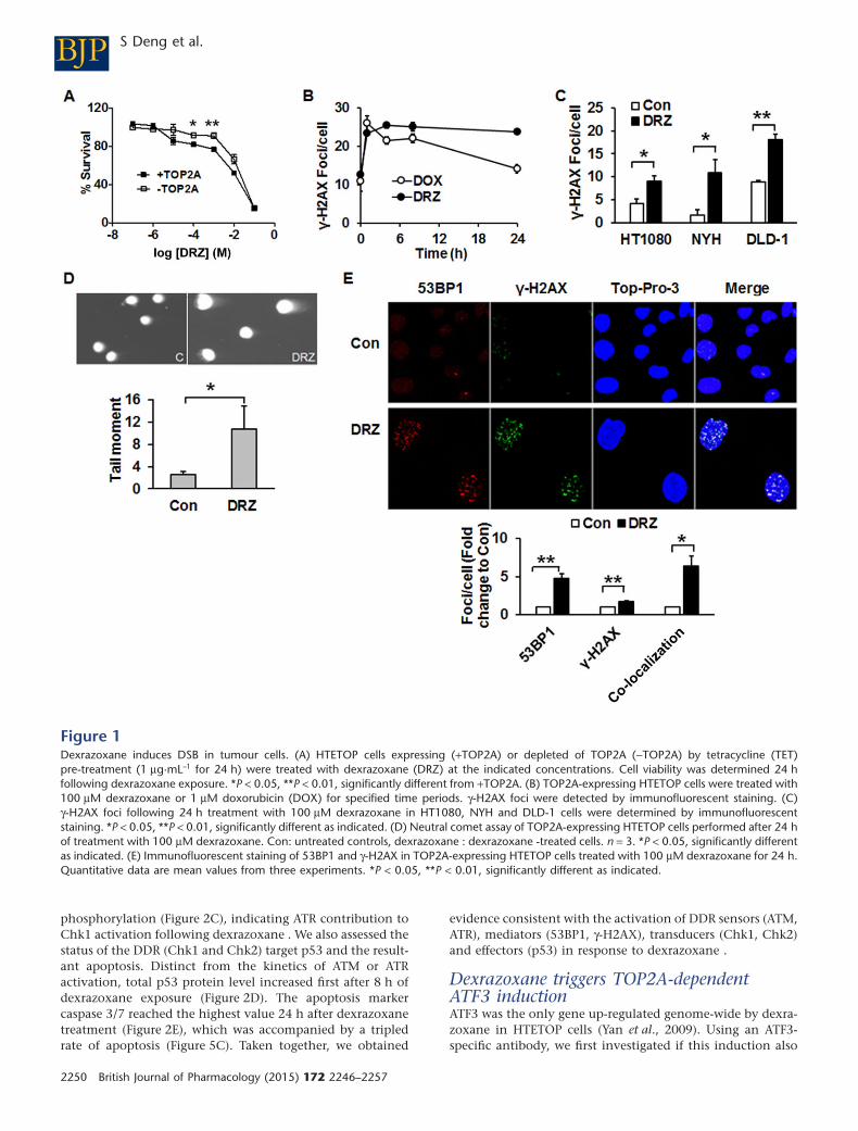
Dexrazoxane induces TOP2A-mediated DSBCell viability in response to dexrazoxane was evaluated inTOP2A expressing and non-expressing HTETOP cells. Higher
percentages of viable cells were observed at 100 μM and1 mM dexrazoxane in TOP2A non-expressing cells comparedwith TOP2A expressing ones (Figure 1A). In the followingexperiments, dexrazoxane was used at 100 μM, which is inthe range of concentrations seen in patients (Hochster et al.,1992; Sparano et al., 1999). Treatment of HTETOP cells withdexrazoxane (100 μM, 24 h) resulted in an accumulation ofthe DSB marker γ-H2AX, which was abolished by TOP2Adepletion with tetracycline (Yan et al., 2009), indicating thatTOP2A inhibition was the primary source of dexrazoxane-induced γ-H2AX. The kinetics of dexrazoxane-induced DSBformation was determined by γ-H2AX foci staining and com-pared with that of the classical TOP2A poison doxorubicin.Enhanced foci formation was observed as early as 1 h aftereither drug (Figure 1B). Foci induced by dexrazoxane lastedlonger than those evoked by doxorubicin, suggesting distinctkinetics of cleavable complex formation. Additionally, weinvestigated whether dexrazoxane-induced DSB formation isa general phenomenon in tumour cells using NYH cellsderived from human small cell lung cancer, LDL-1 cells fromcolorectal adenocarcinoma and fibrosarcoma-derivedHT1080, the parental cell line of HTETOP. All cell lines exhib-ited increased foci formation following dexrazoxane expo-sure (Figure 1C).
However, γ-H2AX is an indirect DSB marker and its induc-tion may also reflect inhibition of DNA synthesis rather thangeneration of free DSB (Kinner et al., 2008). Therefore, wecharacterized the effects of dexrazoxane using the neutralcomet assay, which allows for direct DSB detection in singlecells. Dexrazoxane (100 μM, 24 h) enlarged the size of nucleiof dexrazoxane-treated cells, probably due to endopolyploidycaused by TOP2A inhibition (Hasinoff et al., 2001) or to G2/Mcell cycle arrest (Supporting Information Fig. S1). Dexrazox-ane significantly increased the tail moment as compared tothat of untreated cells (Figure 1D). The formation of DSB bydexrazoxane treatment was further verified by γ-H2AX and53BP1 immunohistochemistry. Dexrazoxane treatment led tothe formation of γ-H2AX and 53BP1 foci, many of whichco-localized (Figure 1E). Both the increased tail moment andthe γ-H2AX/53BP1 co-localization were consistent withdexrazoxan -induced and TOP2A-mediated γ-H2AX beingtrue DSB (FitzGerald et al., 2009).
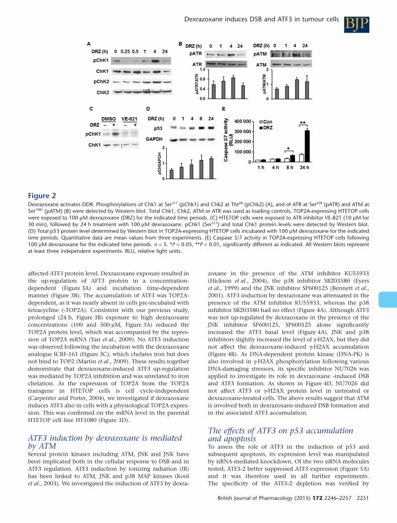
Dexrazoxane activates the DDRWe next investigated if dexrazoxane-induced DSB result inDDR activation. Firstly, we measured the phosphorylation ofDDR transducers and kinases Chk1 and Chk2 at serine 317and threonine 68 respectively. Both kinases were largelyphosphorylated in response to 100 μM dexrazoxane 4 h fol-lowing the drug exposure (Figure 2A). We then investigatedthe phosphorylation status of ATR and ATM, the respectiveactivating kinases of Chk1 and Chk2. The phosphorylation ofATR at Ser428 and of ATM at Ser1981, which act as the markersfor enzyme activation following DNA damage (Sahu et al.,2009), reached peak values 4 h following dexrazoxane expo-sure (Figure 2B). As expected, the accumulation of thedexrazoxane-induced DDR mediator γ-H2AX was nearly abol-ished by the ATM inhibitor KU55933 (Figure 4B) and absentin ATM mutant cells (Figure 4C). Consistent with enhancedATR phosphorylation (Figure 2B), its specific inhibitorVE-821 successfully prevented the downstream target Chk1
BJPDexrazoxane induces DSB and ATF3 in tumour cells
British Journal of Pharmacology (2015) 172 2246–2257 2249
phosphorylation (Figure 2C), indicating ATR contribution toChk1 activation following dexrazoxane . We also assessed thestatus of the DDR (Chk1 and Chk2) target p53 and the result-ant apoptosis. Distinct from the kinetics of ATM or ATRactivation, total p53 protein level increased first after 8 h ofdexrazoxane exposure (Figure 2D). The apoptosis markercaspase 3/7 reached the highest value 24 h after dexrazoxanetreatment (Figure 2E), which was accompanied by a tripledrate of apoptosis (Figure 5C). Taken together, we obtained
evidence consistent with the activation of DDR sensors (ATM,ATR), mediators (53BP1, γ-H2AX), transducers (Chk1, Chk2)and effectors (p53) in response to dexrazoxane .
Dexrazoxane triggers TOP2A-dependentATF3 inductionATF3 was the only gene up-regulated genome-wide by dexra-zoxane in HTETOP cells (Yan et al., 2009). Using an ATF3-specific antibody, we first investigated if this induction also
Figure 1Dexrazoxane induces DSB in tumour cells. (A) HTETOP cells expressing (+TOP2A) or depleted of TOP2A (−TOP2A) by tetracycline (TET)pre-treatment (1 μg·mL–1 for 24 h) were treated with dexrazoxane (DRZ) at the indicated concentrations. Cell viability was determined 24 hfollowing dexrazoxane exposure. *P < 0.05, **P < 0.01, significantly different from +TOP2A. (B) TOP2A-expressing HTETOP cells were treated with100 μM dexrazoxane or 1 μM doxorubicin (DOX) for specified time periods. γ-H2AX foci were detected by immunofluorescent staining. (C)γ-H2AX foci following 24 h treatment with 100 μM dexrazoxane in HT1080, NYH and DLD-1 cells were determined by immunofluorescentstaining. *P < 0.05, **P < 0.01, significantly different as indicated. (D) Neutral comet assay of TOP2A-expressing HTETOP cells performed after 24 hof treatment with 100 μM dexrazoxane. Con: untreated controls, dexrazoxane : dexrazoxane -treated cells. n = 3. *P < 0.05, significantly differentas indicated. (E) Immunofluorescent staining of 53BP1 and γ-H2AX in TOP2A-expressing HTETOP cells treated with 100 μM dexrazoxane for 24 h.Quantitative data are mean values from three experiments. *P < 0.05, **P < 0.01, significantly different as indicated.
BJP S Deng et al.
2250 British Journal of Pharmacology (2015) 172 2246–2257
affected ATF3 protein level. Dexrazoxane exposure resulted inthe up-regulation of AFT3 protein in a concentration-dependent (Figure 3A) and incubation time-dependentmanner (Figure 3B). The accumulation of ATF3 was TOP2A-dependent, as it was nearly absent in cells pre-incubated withtetracycline (-TOP2A). Consistent with our previous study,prolonged (24 h, Figure 3B) exposure to high dexrazoxaneconcentrations (100 and 500 μM, Figure 3A) reduced theTOP2A protein level, which was accompanied by the repres-sion of TOP2A mRNA (Yan et al., 2009). No ATF3 inductionwas observed following the incubation with the dexrazoxaneanalogue ICRF-161 (Figure 3C), which chelates iron but doesnot bind to TOP2 (Martin et al., 2009). These results togetherdemonstrate that dexrazoxane-induced ATF3 up-regulationwas mediated by TOP2A inhibition and was unrelated to ironchelation. As the expression of TOP2A from the TOP2Atransgene in HTETOP cells is cell cycle-independent(Carpenter and Porter, 2004), we investigated if dexrazoxaneinduces ATF3 also in cells with a physiological TOP2A expres-sion. This was confirmed on the mRNA level in the parentalHTETOP cell line HT1080 (Figure 3D).
ATF3 induction by dexrazoxane is mediatedby ATMSeveral protein kinases including ATM, JNK and JNK havebeen implicated both in the cellular response to DSB and inATF3 regulation. ATF3 induction by ionizing radiation (IR)has been linked to ATM, JNK and p38 MAP kinases (Koolet al., 2003). We investigated the induction of ATF3 by dexra-
zoxane in the presence of the ATM inhibitor KU55933(Hickson et al., 2004), the p38 inhibitor SB203580 (Eyerset al., 1999) and the JNK inhibitor SP600125 (Bennett et al.,2001). ATF3 induction by dexrazoxane was attenuated in thepresence of the ATM inhibitor KU55933, whereas the p38inhibitor SB203580 had no effect (Figure 4A). Although ATF3was not up-regulated by dexrazoxane in the presence of theJNK inhibitor SP600125, SP600125 alone significantlyincreased the ATF3 basal level (Figure 4A). JNK and p38inhibitors slightly increased the level of γ-H2AX, but they didnot affect the dexrazoxane-induced γ-H2AX accumulation(Figure 4B). As DNA-dependent protein kinase (DNA-PK) isalso involved in γ-H2AX phosphorylation following variousDNA-damaging stressors, its specific inhibitor NU7026 wasapplied to investigate its role in dexrazoxane -induced DSBand ATF3 formation. As shown in Figure 4D, NU7026 didnot affect ATF3 or γ-H2AX protein level in untreated ordexrazoxane-treated cells. The above results suggest that ATMis involved both in dexrazoxane-induced DSB formation andin the associated ATF3 accumulation.
The effects of ATF3 on p53 accumulationand apoptosisTo assess the role of ATF3 in the induction of p53 andsubsequent apoptosis, its expression level was manipulatedby siRNA-mediated knockdown. Of the two siRNA moleculestested, ATF3-2 better suppressed ATF3 expression (Figure 5A)and it was therefore used in all further experiments.The specificity of the ATF3-2 depletion was verified by
Figure 2Dexrazoxane activates DDR. Phosphorylations of Chk1 at Ser317 (pChk1) and Chk2 at Thr68 (pChk2) (A), and of ATR at Ser428 (pATR) and ATM atSer1981 (pATM) (B) were detected by Western blot. Total Chk1, Chk2, ATM or ATR was used as loading controls. TOP2A-expressing HTETOP cellswere exposed to 100 μM dexrazoxane (DRZ) for the indicated time periods. (C) HTETOP cells were exposed to ATR inhibitor VE-821 (10 μM for30 min), followed by 24 h treatment with 100 μM dexrazoxane. pChk1 (Ser317) and total Chk1 protein levels were detected by Western blot.(D) Total p53 protein level determined by Western blot in TOP2A-expressing HTETOP cells incubated with 100 μM dexrazoxane for the indicatedtime periods. Quantitative data are mean values from three experiments. (E) Caspase 3/7 activity in TOP2A-expressing HTETOP cells following100 μM dexrazoxane for the indicated time periods. n = 5. *P < 0.05, **P < 0.01, significantly different as indicated. All Western blots representat least three independent experiments. RLU, relative light units.
BJPDexrazoxane induces DSB and ATF3 in tumour cells
British Journal of Pharmacology (2015) 172 2246–2257 2251

Figure 3Dexrazoxane, but not ICRF-161, induces TOP2A-dependent ATF3 overexpression. HTETOP cells expressing (+TOP2A) or depleted of TOP2A(−TOP2A) by tetracycline pre-treatment were treated with increasing concentrations of dexrazoxane (DRZ) for 24 h (A) or with 100 μMdexrazoxane for the indicated time periods (B). ATF3 and TOP2A protein levels were analysed by Western blot. (C) ATF3 protein expressiondetermined by Western blot in TOP2A-expressing HTETOP cells following 24 h of ICRF-161 treatment with indicated concentrations. (D) ATF3mRNA expression assessed by TaqMan in HT1080 cells incubated with 100 μM dexrazoxane for 24 h. n = 3, *P < 0.05, significantly different asindicated.
Figure 4The effects of ATM, JNK, p38 and DNA-PK on the induction of ATF3 and γ-H2AX by dexrazoxane . Protein levels of ATF3 (A) and γ-H2AX (B)following dexrazoxane (DRZ; 100 μM, 24 h) in the presence or absence of ATM inhibitor KU55933 (20 μM), JNK inhibitor SP600125 (20 μM) orp38 inhibitor SB203580 (20 μM) were assessed by Western blot in TOP2A-expressing HTETOP cells. The inhibitors were administered 30 min priorto dexrazoxane treatment. Quantitative data are mean values from three experiments. **P < 0.01, significantly different as indicated. (C) γ-H2AXWestern blot was performed in dexrazoxane-treated (100 μM for 24 h) ATM mutant (GM05849) and wild-type (GM637) cells. (D) HTETOP cellswere first incubated with DNA-PK inhibitor NU7026 (10 μM) for 30 min then with dexrazoxane (100 μM) for 4 h. Western blots were used toevaluate ATF3 and γ-H2AX protein levels. *P < 0.05, **P < 0.01, significantly different as indicated.
BJP S Deng et al.
2252 British Journal of Pharmacology (2015) 172 2246–2257
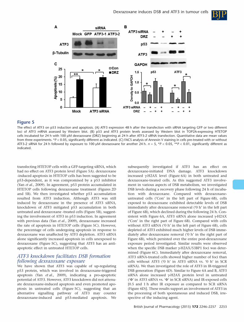
transfecting HTETOP cells with a GFP-targeting siRNA, whichhad no effect on ATF3 protein level (Figure 5A). dexrazoxane-induced apoptosis in HTETOP cells has been suggested to bep53-dependent, as it was compromised by a p53 inhibitor(Yan et al., 2009). In agreement, p53 protein accumulated inHTETOP cells following dexrazoxane treatment (Figures 2Dand 5B). We then investigated whether p53 accumulationresulted from ATF3 induction. Although ATF3 was stillinduced by dexrazoxane in the presence of ATF3 siRNA,knockdown of ATF3 mitigated p53 accumulation in bothuntreated and dexrazoxane -treated cells (Figure 5B), suggest-ing the involvement of ATF3 in p53 induction. In agreementwith previous data (Yan et al., 2009), dexrazoxane increasedthe rate of apoptosis in HTETOP cells (Figure 5C). However,the percentage of cells undergoing apoptosis in response todexrazoxane was unaffected by ATF3 depletion. ATF3 siRNAalone significantly increased apoptosis in cells unexposed todexrazoxane (Figure 5C), suggesting that ATF3 has an anti-apoptotic effect in untreated HTETOP cells.
ATF3 knockdown facilitates DSB formationfollowing dexrazoxane exposureWe have shown that ATF3 was capable of up-regulatingp53 protein, which was involved in dexrazoxane-triggeredapoptosis (Yan et al., 2009), indicating a pro-apoptoticpotential of ATF3. However, ATF3 knockdown did not attenu-ate dexrazoxane-induced apoptosis and even promoted apo-ptosis in untreated cells (Figure 5C), suggesting that analternative signalling pathway of ATF3 may counterdexrazoxane-induced and p53-mediated apoptosis. We
subsequently investigated if ATF3 has an effect ondexrazoxane-initiated DNA damage. ATF3 knockdownincreased γ-H2AX level (Figure 6A) in both untreated anddexrazoxane-treated cells. As this suggested ATF3 involve-ment in various aspects of DSB metabolism, we investigatedDSB levels during a recovery phase following 24 h of incuba-tion with dexrazoxane. Compared with dexrazoxane-untreated cells (‘Con’ in the left part of Figure 6B), cellsexposed to dexrazoxane exhibited detectable levels of DSBimmediately after dexrazoxane removal (‘0 h’ in the left partof Figure 6B), which declined during the following 24 h. Con-sistent with Figure 6A, ATF3 siRNA alone increased γ-H2AX(‘Con’ in the right part of Figure 6B). Compared with cellswithout ATF3 siRNA (‘0 h’ in the left part of Figure 6B), cellsdepleted of ATF3 exhibited much higher levels of DSB imme-diately after dexrazoxane removal (‘0 h’ in the right part ofFigure 6B), which persisted over the entire post-dexrazoxaneexposure period investigated. Similar results were observedwhen the specific DSB marker γ-H2AX/53BP1 foci was deter-mined (Figure 6C). Immediately after dexrazoxane removal,ATF3 siRNA-treated cells showed higher number of foci thancells without ATF3 (‘0 h’ in ATF3 siRNA vs. ‘0 h’ in SCRsiRNA). We than investigated the role of ATF3 in IR-triggeredDSB generation (Figure 6D). Similar to Figure 6A and B, ATF3siRNA alone increased γ-H2AX protein level in untreated(‘Φ’ in ATF3 siRNA vs. ‘Φ’ in SCR siRNA) and IR-exposed cells[0.5 and 1 h after IR exposure as compared to SCR siRNA(Figure 6D)]. These results support an involvement of ATF3 inthe processing of both spontaneous and induced DSB, irre-spective of the inducing agent.
Figure 5The effect of ATF3 on p53 induction and apoptosis. (A) ATF3 expression 48 h after the transfection with siRNA targeting GFP or two differentloci of ATF3 mRNA assessed by Western blot. (B) p53 and ATF3 protein levels assessed by Western blot in TOP2A-expressing HTETOPcells incubated for 24 h with 100 μM dexrazoxane (DRZ) beginning at 24 h after ATF3-2 siRNA transfection. Quantitative data are mean valuesfrom three experiments. *P < 0.05, significantly different as indicated. (C) FACS analysis of Annexin V staining in cells pre-treated with or withoutATF3-2 siRNA for 24 h followed by exposure to 100 μM dexrazoxane for another 24 h. n = 5, *P < 0.05, **P < 0.01, significantly different asindicated.
BJPDexrazoxane induces DSB and ATF3 in tumour cells
British Journal of Pharmacology (2015) 172 2246–2257 2253

Discussion and conclusions
Catalytic TOP2 inhibitors are thought to exert cytotoxiceffects predominantly via inhibiting ATPase activity, forminga closed clamp and blocking the turnover of the enzyme. Asbisdioxopiperazines are specific for TOP2, they are the mostcommonly used catalytic TOP2 inhibitors in human cells.The question of whether bisdioxopiperazines are pure cata-lytic inhibitors has been raised following observations thatbisdioxopiperazine ICRF-193 is able to trap TOP2 covalentcomplexes undetectable by standard procedures (Huanget al., 2001). Using a genetic model of conditional TOP2Aexpression, we demonstrated a correlation between TOP2-mediated DSB marker γ-H2AX and apoptosis in response todexrazoxane (Yan et al., 2009). However, a limitation ofγ-H2AX is that it detects both free DSB and blocked replica-tion forks (Kinner et al., 2008). In the present study, we dem-onstrate that TOP2A inhibition by dexrazoxane induces trueDSB. This is followed by activation of the DDR and by apo-ptosis. dexrazoxane also triggers ATF3 gene expression, whichaffects the level of p53 accumulation as well as dexrazoxan-induced DSB.
Dexrazoxane-induced DSB were demonstrated by γ-H2AXprotein level, by the formation of γ-H2AX foci co-localizationwith 53BP1 and by the tail moment in the neutral cometassay. These data are consistent with the formation of
dexrazoxane–TOP2A cleavable complexes demonstrated byothers (Sehested et al., 1998; Wessel et al., 1999; Lyu et al.,2007). DSB triggered by TOP2A poisons have been shown toactivate the DDR, beginning with the activation of ATRand/or ATM, which signals downstream to Chk1, Chk2 andp53. p53 induces transcriptional activation of pro-apoptoticfactors such as FAS, PUMA and BAX. As dexrazoxane inducedtrue DSB, we investigated if it also activated DDR. This wasconfirmed by increased γ-H2AX/53BP1 co-localization as wellas by the phosphorylation of ATR, ATM, Chk1 and Chk2,which was accompanied by p53 accumulation and apoptosis.The time-course of these events was consistent with γ-H2AXinduction being followed by DDR activation, ATF3, as well asp53 accumulation and ultimately apoptosis (Figure 7).Besides the time-course, the dependency of dexrazoxane-induced apoptosis on p53 in HTETOP cells is supported by itsblock by a p53 inhibitor (Yan et al., 2009). Therefore, inaddition to blocking TOP2 catalytic activity and similar toTOP2 poisons, dexrazoxane treatment clearly results in DSBformation, which is followed by DDR activation and ulti-mately by cell death from p53-dependent apoptosis.
Accumulation of p53 following dexrazoxane treatmentwas dependent upon ATF3 (Yan et al., 2009). ATF3 acts as anadaptive response gene that participates in cellular processesin response to extra- and/or intracellular changes. The induc-tion of ATF3 by dexrazoxane was TOP2A-dependent, as it was
Figure 6The effect of ATF3 on DSB formation in TOP2A-expressing HTETOP cells in response to dexrazoxane (DRZ) or IR. (A) γ-H2AX protein levels wereassessed by Western blot in TOP2A-expressing HTETOP cells. ATF3 siRNA was transfected 24 h before 100 μM dexrazoxane exposure for 24 h. (B)γ-H2AX levels in scrambled (SCR) or ATF3-2 siRNA-transfected HTETOP cells treated 24 later with 100 μM dexrazoxane for another 24 h. Cells weresubsequently washed with PBS, fed with fresh medium and γ-H2AX levels were assessed at the indicated time points after washing. *P < 0.05,significantly different as indicated. (C) The quantification of γ-H2AX/53BP1 foci determined by immunofluorescent staining. The treatment wasthe same as in (B). n = 3, *P < 0.05, significantly different as indicated. (D) γ-H2AX levels in SCR or ATF3-2 siRNA-transfected cells exposed 24 hlater to 10 Gy of IR (Φ: without IR exposure). γ-H2AX levels were determined at the indicated time points after IR exposure. *P < 0.05, significantlydifferent as indicated. All histograms represent mean values from three independent experiments.
BJP S Deng et al.
2254 British Journal of Pharmacology (2015) 172 2246–2257

absent both after TOP2A depletion and upon treatment witha dexrazoxane analogue incapable of TOP2 inhibition. Theinduction of ATF3 also involved the protein kinase ATM, as itwas blocked by a specific ATM inhibitor and in ATM mutantcells. ATM acts as a primary transducer in response to DSBand phosphorylates key factors in DNA damage responsepathways. Among others, it is suggested to interact with andphosphorylate ATF2, which is capable of increasing ATF3expression via promoter activation (Lee et al., 2010). The JNKinhibitor SP600125 alone significantly increased the ATF3level in untreated, but not in dexrazoxane-treated, cells(Figure 4A). This is consistent with the SP600125-inducedATF3 expression in human colon cancer HCT116 cells, via anunknown mechanism (Hackl et al., 2010). Taken together,dexrazoxane-driven accumulation of ATF3 was most likely tobe mediated by TOP2A-generated DSB detected by ATM.
ATF3 has been suggested to be pro-apoptotic (Huanget al., 2008; Kashiwakura et al., 2008) and to function astumour suppressor, as its down-regulation promotes tumourgrowth and metastasis (Hackl et al., 2010). On the contrary,ATF3 overexpression has been found to enhance the tumori-genic potential (Wu et al., 2010) and contribute to the malig-nant growth of tumour cells. Despite many efforts trying toclarify the oncogenic properties of ATF3, ATF3 cannot bedefined exclusively as an oncogene or tumour repressor andthis assessment is consistent with the various effects of ATF3perturbation observed in HTETOP cells. ATF3 was clearly anti-apoptotic in untreated cells, as its knockdown by siRNAcaused significant apoptosis. In contrast, down-regulation ofATF3 could not affect dexrazoxan-induced apoptosis, despiteATF3-mediated p53 accumulation, although the latter one isrequired for dexrazoxane-induced apoptosis (Yan et al.,2009).
How can we explain the apparent contradiction betweenthe dependency of dexrazoxane-induced p53 accumulationon ATF3 and the disparate effects of these proteins on apop-tosis? One possibility is that induction of ATF3 facilitatesapoptosis by up-regulating p53, but detecting this effectrequires more than simple manipulation of the ATF3 level.Alternatively, this contradiction may reflect the involvement
of ATF3 in the processing of dexrazoxane-induced DSB,which may counter the pro-apoptotic effect of p53 stabilizedby ATF3 (Figure 7). ATF3 knockdown led to increased γ-H2AXprotein accumulation in dexrazoxane-treated cells, whichmay be the consequence of ongoing apoptosis rather thanDSB. However, the specific marker of true DSB, γ-H2AX/53BP1 foci, was also enhanced by ATF3 siRNA indexrazoxane-treated cells. The involvement of ATF3 in DSBprocessing would be consistent with the capability of itstranscriptional activator ATF2 to enhance DNA repair(Hayakawa et al., 2003). Consistent with our data, the repres-sion of ATF3 after UV-mediated genotoxic stress impaired theDNA repair process mediated through ATF3 transcriptionaltarget PCNA-associated factor KIAA0101/p15PAF (Turchi et al.,2009). The mechanisms by which ATF3 protects againstdexrazoxane-induced DNA damage need to be further inves-tigated. p15PAF elevation was not observed in dexrazoxane-treated HTETOP cells from the microarray experiments (datanot shown), indicating that this protein may not be involvedin the protective effect of ATF3, or that such an effect is p15PAF
transcription independent. Taken together, the DNA damage-triggered ATF3 induction controls the level of p53 accumula-tion and may as well as regulate DSB repair. This is consistentwith ATF3 serving as a switch between DNA damage repairand cell death following dexrazoxane treatment.
The apparent roles of ATF3 in apoptosis and DNA damageprocess are reminiscent of the puzzling heterogeneity ofcancer-related clinical endpoints of dexrazoxane treatments.Dexrazoxane remains underused due to the reported reduc-tion of the anti-tumour effect of anthracyclines (Swain et al.,1997b) and to its association with secondary malignancies(Tebbi et al., 2007). On the contrary, dexrazoxane hasincreased survival both in patients (Swain et al., 1997a) andin pre-clinical animal cancer models when combined withTOP2 poisons (Hasinoff et al., 1998; Hofland et al., 2005). Thelatter findings are in agreement with dexrazoxane’s capacityto induce DSB, DDR and apoptosis, which resemble and mayenhance the anti-tumour effects of anthracyclines. They arealso consistent with meta-analyses demonstrating undimin-ished anti-tumour efficacy of anthracyclines when combinedwith dexrazoxane (van Dalen et al., 2011b). Finally, basedupon our observation that dexrazoxane is clearly genotoxic,secondary malignancies could arise from dexrazoxane-induced DNA damage in cells with diminished capacity forapoptosis and/or DNA repair. The application of dexrazoxaneas a cardioprotectant may require a consideration of thesevariables in the context of individual patients and tumourentities. These complex relationships in dividing, that is,TOP2A-expressing cells, contrast the apparently morestraightforward mechanism of cardioprotection conferred bydexrazoxane. The predominant cardiac TOP2 isozyme TOP2Bundergoes rapid proteosomal degradation following exposureto dexrazoxane, thereby preventing doxorubicin-inducedand TOP2B-mediated DNA damage (Lyu et al., 2007).
Acknowledgements
This project was supported by the Deutsche Forschungsge-meinschaft (DFG) grant (WO505/3-1).
Figure 7HTETOP cell response to dexrazoxane (DRZ) , including DSB induc-tion, DDR activation, ATF3 accumulation and apoptosis.
BJPDexrazoxane induces DSB and ATF3 in tumour cells
British Journal of Pharmacology (2015) 172 2246–2257 2255

Author Contributions
S. D., B. K. and L. W. participated in research design. S. D.,T. Y., T. N., D. F., A. N. and U. G.-A. conducted the experi-ments. T. N. contributed new reagents or analytical tools.S. D. and T. Y. performed data analysis. S. D., B. K. and L. W.wrote or contributed to the writing of the manuscript.
Conflicts of interest
No conflict of interest is declared.
ReferencesAlexander SPH, Benson HE, Faccenda E, Pawson AJ, Sharman JL,Spedding M et al. (2013). The Concise Guide to PHARMACOLOGY2013/14: Enzymes. Br J Pharmacol, 170: 1797–1867
Bennett BL, Sasaki DT, Murray BW, O’Leary EC, Sakata ST, Xu Wet al. (2001). SP600125, an anthrapyrazolone inhibitor of JunN-terminal kinase. Proc Natl Acad Sci U S A 98: 13681–13686.
Berdelle N, Nikolova T, Quiros S, Efferth T, Kaina B (2011).Artesunate induces oxidative DNA damage, sustained DNAdouble-strand breaks, and the ATM/ATR damage response in cancercells. Mol Cancer Ther 10: 2224–2233.
Bonnefoi HR (2011). Anthracyclines, HER2, and TOP2A: the verdict.Lancet Oncol 12: 1084–1085.
Burgess DJ, Doles J, Zender L, Xue W, Ma B, McCombie WR et al.(2008). Topoisomerase levels determine chemotherapy responsein vitro and in vivo. Proc Natl Acad Sci U S A 105: 9053–9058.
Carpenter AJ, Porter AC (2004). Construction, characterization, andcomplementation of a conditional-lethal DNA topoisomeraseIIalpha mutant human cell line. Mol Biol Cell 15: 5700–5711.
Chen BP, Liang G, Whelan J, Hai T (1994). ATF3 and ATF3 deltaZip. Transcriptional repression versus activation by alternativelyspliced isoforms. J Biol Chem 269: 15819–15826.
Creighton AM, Hellmann K, Whitecross S (1969). Antitumouractivity in a series of bisdiketopiperazines. Nature 222: 384–385.
van Dalen EC, Caron HN, Dickinson HO, Kremer LC (2011a).Cardioprotective interventions for cancer patients receivinganthracyclines. Cochrane Database Syst Rev (6): CD003917.
van Dalen EC, van den Berg H, Raphael MF, Caron HN, Kremer LC(2011b). Should anthracyclines and dexrazoxane be used forchildren with cancer? Lancet Oncol 12: 12–13.
Eich M, Roos WP, Nikolova T, Kaina B (2013). Contribution ofATM and ATR to the resistance of glioblastoma and malignantmelanoma cells to the methylating anticancer drug temozolomide.Mol Cancer Ther 12: 2529–2540.
Eyers PA, van den Ijssel P, Quinlan RA, Goedert M, Cohen P (1999).Use of a drug-resistant mutant of stress-activated protein kinase2a/p38 to validate the in vivo specificity of SB 203580. FEBS Lett451: 191–196.
FitzGerald JE, Grenon M, Lowndes NF (2009). 53BP1: function andmechanisms of focal recruitment. Biochem Soc Trans 37: 897–904.
Hackl C, Lang SA, Moser C, Mori A, Fichtner-Feigl S, Hellerbrand Cet al. (2010). Activating transcription factor-3 (ATF3) functions as atumor suppressor in colon cancer and is up-regulated uponheat-shock protein 90 (Hsp90) inhibition. BMC Cancer 10: 668.
Hasinoff BB, Hellmann K, Herman EH, Ferrans VJ (1998). Chemical,biological and clinical aspects of dexrazoxane and otherbisdioxopiperazines. Curr Med Chem 5: 1–28.
Hasinoff BB, Abram ME, Barnabe N, Khelifa T, Allan WP, YalowichJC (2001). The catalytic DNA topoisomerase II inhibitordexrazoxane (ICRF-187) induces differentiation and apoptosis inhuman leukemia K562 cells. Mol Pharmacol 59: 453–461.
Hayakawa J, Depatie C, Ohmichi M, Mercola D (2003). Theactivation of c-Jun NH2-terminal kinase (JNK) by DNA-damagingagents serves to promote drug resistance via activating transcriptionfactor 2 (ATF2)-dependent enhanced DNA repair. J Biol Chem 278:20582–20592.
Hickson I, Zhao Y, Richardson CJ, Green SJ, Martin NM, Orr AIet al. (2004). Identification and characterization of a novel andspecific inhibitor of the ataxia-telangiectasia mutated kinase ATM.Cancer Res 64: 9152–9159.
van Hille B, Hill BT (1998). Yeast cells expressing differential levelsof human or yeast DNA topoisomerase II: a potent tool foridentification and characterization of topoisomerase II-targetingantitumour agents. Cancer Chemother Pharmacol 42: 345–356.
Hochster H, Liebes L, Wadler S, Oratz R, Wernz JC, Meyers M et al.(1992). Pharmacokinetics of the cardioprotector ADR-529(ICRF-187) in escalating doses combined with fixed-dosedoxorubicin. J Natl Cancer Inst 84: 1725–1730.
Hofland KF, Thougaard AV, Dejligbjerg M, Jensen LH, KristjansenPE, Rengtved P et al. (2005). Combining etoposide and dexrazoxanesynergizes with radiotherapy and improves survival in mice withcentral nervous system tumors. Clin Cancer Res 11: 6722–6729.
Huang KC, Gao H, Yamasaki EF, Grabowski DR, Liu S, Shen LL et al.(2001). Topoisomerase II poisoning by ICRF-193. J Biol Chem 276:44488–44494.
Huang X, Li X, Guo B (2008). KLF6 induces apoptosis in prostatecancer cells through up-regulation of ATF3. J Biol Chem 283:29795–29801.
Janz M, Hummel M, Truss M, Wollert-Wulf B, Mathas S, Johrens Ket al. (2006). Classical Hodgkin lymphoma is characterized by highconstitutive expression of activating transcription factor 3 (ATF3),which promotes viability of Hodgkin/Reed-Sternberg cells. Blood107: 2536–2539.
Jensen LH, Nitiss KC, Rose A, Dong J, Zhou J, Hu T et al. (2000).A novel mechanism of cell killing by anti-topoisomerase IIbisdioxopiperazines. J Biol Chem 275: 2137–2146.
Kashiwakura Y, Ochiai K, Watanabe M, Abarzua F, Sakaguchi M,Takaoka M et al. (2008). Down-regulation of inhibition ofdifferentiation-1 via activation of activating transcription factor 3and Smad regulates REIC/Dickkopf-3-induced apoptosis. Cancer Res68: 8333–8341.
Kinner A, Wu W, Staudt C, Iliakis G (2008). Gamma-H2AX inrecognition and signaling of DNA double-strand breaks in thecontext of chromatin. Nucleic Acids Res 36: 5678–5694.
Kool J, Hamdi M, Cornelissen-Steijger P, van der Eb AJ, Terleth C,van Dam H (2003). Induction of ATF3 by ionizing radiation ismediated via a signaling pathway that includes ATM, Nibrin1,stress-induced MAPkinases and ATF-2. Oncogene 22: 4235–4242.
Larsen AK, Escargueil AE, Skladanowski A (2003). Catalytictopoisomerase II inhibitors in cancer therapy. Pharmacol Ther 99:167–181.
BJP S Deng et al.
2256 British Journal of Pharmacology (2015) 172 2246–2257
Lee SH, Bahn JH, Whitlock NC, Baek SJ (2010). Activatingtranscription factor 2 (ATF2) controls tolfenamic acid-induced ATF3expression via MAP kinase pathways. Oncogene 29: 5182–5192.
Lyu YL, Kerrigan JE, Lin CP, Azarova AM, Tsai YC, Ban Y et al.(2007). Topoisomerase IIbeta mediated DNA double-strand breaks:implications in doxorubicin cardiotoxicity and prevention bydexrazoxane. Cancer Res 67: 8839–8846.
Martin E, Thougaard AV, Grauslund M, Jensen PB, Bjorkling F,Hasinoff BB et al. (2009). Evaluation of the topoisomeraseII-inactive bisdioxopiperazine ICRF-161 as a protectant againstdoxorubicin-induced cardiomyopathy. Toxicology 255: 72–79.
Nikolova T, Dvorak M, Jung F, Adam I, Kramer E, Gerhold-Ay Aet al. (2014). The γH2AX assay for genotoxic and nongenotoxicagents: comparison of H2AX phosphorylation with cell deathresponse. Toxicol Sci 140: 103–117.
Nitiss JL (2009). Targeting DNA topoisomerase II in cancerchemotherapy. Nat Rev Cancer 9: 338–350.
Nobori K, Ito H, Tamamori-Adachi M, Adachi S, Ono Y, Kawauchi Jet al. (2002). ATF3 inhibits doxorubicin-induced apoptosis incardiac myocytes: a novel cardioprotective role of ATF3. J Mol CellCardiol 34: 1387–1397.
Pawson AJ, Sharman JL, Benson HE, Faccenda E, Alexander SP,Buneman OP et al.; NC-IUPHAR (2014). The IUPHAR/BPS Guide toPHARMACOLOGY: an expert-driven knowledge base of drug targetsand their ligands. Nucl. Acids Res. 42 (Database Issue):D1098–1106.
Sahu RP, Batra S, Srivastava SK (2009). Activation of ATM/Chk1 bycurcumin causes cell cycle arrest and apoptosis in humanpancreatic cancer cells. Br J Cancer 100: 1425–1433.
Sehested M, Wessel I, Jensen LH, Holm B, Oliveri RS, Kenwrick Set al. (1998). Chinese hamster ovary cells resistant to thetopoisomerase II catalytic inhibitor ICRF-159: a Tyr49Phe mutationconfers high-level resistance to bisdioxopiperazines. Cancer Res 58:1460–1468.
Snyder RD (2003). Evidence from studies with intact mammaliancells that merbarone and bis(dioxopiperazine)s are topoisomerase IIpoisons. Drug Chem Toxicol 26: 15–22.
Sparano JA, Speyer J, Gradishar WJ, Liebes L, Sridhara R, Mendoza Set al. (1999). Phase I trial of escalating doses of paclitaxel plusdoxorubicin and dexrazoxane in patients with advanced breastcancer. J Clin Oncol 17: 880–886.
Swain SM, Whaley FS, Gerber MC, Ewer MS, Bianchine JR, GamsRA (1997a). Delayed administration of dexrazoxane providescardioprotection for patients with advanced breast cancer treatedwith doxorubicin-containing therapy. J Clin Oncol 15: 1333–1340.
Swain SM, Whaley FS, Gerber MC, Weisberg S, York M, Spicer Det al. (1997b). Cardioprotection with dexrazoxane fordoxorubicin-containing therapy in advanced breast cancer. J ClinOncol 15: 1318–1332.
Tebbi CK, London WB, Friedman D, Villaluna D, De Alarcon PA,Constine LS et al. (2007). Dexrazoxane-associated risk for acutemyeloid leukemia/myelodysplastic syndrome and other secondarymalignancies in pediatric Hodgkin’s disease. J Clin Oncol 25:493–500.
Turchi L, Aberdam E, Mazure N, Pouyssegur J, Deckert M, KitajimaS et al. (2008). Hif-2alpha mediates UV-induced apoptosis through anovel ATF3-dependent death pathway. Cell Death Differ 15:1472–1480.
Turchi L, Fareh M, Aberdam E, Kitajima S, Simpson F, Wicking Cet al. (2009). ATF3 and p15PAF are novel gatekeepers of genomicintegrity upon UV stress. Cell Death Differ 16: 728–737.
Wessel I, Jensen LH, Jensen PB, Falck J, Rose A, Roerth M et al.(1999). Human small cell lung cancer NYH cells selected forresistance to the bisdioxopiperazine topoisomerase II catalyticinhibitor ICRF-187 demonstrate a functional R162Q mutation inthe Walker A consensus ATP binding domain of the alpha isoform.Cancer Res 59: 3442–3450.
Wu X, Nguyen BC, Dziunycz P, Chang S, Brooks Y, Lefort K et al.(2010). Opposing roles for calcineurin and ATF3 in squamous skincancer. Nature 465: 368–372.
Yamada N, Noguchi S, Mori T, Naoe T, Maruo K, Akao Y (2013).Tumor-suppressive microRNA-145 targets catenin delta-1 to regulateWnt/beta-catenin signaling in human colon cancer cells. CancerLett 335: 332–342.
Yan T, Deng S, Metzger A, Godtel-Armbrust U, Porter AC,Wojnowski L (2009). Topoisomerase II{alpha}-dependent and-independent apoptotic effects of dexrazoxane and doxorubicin.Mol Cancer Ther 8: 1075–1085.
Supporting information
Additional Supporting Information may be found in theonline version of this article at the publisher’s web-site:
http://dx.doi.org/10.1111/bph.13046
Figure S1 Dexrazoxane exposure led to cell cycle arrest atG2/M. HTETOP cell was exposed to 100 μM dexrazoxane(DRZ) or DMSO for indicated time periods. DNA was stainedwith PI and cell cycle distribution was examined by FACSanalysis. The PI intensity was plotted against cell numbers.
BJPDexrazoxane induces DSB and ATF3 in tumour cells
British Journal of Pharmacology (2015) 172 2246–2257 2257


![REVIEW Open Access Drug therapy for hereditary cancers · 34. 36]. Another topoisomerase II inhibitor, etoposide, showed selective efficacy againstBRCA-defective cell in all but one](https://img.pdfslide.us/doc/110x75/5f235916b6622875be3207bf/review-open-access-drug-therapy-for-hereditary-cancers-34-36-another-topoisomerase.jpg)