Embed Size (px)
Citation preview

MOLECULAR AND CELLULAR BIOLOGY,0270-7306/00/$04.0010
Jan. 2000, p. 594–603 Vol. 20, No. 2
Copyright © 2000, American Society for Microbiology. All Rights Reserved.
An Antitumor Drug-Induced Topoisomerase Cleavage ComplexBlocks a Bacteriophage T4 Replication Fork In Vivo
GEORGE HONG AND KENNETH N. KREUZER*
Department of Microbiology, Duke University Medical Center, Durham, North Carolina 27710
Received 23 August 1999/Returned for modification 21 September 1999/Accepted 20 October 1999
Many antitumor and antibacterial drugs inhibit DNA topoisomerases by trapping covalent enzyme-DNAcleavage complexes. Formation of cleavage complexes is important for cytotoxicity, but evidence suggests thatcleavage complexes themselves are not sufficient to cause cell death. Rather, active cellular processes such astranscription and/or replication are probably necessary to transform cleavage complexes into cytotoxic lesions.Using defined plasmid substrates and two-dimensional agarose gel analysis, we examined the collision of anactive replication fork with an antitumor drug-trapped cleavage complex. Discrete DNA molecules accumu-lated on the simple Y arc, with branch points very close to the topoisomerase cleavage site. Accumulation ofthe Y-form DNA required the presence of a topoisomerase cleavage site, the antitumor drug, the type IItopoisomerase, and a T4 replication origin on the plasmid. Furthermore, all three arms of the Y-form DNAwere replicated, arguing strongly that these are trapped replication intermediates. The Y-form DNA appearedeven in the absence of two important phage recombination proteins, implying that Y-form DNA is the resultof replication rather than recombination. This is the first direct evidence that a drug-induced topoisomerasecleavage complex blocks the replication fork in vivo. Surprisingly, these blocked replication forks do notcontain DNA breaks at the topoisomerase cleavage site, implying that the replication complex was inactivated(at least temporarily) and that topoisomerase resealed the drug-induced DNA breaks. The replication fork maybehave similarly at other types of DNA lesions, and thus cleavage complexes could represent a useful(site-specific) model for chemical- and radiation-induced DNA damage.
Type II DNA topoisomerases are involved in diverse cellularprocesses such as replication, transcription, recombination,chromosome condensation, and the maintenance of genomestability. These enzymes transform the topological state ofDNA by a strand passage reaction (for reviews, see references6, 65, and 66). After binding to one segment of duplex DNA,a type II topoisomerase cleaves the duplex with a four-basestagger while covalently attaching to both 59 ends via phospho-tyrosine linkages. This reaction intermediate, with the enzymecovalently linked to broken DNA, is referred to as the cleavagecomplex. After passage of a second duplex segment throughthe break, the topoisomerase reverses the phosphotyrosinelinkages and rejoins the cleaved DNA ends.
Mammalian type II topoisomerases are targets of many im-portant antitumor drug classes, including aminoacridines, an-thracenediones, anthracyclines, ellipticines, and epipodophyl-lotoxins (for reviews, see references 10, 13, and 55). Inaddition, bacterial type II topoisomerases are targets of clini-cally important antibacterial agents (quinolones and flouro-quinolones) (17, 28). Remarkably, all these antitumor andantibacterial agents inhibit the enzyme by trapping the cleav-age complex. The simplest model is that the drug, localizedprecisely at the site of DNA cleavage, prevents topoisomerasefrom rejoining DNA breaks.
Results from different systems suggest that the cytotoxicityof topoisomerase inhibitors depends on trapping of the cleav-age complex rather than loss of enzyme activity (for reviews,see references 13, 17, and 44). First, Escherichia coli cells thatcontain both nalidixic acid-sensitive and -resistant DNA gyraseare sensitive to nalidixic acid (24, 33). This result implies that
in the presence of drug, the drug-sensitive gyrase causes cyto-toxicity even though the resistant gyrase is enzymatically active.Second, bacteriophage T7 growth is inhibited by nalidixic acideven though T7 does not require DNA gyrase for growth, andthis inhibition is alleviated by heat inactivation of a thermo-sensitive gyrase subunit A (36). Third, mutational inactivationof bacteriophage T4 topoisomerase, which is not totally essen-tial for growth, causes drug resistance (50). Fourth, overpro-duction of type II topoisomerase in Saccharomyces cerevisiaecauses hypersensitivity to antitumor drugs (52). Fifth, in vari-ous systems, recombinational repair reduces sensitivity to to-poisomerase inhibitors, consistent with the cleavage complex(or some derivative) causing DNA damage (18, 32, 33, 50, 52).
The drug-induced cleavage complex itself is apparently not acytotoxic lesion; cellular processes are probably required tocause cytotoxicity (15). For example, inhibition of protein syn-thesis protects cells from cytotoxicity but does not prevent theformation of cleavage complexes (59). In addition, mammaliancells treated with a topoisomerase poison survive much betterwhen they are simultaneously treated with dinitrophenol, anuncoupler of oxidative phosphorylation that reduces the intra-cellular ATP pool (39). Since dinitrophenol has little effect oncleavage complex formation, an ATP-requiring process (e.g.,transcription or DNA replication) subsequent to cleavagecomplex formation is apparently involved in the cytotoxicity oftopoisomerase poisons.
Transcription appears to play a role in the killing of certainmammalian cells by 49-(9-acridinylamino)methanesulfon-m-anisidide (m-AMSA) because the transcription inhibitorcordycepin protects G1-phase cells (15). The involvement oftranscription, however, may not be universal. Yeast cells ar-rested by a factor are transcription competent and yet resistantto m-AMSA and the epipodophyllotoxin etoposide (53). Thisresult suggests that DNA replication might be important forcytotoxicity. Indeed, S-phase yeast cells are more readily killed
* Corresponding author. Mailing address: Department of Microbi-ology, Box 3020, Duke University Medical Center, Durham, NC 27710.Phone: (919) 684-6466. Fax: (919) 681-8911. E-mail: [email protected].
594
on April 3, 2018 by guest
http://mcb.asm
.org/D
ownloaded from

by m-AMSA than are G1-phase yeast cells (53). Furthermore,the cytotoxicity of m-AMSA can be reduced or abolished ifmammalian S-phase cells are cotreated with the DNA poly-merase inhibitor aphidicolin (15, 27, 67). These results arguethat some interaction between the drug-induced cleavage com-plex and the replication apparatus can be very important in celldeath mediated by topoisomerase inhibitors.
We analyzed the collision of a replication fork with a drug-stabilized cleavage complex in vivo by using the bacteriophageT4 model system. Phage T4 encodes a type II DNA topoisom-erase that is inhibited by many of the same antitumor drugsthat block the mammalian enzyme. The T4 topoisomerase wasdemonstrated to be the physiological target for m-AMSA (31),and inhibition of phage growth is probably dependent on cleav-age complex formation (see above) (50). Recombinational re-pair of damage derived from the drug-induced cleavage com-plex is important in both the bacteriophage T4 and mammaliansystems, which also argues that T4 is a useful model system forthese studies. Bacteriophage T4 is obviously not a suitablemodel system for all aspects of topoisomerase inhibitor action,since it lacks the cell cycle checkpoints and programmed celldeath responses seen in the more complex mammalian sys-tems. Nevertheless, T4 provides many experimental advan-tages for this study, including the availability of mutants in allimportant replication and recombination genes, well-definedreplication origins, a unique cytosine modification that marksDNA at the time of replication, and a type II topoisomerasethat has been carefully analyzed for DNA sequence recogni-tion (19, 38). Here, we use a strong topoisomerase cleavagesite and a cloned T4 replication origin to examine the in vivoconsequence of a collision between a replication fork and anm-AMSA-induced type II topoisomerase cleavage complex.
MATERIALS AND METHODS
Materials. m-AMSA was provided by the Drug Synthesis and ChemistryBranch, National Cancer Institute. Oligonucleotides were purchased from Na-tional Biosciences. Nylon blotting membrane was obtained from Schleicher &Schuell, the random-primed labeling kit was obtained from Boehringer Mann-heim, and Sequenase version 2.0 was obtained from United States Biochemicals.Restriction enzymes and T4 ligase were purchased from New England Biolabs.
Bacterial and phage strains. The host for all infections was an acrA::Tn10-kanderivative of E. coli NapIV (hsdMK
1 hsdRK2 hsdSK
1 rglB1 nonsuppressing)(51). The acrA mutation eliminates a multidrug efflux pump so that the infectedcells are more sensitive to m-AMSA (J. George and K. Kreuzer, unpublisheddata). Phage strains used in this study were T4 K10 (amB262 [gene 38] amS29[gene 51] nd28 [denA] rIIPT8 [denB-rII deletion]) (61), K10-46/uvsX (as K10,with amB14 [gene 46] and am11 [gene uvsX]) (37), K10-39 (as K10, with amN116[gene 39]) (49) and T4 dC (amC87 [gene 42], amE51 [gene 56], NB5060 [denB-rIIdeletion], W7 [gene alc], and probably an uncharacterized denA mutation) (62).
Plasmid construction. For these studies, we used a strong m-AMSA-inducibletopoisomerase cleavage site (topo site) that had been designed based on ananalysis of T4 topoisomerase sequence recognition (19, 20). Plasmid pGH2,containing this strong site, was constructed by ligating the following duplexoligonucleotide to the large BamHI-SalI fragment of pBR322:
59GATCCAAGCTAAAGTTATATAACTTTATTCAAGG3939GTTCGATTTCAATATATTGAAATAAGTTCCAGCT59
Plasmid pGH2-01 was constructed by ligating a purified PstI-ClaI fragment[containing the T4 origin, ori(34) (ori)] from plasmid pKK061-6 (46) to the largepurified PstI-ClaI fragment of pGH2 (which contains the topo site). A controlplasmid, pGH4, lacking both the topo site and the T4 origin, was constructed bycleaving pGH2 with BamHI and SalI, filling in the ends with Klenow polymerase,purifying the large fragment from low-melting-temperature agarose, and thenligating the fragment into a circle. Another control plasmid, pGH4-01, which hasthe ori but not the topo site, was constructed as follows. Plasmid pGH2-01 wascleaved with PstI and ClaI, and the small fragment containing the T4 origin waspurified. Similarly, plasmid pGH4 was cleaved with the same two enzymes andthe large fragment containing the BamHI-SalI deletion was purified. The twofragments were ligated and transformed.
T4 infections and DNA preparations. NapIV acrA cells containing the indi-cated plasmid were grown with vigorous shaking at 37°C to a cell density of 4 3108 per ml and then infected with the indicated bacteriophage T4 strain at amultiplicity of 3 PFU per cell. After 4 min at 37°C, without shaking, for adsorp-
tion, the cells were incubated for 2 min with shaking and then m-AMSA wasadded at 10 mg/ml (unless otherwise indicated). The infected cells were thenincubated with vigorous shaking at 37°C for 18 min. Infected cells from 1 ml ofthe culture were collected by centrifugation, and the pellet was frozen in a dryice-ethanol bath. The frozen pellet was thawed in 300 ml of cleavage lysis buffer(50 mM Tris-HCl [pH 7.8], 10 mM disodium EDTA, 100 mM NaCl, 0.2%sodium dodecyl sulfate [SDS]). Proteinase K was added to 0.5 mg/ml, and thesuspension was incubated at 65°C for 2 h. The total nucleic acid was extractedsequentially with phenol and chloroform-isoamyl alcohol (24:1) and then dia-lyzed overnight at 4°C against TE buffer (10 mM Tris-HCl [pH 7.8], 1 mMdisodium EDTA).
The experiment in Fig. 8 compared two different extraction conditions. Onealiquot (1 ml) of infected cells was added to 1 ml of 23 reversal lysis buffer (finalconcentrations: 50 mM Tris-HCl [pH 7.8], 10 mM disodium EDTA, 100 mMNaCl, 1% Triton X-100, 1.8 mg of lysozyme per ml) and placed at 65°C for 20min. After this incubation for reversal of cleavage complexes, SDS (0.2%) andproteinase K (0.5 mg/ml) were added and the sample was incubated at 65°C foran additional 100 min. A second aliquot (1 ml) of infected cells was added to 200ml of 63 cleavage lysis buffer (final concentrations: 50 mM Tris-HCl [pH 7.8], 10mM disodium EDTA, 100 mM NaCl, 0.2% SDS) and incubated at 37°C for 20min. The sample was then incubated at 65°C for an additional 100 min. Nucleicacids from both samples were purified as described above.
Agarose gel electrophoresis and Southern hybridization. Nucleic acid sampleswere treated with the indicated restriction enzymes before being subjected to gelelectrophoresis. The one-dimension gels contained 1% agarose and were run in0.53 TBE buffer (13 TBE contains 89 mM Tris-HCl, 89 mM borate, and 2.0 mMdisodium EDTA) at 2.8 V/cm for 15 h. The two-dimension gel protocol was fromFriedman and Brewer (21). Briefly, the first-dimension gel was a 0.4% agarosegel run in 13 TBE buffer for 30 h at 1 V/cm at room temperature (21°C). Thedesired gel lane was sliced from the first-dimension gel and cast across the top ofa second-dimension 1% agarose gel, which was run in 13 TBE containingethidium bromide (0.3 mg/ml) for 15 h at 6 V/cm in the cold (4°C). For Southernhybridization, agarose gels were transferred to a nylon blotting membrane by thedownward sponge method (adapted from reference 48). The probe for theSouthern blots consisted of pBR322 DNA labeled with [a-32P]dATP by using therandom-primed DNA-labeling kit.
RESULTS
Inhibition of DNA replication by m-AMSA. The antitumordrug m-AMSA blocks T4 growth by targeting the phage-en-coded type II topoisomerase (31). Nevertheless, this enzyme isnot essential for T4 DNA replication, presumably because thehost DNA gyrase can substitute for it (45). We therefore beganby testing whether m-AMSA inhibits T4 DNA replication and,if so, whether the inhibition is caused by reduction of thetopoisomerase catalytic activity or stabilization of the cleavagecomplex. Bacterial cells were infected with either topoisomer-ase-proficient bacteriophage T4 (strain K10) or an isogenictopoisomerase-deficient mutant (K10-39am) and treated withvarious levels of m-AMSA. DNA was collected after 24 min ofinfection and analyzed by restriction enzyme digestion fol-lowed by gel electrophoresis. A large majority of the ethidiumbromide-stained DNA in this gel consists of phage DNA thathas been replicated during this infection, since T4 replicationpredominates during the infection and E. coli DNA replicationis shut off. Even at the lowest level of m-AMSA, T4 DNAreplication was greatly inhibited in the K10 infection (Fig. 1A).However, m-AMSA had much less effect on DNA replicationin the K10-39am infection (compare Fig. 1A and B). Theseresults argue that m-AMSA does not inhibit T4 DNA replica-tion by reducing the topoisomerase catalytic activity. If this hadbeen true, the extent of T4 DNA replication should have beenequal in the K10-39am infection with or without drug and in theK10 infection with m-AMSA. Instead, the absence of topo-isomerase actually protected phage DNA replication from m-AMSA. These results argue strongly that formation of thecleavage complex inhibits T4 DNA replication.
Plasmid replication and m-AMSA-induced cleavage duringT4 infection. To examine the encounter of replication forksand m-AMSA-induced cleavage complexes, a plasmid modelsystem was used because proper controls can easily be con-
VOL. 20, 2000 CLEAVAGE COMPLEX BLOCKS A REPLICATION FORK IN VIVO 595
on April 3, 2018 by guest
http://mcb.asm
.org/D
ownloaded from

structed and because larger amounts of DNA can be visual-ized. We constructed a series of closely related plasmids thatdiffer only in whether they contain a cloned strong m-AMSA-inducibe topoisomerase cleavage site (topo site) and/or the T4replication origin ori(34) (ori) (20, 46). Drug-induced cleavageof the plasmids was analyzed after plasmid-bearing cells wereinfected with T4 strain K10, which produces modified DNAduring replication (see below). Although these plasmids con-tain the ColE1 origin, they do not replicate during a T4 infec-tion unless a T4 origin is present on the plasmid (5) (seebelow).
DNA from the drug-free control infections produced theexpected linear AseI fragments from all four plasmids (Fig. 2A,lanes 1 to 4). For the two ori-containing plasmids, this bandconsists of both T4-replicated plasmid and residual plasmidthat was not replicated during the infection. To visualize onlythe plasmid DNA that had been replicated by T4, we tookadvantage of the cytosine modifications that are introducedduring T4-directed DNA replication (by direct incorporationof 5-hydroxymethyl dCMP [Hm-dCMP]). AseI, which was usedto linearize the plasmid DNA, cleaves DNA regardless ofwhether the cytosine residues are modified. However, HaeIIIcannot cleave Hm-dCMP-containing DNA, and the plasmidscontain numerous HaeIII recognition sites. Therefore, by in-cluding HaeIII in the digest, all unreplicated plasmid DNA is
cleaved into small fragments that run off the gel but all T4-replicated plasmid DNA is unaffected. In this case, Southernhybridization with the plasmid probe revealed only T4-repli-cated DNA. As expected, T4-replicated plasmid DNA wasgenerated only with the ori-containing plasmids (Fig. 2A, lanes5 to 8).
When m-AMSA was added to the infections, topoisomerasecleavage products smaller than the linear plasmid DNA wereevident (Fig. 2B). Consider first the non-ori plasmids. Whenthe non-ori plasmid contained the cloned topo site, most of them-AMSA-induced cleavage was at this site, generating the twoexpected partner fragments after AseI digestion; these twofragments disappeared when the topo site was absent (Fig. 2B,lanes 1 and 2). The patterns with the ori-containing plasmidsare more complex. In the AseI digest of the ori- and toposite-containing plasmid, the same two partner fragments weredetected, along with several additional sublinear fragments(Fig. 2B, lane 3). These include fragments generated by at leasttwo other relatively strong topo sites in the vector (Fig. 2B,lanes 3 and 4). The additional cleavage products must havebeen generated from the replicated plasmid DNA, becausethey were resistant to HaeIII (lanes 7 and 8). Furthermore,these additional cleavage fragments were not detected whenthe DNA was treated with only PstI, which linearizes unrepli-cated DNA but cannot cleave T4-modified DNA (data notshown).
One model to explain the additional sites of m-AMSA-in-duced cleavage in the replicated (modified) DNA is that the T4topoisomerase recognizes additional sites when DNA containsmodified cytosine residues (35, 56). If this is true, these addi-tional sites should not be recognized when the replicated DNAis generated during infections by T4 dC, a multiple-mutantphage strain which replicates with unmodified cytosine resi-dues (40). The ori-containing plasmids replicated extensively inT4 dC infections, as judged by the ori-dependent increase inthe amount of linearized plasmid DNA in the AseI digests (Fig.2C, lanes 1 to 4). Also as expected, the replicated plasmidDNA was sensitive to HaeIII (data not shown). The m-AMSA-induced cleavage patterns from the T4 dC infections were verysimple, with only the cloned topo site generating the two ex-pected partner fragments regardless of the presence of the ori(Fig. 2D, lanes 1 and 3). Therefore, the T4 cytosine modifica-tions led to recognition of the additional cleavage sites by T4topoisomerase in the K10 infections above. This conclusionwas confirmed by in vitro cleavage assays with purified T4topoisomerase (data not shown). For the experiments de-scribed below, the most important conclusion is that the clonedtopo site is the only strong site in the plasmid during T4 dCinfections and is one of about three strong sites during infec-tions by phages that make modified DNA (e.g., strain K10).
Unique Y-form DNAs generated at the topo site in the pres-ence of m-AMSA. Since the m-AMSA-induced cleavage com-plexes formed at the cloned topo site can be visualized easily,we next analyzed the collision between a replication fork andthe topoisomerase cleavage complex by two-dimensional (neu-tral-neutral) gel electrophoresis (8, 21). In this method, DNAfragments are separated by mass in the first dimension (left toright) and by both mass and shape in the second dimension(top to bottom). Because of their branched structures, a seriesof replicative intermediates generates unique arc shapes withreduced migration in the second dimension.
The branched DNA structures generated from T4 ori-con-taining plasmids during T4 infection are primarily intermedi-ates of rolling-circle replication. Indirect evidence suggeststhat such plasmids begin replication in the theta forms, whichare then converted into rolling circles that replicate in either
FIG. 1. Inhibition of T4 replication by m-AMSA. Nonsuppressing E. colicells were infected with either bacteriophage K10 (encoding wild-type topoisom-erase) (A) or K10-39am (topoisomerase-deficient mutant) (B). Phage attachmentwas allowed for 4 min, and m-AMSA was added 2 min later (at 0, 5, 15, and 50mg/ml in lanes 1 to 4, respectively). After an additional 18 min of infection, DNAwas harvested. The DNA was purified, digested with AseI and HaeIII, andsubjected to electrophoresis through a 1% agarose gel, and the resulting frag-ments were visualized by ethidium bromide staining. These bacterial cells alsoharbored a plasmid containing a T4 origin; plasmid replication is analyzed indetail below (Fig. 2).
596 HONG AND KREUZER MOL. CELL. BIOL.
on April 3, 2018 by guest
http://mcb.asm
.org/D
ownloaded from

direction (2–4) (Fig. 3A). The two different directions of roll-ing-circle replication generate two families of Y-form interme-diates after the plasmid is linearized by restriction digests invitro (Fig. 3E), but both families fall on the simple Y arc in thetwo-dimensional gel. This arc emanates from the linear mono-mer spot, reaches a peak that contains intermediates withbranches near the middle of the restriction fragment, and thenreturns to the diagonal of linear DNA at twice the size of theplasmid (almost fully replicated intermediates).
When DNA was prepared from K10-infected cells harboringplasmids that contained both the ori and the topo site, thesimple Y arc was greatly enhanced by the presence of m-AMSA (compare Fig. 4A and B). Furthermore, a series ofunique Y-form DNAs appeared as discrete spots along thesimple Y arc in the infections with m-AMSA. Importantly, twoof these spots depended on the presence of the cloned toposite (compare Fig. 4B and C). The migration of these twoY-form DNAs in the first-dimension gel (relative to size mark-ers) indicated that each contains a branch close to the clonedtopo site. The two spots represent the two different orienta-tions of the branch, presumably resulting from rolling circles
that replicate in each of two directions. The additional spotsmost probably reflect Y-form DNA with the branches at theadditional strong topoisomerase cleavage sites in the plasmidvector (see the cleavage site analysis [above]). Since the uniqueY-form DNAs depend on the presence of m-AMSA and ac-cumulate at the topo site, we propose that they consist ofblocked replication forks. The proposed pathway for the for-mation of blocked replication forks is diagrammed in Fig. 3,and various aspects of this pathway are discussed and defendedthroughout this paper. Ignoring the strong spots, the entirelength of the Y arc is much stronger in the presence of m-AMSA than in its absence (compare Fig. 4A and B). Webelieve that this increased Y-form DNA is also caused byblocked replication forks. The plasmid vector contains numer-ous weak topoisomerase cleavage sites, and cleavage com-plexes located at these sites could produce a relatively smoothY arc by blocking replication forks. (In addition, the analysis ofa topoisomerase-deficient mutant [below] argues that the in-crease in the Y arc is not caused by a loss of topoisomeraseactivity [see Fig. 6].) Overexposure of a blot from a K10 infec-tion without m-AMSA showed a simple Y arc without spots,
FIG. 2. Replication and topoisomerase cleavage of plasmid substrates. DNA was purified after T4 infection either in the presence (B and D) or absence (A andC) of m-AMSA (10 mg/ml for K10 and 2.5 mg/ml for T4 dC infections [same conditions as described in Fig. 1 and Materials and Methods]). The infecting phage wasT4 K10, which produces modified DNA (A and B), or T4 dC, which produces unmodified DNA (C and D). The presence or absence of the topo site and the ori oneach plasmid is indicated. Each sample was digested with the indicated restriction enzymes and subjected to agarose gel electrophoresis and Southern hybridizationwith pBR322 as the probe. The cleavage products from the cloned m-AMSA-induced cleavage site are indicated by asterisks. One relatively strong m-AMSA-inducedcleavage site in the vector generates two partner fragments, indicated by daggers, whereas another generates two comigrating partner fragments, indicated by a doubledagger. In comparing the cleavage products generated from the cloned topo site in the ori versus non-ori plasmids (highlighted with asterisks in lanes 1 and 3 of panelsB and D), the smaller cleavage products differed in size due to the presence or absence of the cloned ori. In the K10 infections (panel B), the larger cleavage productfrom the ori-containing plasmid migrated more slowly than that from the non-ori plasmid because the former DNA is modified.
VOL. 20, 2000 CLEAVAGE COMPLEX BLOCKS A REPLICATION FORK IN VIVO 597
on April 3, 2018 by guest
http://mcb.asm
.org/D
ownloaded from
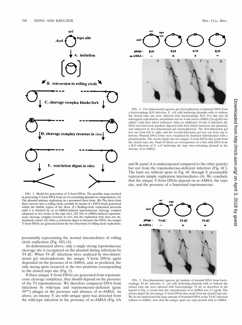
presumably representing the normal intermediates of rollingcircle replication (Fig. 4D) (4).
As demonstrated above, only a single strong topoisomerasecleavage site is recognized on the plasmid during infections byT4 dC. When T4 dC infections were analyzed by two-dimen-sional gel electrophoresis, the unique Y-form DNAs againdepended on the presence of m-AMSA, and, as predicted, theonly strong spots occurred at the two positions correspondingto the cloned topo site (Fig. 5).
If these unique Y-form DNAs are generated from topoisom-erase cleavage complexes, they should depend on the presenceof the T4 topoisomerase. We therefore compared DNA frominfections by wild-type and topoisomerase-deficient (gene39am) phages in the presence and absence of m-AMSA. Asabove, an intense Y arc with unique spots was detected fromthe wild-type infection in the presence of m-AMSA (Fig. 6A
and B; panel A is underexposed compared to the other panels)but not from the topoisomerase-deficient infection (Fig. 6C).The faint arc without spots in Fig. 6C through E presumablyrepresents simple replication intermediates (4). We concludethat the unique Y-form DNAs depend on m-AMSA, the toposite, and the presence of a functional topoisomerase.
FIG. 3. Model for generation of Y-form DNAs. The possible steps involvedin generating Y-form DNA from an ori-containing plasmid are diagrammed. (A)The plasmid initiates replication in a presumed theta form. (B) The theta formthen converts into a rolling circle, possibly by means of a DNA break generatedwithin the bubble region of the theta. (C) Rolling-circle replication proceedsuntil it is blocked by an m-AMSA-induced topoisomerase cleavage complex(depicted as two circles at the topo site). (D) The m-AMSA-induced topoisom-erase cleavage complex reverses in vivo, but the replication fork does not im-mediately restart. (E) After a restriction digest to linearize the DNA, two uniqueY-form DNAs are generated from the two directions of rolling-circle replication.
FIG. 4. Two-dimensional agarose gel electrophoresis of plasmid DNA froma bacteriophage K10 infection. E. coli cells harboring plasmids with or withoutthe cloned topo site were infected with bacteriophage K10. For this and allsubsequent experiments, attachment was for 4 min and m-AMSA (10 mg/ml) wasadded 2 min later where indicated. After an additional 18 min of infection, theDNA was harvested, purified, digested with AseI (which linearizes the plasmid),and subjected to two-dimensional gel electrophoresis. The first-dimension gelwas run from left to right, and the second-dimension gel was run from top tobottom. Plasmid DNA forms were visualized by Southern hybridization with aplasmid probe. The arrows depict the two unique Y-form DNAs that result fromthe cloned topo site. Panel D shows an overexposure of a blot with DNA froma K10 infection of E. coli harboring the topo site-containing plasmid in theabsence of m-AMSA.
FIG. 5. Two-dimensional agarose gel analysis of plasmid DNA from bacte-riophage T4 dC infection. E. coli cells harboring plasmids with or without thecloned topo site were infected with bacteriophage T4 dC as described in thelegend to Fig. 4, except that the concentration of m-AMSA was 2.5 mg/ml. Thearrows depict the two unique Y-form DNAs that result from the cloned topo site.We do not understand the large amount of branched DNA in the T4 dC infectionwithout m-AMSA; note that the unique spots are only present with m-AMSA.
598 HONG AND KREUZER MOL. CELL. BIOL.
on April 3, 2018 by guest
http://mcb.asm
.org/D
ownloaded from

Replication is needed for generation of Y-form DNAs. Wenext asked whether the cloned ori is also important in thegeneration of the unique Y-form DNAs during infection byeither T4 K10 or T4 dC. In both infections, only the ori-containing plasmids produced the intense Y-arc with strongspots (Fig. 7A, C, and E), consistent with the model that theserepresent blocked replication forks.
We also asked whether the DNA within the spots on the Yarc had been replicated by treating DNA from the K10 infec-tions with HaeIII, which cleaves unreplicated DNA into smallpieces (see above). The pattern was unchanged (compare Fig.7A and C), and therefore the branched Y-form molecules musthave been replicated throughout the length of all three arms.The origin dependence and the replicated status of the Y-formDNA strongly support the conclusion that the Y-form DNAsarise from blocked replication forks.
Why are Y-form DNAs not cleaved at the topo site? If theunique Y-form DNAs arise from replication forks that wereblocked at a cleavage complex, why is the Y form intact? Theinfected cells were lysed in the presence of SDS, which shoulddisrupt any topoisomerase cleavage complex and reveal thestaggered double-stranded break. One explanation is that thetopoisomerase resealed the cleaved DNA within the cleavagecomplex before cell lysis and yet the replication fork did notrestart (Fig. 3D) (see Discussion). A different explanation isthat the topoisomerase resealed the cleaved DNA during celllysis in some fraction of the Y molecules, in spite of the pres-ence of SDS. In this case, perhaps a large proportion of Ymolecules are lost during extraction due to SDS-induced DNAcleavage, and a large increase in the amount of Y-form DNAwould be obtained if we could prevent this SDS-induced cleav-age. To explore these two possibilities, we set up cell lysis
FIG. 6. Requirement for T4 topoisomerase. E. coli cells harboring the plasmid with both the ori and the topo site were infected with either K10 (wild type) orK10-39am (topoisomerase-deficient mutant) in the presence or absence of m-AMSA. DNA was purified, digested with AseI, and subjected to two-dimensional gelanalysis. Panel A is a lighter exposure of panel B, while panels B to E are matched exposures.
FIG. 7. Requirement for T4 origin and DNA replication. E. coli cells harboring plasmids with the topo site, either with or without the ori, were infected with eitherK10 (A through D) or T4 dC (E and F). DNA samples were digested with AseI, in either the presence or absence of HaeIII, and subjected to two-dimensional gelelectrophoresis.
VOL. 20, 2000 CLEAVAGE COMPLEX BLOCKS A REPLICATION FORK IN VIVO 599
on April 3, 2018 by guest
http://mcb.asm
.org/D
ownloaded from

conditions which would disfavor SDS-induced cleavage butfavor resealing of the DNA within the cleavage complex.
Purified topoisomerase can reseal broken DNA within thecleavage complex in vitro upon dilution of drug, brief heatingto 65°C, or treatment with EDTA and/or high salt (a phenom-enon known as reversal) (29, 54, 57, 63). In addition, at leastupon brief heat treatment at 65°C, cleavage complexes canreverse on chromosomal DNA within mammalian cells (30).
We tested reversal conditions with purified T4 topoisomer-ase in vitro and found that a large fraction of cleavage com-plexes could be reversed to intact plasmid DNA by treatmentwith Triton X-100 detergent (1%) at 65°C (data not shown).We next compared both DNA cleavage and production ofY-form DNA when m-AMSA-treated, infected cells were lysedeither under these reversal conditions or under the standardconditions that should not favor resealing. About 80% of thetotal cleavage complexes underwent reversal, demonstratingthat the reversal conditions worked well for in vivo samples(Fig. 8A). However, the unique Y-form DNAs appeared insimilar amounts in the two samples (compare Fig. 8B and C).These results argue that intact Y-form DNA is not created byresealing during the cell lysis procedure. Rather, we believethat topoisomerase resealing happens in vivo prior to cell lysis(see Discussion).
Y-form DNAs are not dependent on recombinational repair.One concern about our interpretation of these results is that, intheory, Y-form DNAs could also arise from recombination.The m-AMSA-induced topoisomerase cleavage complex orsome derivative thereof is repaired by a recombinational mech-anism (see the introduction), which could perhaps lead toY-form intermediates with the branches near topoisomerasecleavage sites.
To ask whether the Y-form DNAs are recombination inter-mediates, we analyzed a T4 K10-46/uvsX double mutant, whichis strongly deficient in recombination and recombinational re-pair. Since the majority of T4 DNA replication occurs througha recombination-dependent mode and therefore relies on re-
combination proteins gp46 and UvsX, we began by comparingthe extent of plasmid replication as a function of the m-AMSAdose in the wild type (K10) and the recombination mutant(K10-46/uvsX). Particularly at the higher levels of drug, therecombination mutant produced much less replicated plasmidDNA (Fig. 9) (see Discussion). The ratio of intact linear DNAto topoisomerase-cleaved DNA was also lower for the recom-bination mutant than for the wild-type as the m-AMSA doseincreased.
For the analysis of Y-form DNA above, our standard level ofm-AMSA was 10 mg/ml (equivalent to lane 6 in Fig. 9). How-ever, for comparisons of K10 and K10-46/uvsX infections, thislevel is problematic for the following reasons. First, there wasvery little replicated plasmid DNA at the higher drug level inthe recombination mutant infection. Second, the mutant infec-tion was probably more aberrant than the wild-type infection athigh levels of drug, because phage DNA replication was morestrongly inhibited (data not shown) (the stronger inhibition ofphage DNA replication would presumably lead to lower levelsof replication and recombination proteins). Third, a higherfraction of the replicated DNA was cleaved by topoisomeraseat the high concentration of m-AMSA in the mutant infection,and thus a larger proportion of the Y-form DNA would pre-sumably be destroyed due to topoisomerase cleavage com-plexes in one of the three arms (unrelated to the cleavagecomplex that led to branch formation). For these reasons, we
FIG. 8. Reversal of topoisomerase cleavage does not affect the production ofbranched DNA. E. coli cells harboring plasmids containing both the ori and thetopo site were infected with K10, treated with m-AMSA, and then lysed witheither reversal lysis buffer (Rev.) or cleavage lysis buffer (Cl.) as described inMaterials and Methods. The AseI-digested samples were analyzed on a one-dimensional gel (A) and on a two-dimensional gel (B and C). Note that most ofthe topoisomerase-mediated cleavage was reversed by the reversal conditions(A), yet the amount of unique branched DNA did not increase (B and C).
FIG. 9. Titration of m-AMSA in wild-type and recombination-deficient mu-tant infection. E. coli cells harboring the plasmid with both the ori and topo sitewere infected with either K10 or K10-46/uvsX in the presence of increasingconcentrations of m-AMSA (0, 1, 2, 3, 5, and 10 mg/ml in lanes 1 through 6,respectively). DNA was purified from the infected cells, digested with AseI andHaeIII, and subjected to agarose gel electrophoresis. The resulting fragmentswere visualized by Southern hybridization with pBR322 as the probe.
600 HONG AND KREUZER MOL. CELL. BIOL.
on April 3, 2018 by guest
http://mcb.asm
.org/D
ownloaded from

analyzed Y-form DNA by two-dimensional gel electrophoresisof DNA from infections with a relatively low drug level, 2mg/ml (equivalent to lane 3 in Fig. 9). When equal volumes ofDNA were compared, the recombination mutant infectionswere found to produce somewhat less Y-form DNA than thewild-type infections did (Fig. 10A and B). However, Y-formDNA cannot exist without unit-length plasmid DNA, and therecombination mutant infection has less replicated unit-lengthplasmid DNA even at this low drug level (see Fig. 9). It seemsmore fair to compare the ratios between the amount of Y-formDNA and replicated unit-length linear DNA. We thereforequantitated the replicated unit-length plasmid DNA andequalized this amount between the pair of infections by load-ing decreased amounts of the wild-type DNA sample. Whenthis equalization was applied, the amount of Y-form DNA wasunaffected by the recombination mutations (Fig. 10B and C).These results argue that the unique Y-form DNAs seen on thesimple Y arc are not recombination intermediates.
DISCUSSION
Type II topoisomerases such as bacterial DNA gyrase act infront of DNA replication forks to relieve the overwindingproduced from unwinding of the parental helix (42). Here weshowed that m-AMSA-induced cleavage complexes can arrestthe progression of T4 replication forks in vivo, consistent withthe placement of the type II topoisomerase in front of thereplication fork. Since DNA molecules containing the arrestedreplication forks were found to be intact, we infer that thereplication apparatus must have been inactivated upon en-countering the cleavage complex and that the topoisomerasesubsequently resealed the DNA breaks prior to cell lysis (Fig.3). Since the cytotoxicity of topoisomerase inhibitors is knownto involve DNA replication, these arrested replication forksmay become cytotoxic lesions if they are not restarted or re-paired.
Previous reports have shown that replication forks can beblocked by bound proteins, such as terminator protein binding
at ter sites or centromere protein-DNA complexes (1, 23, 26).The replication fork barrier in yeast or human rDNA is alsoprobably a protein-mediated blockage (9, 34, 43). In addition,two previous reports suggest that a drug-induced type II topo-isomerase cleavage complex can block a replication fork. Usingmammalian cells, Catapano et al. (11) measured a reduction inthe extent of replication downstream from a cleavage site butdid not directly analyze the state of DNA at the cleavage site.Using an in vitro E. coli replication system, Hiasa et al. (25)presented evidence that replication forks are blocked by quin-olone-induced topoisomerase IV cleavage complexes; they didnot attempt to map the position of the blocked fork withrespect to particular topoisomerase IV cleavage sites.
In this study, we detected an accumulation of unique Y-formDNA, appearing as spots along the simple Y arc, when m-AMSA, topoisomerase, and a topoisomerase cleavage sitewere all present. Several results argue strongly that the uniqueY-form DNA originates from replication forks blocked at to-poisomerase cleavage complexes. First, a T4 origin was re-quired on the plasmid to generate the unique Y-form DNAmolecules. Second, the unique Y-form DNA spots persistedafter HaeIII digestion, demonstrating that all three arms of thebranched DNA were replicated during the T4 infection. Third,deletion of the strong topo site led to the disappearance of twospots (one for each direction of replication). Fourth, the spotswere present in the analysis of DNA from a recombination-deficient infection, arguing against the possibility that recom-bination generates the unique Y-form DNA.
A somewhat surprising aspect of our results is that the Y-form DNA molecules are not actually cleaved at the topo site.Instead, the results indicate that the topoisomerase-mediatedDNA cleavages are resealed after fork blockage but before celllysis. There was no noticeable increase in the amounts ofunique Y-form DNAs when the cells were lysed under condi-tions that favored resealing of the enzyme-mediated breaks. Invivo resealing is a reasonable possibility because drug-inducedcleavage complex formation is an equilibrium process ratherthan an absolute trapping of the cleaved intermediate. If theresealing event happens in vivo, why do the Y-form DNAs notdisappear due to the restart of the replication fork? Perhapsthe collision between the replication fork and the topoisomer-ase cleavage complex leads to disassembly of one or morecomponents of the replication complex such that restart isdelayed or prevented.
The above model assumes that the fork-blocking lesion atthe topo site is a cleavage complex complete with topoisomer-ase-bound DNA breaks. A different interpretation is that m-AMSA stabilizes another type of topoisomerase-DNA com-plex that does not contain any DNA breaks. To our knowledge,no one has detected the induction of stable complexes withoutDNA breaks by this general class of topoisomerase inhibitors.Furthermore, in their in vitro study, Hiasa et al. (25) found thata mutant topoisomerase IV, which binds DNA but cannotcleave, does not block replication in the presence of inhibitor.
The topoisomerase cleavage complex, or some derivativethereof, can be repaired by a recombinational mechanism (seethe introduction). During phage T4 infection, this repair de-pends on the products of the 46 and uvsX genes (50). Sincerecombination could, in principle, produce Y-form DNA in-termediates, we tested the involvement of gp46 and UvsX inthe production of the Y-form DNA. When we equalized thereplicated monomer fragments between the wild-type and 46/uvsX infections, we found a comparable intensity of the Y-formDNAs. This result argues strongly that the Y-form DNAs donot arise from recombination and is consistent with the stalled-fork model presented above. Nevertheless, there was a fairly
FIG. 10. Branched DNA in the recombination-deficient mutant infection. E.coli cells harboring the plasmid with both the ori and the topo site were infectedwith either K10 or K10-46/uvsX in the presence of m-AMSA at 2 mg/ml. Equalvolumes of DNA (50 ml) were compared in panels A and B. To allow a com-parison of wild-type and mutant infections at similar levels of intact replicatedmonomer plasmid, samples from each infection were equalized by changing thevolume of the K10 infection DNA that was digested with AseI. Equalization wascalculated after quantitating the unit-length linear fragments in Fig. 9 (lane 3) byusing an Ambis direct radioisotope imaging system. Note that the unit-lengthlinear spots are approximately the same intensity in panels B and C.
VOL. 20, 2000 CLEAVAGE COMPLEX BLOCKS A REPLICATION FORK IN VIVO 601
on April 3, 2018 by guest
http://mcb.asm
.org/D
ownloaded from

dramatic difference in the level of plasmid replication betweenthe recombination mutant and wild-type infections, particu-larly as the concentration of m-AMSA increased (Fig. 9). Thisresult suggests that the continued replication of the ori-con-taining plasmid in the presence of m-AMSA depends on therecombinational repair system that is blocked in the 46/uvsXinfection. In other words, we propose that these recombinationproteins assist the restart of replication after the blockageevent. One plausible scenario is that the Y-form DNA is con-verted into an overt DNA break and that the DNA break (aftera strand invasion reaction) serves to initiate a new round ofrecombination-dependent plasmid replication. Previous stud-ies demonstrated that recombinational repair of endonuclease-generated breaks involves extensive DNA replication triggeredfrom the break (22, 49).
Several alternatives for processing topoisomerase cleavagecomplexes have been suggested from in vitro experiments,although none have yet been shown to be important in vivo.Howard et al. (29) showed that a helicase can disrupt a drug-induced cleavage complex, generating overt DNA breaks, andit is conceivable that a complete replication complex can do thesame. Yang et al. (68) purified a yeast enzyme that is able tocleave the phosphotyrosine bond between a type I topoisom-erase and DNA, generating a DNA nick. If such an enzyme isinvolved in processing cleavage complexes in vivo, the resultingnicks could be resealed by DNA ligase. If a similar enzyme actson type II topoisomerase cleavage complexes, a double-strandbreak would probably result. Sastry and Ross (58) detected anapparently similar phosphodiesterase from human cells andalso presented some evidence for a nuclease that creates dou-ble-strand breaks flanking topoisomerase cleavage complexes.Any of these processes could play important roles in the cyto-toxicity and/or repair of topoisomerase-mediated DNA dam-age.
Based on the results presented here and prior work of othergroups described above, we suggest that blocked replicationforks might be the most important lesion in cytotoxicity andDNA repair of topoisomerase-mediated damage. Understand-ing the repair pathway could improve chemotherapy, perhapsleading to compounds that inhibit repair and thereby potenti-ate the topoisomerase inhibitors.
In a broader sense, the process that we are analyzing at thetopoisomerase cleavage sites may occur with other forms ofDNA damage. Our use of topoisomerase cleavage complexesprovided a unique opportunity to analyze DNA damage in asite-specific manner in vivo. Various studies argue that repli-cation forks are blocked by other forms of DNA damage, forexample pyrimidine dimers from UV treatment (5, 64). Inaddition, E. coli chromosomal forks can be artificially stalled bya replicative helicase defect, perhaps mimicking DNA (or rep-lication protein) damage (7, 41, 47, 60). Blocked forks, or somederivative thereof, may be generally cytotoxic unless repaired.Our results may also be relevant in considering the process ofreplication fork blockage in normal cells without overt DNAdamage, which has recently become a topic of great interest. Avariety of indirect results argue that many, perhaps most, E.coli replication forks initiated at oriC under normal growthconditions become blocked and need to be restarted by arecombinational process to complete replication (14). Further-more, replication fork blockage has recently been implicated inaging: elimination of the Fob1p replication block protein pro-longs the life span of yeast mother cells by preventing forkblockage (and subsequent events) in the rDNA (16). Finally,the helicases defective in the premature-aging disease Wern-er’s syndrome and the cancer predisposition disease Bloom’ssyndrome have been proposed to play a role in recognizing or
correcting aberrant replication structures such as blocked forks(12).
ACKNOWLEDGMENTS
We thank Michael Been, Jeffrey Dawson, Mariano Garcia-Blanco,Tao-shih Hsieh, and David Pickup for insightful discussions.
This work was supported by research grant CA60836 from NationalInstitutes of Health/National Cancer Institute, and George Hong wassupported in part by National Research Science Award 5 T32CA09111.
REFERENCES
1. Bastia, D., and B. Mohanty. 1996. Mechanisms for completing replication, p.177–215. In M. DePamphilis (ed.), DNA replication in eukaryotic cells. ColdSpring Harbor Press, Cold Spring Harbor, N.Y.
2. Belanger, K. G. 1997. Origin-dependent DNA replication in bacteriophageT4. Ph.D. thesis. Duke University, Durham, N.C.
3. Belanger, K. G., and K. N. Kreuzer. 1998. Bacteriophage T4 initiates bidi-rectional DNA replication through a two-step process. Mol. Cell 2:693–701.
4. Belanger, K. G., C. Mirzayan, H. E. Kreuzer, B. M. Alberts, and K. N.Kreuzer. 1996. Two-dimensional gel analysis of rolling circle replication inthe presence and absence of bacteriophage T4 primase. Nucleic Acids Res.24:2166–2175.
5. Berger, C. A., and H. J. Edenberg. 1996. Pyrimidine dimers block simianvirus 40 replication forks. Mol. Cell. Biol. 6:3443–3450.
6. Berger, J. M. 1998. Type II DNA topoisomerases. Curr. Opin. Struct. Biol.8:26–32.
7. Bierne, H., and B. Michel. 1994. When replication forks stop. Mol. Micro-biol. 13:17–23.
8. Brewer, B. J., and W. L. Fangman. 1987. The localization of replicationorigins on ARS plasmids in S. cerevisiae. Cell 51:463–471.
9. Brewer, B. J., D. Lockshon, and W. L. Fangman. 1992. The arrest of repli-cation forks in the rDNA of yeast occurs independently of transcription. Cell71:267–276.
10. Burden, D. A., and N. Osheroff. 1998. Mechanism of action of eukaryotictopoisomerase II and drugs targeted to the enzyme. Biochim. Biophys. Acta1400:139–154.
11. Catapano, C. V., G. M. R. Carbone, F. Pisani, J. Qiu, and D. J. Fernandes.1997. Arrest of replication fork progression at sites of topoisomerase II-mediated DNA cleavage in human leukemia CEM cells incubated withVM-26. Biochemistry 36:5739–5748.
12. Chakraverty, R. K., and I. D. Hickson. 1999. Defending genome integrityduring DNA replication: a proposed role for RecQ family helicases. Bioes-says 21:286–294.
13. Chen, A. Y., and L. F. Liu. 1994. DNA topoisomerases: essential enzymesand lethal targets. Annu. Rev. Pharmacol. Toxicol. 34:191–218.
14. Cox, M. M. 1999. Recombinational DNA repair in bacteria and the RecAprotein. Nucleic Acids Res. Mol. Biol. 63:310–366.
15. D’Arpa, P., C. Beardmore, and L. F. Liu. 1990. Involvement of nucleic acidsynthesis in cell killing mechanisms of topoisomerase poisons. Cancer Res.50:6919–6924.
16. Defossez, P. A., R. Prusty, M. Kaeberlein, S. J. Lin, P. Ferrigno, P. A. Silver,R. L. Keil, and L. Guarente. 1999. Elimination of replication block proteinFob1 extends the life span of yeast mother cells. Mol. Cell 3:447–455.
17. Drlica, K., and X. L. Zhao. 1997. DNA gyrase, topoisomerase IV, and the4-quinolones. Microbiol. Rev. 61:377–392.
18. Eng, W.-K., L. Faucette, R. K. Johnson, and R. Sternglanz. 1989. Evidencethat DNA topoisomerase I is necessary for the cytotoxic effects of campto-thecin. Mol. Pharmacol. 34:755–760.
19. Freudenreich, C. H., and K. N. Kreuzer. 1993. Mutational analysis of a typeII topoisomerase cleavage site: Distinct requirements for enzyme and inhib-itors. EMBO J. 12:2085–2097.
20. Freudenreich, C. H., and K. N. Kreuzer. 1994. Localization of an amino-acridine antitumor agent in a type II topoisomerase-DNA complex. Proc.Natl. Acad. Sci. USA 91:11007–11011.
21. Friedman, K. L., and B. J. Brewer. 1995. Analysis of replication intermedi-ates by two-dimensional agarose gel electrophoresis. Methods Enzymol.262:613–627.
22. George, J. W., and K. N. Kreuzer. 1996. Repair of double-strand breaks inbacteriophage T4 by a mechanism that involves extensive DNA replication.Genetics 143:1507–1520.
23. Greenfeder, S. A., and C. S. Newlon. 1992. Replication forks pause at yeastcentromeres. Mol. Cell. Biol. 12:4056–4066.
24. Hane, M. W., and T. H. Wood. 1969. Escherichia coli K-12 mutants resistantto nalidixic acid: genetic mapping and dominance studies. J. Bacteriol. 99:238–241.
25. Hiasa, H., D. O. Yousef, and K. J. Marians. 1996. DNA strand cleavage isrequired for replication fork arrest by a frozen topoisomerase-quinolone-DNA ternary complex. J. Biol. Chem. 271:26424–26429.
602 HONG AND KREUZER MOL. CELL. BIOL.
on April 3, 2018 by guest
http://mcb.asm
.org/D
ownloaded from

26. Hill, T. M. 1992. Arrest of bacterial DNA replication. Annu. Rev. Microbiol.46:603–633.
27. Holm, C., J. M. Covey, D. Kerrigan, and Y. Pommier. 1989. Differentialrequirement of DNA replication for the cytotoxicity of DNA topoisomeraseI and II inhibitors in Chinese hamster DC3F cells. Cancer Res. 49:6365–6368.
28. Hooper, D. C. 1998. Clinical applications of quinolones. Biochim. Biophys.Acta 1400:45–61.
29. Howard, M. T., S. H. Neece, S. W. Matson, and K. N. Kreuzer. 1994.Disruption of a topoisomerase-DNA cleavage complex by a DNA helicase.Proc. Natl. Acad. Sci. USA 91:12031–12035.
30. Hsiang, Y.-H., and L. F. Liu. 1989. Evidence for the reversibility of cellularDNA lesion induced by mammalian topoisomerase II poisons. J. Biol. Chem.264:9713–9715.
31. Huff, A. C., J. K. Leatherwood, and K. N. Kreuzer. 1989. Bacteriophage T4DNA topoisomerase is the target of antitumor agent 49-(9-acridinylamino)methanesulfon-m-anisidide (m-AMSA) in T4-infected Escherichia coli. Proc.Natl. Acad. Sci. USA 86:1307–1311.
32. Jeggo, P. A., K. Caldecott, S. Pidsley, and G. R. Banks. 1989. Sensitivity ofChinese hamster ovary mutants defective in DNA double strand break repairto topoisomerase II inhibitors. Cancer Res. 49:7057–7063.
33. Khodursky, A. B., and N. R. Cozzarelli. 1998. The mechanism of inhibitionof topoisomerase IV by quinolone antibacterials. J. Biol. Chem. 273:27668–27677.
34. Kobayashi, T., and T. Horiuchi. 1996. A yeast gene product, Fob1 protein,required for both replication fork blocking and recombinational hotspotactivities. Genes Cells 1:465–474.
35. Kreuzer, K. N., and B. M. Alberts. 1984. Site-specific recognition of bacte-riophage T4 DNA by T4 type II DNA topoisomerase and Escherichia coliDNA gyrase. J. Biol. Chem. 259:5339–5346.
36. Kreuzer, K. N., and N. R. Cozzarelli. 1979. Escherichia coli mutants ther-mosensitive for deoxyribonucleic acid gyrase subunit A: effects on deoxyri-bonucleic acid replication, transcription, and bacteriophage growth. J. Bac-teriol. 140:424–435.
37. Kreuzer, K. N., H. W. Engman, and W. Y. Yap. 1988. Tertiary initiation ofreplication in bacteriophage T4. Deletion of the overlapping uvsY promoter/replication origin from the phage genome. J. Biol. Chem. 263:11348–11357.
38. Kreuzer, K. N., and S. W. Morrical. 1994. Initiation of DNA replication, p.28–42. In J. D. Karam (ed.), Molecular biology of bacteriophage T4. ASMPress, Washington, D.C.
39. Kupfer, G., A. L. Bodley, and L. F. Liu. 1987. Involvement of intracellularATP in cytotoxicity of topoisomerase II-targeting antitumor drugs. NCIMonogr. 4:37–40.
40. Kutter, E., and L. Snyder. 1983. Preparation of cytosine-containing T4phage, p. 56–57. In C. K. Mathews, E. M. Kutter, G. Mosig, and P. B. Berget(ed.), Bacteriophage T4. ASM Press, Washington, D.C.
41. Kuzminov, A. 1995. Collapse and repair of replication forks in Escherichiacoli. Mol. Microbiol. 16:373–384.
42. Levine, C., H. Hiasa, and K. J. Marians. 1998. DNA gyrase and topoisom-erase IV: Biochemical activities, physiological roles during chromosomereplication, and drug sensitivities. Biochim. Biophys. Acta 1400:29–43.
43. Little, R. D., T. H. Platt, and C. L. Schildkraut. 1993. Initiation and termi-nation of DNA replication in human rRNA genes. Mol. Cell. Biol. 13:6600–6613.
44. Liu, L. F. 1989. DNA topoisomerase poisons as antitumor drugs. Annu. Rev.Biochem. 58:351–375.
45. McCarthy, D. 1979. Gyrase-dependent initiation of bacteriophage T4 DNAreplication: interactions of Escherichia coli gyrase with novobiocin, coumer-mycin and phage DNA-delay gene products. J. Mol. Biol. 127:265–283.
46. Menkens, A. E., and K. N. Kreuzer. 1988. Deletion analysis of bacteriophageT4 tertiary origins. J. Biol. Chem. 263:11358–11365.
47. Michel, B., S. D. Ehrlich, and M. Uzest. 1997. DNA double-strand breakscaused by replication arrest. EMBO J. 16:430–438.
48. Ming, Y. Z., X. Di, E. P. Gomez-Sanchez, and C. E. Gomez-Sanchez. 1994.Improved downward capillary transfer for blotting of DNA and RNA. Bio-Techniques 16:58–60.
49. Mueller, J. E., J. Clyman, Y. J. Huang, M. M. Parker, and M. Belfort. 1996.Intron mobility in phage T4 occurs in the context of recombination-depen-dent DNA replication by way of multiple pathways. Genes Dev. 10:351–364.
50. Neece, S. H., K. Carles-Kinch, D. J. Tomso, and K. N. Kreuzer. 1996. Roleof recombinational repair in sensitivity to an antitumor agent that inhibitsbacteriophage T4 type II DNA topoisomerase. Mol. Microbiol. 20:1145–1154.
51. Nelson, M. A., M. Ericson, L. Gold, and J. F. Pulitzer. 1982. The isolationand characterization of TabR bacteria: hosts that restrict bacteriophage T4rII mutants. Mol. Gen. Genet. 188:60–68.
52. Nitiss, J., and J. C. Wang. 1988. DNA topoisomerase-targeting antitumordrugs can be studied in yeast. Proc. Natl. Acad. Sci. USA 85:7501–7505.
53. Nitiss, J. L., and J. C. Wang. 1996. Mechanisms of cell killing by drugs thattrap covalent complexes between DNA topoisomerases and DNA. Mol.Pharmacol. 50:1095–1102.
54. Osheroff, N., and E. L. Zechiedrich. 1987. Calcium-promoted DNA cleavageby eukaryotic topoisomerase II: Trapping the covalent enzyme-DNA com-plex in an active form. Biochemistry 26:4303–4309.
55. Pommier, Y. 1993. DNA topoisomerase I and II in cancer chemotherapy:update and perspectives. Cancer Chemother. Pharmacol. 32:103–108.
56. Ripley, L. S., J. S. Dubins, J. G. deBoer, D. M. DeMarini, A. M. Bogerd, andK. N. Kreuzer. 1988. Hotspot sites for acridine-induced frameshift mutationsin bacteriophage T4 correspond to sites of action of the T4 type II topo-isomerase. J. Mol. Biol. 200:665–680.
57. Sander, M., and T.-S. Hsieh. 1983. Double strand DNA cleavage by type IIDNA topoisomerase from Drosophila melanogaster. J. Biol. Chem. 258:8421–8428.
58. Sastry, S., and B. M. Ross. 1998. Mechanisms for the processing of a frozentopoisomerase-DNA conjugate by human cell-free extracts. J. Biol. Chem.273:9942–9950.
59. Schneider, E., P. A. Lawson, and R. K. Ralph. 1989. Inhibition of proteinsynthesis reduces the cytotoxicity of 49-(9-acridinylamino)methanesulfon-m-anisidide without affecting DNA breakage and DNA topoisomerase II in amurine mastocytoma cell line. Biochem. Pharmacol. 38:263–269.
60. Seigneur, M., V. Bidnenko, S. D. Ehrlich, and B. Michel. 1998. RuvAB actsat arrested replication forks. Cell 95:419–430.
61. Selick, H. E., K. N. Kreuzer, and B. M. Alberts. 1988. The bacteriophage T4insertion/substitution vector system. A method for introducing site-specificmutations into the virus chromosome. J. Biol. Chem. 263:11336–11347.
62. Snustad, D. P., L. Snyder, and E. M. Kutter. 1983. Effects on host genomestructure and expression, p. 40–57. In C. K. Mathews, E. M. Kutter, G.Mosig, and P. B. Berget (ed.), Bacteriophage T4. ASM Press, Washington,D.C.
63. Tewey, K. M., G. L. Chen, E. M. Nelson, and L. F. Liu. 1984. Intercalativeantitumor drugs interfere with the breakage-reunion reaction of mammalianDNA topoisomerase II. J. Biol. Chem. 259:9182–9187.
64. Villani, G., S. Boiteux, and M. Radman. 1978. Mechanism of ultraviolet-induced mutagenesis: Extent and fidelity of in vitro DNA synthesis on irra-diated templates. Proc. Natl. Acad. Sci. USA 75:3037–3041.
65. Wang, J. C. 1996. DNA topoisomerases. Annu. Rev. Biochem. 65:635–692.66. Wigley, D. B. 1995. Structure and mechanism of DNA topoisomerases.
Annu. Rev. Biophys. Biomol. Struct. 24:185–208.67. Wilson, W. R., and G. F. Whitmore. 1981. Cell-cycle-stage specificity of
49-(9-acridinylamino)methanesulfon-m-anisidide (m-AMSA) and interac-tion with ionizing radiation in mammalian cell cultures. Radiat. Res. 87:121–136.
68. Yang, S. W., A. B. Burgin, Jr., B. N. Huizenga, C. A. Robertson, K. C. Yao,and H. A. Nash. 1996. A eukaryotic enzyme that can disjoin dead-endcovalent complexes between DNA and type I topoisomerases. Proc. Natl.Acad. Sci. USA 93:11534–11539.
VOL. 20, 2000 CLEAVAGE COMPLEX BLOCKS A REPLICATION FORK IN VIVO 603
on April 3, 2018 by guest
http://mcb.asm
.org/D
ownloaded from