Embed Size (px)
Citation preview
356
11
When most people hear the term cardio-vascular system, they immediately think of the heart. We have all felt our own
heart “pound” from time to time when we are ner-vous. The crucial importance of the heart has been recognized for ages. However, the cardiovascular system is much more than just the heart, and from a scientific and medical standpoint, it is important to understand why this system is so vital to life.
Night and day, minute after minute, our tril-lions of cells take up nutrients and excrete wastes. Although the pace of these exchanges slows dur-ing sleep, they must go on continuously: when they stop, we die. Cells can make such exchanges
only with the interstitial fluid in their immediate vicinity. Thus, some means of changing and “refreshing” these fluids is necessary to renew the nutrients and prevent pollution caused by the buildup of wastes. Like a bustling factory, the body must have a transportation system to carry its various “cargoes” back and forth. Instead of roads, railway tracks, and subways, the body’s delivery routes are its hollow blood vessels.
Most simply stated, the major function of the cardiovascular system is transportation. Using blood as the transport vehicle, the system carries oxygen, nutrients, cell wastes, hormones, and many other substances vital for body homeostasis to and from the cells. The force to move the blood
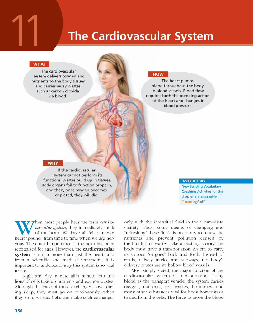
The Cardiovascular System
WHAT
HOW
WHY
The cardiovascular system delivers oxygen and
nutrients to the body tissues and carries away wastes such as carbon dioxide
via blood.
The heart pumps blood throughout the body in blood vessels. Blood flow
requires both the pumping action of the heart and changes in
blood pressure.
If the cardiovascular system cannot perform its
functions, wastes build up in tissues. Body organs fail to function properly,
and then, once oxygen becomes depleted, they will die.
INSTRUCTORS
New Building Vocabulary Coaching Activities for this chapter are assignable in
Chapter 11: The Cardiovascular System 357
11
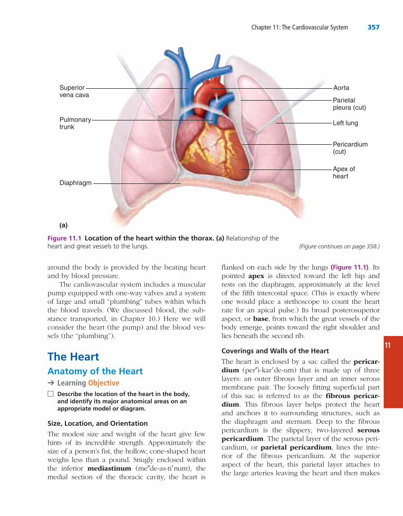
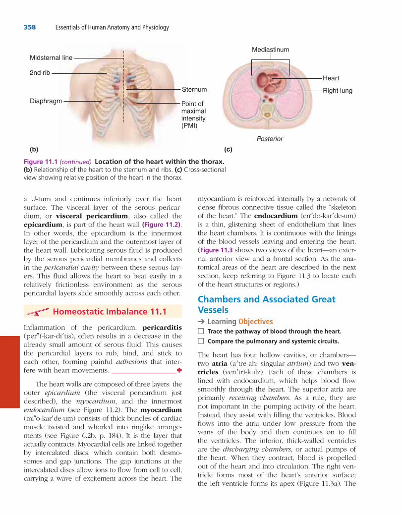
flanked on each side by the lungs (Figure 11.1). Its pointed apex is directed toward the left hip and rests on the diaphragm, approximately at the level of the fifth intercostal space. (This is exactly where one would place a stethoscope to count the heart rate for an apical pulse.) Its broad posterosuperior aspect, or base, from which the great vessels of the body emerge, points toward the right shoulder and lies beneath the second rib.
Coverings and Walls of the Heart
The heart is enclosed by a sac called the pericar-dium (per″i-kar′de-um) that is made up of three layers: an outer fibrous layer and an inner serous membrane pair. The loosely fitting superficial part of this sac is referred to as the fibrous pericar-dium. This fibrous layer helps protect the heart and anchors it to surrounding structures, such as the diaphragm and sternum. Deep to the fibrous pericardium is the slippery, two-layered serous pericardium. The parietal layer of the serous peri-cardium, or parietal pericardium, lines the inte-rior of the fibrous pericardium. At the superior aspect of the heart, this parietal layer attaches to the large arteries leaving the heart and then makes
around the body is provided by the beating heart and by blood pressure.
The cardiovascular system includes a muscular pump equipped with one-way valves and a system of large and small “plumbing” tubes within which the blood travels. (We discussed blood, the sub-stance transported, in Chapter 10.) Here we will consider the heart (the pump) and the blood ves-sels (the “plumbing”).
The HeartAnatomy of the Heart➔ Learning Objective
□ Describe the location of the heart in the body,
appropriate model or diagram.
Size, Location, and Orientation
The modest size and weight of the heart give few hints of its incredible strength. Approximately the size of a person’s fist, the hollow, cone-shaped heart weighs less than a pound. Snugly enclosed within the inferior mediastinum (me″de-as-ti′num), the medial section of the thoracic cavity, the heart is
(a)
Superiorvena cava
Left lung
Aorta
Parietalpleura (cut)
Pericardium(cut)
Pulmonarytrunk
Diaphragm
Apex ofheart
Figure 11.1 Location of the heart within the thorax. (a) Relationship of the (Figure continues on page 358.)
358 Essentials of Human Anatomy and Physiology
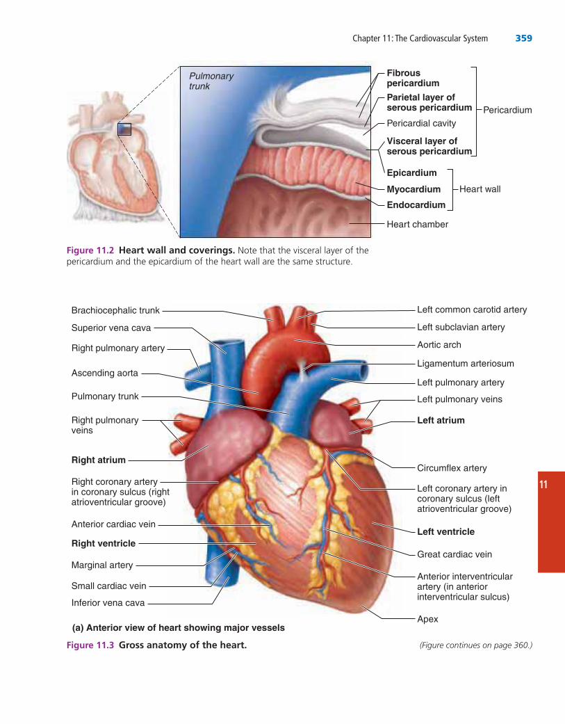
myocardium is reinforced internally by a network of dense fibrous connective tissue called the “skeleton of the heart.” The endocardium (en″do-kar′de-um) is a thin, glistening sheet of endothelium that lines the heart chambers. It is continuous with the linings of the blood vessels leaving and entering the heart. (Figure 11.3 shows two views of the heart—an exter-nal anterior view and a frontal section. As the ana-tomical areas of the heart are described in the next section, keep referring to Figure 11.3 to locate each of the heart structures or regions.)
Chambers and Associated Great Vessels➔ Learning Objectives
□ Trace the pathway of blood through the heart.
□ Compare the pulmonary and systemic circuits.
The heart has four hollow cavities, or chambers—two atria (a′tre-ah; singular atrium) and two ven-tricles (ven′trı-kulz). Each of these chambers is lined with endocardium, which helps blood flow smoothly through the heart. The superior atria are primarily receiving chambers. As a rule, they are not important in the pumping activity of the heart. Instead, they assist with filling the ventricles. Blood flows into the atria under low pressure from the veins of the body and then continues on to fill the ventricles. The inferior, thick-walled ventricles are the discharging chambers, or actual pumps of the heart. When they contract, blood is propelled out of the heart and into circulation. The right ven-tricle forms most of the heart’s anterior surface; the left ventricle forms its apex (Figure 11.3a). The
a U-turn and continues inferiorly over the heart surface. The visceral layer of the serous pericar-dium, or visceral pericardium, also called the epicardium, is part of the heart wall (Figure 11.2). In other words, the epicardium is the innermost layer of the pericardium and the outermost layer of the heart wall. Lubricating serous fluid is produced by the serous pericardial membranes and collects in the pericardial cavity between these serous lay-ers. This fluid allows the heart to beat easily in a relatively frictionless environment as the serous pericardial layers slide smoothly across each other.
Homeostatic Imbalance 11.1
Inflammation of the pericardium, pericarditis (per″ ı-kar-di′tis), often results in a decrease in the already small amount of serous fluid. This causes the pericardial layers to rub, bind, and stick to each other, forming painful adhesions that inter-fere with heart movements. ___________________✚
The heart walls are composed of three layers: the outer epicardium (the visceral pericardium just described), the myocardium, and the innermost endocardium (see Figure 11.2). The myocardium (mi″o-kar′de-um) consists of thick bundles of cardiac muscle twisted and whorled into ringlike arrange-ments (see Figure 6.2b, p. 184). It is the layer that actually contracts. Myocardial cells are linked together by intercalated discs, which contain both desmo-somes and gap junctions. The gap junctions at the intercalated discs allow ions to flow from cell to cell, carrying a wave of excitement across the heart. The
Heart
Posterior
Right lung
Point ofmaximalintensity(PMI)
Diaphragm
(b) (c)
Sternum
2nd rib
Midsternal lineMediastinum
Figure 11.1 (continued) Location of the heart within the thorax. (b) (c) Cross-sectional
Chapter 11: The Cardiovascular System 359
11
Left subclavian artery
Aortic arch
Left common carotid artery
Ligamentum arteriosum
Left pulmonary veins
Left atrium
Circumflex artery
Left coronary artery in coronary sulcus (left atrioventricular groove)
Left ventricle
Great cardiac vein
Left pulmonary artery
Apex
Anterior interventricularartery (in anterior interventricular sulcus)
(a) Anterior view of heart showing major vessels
Superior vena cava
Right pulmonary artery
Brachiocephalic trunk
Ascending aorta
Pulmonary trunk
Right pulmonary veins
Right atrium
Right coronary arteryin coronary sulcus (right atrioventricular groove)
Anterior cardiac vein
Right ventricle
Marginal artery
Inferior vena cava
Small cardiac vein
Fibrouspericardium
Parietal layer ofserous pericardium
Pericardial cavity
Visceral layer ofserous pericardium
Epicardium
Myocardium
Endocardium
Pulmonarytrunk
Heart chamber
Heart wall
Pericardium
Figure 11.2 Heart wall and coverings.
Figure 11.3 Gross anatomy of the heart. (Figure continues on page 360.)
360 Essentials of Human Anatomy and Physiology
(oxygen enters the blood and carbon dioxide enters the lungs) and then return it to the heart.
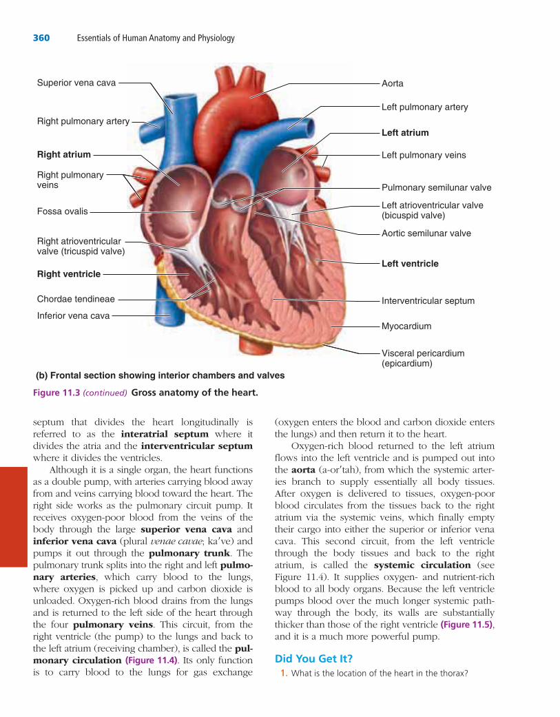
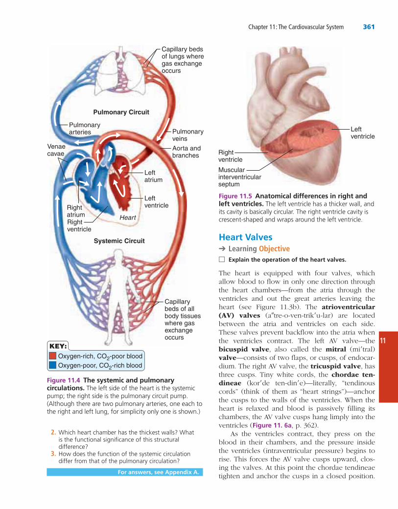
Oxygen-rich blood returned to the left atrium flows into the left ventricle and is pumped out into the aorta (a-or′tah), from which the systemic arter-ies branch to supply essentially all body tissues. After oxygen is delivered to tissues, oxygen-poor blood circulates from the tissues back to the right atrium via the systemic veins, which finally empty their cargo into either the superior or inferior vena cava. This second circuit, from the left ventricle through the body tissues and back to the right atrium, is called the systemic circulation (see Figure 11.4). It supplies oxygen- and nutrient-rich blood to all body organs. Because the left ventricle pumps blood over the much longer systemic path-way through the body, its walls are substantially thicker than those of the right ventricle (Figure 11.5), and it is a much more powerful pump.
Did You Get It?1.
septum that divides the heart longitudinally is referred to as the interatrial septum where it divides the atria and the interventricular septum where it divides the ventricles.
Although it is a single organ, the heart functions as a double pump, with arteries carrying blood away from and veins carrying blood toward the heart. The right side works as the pulmonary circuit pump. It receives oxygen-poor blood from the veins of the body through the large superior vena cava and inferior vena cava (plural venae cavae; ka′ve) and pumps it out through the pulmonary trunk. The pulmonary trunk splits into the right and left pulmo-nary arteries, which carry blood to the lungs, where oxygen is picked up and carbon dioxide is unloaded. Oxygen-rich blood drains from the lungs and is returned to the left side of the heart through the four pulmonary veins. This circuit, from the right ventricle (the pump) to the lungs and back to the left atrium (receiving chamber), is called the pul-monary circulation (Figure 11.4). Its only function is to carry blood to the lungs for gas exchange
Left pulmonary artery
Left atrium
Aorta
Left pulmonary veins
Left atrioventricular valve(bicuspid valve)
Aortic semilunar valve
Left ventricle
Interventricular septum
Pulmonary semilunar valve
Visceral pericardium(epicardium)
Myocardium
(b) Frontal section showing interior chambers and valves
Right pulmonary artery
Superior vena cava
Right atrium
Right pulmonary veins
Fossa ovalis
Right atrioventricularvalve (tricuspid valve)
Right ventricle
Chordae tendineae
Inferior vena cava
Figure 11.3 (continued) Gross anatomy of the heart.
Chapter 11: The Cardiovascular System 361
11
Heart Valves➔ Learning Objective
□ Explain the operation of the heart valves.
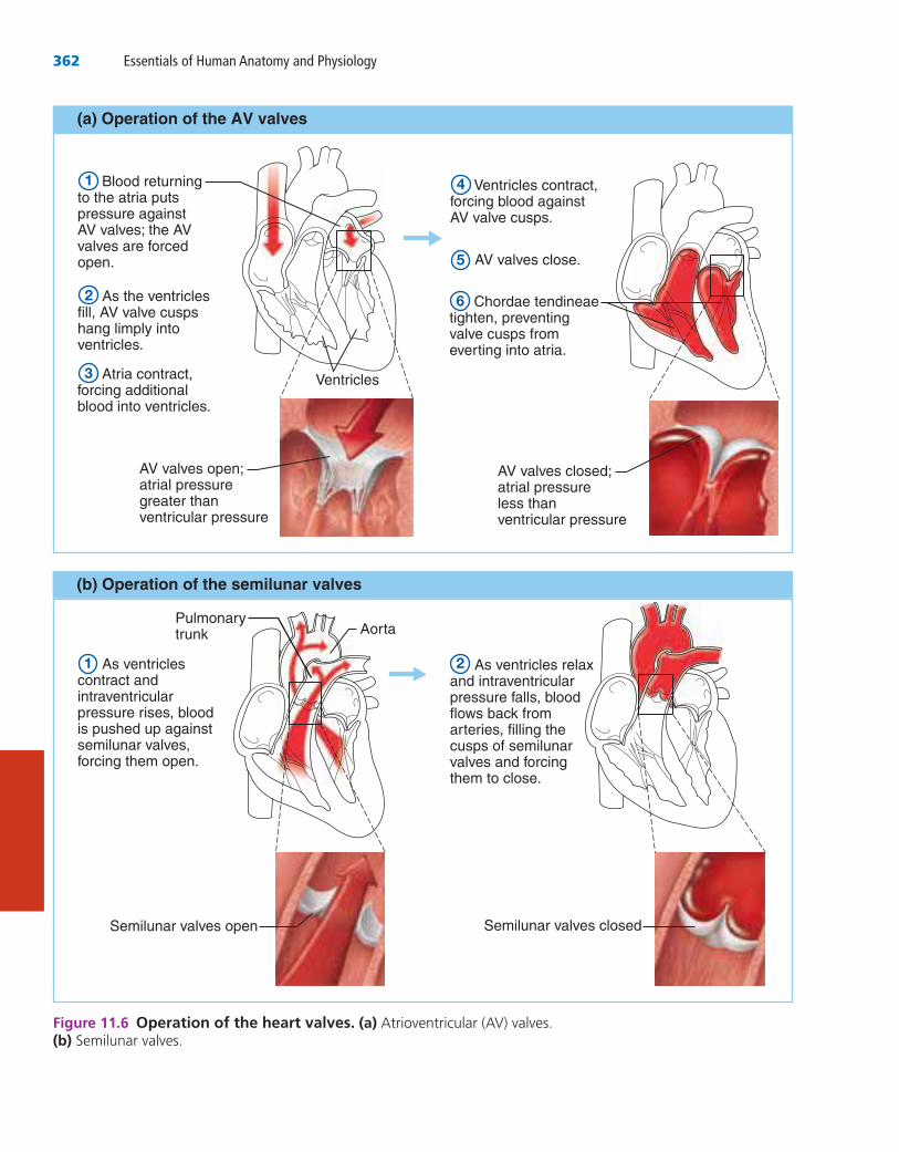
The heart is equipped with four valves, which allow blood to flow in only one direction through the heart chambers—from the atria through the ventricles and out the great arteries leaving the heart (see Figure 11.3b). The atrioventricular (AV) valves (a″tre-o-ven-trik′u-lar) are located between the atria and ventricles on each side. These valves prevent backflow into the atria when the ventricles contract. The left AV valve—the bicuspid valve, also called the mitral (mi′tral) valve—consists of two flaps, or cusps, of endocar-dium. The right AV valve, the tricuspid valve, has three cusps. Tiny white cords, the chordae ten-dineae (kor′de ten-din′e)—literally, “tendinous cords” (think of them as “heart strings”)—anchor the cusps to the walls of the ventricles. When the heart is relaxed and blood is passively filling its chambers, the AV valve cusps hang limply into the ventricles (Figure 11. 6a, p. 362).
As the ventricles contract, they press on the blood in their chambers, and the pressure inside the ventricles (intraventricular pressure) begins to rise. This forces the AV valve cusps upward, clos-ing the valves. At this point the chordae tendineae tighten and anchor the cusps in a closed position.
2.
3.
For answers, see Appendix A.
Oxygen-rich, CO2-poor blood Oxygen-poor, CO2-rich blood
Capillary bedsof lungs wheregas exchangeoccurs
Capillarybeds of allbody tissueswhere gasexchangeoccurs
Pulmonaryveins
Pulmonaryarteries
Pulmonary Circuit
Systemic Circuit
Aorta and branches
Leftatrium
Heart
LeftventricleRight
atriumRightventricle
Venaecavae
KEY:
Figure 11.4 The systemic and pulmonary circulations.
Rightventricle
Leftventricle
Muscularinterventricularseptum
Figure 11.5 Anatomical differences in right and left ventricles.
362 Essentials of Human Anatomy and Physiology
Ventricles contract,forcing blood against AV valve cusps.
AV valves close.
Chordae tendineaetighten, preventingvalve cusps fromeverting into atria.
Blood returning to the atria puts pressure againstAV valves; the AVvalves are forcedopen.
As the ventriclesfill, AV valve cuspshang limply intoventricles.
Atria contract,forcing additionalblood into ventricles.
Ventricles
AV valves open;atrial pressuregreater thanventricular pressure
AV valves closed;atrial pressureless thanventricular pressure
As ventricles contract and intraventricularpressure rises, bloodis pushed up againstsemilunar valves,forcing them open.
AortaPulmonarytrunk
Semilunar valves open Semilunar valves closed
As ventricles relaxand intraventricularpressure falls, bloodflows back fromarteries, filling thecusps of semilunarvalves and forcingthem to close.
(a) Operation of the AV valves
(b) Operation of the semilunar valves
1
2
3
4
5
6
1 2
Figure 11.6 Operation of the heart valves. (a)(b)
Chapter 11: The Cardiovascular System 363
11
_____________________________________________✚
Cardiac Circulation
➔ Learning Objective □ Name the functional blood supply of the heart.
Although the heart chambers are bathed with blood almost continuously, the blood contained in the heart does not nourish the myocardium. The func-tional blood supply that oxygenates and nourishes the myocardium is provided by the right and left coronary arteries. The coronary arteries branch from the base of the aorta and encircle the heart in the coronary sulcus (atrioventricular groove) at the junction of the atria and ventricles (see Figure 11.3a). The coronary arteries and their major branches (the anterior interventricular artery and circumflex artery on the left, and the poste-rior interventricular artery and marginal artery on the right) are compressed (flow is inhibited, not stopped completely) when the ventricles are con-tracting and fill when the heart is relaxed. The myo-cardium is drained by several cardiac veins, which empty into an enlarged vessel on the posterior of the heart called the coronary sinus. The coronary sinus, in turn, empties into the right atrium.
Homeostatic Imbalance 11.3
When the heart beats at a very rapid rate, the myocardium may receive an inadequate blood supply because the relaxation periods (when the blood is able to flow to the heart tissue) are short-ened. Situations in which the myocardium is deprived of oxygen often result in crushing chest pain called angina pectoris (an-ji′nah pek′tor-is). This pain is a warning that should never be ignored, because if angina is prolonged, the oxygen- deprived heart cells may die, forming an area called an infarct. The resulting myocardial infarction (in-fark′shun), or MI, is commonly called a “heart attack” or a “coronary.” _________✚
If the cusps were unanchored, they would blow upward into the atria like an umbrella being turned inside out by a gusty wind. In this manner, the AV valves prevent backflow into the atria when the ventricles are contracting.
The second set of valves, the semilunar (sem″ ı-lu′nar) valves, guards the bases of the two large arteries leaving the ventricular chambers. Thus, they are known as the pulmonary semilu-nar valve and aortic semilunar valve (see Figure 11.3b). Each semilunar valve has three cusps that fit tightly together when the valves are closed. When the ventricles are contracting and forcing blood out of the heart, the cusps are forced open and flattened against the walls of the arteries by the tremendous force of rushing blood (Figure 11.6b). Then, when the ventricles relax, the blood begins to flow backward toward the heart, and the cusps fill with blood like a para-chute filling with air, closing the valves. This pre-vents arterial blood from reentering the heart.
Each set of valves operates at a different time. The AV valves are open during heart relaxation and closed when the ventricles are contracting. The semilunar valves are closed during heart relaxation and are forced open when the ventri-cles contract. The valves force blood to continually move forward through the heart by opening and closing in response to pressure changes in the heart.
Homeostatic Imbalance 11.2
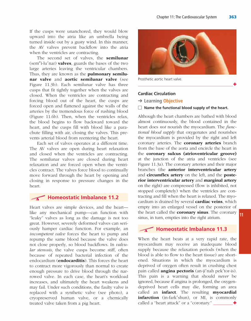
Heart valves are simple devices, and the heart—like any mechanical pump—can function with “leaky” valves as long as the damage is not too great. However, severely deformed valves can seri-ously hamper cardiac function. For example, an incompetent valve forces the heart to pump and repump the same blood because the valve does not close properly, so blood backflows. In valvu-lar stenosis, the valve cusps become stiff, often because of repeated bacterial infection of the endocardium (endocarditis). This forces the heart to contract more vigorously than normal to create enough pressure to drive blood through the nar-rowed valve. In each case, the heart’s workload increases, and ultimately the heart weakens and may fail. Under such conditions, the faulty valve is replaced with a synthetic valve (see photo), a cryopreserved human valve, or a chemically treated valve taken from a pig heart.
364 Essentials of Human Anatomy and Physiology
before they will contract, cardiac muscle cells can and do contract spontaneously and independently, even if all nervous connections are severed. Moreover, these spontaneous contractions occur in a regular and continuous way. Although cardiac muscle can beat independently, the muscle cells in different areas of the heart have different rhythms. Atrial cells beat about 60 times per min-ute, but ventricular cells contract more slowly (20–40 times per minute). Therefore, without some type of unifying control system, the heart would be an uncoordinated and inefficient pump.
Two systems act to regulate heart activity. One of these involves the nerves of the auto-nomic nervous system, which act like brakes and gas pedals to decrease or increase the heart rate, depending on which division is activated. We consider this topic later (see p. 368). The second system is the intrinsic conduction system, or nodal system, that is built into the heart tissue (Figure 11.7) and sets its basic rhythm like a drummer sets the beat for a rock band playing a song. The intrinsic conduction system is com-posed of a special tissue found nowhere else in the body; it is much like a cross between muscle and nervous tissue. This system causes heart muscle depolarization in only one direction—from the atria to the ventricles.
Did You Get It?4. 5.
For answers, see Appendix A.
Physiology of the HeartAs the heart beats, or contracts, the blood makes continuous round-trips—into and out of the heart, through the rest of the body, and then back to the heart—only to be sent out again. The amount of work that a heart does is almost too incredible to believe. In one day it pushes the body’s supply of 6 quarts or so of blood (6 liters [L]) through the blood vessels over 1,000 times, meaning that it actually pumps about 6,000 quarts of blood (1500 gallons) in a single day!
Intrinsic Conduction System of the Heart: Setting the Basic Rhythm
➔ Learning Objectives □ Name the elements of the intrinsic conduction
system of the heart, and describe the pathway of impulses through this system.
□ Explain what information can be gained from an electrocardiogram.
What makes the heart beat? Unlike skeletal muscle cells, which must be stimulated by nerve impulses
Sinoatrial (SA)node (pacemaker)
Atrioventricular(AV) node
Atrioventricular(AV) bundle(bundle of His)
Bundle branches
Purkinje fibers
Superior vena cava
Right atrium
Left atrium
Purkinje fibers
Interventricularseptum
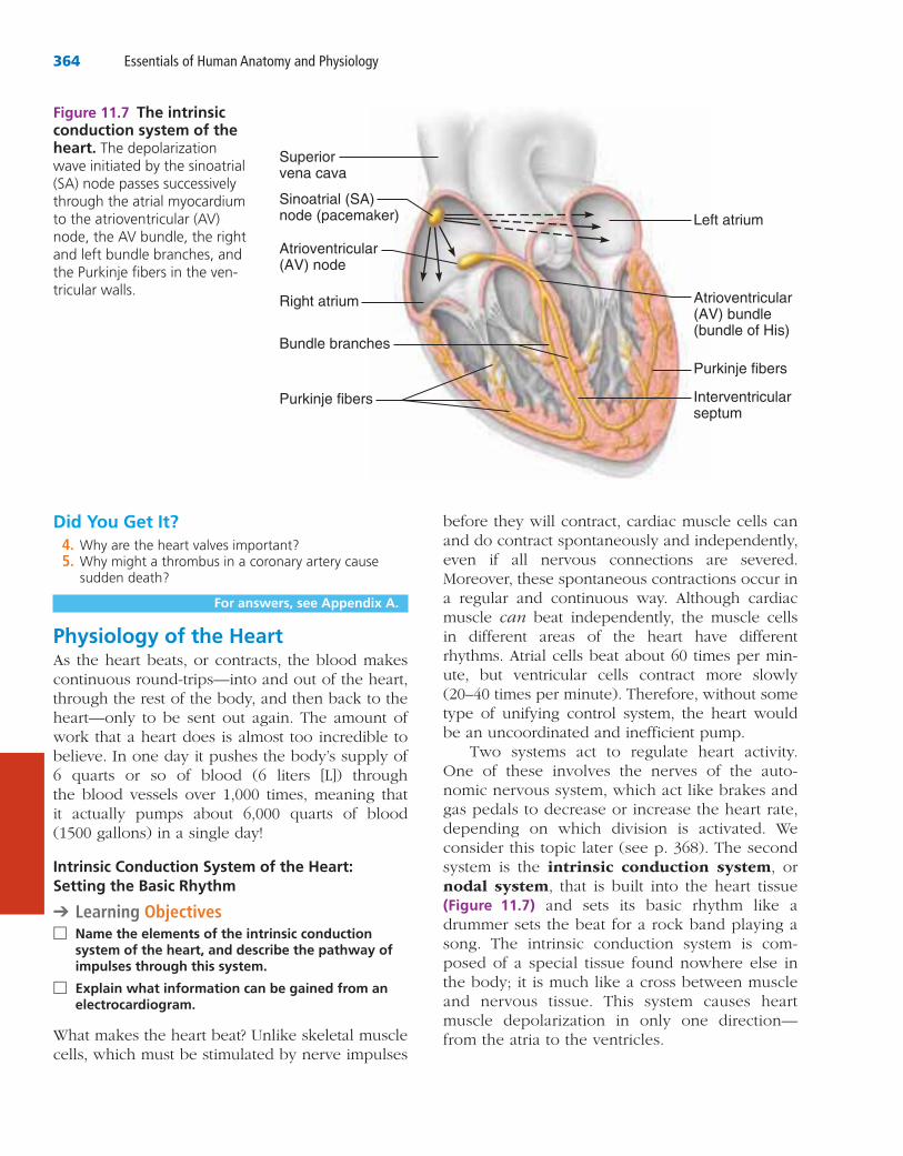
Figure 11.7 The intrinsic conduction system of the heart.
-
Chapter 11: The Cardiovascular System 365
11
There are other conditions that can interfere with the regular conduction of impulses across the heart—for example, damage to the SA node results in a slower heart rate. When this is a problem, arti-ficial pacemakers are usually installed surgically.
Ischemia (is-ke′me-ah), or lack of an ade-quate blood supply to the heart muscle, may lead to fibrillation—a rapid, uncoordinated quivering of the ventricles (it looks like a bag of wiggling worms). Fibrillation makes the heart unable to pump any blood and so is a major cause of death from heart attacks in adults. Many businesses train their employees in the use of AEDs (automatic external defibrillators), which has proven to be lifesaving in many cases. _____________________✚
Tachycardia (tak″e-kar′de-ah) is a rapid heart rate (over 100 beats per minute). Bradycardia (brad″e-kar′de-ah) is a heart rate that is substan-tially slower than normal (less than 60 beats per minute). Neither condition is pathological, but prolonged tachycardia may progress to fibrillation.
Cardiac Cycle and Heart Sounds
➔ Learning Objective □ Define systole, diastole, stroke volume, cardiac
cycle, heart sounds, and murmur.
In a healthy heart, the atria contract simultaneously. Then, as they start to relax, the ventricles begin to contract. Systole (sis′to-le) and diastole (di-as′to-le) mean heart contraction and relaxation, respectively. Because most of the pumping work is done by the ventricles, these terms refer to the contraction and relaxation of the ventricles unless otherwise stated.
The term cardiac cycle refers to the events of one complete heartbeat, during which both atria and ventricles contract and then relax. The aver-age heart beats approximately 75 times per min-ute, so the length of the cardiac cycle is normally about 0.8 second. We will consider the cardiac cycle in terms of events occurring during five peri-ods (Figure 11.8, p. 366).
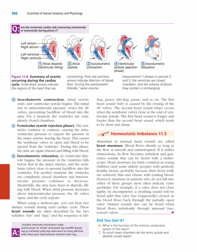
1 Atrial diastole (ventricular filling). Our dis-cussion begins with the heart completely relaxed. Pressure in the heart is low, the AV valves are open, and blood is flowing pas-sively through the atria into the ventricles. The semilunar valves are closed.
2 Atrial systole. The ventricles remain in dias-tole as the atria contract, forcing blood into the ventricles to complete ventricular filling.
CONCEPTLINK
➔
In addition, the intrinsic conduction system enforces a contraction rate of approximately 75 beats per minute on the heart; thus, the heart beats as a coordinated unit.
One of the most important parts of the intrin-sic conduction system is a crescent-shaped node of tissue called the sinoatrial (si″no-a′tre-al) (SA) node, located in the right atrium. Other compo-nents include the atrioventricular (AV) node at the junction of the atria and ventricles, the atrio-ventricular (AV) bundle (bundle of His) and the right and left bundle branches located in the interventricular septum, and finally the Purkinje (pur-kin′je) fibers, which spread within the myo-cardium of the ventricle walls.
The SA node is a tiny cell mass with a mammoth job. Because it has the highest rate of depolarization in the whole system, it starts each heartbeat and sets the pace for the whole heart. Consequently, the SA node is often called the pacemaker. From the SA node, the impulse spreads through the atria to the AV node, and then the atria contract. At the AV node, the impulse is delayed briefly to give the atria time to finish contracting. It then passes rapidly through the AV bundle, the bundle branches, and the Purkinje fibers, resulting in a “wringing” contrac-tion of the ventricles that begins at the heart apex and moves toward the atria. This contraction effec-tively ejects blood superiorly into the large arteries leaving the heart. “A Closer Look” (on p. 367) describes electrocardiography, the clinical proce-dure for mapping the electrical activity of the heart.
Homeostatic Imbalance 11.4
Because the atria and ventricles are separated from one another by “insulating” connective tissue, which is part of the fibrous skeleton of the heart, depolarization waves can reach the ventricles only by traveling through the AV node. Thus, any dam-age to the AV node can partially or totally block the ventricles from the control of the SA node. When this occurs, the ventricles begin to beat at their own rate, which is much slower, some or all of the time. This condition is called heart block.
366 Essentials of Human Anatomy and Physiology
dup, pause, lub-dup, pause, and so on. The first heart sound (lub) is caused by the closing of the AV valves. The second heart sound (dup) occurs when the semilunar valves close at the end of ven-tricular systole. The first heart sound is longer and louder than the second heart sound, which tends to be short and sharp.
Homeostatic Imbalance 11.5
Abnormal or unusual heart sounds are called heart murmurs. Blood flows silently as long as the flow is smooth and uninterrupted. If it strikes obstructions, its flow becomes turbulent and gen-erates sounds that can be heard with a stetho-scope. Heart murmurs are fairly common in young children (and some elderly people) with perfectly healthy hearts, probably because their heart walls are relatively thin and vibrate with rushing blood. However, murmurs in patients who do not fall into either of these groups most often indicate valve problems. For example, if a valve does not close tightly (is incompetent), a swishing sound will be heard after that valve has (supposedly) closed, as the blood flows back through the partially open valve. Distinct sounds also can be heard when blood flows turbulently through stenosed (nar-rowed) valves. ______________________________✚
Did You Get It?6.
7. systolediastole
3 Isovolumetric contraction. Atrial systole ends, and ventricular systole begins. The initial rise in intraventricular pressure closes the AV valves, preventing backflow of blood into the atria. For a moment, the ventricles are com-pletely closed chambers.
4 Ventricular systole (ejection phase). The ven- tricles continue to contract, causing the intra-ventricular pressure to surpass the pressure in the major arteries leaving the heart. This causes the semilunar valves to open and blood to be ejected from the ventricles. During this phase, the atria are again relaxed and filling with blood.
5 Isovolumetric relaxation. As ventricular dias-tole begins, the pressure in the ventricles falls below that in the major arteries, and the semi-lunar valves close to prevent backflow into the ventricles. For another moment, the ventricles are completely closed chambers and intraven-tricular pressure continues to decrease. Meanwhile, the atria have been in diastole, fill-ing with blood. When atrial pressure increases above intraventricular pressure, the AV valves open, and the cycle repeats.
When using a stethoscope, you can hear two distinct sounds during each cardiac cycle. These heart sounds are often described by the two syllables “lub” and “dup,” and the sequence is lub-
Right atrium
Left ventricleRight ventricle
Left atrium
1 Atrial diastole (ventricular filling)
2 Atrial systole
3 Isovolumetric contraction
4 Ventricular systole (ejection phase)
5 Isovolumetric relaxation
The cells contract isometrically until they have enough force to overcome the back pressure of the blood against the semilunar valves, at which point
their contraction becomes isotonic.
A:
Are the ventricular cardiac cells contracting isometrically or isotonically during phase 3?Q:
Figure 11.8 Summary of events occurring during the cardiac cycle. isovolumetric
367
A CLOSER
LOOK
W
electrocardiographelectro-
cardiogram (ECG)-
--
-
QRS complex
-T wave
myocardial infarct (present
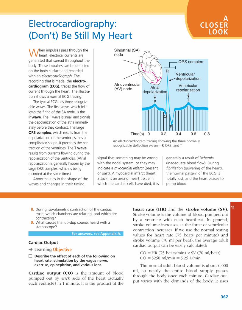
Electrocardiography: (Don’t) Be Still My Heart
ischemia
fibrillation
heart rate (HR) and the stroke volume (SV). Stroke volume is the volume of blood pumped out by a ventricle with each heartbeat. In general, stroke volume increases as the force of ventricular contraction increases. If we use the normal resting values for heart rate (75 beats per minute) and stroke volume (70 ml per beat), the average adult cardiac output can be easily calculated:
CO = HR (75 beats/min) × SV (70 ml/beat)CO = 5250 ml/min = 5.25 L/min
The normal adult blood volume is about 6,000 ml, so nearly the entire blood supply passes through the body once each minute. Cardiac out-put varies with the demands of the body. It rises
8.
9.
For answers, see Appendix A.
Cardiac Output
➔ Learning Objective □ Describe the effect of each of the following on
heart rate: stimulation by the vagus nerve, exercise, epinephrine, and various ions.
Cardiac output (CO) is the amount of blood pumped out by each side of the heart (actually each ventricle) in 1 minute. It is the product of the
Sinoatrial (SA)node
Atrioventricular(AV) node
Time(s) 0 0.2 0.4 0.6 0.8
Atrialdepolarization
P
R
Q
S
T
QRS complex
Ventriculardepolarization
Ventricularrepolarization
11
368 Essentials of Human Anatomy and Physiology
1. Neural (ANS) controls. During times of physical or emotional stress, the nerves of the sympathetic division of the autonomic nervous system more strongly stimulate the SA and AV nodes and the cardiac muscle itself. As a result, the heart beats more rap-idly. This is a familiar phenomenon to any-one who has ever been frightened or has had to run to catch a bus. As fast as the heart pumps under ordinary conditions, it really speeds up when special demands are placed on it. Because a faster blood flow increases the rate at which fresh blood reaches body cells, more oxygen and glucose are made available to them during periods of stress. When demand declines, the heart adjusts accordingly. Parasympathetic nerves, primar-ily vagus nerve fibers, slow and steady the heart, giving it more time to rest during non-crisis times.
2. Hormones and ions. Various hormones and ions can have a dramatic effect on heart activ-ity. Both epinephrine, which mimics sympa-thetic nerves and is released in response to sympathetic nerve stimulation, and thyroxine, a thyroid hormone, increase heart rate. Electrolyte imbalances pose a real threat to the heart. For example, recall that calcium ions are required for muscle contraction. A reduced level of ionic calcium in the blood depresses the heartbeat, whereas an excessive level of blood calcium ions causes such pro-longed contractions that the heart may stop entirely. Either excess or lack of needed ions such as sodium and potassium also modifies heart activity. A deficit of potassium ions in the blood, for example, causes the heart to beat feebly, and abnormal heart rhythms appear.
3. Physical factors. A number of physical fac-tors, including age, gender, exercise, and body temperature, influence heart rate. Resting heart rate is fastest in the fetus (140–160 beats per minute) and then gradually decreases throughout life. The average adult heart rate is faster in females (72–80 beats per minute) than in males (64–72 beats per minute). Heat increases heart rate by boosting the metabolic rate of heart cells. This explains the rapid, pounding heartbeat you feel when you have a
when the stroke volume is increased or the heart beats faster or both; it drops when either or both of these factors decrease. Let’s take a look at how stroke volume and heart rate are regulated.
Regulation of Stroke Volume A healthy heart pumps out about 60 percent of the blood present in its ventricles. As noted previously, this is approx-imately 70 ml (about 2 ounces) with each heart-beat. According to Starling’s law of the heart, the critical factor controlling stroke volume is how much the cardiac muscle cells are stretched by the filling of the chambers just before they contract. The more they are stretched, the stronger the con-traction will be. The important factor stretching the heart muscle is venous return, the amount of blood entering the heart and distending its ventri-cles. If one side of the heart suddenly begins to pump more blood than the other, the increased venous return to the opposite ventricle will force it to pump out an equal amount, thus preventing backup of blood in the circulation.
Anything that increases the volume or speed of venous return also increases stroke volume and force of contraction (Figure 11.9). For example, a slow heartbeat allows more time for the ventricles to fill. Exercise speeds venous return because it results in increased heart rate and force; the enhanced squeezing action of active skeletal mus-cles on the veins helps return blood to the heart. This so-called muscular pump also plays a major role in increasing the venous return. In contrast, low venous return, such as might result from severe blood loss or an abnormally rapid heart rate, decreases stroke volume, causing the heart to beat less forcefully.
Factors Modifying Basic Heart Rate In healthy people, stroke volume tends to be relatively con-stant. However, when blood volume drops sud-denly or when the heart has been seriously weakened, stroke volume declines, and cardiac output is maintained by a faster heartbeat. Although heart contraction does not depend on the nervous system, its rate can be changed tem-porarily by the autonomic nerves. Indeed, the most important external influence on heart rate is the activity of the autonomic nervous system. Several chemicals, hormones, and ions also mod-ify heart rate. Some of these factors are discussed next (see also Figure 11.9).

Chapter 11: The Cardiovascular System 369
11
progressive condition that reflects weakening of the heart by coronary atherosclerosis (clogging of the coronary vessels with fatty buildup), hyperten-sive heart disease, or multiple myocardial infarc-tions (repaired with noncontracting scar tissue). In these patients, the heart pumps weakly and is nearly “worn out.” The weak contractions of a heart in CHF result in a lower stroke volume. For those patients, the drug digitalis is routinely pre-scribed. It enhances contractile force and stroke volume of the heart, resulting in greater cardiac output.
Because the heart is a double pump, each side can fail independently of the other. If the left heart fails, pulmonary congestion occurs. The right side of the heart continues to propel blood to the lungs, but the left side is unable to eject the returning blood into the systemic circulation. As
high fever and accounts in part for the effect of exercise on heart rate (remember, working muscles generate heat). Cold has the opposite effect; it directly decreases heart rate. As noted previously, exercise acts through ner-vous system controls (sympathetic division) to increase heart rate (and also, through the action of the muscular pump, to increase stroke volume).
Homeostatic Imbalance 11.6
The pumping action of the healthy heart maintains a balance between cardiac output and venous return. But when the pumping efficiency of the heart is reduced so that circulation is inadequate to meet tissue needs, congestive heart failure (CHF) occurs. Congestive heart failure is usually a
Low bloodpressure
Hormones:epinephrine,thyroxine
Crisis haspassed
High bloodpressureor bloodvolume
Exercise Decreasedblood volume(hemorrhage)
Crisis stressors(physical or emotional trauma; increased bodytemperature; exercise)
Sympathetic nervous system activity
Activation ofskeletal muscleand respiratory“pumps”
Parasympatheticnervous systemcontrols (via vagus nerves)
Increased contractileforce of cardiac muscle
Increased venousreturn
Stroke volume (ml/beat)Heart rate (beats/min)
Cardiac output (ml/min)
Decreased venousreturn
KEY:Increases, stimulatesReduces, inhibitsInitial stimulusPhysiological responseEnd result
Figure 11.9 Influence of selected factors on cardiac output.
370 Essentials of Human Anatomy and Physiology
which carry blood away from the heart, and veins, which drain the tissues and return the blood to the heart, are simply conducting vessels—the free-ways and secondary roads. Only the tiny hairlike capillaries, which extend and branch through the tissues and connect the smallest arteries (arteri-oles) to the smallest veins (venules), directly serve the needs of the body cells. The capillaries are the side streets or alleys that intimately intertwine among the body cells and provide access to individual “homes.” It is only through their walls that exchanges between tissue cells and the blood can occur.
Notice that we routinely depict arteries in red and veins in blue. By convention, red indicates oxygen-rich blood, the normal status of blood in most of the body’s arteries, and blue indicates rela-tively oxygen-depleted, carbon dioxide–rich blood, the normal status of blood in most of the veins. However, there are exceptions. For instance, we have seen that oxygen-poor blood is carried in the pulmonary trunk, an artery, while oxygen-rich blood is transported back to the heart in pulmonary veins. An easy way to remember this difference is the following: Arteries are red and veins are blue, but for the lungs there’s an exception of two.
Microscopic Anatomy of Blood VesselsTunics
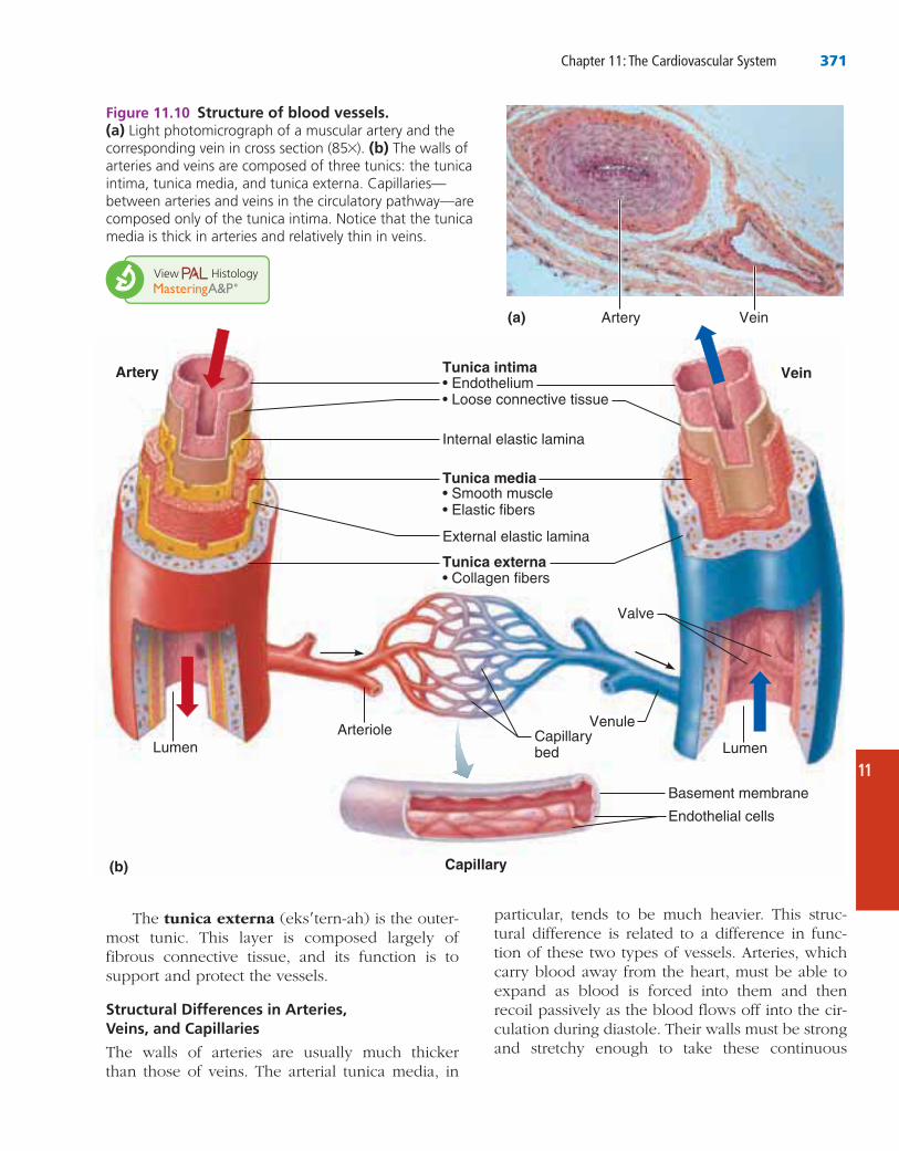
Except for the microscopic capillaries (which have only one layer), the walls of blood vessels have three layers, or tunics (Figure 11.10). The tunica intima (tu′nı-kah in-tim′ah), which lines the lumen, or interior, of the vessels, is a thin layer of endo-thelium (squamous epithelial cells) resting on a basement membrane. Its cells fit closely together and form a slick surface that decreases friction as blood flows through the vessel lumen.
The tunica media (me′de-ah) is the bulky middle layer, made up mostly of smooth muscle and elastic fibers. Some of the larger arteries have elastic laminae, sheets of elastic tissue, in addi-tion to the scattered elastic fibers. The smooth muscle, which is controlled by the sympathetic nervous system, is active in changing the diame-ter of the vessels. As the vessels constrict or dilate, blood pressure increases or decreases, respectively.
blood “backs up” in the lungs, they become swol-len with blood, the pressure within them increases, and fluid leaks into the lung tissue, causing pul-monary edema. If untreated, the person “drowns” in these fluids.
If the right side of the heart fails, peripheral congestion occurs as blood backs up in the sys-temic circulation. Edema is most noticeable in the distal parts of the body: The feet, ankles, and fin-gers become swollen and puffy. Failure of one side of the heart puts a greater strain on the opposite side, and eventually the whole heart fails. _______________________________________ ✚
Did You Get It?10. cardiac output11.
12.
For answers, see Appendix A.
Blood Vessels➔ Learning Objective
□ Compare and contrast the structure and function of arteries, veins, and capillaries.
Blood circulates inside the blood vessels, which form a closed transport system called the vascu-lar system. The idea that blood circulates, or “makes rounds,” through the body is only about 300 years old. The ancient Greeks believed that blood moved through the body like an ocean tide, first moving out from the heart and then ebbing back to it in the same vessels to get rid of its impurities in the lungs. It was not until the sev-enteenth century that William Harvey, an English physician, proved that blood did, in fact, move in circles.
Like a system of roads, the vascular system has its freeways, secondary roads, and alleys. As the heart beats, it propels blood into the large arter-ies leaving the heart. As the large arteries branch, blood moves into successively smaller and smaller arteries and then into the arterioles (ar-ter′e-olz), which feed the capillary (kap′ ı-lar″e) beds in the tissues. Capillary beds are drained by venules (ven′ulz), which in turn empty into veins that merge and finally empty into the great veins (venae cavae) entering the heart. Thus arteries,
Chapter 11: The Cardiovascular System 371
11
particular, tends to be much heavier. This struc-tural difference is related to a difference in func-tion of these two types of vessels. Arteries, which carry blood away from the heart, must be able to expand as blood is forced into them and then recoil passively as the blood flows off into the cir-culation during diastole. Their walls must be strong and stretchy enough to take these continuous
The tunica externa (eks′tern-ah) is the outer-most tunic. This layer is composed largely of fibrous connective tissue, and its function is to support and protect the vessels.
Structural Differences in Arteries, Veins, and Capillaries
The walls of arteries are usually much thicker than those of veins. The arterial tunica media, in
Tunica intima
Tunica media
Tunica externa
Artery Vein
(a)
(b) Capillary
Figure 11.10 Structure of blood vessels. (a)
* (b)
372 Essentials of Human Anatomy and Physiology
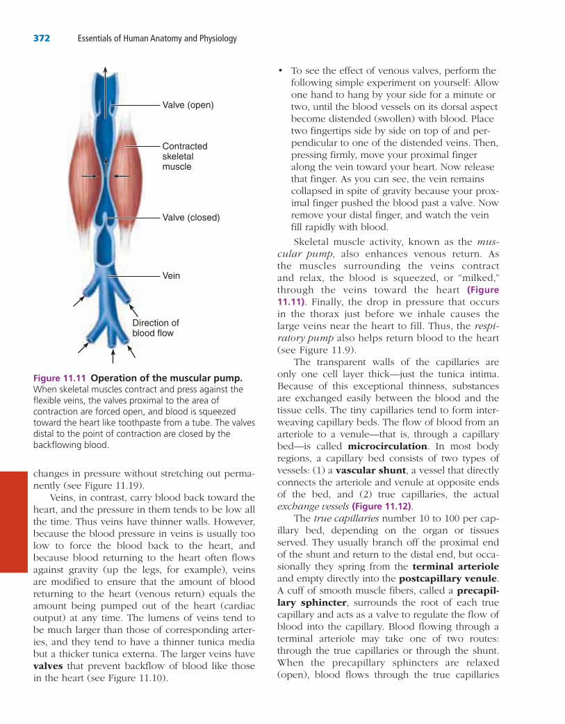
• To see the effect of venous valves, perform the following simple experiment on yourself: Allow one hand to hang by your side for a minute or two, until the blood vessels on its dorsal aspect become distended (swollen) with blood. Place two fingertips side by side on top of and per-pendicular to one of the distended veins. Then, pressing firmly, move your proximal finger along the vein toward your heart. Now release that finger. As you can see, the vein remains collapsed in spite of gravity because your prox-imal finger pushed the blood past a valve. Now remove your distal finger, and watch the vein fill rapidly with blood.
Skeletal muscle activity, known as the mus-cular pump, also enhances venous return. As the muscles surrounding the veins contract and relax, the blood is squeezed, or “milked,” through the veins toward the heart (Figure 11.11). Finally, the drop in pressure that occurs in the thorax just before we inhale causes the large veins near the heart to fill. Thus, the respi-ratory pump also helps return blood to the heart (see Figure 11.9).
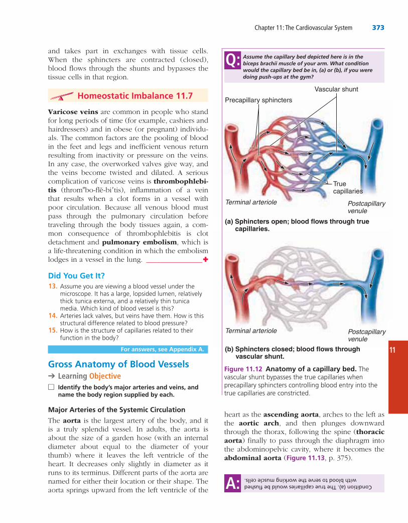
The transparent walls of the capillaries are only one cell layer thick—just the tunica intima. Because of this exceptional thinness, substances are exchanged easily between the blood and the tissue cells. The tiny capillaries tend to form inter-weaving capillary beds. The flow of blood from an arteriole to a venule—that is, through a capillary bed—is called microcirculation. In most body regions, a capillary bed consists of two types of vessels: (1) a vascular shunt, a vessel that directly connects the arteriole and venule at opposite ends of the bed, and (2) true capillaries, the actual exchange vessels (Figure 11.12).
The true capillaries number 10 to 100 per cap-illary bed, depending on the organ or tissues served. They usually branch off the proximal end of the shunt and return to the distal end, but occa-sionally they spring from the terminal arteriole and empty directly into the postcapillary venule. A cuff of smooth muscle fibers, called a precapil-lary sphincter, surrounds the root of each true capillary and acts as a valve to regulate the flow of blood into the capillary. Blood flowing through a terminal arteriole may take one of two routes: through the true capillaries or through the shunt. When the precapillary sphincters are relaxed (open), blood flows through the true capillaries
changes in pressure without stretching out perma-nently (see Figure 11.19).
Veins, in contrast, carry blood back toward the heart, and the pressure in them tends to be low all the time. Thus veins have thinner walls. However, because the blood pressure in veins is usually too low to force the blood back to the heart, and because blood returning to the heart often flows against gravity (up the legs, for example), veins are modified to ensure that the amount of blood returning to the heart (venous return) equals the amount being pumped out of the heart (cardiac output) at any time. The lumens of veins tend to be much larger than those of corresponding arter-ies, and they tend to have a thinner tunica media but a thicker tunica externa. The larger veins have valves that prevent backflow of blood like those in the heart (see Figure 11.10).
Valve (open)
Contractedskeletalmuscle
Valve (closed)
Vein
Direction ofblood flow
Figure 11.11 Operation of the muscular pump.
Chapter 11: The Cardiovascular System 373
11
heart as the ascending aorta, arches to the left as the aortic arch, and then plunges downward through the thorax, following the spine (thoracic aorta) finally to pass through the diaphragm into the abdominopelvic cavity, where it becomes the abdominal aorta (Figure 11.13, p. 375).
and takes part in exchanges with tissue cells. When the sphincters are contracted (closed), blood flows through the shunts and bypasses the tissue cells in that region.
Homeostatic Imbalance 11.7
Varicose veins are common in people who stand for long periods of time (for example, cashiers and hairdressers) and in obese (or pregnant) individu-als. The common factors are the pooling of blood in the feet and legs and inefficient venous return resulting from inactivity or pressure on the veins. In any case, the overworked valves give way, and the veins become twisted and dilated. A serious complication of varicose veins is thrombophlebi-tis (throm″bo-fle-bi′tis), inflammation of a vein that results when a clot forms in a vessel with poor circulation. Because all venous blood must pass through the pulmonary circulation before traveling through the body tissues again, a com-mon consequence of thrombophlebitis is clot detachment and pulmonary embolism, which is a life-threatening condition in which the embolism lodges in a vessel in the lung. ________________✚
Did You Get It?13.
14.
15.
For answers, see Appendix A.
Gross Anatomy of Blood Vessels➔ Learning Objective
□name the body region supplied by each.
The aorta is the largest artery of the body, and it is a truly splendid vessel. In adults, the aorta is about the size of a garden hose (with an internal diameter about equal to the diameter of your thumb) where it leaves the left ventricle of the heart. It decreases only slightly in diameter as it runs to its terminus. Different parts of the aorta are named for either their location or their shape. The aorta springs upward from the left ventricle of the
Condition (a). The true capillaries would be flushed with blood to serve the working muscle cells.A:
(a) Sphincters open; blood flows through true capillaries.
(b) Sphincters closed; blood flows throughvascular shunt.
Precapillary sphincters
Vascular shunt
Terminal arteriole Postcapillary venule
Terminal arteriole Postcapillary venule
Truecapillaries
Figure 11.12 Anatomy of a capillary bed. The
Assume the capillary bed depicted here is in the biceps brachii muscle of your arm. What condition would the capillary bed be in, (a) or (b), if you were doing push-ups at the gym?
Q:
374 Essentials of Human Anatomy and Physiology
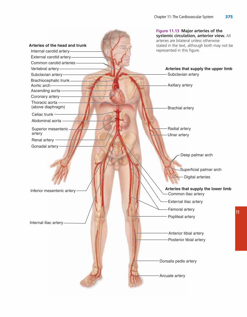
three branches: the L. gastric artery, which supplies the stomach; the splenic artery, which supplies the spleen; and the common hepatic artery, which supplies the liver.
• The unpaired superior mesenteric (mes″en-ter′ik) artery supplies most of the small intes-tine and the first half of the large intestine, or colon.
• The renal (R. and L.) arteries serve the kidneys.
• The gonadal (R. and L.) arteries supply the gonads. They are called the ovarian arteries in females (serving the ovaries) and the testicular arteries in males (serving the testes).
• The lumbar arteries (not illustrated in Figure 11.13) are several pairs of arteries serving the heavy muscles of the abdomen and trunk walls.
• The inferior mesenteric artery is a small, unpaired artery supplying the second half of the large intestine.
• The common iliac (R. and L.) arteries are the final branches of the abdominal aorta. Each di-vides into an internal iliac artery, which sup-plies the pelvic organs (bladder, rectum, and so on), and an external iliac artery, which enters the thigh, where it becomes the femoral ar-tery. The femoral artery and its branch, the deep artery of the thigh, serve the thigh. At the knee, the femoral artery becomes the popli-teal artery, which then splits into the anterior tibial artery and posterior tibial artery, which supply the leg and foot. The anterior tib-ial artery terminates in the dorsalis pedis ar-tery, which via the arcuate artery supplies the dorsum of the foot. (The dorsalis pedis is often palpated in patients with circulatory problems of the legs to determine whether the distal part of the leg has adequate circulation.)
Although arteries are generally located in deep, well-protected body areas, many veins are more superficial, and some are easily seen and palpated on the body surface. Most deep veins follow the course of the major arteries, and with a few excep-tions, the naming of these veins is identical to that of their companion arteries. Major systemic arteries branch off the aorta, whereas the veins converge on the venae cavae, which enter the right atrium of the heart. Veins draining the head and arms empty
The major branches of the aorta and the organs they serve are listed next in sequence from the heart. (Figure 11.13 shows the course of the aorta and its major branches.) As you locate the arteries on the figure, use what you already know to make your learning easier. In many cases the name of the artery tells you the body region or organs served (for example, renal artery, brachial artery, and coronary artery) or the bone followed (femoral artery and ulnar artery).
Arterial Branches of the Ascending Aorta
• The only branches of the ascending aorta are the right (R.) coronary artery and left (L.) coronary artery, which serve the heart.
Arterial Branches of the Aortic Arch
• The brachiocephalic (bra″ke-o-se-fal′ik) trunk (the first branch off the aortic arch) splits into the R. common carotid (kah- ro′tid) artery, which further branches into the R. internal and R. external carotid arteries, and the R. subclavian (sub-kla′ve-an) artery. (See same-named vessels on the left side of the body for organs served.)
• The L. common carotid artery is the second branch off the aortic arch. It divides, forming the L. internal carotid, which serves the brain, and the L. external carotid, which serves the skin and muscles of the head and neck.
• The third branch of the aortic arch, the L. subclavian artery, gives off an important branch—the vertebral artery, which serves part of the brain. In the axilla, the subclavian artery becomes the axillary artery and then continues into the arm as the brachial artery, which supplies the arm. At the elbow, the bra-chial artery splits to form the radial artery and ulnar artery, which serve the forearm.
Arterial Branches of the Thoracic Aorta
• The intercostal arteries (10 pairs) supply the muscles of the thorax wall. Other branches of the thoracic aorta supply the lungs (bronchial arteries), the esophagus (esophageal arteries), and the diaphragm (phrenic arteries). (These arteries are not illustrated in Figure 11.13.)
Arterial Branches of the Abdominal Aorta
• The celiac trunk is the first branch of the ab-dominal aorta. It is a single vessel that has
Chapter 11: The Cardiovascular System 375
11
Internal carotid artery
Common carotid arteries
Subclavian artery Subclavian artery
Aortic archAscending aortaCoronary artery
Thoracic aorta (above diaphragm)
Renal artery
Superficial palmar arch
Radial artery
Ulnar artery
Internal iliac artery
Deep palmar arch
Vertebral artery
Brachiocephalic trunkAxillary artery
Brachial artery
Abdominal aorta
Superior mesenteric artery
Gonadal artery
External iliac artery
Digital arteries
Femoral artery
Popliteal artery
Anterior tibial artery
Posterior tibial artery
Arcuate artery
Inferior mesenteric artery
Celiac trunk
External carotid artery
Arteries of the head and trunk
Arteries that supply the upper limb
Arteries that supply the lower limb
Dorsalis pedis artery
Common iliac artery
Figure 11.13 All
376 Essentials of Human Anatomy and Physiology
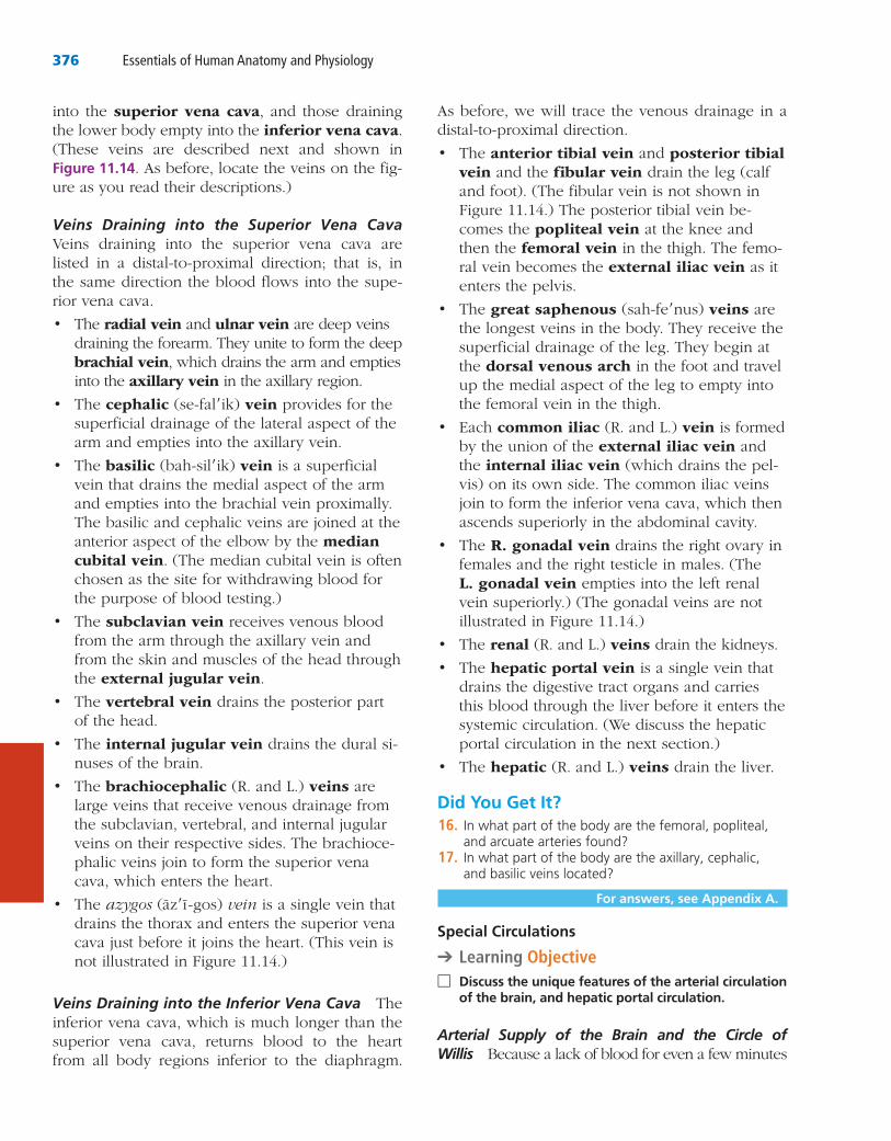
into the superior vena cava, and those draining the lower body empty into the inferior vena cava. (These veins are described next and shown in Figure 11.14. As before, locate the veins on the fig-ure as you read their descriptions.)
Veins Draining into the Superior Vena Cava Veins draining into the superior vena cava are listed in a distal-to-proximal direction; that is, in the same direction the blood flows into the supe-rior vena cava.
• The radial vein and ulnar vein are deep veins draining the forearm. They unite to form the deep brachial vein, which drains the arm and empties into the axillary vein in the axillary region.
• The cephalic (se-fal′ik) vein provides for the superficial drainage of the lateral aspect of the arm and empties into the axillary vein.
• The basilic (bah-sil′ik) vein is a superficial vein that drains the medial aspect of the arm and empties into the brachial vein proximally. The basilic and cephalic veins are joined at the anterior aspect of the elbow by the median cubital vein. (The median cubital vein is often chosen as the site for withdrawing blood for the purpose of blood testing.)
• The subclavian vein receives venous blood from the arm through the axillary vein and from the skin and muscles of the head through the external jugular vein.
• The vertebral vein drains the posterior part of the head.
• The internal jugular vein drains the dural si-nuses of the brain.
• The brachiocephalic (R. and L.) veins are large veins that receive venous drainage from the subclavian, vertebral, and internal jugular veins on their respective sides. The brachioce-phalic veins join to form the superior vena cava, which enters the heart.
• The azygos (az′ı -gos) vein is a single vein that drains the thorax and enters the superior vena cava just before it joins the heart. (This vein is not illustrated in Figure 11.14.)
Veins Draining into the Inferior Vena Cava The inferior vena cava, which is much longer than the superior vena cava, returns blood to the heart from all body regions inferior to the diaphragm.
As before, we will trace the venous drainage in a distal-to-proximal direction.
• The anterior tibial vein and posterior tibial vein and the fibular vein drain the leg (calf and foot). (The fibular vein is not shown in Figure 11.14.) The posterior tibial vein be-comes the popliteal vein at the knee and then the femoral vein in the thigh. The femo-ral vein becomes the external iliac vein as it enters the pelvis.
• The great saphenous (sah-fe′nus) veins are the longest veins in the body. They receive the superficial drainage of the leg. They begin at the dorsal venous arch in the foot and travel up the medial aspect of the leg to empty into the femoral vein in the thigh.
• Each common iliac (R. and L.) vein is formed by the union of the external iliac vein and the internal iliac vein (which drains the pel-vis) on its own side. The common iliac veins join to form the inferior vena cava, which then ascends superiorly in the abdominal cavity.
• The R. gonadal vein drains the right ovary in females and the right testicle in males. (The L. gonadal vein empties into the left renal vein superiorly.) (The gonadal veins are not illustrated in Figure 11.14.)
• The renal (R. and L.) veins drain the kidneys.
• The hepatic portal vein is a single vein that drains the digestive tract organs and carries this blood through the liver before it enters the systemic circulation. (We discuss the hepatic portal circulation in the next section.)
• The hepatic (R. and L.) veins drain the liver.
Did You Get It?16.
17.
For answers, see Appendix A.
Special Circulations
➔ Learning Objective □ Discuss the unique features of the arterial circulation
of the brain, and hepatic portal circulation.
Arterial Supply of the Brain and the Circle of Willis Because a lack of blood for even a few minutes
Chapter 11: The Cardiovascular System 377
11
Renal vein
Splenic vein
Basilic veinBrachial veinCephalic vein
Dural venous sinuses
External jugular veinVertebral veinInternal jugular vein
Superior vena cava
Right and leftbrachiocephalic veins Axillary vein
Great cardiac vein
Hepatic veins
Hepatic portal vein
Superior mesenteric vein
Inferior vena cava
Ulnar veinRadial vein
Common iliac vein
External iliac vein
Internal iliac vein
Digital veins
Femoral vein
Great saphenous vein
Popliteal vein
Posterior tibial vein
Anterior tibial vein
Small saphenous vein
Dorsal venous archDorsal metatarsal veins
Inferior mesenteric vein
Median cubital vein
Subclavian vein
Veins of the head and trunk
Veins that drain the upper limb
Veins that drain the lower limb
Figure 11.14 circulation, anterior view.
378 Essentials of Human Anatomy and Physiology
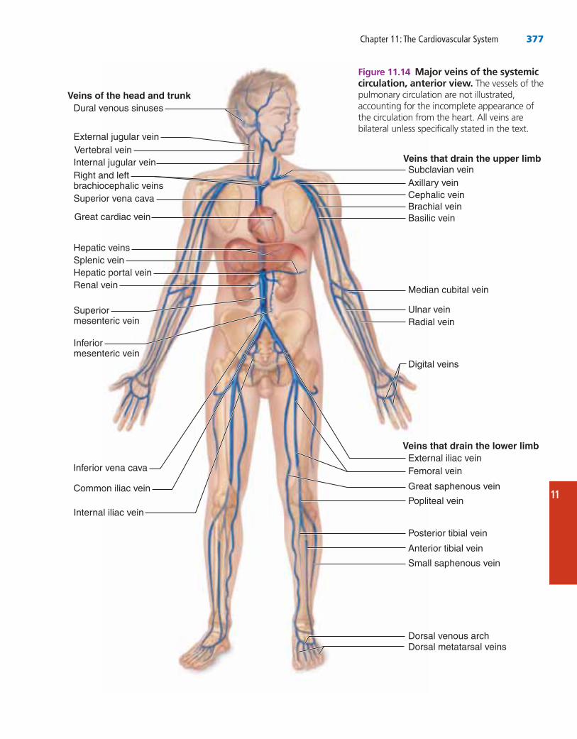
The paired vertebral arteries pass upward from the subclavian arteries at the base of the neck. Within the skull, the vertebral arteries join to form the single basilar artery. This artery serves the brain stem and cerebellum as it travels upward. At the base of the cerebrum, the basilar artery divides to form the posterior cerebral arteries, which supply the posterior part of the cerebrum.
causes the delicate brain cells to die, a continuous blood supply to the brain is crucial. The brain is supplied by two pairs of arteries, the internal carotid arteries and the vertebral arteries (Figure 11.15).
The internal carotid arteries, branches of the common carotid arteries, run through the neck and enter the skull through the temporal bone. Once inside the cranium, each divides into the anterior cerebral artery and middle cerebral artery, which supply most of the cerebrum.
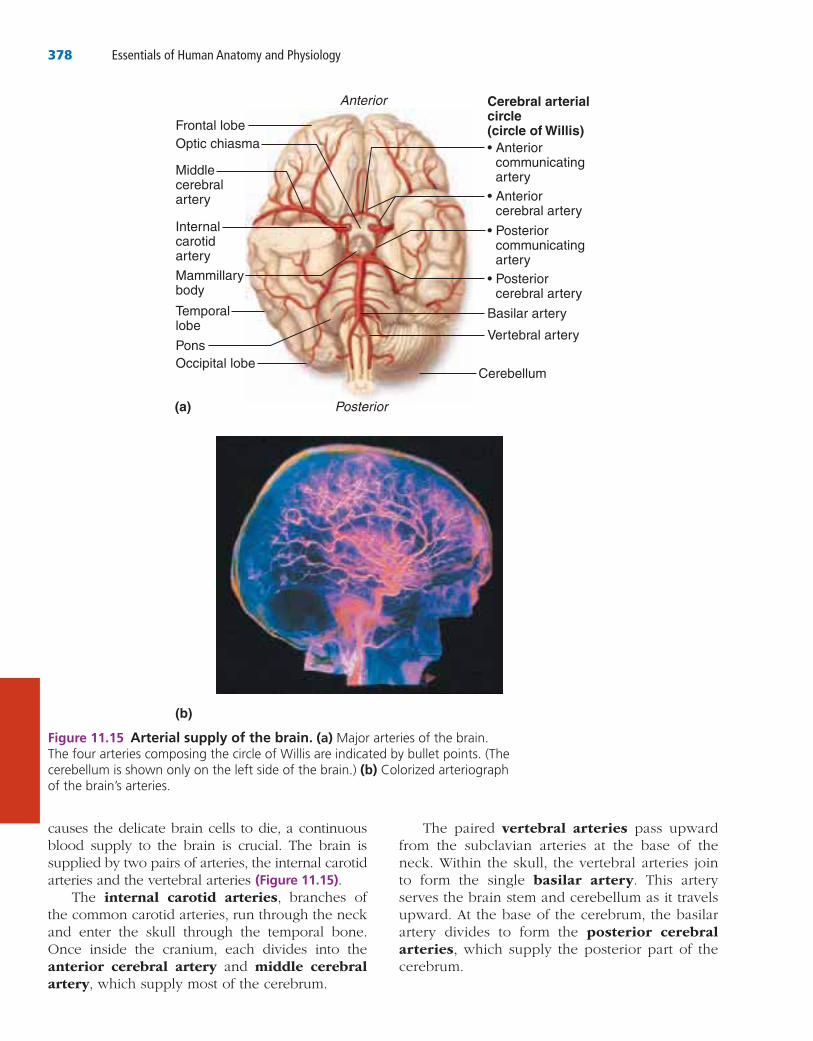
(b)
Frontal lobe
Anterior
Posterior
Optic chiasma
Middle cerebral artery
Internal carotid artery
Mammillarybody
Temporal lobe
Occipital lobe
Cerebral arterialcircle (circle of Willis)
cerebral artery
Basilar artery
Vertebral artery
Cerebellum
communicating artery
(a)
cerebral artery
communicating artery
Figure 11.15 Arterial supply of the brain. (a)
(b)
Chapter 11: The Cardiovascular System 379
11
The anterior and posterior blood supplies of the brain are united by small communicating arte-rial branches. The result is a complete circle of connecting blood vessels called the cerebral arte-rial circle or the circle of Willis, which sur-rounds the base of the brain. The cerebral arterial circle protects the brain by providing more than one route for blood to reach brain tissue in case of a clot or impaired blood flow anywhere in the system.
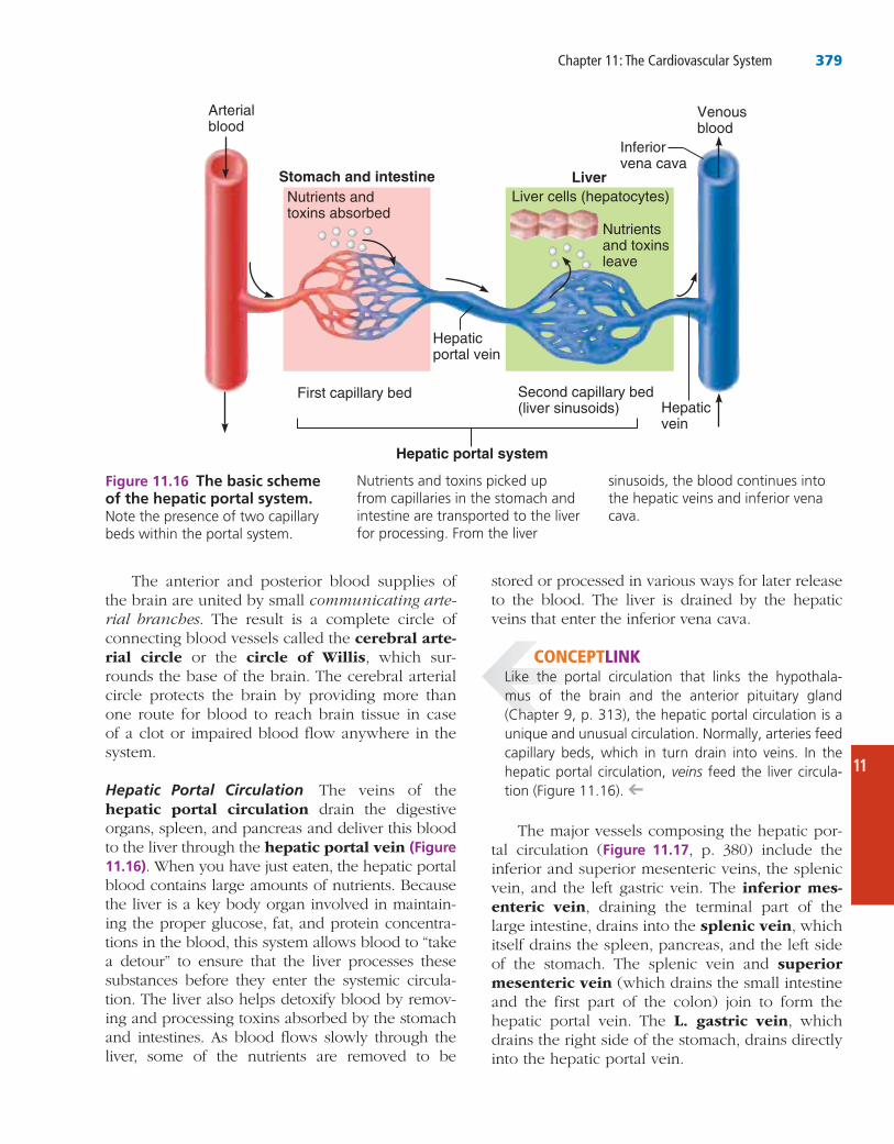
Hepatic Portal Circulation The veins of the hepatic portal circulation drain the digestive organs, spleen, and pancreas and deliver this blood to the liver through the hepatic portal vein (Figure 11.16). When you have just eaten, the hepatic portal blood contains large amounts of nutrients. Because the liver is a key body organ involved in maintain-ing the proper glucose, fat, and protein concentra-tions in the blood, this system allows blood to “take a detour” to ensure that the liver processes these substances before they enter the systemic circula-tion. The liver also helps detoxify blood by remov-ing and processing toxins absorbed by the stomach and intestines. As blood flows slowly through the liver, some of the nutrients are removed to be
Hepatic portal system
Arterialblood
Venousblood
Hepaticvein
Inferiorvena cava
Hepaticportal vein
First capillary bed
Nutrients andtoxins absorbed
Liver cells (hepatocytes)
Nutrientsand toxinsleave
Second capillary bed(liver sinusoids)
Stomach and intestine Liver
Figure 11.16 The basic scheme of the hepatic portal system.
CONCEPTLINK-
veins -➔
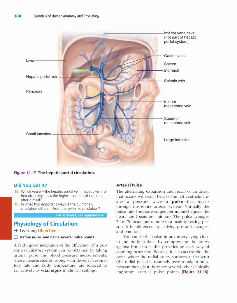
The major vessels composing the hepatic por-tal circulation (Figure 11.17, p. 380) include the inferior and superior mesenteric veins, the splenic vein, and the left gastric vein. The inferior mes-enteric vein, draining the terminal part of the large intestine, drains into the splenic vein, which itself drains the spleen, pancreas, and the left side of the stomach. The splenic vein and superior mesenteric vein (which drains the small intestine and the first part of the colon) join to form the hepatic portal vein. The L. gastric vein, which drains the right side of the stomach, drains directly into the hepatic portal vein.
stored or processed in various ways for later release to the blood. The liver is drained by the hepatic veins that enter the inferior vena cava.
380 Essentials of Human Anatomy and Physiology
Arterial Pulse
The alternating expansion and recoil of an artery that occurs with each beat of the left ventricle cre-ates a pressure wave—a pulse—that travels through the entire arterial system. Normally the pulse rate (pressure surges per minute) equals the heart rate (beats per minute). The pulse averages 70 to 76 beats per minute in a healthy resting per-son. It is influenced by activity, postural changes, and emotions.
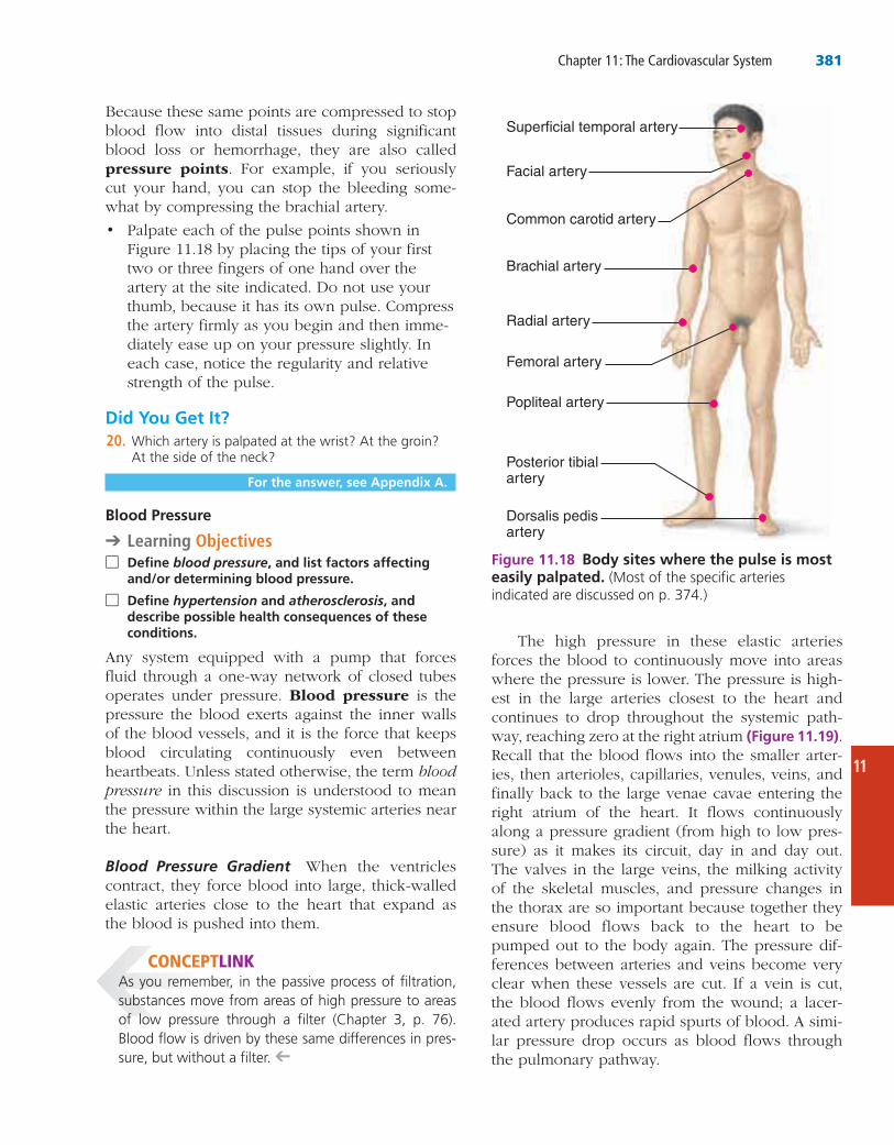
You can feel a pulse in any artery lying close to the body surface by compressing the artery against firm tissue; this provides an easy way of counting heart rate. Because it is so accessible, the point where the radial artery surfaces at the wrist (the radial pulse) is routinely used to take a pulse measurement, but there are several other clinically important arterial pulse points (Figure 11.18).
Did You Get It?18.
19.
For answers, see Appendix A.
Physiology of Circulation➔ Learning Objective
□ Define pulse, and name several pulse points.
A fairly good indication of the efficiency of a per-son’s circulatory system can be obtained by taking arterial pulse and blood pressure measurements. These measurements, along with those of respira-tory rate and body temperature, are referred to collectively as vital signs in clinical settings.
LiverSpleen
Gastric veins
Inferior vena cava(not part of hepaticportal system)
Splenic vein
Inferiormesenteric vein
Superiormesenteric vein
Large intestine
Hepatic portal vein
Small intestine
Pancreas
Stomach
Figure 11.17 The hepatic portal circulation.
Chapter 11: The Cardiovascular System 381
11
Because these same points are compressed to stop blood flow into distal tissues during significant blood loss or hemorrhage, they are also called pressure points. For example, if you seriously cut your hand, you can stop the bleeding some-what by compressing the brachial artery.
• Palpate each of the pulse points shown in Figure 11.18 by placing the tips of your first two or three fingers of one hand over the artery at the site indicated. Do not use your thumb, because it has its own pulse. Compress the artery firmly as you begin and then imme-diately ease up on your pressure slightly. In each case, notice the regularity and relative strength of the pulse.
Did You Get It?20.
For the answer, see Appendix A.
Blood Pressure
➔ Learning Objectives □ Define blood pressure, and list factors affecting
and/or determining blood pressure.
□ Define hypertension and atherosclerosis, and describe possible health consequences of these conditions.
Any system equipped with a pump that forces fluid through a one-way network of closed tubes operates under pressure. Blood pressure is the pressure the blood exerts against the inner walls of the blood vessels, and it is the force that keeps blood circulating continuously even between heartbeats. Unless stated otherwise, the term blood pressure in this discussion is understood to mean the pressure within the large systemic arteries near the heart.
Blood Pressure Gradient When the ventricles contract, they force blood into large, thick-walled elastic arteries close to the heart that expand as the blood is pushed into them.
Common carotid artery
Brachial artery
Radial artery
Femoral artery
Popliteal artery
Posterior tibialartery
Dorsalis pedisartery
Superficial temporal artery
Facial artery
Figure 11.18 Body sites where the pulse is most easily palpated. (Most of the specific arteries
CONCEPTLINK
-➔
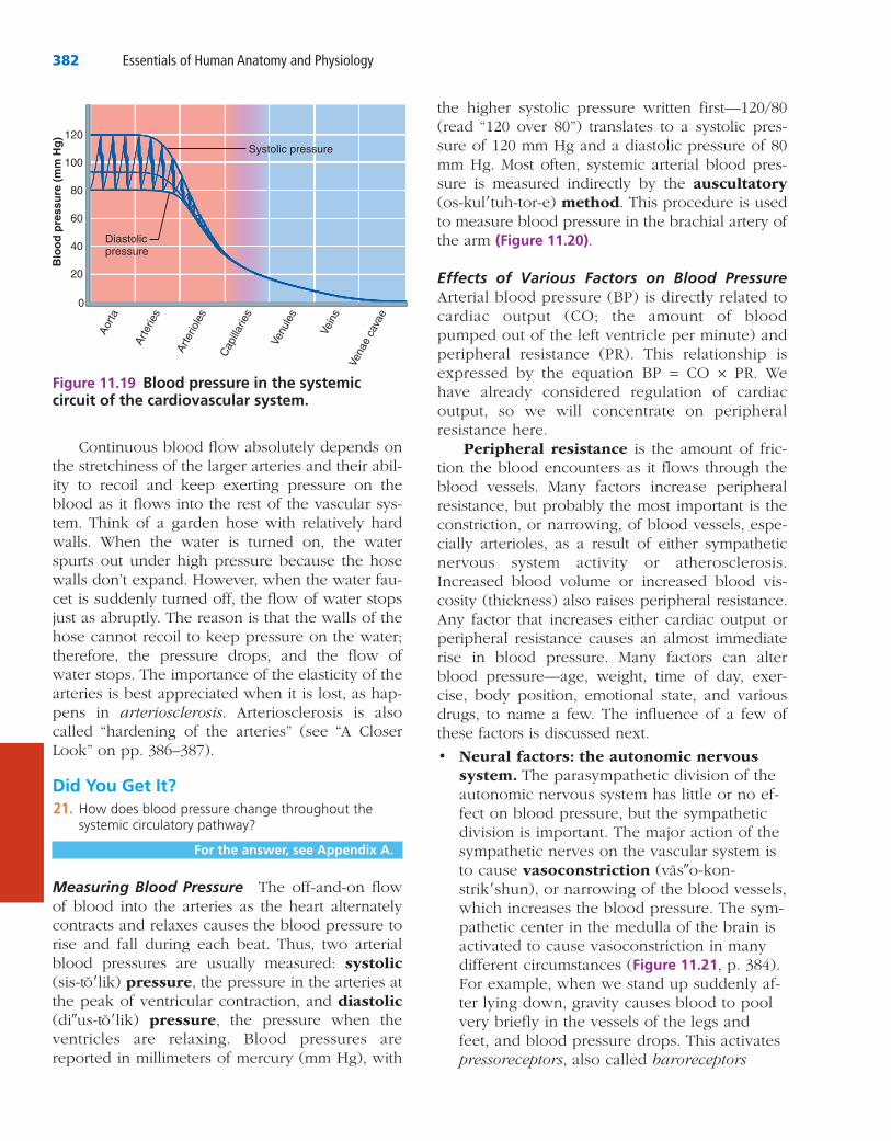
The high pressure in these elastic arteries forces the blood to continuously move into areas where the pressure is lower. The pressure is high-est in the large arteries closest to the heart and continues to drop throughout the systemic path-way, reaching zero at the right atrium 11.19). Recall that the blood flows into the smaller arter-ies, then arterioles, capillaries, venules, veins, and finally back to the large venae cavae entering the right atrium of the heart. It flows continuously along a pressure gradient (from high to low pres-sure) as it makes its circuit, day in and day out. The valves in the large veins, the milking activity of the skeletal muscles, and pressure changes in the thorax are so important because together they ensure blood flows back to the heart to be pumped out to the body again. The pressure dif-ferences between arteries and veins become very clear when these vessels are cut. If a vein is cut, the blood flows evenly from the wound; a lacer-ated artery produces rapid spurts of blood. A simi-lar pressure drop occurs as blood flows through the pulmonary pathway.
382 Essentials of Human Anatomy and Physiology
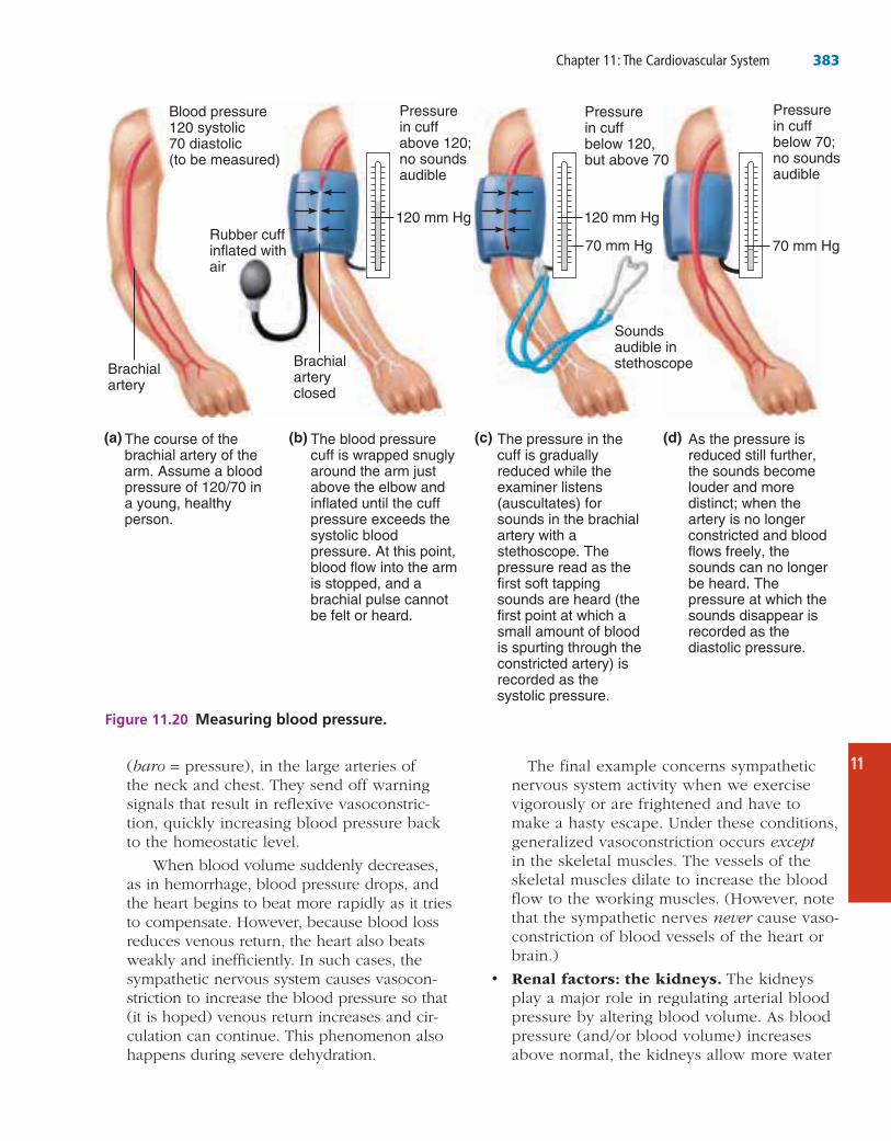
the higher systolic pressure written first—120/80 (read “120 over 80”) translates to a systolic pres-sure of 120 mm Hg and a diastolic pressure of 80 mm Hg. Most often, systemic arterial blood pres-sure is measured indirectly by the auscultatory (os-kul′tuh-tor-e) method. This procedure is used to measure blood pressure in the brachial artery of the arm (Figure 11.20).
Effects of Various Factors on Blood Pressure Arterial blood pressure (BP) is directly related to cardiac output (CO; the amount of blood pumped out of the left ventricle per minute) and peripheral resistance (PR). This relationship is expressed by the equation BP = CO × PR. We have already considered regulation of cardiac output, so we will concentrate on peripheral resistance here.
Peripheral resistance is the amount of fric-tion the blood encounters as it flows through the blood vessels. Many factors increase peripheral resistance, but probably the most important is the constriction, or narrowing, of blood vessels, espe-cially arterioles, as a result of either sympathetic nervous system activity or atherosclerosis. Increased blood volume or increased blood vis-cosity (thickness) also raises peripheral resistance. Any factor that increases either cardiac output or peripheral resistance causes an almost immediate rise in blood pressure. Many factors can alter blood pressure—age, weight, time of day, exer-cise, body position, emotional state, and various drugs, to name a few. The influence of a few of these factors is discussed next.
• Neural factors: the autonomic nervous system. The parasympathetic division of the autonomic nervous system has little or no ef-fect on blood pressure, but the sympathetic division is important. The major action of the sympathetic nerves on the vascular system is to cause vasoconstriction (vas″o-kon-strik′shun), or narrowing of the blood vessels, which increases the blood pressure. The sym-pathetic center in the medulla of the brain is activated to cause vasoconstriction in many different circumstances (Figure 11.21, p. 384). For example, when we stand up suddenly af-ter lying down, gravity causes blood to pool very briefly in the vessels of the legs and feet, and blood pressure drops. This activates pressoreceptors, also called baroreceptors
Continuous blood flow absolutely depends on the stretchiness of the larger arteries and their abil-ity to recoil and keep exerting pressure on the blood as it flows into the rest of the vascular sys-tem. Think of a garden hose with relatively hard walls. When the water is turned on, the water spurts out under high pressure because the hose walls don’t expand. However, when the water fau-cet is suddenly turned off, the flow of water stops just as abruptly. The reason is that the walls of the hose cannot recoil to keep pressure on the water; therefore, the pressure drops, and the flow of water stops. The importance of the elasticity of the arteries is best appreciated when it is lost, as hap-pens in arteriosclerosis. Arteriosclerosis is also called “hardening of the arteries” (see “A Closer Look” on pp. 386–387).
Did You Get It?21.
For the answer, see Appendix A.
Measuring Blood Pressure The off-and-on flow of blood into the arteries as the heart alternately contracts and relaxes causes the blood pressure to rise and fall during each beat. Thus, two arterial blood pressures are usually measured: systolic (sis-to′lik) pressure, the pressure in the arteries at the peak of ventricular contraction, and diastolic (di″us-to′lik) pressure, the pressure when the ventricles are relaxing. Blood pressures are reported in millimeters of mercury (mm Hg), with
Aorta
Arte
ries
Arte
riole
s
Cap
illarie
s
Venu
les
Vein
s
Vena
e ca
vae
Blo
od p
ress
ure
(mm
Hg) Systolic pressure
Diastolicpressure
0
20
40
60
80
100
120
Figure 11.19 Blood pressure in the systemic circuit of the cardiovascular system.
Chapter 11: The Cardiovascular System 383
11The final example concerns sympathetic nervous system activity when we exercise vigorously or are frightened and have to make a hasty escape. Under these conditions, generalized vasoconstriction occurs except in the skeletal muscles. The vessels of the skeletal muscles dilate to increase the blood flow to the working muscles. (However, note that the sympathetic nerves never cause vaso-constriction of blood vessels of the heart or brain.)
• Renal factors: the kidneys. The kidneys play a major role in regulating arterial blood pressure by altering blood volume. As blood pressure (and/or blood volume) increases above normal, the kidneys allow more water
(baro = pressure), in the large arteries of the neck and chest. They send off warning signals that result in reflexive vasoconstric-tion, quickly increasing blood pressure back to the homeostatic level.
When blood volume suddenly decreases, as in hemorrhage, blood pressure drops, and the heart begins to beat more rapidly as it tries to compensate. However, because blood loss reduces venous return, the heart also beats weakly and inefficiently. In such cases, the sympathetic nervous system causes vasocon-striction to increase the blood pressure so that (it is hoped) venous return increases and cir-culation can continue. This phenomenon also happens during severe dehydration.
Blood pressure120 systolic70 diastolic(to be measured)
Rubber cuffinflated with air
Brachialartery
Brachialarteryclosed
Pressurein cuffabove 120; no soundsaudible
120 mm Hg
Pressurein cuffbelow 120,but above 70
120 mm Hg
Soundsaudible instethoscope
Pressurein cuffbelow 70;no soundsaudible
70 mm Hg
(a) (b) (c) (d)
70 mm Hg
The course of the brachial artery of the arm. Assume a blood pressure of 120/70 in a young, healthy person.
The blood pressure cuff is wrapped snugly around the arm just above the elbow and inflated until the cuff pressure exceeds the systolic blood pressure. At this point, blood flow into the arm is stopped, and a brachial pulse cannot be felt or heard.
The pressure in the cuff is gradually reduced while the examiner listens (auscultates) for sounds in the brachial artery with a stethoscope. The pressure read as the first soft tapping sounds are heard (the first point at which a small amount of blood is spurting through the constricted artery) is recorded as the systolic pressure.
As the pressure is reduced still further, the sounds become louder and more distinct; when the artery is no longer constricted and blood flows freely, the sounds can no longer be heard. The pressure at which the sounds disappear is recorded as the diastolic pressure.
Figure 11.20 Measuring blood pressure.
384 Essentials of Human Anatomy and Physiology
the blood, water follows. Thus, blood volume and blood pressure both rise in response to aldosterone.
• Temperature. In general, cold has a vaso-constricting effect. This is why your exposed skin feels cold to the touch on a winter day and why cold compresses are recommended to prevent swelling of a bruised area. Heat has a vasodilating effect. This explains why skin reddens during exercise as body temper-ature increases and why warm compresses are used to speed the circulation into an in-flamed area.
• Chemicals. The effects of chemical sub-stances, many of which are drugs, on blood pressure are widespread and well known in many cases. We will give just a few examples here. Epinephrine increases both heart rate and blood pressure.
to leave the body in the urine. Because the source of this water is the bloodstream, blood volume decreases, which in turn de-creases blood pressure. However, when arte-rial blood pressure falls, the kidneys retain body water, maintaining blood volume and blood pressure (see Figure 11.21). In order to increase blood volume and blood pressure, fluids must be ingested or administered intra-venously.
In addition, when arterial blood pressure is low, certain kidney cells release the enzyme renin into the blood. Renin triggers a series of chemical reactions that result in the forma-tion of angiotensin II, a potent vasoconstric-tor chemical. Angiotensin also stimulates the adrenal cortex to release aldosterone, a hor-mone that enhances sodium ion reabsorption by the kidneys. As sodium ions move into
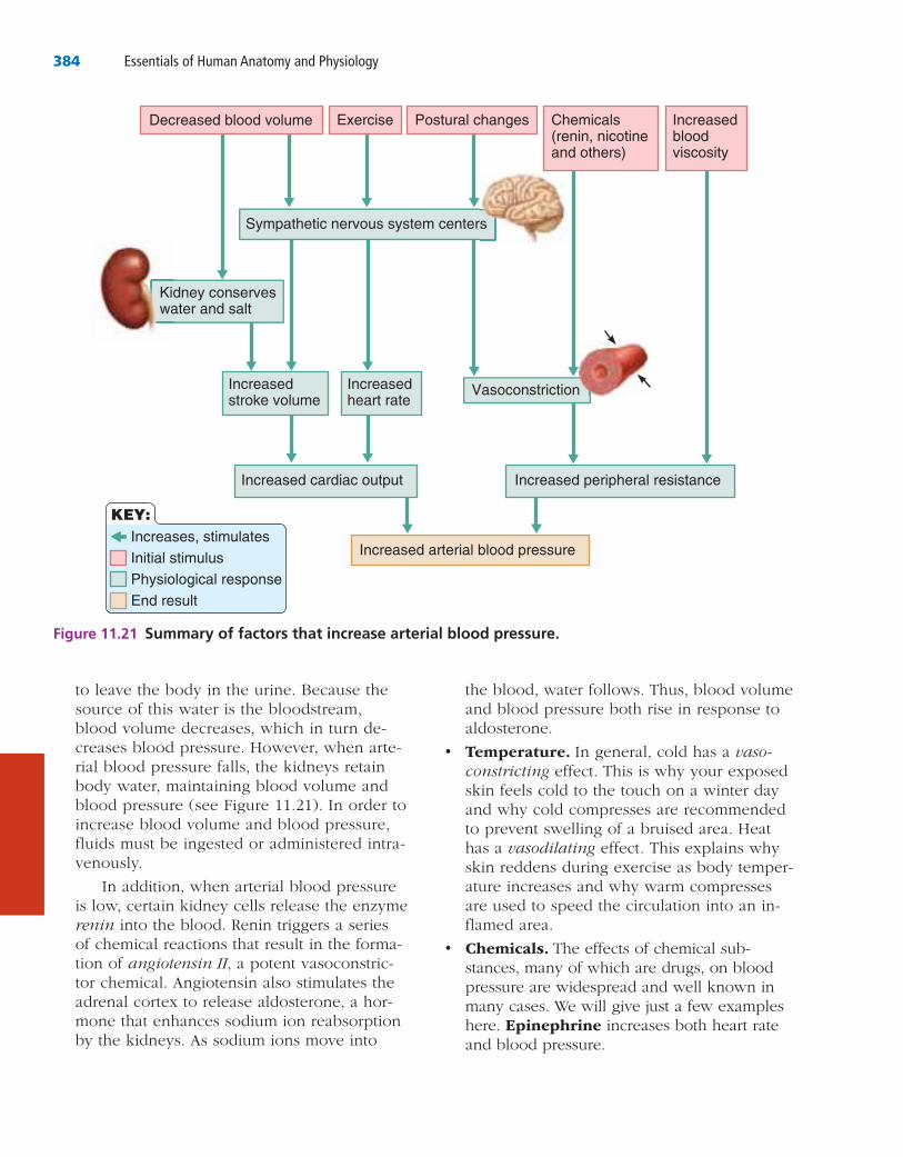
Increases, stimulatesInitial stimulusPhysiological responseEnd result
Increased arterial blood pressure
Exercise Postural changes Chemicals(renin, nicotineand others)
Increasedbloodviscosity
Decreased blood volume
Sympathetic nervous system centers
Kidney conserveswater and salt
Increasedstroke volume
Increasedheart rate
Vasoconstriction
Increased peripheral resistanceIncreased cardiac output
KEY:
Figure 11.21 Summary of factors that increase arterial blood pressure.
Chapter 11: The Cardiovascular System 385
11
tion called orthostatic hypotension. Because an aging sympathetic nervous system reacts more slowly to postural changes, blood pools briefly in the lower limbs, reducing blood pressure and, consequently, blood delivery to the brain. Making postural changes more slowly to give the nervous system time to make the necessary adjustments usually prevents this problem. ________________✚
Chronic hypotension (not explained by physi-cal conditioning) may hint at poor nutrition and inadequate levels of blood proteins. Because blood viscosity is low, blood pressure is also lower than normal. Acute hypotension is one of the most important warnings of circulatory shock, a con-dition in which the blood vessels are inadequately filled and blood cannot circulate normally. The most common cause is blood loss.
A brief elevation in blood pressure is a normal response to fever, physical exertion, and emo-tional upset, such as anger or fear. Persistent hypertension (high blood pressure), is patho-logical and is defined as a condition of sustained elevated arterial pressure of 140/90 or higher.
Homeostatic Imbalance 11.9
Chronic hypertension is a common and danger-ous disease that warns of increased peripheral resistance. Although it progresses without symp-toms for the first 10 to 20 years, it slowly and surely strains the heart and damages the arteries. For this reason, hypertension is often called the “silent killer.” Because the heart is forced to pump against increased resistance, it must work harder, and in time, the myocardium enlarges. When finally strained beyond its capacity to respond, the heart weakens and its walls become flabby. Hypertension also ravages blood vessels, causing small tears in the endothelium that accelerate the progress of atherosclerosis (the early stage of arte-riosclerosis).
Although hypertension and atherosclerosis are often linked, it is difficult to blame hypertension on any distinct anatomical pathology. In fact, about 90 percent of hypertensive people have primary (essential) hypertension, which cannot be attrib-uted to any specific organic cause. However, factors such as diet, obesity, heredity, race, and stress appear to be involved. For instance, more women than men and more blacks than whites are hypertensive.
Nicotine increases blood pressure by caus-ing vasoconstriction. Both alcohol and hista-mine cause vasodilation and decrease blood pressure. The reason a person who has “one too many” becomes flushed is that alcohol di-lates the skin vessels.
• Diet. Although medical opinions tend to change and are at odds from time to time, it is generally thought that a diet low in salt, satu-rated fats, and cholesterol helps to prevent hy-pertension, or high blood pressure.
Did You Get It?22.
For the answer, see Appendix A.
Variations in Blood Pressure In normal adults at rest, systolic blood pressure varies between 110 and 140 mm Hg, and diastolic pressure between 70 and 80 mm Hg—but blood pressure varies considerably from one person to another and cycles over a 24-hour period, peaking in the morning. What is normal for you may not be nor-mal for your grandfather or your neighbor. Blood pressure varies with age, weight, race, mood, physical activity, and posture. Nearly all these variations can be explained in terms of the factors affecting blood pressure that we have already discussed.
Hypotension, or low blood pressure, is gen-erally considered to be a systolic blood pressure below 100 mm Hg. In many cases, it simply reflects individual differences and is no cause for concern. In fact, low blood pressure is an expected result of physical conditioning and is often associated with long life and an old age free of illness.
Homeostatic Imbalance 11.8
Elderly people may experience temporary low blood pressure and dizziness when they rise sud-denly from a reclining or sitting position—a condi-
CONCEPTLINKRecall that epinephrine -
➔
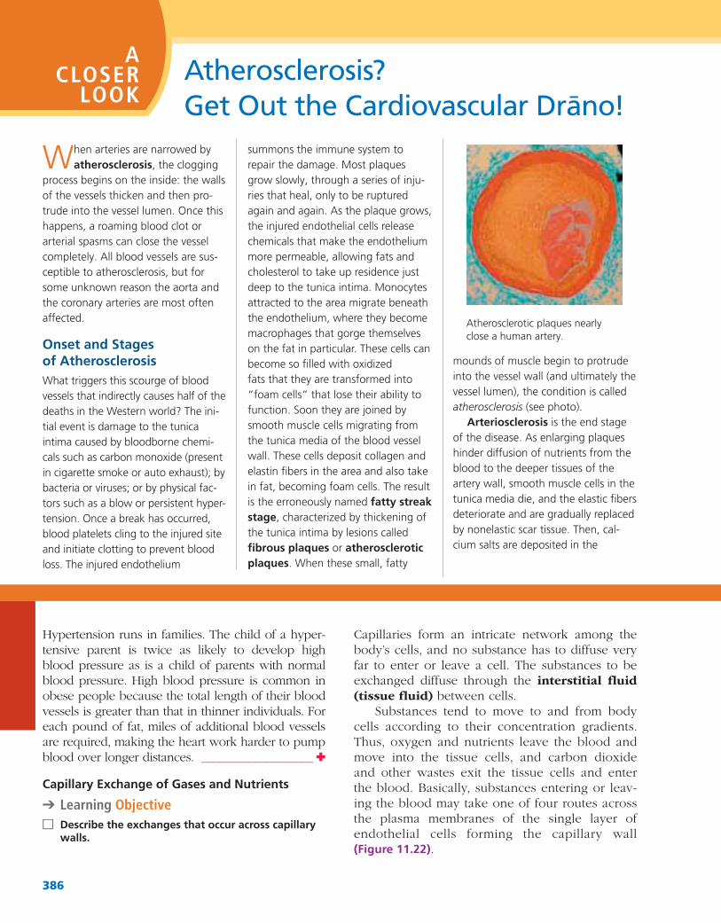
386
mounds of muscle begin to protrude into the vessel wall (and ultimately the vessel lumen), the condition is called atherosclerosis (see photo).
Arteriosclerosis is the end stage of the disease. As enlarging plaques hinder diffusion of nutrients from the blood to the deeper tissues of the artery wall, smooth muscle cells in the tunica media die, and the elastic fibers deteriorate and are gradually replaced by nonelastic scar tissue. Then, cal-cium salts are deposited in the
summons the immune system to repair the damage. Most plaques grow slowly, through a series of inju-ries that heal, only to be ruptured again and again. As the plaque grows, the injured endothelial cells release chemicals that make the endothelium more permeable, allowing fats and cholesterol to take up residence just deep to the tunica intima. Monocytes attracted to the area migrate beneath the endothelium, where they become macrophages that gorge themselves on the fat in particular. These cells can become so filled with oxidized
“foam cells” that lose their ability to function. Soon they are joined by smooth muscle cells migrating from the tunica media of the blood vessel wall. These cells deposit collagen and elastin fibers in the area and also take in fat, becoming foam cells. The result is the erroneously named fatty streak stage, characterized by thickening of the tunica intima by lesions called fibrous plaques or atherosclerotic plaques
When arteries are narrowed by atherosclerosis, the clogging
process begins on the inside: the walls of the vessels thicken and then pro-trude into the vessel lumen. Once this happens, a roaming blood clot or arterial spasms can close the vessel completely. All blood vessels are sus-ceptible to atherosclerosis, but for some unknown reason the aorta and the coronary arteries are most often affected.
Onset and Stages
What triggers this scourge of blood vessels that indirectly causes half of the deaths in the Western world? The ini-tial event is damage to the tunica intima caused by bloodborne chemi-cals such as carbon monoxide (present in cigarette smoke or auto exhaust); by bacteria or viruses; or by physical fac-tors such as a blow or persistent hyper-tension. Once a break has occurred, blood platelets cling to the injured site and initiate clotting to prevent blood loss. The injured endothelium
Atherosclerosis? A
CLOSER LOOK
Atherosclerotic plaques nearly close a human artery.
Hypertension runs in families. The child of a hyper-tensive parent is twice as likely to develop high blood pressure as is a child of parents with normal blood pressure. High blood pressure is common in obese people because the total length of their blood vessels is greater than that in thinner individuals. For each pound of fat, miles of additional blood vessels are required, making the heart work harder to pump blood over longer distances. ___________________ ✚
Capillary Exchange of Gases and Nutrients
➔ Learning Objective □ Describe the exchanges that occur across capillary
walls.
Capillaries form an intricate network among the body’s cells, and no substance has to diffuse very far to enter or leave a cell. The substances to be exchanged diffuse through the interstitial fluid (tissue fluid) between cells.
Substances tend to move to and from body cells according to their concentration gradients. Thus, oxygen and nutrients leave the blood and move into the tissue cells, and carbon dioxide and other wastes exit the tissue cells and enter the blood. Basically, substances entering or leav-ing the blood may take one of four routes across the plasma membranes of the single layer of endothelial cells forming the capillary wall
11.22).
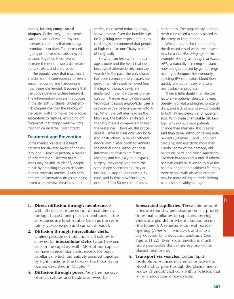
387
Sometimes after angioplasty, a metal-mesh tube called a stent is placed in the artery to keep it open.
When a blood clot is trapped by the diseased vessel walls, the answer may be a clot-dissolving agent, for example, tissue plasminogen activator (tPA), a naturally occurring substance now being produced by genetic engi-neering techniques. Intravenously injecting tPA can restore blood flow quickly and put an early end to a heart attack in progress.
There is little doubt that lifestyle factors—emotional stress, smoking, obesity, high-fat and high-cholesterol diets, and lack of exercise—contribute to both atherosclerosis and hyperten-sion. With these changeable risk fac-tors, why not just have patients change their lifestyle? This is easier said than done. Although taking anti-oxidants (vitamins C and E and beta carotene) and exercising more may “undo” some of the damage, old habits die hard, and North Americans like their burgers and butter. If athero-sclerosis could be reversed to give the heart a longer and healthier life, many more people with diseased arteries may be more willing to trade lifelong habits for a healthy old age!
statins, cholesterol-reducing drugs, show promise. Even the humble aspi-rin is gaining new respect, and many cardiologists recommend that people at high risk take one “baby aspirin” (81 mg) daily.
So what can help when the dam-age is done and the heart is at risk because of atherosclerotic coronary vessels? In the past, the only choice has been coronary artery bypass sur-gery, in which vessels removed from the legs or thoracic cavity are implanted in the heart to restore cir-culation. A more recently developed technique, balloon angioplasty, uses a catheter with a balloon packed into its tip. When the catheter reaches the blockage, the balloon is inflated, and the fatty mass is compressed against the vessel wall. However, this proce-dure is useful to clear only very local-ized obstructions. A newer catheter device uses a laser beam to vaporize the arterial clogs. Although these intravascular devices are faster, cheaper, and less risky than bypass surgery, they carry with them the same major shortcoming: they do nothing to stop the underlying dis-ease, and in time new blockages occur in 30 to 50 percent of cases.
complicated plaques. Collectively, these events cause the arterial wall to fray and ulcerate, conditions that encourage thrombus formation. The increased rigidity of the vessels leads to hyper-tension. Together, these events increase the risk of myocardial infarc-tions, strokes, and aneurysms.
The popular view that most heart attacks are the consequence of severe vessel narrowing and hardening is now being challenged. It appears that the body’s defense system betrays it. The inflammatory process that occurs in the still soft, unstable, cholesterol-rich plaques changes the biology of the vessel wall and makes the plaques susceptible to rupture, exploding off fragments that trigger massive clots that can cause lethal heart attacks.
Treatment and PreventionSome medical centers test heart patients for elevated levels of choles-terol and C-reactive protein, a marker of inflammation. Electron beam CT scans may be able to identify people
in their coronary arteries. Antibiotics and anti-inflammatory drugs are being tested as preventive measures, and
1. Direct diffusion through membrane. As with all cells, substances can diffuse directly through (cross) their plasma membranes if the substances are lipid-soluble (such as the respi-ratory gases oxygen and carbon dioxide).
2. Diffusion through intercellular clefts. Limited passage of fluid and small solutes is allowed by intercellular clefts (gaps between cells in the capillary wall). Most of our capillar-ies have intercellular clefts, except for brain capillaries, which are entirely secured together by tight junctions (the basis of the blood-brain barrier, described in Chapter 7).
3. Diffusion through pores. Very free passage of small solutes and fluids is allowed by
fenestrated capillaries. These unique capil-laries are found where absorption is a priority (intestinal capillaries or capillaries serving endocrine glands) or where filtration occurs (the kidney). A fenestra is an oval pore, or opening (fenestra = window), and is usu-ally covered by a delicate membrane (see Figure 11.22). Even so, a fenestra is much more permeable than other regions of the plasma membrane.
4. Transport via vesicles. Certain lipid- insoluble substances may enter or leave the blood and/or pass through the plasma mem-branes of endothelial cells within vesicles, that is, by endocytosis or exocytosis.
11
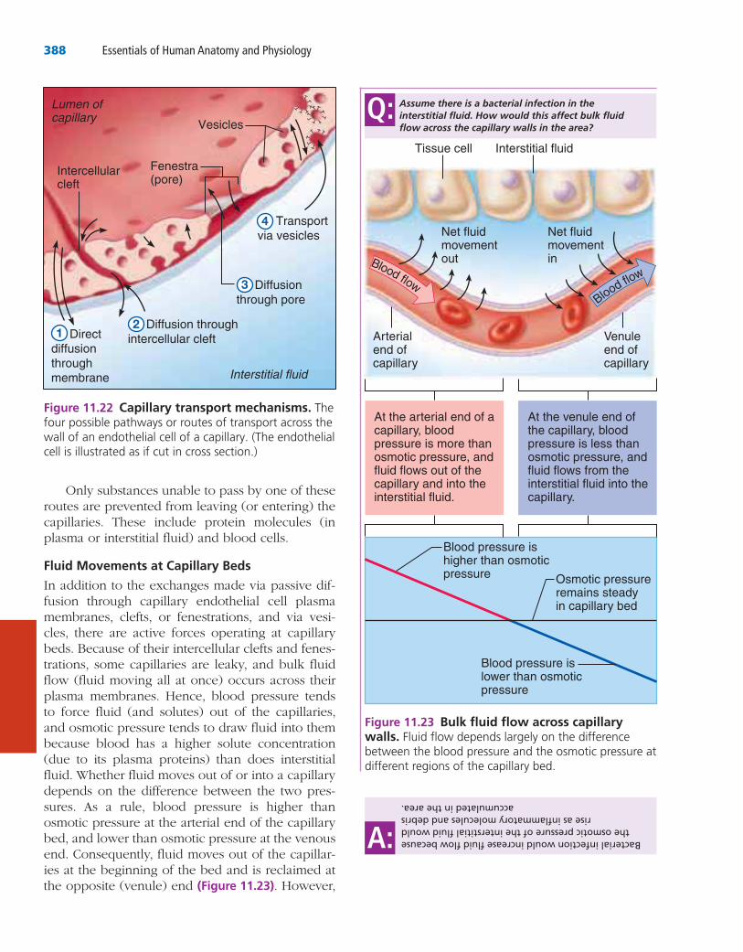
388 Essentials of Human Anatomy and Physiology
Only substances unable to pass by one of these routes are prevented from leaving (or entering) the capillaries. These include protein molecules (in plasma or interstitial fluid) and blood cells.
Fluid Movements at Capillary Beds
In addition to the exchanges made via passive dif-fusion through capillary endothelial cell plasma membranes, clefts, or fenestrations, and via vesi-cles, there are active forces operating at capillary beds. Because of their intercellular clefts and fenes-trations, some capillaries are leaky, and bulk fluid flow (fluid moving all at once) occurs across their plasma membranes. Hence, blood pressure tends to force fluid (and solutes) out of the capillaries, and osmotic pressure tends to draw fluid into them because blood has a higher solute concentration (due to its plasma proteins) than does interstitial fluid. Whether fluid moves out of or into a capillary depends on the difference between the two pres-sures. As a rule, blood pressure is higher than osmotic pressure at the arterial end of the capillary bed, and lower than osmotic pressure at the venous end. Consequently, fluid moves out of the capillar-ies at the beginning of the bed and is reclaimed at the opposite (venule) end (Figure 11.23). However,
Fenestra(pore)
Intercellularcleft
Lumen ofcapillary
Interstitial fluid
Vesicles
1 Direct diffusion through membrane
2 Diffusion through intercellular cleft
3 Diffusion through pore
4 Transport via vesicles
Figure 11.23 Bulk fluid flow across capillary walls.
Blood pressure ishigher than osmoticpressure Osmotic pressure
remains steadyin capillary bed
Blood pressure islower than osmoticpressure
At the arterial end of a capillary, blood pressure is more than osmotic pressure, and fluid flows out of the capillary and into the interstitial fluid.
At the venule end of the capillary, blood pressure is less than osmotic pressure, and fluid flows from the interstitial fluid into the capillary.
Net fluidmovement out
Arterialend ofcapillary
Venuleend ofcapillary
Net fluidmovement inBlood flow
Blood flow
Tissue cell Interstitial fluid
Assume there is a bacterial infection in the interstitial fluid. How would this affect bulk fluid flow across the capillary walls in the area?
Q:Bacterial infection would increase fluid flow because
the osmotic pressure of the interstitial fluid would rise as inflammatory molecules and debris
accumulated in the area.
A:
Figure 11.22 Capillary transport mechanisms. The
Chapter 11: The Cardiovascular System 389
11
directly into the left atrium through the foramen ovale (fo-ra′men o-val′e), a flaplike opening in the interatrial septum. Blood that does manage to enter the right ventricle is pumped out to the pul-monary trunk, where it meets a second shunt, the ductus arteriosus (ar-ter″e-o′sus), a short vessel that connects the aorta and the pulmonary trunk. Because the collapsed lungs are a high-pressure area, blood tends to enter the systemic circulation through the ductus arteriosus. The aorta carries blood to the tissues of the fetal body and ulti-mately back to the placenta through the umbilical arteries.
At birth, or shortly after, the foramen ovale closes. Its remnant, the fossa ovalis, is visible in the right atrium (see Figure 11.3b). The ductus arteriosus collapses and is converted to the fibrous ligamentum arteriosum (lig″ah-men′tum ar- ter″e-o′sum) (see Figure 11.3a). As blood stops flowing through the umbilical vessels, they col-lapse, and the circulatory pattern converts to that of an adult.
Homeostatic Imbalance 11.10
Congenital heart defects account for about half of infant deaths resulting from all congenital defects. Environmental interferences, such as maternal infec-tion and drugs ingested during the first 3 months of pregnancy (when the embryonic heart is forming), seem to be the major causes of such problems. Congenital heart defects may include a ductus arte-riosus that does not close, septal openings, and other structural abnormalities of the heart. Such problems can usually be corrected surgically. ____ ✚
In the absence of congenital heart problems, the heart usually functions smoothly throughout a long lifetime for most people. Homeostatic mecha-nisms are so effective that we rarely are aware of when the heart is working harder. The heart will hypertrophy and its cardiac output will increase substantially if we exercise regularly and aerobi-cally (that is, vigorously enough to force it to beat at a higher-than-normal rate for extended periods of time). The heart becomes not only a more pow-erful pump but also a more efficient one: pulse rate and blood pressure decrease. An added ben-efit of aerobic exercise is that it clears fatty depos-its from the blood vessel walls, helping to slow the progress of atherosclerosis. However, let’s raise a
not quite all of the fluid forced out of the blood is reclaimed at the venule end. Returning fluid left in tissues to the blood is the chore of the lymphatic system (discussed in Chapter 12).
Did You Get It?23.
For the answer, see Appendix A.
Developmental Aspects of the Cardiovascular System➔ Learning Objectives
□ Briefly describe the development of the cardiovascular system.
□ Name the fetal vascular modifications, or “fetal shunts,” and describe their function before birth.
□ Describe changes in the cardiovascular system that occur with aging, and list several factors that help maintain cardiovascular health.
The heart begins as a simple tube in the embryo. It is beating and busily pumping blood by the fourth week of pregnancy. During the next 3 weeks, the heart continues to change and mature, finally becoming a four-chambered structure capa-ble of acting as a double pump—all without miss-ing a beat!
Because the lungs and digestive system are immature and not functioning in a fetus, all nutri-ent, excretory, and gas exchanges occur through the placenta. Nutrients and oxygen move from the mother’s blood into the fetal blood, and fetal wastes move in the opposite direction. The umbil-ical cord contains three blood vessels: one large umbilical vein and two smaller umbilical arter-ies (Figure 11.24, p. 390). The umbilical vein car-ries blood rich in nutrients and oxygen to the fetus. The umbilical arteries carry blood laden with carbon dioxide and metabolic waste products from the fetus to the placenta. As blood flows superiorly toward the heart of the fetus, most of it bypasses the immature liver through the ductus venosus (duk′tus ve-no′sus) and enters the infe-rior vena cava, which carries the blood to the right atrium of the heart.
Because the nonfunctional fetal lungs are col-lapsed, two shunts (diverting vessels) make sure that they are almost entirely bypassed. Some of the blood entering the right atrium is shunted
(Text continues on page 392.)
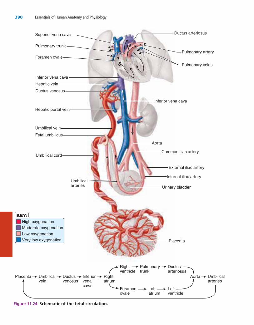
390 Essentials of Human Anatomy and Physiology
Superior vena cava
Foramen ovale
Pulmonary trunk
Inferior vena cava
Hepatic vein
Ductus venosus
Hepatic portal vein
Umbilical vein
Fetal umbilicus
Umbilical cord
Umbilicalarteries
Ductus arteriosus
Pulmonary veins
Pulmonary artery
Inferior vena cava
Aorta
Common iliac artery
External iliac artery
Internal iliac artery
Urinary bladder
Placenta
Placenta Umbilicalvein
Ductusvenosus
Inferiorvenacava
Rightatrium
Aorta Umbilicalarteries
Rightventricle
Pulmonarytrunk
Ductusarteriosus
Foramenovale
Leftatrium
Leftventricle
High oxygenation
Moderate oxygenation
Low oxygenation
Very low oxygenation
KEY:
Figure 11.24 Schematic of the fetal circulation.
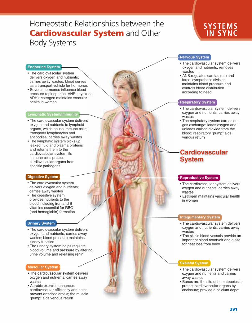
Cardiovascular SystemSYSTEMS IN SYNC
391
Integumentary System
Urinary System
Muscular System
Lymphatic System/Immunity
Digestive System Reproductive System
Respiratory System
Skeletal System
Endocrine System
Nervous System
CardiovascularSystem
392 Essentials of Human Anatomy and Physiology
hypertensive heart disease. The insidious filling of the blood vessels with fatty, calcified deposits leads most commonly to coronary artery dis-ease. Also, the roughening of the vessel walls encourages thrombus formation (see Chapter 10). At least 30 percent of the population in the United States has hypertension by the age of 50, and car-diovascular disease causes more than one-half of the deaths in people over age 65. Although the aging process itself contributes to changes in the walls of the blood vessels that can lead to strokes or myocardial infarctions, most researchers feel that diet, not aging, is the single most important contributing factor to cardiovascular diseases. There is some agreement that the risk is lowered if people eat less animal fat, cholesterol, and salt. Other recommendations include avoiding stress, eliminating cigarette smoking, and taking part in a regular, moderate exercise program.
caution flag here: The once-a-month or once-a-year tennis player or snow shoveler has not built up this type of heart endurance and strength. When such an individual pushes his or her heart too much, it may not be able to cope with the sud-den demand. This is why many “weekend ath-letes” are myocardial infarction (heart attack) victims.
As we get older, more and more signs of car-diovascular system disturbances start to appear. In some people, the venous valves weaken, and pur-ple, snakelike varicose veins appear. Not everyone has varicose veins, but we all have progressive atherosclerosis. Some say the process begins at birth, and even children’s arteries show atheroscle-rotic plaques. There’s an old saying, “You are only as old as your arteries,” referring to this degenerative process. The gradual loss in elasticity of the blood vessels leads to hypertension and
of the right and left coronary arteries and their
branches, and is drained by the cardiac veins and
the coronary sinus.
6. Cardiac muscle is able to initiate its own contrac-
tion in a regular way, but its rate is influenced by
both intrinsic and extrinsic factors. The intrinsic
conduction system increases the rate of heart
contraction and ensures that the heart beats as a
unit. The SA node is the heart’s pacemaker.
Extrinsic factors include neural and hormonal
stimuli.
Summary
The Heart (pp. 357–370)
1. The heart, located in the thorax, is flanked laterally
by the lungs and enclosed in a multi-layered peri-
cardium.
2. The bulk of the heart wall (myocardium) is com-
posed of cardiac muscle. The heart has four hollow
chambers—two atria (receiving chambers) and two
ventricles (discharging chambers), each lined with
endocardium. (The heart is divided longitudinally
by a septum.)
3. The heart functions as a double pump. The right
ventricle is the pulmonary pump (right ventricle to
lungs to left atrium). The left ventricle is the systemic
pump (left ventricle to body tissues to right atrium).
4. Four valves prevent backflow of blood in the
heart. The AV valves (mitral, or bicuspid, and tri-
cuspid) prevent backflow into the atria when the
ventricles are contracting. The semilunar valves
(pulmonary and aortic) prevent backflow into the
ventricles when the heart is relaxing. The valves
open and close in response to pressure changes in
the heart.
5. The myocardium is nourished by the coronary cir-
culation, which branches off the aorta and consists
Complete an interactive tutorial: > Study Area > Interactive Physiology > Cardiovascular System > Electrical Activity of the Heart
7. The time and events occurring from one heartbeat
to the next are the cardiac cycle.
8. As the heart beats, sounds resulting from the clos-
ing of the valves (“lub-dup”) can be heard. Faulty
valves reduce the efficiency of the heart as a pump
and result in abnormal heart sounds (murmurs).
Complete an interactive tutorial: > Study Area > Interactive Physiology > Cardiovascular System > Cardiac Cycle
Chapter 11: The Cardiovascular System 393
11
veins superior to the diaphragm drain into the
superior vena cava, and those inferior to the
diaphragm drain into the inferior vena cava. Both
venae cavae enter the right atrium of the heart. (See
pp. 374–377 for the names and locations of the sys-
temic veins.)
8. The arterial circulation of the brain is formed by
branches of paired vertebral and internal carotid
arteries. The cerebral arterial circle provides alter-
nate routes for blood flow in case of a blockage in
the brain’s arterial supply.
9. The hepatic portal circulation is formed by veins
draining the digestive organs, which empty into the
hepatic portal vein. The hepatic portal vein carries
the nutrient-rich blood to the liver, where it is pro-
cessed before the blood is allowed to enter the
systemic circulation.
10. The pulse is the alternating expansion and recoil of
a blood vessel wall (the pressure wave) that occurs
as the heart beats. It may be felt easily over any
superficial artery; such sites are called pressure
points.
11. Blood pressure is the pressure that blood exerts on
the walls of the blood vessels. It is the force that
causes blood to flow down its pressure gradient in
the blood vessels. It is high in the arteries, lower in
the capillaries, and lowest in the right atrium. Both
systolic and diastolic pressures are recorded when
measuring blood pressure.
12. Arterial blood pressure is directly influenced by
heart activity (increased heart rate leads to
increased blood pressure) and by resistance to
blood flow. The most important factors increasing
the peripheral resistance are a decrease in the
diameter or stretchiness of the arteries and arteri-
oles and an increase in blood viscosity.
13. Many factors influence blood pressure, including
the activity of the sympathetic nerves and kidneys,
drugs, and diet.
10. SV rises or falls with the volume of venous return.
HR is influenced by the nerves of the autonomic
nervous system, drugs (and other chemicals), and
ion levels in the blood.
Blood Vessels (pp. 370–389)
1. Arteries, which transport blood away from the
heart, and veins, which carry blood back to the
heart, are conducting vessels. Only capillaries play
a role in actual exchanges with tissue cells.
2. Except for capillaries, blood vessels are composed
of three tunics: The tunica intima forms a friction-
reducing lining for the vessel. The tunica media is
the bulky middle layer of muscle and elastic tissue.
The tunica externa is the protective, outermost con-
nective tissue layer. Capillary walls are formed of
the tunica intima only.
3. Artery walls are thick and strong to withstand
blood pressure fluctuations. They expand and
recoil as the heart beats. Vein walls are thinner,
their lumens are larger, and they are equipped with
valves. These modifications reflect the low pressure
of the blood flowing in veins.
4. Capillary beds have two types of vessels—a vascu-
lar shunt and true capillaries. Blood flow into true
capillaries is controlled by precapillary sphincters.
Exchanges with tissue cells occur across the walls
of the true capillaries. When the precapillary
sphincters are closed, blood flows directly through
the vascular shunt.
5. Varicose veins, a structural defect due to incompe-
tent valves, is a common vascular problem, espe-
cially in people who are obese or who stand for
long hours. It is a predisposing factor for thrombo-
phlebitis.
6. All the major arteries of the systemic circulation are
branches of the aorta, which leaves the left ventri-
cle. They branch into smaller arteries and then into
the arterioles, which feed the capillary beds of the
body tissues. (For the names and locations of the
systemic arteries, see pp. 373–375.)
7. The major veins of the systemic circulation ulti-
mately converge on one of the venae cavae. All
Complete an interactive tutorial: > Study Area > Interactive Physiology > Cardiovascular System > Factors that Affect Blood Pressure
14. Hypertension, which reflects an increase in periph-
eral resistance, strains the heart and damages blood
vessels. In most cases, the precise cause is
unknown.
15. Substances move to and from the blood and tissue
cells through capillary walls. Some substances
are transported in vesicles, but most move by
diffusion—directly through the endothelial cell
Complete an interactive tutorial: > Study Area > Interactive Physiology > Cardiovascular System > Cardiac Output
9. Cardiac output, the amount of blood pumped out by
each ventricle in one minute, is the product of heart
rate (HR) × stroke volume (SV). SV is the amount of
blood ejected by a ventricle with each beat.
394 Essentials of Human Anatomy and Physiology
the fetus to the placenta. Shunts bypassing the
lungs and liver are also present.
3. Congenital heart defects account for half of all
infant deaths resulting from congenital problems.
4. Arteriosclerosis is an expected consequence of
aging. Gradual loss of elasticity in the arteries leads
to hypertension and hypertensive heart disease,
and clogging of the vessels with fatty substances
leads to coronary artery disease and stroke.
Cardiovascular disease is an important cause of
death in individuals over age 65.
5. A healthy diet low in fat, cholesterol, and salt; stop-
ping smoking; and regular aerobic exercise may
help to reverse the atherosclerotic process and pro-
long life.
plasma membranes, through intercellular clefts, or
through fenestrations. Fluid is forced from the
bloodstream by blood pressure and drawn back
into the blood by osmotic pressure.
Developmental Aspects of the Cardiovascular System (pp. 389–392)
1. The heart begins as a tubelike structure that is beat-
ing and pumping blood by the fourth week of
embryonic development.
2. The fetal circulation is a temporary circulation
seen only in the fetus. It consists primarily of three
special vessels: the single umbilical vein that carries
nutrient- and oxygen-laden blood to the fetus from
the placenta, and the two umbilical arteries that
carry carbon dioxide and waste-laden blood from
Multiple Choice
More than one choice may apply.
1. Pulmonary veins deliver freshly oxygenated blood
from the lungs to the
a. right ventricle.
b. left ventricle.
c. right atrium.
d. left atrium.
2. Given a volume of 150 ml at the end of diastole, a
volume of 50 ml at the end of systole, and a heart
rate of 60 bpm, the cardiac output is
a. 600 ml/min.
b. 6 liters/min.
c. 1200 ml/min.
d. 3 liters/min.
3. Which of the following depolarizes next after the
AV node?
a. Atrial myocardium
b. Ventricular myocardium
c. Bundle branches
d. AV bundle
4. During atrial systole,
a. the atrial pressure exceeds ventricular pressure.
b. 70 percent of ventricular filling occurs.
c. the AV valves are open.
d. valves prevent backflow into the great veins.
5. Atrial repolarization coincides in time with the
a. P wave.
b. T wave.
c. QRS wave.
d. P-Q interval.
6. Soon after the onset of ventricular systole, the
a. AV valves close.
b. semilunar valves open.
c. first heart sound is heard.
d. aortic pressure increases.
7. The base of the heart is its ______ surface.
a. diaphragmatic
b. superior
c. anterior
d. inferior
Review Questions Access additional practice questions using your smartphone, tablet, or computer: > Study Area > Practice Tests & Quizzes
Chapter 11: The Cardiovascular System 395
11
15. The most external part of the pericardium is the
a. parietal layer of serous pericardium.
b. fibrous pericardium.
c. visceral layer of serous pericardium.
d. epicardium.
16. Which heart chamber pumps blood with the
greatest amount of force?
a. Right atrium
b. Right ventricle
c. Left atrium
d. Left ventricle
17. How many cusps does the right atrioventricular
valve have?
a. Two
b. Three
c. Four
d. Six
18. Which layer of the heart wall is an endothelium?
a. Endocardium
b. Myocardium
c. Epicardium
d. Pericardium
Short Answer Essay
19. Draw a diagram of the heart showing the three
layers composing its wall and its four chambers.
Label each. Show where the AV and semilunar
valves are, and name them. Show and label all
blood vessels entering and leaving the heart
chambers.
20. Trace one drop of blood from the time it enters the
right atrium of the heart until it enters the left
atrium. What is this circuit called?
21. What is the function of the fluid that fills the
pericardial sac?
22. Define systole and diastole.
23. Define stroke volume and cardiac cycle.
24. How does the heart’s ability to contract differ from
that of other muscles of the body?
25. Name the elements of the intrinsic conduction
system, in order, beginning with the pacemaker.
26. Name three different factors that increase heart
rate.
8. In comparing a parallel artery and vein, you would
find that
a. the artery wall is thicker.
b. the artery diameter is greater.
c. the artery lumen is smaller.
d. the artery endothelium is thicker.
9. Which of these vessels is bilaterally symmetrical
(i.e., one vessel of the pair occurs on each side of
the body)?
a. Internal carotid artery
b. Brachiocephalic trunk
c. Azygos vein
d. Renal vein
10. A stroke that occludes a posterior cerebral artery
will most likely affect
a. hearing.
b. vision.
c. smell.
d. higher thought processes.
11. Vessels involved in the circulatory pathway to and/
or from the brain are the
a. brachiocephalic trunk.
b. subclavian artery.
c. internal jugular vein.
d. internal carotid artery.
12. Which layer of the artery wall thickens most in
atherosclerosis?
a. Tunica media
b. Tunica intima
c. Tunica adventitia
d. Tunica externa
13. Which of the following are associated with aging?
a. Increasing blood pressure
b. Weakening of venous valves
c. Arteriosclerosis
d. Collapse of the ductus arteriosus
14. An increase in BP would be caused by all of the
following except
a. increase in SV.
b. increase in heart rate.
c. hemorrhage.
d. vasoconstriction of the arterioles.
396 Essentials of Human Anatomy and Physiology
36. Which artery is palpated at the front of the ear? At
the back of the knee?
37. Define systolic pressure and diastolic pressure.
38. Two elements determine blood pressure—the
cardiac output of the heart and the peripheral
resistance, or friction, in the blood vessels. Name
two factors that increase cardiac output. Name two
factors that increase peripheral resistance.
39. In which position—sitting, lying down, or standing—
is the blood pressure normally highest? Lowest?
Explain why.
40. What is different about the capillary exchanges
seen in a capillary with fenestrations and in-
tercellular clefts and the exchanges seen in a cap-
illary lacking those modifications?
41. What are varicose veins? What factors seem to
promote their formation?
42. Explain why blood flow in arteries is pulsatile and
blood flow in veins is not.
43. What is the relationship between cross-sectional
area of a blood vessel and velocity (speed) of
blood flow in that vessel?
44. Which type of blood vessel is most important in
regulating vascular resistance, and how does it
achieve this?
45. What is the ductus venosus, and what is its
function?
27. Name and describe, from the inside out, the three
tunics making up the walls of arteries and veins,
and give the most important function of each layer.
28. Describe the structure of capillary walls.
29. Why are artery walls so much thicker than those of
corresponding veins?
30. Name three factors that are important in promoting
venous return.
31. Arteries are often described as vessels that carry
oxygen-rich blood, and veins are said to carry
oxygen-poor (carbon dioxide–rich) blood. Name
two sets of exceptions to this rule that were
discussed in this chapter.
32. Trace a drop of blood from the left ventricle of the
heart to the wrist of the right hand and back to the
heart. Now trace it to the dorsum of the right foot
and back to the right heart.
33. What is the function of the hepatic portal
circulation? In what way is a portal circulation a
“strange” circulation?
34. In a fetus, the liver and lungs are almost entirely
bypassed by blood. Why is this? Name the vessel
that bypasses the liver. Name two lung bypasses.
Three vessels travel in the umbilical cord; which of
these carries oxygen- and nutrient-rich blood?
35. Define pulse. Palpate your pulse. Which pulse point
did you use?
46. John is a 30-year-old man who is overweight and
smokes. He has been diagnosed with hypertension
and arteriosclerosis. Define each of these conditions.
How are they often related? Why is hypertension
called the “silent killer”? Name three changes in
your lifestyle that might help prevent cardiovascular
disease in your old age.
47. Mrs. Hamad, a middle-aged woman, is admitted to
the coronary care unit with a diagnosis of left
ventricular failure resulting from a myocardial
infarction. Her chart indicates that she was
awakened in the middle of the night by severe
chest pain. Her skin is pale and cold, and moist
sounds of pulmonary edema are heard over the
lower regions of both lungs. Explain how failure of
the left ventricle might cause these signs and
symptoms.
48. Hannah, a 14-year-old girl undergoing a physical
examination before being admitted to summer
camp, was found to have a loud heart murmur at
the second intercostal space on the left side of the
sternum. The murmur takes the form of a swishing
sound with no high-pitched whistle. What, exactly,
is producing the murmur?
49. Mrs. Rees is brought to the emergency room after
being involved in an auto accident. She is
hemorrhaging and has a rapid pulse that can barely
be felt, but her blood pressure is still within normal
limits. Describe the compensatory mechanisms that
are maintaining her blood pressure in the face of
blood loss.
50. During a lethal heart attack, a blood clot lodges in
the first part of the circumflex branch of the left
Critical Thinking and Clinical Application Questions
Chapter 11: The Cardiovascular System 397
11
52. Grandma tells Hailey not to swim for 30 minutes
after eating. Explain why taking a vigorous swim
immediately after lunch is more likely to cause
indigestion than cramping of your muscles.
53. The guards at the royal palace in London stand at
attention while on duty. On a very hot day, it is not
unusual for one (or more) to become lightheaded
and faint. Explain this phenomenon.
coronary artery, blocking blood flow through this
vessel. What parts of the heart will become
ischemic and die?
51. Mr. Grimaldi was previously diagnosed as having a
posterior pituitary tumor that causes hypersecretion
of ADH. He comes to the clinic regularly to have
his blood pressure checked. Would you expect his
blood pressure to be chronically elevated or
depressed? Why?