Embed Size (px)
Citation preview
The Cardiovascular system
Functions of the circulatory system• Transport
– Erythrocytes (red blood cells, RBC’s) carry oxygen from lungs, remove CO2 from tissues
– Nutrients, hormones etc. all carried by the fluid portion of blood (NOT RBC’s)
– Metabolic wastes from body tissues delivered to renals
• Protection– “White” cells (immune cells)– Antibodies, inflammatory mediators (cytokines), blood clotting factors
• Regulation– Constant flow helps to stabilize fluid and fluid ingredient distribution
(mixes everything equally)– Buffers pH changes in tissue– Buffers temperature changes

The Heart
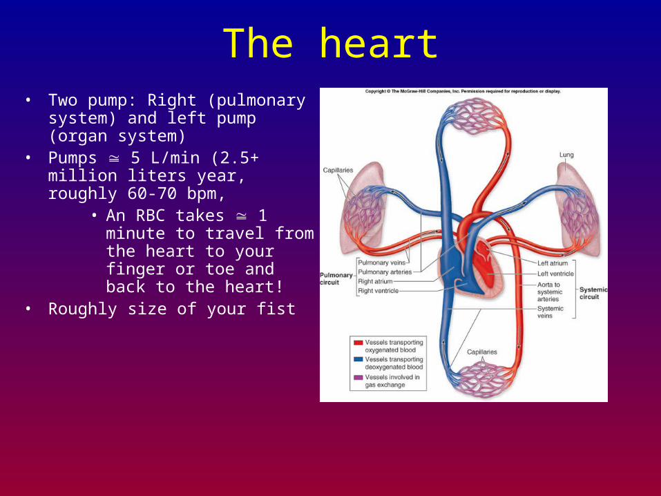
The heart• Two pump: Right (pulmonary system)
and left pump (organ system)• Pumps 5 L/min (2.5+ million liters
year, roughly 60-70 bpm, • An RBC takes 1 minute to
travel from the heart to your finger or toe and back to the heart!
• Roughly size of your fist
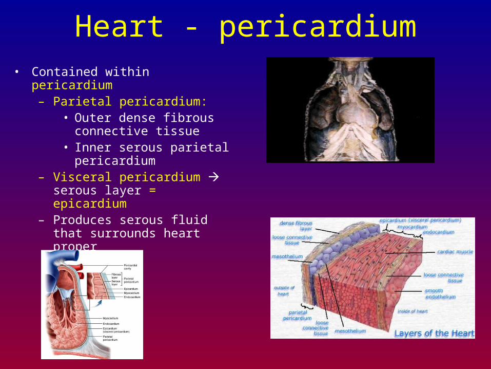
Heart - pericardium• Contained within pericardium
– Parietal pericardium: • Outer dense fibrous connective
tissue• Inner serous parietal
pericardium– Visceral pericardium serous layer
= epicardium– Produces serous fluid that surrounds
heart proper
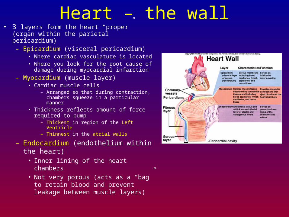
Heart – the wall• 3 layers form the heart “proper” (organ within
the parietal pericardium)– Epicardium (visceral pericardium)
• Where cardiac vasculature is located• Where you look for the root cause of
damage during myocardial infarction
– Myocardium (muscle layer)• Cardiac muscle cells
– Arranged so that during contraction, chambers squeeze in a particular manner
• Thickness reflects amount of force required to pump
– Thickest in region of the Left Ventricle– Thinnest in the atrial walls
– Endocardium (endothelium within the heart)
• Inner lining of the heart chambers
• Not very porous (acts as a “bag” to retain blood and prevent leakage between muscle layers)
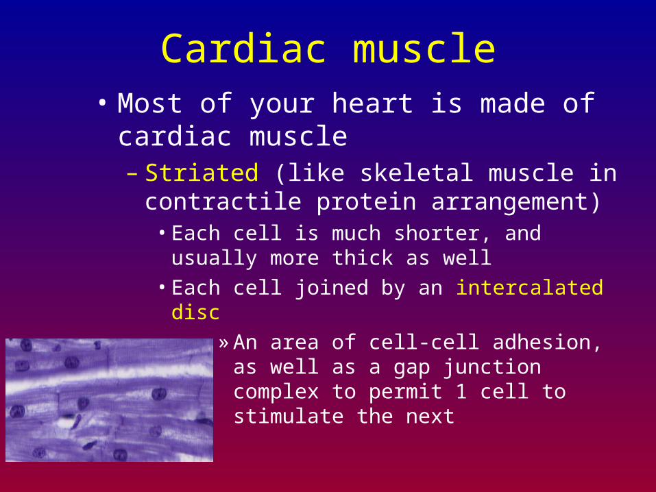
Cardiac muscle• Most of your heart is made of cardiac
muscle– Striated (like skeletal muscle in contractile
protein arrangement)• Each cell is much shorter, and usually more thick as
well
• Each cell joined by an intercalated disc
» An area of cell-cell adhesion, as well as a gap junction complex to permit 1 cell to stimulate the next
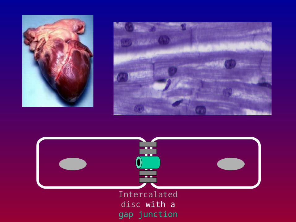
Intercalated disc with a gap junction
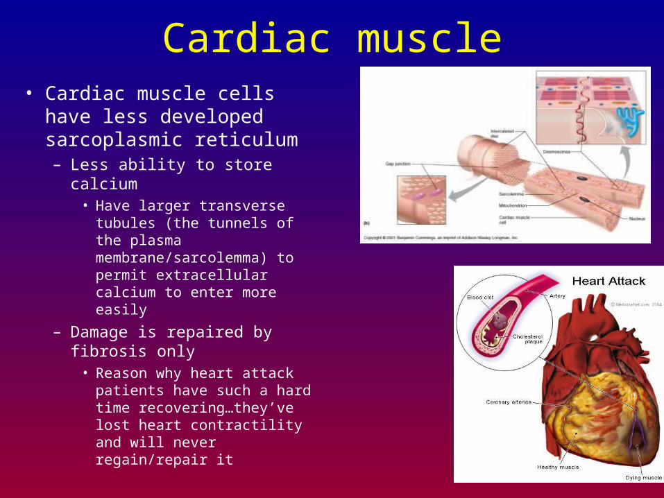
Cardiac muscle• Cardiac muscle cells have less
developed sarcoplasmic reticulum– Less ability to store calcium
• Have larger transverse tubules (the tunnels of the plasma membrane/sarcolemma) to permit extracellular calcium to enter more easily
– Damage is repaired by fibrosis only
• Reason why heart attack patients have such a hard time recovering…they’ve lost heart contractility and will never regain/repair it
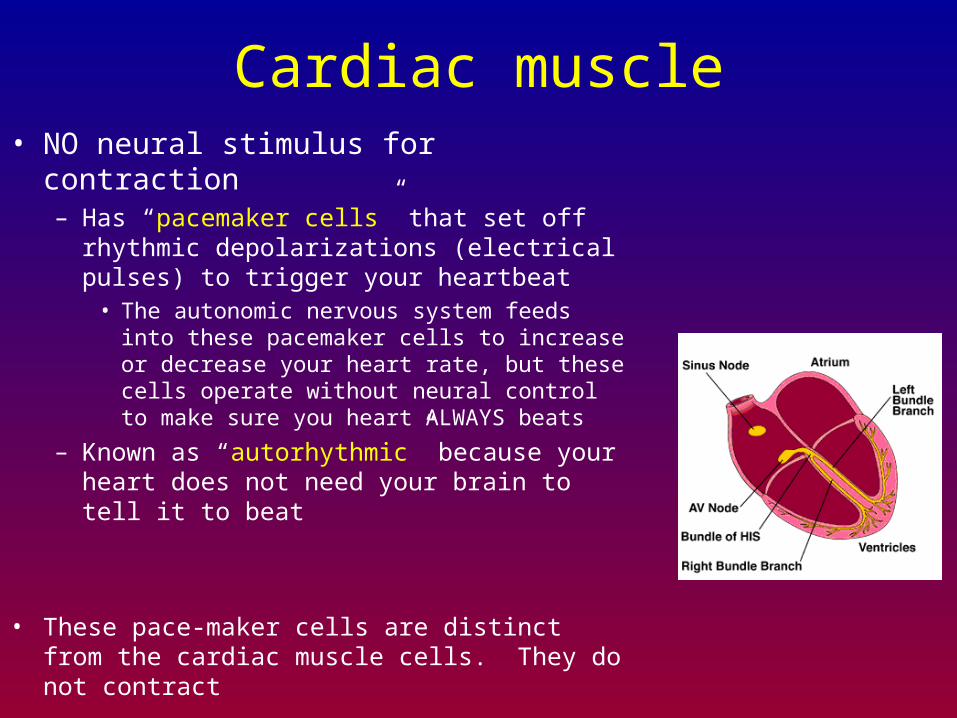
Cardiac muscle• NO neural stimulus for contraction
– Has “pacemaker cells” that set off rhythmic depolarizations (electrical pulses) to trigger your heartbeat
• The autonomic nervous system feeds into these pacemaker cells to increase or decrease your heart rate, but these cells operate without neural control to make sure you heart ALWAYS beats
– Known as “autorhythmic” because your heart does not need your brain to tell it to beat
• These pace-maker cells are distinct from the cardiac muscle cells. They do not contract
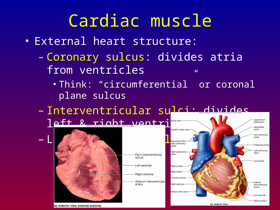
Cardiac muscle• External heart structure:
– Coronary sulcus: divides atria from ventricles• Think: “circumferential” or coronal plane sulcus
– Interventricular sulci: divides left & right ventricles
– Look for “adipose lines”

Cardiac muscle• External heart structure:
– Various sulci serve as “routes” for cardiac blood vessels
• Cardiac muscle reliant on cardiac blood vessels for blood supply (endocardium does not allow for much fluid or gas exchange within the heart)
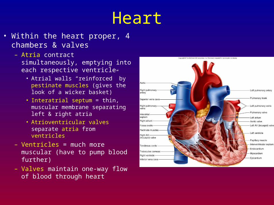
Heart• Within the heart proper, 4
chambers & valves– Atria contract simultaneously,
emptying into each respective ventricle
• Atrial walls “reinforced” by pestinate muscles (gives the look of a wicker basket)
• Interatrial septum = thin, muscular membrane separating left & right atria
• Atrioventricular valves separate atria from ventricles
– Ventricles = much more muscular (have to pump blood further)
– Valves maintain one-way flow of blood through heart
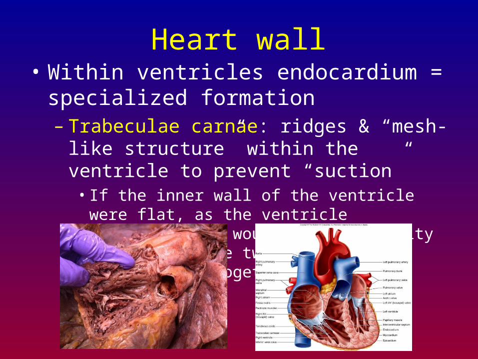
Heart wall• Within ventricles endocardium = specialized
formation– Trabeculae carnae: ridges & “mesh-like structure”
within the ventricle to prevent “suction”• If the inner wall of the ventricle were flat, as the
ventricle contracted, it would have difficulty relaxing as the two flat surfaces would adhere together
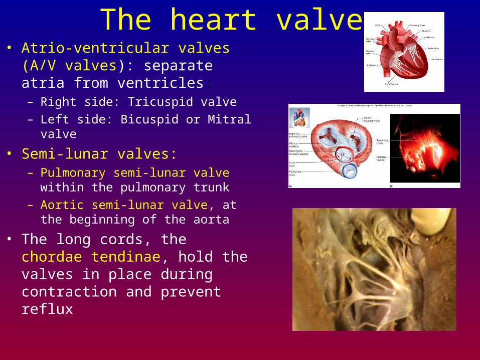
The heart valves• Atrio-ventricular valves (A/V
valves): separate atria from ventricles:– Right side: Tricuspid valve
– Left side: Bicuspid or Mitral valve
• Semi-lunar valves:– Pulmonary semi-lunar valve within the
pulmonary trunk
– Aortic semi-lunar valve, at the beginning of the aorta
• The long cords, the chordae tendinae, hold the valves in place during contraction and prevent reflux
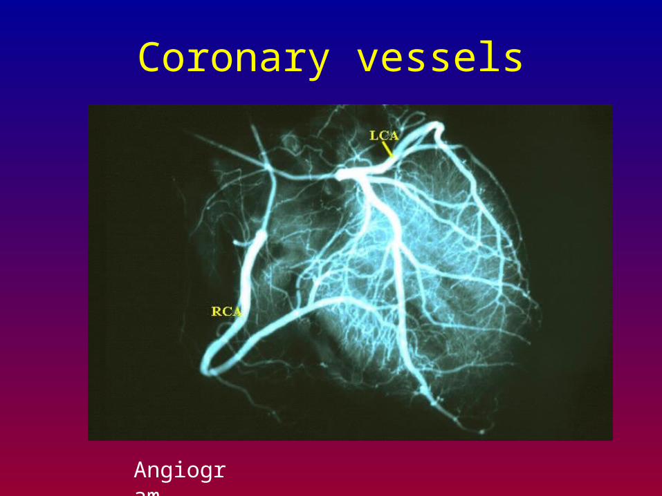
Coronary vessels
Angiogram
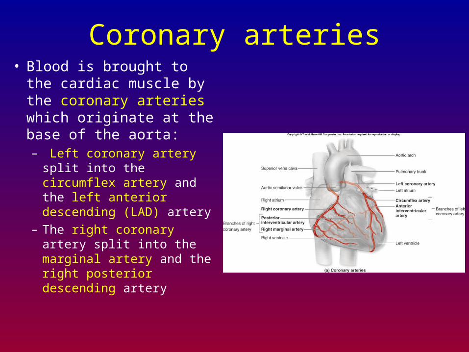
Coronary arteries• Blood is brought to the
cardiac muscle by the coronary arteries which originate at the base of the aorta:– Left coronary artery split
into the circumflex artery and the left anterior descending (LAD) artery
– The right coronary artery split into the marginal artery and the right posterior descending artery
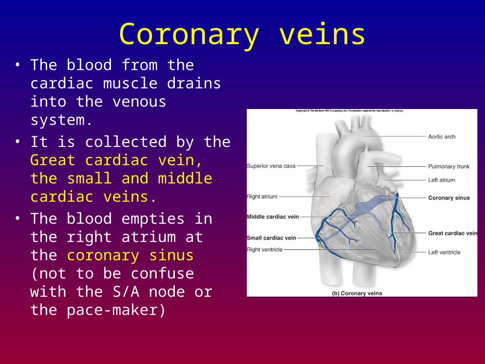
Coronary veins• The blood from the cardiac
muscle drains into the venous system.
• It is collected by the Great cardiac vein, the small and middle cardiac veins.
• The blood empties in the right atrium at the coronary sinus (not to be confuse with the S/A node or the pace-maker)
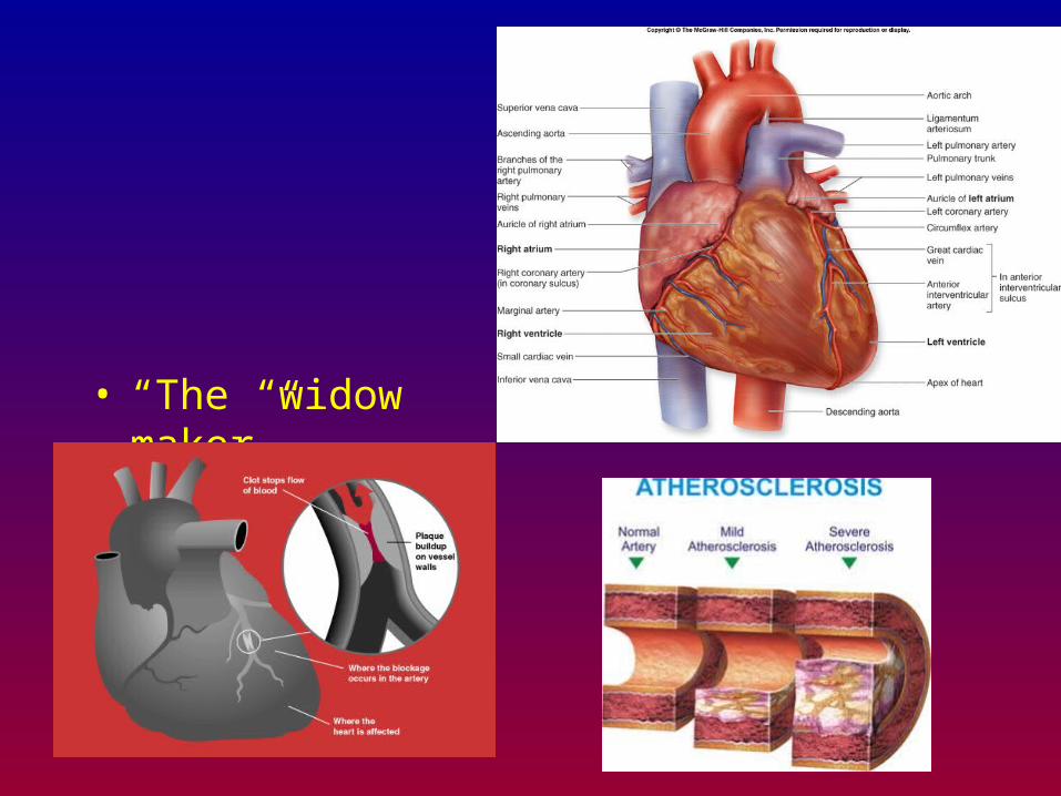
• “The “widow maker”
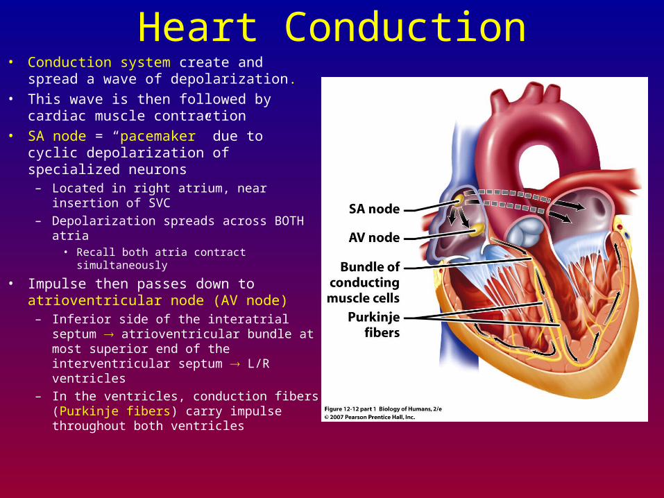
Heart Conduction• Conduction system create and spread a
wave of depolarization.
• This wave is then followed by cardiac muscle contraction
• SA node = “pacemaker” due to cyclic depolarization of specialized neurons
– Located in right atrium, near insertion of SVC
– Depolarization spreads across BOTH atria• Recall both atria contract simultaneously
• Impulse then passes down to atrioventricular node (AV node)
– Inferior side of the interatrial septum atrioventricular bundle at most superior end of the interventricular septum L/R ventricles
– In the ventricles, conduction fibers (Purkinje fibers) carry impulse throughout both ventricles
Heart conduction
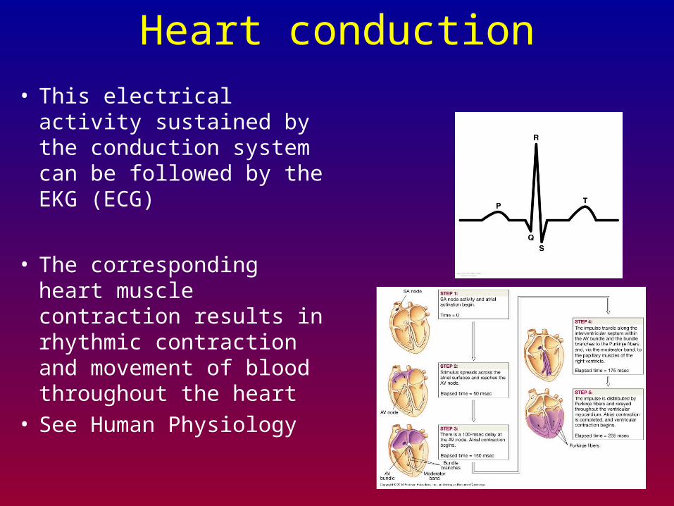
• This electrical activity sustained by the conduction system can be followed by the EKG (ECG)
• The corresponding heart muscle contraction results in rhythmic contraction and movement of blood throughout the heart
• See Human Physiology

“lub”
“dub”
“systole”
“diastole”
Blood vessels
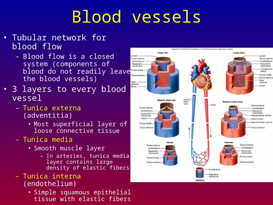
Blood vessels• Tubular network for blood flow
– Blood flow is a closed system (components of blood do not readily leave the blood vessels)
• 3 layers to every blood vessel– Tunica externa (adventitia)
• Most superficial layer of loose connective tissue
– Tunica media• Smooth muscle layer
– In arteries, tunica media layer contains large density of elastic fibers
– Tunica interna (endothelium)• Simple squamous epithelial tissue
with elastic fibers

ArteriesElastic fibers within the tunica
media– Allows artery to expand when
heart expels blood
– Elastic fibers permit recoil to original shape following expulsion of blood from heart
– Expansion & recoil (elasticity) acts to smooth out the blood flow (less pulsing)
– As arteries reduce in size, they become less elastic
• Small arteries, arterioles are less elastic than arteries
• Capillary = 7-10 m diameter
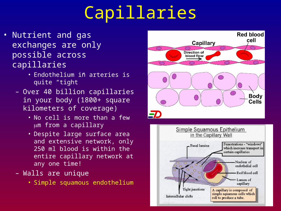
Capillaries• Nutrient and gas exchanges are
only possible across capillaries• Endothelium in arteries is quite
“tight”
– Over 40 billion capillaries in your body (1800+ square kilometers of coverage)
• No cell is more than a few m from a capillary
• Despite large surface area and extensive network, only 250 ml blood is within the entire capillary network at any one time!
– Walls are unique• Simple squamous endothelium
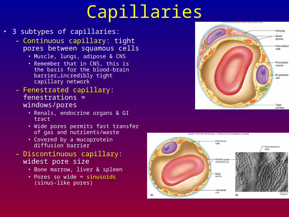
Capillaries• 3 subtypes of capillaries:
– Continuous capillary: tight pores between squamous cells
• Muscle, lungs, adipose & CNS• Remember that in CNS, this is the
basis for the blood-brain barrier…incredibly tight capillary network
– Fenestrated capillary: fenestrations = windows/pores
• Renals, endocrine organs & GI tract• Wide pores permits fast transfer of gas
and nutrients/waste• Covered by a mucoprotein diffusion
barrier
– Discontinuous capillary: widest pore size
• Bone marrow, liver & spleen• Pores so wide = sinusoids (sinus-like
pores)
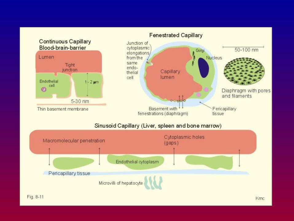
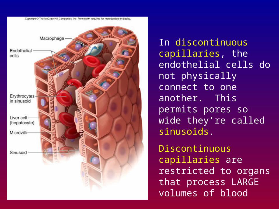
In discontinuous capillaries, the endothelial cells do not physically connect to one another. This permits pores so wide they’re called sinusoids.
Discontinuous capillaries are restricted to organs that process LARGE volumes of blood
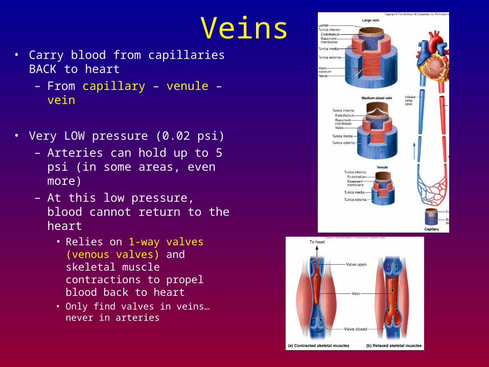
Veins• Carry blood from capillaries BACK
to heart
– From capillary – venule – vein
• Very LOW pressure (0.02 psi)
– Arteries can hold up to 5 psi (in some areas, even more)
– At this low pressure, blood cannot return to the heart
• Relies on 1-way valves (venous valves) and skeletal muscle contractions to propel blood back to heart
• Only find valves in veins…never in arteries
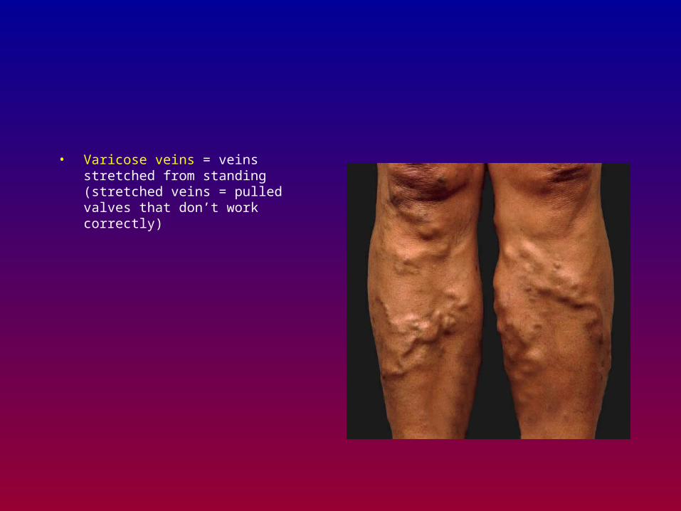
• Varicose veins = veins stretched from standing (stretched veins = pulled valves that don’t work correctly)
Circulation
• Coronary circulation
• Cardiac blood flow
• Pulmonary circulation
• Systemic circulation
• Fetal circulation
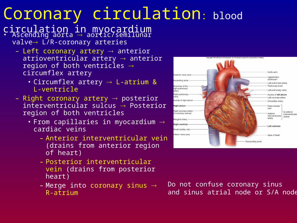
Coronary circulation: blood circulation in myocardium• Ascending aorta aortic/semilunar valve L/R-
coronary arteries – Left coronary artery anterior
atrioventricular artery anterior region of both ventricles circumflex artery
• Circumflex artery L-atrium & L-ventricle
– Right coronary artery posterior interventricular sulcus Posterior region of both ventricles
• From capillaries in myocardium cardiac veins
– Anterior interventricular vein (drains from anterior region of heart)
– Posterior interventricular vein (drains from posterior heart)
– Merge into coronary sinus R-atrium
Do not confuse coronary sinus and sinus atrial node or S/A node
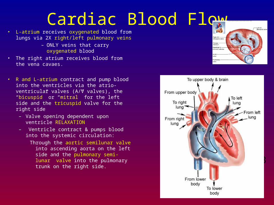
Cardiac Blood Flow• L-atrium receives oxygenated blood from lungs
via 2X right/left pulmonary veins
– ONLY veins that carry oxygenated blood
• The right atrium receives blood from the vena cavaes.
• R and L-atrium contract and pump blood into the ventricles via the atrio-ventricular valves (A/V valves), the “bicuspid” or “mitral” for the left side and the tricuspid valve for the right side
– Valve opening dependent upon ventricle RELAXATION
– Ventricle contract & pumps blood into the systemic circulation:
Through the aortic semilunar valve into ascending aorta on the left side and the pulmonary semi-lunar valve into the pulmonary trunk on the right side.
3
4
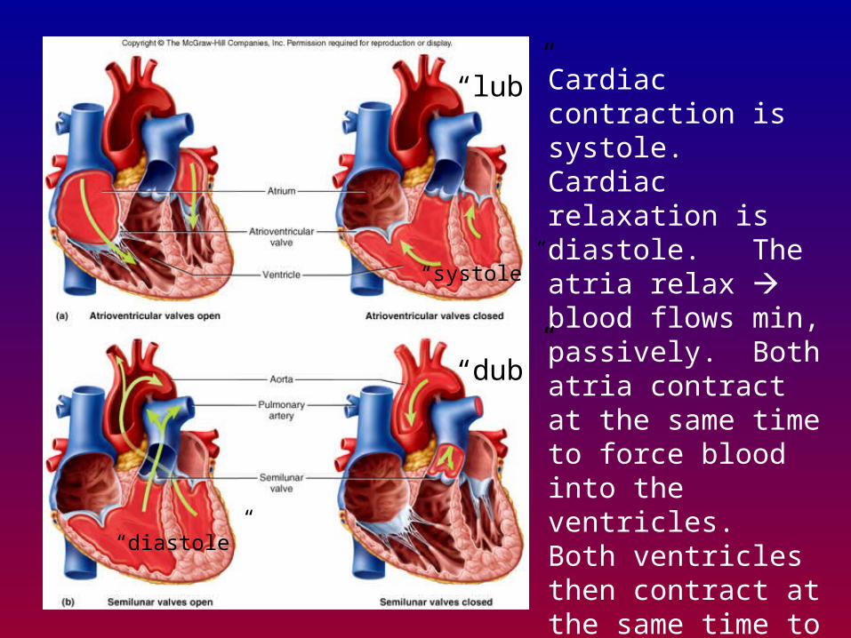
Cardiac contraction is systole. Cardiac relaxation is diastole. The atria relax blood flows min, passively. Both atria contract at the same time to force blood into the ventricles. Both ventricles then contract at the same time to propel blood towards the pulmonary artery or the aorta.
“lub”
“dub”
“systole”
“diastole”
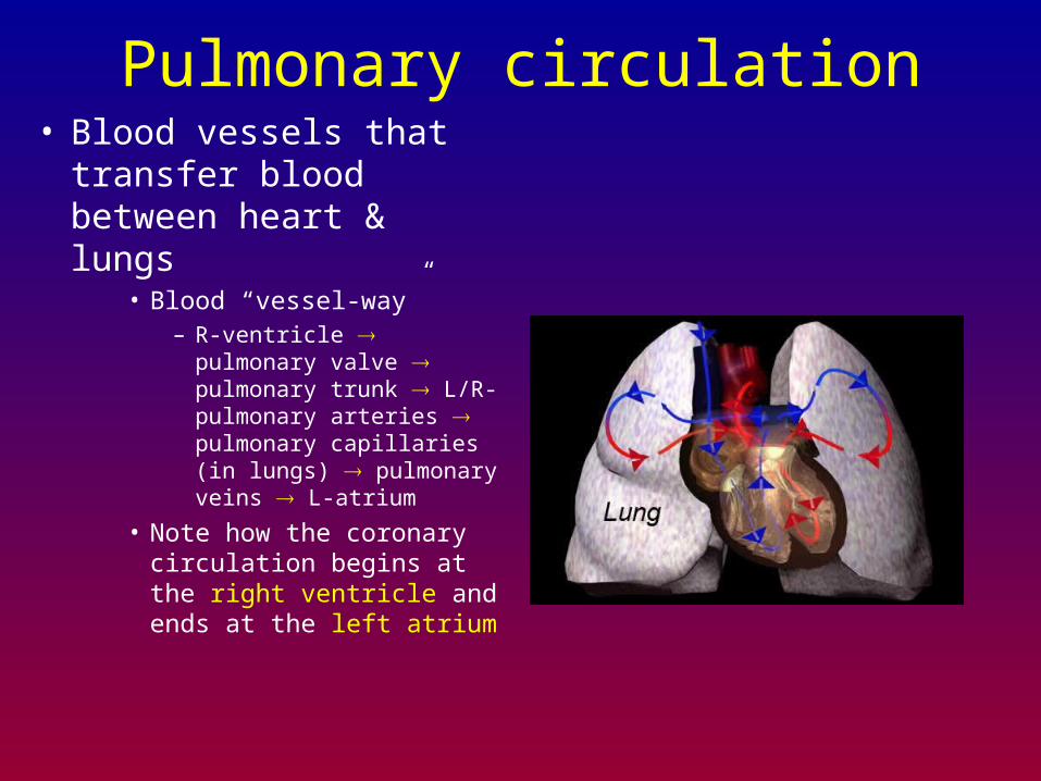
Pulmonary circulation• Blood vessels that
transfer blood between heart & lungs
• Blood “vessel-way”– R-ventricle pulmonary
valve pulmonary trunk L/R-pulmonary arteries pulmonary capillaries (in lungs) pulmonary veins L-atrium
• Note how the coronary circulation begins at the right ventricle and ends at the left atrium
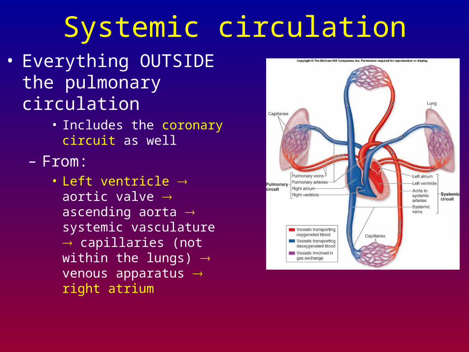
Systemic circulation• Everything OUTSIDE the
pulmonary circulation• Includes the coronary circuit
as well
– From: • Left ventricle aortic valve
ascending aorta systemic vasculature capillaries (not within the lungs) venous apparatus right atrium
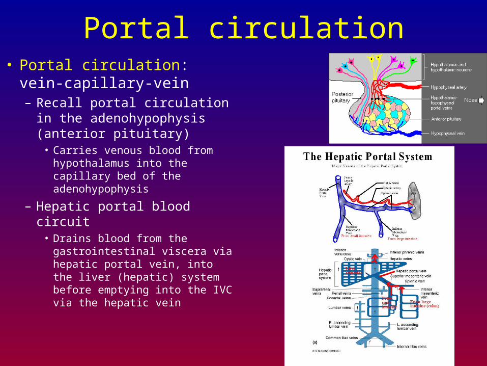
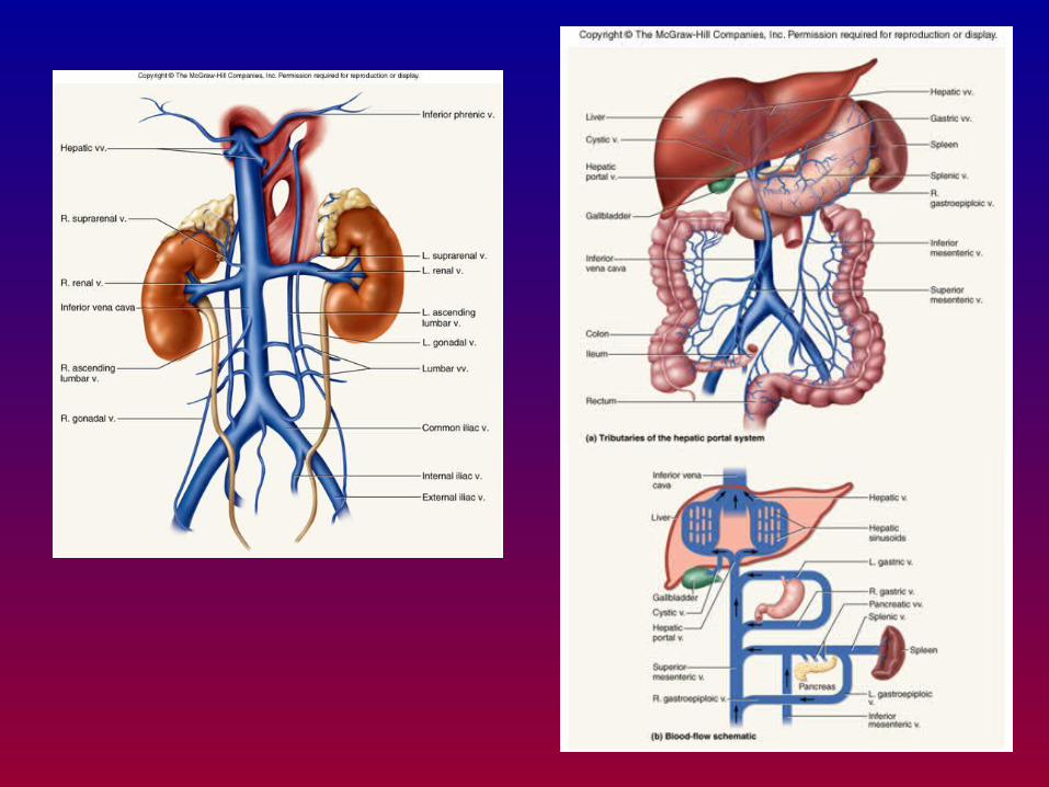
Portal circulation• Portal circulation: vein-
capillary-vein– Recall portal circulation in
the adenohypophysis (anterior pituitary)
• Carries venous blood from hypothalamus into the capillary bed of the adenohypophysis
– Hepatic portal blood circuit• Drains blood from the
gastrointestinal viscera via hepatic portal vein, into the liver (hepatic) system before emptying into the IVC via the hepatic vein
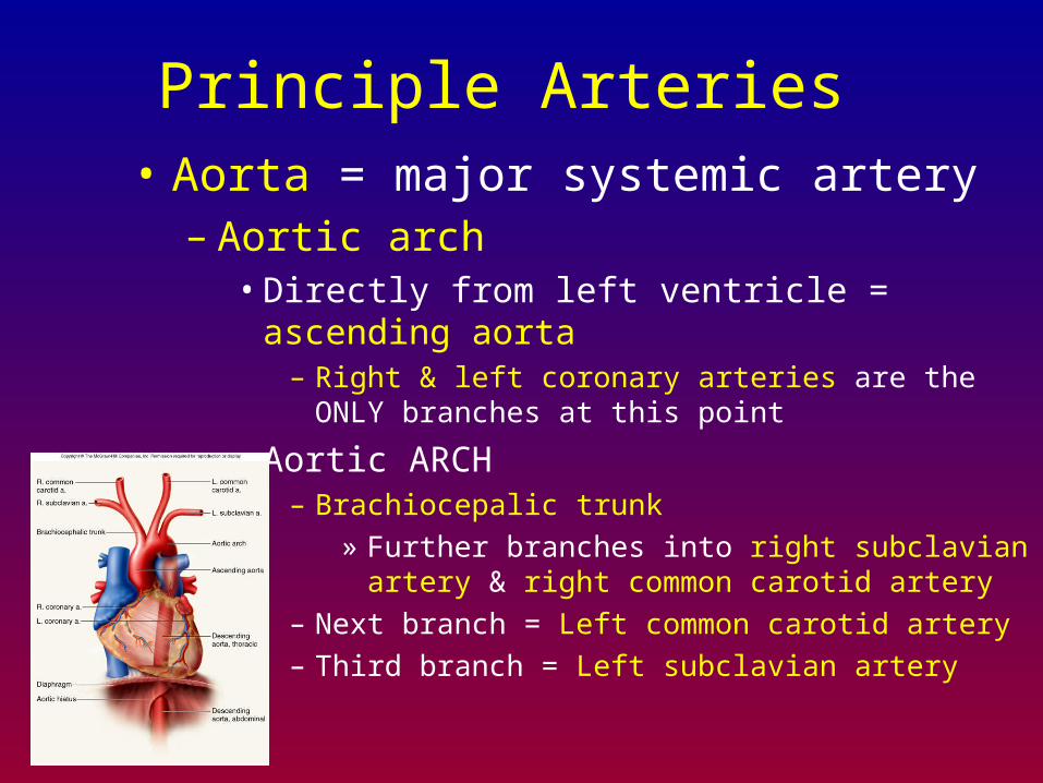
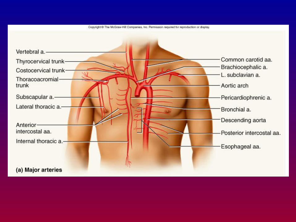
Principle Arteries • Aorta = major systemic artery
– Aortic arch• Directly from left ventricle = ascending aorta
– Right & left coronary arteries are the ONLY branches at this point
• Aortic ARCH – Brachiocepalic trunk
» Further branches into right subclavian artery & right common carotid artery
– Next branch = Left common carotid artery
– Third branch = Left subclavian artery
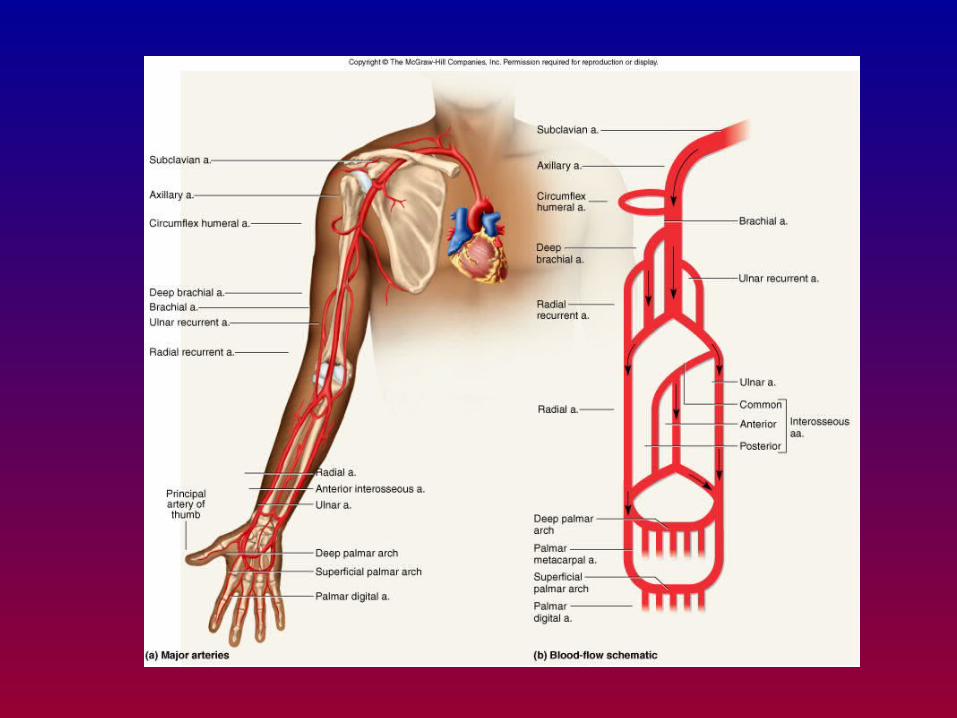
Principle Arteries • Towards upper appendage via subclavian artery
– Arterial blood from subclavian artery has a number of “choices”
• Vertebral artery = towards cranium via transverse foramen of the cervical vertebrae & enters cranium via foramen magnum
• Thyrocervical trunk = destined for thyroid
• Internal thoracic artery = destined for thymus & pericardium
• Costovertebral trunk = destined for intercostal muscles & spinal meninges
Principle Arteries • Towards upper appendage via subclavian artery
– Arterial blood from subclavian artery has a number of “choices”
– If destined for the appendage:• Subclavian artery = axillary artery between 1st rib &
median edge of the humerus– Past medial side of humerus = Brachial artery
– Around humerus = anterior & posterior humeral circumflex arteries
» Ring of arteries around humeral muscles
– Bifurcates into radial & ulnar arteries proximal to cubital fossa
» Radial = pulse at the wrist
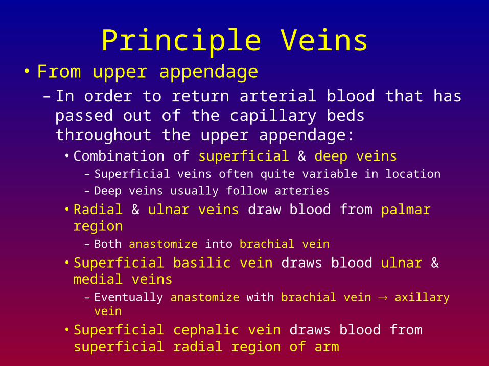
Principle Veins • From upper appendage
– In order to return arterial blood that has passed out of the capillary beds throughout the upper appendage:
• Combination of superficial & deep veins– Superficial veins often quite variable in location
– Deep veins usually follow arteries
• Radial & ulnar veins draw blood from palmar region– Both anastomize into brachial vein
• Superficial basilic vein draws blood ulnar & medial veins– Eventually anastomize with brachial vein axillary vein
• Superficial cephalic vein draws blood from superficial radial region of arm
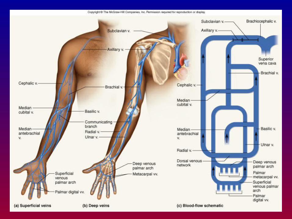
Principle Veins • From upper appendage
– Once all upper appendicular veins have anastomized into axillary vein:
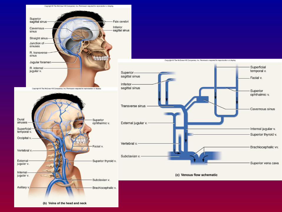
– Axillary vein subclavian vein• Receives venous drainage from cranium as well
– External jugular vein (“external” skull)
– Internal jugular vein (brain, meninges etc.)
» Physically larger than external jugular
» Adjacent to common carotid artery
• Where internal jugular vein merges/anastomizes with subclavian vein = brachiocephalic vein
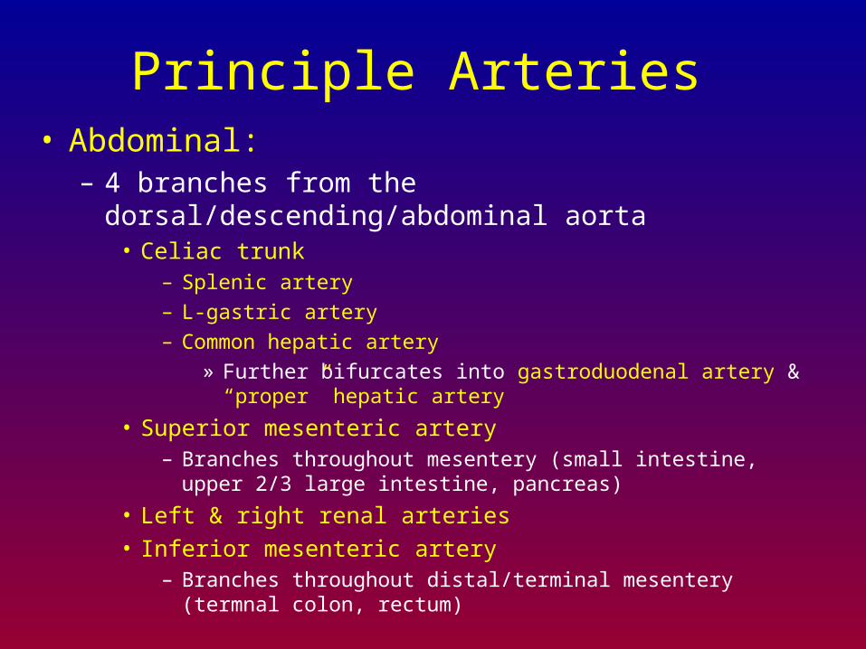
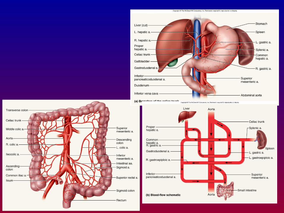
Principle Arteries • Abdominal:
– 4 branches from the dorsal/descending/abdominal aorta• Celiac trunk
– Splenic artery
– L-gastric artery
– Common hepatic artery
» Further bifurcates into gastroduodenal artery & “proper” hepatic artery
• Superior mesenteric artery– Branches throughout mesentery (small intestine, upper 2/3 large
intestine, pancreas)
• Left & right renal arteries
• Inferior mesenteric artery– Branches throughout distal/terminal mesentery (termnal colon, rectum)
Principle Arteries • Abdominal:
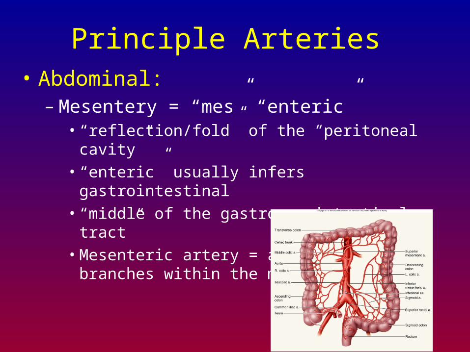
– Mesentery = “mes” “enteric”• “reflection/fold” of the “peritoneal cavity”
• “enteric” usually infers gastrointestinal
• “middle of the gastro or intestinal tract”
• Mesenteric artery = artery that branches within the mesentery
Principle Veins • Abdominal veins:
– Absorptive viscera do not directly drain into the inferior vena cava
• Lower extremities, renals & reproductive organs are the only organs that directly drain into the IVC
• Absorptive viscera drain into hepatic portal vein– All venous blood from GI tract drains into liver via hepatic
portal vein
– Liver processes venous blood, then delivers back to inferior vena cava (cranial to diaphragm) via hepatic vein (NOTE: not the hepatic PORTAL vein)
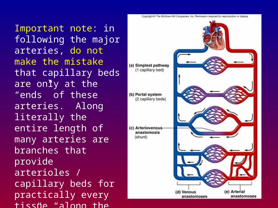
Important note: in following the major arteries, do not make the mistake that capillary beds are only at the “ends” of these arteries. Along literally the entire length of many arteries are branches that provide arterioles / capillary beds for practically every tissue “along the way”. Remember that practically every cell in your body is mere microns (m) from a capillary bed.
Circulatory “collaterals” • Throughout your circulatory system, there are
“collaterals” – Pools or “supplies” of blood that can be “mobilized”
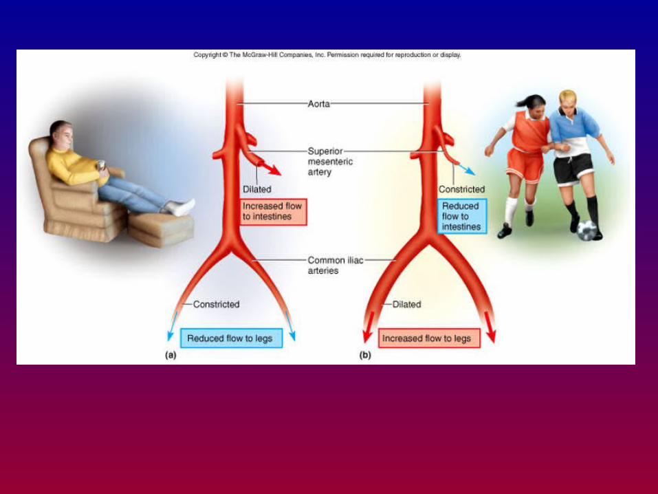
when called for• GI tract retains 50-70% blood volume during rest
– During high activity/trauma, GI tract innervated by sympathetic nervous system is triggered to vasoconstrict (provide more blood for vitals and skeletal muscle)
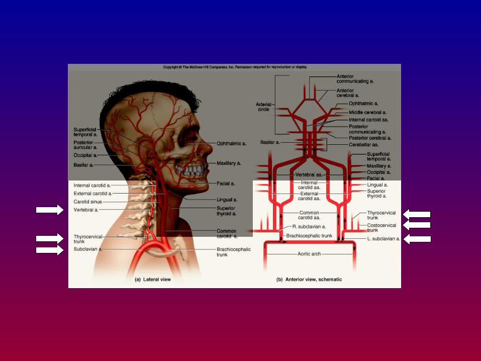
• Within brain: “circle of Willis” provides a similar function for the brain
– Paired carotid arteries, paired vertebral arteries provides at 4 different pathways for arterial blood to enter the brain
– Pairs of vessels span many joints• Allows flexion of the joint while maintaining blood flow
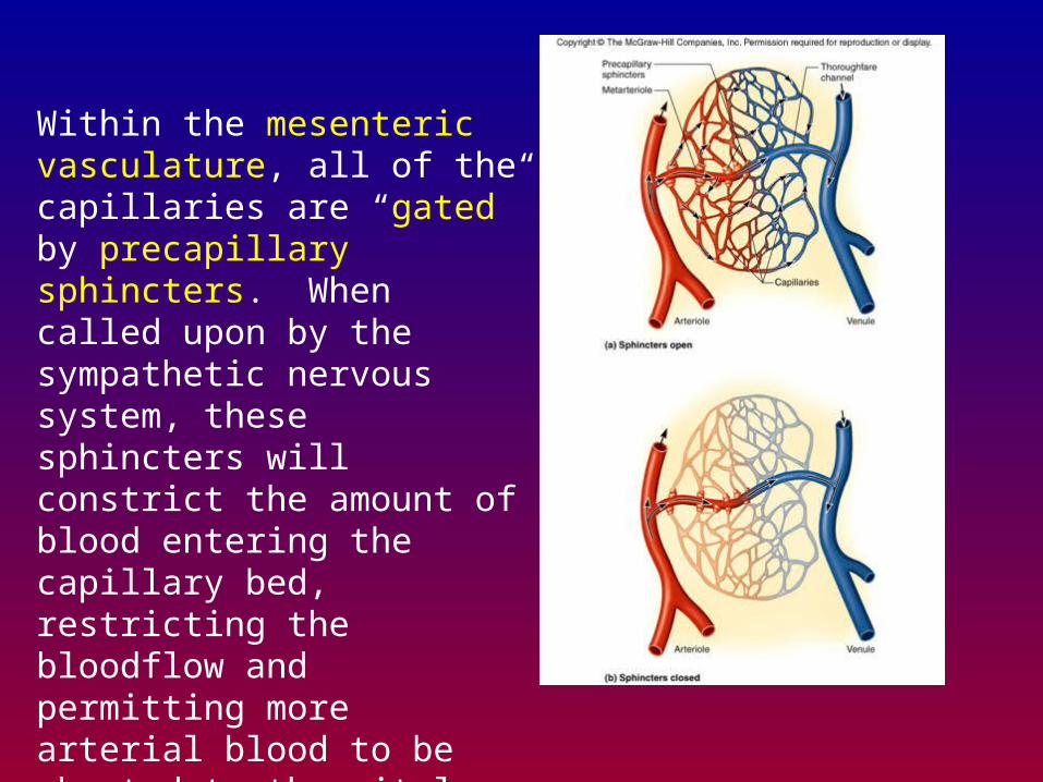
Within the mesenteric vasculature, all of the capillaries are “gated” by precapillary sphincters. When called upon by the sympathetic nervous system, these sphincters will constrict the amount of blood entering the capillary bed, restricting the bloodflow and permitting more arterial blood to be shunted to the vitals.
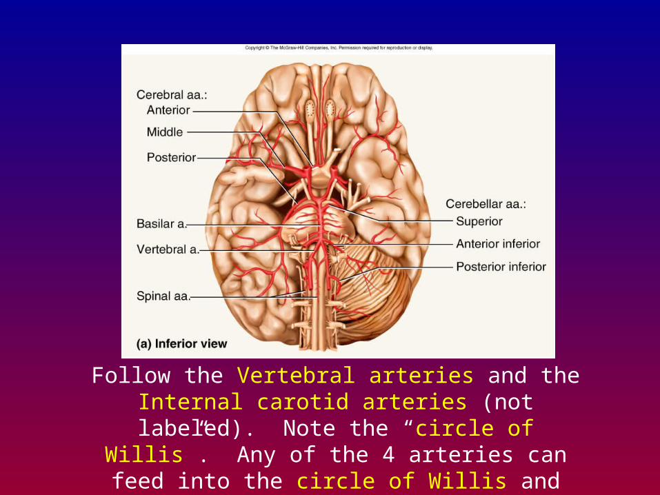
Follow the Vertebral arteries and the Internal carotid arteries (not labeled). Note the “circle of Willis”. Any of the 4 arteries can feed into the circle of Willis and
keep the brain supplied with arterial blood.
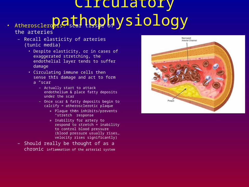
Circulatory pathophysiology • Atherosclerosis: scar tissue of the arteries
– Recall elasticity of arteries (tunic media)• Despite elasticity, or in cases of
exaggerated stretching, the endothelial layer tends to suffer damage
• Circulating immune cells then sense this damage and act to form a “scar”
– Actually start to attack endothelium & place fatty deposits under the scar
– Once scar & fatty deposits begin to calcify = atherosclerotic plaque
» Plaque then inhibits/prevents “stretch” response
» Inability for artery to respond to stretch = inability to control blood pressure (blood pressure usually rises…velocity rises significantly)
– Should really be thought of as a chronic inflammation of the arterial system
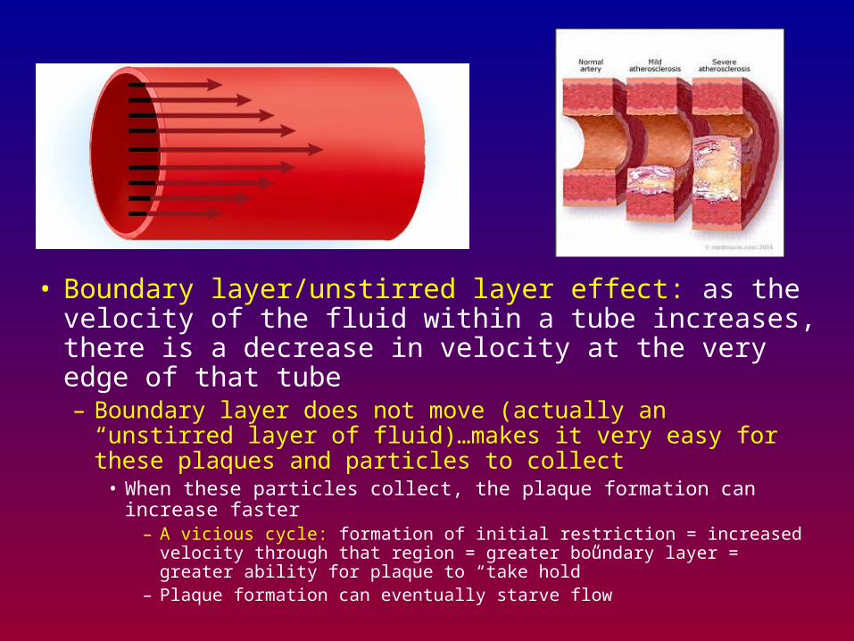
• Boundary layer/unstirred layer effect: as the velocity of the fluid within a tube increases, there is a decrease in velocity at the very edge of that tube– Boundary layer does not move (actually an “unstirred layer of
fluid)…makes it very easy for these plaques and particles to collect
• When these particles collect, the plaque formation can increase faster– A vicious cycle: formation of initial restriction = increased velocity
through that region = greater boundary layer = greater ability for plaque to “take hold”
– Plaque formation can eventually starve flow
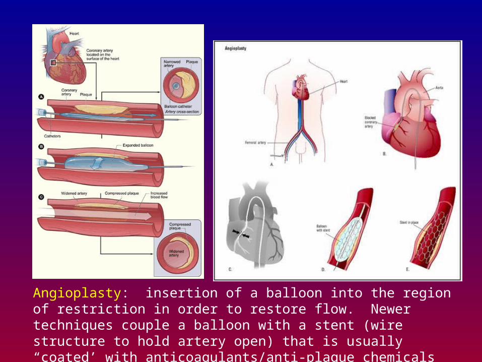
Angioplasty: insertion of a balloon into the region of restriction in order to restore flow. Newer techniques couple a balloon with a stent (wire structure to hold artery open) that is usually “coated’ with anticoagulants/anti-plaque chemicals
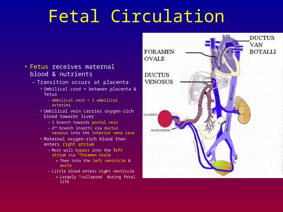
Fetal Circulation
• Fetus receives maternal blood & nutrients– Transition occurs at placenta
• Umbilical cord = between placenta & fetus
– Umbilical vein + 2 umbilical arteries
• Umbilical vein carries oxygen-rich blood towards liver
– 1 branch towards portal vein
– 2nd branch inserts via ductus venosus into the interior vena cava
• Maternal oxygen-rich blood then enters right atrium
– Most will bypass into the left atrium via “foramen ovale”
» Then into the left ventricle & aorta
– Little blood enters right ventricle
» Largely “collapsed” during fetal life
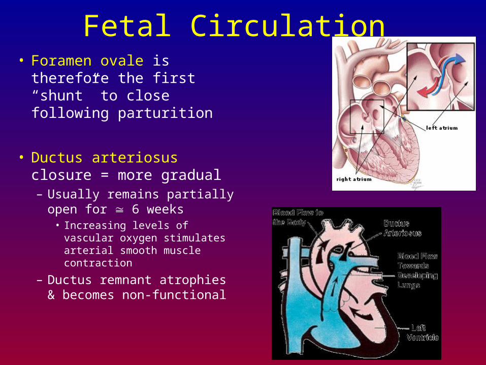
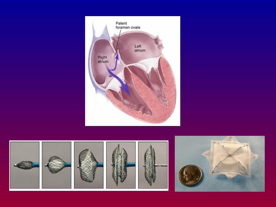
Fetal Circulation • Foramen ovale is therefore
the first “shunt” to close following parturition
• Ductus arteriosus closure = more gradual– Usually remains partially
open for 6 weeks• Increasing levels of vascular
oxygen stimulates arterial smooth muscle contraction
– Ductus remnant atrophies & becomes non-functional


![ERYTHROCYTES [RBCs]](https://img.pdfslide.us/doc/110x75/56813dc0550346895da78963/erythrocytes-rbcs-56ea22b2e2743.jpg)