Embed Size (px)
Citation preview
The Cardiovascular
System
Chapter 17, 18, 19
The Heart, Blood Vessels, Blood Types
BLOOD
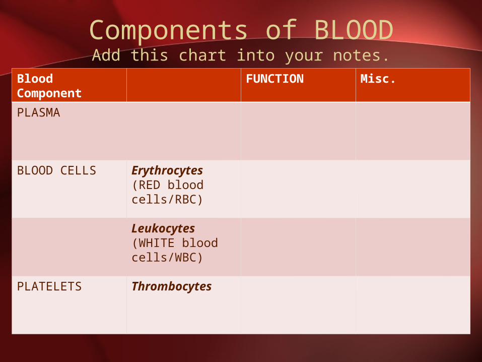
Components of BLOODAdd this chart into your notes.
Blood Component
FUNCTION Misc.
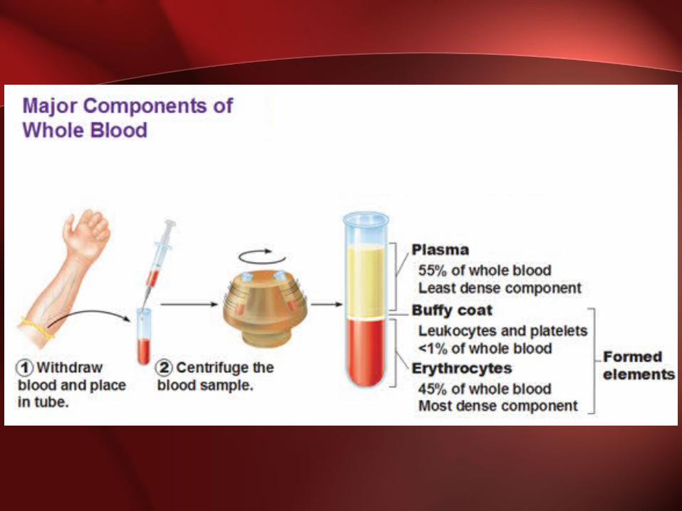
PLASMA
BLOOD CELLS Erythrocytes(RED blood cells/RBC)
Leukocytes(WHITE blood cells/WBC)
PLATELETS Thrombocytes
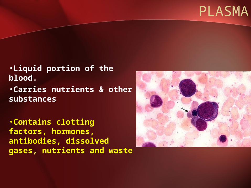
PLASMA
•Liquid portion of the blood.•Carries nutrients & other substances
•Contains clotting factors, hormones, antibodies, dissolved gases, nutrients and waste
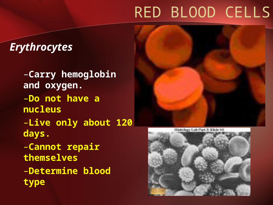
RED BLOOD CELLS
Erythrocytes
–Carry hemoglobin and oxygen. –Do not have a nucleus –Live only about 120 days.–Cannot repair themselves–Determine blood type
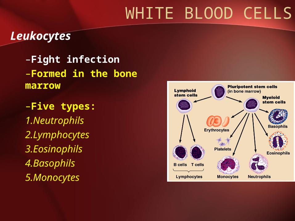
WHITE BLOOD CELLSLeukocytes
–Fight infection –Formed in the bone marrow
–Five types: 1.Neutrophils2.Lymphocytes3.Eosinophils4.Basophils 5.Monocytes
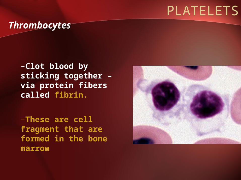
PLATELETSThrombocytes
–Clot blood by sticking together – via protein fibers called fibrin.
–These are cell fragment that are formed in the bone marrow
ABO BLOOD TYPES
• Blood type is determines by the presence or absence of blood proteins called ANTIGENS located on the surface of the erythrocytes.
• There are also ANTIBODIES on the surface of erythrocytes.
• When incompatible blood proteins mix, agglutination (blood clumping) results.
BLOOD TYPES
• There are 3 alleles that determine 4 blood types:– A (dominant)
– B (dominant)
– O (recessive)
Type A = AA, AoType B = BB, BoType AB = ABType O = oo
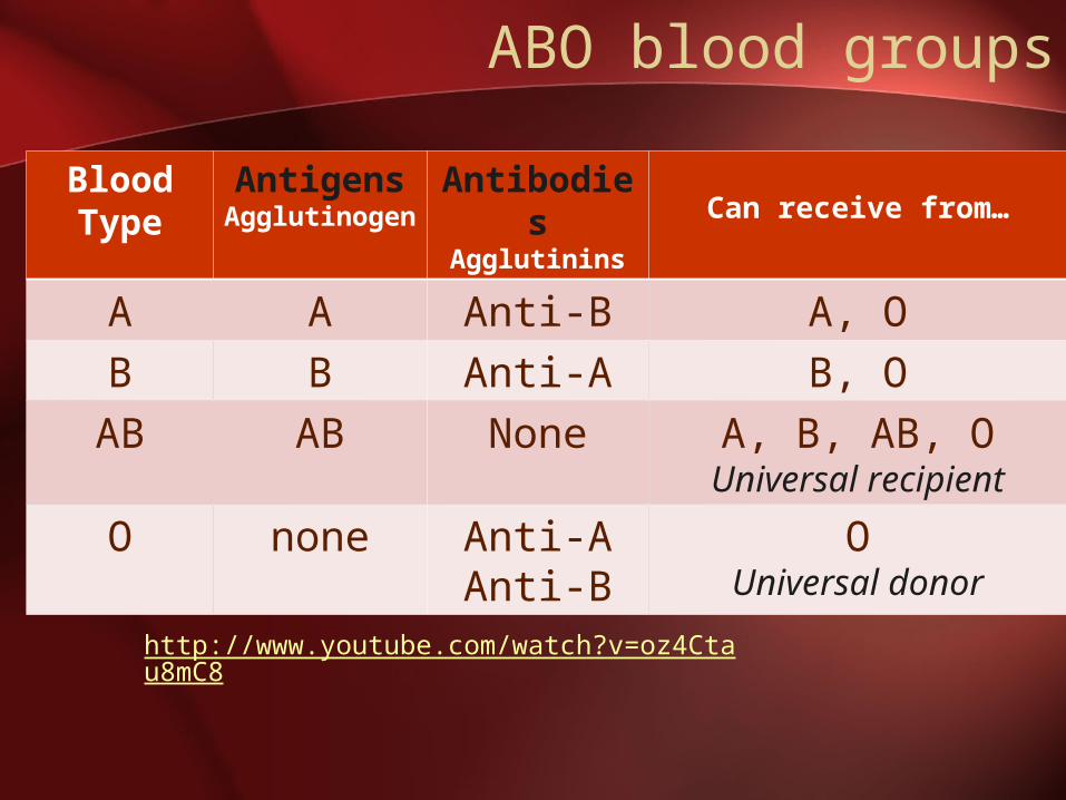
ABO blood groups
Blood Type
AntigensAgglutinoge
n
Antibodies
Agglutinins
Can receive from…
A A Anti-B A, O
B B Anti-A B, O
AB AB None A, B, AB, OUniversal recipient
O none Anti-AAnti-B
OUniversal donor
http://www.youtube.com/watch?v=oz4Ctau8mC8

Eucharistic Miracles
• http://www.youtube.com/watch?v=N6SH93arrIE

When Things Go Wrong: BLOOD
Complete handout. Then cut & paste into NB.
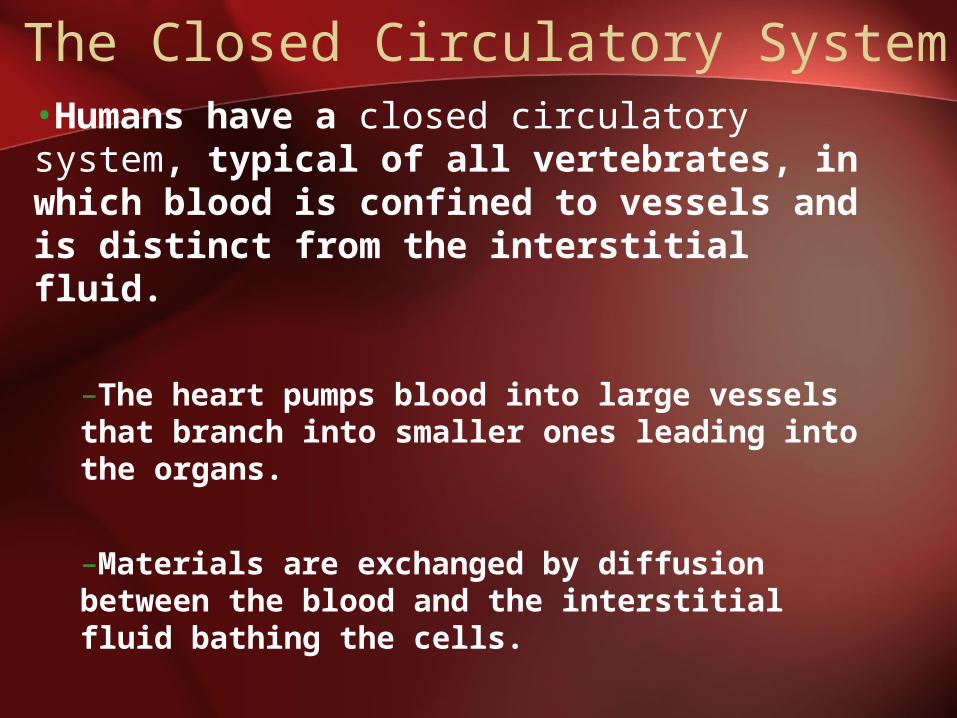
The Closed Circulatory System•Humans have a closed circulatory system, typical of all vertebrates, in which blood is confined to vessels and is distinct from the interstitial fluid.
–The heart pumps blood into large vessels that branch into smaller ones leading into the organs.
–Materials are exchanged by diffusion between the blood and the interstitial fluid bathing the cells.
BLOOD VESSELS
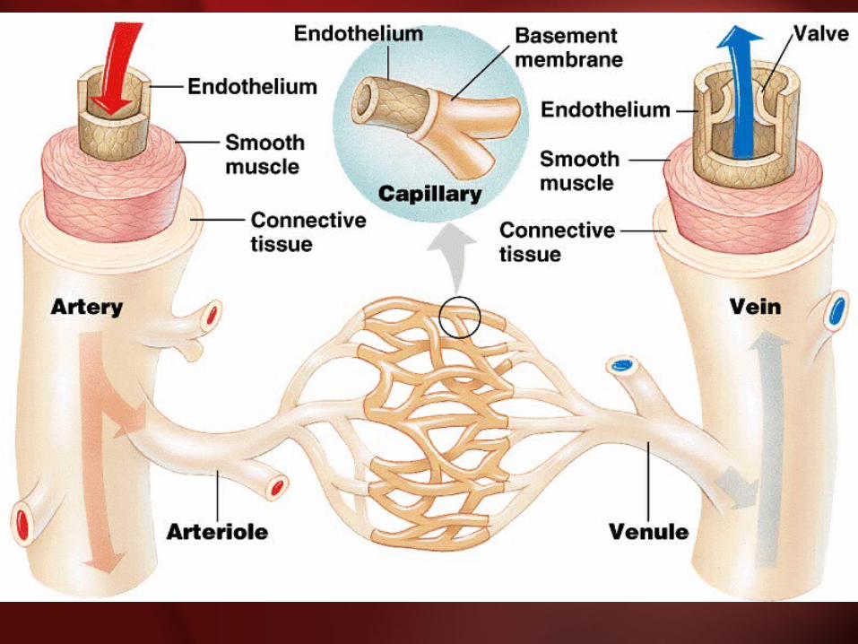
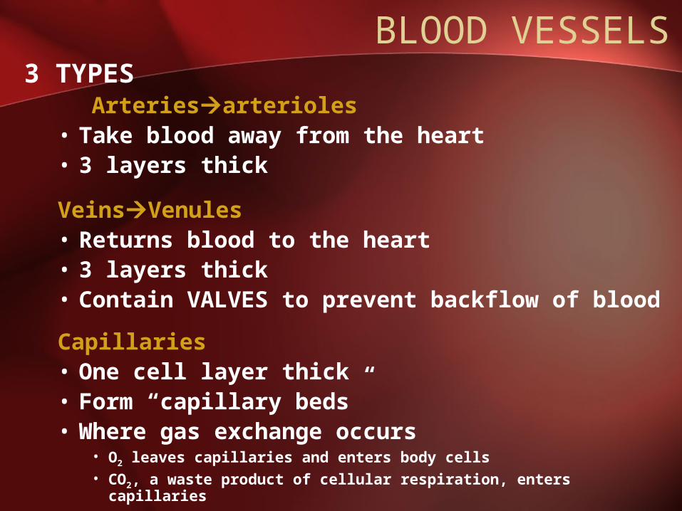
BLOOD VESSELS3 TYPES Arteriesarterioles • Take blood away from the heart• 3 layers thick
VeinsVenules • Returns blood to the heart• 3 layers thick• Contain VALVES to prevent backflow of blood
Capillaries • One cell layer thick• Form “capillary beds”• Where gas exchange occurs
• O2 leaves capillaries and enters body cells• CO2, a waste product of cellular respiration, enters capillaries
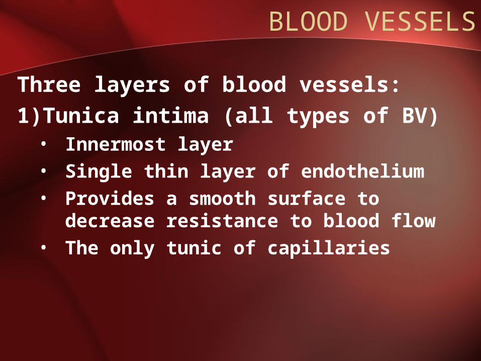
BLOOD VESSELS
Three layers of blood vessels:1)Tunica intima (all types of BV)• Innermost layer• Single thin layer of endothelium• Provides a smooth surface to decrease
resistance to blood flow• The only tunic of capillaries
BLOOD VESSELS
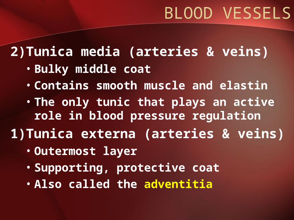
2)Tunica media (arteries & veins)• Bulky middle coat• Contains smooth muscle and elastin• The only tunic that plays an active role
in blood pressure regulation
1)Tunica externa (arteries & veins)• Outermost layer• Supporting, protective coat• Also called the adventitia
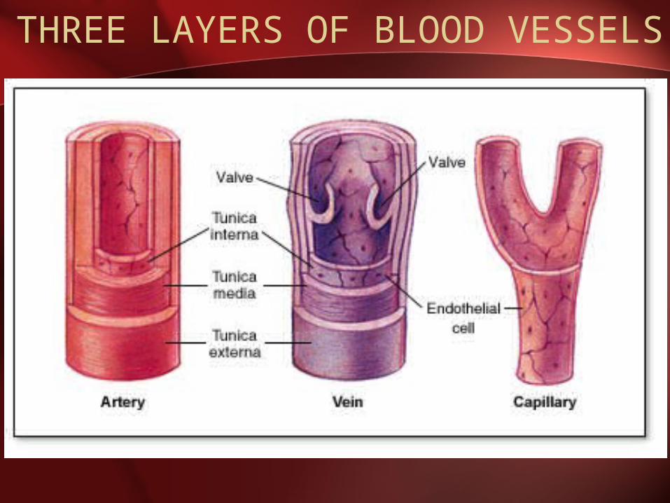
THREE LAYERS OF BLOOD VESSELS
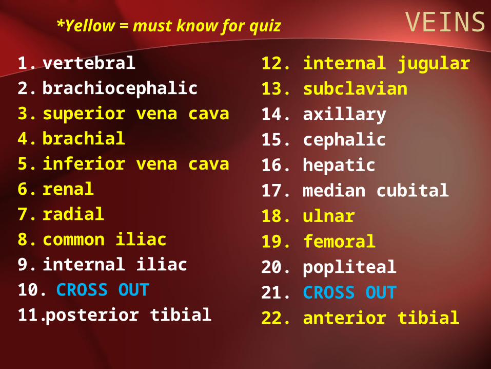
*Yellow = must know for quiz
VEINS1. vertebral2. brachiocephalic3. superior vena cava4. brachial5. inferior vena cava6. renal7. radial8. common iliac9. internal iliac10. CROSS OUT11.posterior tibial
12. internal jugular13. subclavian14. axillary15. cephalic16. hepatic17. median cubital18. ulnar19. femoral20. popliteal21. CROSS OUT22. anterior tibial
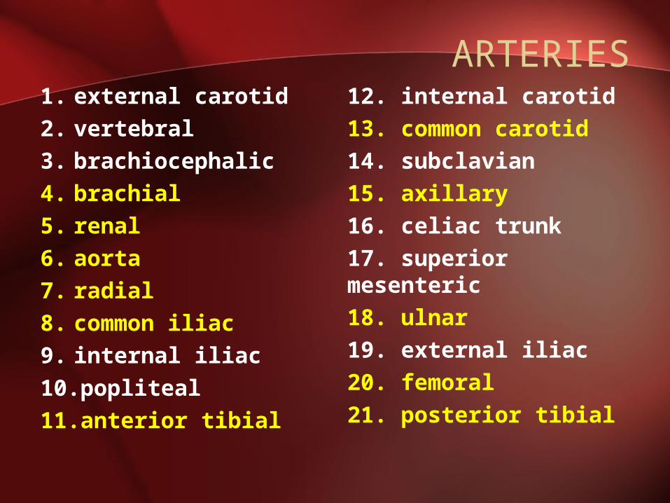
ARTERIES1. external carotid2. vertebral3. brachiocephalic4. brachial5. renal6. aorta7. radial8. common iliac9. internal iliac10.popliteal11.anterior tibial
12. internal carotid13. common carotid14. subclavian15. axillary16. celiac trunk17. superior mesenteric18. ulnar19. external iliac20. femoral21. posterior tibial

When Things Go Wrong: BLOOD VESSELS
Complete handout. Then cut & paste into NB.
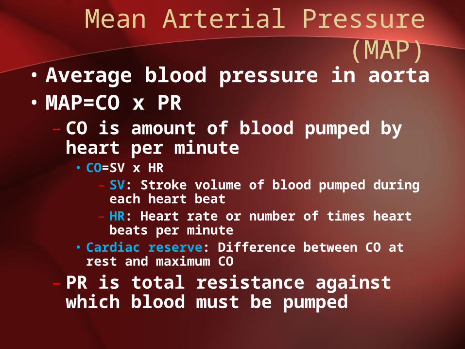
Mean Arterial Pressure (MAP)
• Average blood pressure in aorta• MAP=CO x PR
– CO is amount of blood pumped by heart per minute
• CO=SV x HR– SV: Stroke volume of blood pumped during
each heart beat– HR: Heart rate or number of times heart
beats per minute• Cardiac reserve: Difference between CO at rest
and maximum CO
– PR is total resistance against which blood must be pumped
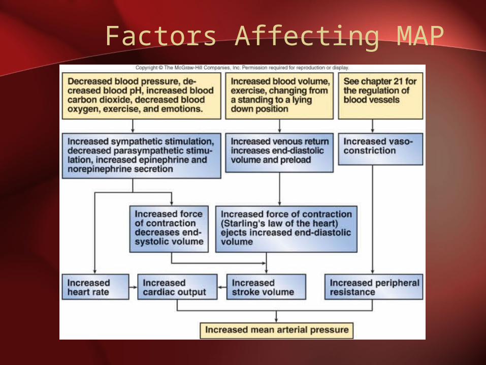
Factors Affecting MAP
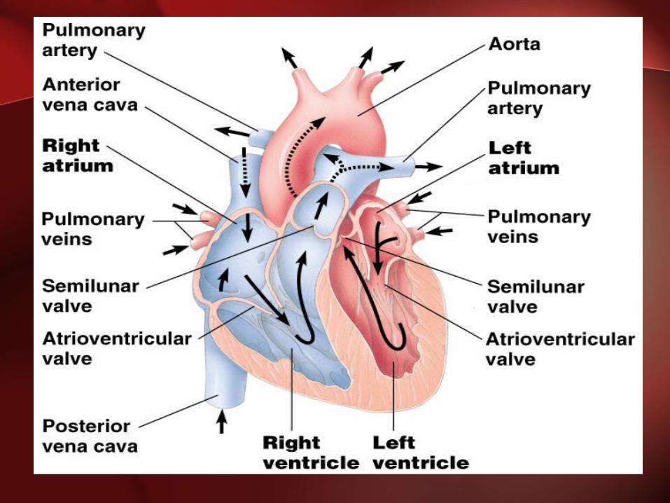
THE HEART
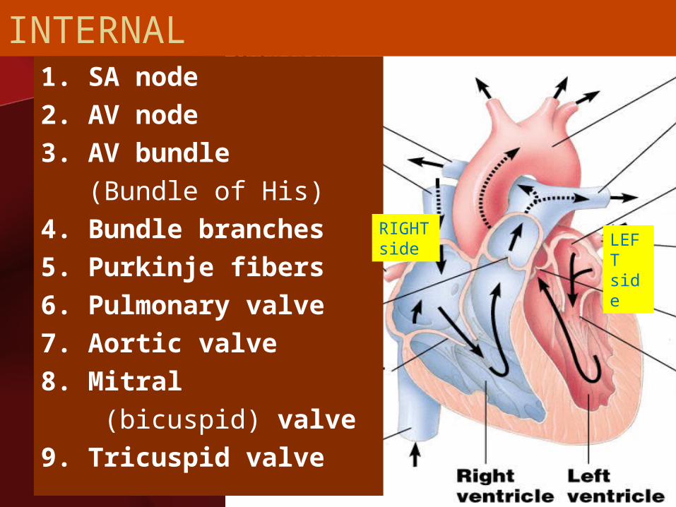
INTERNAL1. SA node 2. AV node3. AV bundle (Bundle of His)4. Bundle branches5. Purkinje fibers6. Pulmonary valve7. Aortic valve8. Mitral (bicuspid) valve9. Tricuspid valve
RIGHT side LEF
T side
8. Mitral (bicuspid) valve
9. Tricuspid valve
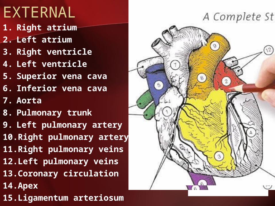
EXTERNAL1. Right atrium2. Left atrium3. Right ventricle4. Left ventricle5. Superior vena cava6. Inferior vena cava7. Aorta8. Pulmonary trunk9. Left pulmonary artery10.Right pulmonary artery11.Right pulmonary veins12.Left pulmonary veins13.Coronary circulation14.Apex15.Ligamentum
arteriosum
The HEART
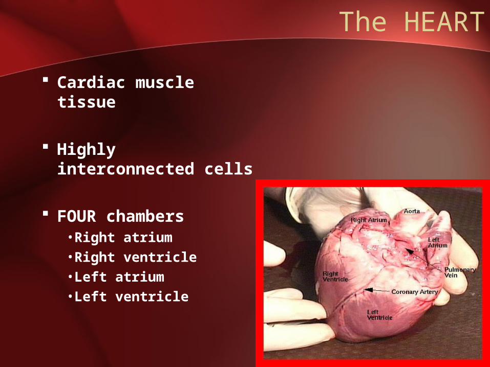
Cardiac muscle tissue
Highly interconnected cells
FOUR chambers•Right atrium•Right ventricle•Left atrium•Left ventricle
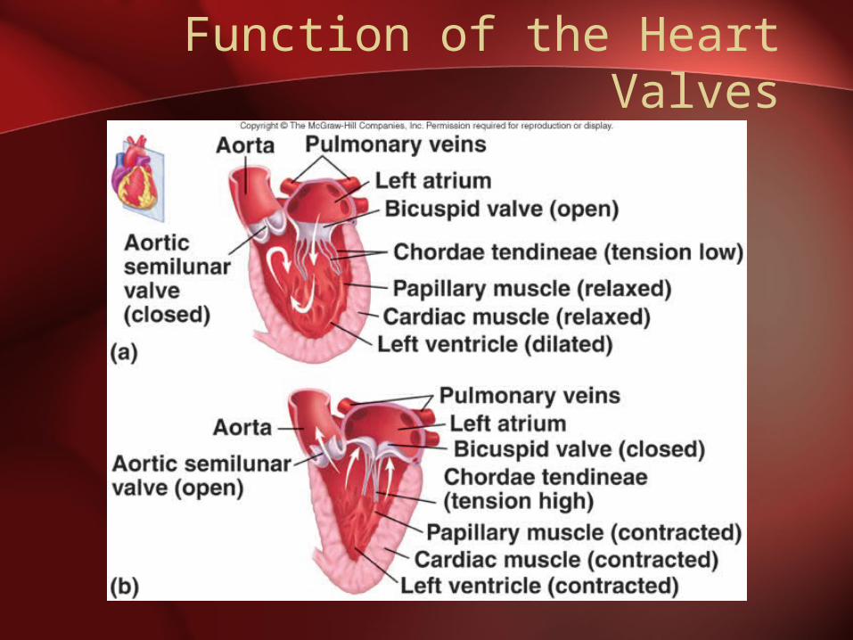
Function of the Heart Valves
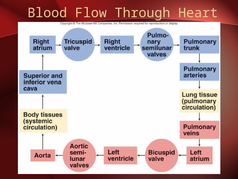
Blood Flow Through Heart
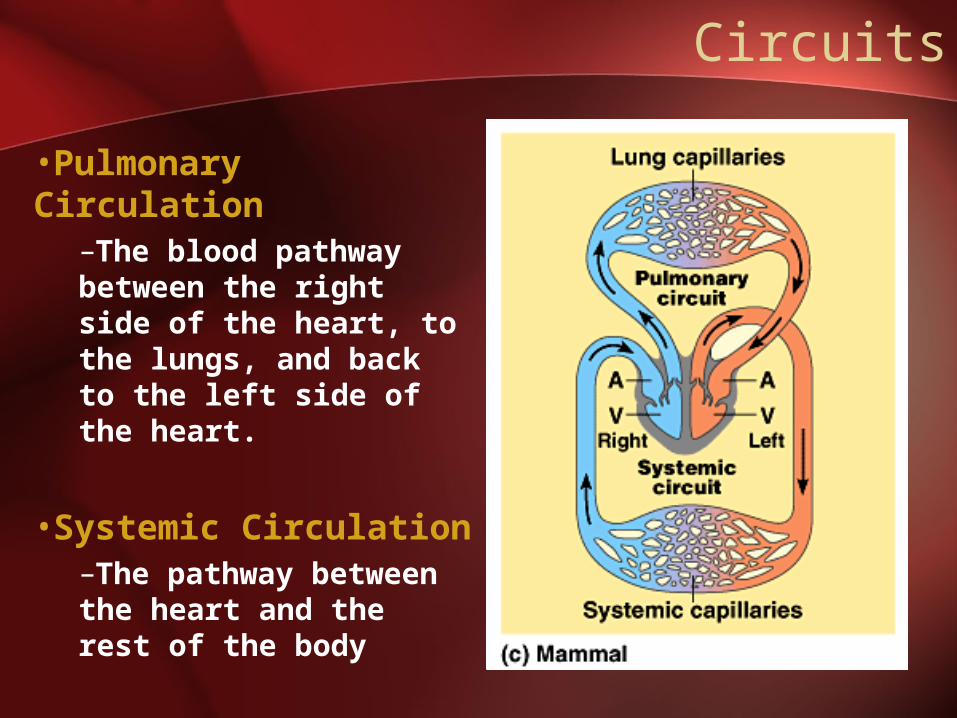
Circuits
•Pulmonary Circulation
–The blood pathway between the right side of the heart, to the lungs, and back to the left side of the heart.
•Systemic Circulation–The pathway between the heart and the rest of the body
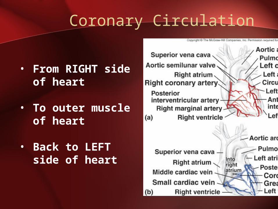
Coronary Circulation
• From RIGHT side of heart
• To outer muscle of heart
• Back to LEFT side of heart
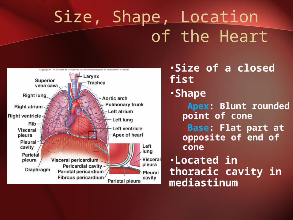
Size, Shape, Location of the Heart
•Size of a closed fist•Shape
–Apex: Blunt rounded point of cone–Base: Flat part at opposite of end of cone
•Located in thoracic cavity in mediastinum
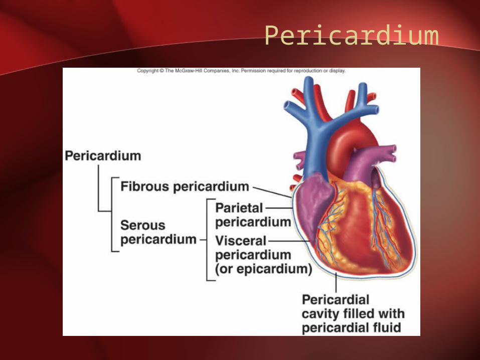
Pericardium
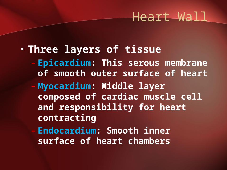
Heart Wall
• Three layers of tissue– Epicardium: This serous membrane
of smooth outer surface of heart– Myocardium: Middle layer
composed of cardiac muscle cell and responsibility for heart contracting
– Endocardium: Smooth inner surface of heart chambers
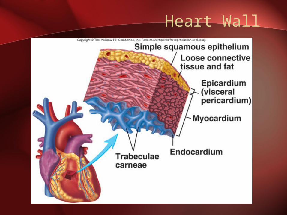
Heart Wall
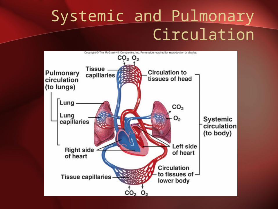
Systemic and PulmonaryCirculation
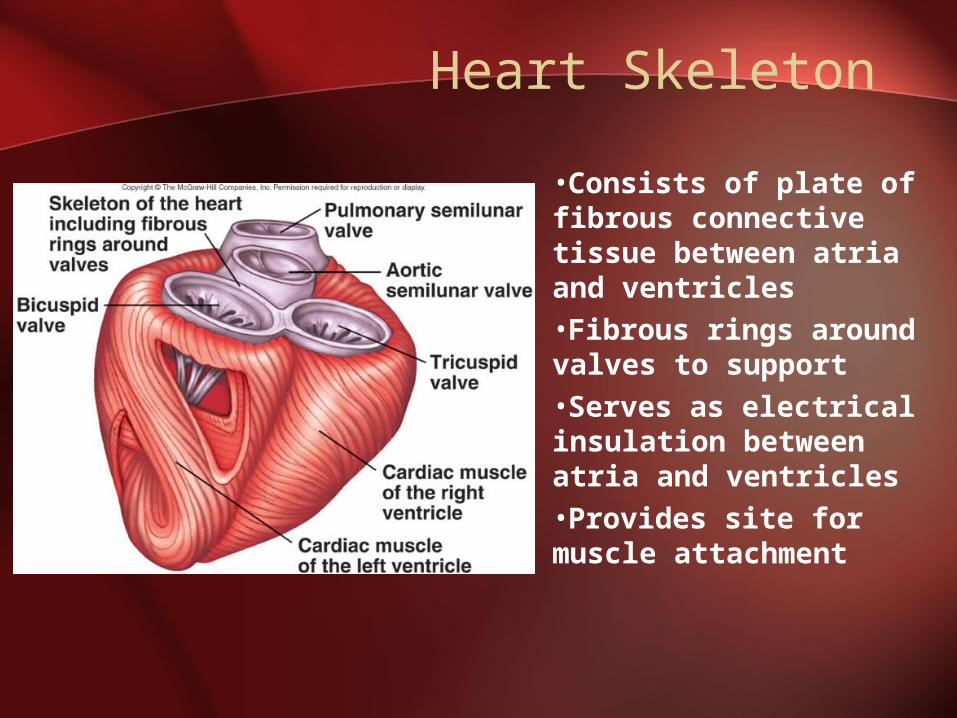
Heart Skeleton
•Consists of plate of fibrous connective tissue between atria and ventricles•Fibrous rings around valves to support•Serves as electrical insulation between atria and ventricles•Provides site for muscle attachment
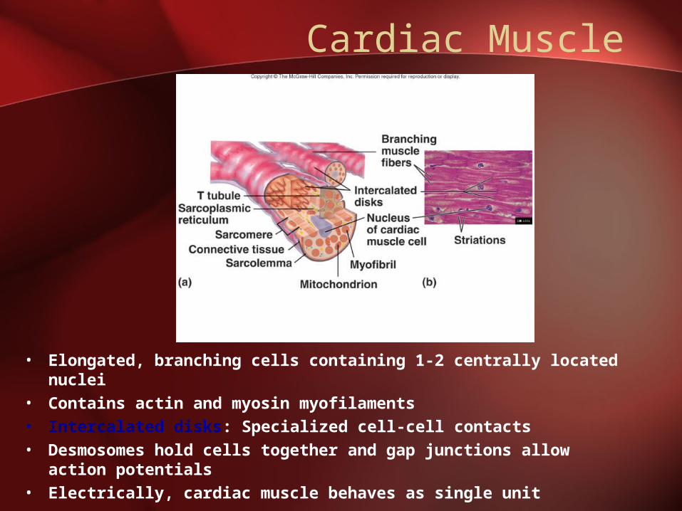
Cardiac Muscle
• Elongated, branching cells containing 1-2 centrally located nuclei
• Contains actin and myosin myofilaments • Intercalated disks: Specialized cell-cell contacts• Desmosomes hold cells together and gap junctions allow
action potentials• Electrically, cardiac muscle behaves as single unit
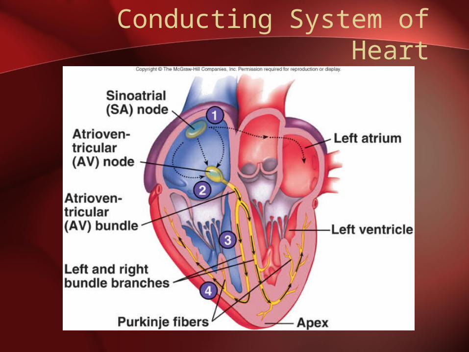
Conducting System of Heart
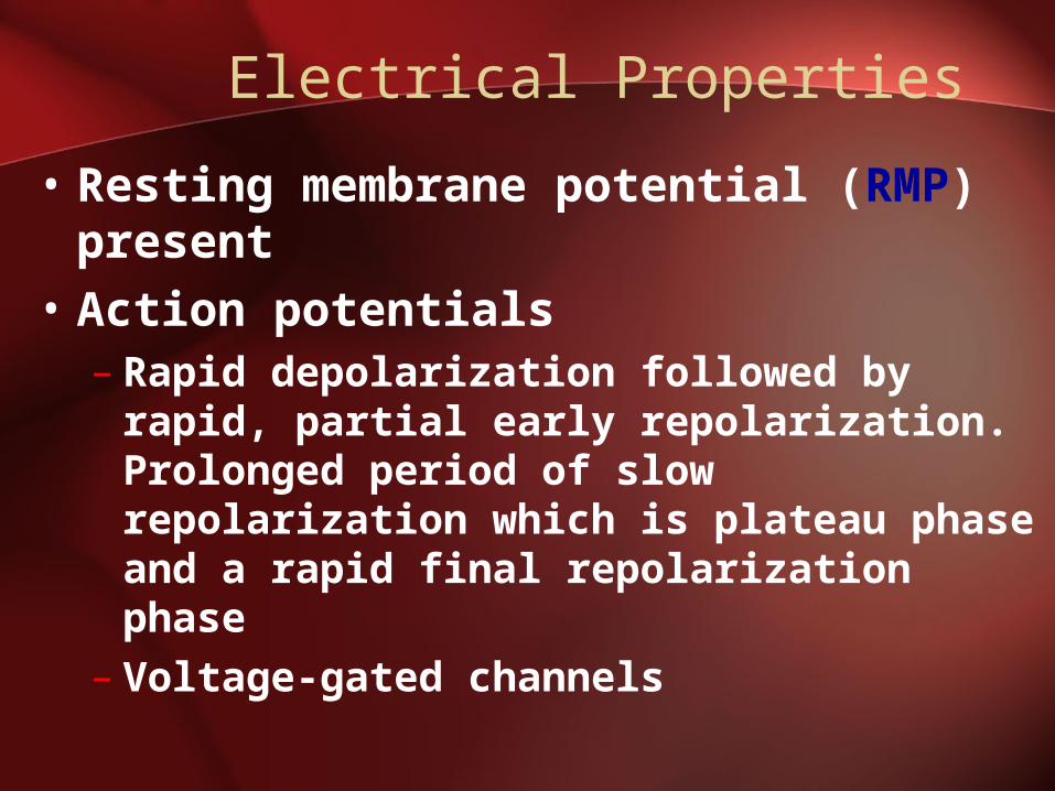
Electrical Properties
• Resting membrane potential (RMP) present
• Action potentials– Rapid depolarization followed by rapid,
partial early repolarization. Prolonged period of slow repolarization which is plateau phase and a rapid final repolarization phase
– Voltage-gated channels
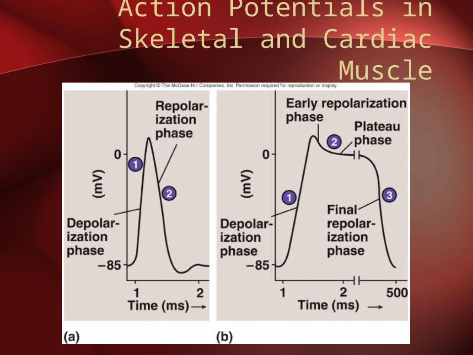
Action Potentials inSkeletal and Cardiac Muscle
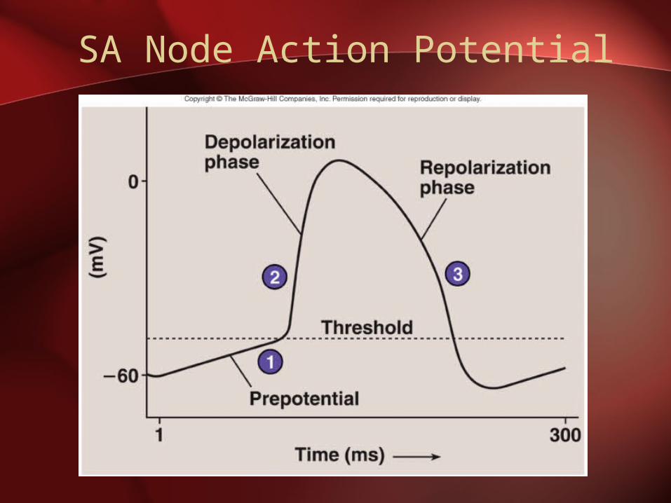
SA Node Action Potential
Refractory Period
• Absolute: Cardiac muscle cell completely insensitive to further stimulation
• Relative: Cell exhibits reduced sensitivity to additional stimulation
• Long refractory period prevents tetanic contractions
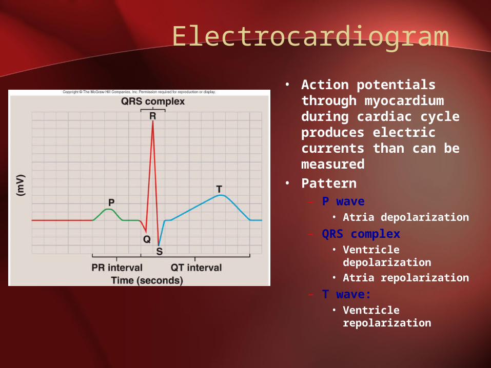
Electrocardiogram
• Action potentials through myocardium during cardiac cycle produces electric currents than can be measured
• Pattern– P wave
• Atria depolarization
– QRS complex• Ventricle
depolarization• Atria repolarization
– T wave: • Ventricle
repolarization
Cardiac Arrhythmias
• Tachycardia: Heart rate in excess of 100bpm
• Bradycardia: Heart rate less than 60 bpm
• Sinus arrhythmia: Heart rate varies 5% during respiratory cycle and up to 30% during deep respiration
• Premature atrial contractions: Occasional shortened intervals between one contraction and succeeding, frequently occurs in healthy people
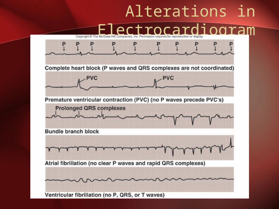
Alterations in Electrocardiogram
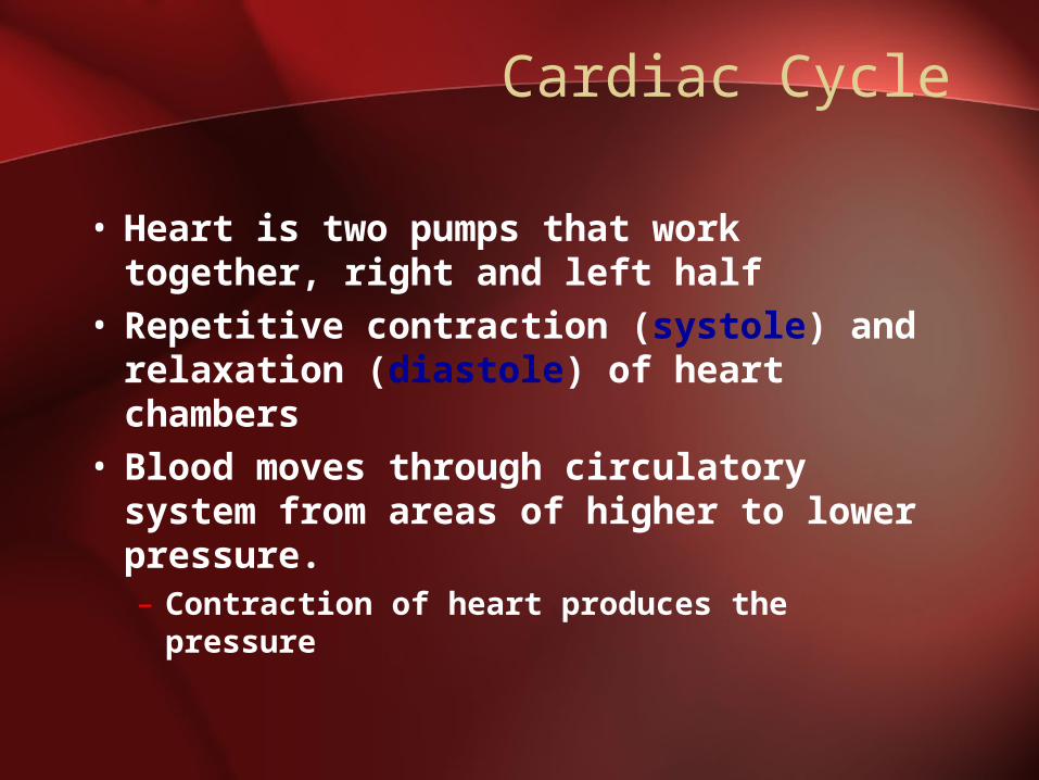
Cardiac Cycle
• Heart is two pumps that work together, right and left half
• Repetitive contraction (systole) and relaxation (diastole) of heart chambers
• Blood moves through circulatory system from areas of higher to lower pressure.– Contraction of heart produces the
pressure
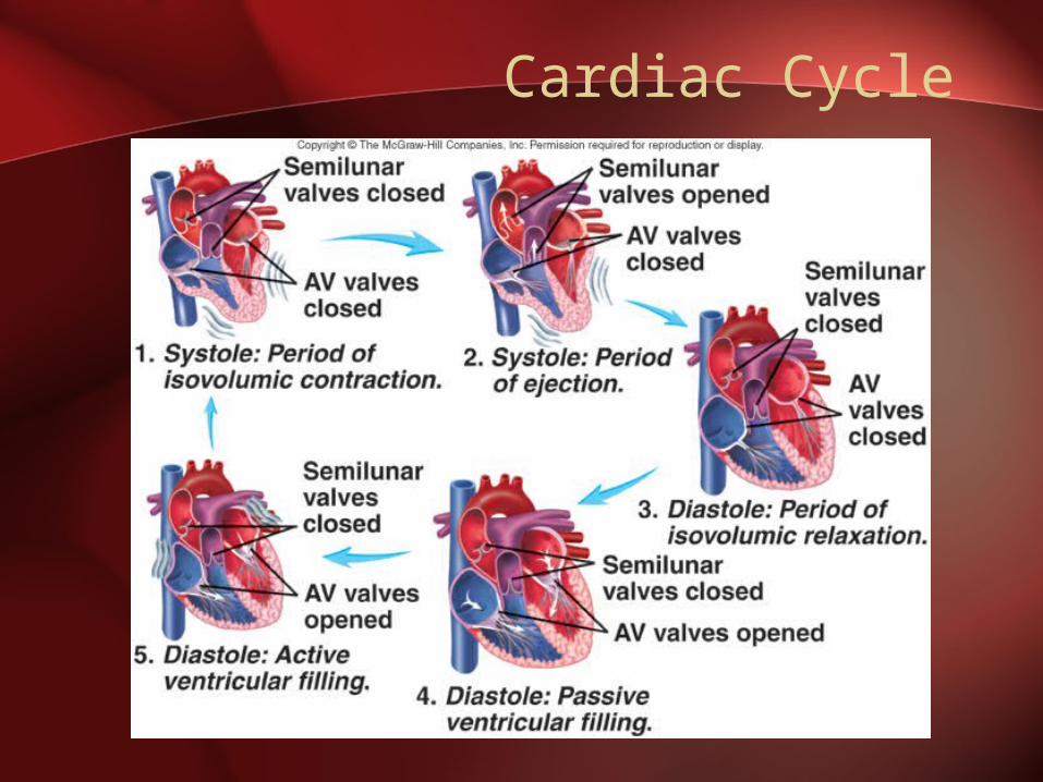
Cardiac Cycle
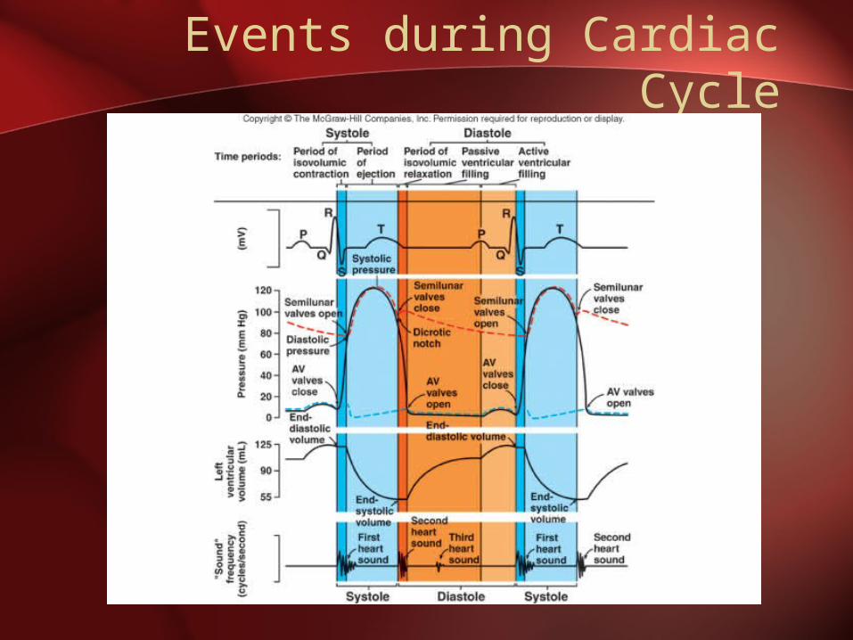
Events during Cardiac Cycle
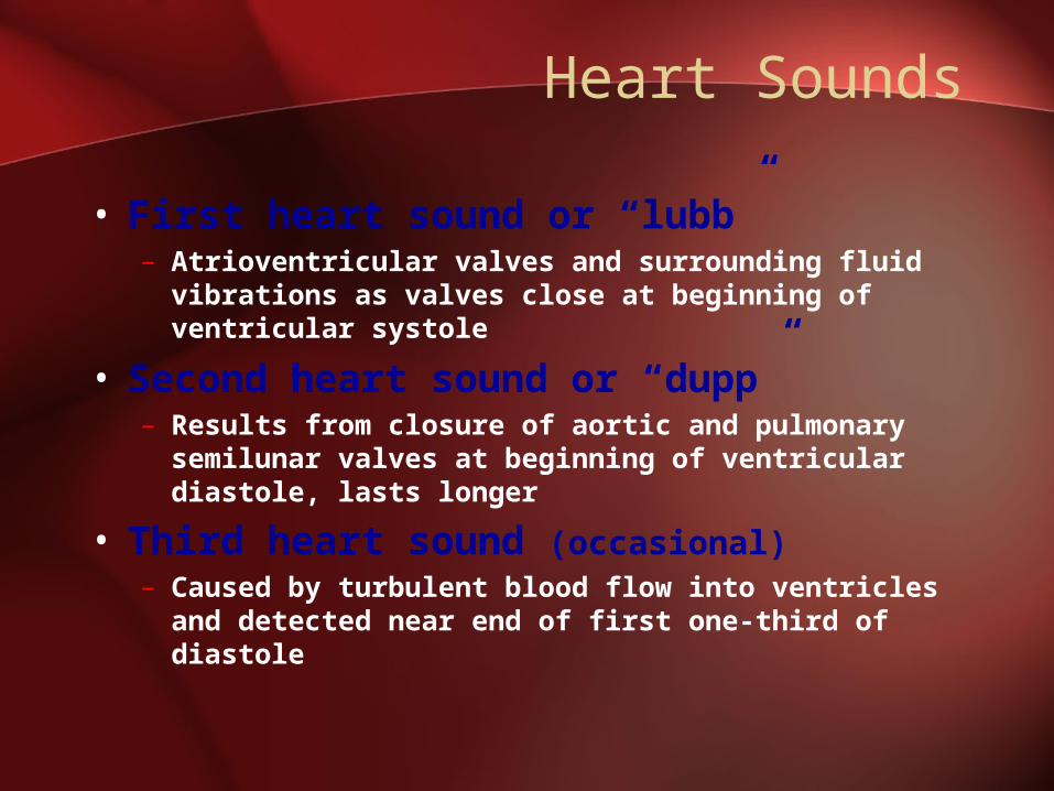
Heart Sounds
• First heart sound or “lubb”– Atrioventricular valves and surrounding fluid
vibrations as valves close at beginning of ventricular systole
• Second heart sound or “dupp”– Results from closure of aortic and pulmonary
semilunar valves at beginning of ventricular diastole, lasts longer
• Third heart sound (occasional)– Caused by turbulent blood flow into ventricles
and detected near end of first one-third of diastole
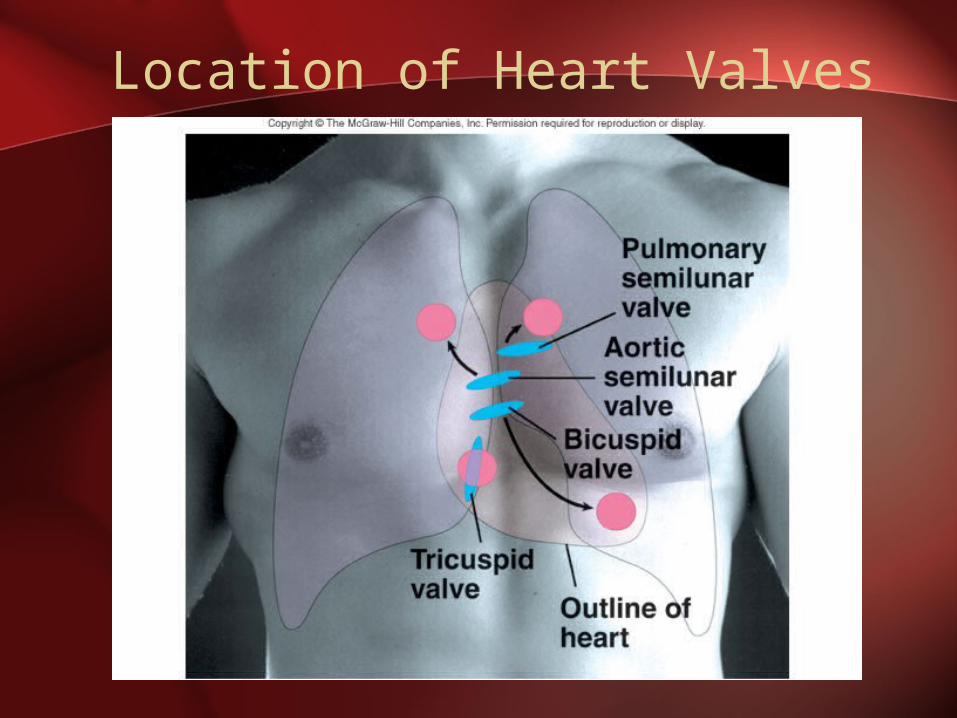
Location of Heart Valves

When Things Go Wrong: HEART
Complete handout. Then cut & paste into NB.
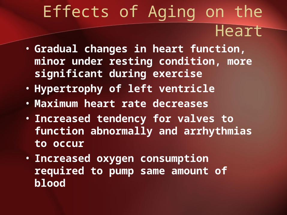
Effects of Aging on the Heart
• Gradual changes in heart function, minor under resting condition, more significant during exercise
• Hypertrophy of left ventricle• Maximum heart rate decreases• Increased tendency for valves to
function abnormally and arrhythmias to occur
• Increased oxygen consumption required to pump same amount of blood