Embed Size (px)
Citation preview
Chapter 19, Cardiovascular System - Blood Vessel
1
19The Cardiovascular System:
Blood Vessels
Anatomy
Chapter 19, Cardiovascular System - Blood Vessel
2
Blood Vessels
Blood is carried in a closed system of vessels that begins and ends at the heart
The three major types of vessels are arteries, capillaries, and veins
Arteries carry blood away from the heart, veins carry blood toward the heart
Capillaries contact tissue cells and directly serve cellular needs
Chapter 19, Cardiovascular System - Blood Vessel
3
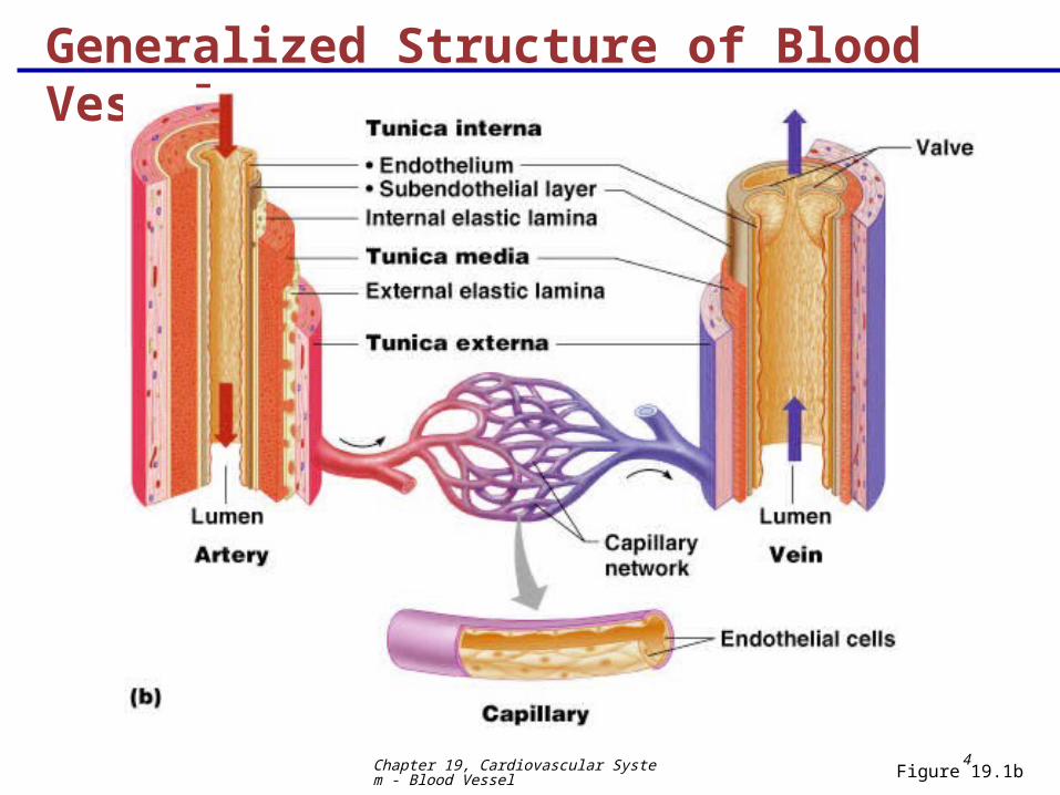
Generalized Structure of Blood Vessels
Arteries and veins are composed of three tunics – tunica interna, tunica media, and tunica externa
Lumen – central blood-containing space surrounded by tunics
Capillaries are composed of endothelium with sparse basal lamina
Chapter 19, Cardiovascular System - Blood Vessel
4
Generalized Structure of Blood Vessels
Figure 19.1b

Chapter 19, Cardiovascular System - Blood Vessel
5
Tunics
Tunica interna (tunica intima)
Endothelial layer that lines the lumen of all vessels
In vessels larger than 1 mm, a subendothelial connective tissue basement membrane is present
Tunica media
Smooth muscle and elastic fiber layer, regulated by sympathetic nervous system
Controls vasoconstriction/vasodilation of vessels
Chapter 19, Cardiovascular System - Blood Vessel
6
Tunics
Tunica externa (tunica adventitia)
Collagen fibers that protect and reinforce vessels
Larger vessels contain vasa vasorum
Chapter 19, Cardiovascular System - Blood Vessel
7
Elastic (Conducting) Arteries
Thick-walled arteries near the heart; the aorta and its major branches
Large lumen allow low-resistance conduction of blood
Contain elastin in all three tunics
Withstand and smooth out large blood pressure fluctuations
Allow blood to flow fairly continuously through the body
Chapter 19, Cardiovascular System - Blood Vessel
8
Muscular (Distributing) Arteries and Arterioles Muscular arteries – distal to elastic arteries; deliver
blood to body organs
Have thick tunica media with more smooth muscle and less elastic tissue
Active in vasoconstriction
Arterioles – smallest arteries; lead to capillary beds
Control flow into capillary beds via vasodilation and constriction
Chapter 19, Cardiovascular System - Blood Vessel
9
Capillaries Primary function is to permit the exchange of
nutrients and gases between the blood and tissue cells.
Capillaries are the smallest blood vessels
Walls consisting of a thin tunica interna, one cell thick
Allow only a single RBC to pass at a time
Pericytes on the outer surface stabilize their walls
There are three structural types of capillaries: continuous, fenestrated, and sinusoids
Chapter 19, Cardiovascular System - Blood Vessel
10
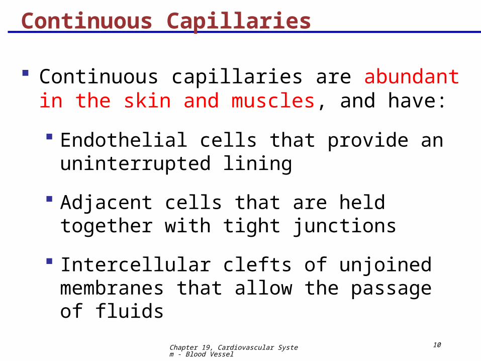
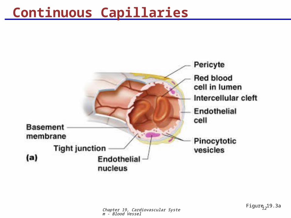
Continuous Capillaries
Continuous capillaries are abundant in the skin and muscles, and have:
Endothelial cells that provide an uninterrupted lining
Adjacent cells that are held together with tight junctions
Intercellular clefts of unjoined membranes that allow the passage of fluids
Chapter 19, Cardiovascular System - Blood Vessel
11
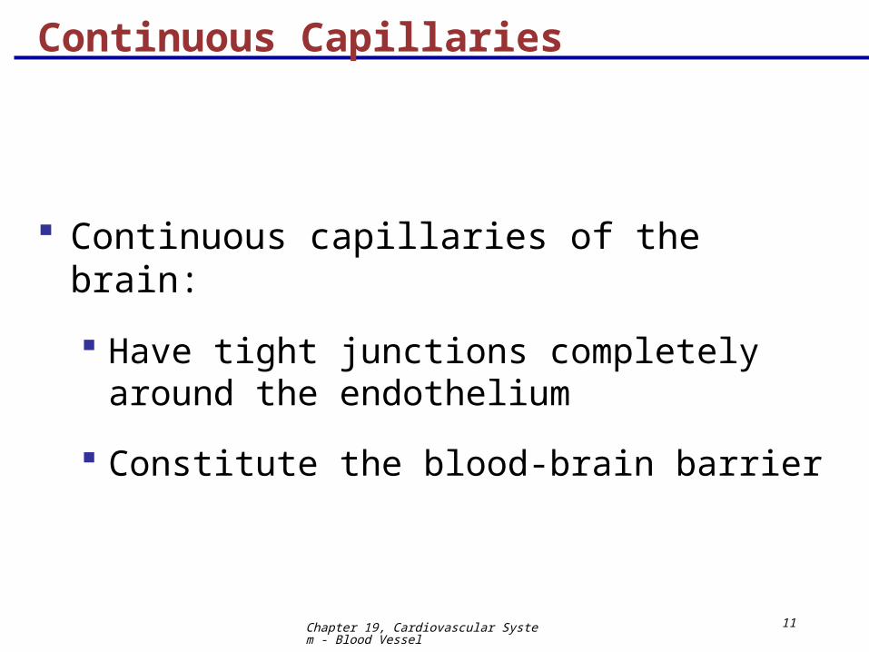
Continuous Capillaries
Continuous capillaries of the brain:
Have tight junctions completely around the endothelium
Constitute the blood-brain barrier
Chapter 19, Cardiovascular System - Blood Vessel
12
Continuous Capillaries
Figure 19.3a
Chapter 19, Cardiovascular System - Blood Vessel
13
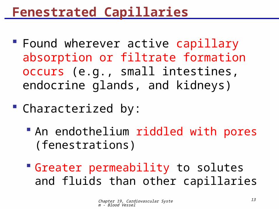
Fenestrated Capillaries
Found wherever active capillary absorption or filtrate formation occurs (e.g., small intestines, endocrine glands, and kidneys)
Characterized by:
An endothelium riddled with pores (fenestrations)
Greater permeability to solutes and fluids than other capillaries
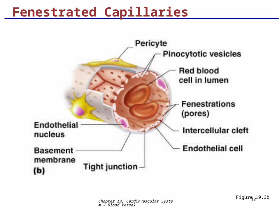
Chapter 19, Cardiovascular System - Blood Vessel
14
Fenestrated Capillaries
Figure 19.3b
Chapter 19, Cardiovascular System - Blood Vessel
15
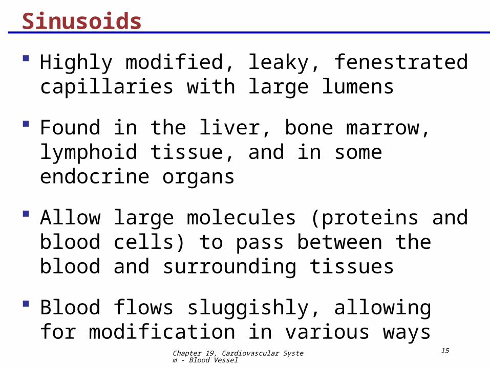
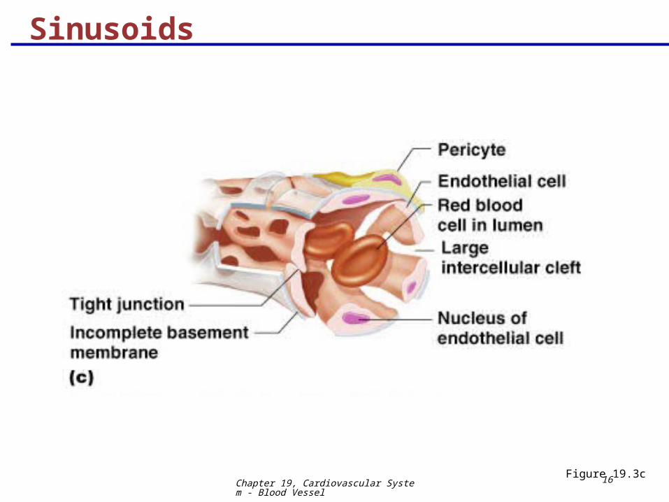
Sinusoids
Highly modified, leaky, fenestrated capillaries with large lumens
Found in the liver, bone marrow, lymphoid tissue, and in some endocrine organs
Allow large molecules (proteins and blood cells) to pass between the blood and surrounding tissues
Blood flows sluggishly, allowing for modification in various ways
Chapter 19, Cardiovascular System - Blood Vessel
16
Sinusoids
Figure 19.3c

Chapter 19, Cardiovascular System - Blood Vessel
17
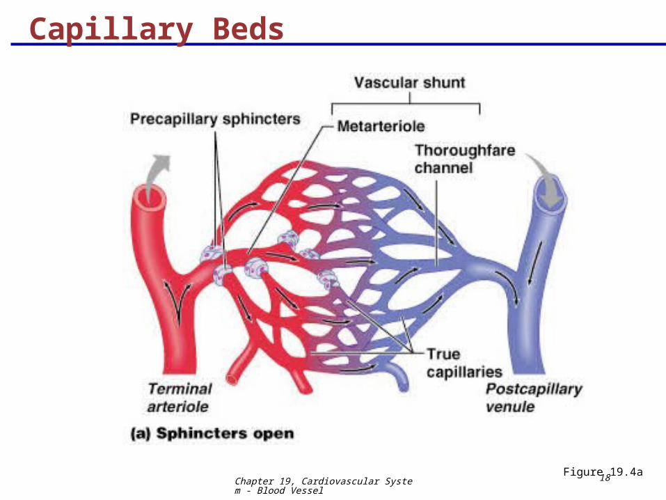
Capillary Beds
A microcirculation of interwoven networks of capillaries, consisting of:
Vascular shunts – metarteriole–thoroughfare channel connecting an arteriole directly with a postcapillary venule
True capillaries – 10 to 100 per capillary bed, capillaries branch off the metarteriole and return to the thoroughfare channel at the distal end of the bed
Chapter 19, Cardiovascular System - Blood Vessel
18
Capillary Beds
Figure 19.4a
Chapter 19, Cardiovascular System - Blood Vessel
19
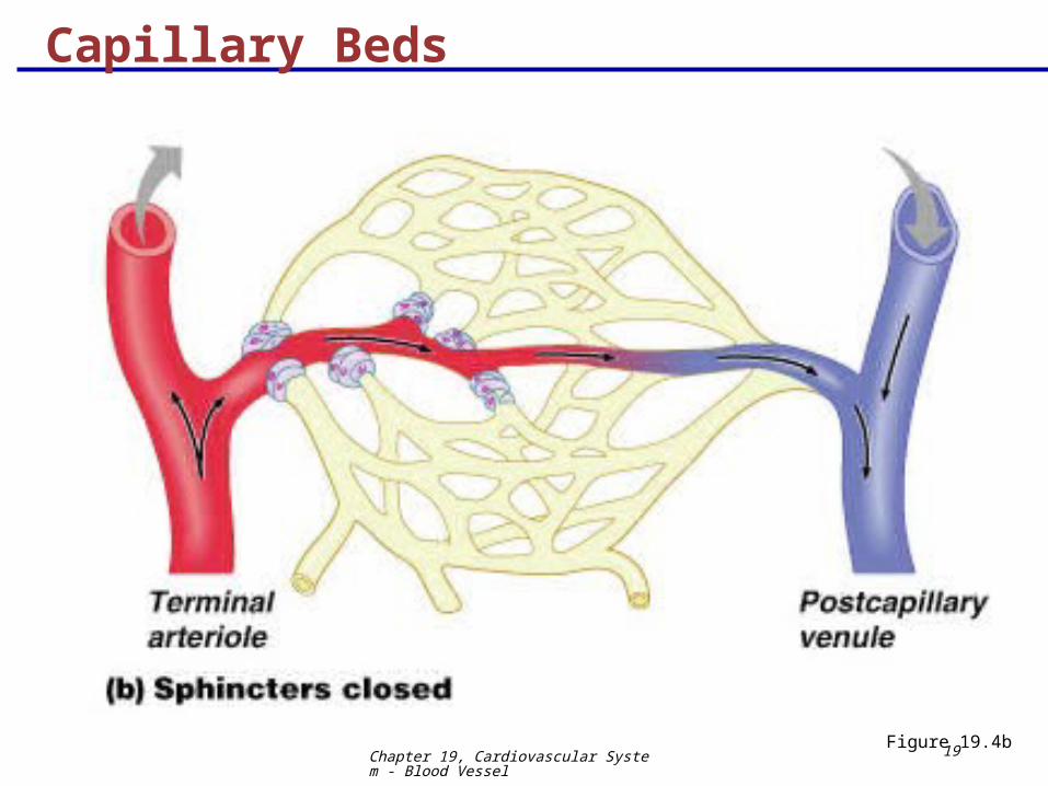
Capillary Beds
Figure 19.4b
Chapter 19, Cardiovascular System - Blood Vessel
20
Blood Flow Through Capillary Beds
Precapillary sphincter
Cuff of smooth muscle that surrounds each true capillary
Regulates blood flow into the capillary
Blood flow is regulated by vasomotor nerves and local chemical conditions, so it can either bypass or flood the capillary bed

Chapter 19, Cardiovascular System - Blood Vessel
21
Venous System: Venules
Are formed when capillary beds unite
Allow fluids and WBCs to pass from the bloodstream to tissues
Postcapillary venules – smallest venules, composed of endothelium and a few pericytes
Large venules have one or two layers of smooth muscle (tunica media)
Chapter 19, Cardiovascular System - Blood Vessel
22
Venous System: Veins
Veins are:
Formed when venules converge
Composed of three tunics, with a thin tunica media and a thick tunica externa consisting of collagen fibers and elastic networks
Capacitance vessels (blood reservoirs) that contain 65% of the blood supply
Chapter 19, Cardiovascular System - Blood Vessel
23
Venous System: Veins Veins have much lower blood pressure and thinner
walls than arteries
To return blood to the heart, veins have special adaptations
Large-diameter lumens, which offer little resistance to flow
Valves (resembling semilunar heart valves), which prevent backflow of blood
Venous sinuses – specialized, flattened veins with extremely thin walls (e.g., coronary sinus of the heart and dural sinuses of the brain)
Chapter 19, Cardiovascular System - Blood Vessel
24
Vascular Anastomoses
Merging blood vessels, more common in veins than arteries
Arterial anastomoses provide alternate pathways (collateral channels) for blood to reach a given body region
If one branch is blocked, the collateral channel can supply the area with adequate blood supply
Thoroughfare channels are examples of arteriovenous anastomoses
Chapter 19, Cardiovascular System - Blood Vessel
25
Blood Flow
Actual volume of blood flowing through a vessel, an organ, or the entire circulation in a given period:
Is measured in ml per min.
Is equivalent to cardiac output (CO), considering the entire vascular system
Is relatively constant when at rest
Varies widely through individual organs, according to immediate needs
Chapter 19, Cardiovascular System - Blood Vessel
26
19The Cardiovascular System:
Blood Vessels
Physiology
Chapter 19, Cardiovascular System - Blood Vessel
27
Blood Pressure (BP)
Force per unit area exerted on the wall of a blood vessel by its contained blood
Expressed in millimeters of mercury (mm Hg)
Measured in reference to systemic arterial BP in large arteries near the heart
The differences in BP within the vascular system provide the driving force that keeps blood moving from higher to lower pressure areas
Chapter 19, Cardiovascular System - Blood Vessel
28
Resistance
Resistance – opposition to flow
Measure of the amount of friction blood encounters as it passes through vessels
Generally encountered in the systemic circulation
Referred to as peripheral resistance (PR)
The three important sources of resistance are blood viscosity, total blood vessel length, and blood vessel diameter
Chapter 19, Cardiovascular System - Blood Vessel
29
Resistance factors that remain relatively constant are:
Blood viscosity – thickness or “stickiness” of the blood
Blood vessel length – the longer the vessel, the greater the resistance encountered
Resistance Factors: Viscosity and Vessel Length
Chapter 19, Cardiovascular System - Blood Vessel
30
Resistance Factors: Blood Vessel Diameter
Changes in vessel diameter are frequent and significantly alter peripheral resistance
Resistance varies inversely with the fourth power of vessel radius (one-half the diameter)
For example, if the radius is doubled, the resistance is 1/16 as much
Chapter 19, Cardiovascular System - Blood Vessel
31
Resistance Factors: Blood Vessel Diameter
Small-diameter arterioles are the major determinants of peripheral resistance
Fatty plaques from atherosclerosis:
Cause turbulent blood flow
Dramatically increase resistance due to turbulence
Chapter 19, Cardiovascular System - Blood Vessel
32
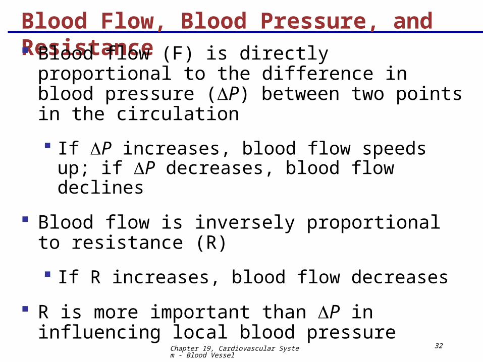
Blood Flow, Blood Pressure, and Resistance
Blood flow (F) is directly proportional to the difference in blood pressure (P) between two points in the circulation
If P increases, blood flow speeds up; if P decreases, blood flow declines
Blood flow is inversely proportional to resistance (R)
If R increases, blood flow decreases
R is more important than P in influencing local blood pressure

Chapter 19, Cardiovascular System - Blood Vessel
33
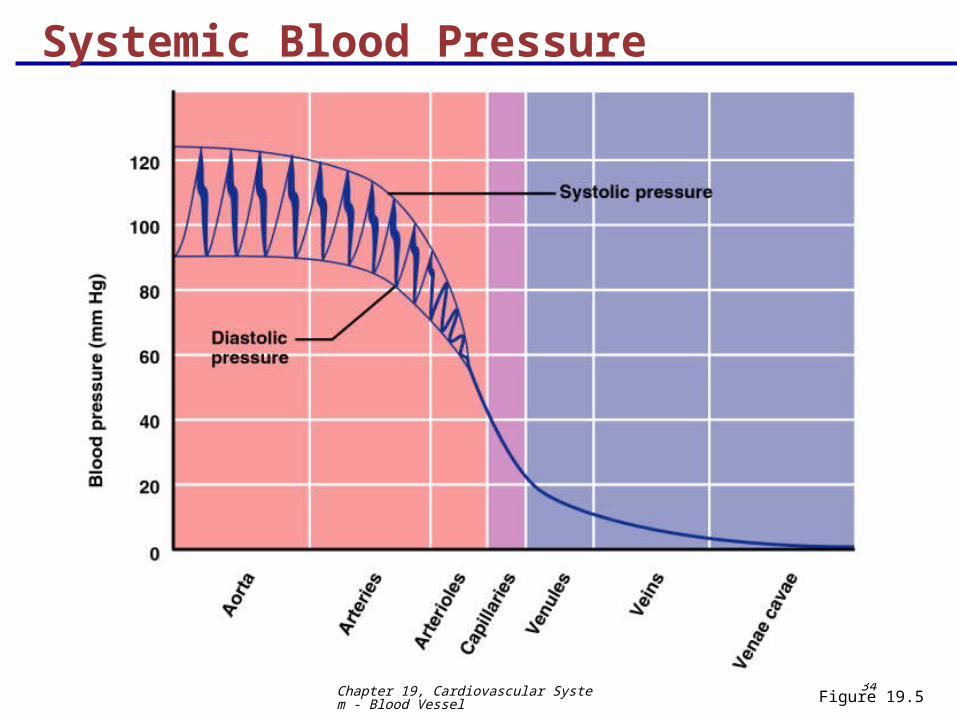
Systemic Blood Pressure
The pumping action of the heart generates blood flow through the vessels along a pressure gradient, always moving from higher- to lower-pressure areas
Pressure results when flow is opposed by resistance
Systemic pressure:
Is highest in the aorta
Declines throughout the length of the pathway
Is 0 mm Hg in the right atrium
The steepest change in blood pressure occurs in the arterioles
Chapter 19, Cardiovascular System - Blood Vessel
34
Systemic Blood Pressure
Figure 19.5
Chapter 19, Cardiovascular System - Blood Vessel
35
Arterial Blood Pressure
Arterial BP reflects two factors of the arteries close to the heart
Their elasticity (compliance or distensibility)
The amount of blood forced into them at any given time
Blood pressure in elastic arteries near the heart is pulsatile (BP rises and falls)
Chapter 19, Cardiovascular System - Blood Vessel
36
Arterial Blood Pressure
Systolic pressure – pressure exerted on arterial walls during ventricular contraction
Diastolic pressure – lowest level of arterial pressure during a ventricular cycle
Pulse pressure – the difference between systolic and diastolic pressure
Mean arterial pressure (MAP) – pressure that propels the blood to the tissues
MAP = diastolic pressure + 1/3 pulse pressure
Chapter 19, Cardiovascular System - Blood Vessel
37
Capillary Blood Pressure
Capillary BP ranges from 20 to 40 mm Hg
Low capillary pressure is desirable because high BP would rupture fragile, thin-walled capillaries
Low BP is sufficient to force filtrate out into interstitial space and distribute nutrients, gases, and hormones between blood and tissues
Chapter 19, Cardiovascular System - Blood Vessel
38
Venous Blood Pressure
Venous BP is steady and changes little during the cardiac cycle
The pressure gradient in the venous system is only about 20 mm Hg
A cut vein has even blood flow; a lacerated artery flows in spurts
Chapter 19, Cardiovascular System - Blood Vessel
39
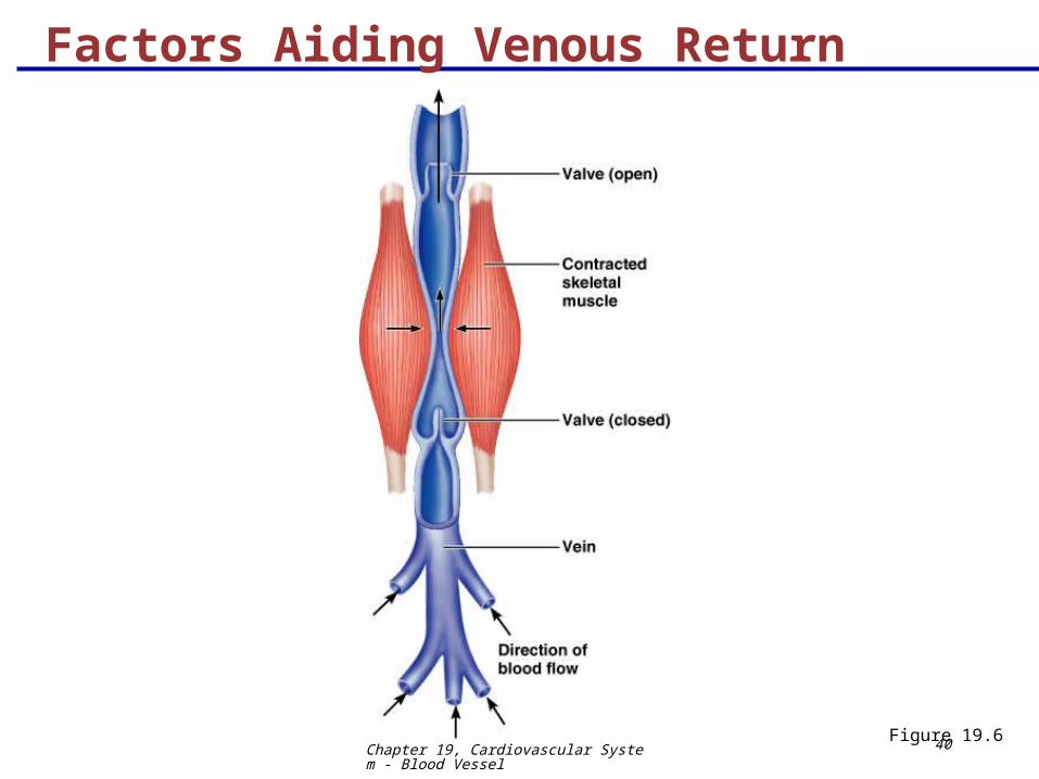
Factors Aiding Venous Return
Venous BP alone is too low to promote adequate blood return and is aided by the:
Respiratory “pump” – pressure changes created during breathing suck blood toward the heart by squeezing local veins
Muscular “pump” – contraction of skeletal muscles “milk” blood toward the heart
Valves prevent backflow during venous return
InterActive Physiology®: Cardiovascular System: Anatomy Review: Blood Vessel Structure and FunctionPLAYPLAY
Chapter 19, Cardiovascular System - Blood Vessel
40
Factors Aiding Venous Return
Figure 19.6
Chapter 19, Cardiovascular System - Blood Vessel
41
Maintaining Blood Pressure
Maintaining blood pressure requires:
Cooperation of the heart, blood vessels, and kidneys
Supervision of the brain

Chapter 19, Cardiovascular System - Blood Vessel
42
Maintaining Blood Pressure
The main factors influencing blood pressure are:
Cardiac output (CO)
Peripheral resistance (PR)
Blood volume
Blood pressure = CO x PR
Blood pressure varies directly with CO, PR, and blood volume
Chapter 19, Cardiovascular System - Blood Vessel
43
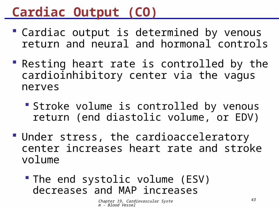
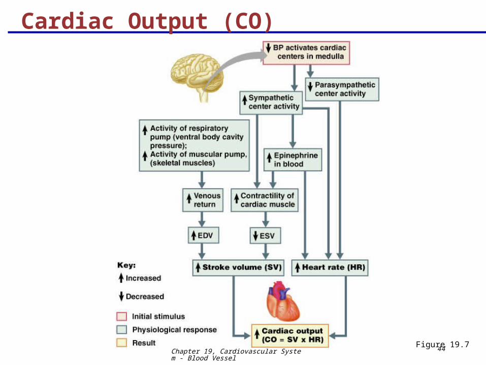
Cardiac Output (CO)
Cardiac output is determined by venous return and neural and hormonal controls
Resting heart rate is controlled by the cardioinhibitory center via the vagus nerves
Stroke volume is controlled by venous return (end diastolic volume, or EDV)
Under stress, the cardioacceleratory center increases heart rate and stroke volume
The end systolic volume (ESV) decreases and MAP increases
Chapter 19, Cardiovascular System - Blood Vessel
44
Cardiac Output (CO)
Figure 19.7
Chapter 19, Cardiovascular System - Blood Vessel
45
Controls of Blood Pressure
Short-term controls:
Are mediated by the nervous system and blood-borne chemicals
Counteract moment-to-moment fluctuations in blood pressure by altering peripheral resistance
Long-term controls regulate blood volume

Chapter 19, Cardiovascular System - Blood Vessel
46
Short-Term Mechanisms: Neural Controls
Neural controls of peripheral resistance:
Alter blood distribution to respond to specific demands
Maintain MAP by altering blood vessel diameter
Neural controls operate via reflex arcs involving:
Baroreceptors
Vasomotor centers of the medulla and vasomotor fibers
Vascular smooth muscle
Chapter 19, Cardiovascular System - Blood Vessel
47
Short-Term Mechanisms: Vasomotor Center
Vasomotor center – a cluster of sympathetic neurons in the medulla that oversees changes in blood vessel diameter
Maintains blood vessel tone by innervating smooth muscles of blood vessels, especially arterioles
Cardiovascular center – vasomotor center plus the cardiac centers that integrate blood pressure control by altering cardiac output and blood vessel diameter

Chapter 19, Cardiovascular System - Blood Vessel
48
Short-Term Mechanisms: Vasomotor Activity
Sympathetic activity causes:
Vasoconstriction and a rise in blood pressure if increased
Blood pressure to decline to basal levels if decreased
Vasomotor activity is modified by:
Baroreceptors (pressure-sensitive), chemoreceptors (O2, CO2, and H+ sensitive), higher brain centers, bloodborne chemicals, and hormones
Chapter 19, Cardiovascular System - Blood Vessel
49
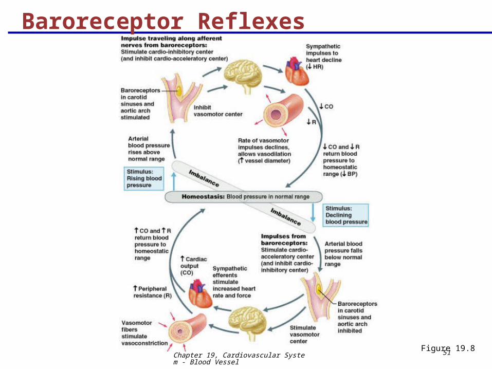
Increased blood pressure stimulates the cardioinhibitory center to:
Increase vessel diameter
Decrease heart rate, cardiac output, peripheral resistance, and blood pressure
Short-Term Mechanisms: Baroreceptor-Initiated Reflexes
Chapter 19, Cardiovascular System - Blood Vessel
50
Declining blood pressure stimulates the cardioacceleratory center to:
Increase cardiac output and peripheral resistance
Low blood pressure also stimulates the vasomotor center to constrict blood vessels
Short-Term Mechanisms: Baroreceptor-Initiated Reflexes
Chapter 19, Cardiovascular System - Blood Vessel
51Figure 19.8
Baroreceptor Reflexes
Chapter 19, Cardiovascular System - Blood Vessel
52
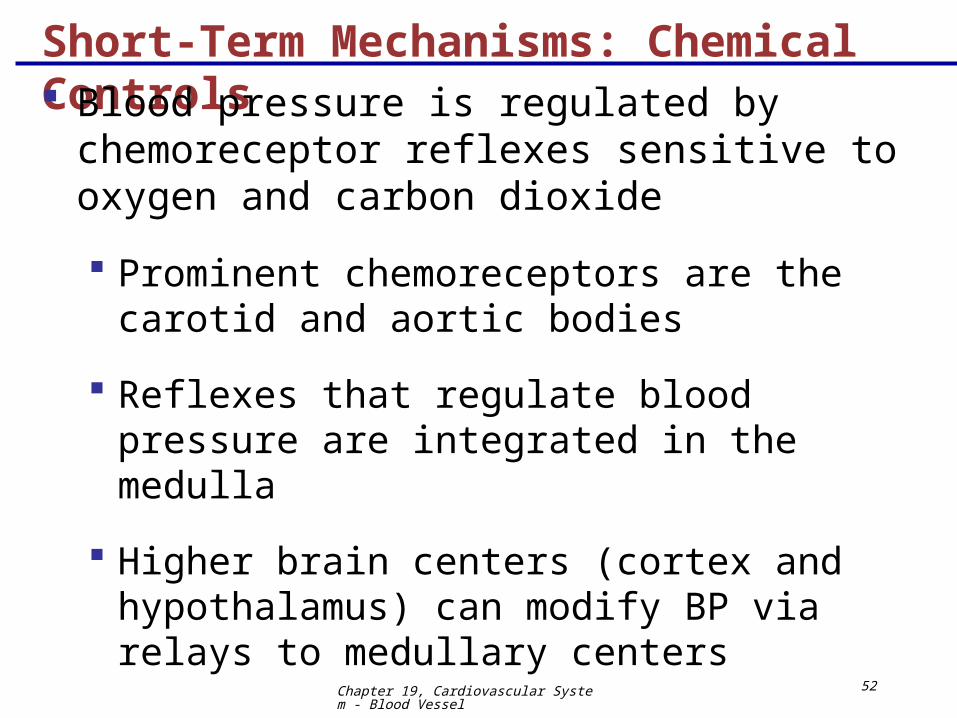
Short-Term Mechanisms: Chemical Controls
Blood pressure is regulated by chemoreceptor reflexes sensitive to oxygen and carbon dioxide
Prominent chemoreceptors are the carotid and aortic bodies
Reflexes that regulate blood pressure are integrated in the medulla
Higher brain centers (cortex and hypothalamus) can modify BP via relays to medullary centers
Chapter 19, Cardiovascular System - Blood Vessel
53
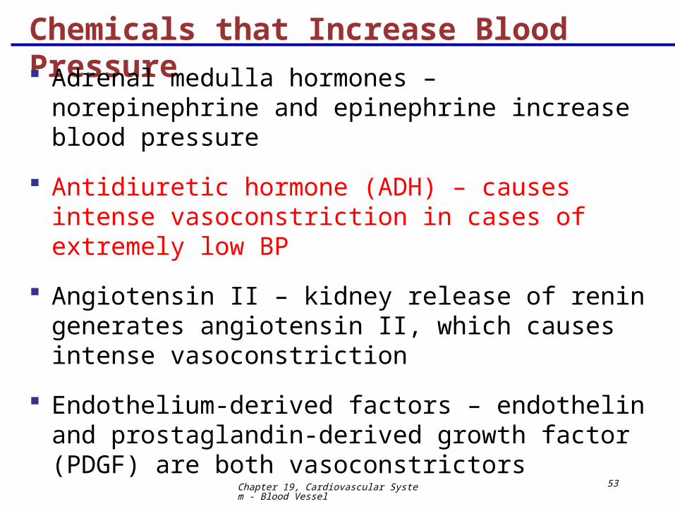
Chemicals that Increase Blood Pressure
Adrenal medulla hormones – norepinephrine and epinephrine increase blood pressure
Antidiuretic hormone (ADH) – causes intense vasoconstriction in cases of extremely low BP
Angiotensin II – kidney release of renin generates angiotensin II, which causes intense vasoconstriction
Endothelium-derived factors – endothelin and prostaglandin-derived growth factor (PDGF) are both vasoconstrictors
Chapter 19, Cardiovascular System - Blood Vessel
54
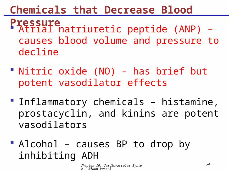
Chemicals that Decrease Blood Pressure
Atrial natriuretic peptide (ANP) – causes blood volume and pressure to decline
Nitric oxide (NO) – has brief but potent vasodilator effects
Inflammatory chemicals – histamine, prostacyclin, and kinins are potent vasodilators
Alcohol – causes BP to drop by inhibiting ADH
Chapter 19, Cardiovascular System - Blood Vessel
55
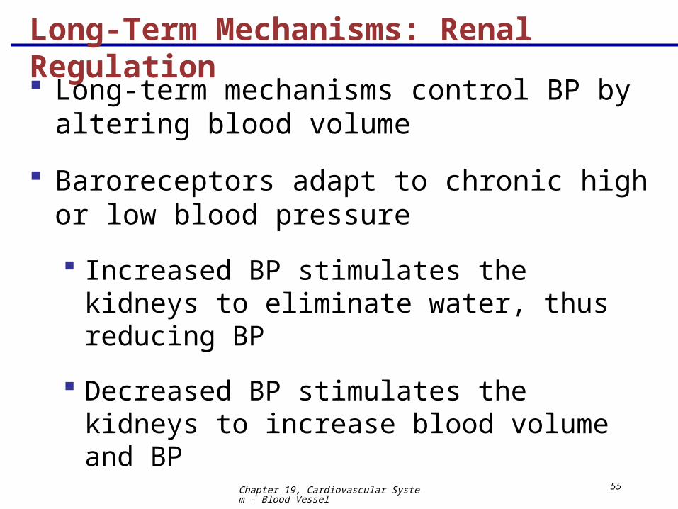
Long-Term Mechanisms: Renal Regulation
Long-term mechanisms control BP by altering blood volume
Baroreceptors adapt to chronic high or low blood pressure
Increased BP stimulates the kidneys to eliminate water, thus reducing BP
Decreased BP stimulates the kidneys to increase blood volume and BP
Chapter 19, Cardiovascular System - Blood Vessel
56
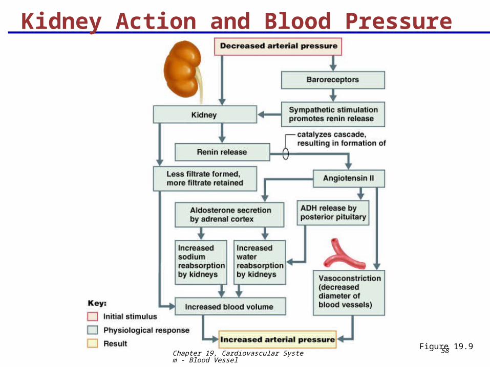
Kidney Action and Blood Pressure
Kidneys act directly and indirectly to maintain long-term blood pressure
Direct renal mechanism alters blood volume
Indirect renal mechanism involves the renin-angiotensin mechanism
Chapter 19, Cardiovascular System - Blood Vessel
57
Kidney Action and Blood Pressure
Declining BP causes the release of renin, which triggers the release of angiotensin II
Angiotensin II is a potent vasoconstrictor that stimulates aldosterone secretion
Aldosterone enhances renal reabsorption and stimulates ADH release
InterActive Physiology®: Cardiovascular System: Blood Pressure RegulationPLAYPLAY
Chapter 19, Cardiovascular System - Blood Vessel
58
Kidney Action and Blood Pressure
Figure 19.9
Chapter 19, Cardiovascular System - Blood Vessel
59
Monitoring Circulatory Efficiency
Efficiency of the circulation can be assessed by taking pulse and blood pressure measurements
Vital signs – pulse and blood pressure, along with respiratory rate and body temperature
Pulse – pressure wave caused by the expansion and recoil of elastic arteries
Radial pulse (taken on the radial artery at the wrist) is routinely used
Varies with health, body position, and activity
Chapter 19, Cardiovascular System - Blood Vessel
60
Measuring Blood Pressure
Systemic arterial BP is measured indirectly with the auscultatory method
A sphygmomanometer is placed on the arm superior to the elbow
Pressure is increased in the cuff until it is greater than systolic pressure in the brachial artery
Pressure is released slowly and the examiner listens with a stethoscope
Chapter 19, Cardiovascular System - Blood Vessel
61
Measuring Blood Pressure
The first sound heard is recorded as the systolic pressure
The pressure when sound disappears is recorded as the diastolic pressure
InterActive Physiology®: Cardiovascular System: Measuring Blood Pressure
PLAYPLAY

Chapter 19, Cardiovascular System - Blood Vessel
62
Variations in Blood Pressure
Blood pressure cycles over a 24-hour period
BP peaks in the morning due to waxing and waning levels of retinoic acid
Extrinsic factors such as age, sex, weight, race, mood, posture, socioeconomic status, and physical activity may also cause BP to vary
Chapter 19, Cardiovascular System - Blood Vessel
63
Alterations in Blood Pressure
Hypotension – low BP in which systolic pressure is below 100 mm Hg
Hypertension – condition of sustained elevated arterial pressure of 140/90 or higher
Transient elevations are normal and can be caused by fever, physical exertion, and emotional upset
Chronic elevation is a major cause of heart failure, vascular disease, renal failure, and stroke
Chapter 19, Cardiovascular System - Blood Vessel
64
Hypotension
Orthostatic hypotension – temporary low BP and dizziness when suddenly rising from a sitting or reclining position
Chronic hypotension – hint of poor nutrition and warning sign for Addison’s disease
Acute hypotension – important sign of circulatory shock
Threat to patients undergoing surgery and those in intensive care units
Chapter 19, Cardiovascular System - Blood Vessel
65
Hypertension
Hypertension maybe transient or persistent
Primary or essential hypertension – risk factors in primary hypertension include diet, obesity, age, race, heredity, stress, and smoking
Secondary hypertension – due to identifiable disorders, including excessive renin secretion, arteriosclerosis, and endocrine disorders
Chapter 19, Cardiovascular System - Blood Vessel
66
Blood Flow Through Tissues
Blood flow, or tissue perfusion, is involved in:
Delivery of oxygen and nutrients to, and removal of wastes from, tissue cells
Gas exchange in the lungs
Absorption of nutrients from the digestive tract
Urine formation by the kidneys
Blood flow is precisely the right amount to provide proper tissue function
Chapter 19, Cardiovascular System - Blood Vessel
67
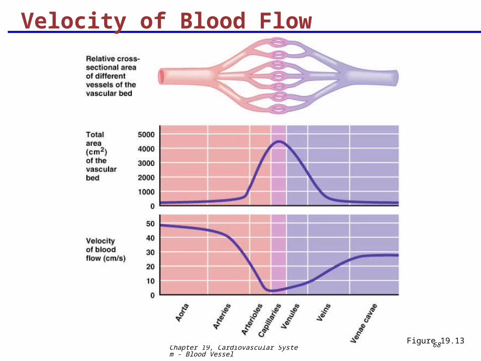
Velocity of Blood Flow
Blood velocity:
Changes as it travels through the systemic circulation
Is inversely proportional to the cross-sectional area
Slow capillary flow allows adequate time for exchange between blood and tissues
Chapter 19, Cardiovascular System - Blood Vessel
68
Velocity of Blood Flow
Figure 19.13
Chapter 19, Cardiovascular System - Blood Vessel
69
Autoregulation: Local Regulation of Blood Flow Autoregulation – automatic adjustment of blood
flow to each tissue in proportion to its requirements at any given point in time
Blood flow through an individual organ is intrinsically controlled by modifying the diameter of local arterioles feeding its capillaries
MAP remains constant, while local demands regulate the amount of blood delivered to various areas according to need
Chapter 19, Cardiovascular System - Blood Vessel
70
Metabolic Controls
Declining tissue nutrient and oxygen levels are stimuli for autoregulation
Hemoglobin delivers nitric oxide (NO) as well as oxygen to tissues
Nitric oxide induces vasodilation at the capillaries to help get oxygen to tissue cells
Other autoregulatory substances include: potassium and hydrogen ions, adenosine, lactic acid, histamines, kinins, and prostaglandins
Chapter 19, Cardiovascular System - Blood Vessel
71
Myogenic Controls
Inadequate blood perfusion or excessively high arterial pressure:
Are autoregulatory
Provoke myogenic responses – stimulation of vascular smooth muscle
Vascular muscle responds directly to:
Increased vascular pressure with increased tone, which causes vasoconstriction
Reduced stretch with vasodilation, which promotes increased blood flow to the tissue
Chapter 19, Cardiovascular System - Blood Vessel
72
Long-Term Autoregulation
Is evoked when short-term autoregulation cannot meet tissue nutrient requirements
May evolve over weeks or months to enrich local blood flow
Angiogenesis takes place:
As the number of vessels to a region increases
When existing vessels enlarge
When a heart vessel becomes partly occluded
Routinely in people in high altitudes, where oxygen content of the air is low
Chapter 19, Cardiovascular System - Blood Vessel
73
Blood Flow: Skeletal Muscles
Resting muscle blood flow is regulated by myogenic and general neural mechanisms in response to oxygen and carbon dioxide levels
When muscles become active, hyperemia is directly proportional to greater metabolic activity of the muscle (active or exercise hyperemia)
Arterioles in muscles have cholinergic, and alpha () and beta () adrenergic receptors
and adrenergic receptors bind to epinephrine
Chapter 19, Cardiovascular System - Blood Vessel
74
Blood Flow: Skeletal Muscle Regulation Muscle blood flow can increase tenfold or more
during physical activity as vasodilation occurs
Low levels of epinephrine bind to receptors
Cholinergic receptors are occupied
Intense exercise or sympathetic nervous system activation results in high levels of epinephrine
High levels of epinephrine bind to receptors and cause vasoconstriction
This is a protective response to prevent muscle oxygen demands from exceeding cardiac pumping ability
Chapter 19, Cardiovascular System - Blood Vessel
75
Blood Flow: Brain Blood flow to the brain is constant, as neurons are
intolerant of ischemia
Metabolic controls – brain tissue is extremely sensitive to declines in pH, and increased carbon dioxide causes marked vasodilation
Myogenic controls protect the brain from damaging changes in blood pressure
Decreases in MAP cause cerebral vessels to dilate to ensure adequate perfusion
Increases in MAP cause cerebral vessels to constrict
Chapter 19, Cardiovascular System - Blood Vessel
76
Blood Flow: Brain
The brain can regulate its own blood flow in certain circumstances, such as ischemia caused by a tumor
The brain is vulnerable under extreme systemic pressure changes
MAP below 60mm Hg can cause syncope (fainting)
MAP above 160 can result in cerebral edema
Chapter 19, Cardiovascular System - Blood Vessel
77
Blood Flow: Skin
Blood flow through the skin:
Supplies nutrients to cells in response to oxygen need
Helps maintain body temperature
Provides a blood reservoir
Chapter 19, Cardiovascular System - Blood Vessel
78
Blood Flow: Skin
Blood flow to venous plexuses below the skin surface:
Varies from 50 ml/min to 2500 ml/min, depending on body temperature
Is controlled by sympathetic nervous system reflexes initiated by temperature receptors and the central nervous system
Chapter 19, Cardiovascular System - Blood Vessel
79
Temperature Regulation
As temperature rises (e.g., heat exposure, fever, vigorous exercise):
Hypothalamic signals reduce vasomotor stimulation of the skin vessels
Heat radiates from the skin
Sweat also causes vasodilation via bradykinin in perspiration
Bradykinin stimulates the release of NO
As temperature decreases, blood is shunted to deeper, more vital organs
Chapter 19, Cardiovascular System - Blood Vessel
80
Blood Flow: Lungs
Blood flow in the pulmonary circulation is unusual in that:
The pathway is short
Arteries/arterioles are more like veins/venules (thin-walled, with large lumens)
They have a much lower arterial pressure (24/8 mm Hg versus 120/80 mm Hg)
Chapter 19, Cardiovascular System - Blood Vessel
81
Blood Flow: Lungs
The autoregulatory mechanism is exactly opposite of that in most tissues
Low oxygen levels cause vasoconstriction; high levels promote vasodilation
This allows for proper oxygen loading in the lungs
Chapter 19, Cardiovascular System - Blood Vessel
82
Blood Flow: Heart
Small vessel coronary circulation is influenced by:
Aortic pressure
The pumping activity of the ventricles
During ventricular systole:
Coronary vessels compress
Myocardial blood flow ceases
Stored myoglobin supplies sufficient oxygen
During ventricular diastole, oxygen and nutrients are carried to the heart
Chapter 19, Cardiovascular System - Blood Vessel
83
Blood Flow: Heart
Under resting conditions, blood flow through the heart may be controlled by a myogenic mechanism
During strenuous exercise:
Coronary vessels dilate in response to local accumulation of carbon dioxide
Blood flow may increase three to four times
Blood flow remains constant despite wide variation in coronary perfusion pressure
Chapter 19, Cardiovascular System - Blood Vessel
84
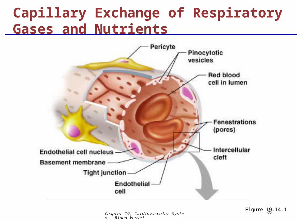
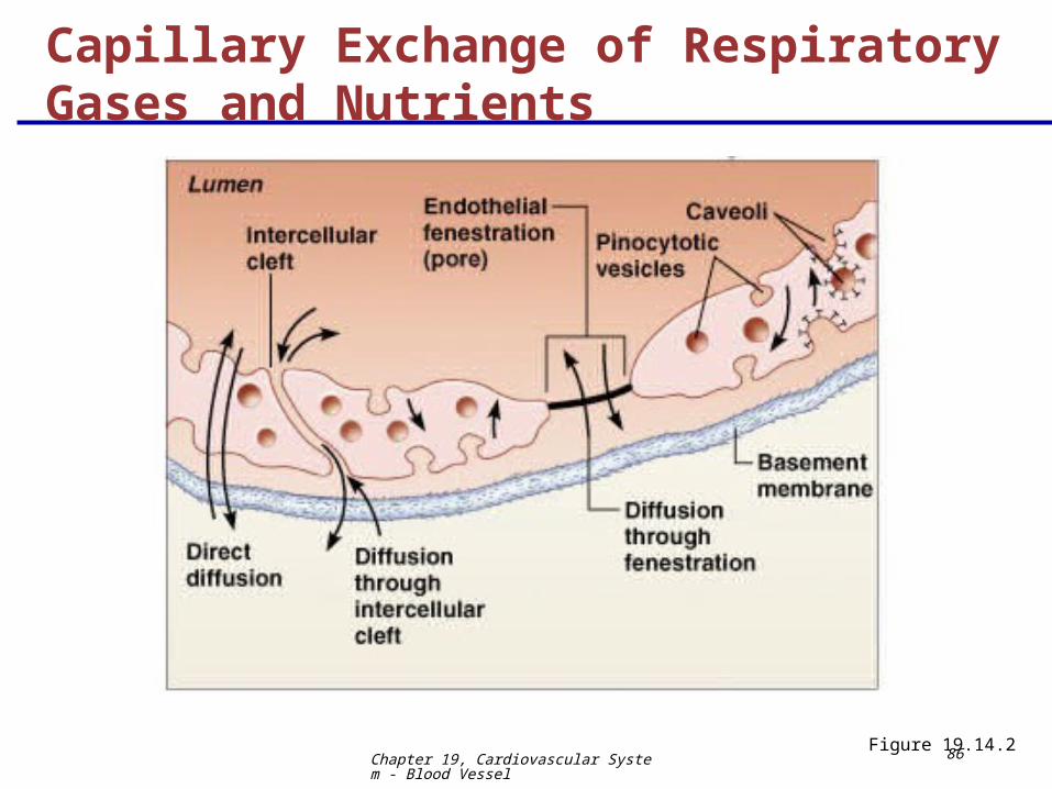
Oxygen, carbon dioxide, nutrients, and metabolic wastes diffuse between the blood and interstitial fluid along concentration gradients
Oxygen and nutrients pass from the blood to tissues
Carbon dioxide and metabolic wastes pass from tissues to the blood
Water-soluble solutes pass through clefts and fenestrations
Lipid-soluble molecules diffuse directly through endothelial membranes
Capillary Exchange of Respiratory Gases and Nutrients
Chapter 19, Cardiovascular System - Blood Vessel
85Figure 19.14.1
Capillary Exchange of Respiratory Gases and Nutrients
Chapter 19, Cardiovascular System - Blood Vessel
86Figure 19.14.2
Capillary Exchange of Respiratory Gases and Nutrients
Chapter 19, Cardiovascular System - Blood Vessel
87
Direction and amount of fluid flow depends upon the difference between:
Capillary hydrostatic pressure (HPc)
Capillary colloid osmotic pressure (OPc)
HPc – pressure of blood against the capillary walls:
Tends to force fluids through the capillary walls
Is greater at the arterial end of a bed than at the venule end
OPc– created by nondiffusible plasma proteins, which draw water toward themselves
Capillary Exchange: Fluid Movements
Chapter 19, Cardiovascular System - Blood Vessel
88
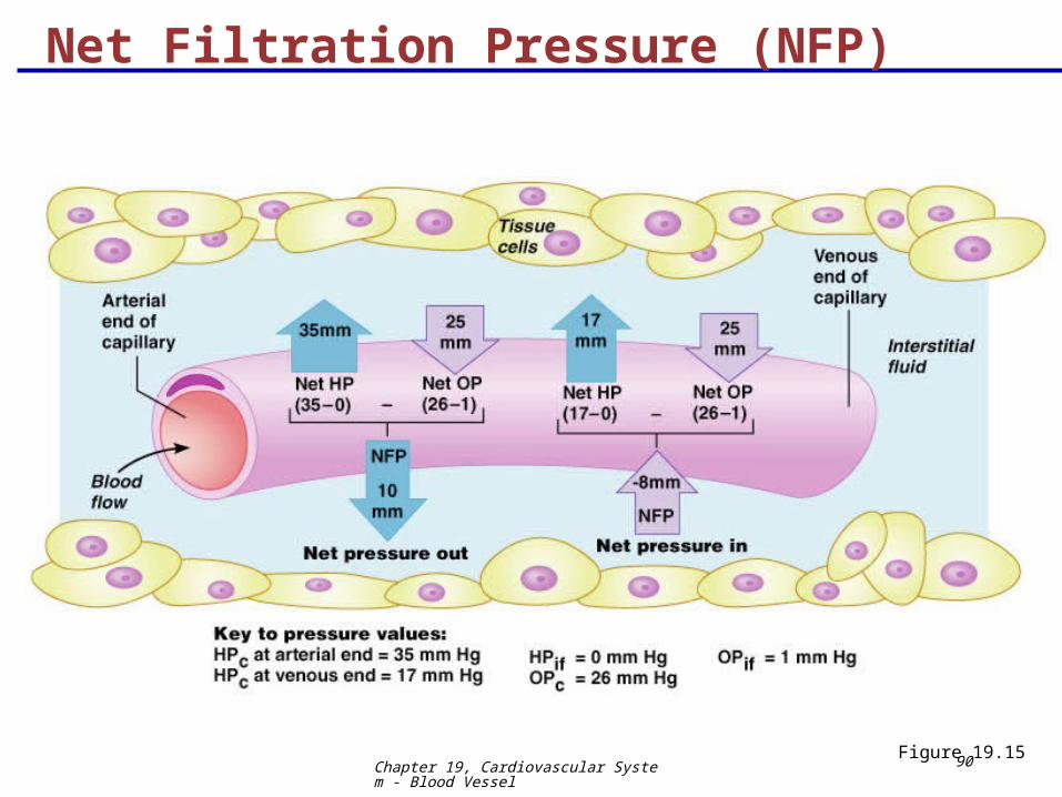
Net Filtration Pressure (NFP)
NFP – considers all the forces acting on a capillary bed
NFP = (HPc – HPif) – (OPc – OPif)
At the arterial end of a bed, hydrostatic forces dominate (fluids flow out)
Chapter 19, Cardiovascular System - Blood Vessel
89
Net Filtration Pressure (NFP)
At the venous end of a bed, osmotic forces dominate (fluids flow in)
More fluids enter the tissue beds than return blood, and the excess fluid is returned to the blood via the lymphatic system
InterActive Physiology®: Cardiovascular System: Autoregulation and Capillary DynamicsPLAYPLAY
Chapter 19, Cardiovascular System - Blood Vessel
90Figure 19.15
Net Filtration Pressure (NFP)
Chapter 19, Cardiovascular System - Blood Vessel
91
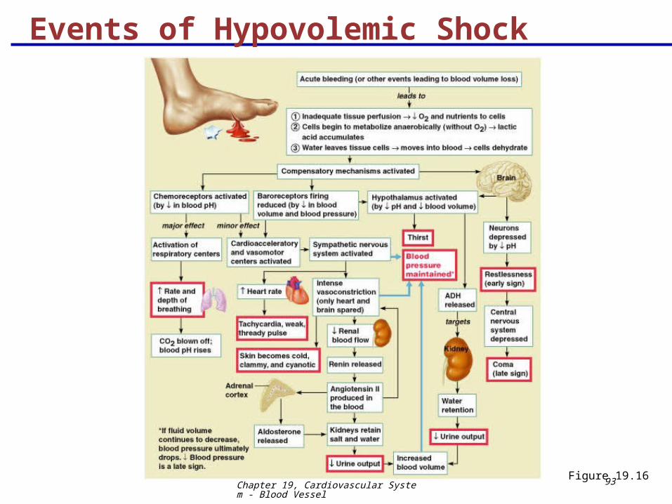
Circulatory shock – any condition in which blood vessels are inadequately filled and blood cannot circulate normally
Results in inadequate blood flow to meet tissue needs
Circulatory Shock
Chapter 19, Cardiovascular System - Blood Vessel
92
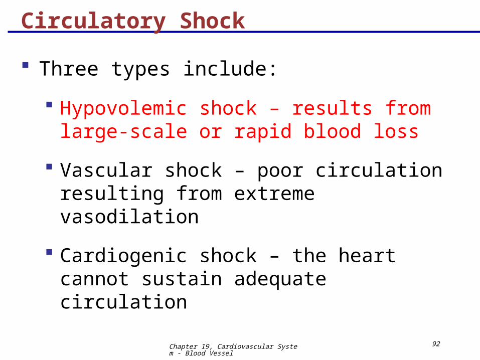
Three types include:
Hypovolemic shock – results from large-scale or rapid blood loss
Vascular shock – poor circulation resulting from extreme vasodilation
Cardiogenic shock – the heart cannot sustain adequate circulation
Circulatory Shock
Chapter 19, Cardiovascular System - Blood Vessel
93Figure 19.16
Events of Hypovolemic Shock
Chapter 19, Cardiovascular System - Blood Vessel
94
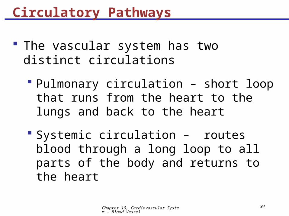
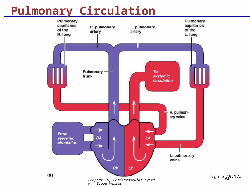
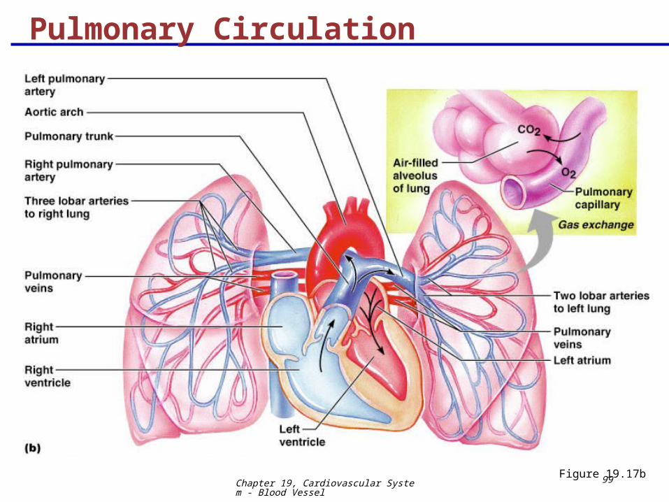
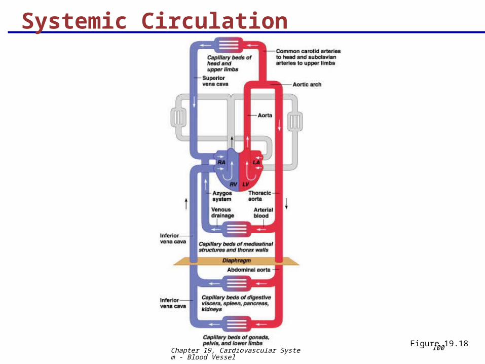
The vascular system has two distinct circulations
Pulmonary circulation – short loop that runs from the heart to the lungs and back to the heart
Systemic circulation – routes blood through a long loop to all parts of the body and returns to the heart
Circulatory Pathways
Chapter 19, Cardiovascular System - Blood Vessel
95
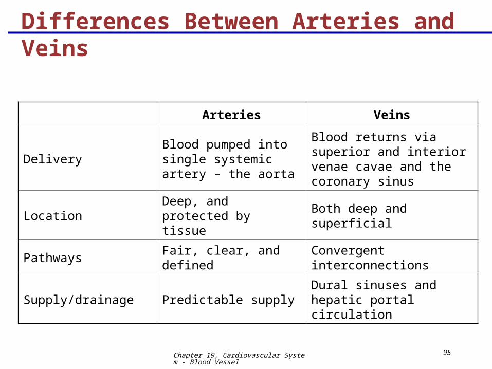
Differences Between Arteries and Veins
Arteries Veins
DeliveryBlood pumped into single systemic artery – the aorta
Blood returns via superior and interior venae cavae and the coronary sinus
LocationDeep, and protected by tissue
Both deep and superficial
Pathways Fair, clear, and defined Convergent interconnections
Supply/drainage Predictable supplyDural sinuses and hepatic portal circulation
Chapter 19, Cardiovascular System - Blood Vessel
96
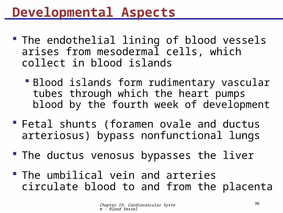
The endothelial lining of blood vessels arises from mesodermal cells, which collect in blood islands
Blood islands form rudimentary vascular tubes through which the heart pumps blood by the fourth week of development
Fetal shunts (foramen ovale and ductus arteriosus) bypass nonfunctional lungs
The ductus venosus bypasses the liver
The umbilical vein and arteries circulate blood to and from the placenta
Developmental Aspects
Chapter 19, Cardiovascular System - Blood Vessel
97
Developmental Aspects
Blood vessels are trouble-free during youth
Vessel formation occurs:
As needed to support body growth
For wound healing
To rebuild vessels lost during menstrual cycles
With aging, varicose veins, atherosclerosis, and increased blood pressure may arise
Chapter 19, Cardiovascular System - Blood Vessel
98Figure 19.17a
Pulmonary Circulation
Chapter 19, Cardiovascular System - Blood Vessel
99Figure 19.17b
Pulmonary Circulation
Chapter 19, Cardiovascular System - Blood Vessel
100Figure 19.18
Systemic Circulation
Chapter 19, Cardiovascular System - Blood Vessel
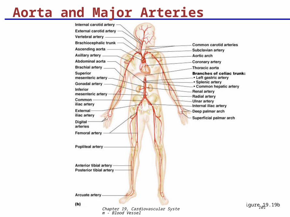
101Figure 19.19b
Aorta and Major Arteries
Chapter 19, Cardiovascular System - Blood Vessel
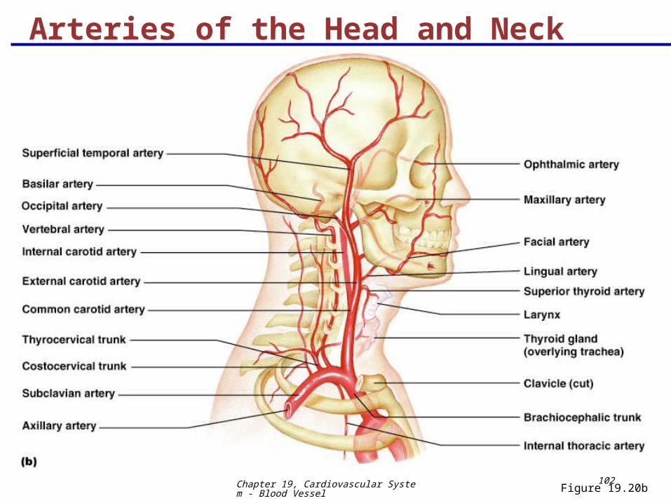
102Figure 19.20b
Arteries of the Head and Neck
Chapter 19, Cardiovascular System - Blood Vessel
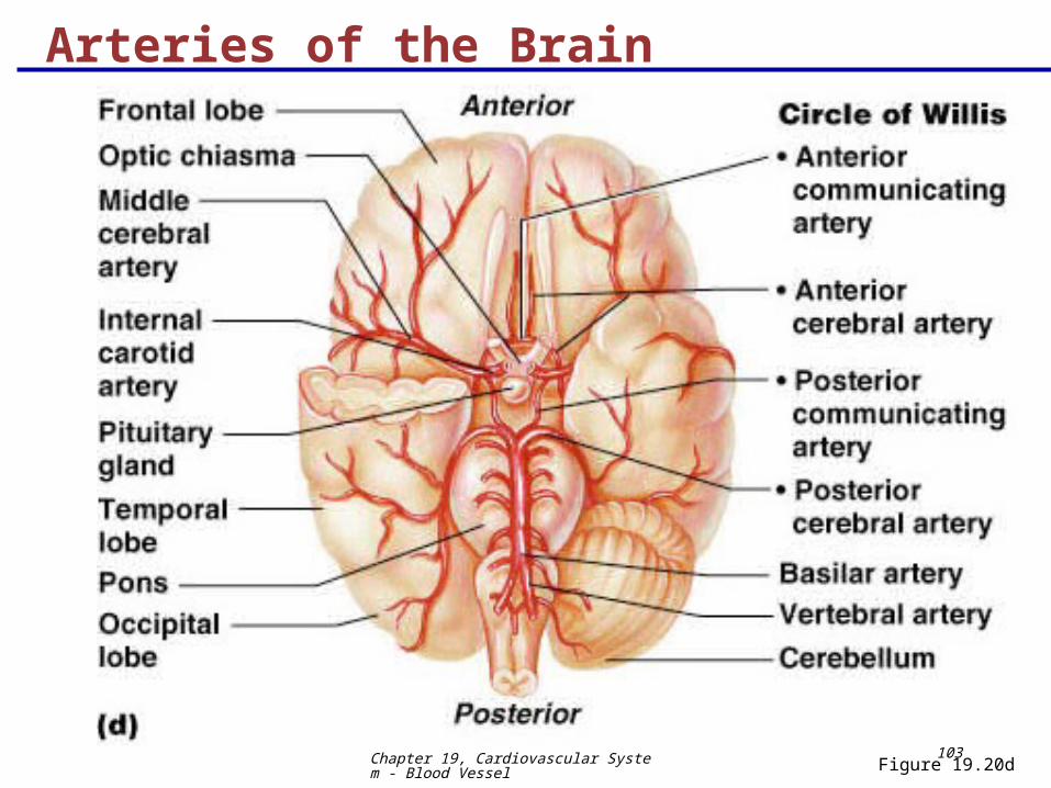
103Figure 19.20d
Arteries of the Brain
Chapter 19, Cardiovascular System - Blood Vessel
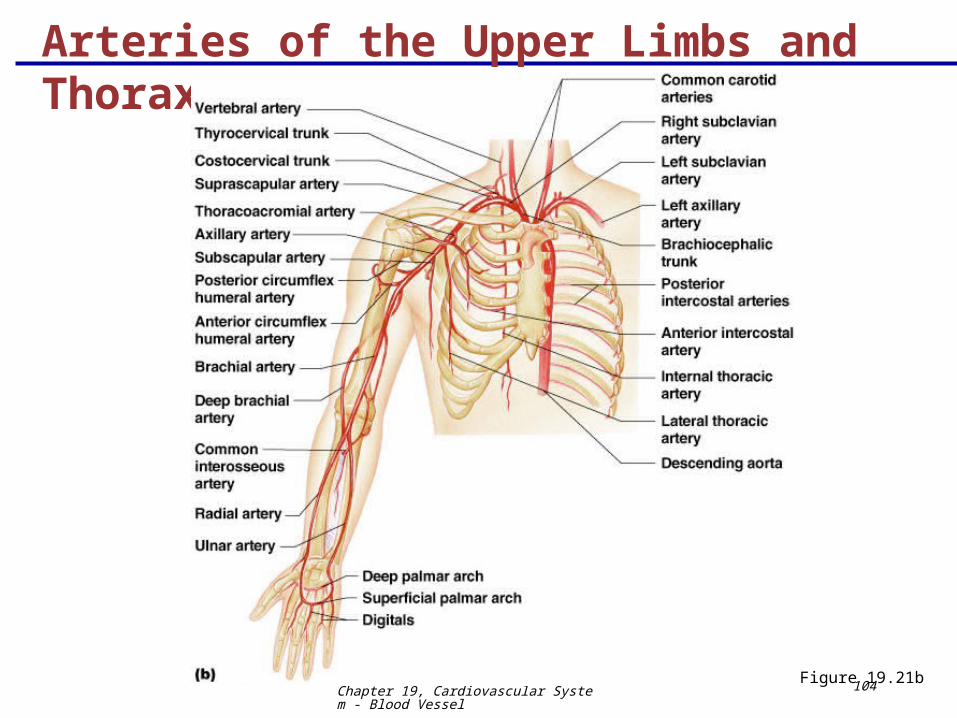
104Figure 19.21b
Arteries of the Upper Limbs and Thorax