Embed Size (px)
Citation preview
MED 1: Cardiovascular Exam Bambam2017
UST-FMS Batch 2017 Section D [amfv] 1
The Cardiovascular Exam Sources: UST-FMS Med1 Lecture (October 8, 2014), Mosby’s, KaiMM notes, Netter’s Anatomy, Berne and Levy Physiology
MOSBY’S NOTES
Heart: ANATOMY AND PHYSIOLOGY Cone-shaped 12 cm long, 8 cm wide, 6 cm AP diameter Broader upper portion: BASE Narrower lower tip: APEX Area overlying the heart: PRECORDIUM Lies in the mediastinum, to the left of midline,
just above the diaphragm, and is cradled between the medial and lower borders of the lungs
Positioned behind the sternum and the contiguous parts of the 3rd to 6th costal cartilages
In a tall person: heart tends to hang vertically and positioned more centrally
In a stocky or short person: heart tends to be more left and horizontally
Dextrocardia: heart on the right Situs Inversus: heart and stomach on the right,
liver on the left Structure
Four chambers: two atria and two ventricles Pericardium: tough, double-walled, fibrous sac
encasing the heart; has 2 layers with fluid inside providing for easy, low friction movement
Epicardium – thin outermost muscle layer Myocardium – thick muscular layer Endocardium – innermost layer, lines the
chambers and valves Cardiac Septum – divides the heart into left and
right Atrium – small, thin-walled Ventricle – large, thick-walled Primary muscle mass of the heart: ventricles
Surface Anatomy
Anterior: RV Left: LV Right: RA Posterior: LA
Valves
AV Valves: Mitral and Tricuspid Mitral (left): 2 cusps Tricuspid (right): 3 cusps
Semilunar Valves: Aortic and Pulmonary (both have 3 cusps) Aortic: between Left ventricle and Aorta Pulmonary: between right ventricle and pulmonary artery
MED 1: Cardiovascular Exam Bambam2017
UST-FMS Batch 2017 Section D [amfv] 2
Ask patient to lie down and remove his/her T-shirt. Introduce yourself to the patient. We should always do CV exam with the patient lying. We stay on the right side of the patient. WE DO IT IN THIS SEQUENCE:
1. General Survey 2. Vital signs 3. JVP 4. Carotid pulse 5. Peripheral pulse 6. Precordial Exam
Just for this afternoon, we’ll start with precordial exam. We expose only the area we want to examine. The heart is in the middle, pointing to the left.
R cardiac border: RA Most anterior portion: RV L cardiac border: LV Most posterior chamber: LA (when it enlarges, it
will push anterior structures forward)
The Flow of Blood
1. SVC and IVC 2. RA 3. Tricuspid valve 4. RV 5. Pulmonic valve 6. Pulmonary artery 7. Lungs (to be oxygenated) 8. Pulmonary veins 9. LA 10. Mitral valve 11. LV 12. Aortic valve 13. Aorta
In essence, there are 2 simultaneous circulation: (1) pulmonary and (2) systemic. Our heart as a pump has to be depolarized. There is electrical and mechanical event. MOSBY’S NOTES
ECG The heart is autonomous An intrinsic electrical conduction system enables
it to contract within itself Electrocardiogram: graphic recording of the
electrical activity of the heart; depolarization and repolarization
P wave – the spread of stimulus through the atria (atrial depolarization)
PR interval – the time from initial stimulation of the atria to initial stimulation of the ventricles, usually 0.12 to 0.20 s
QRS complex – the spread of stimulus to the ventricles (ventricular depolarization), usually less than 0.10 s
ST segment and T wave – the return of stimulated ventricular muscle to a resting state (ventricular repolarization)
U wave – a small deflection sometimes seen just after the T wave
QT interval – the time elapsed from the onset of ventricular depolarization until the completion of ventricular repolarization. Interval varies with cardiac rate
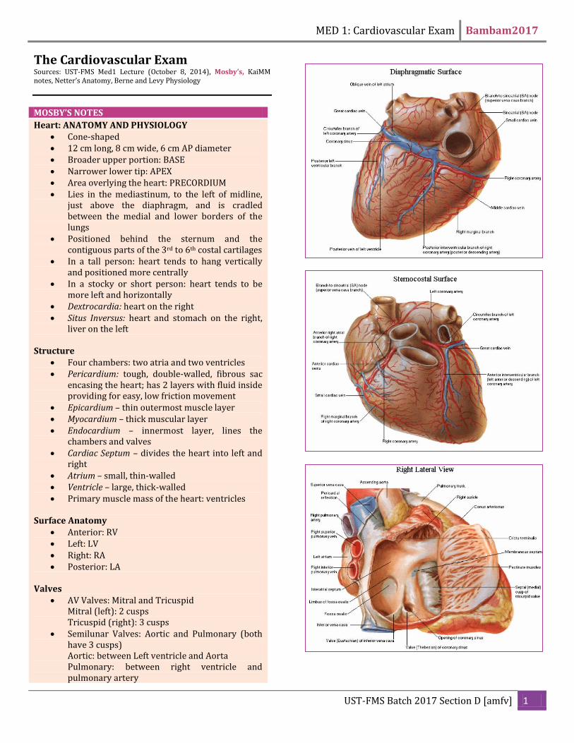
Conduction of Impulse
SA node (pacemaker): located in the wall of RA AV node: located at the atrial septum Bundle of His Purkinje fibers: in the ventricular myocardium Ventricular contraction starts at the apex to the
base
MED 1: Cardiovascular Exam Bambam2017
UST-FMS Batch 2017 Section D [amfv] 3
PRECORDIAL EXAMINATION INSPECTION Look at the chest and check for any deformity:
Pectus excavatum – Sternum naka-uka papaloob Pectus carinatum – sternum protruding out These deformities may be associated with heart
problems
Check the back, there is usually slight thoracic Kyphosis
If there’s no kyphosis Straight back syndrome (associated with heart problems)
Check the precordium for visible pulsations.
Adynamic: no visible pulsations Dynamic: 1 visible pulse Hyperdynamic: 2 or more visible pulse
Check visible pulsations at eye level or use white light (flash it tangentially)
Apex L lower parasternum and Epigastrium Midprecordium 2nd ICS, left parasternum 2nd ICS, right parasternum
Landmarks
Midsternum Parasternum Midclavicular line (do not use the nipple) Anterior axillary line Midaxillary line Posterior axillary line Angle of Louis (palpate from suprasternal notch,
slide your finger down): 2nd rib is attached
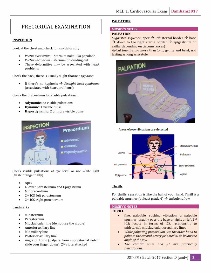
PALPATION MOSBY’S NOTES
PALPATION Suggested sequence: apex left sternal border base down to the right sterna border epigastrium or axilla (depending on circumstances) Apical Impulse: no more than 1cm, gentle and brief, not lasting as long as systole
Thrills For thrills, sensation is like the ball of your hand. Thrill is a palpable murmur (at least grade 4) turbulent flow MOSBY’S NOTES
THRILL fine, palpable, rushing vibration, a palpable
murmur; usually over the base or right or left 2nd ICS; locate in terms of ICS, relationship to midsternal, midclavicular, or axillary lines
While palpating precordium, use the other hand to palpate the carotid artery just medial or below the angle of the jaw.
The carotid pulse and S1 are practically synchronous.
MED 1: Cardiovascular Exam Bambam2017
UST-FMS Batch 2017 Section D [amfv] 4
The murmur of grade IV level or more can be felt. The sensation is called the thrill, It can be appreciated in systole or diastole. The following are common: Timing Location Probable Cause Systole Suprasternal notch
and/or 2nd and 3rd right ICS Suprasternal notch and/or 2nd and 3rd left ICS 4th left ICS Apex Left lower sternal border Left upper sternal border, often with extensive radiation
Aortic stenosis Pulmonic stenosis VSD Mitral regurgitation Tetralogy of Fallot PDA
Diastole Right sternal border Apex
Aortic regurgitation Aneurysm of ascending aorta Mitral stenosis
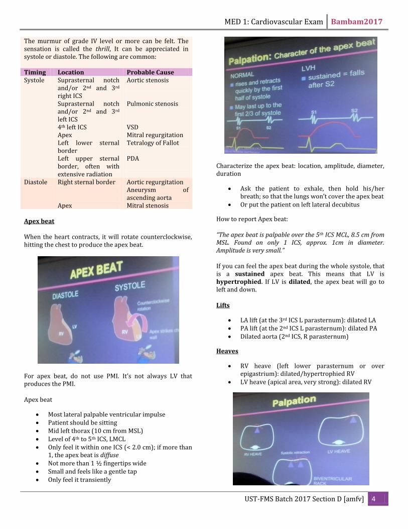
Apex beat When the heart contracts, it will rotate counterclockwise, hitting the chest to produce the apex beat.
For apex beat, do not use PMI. It’s not always LV that produces the PMI. Apex beat
Most lateral palpable ventricular impulse Patient should be sitting Mid left thorax (10 cm from MSL) Level of 4th to 5th ICS, LMCL Only feel it within one ICS (< 2.0 cm); if more than
1, the apex beat is diffuse Not more than 1 ½ fingertips wide Small and feels like a gentle tap Only feel it transiently
Characterize the apex beat: location, amplitude, diameter, duration
Ask the patient to exhale, then hold his/her breath; so that the lungs won’t cover the apex beat
Or put the patient on left lateral decubitus
How to report Apex beat: “The apex beat is palpable over the 5th ICS MCL, 8.5 cm from MSL. Found on only 1 ICS, approx. 1cm in diameter. Amplitude is very small.” If you can feel the apex beat during the whole systole, that is a sustained apex beat. This means that LV is hypertrophied. If LV is dilated, the apex beat will go to left and down. Lifts
LA lift (at the 3rd ICS L parasternum): dilated LA PA lift (at the 2nd ICS L parasternum): dilated PA Dilated aorta (2nd ICS, R parasternum)
Heaves
RV heave (left lower parasternum or over epigastrium): dilated/hypertrophied RV
LV heave (apical area, very strong): dilated RV
MED 1: Cardiovascular Exam Bambam2017
UST-FMS Batch 2017 Section D [amfv] 5
AUSCULTATION Auscultatory Areas
1. Mitral Valve: Apex, 5th ICS MCL 2. Tricuspid Valve: Left lower parasternum, 4th ICS 3. Pulmonic Valve: Left parasternum, 2nd ICS 4. 2nd Pulmonic Valve: Left sternal border, 3rd ICS 5. Aortic Valve: Right parasternum, 2nd ICS
Ideal Stethoscope
Largest ear tips possible Adjustable head pieces Vinyl tubing Not more than 25 cms 3/16 internal diameter Shallow large diameter bell Smooth stiff, thin diaphragm
The Cardiac Cycle
1. Isovolumetric contraction a. Because systemic pressure is always
HIGHER than ventricular pressure, ventricles build pressure by contracting
b. No movement of blood
c. Contraction against closed valve = builds
up pressure
2. Rapid ejection a. Rapid movement of blood upon opening
of aortic valve b. LV pressure drops -> aortic & pulmonic
valve will close (S2) 3. Isovolumetric relaxation
a. Atria cannot build up pressures as high as ventricles Not pressure builders
b. They need the ventricles to actively relax c. When ventricular pressure drops and
becomes less than atrial pressure, mitral & tricuspid valves open
4. Rapid filling phase a. During ventricular systole, large amounts
of blood accumulate in the right and left atria because of the closed A-V valves.
b. Therefore, as soon as systole is over and the ventricular pressures fall again to their low diastolic values, the moderately increased pressures that have developed in the atria immediately push the AV valves open and allow blood to flow rapidly into the ventricles.
5. Slow filling phase a. When pressures between atria and
ventricles equilibriate 6. Atrial depolarization
a. Cause atrial contraction and eject any residual blood
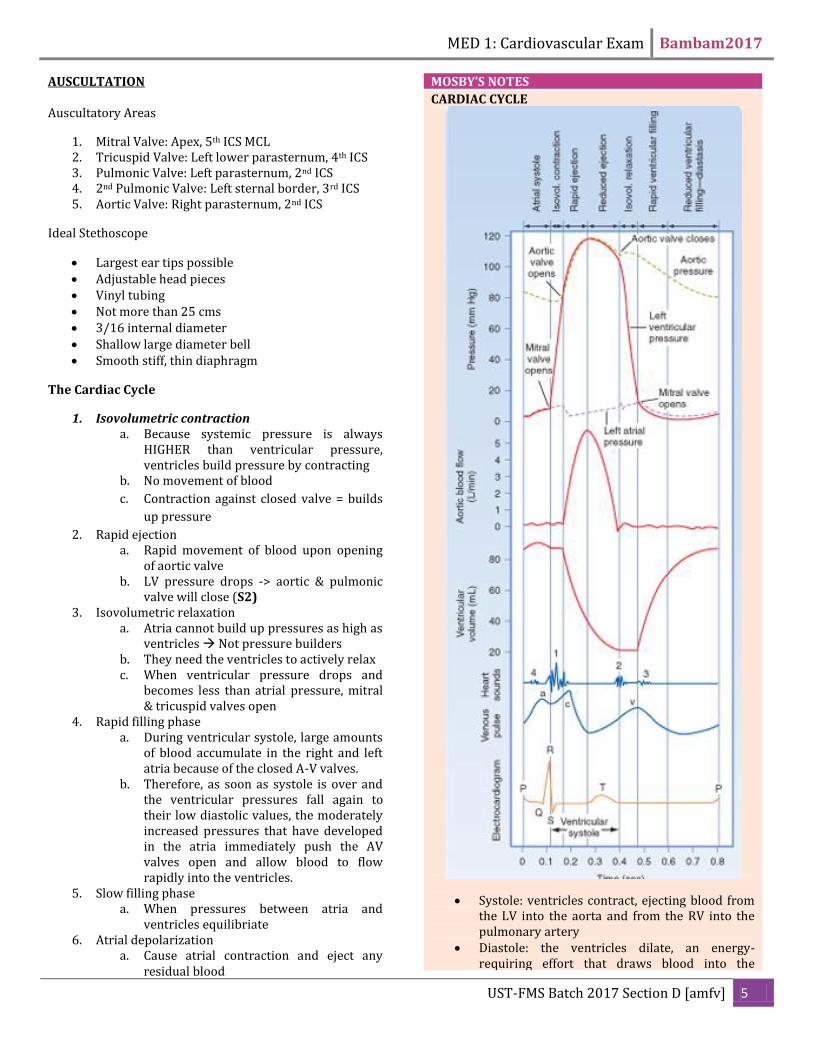
MOSBY’S NOTES
CARDIAC CYCLE
Systole: ventricles contract, ejecting blood from the LV into the aorta and from the RV into the pulmonary artery
Diastole: the ventricles dilate, an energy-requiring effort that draws blood into the
MED 1: Cardiovascular Exam Bambam2017
UST-FMS Batch 2017 Section D [amfv] 6
ventricles as the atria contract
Heart Sounds S1, “lubb”: closure of AV valves S2, “dubb”: closure of semilunar valves, has 2
components; A2(aortic valve closure) and P2 (pulmonary valve closure)
S3: ventricular filling/ diastole S4: atrial contraction
Generalities
Pressures in the RV, RA and PA are LOWER than the left side of the heart
The events occur slightly LATER on the right side than on the left side; thus heart sounds sometimes have two components E.g. A2 and P2: “split S2” (physiologic)
The simultaneous muscular tension and flow of blood give “body” to the sounds
The sounds are best heard in the direction of blood flow
S1 (1st Heart Sound)
Etiology o Closure of mitral valve o Closure of tricuspid valve o Ejection into the aortic root
Quality (apex) o Loud o High pitch
Timing o Coincides with the apex beat
How to Identify S1 from S2 o S1 Coincides with apex upstroke o S1 is heard immediately before carotid
upstroke o S1 with shorter interval from S2 o Apex - S1 louder than S2 o Base - S2 louder than S1, S2 splits on
inspiration Factors affecting the loudness of S1
o Rate of rise of LV pressure o Timing of MV closure in relation to onset
of ventricular contraction o Position of the MV at the beginning of
ventricular contraction o The more open the mitral valve is at the
end of diastole, the louder the S1(parang pinto. Kapag binuksan mo ng malaki, malakas ang kalabog pag padabog mong sinara)
o The stronger the LV contracts, the louder the S1
Splitting of S1: usually over tricuspid area
S2 (2nd Heart sound)
Etiology o Initiation of diastole o Sudden deceleration of forward flow
during aortic and pulmonary valve closure
o Best heard at the base of the heart o Normally widens on inspiration
Physiologic Splitting o When a person inhales, the intrathoracic
pressure becomes more NEGATIVE increases the blood returning to the right side of the heart. Therefore in SYSTOLE, more blood is present in the right atrium LONGER time is needed for blood to empty from the right atrium to the right ventricle
o Component delayed is the pulmonic component
MED 1: Cardiovascular Exam Bambam2017
UST-FMS Batch 2017 Section D [amfv] 7
S3 (3rd Heart sound)
Rapid filling sound Early filling gallop sound Occurs at the end of the rapid expansion phase of
the ventricle Heard best at or near the apex Heart best with bell applied with light pressure Physiologic S3: due to increase in velocity of
ventricular expansion (tachycardia, nervousness) Pathologic S3: loss of compliance/distensibility
(heart failure)
S4 (4th Heart Sound)
Atrial gallop, presystolic gallop, S4 gallop rarely physiological commonly pathological - decreased distensibility
or compliance of the LV o During atrial Contraction
best heard with the use of bell at the apex with the patient on left lateral decubitus
Murmurs
Abnormal sounds Pericardial friction rub Prosthetic Valve sounds Pacemaker sounds
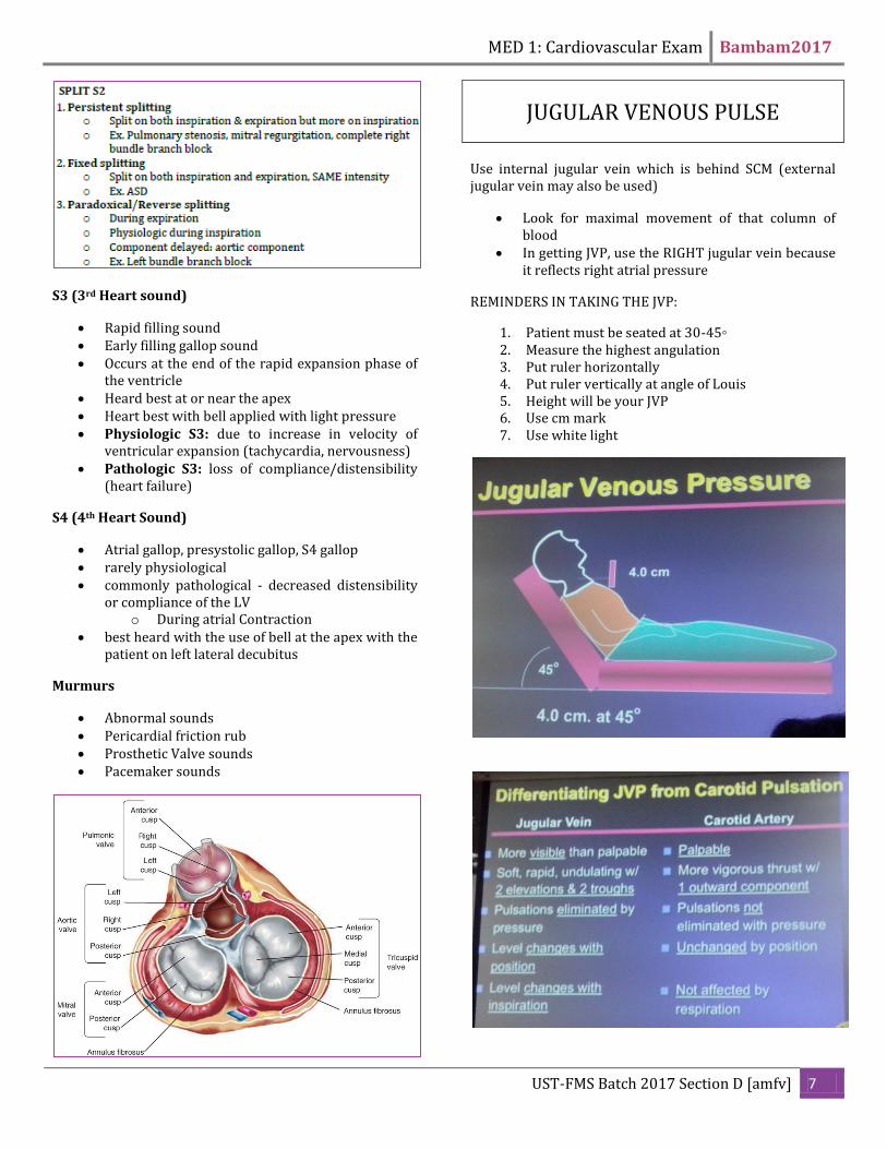
JUGULAR VENOUS PULSE Use internal jugular vein which is behind SCM (external jugular vein may also be used)
Look for maximal movement of that column of blood
In getting JVP, use the RIGHT jugular vein because it reflects right atrial pressure
REMINDERS IN TAKING THE JVP:
1. Patient must be seated at 30-45◦ 2. Measure the highest angulation 3. Put ruler horizontally 4. Put ruler vertically at angle of Louis 5. Height will be your JVP 6. Use cm mark 7. Use white light
MED 1: Cardiovascular Exam Bambam2017
UST-FMS Batch 2017 Section D [amfv] 8
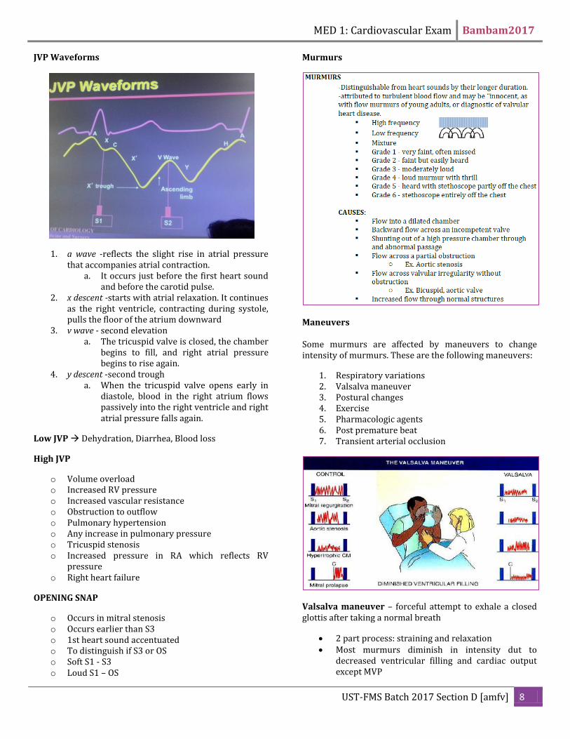
JVP Waveforms
1. a wave -reflects the slight rise in atrial pressure that accompanies atrial contraction.
a. It occurs just before the first heart sound and before the carotid pulse.
2. x descent -starts with atrial relaxation. It continues as the right ventricle, contracting during systole, pulls the floor of the atrium downward
3. v wave - second elevation a. The tricuspid valve is closed, the chamber
begins to fill, and right atrial pressure begins to rise again.
4. y descent -second trough a. When the tricuspid valve opens early in
diastole, blood in the right atrium flows passively into the right ventricle and right atrial pressure falls again.
Low JVP Dehydration, Diarrhea, Blood loss
High JVP
o Volume overload o Increased RV pressure o Increased vascular resistance o Obstruction to outflow o Pulmonary hypertension o Any increase in pulmonary pressure o Tricuspid stenosis o Increased pressure in RA which reflects RV
pressure o Right heart failure
OPENING SNAP
o Occurs in mitral stenosis o Occurs earlier than S3 o 1st heart sound accentuated o To distinguish if S3 or OS o Soft S1 - S3 o Loud S1 – OS
Murmurs
Maneuvers Some murmurs are affected by maneuvers to change intensity of murmurs. These are the following maneuvers:
1. Respiratory variations 2. Valsalva maneuver 3. Postural changes 4. Exercise 5. Pharmacologic agents 6. Post premature beat 7. Transient arterial occlusion
Valsalva maneuver – forceful attempt to exhale a closed glottis after taking a normal breath
2 part process: straining and relaxation Most murmurs diminish in intensity dut to
decreased ventricular filling and cardiac output except MVP
MED 1: Cardiovascular Exam Bambam2017
UST-FMS Batch 2017 Section D [amfv] 9
Classification of Murmurs
1. Systolic Murmurs a. Systolic Ejection murmurs
i. Produced by blood flowing forward through a semilunar valve
ii. starts with final component of S1 iii. crescendo - decrescendo iv. finishes before S2 v. PULMONIC & AORTIC STENOSIS
b. Systolic Regurgitant Murmurs i. Produced by retrograde flow from a high
pressure area through some abnormal opening into an area of lower pressure
ii. always start with S1 if early iii. always go to or beyond S2 if late iv. predominantly high pitch and blowing
when soft
2. Diastolic Murmurs a. Diastolic Atrioventricular Valve Murmurs
i. low pitch ii. rumbling
iii. starts with an opening snap iv. after short crescendo, it is decrescendo,
followed by crescendo to S1 (presystolic accentuation)
b. Diastolic Semilunar Valve Murmurs i. Begins with S2
ii. decrescendo iii. blowing
1. Pulmonary Regurgitation 2. Aortic Regurgitation
a. Sit the patient up b. Lean patient forward c. Press hard with diaphragm
during held expiration d. "Lub kitah"
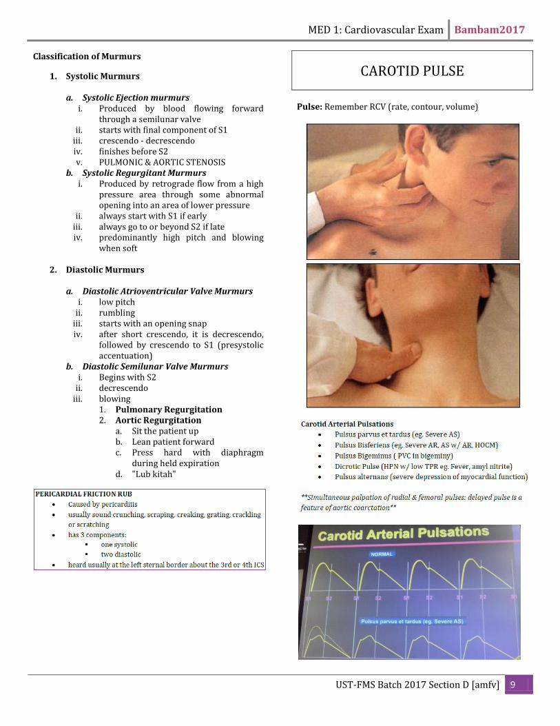
CAROTID PULSE Pulse: Remember RCV (rate, contour, volume)
MED 1: Cardiovascular Exam Bambam2017
UST-FMS Batch 2017 Section D [amfv] 10
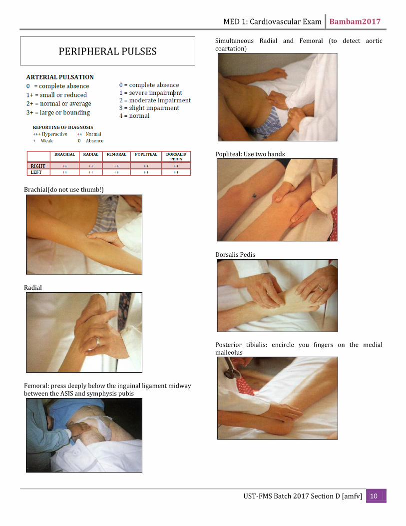
PERIPHERAL PULSES
Brachial(do not use thumb!)
Radial
Femoral: press deeply below the inguinal ligament midway between the ASIS and symphysis pubis
Simultaneous Radial and Femoral (to detect aortic coartation)
Popliteal: Use two hands
Dorsalis Pedis
Posterior tibialis: encircle you fingers on the medial malleolus

MED 1: Cardiovascular Exam Bambam2017
UST-FMS Batch 2017 Section D [amfv] 11
The Auscultogram
Whatever you cannot draw, write (e.g. yung precordium, lagay nyo sa baba) At the apex, louder si S1 At the base, louder si S2 Between S1 and S2 are systolic events. S1 split heard best on tricuspid. Left has higher pressure, so M1 is taller than T1. S2 split heard best at pulmonic, heard at inspiration (indicate). Pulmonic softer than aortic. For JVP, write the measurement. Normally the a-wave is higher. For CAP, normally upstroke is rapid and at S1. Gradually it will decline.


![ECG Signal processing (2) ECE, UA. ECG signal processing - Case [1] Diagnosis of Cardiovascular Abnormalities From Compressed ECG: A Data Mining-Based](https://img.pdfslide.us/doc/110x75/56649dbf5503460f94ab3016/ecg-signal-processing-2-ece-ua-ecg-signal-processing-case-1-diagnosis.jpg)