Embed Size (px)
Citation preview
1
THE C-TERMINUS OF RPT3, AN ATPase SUBUNIT OF PA700 (19S) REGULTORY COMPLEX, IS ESSENTIAL FOR 26S PROTEASOME ASSEMBLY
BUT NOT FOR ACTIVATION Brajesh Kumar, Young-Chan Kim and George N. DeMartino
From the Department of Physiology, University of Texas Southwestern Medical Center, 5323 Harry Hines Boulevard, Dallas, Texas 75390-9040 Running Head: Rpt3 and 26S proteasome assembly
Address correspondence to: George N. DeMartino, Department of Physiology, University of Texas Southwestern Medical Center, 5323 Harry Hines Boulevard, Dallas, TX
75390-9040 Telephone: 214.645.6024; Fax: 214.645.6019; Email: [email protected]
PA700, the 19S regulatory subcomplex of the 26S proteasome, contains a heterohexamric ring of AAA subunits (Rpt1-6) that forms the binding interface with a heteroheptameric ring of α subunits (α1-7) of the 20S proteasome. Binding of these subcomplexes is mediated by interactions of C-termini of certain Rpt sub-units with cognate binding sites on the 20S pro-teasome. Binding of two Rpt subunits (Rpt2 and Rpt5) depends on their last three residues, which share an Hb-Y-X motif and open sub-strate access gates in the center of the α ring. The relative roles of other Rpt subunits for pro-teasome binding and activation remain poorly understood. Here we demonstrate that the C-terminal Hb-Y-X motif of Rpt3 binds to the 20S proteasome but does not promote proteasome gating. Binding requires the last three residues and occurs at a dedicated site on the protea-some. A C-terminal peptide of Rpt3 blocked ATP-dependent in vitro assembly of 26S proteasome from PA700 and 20S proteasome. In HEK293 cells, wild-type Rpt3, but not Rpt3 lacking the Hb-Y-X motif was incorporated into 26S pro-teasome. These results indicate that the C-terminus of Rpt3 was required for cellular as-sembly of this subunit into 26S proteasome. Mutant Rpt3 was assembled into intact PA700. This result indicates that intact PA700 can be assembled independently of association with 20S proteasome and thus may be a direct pre-cursor for 26S proteasome assembly under normal conditions. These results provide new insights to the non-equivalent roles of Rpt sub-units in 26S proteasome function and identify specific roles for Rpt3.
ATP-dependent protease complexes com-monly comprise two distinct subcomplexes: a cyl-inder-shaped protease with internally sequestered catalytic sites, and an ATPase regulatory module required for delivery of substrates to those sites (1-3). The eukaryotic 26S proteasome represents the most structurally and functionally elaborate exam-ple of such complexes (4;5). Its protease subcom-plex, the 20S proteasome, contains two copies each of 14 different gene products arranged as four axially-stacked heteroheptameric rings (6;7). Each identical outer ring contains seven different α-type subunits (α1-α7) and each identical inner ring contains seven different β-type subunits (β1-β7). Three of the seven β-type subunits feature N-terminal threonine residues that serve as catalytic nucleophiles and line an interior chamber in the center of the barrel-shaped structure (8;9). The regulatory subcomplex of 26S proteasome, known as PA700 or 19S regulator, contains about 20 dif-ferent gene products including six distinct AAA (ATPases associated with various activities) sub-units (Rpt 1-6) (4;10). The Rpt subunits are ar-ranged in a hexameric ring that forms the binding interface of PA700 with the α rings of the 20S proteasome (11-14). Binding of PA700 to the 20S proteasome results in repositioning of interlaced N-terminal peptides of α subunits that normally occlude a narrow pore in the center of the α ring (15-19). This conformational rearrangement opens a route for substrates to reach the otherwise inac-cessible catalytic sites in the interior of the protea-some. Although short peptides and some unstruc-tured proteins pass the opened pore by simple dif-fusion, most physiological substrates of the 26S proteasome are folded proteins covalently modi-fied with a polyubiquitin chain (20-22). Polyubiq-
http://www.jbc.org/cgi/doi/10.1074/jbc.M110.153627The latest version is at JBC Papers in Press. Published on October 11, 2010 as Manuscript M110.153627
Copyright 2010 by The American Society for Biochemistry and Molecular Biology, Inc.
by guest on April 5, 2018
http://ww
w.jbc.org/
Dow
nloaded from
2
uitin serves as the principal method of targeting protein substrates to the proteasome via polyubiq-uitin binding subunits of PA700, but its client sub-strates require additional processing by PA700 for delivery to the sites of proteolysis (23;24). Sub-strate processing includes unfolding, detachment from the polyubiquitin chain by resident deubiq-uitylating subunits, and translocation through the open pore. These coordinated activities appear mechanistically linked to one another and to Rpt-catalyzed ATP hydrolysis (21;25;26). Although molecular details of this coordination and linkage remain poorly understood, the Rpt subunits of PA700 are topologically situated and functionally suited to play a central role in proteasome func-tion.
In addition to the obligatory role of ATP for 26S proteasome degradation of polyubiquitylated proteins, ATP also is necessary for PA700 binding to and activation of the 20S proteasome (27;28). However, unlike the former process, the latter re-quires ATP-binding but not hydrolysis (21;22). Thus, the ATP-bound state of one or more Rpt subunits likely promotes a conformation in the Rpt subunit ring that optimizes its interaction with cognate binding sites on the α subunit ring of the 20S proteasome. Considerable insight into the molecular details of binding and consequent pro-teasome activation has been achieved from studies of 20S proteasome-ATPase regulatory complexes in archaea. This structurally simpler system fea-tures a 20S proteasome composed of a homohep-tameric α ring and an ATPase regulator, PAN (proteasome-activating nucletidase), composed of a homohexameric AAA-subunit ring lacking addi-tional non-ATPase subunits (3;29;30). These properties have facilitated imaging and crystallo-graphic analysis of the resulting complex, reveal-ing that residues at the extreme C-terminus of PAN bind to pockets between adjacent α subunits and induce proteasome gate opening (18;19;31;32) Remarkably, a seven-residue peptide correspond-ing to the C-terminus of PAN is sufficient for both binding and activation of archaeal, as well as eu-karyotic proteasomes (33). The carboxyl group of the C-terminal karginine of PAN makes an essen-tial interaction with an ε-amino side chain of a ly-sine residue on one α subunit while the hydroxyl of the penultimate tyrosine residue interacts with residues on the adjacent α subunit. Although con-
flicting data have been presented about the exact identity of these latter contacts, there is general agreement that the interactions stabilize a proline-containing reverse loop in an open gate conforma-tion of the proteasome (18;19;32). This general mechanism explains how the C-terminus of PAN participates in proteasome binding and activation. Notably, tyrosine also is the penultimate residue in four of the six distinct Rpt subunits in eukaryotic PA700; three of the Rpt subunits share with PAN an Hb-Y-X motif (where Hb is a hydrophobic amino acid, Y is tyrosine, and X is any amino acid) at the last three residues. Previous work by us and others showed that the different Rpt sub-units of eukaryotic PA700 have at least some non-equivalent roles with respect to proteasome bind-ing and activation. For example, enzymatic re-moval of the Hb-Y-X motifs from only two (Rpt2 and Rpt5) of the six Rpt subunits of PA700 com-pletely inhibited PA700 binding to and activation of the proteasome (33;34). Moreover, as with PAN, peptides corresponding to the C-terminus of Rpt2 and Rpt5 were each sufficient to bind to and activate the 20S proteasome in a manner that de-pended on an intact Hb-Y-X motif. Finally, bind-ing of the Rpt2 and Rpt5 peptides occurred at dis-tinct and dedicated sites on the fixed-order het-eromeric α ring, as judged by chemical crosslink-ing (34). However, a C-terminal peptide of an-other Hb-Y-X motif-containing subunit, Rpt3, as well as those of the three non-Hb-Y-X, had no demonstrable proteasome activating activity. The lack of activating function of non-activating pep-tides could reflect either their lack of proteasome binding or their inability to induce conformational changes required for gating after binding. The purpose of this work was to explore roles for non-activating Rpt subunits of 26S proteasome. EXPERIMENTAL PROCEDURES Proteins. PA700, PA700 subassemblies (PS-1, PS-2, and PS-3), 20S proteasome, and 26S protea-some were purified from bovine red blood cells as described previously (21;28;34-37). SUMO-Rpt peptide fusion proteins were generated by amplifi-cation of the whole Pet28a-SUMO cassette with primers containing nucleotides appropriate for amino acid sequences of desired peptides. The re-sulting His-tagged recombinant SUMO-Rpt pep-tide fusion proteins were expressed in E.coli BL21
by guest on April 5, 2018
http://ww
w.jbc.org/
Dow
nloaded from
3
(DE3) cells at 15oC overnight and purified by af-finity chromatography utilizing Ni-nitrilotriacetic acid beads (Qiagen). SUMO-Rpt-chimeric peptide fusion proteins were produced by generating two point mutations in the Hb-Y-X motif of a SUMO-Rpt peptide fusion protein sequence. The SUMO-chimeric fusion proteins were expressed and puri-fied as described for the SUMO-Rpt peptide fusion proteins. Peptide Synthesis. Peptides corresponding to the sequences (or variants thereof) of C-termini of Rpt subunits of PA700 were synthesized using Fmoc (N-[9-fluorenyl]methoxycarbonyl) chemistry and purified using HPLC by the Protein Core Facility at UT Southwestern Medical Center. Sequences of all peptides were verified by mass spectrometry. The sequences of these peptides from N- to C- termini are: Rpt1, SATPRYMTYN; Rpt2, QEGTPEGLYL; Rpt3, KDEQEHEFYK; Rpt4, LESKLDYKPV; Rpt5, KKKANLQYYA; Rpt6, KNMSIKKLWK; Rpt3(-C3), KDEQEHE; Rpt3-Rpt1, KDEQEHETYN; Rpt1-Rpt3, SATPRYM-FYK; Rpt5-Rpt1, KKKANLQTYN; Rpt1-Rpt5, SATPRYMYYA; Rpt5-Rpt3, KKKANLQFYK; Rpt3-Rpt5, KDEQEHEYYA. For crosslinking studies peptides containing Dopa (3,4- dihydroxy-phenylalanine) and either biotin or fluorescein were synthesized and had the following sequences: biotin-Dopa-Rpt3, biotin-Dopa-GSKDEQEHEFYK; Biotin-Dopa-Rpt3(-3C), bio-tin-Dopa-GSKDEQEHE; fluorescein-Dopa-Rpt3, fluorescein- GGG-DOPA-GSKDEQEHEFYK; fluorescein-Dopa-Rpt3(-3C), fluorescein-GGG-Dopa-GSKDEQEHE. Proteasome activity and activation assays. Pro-teasome activity was measured by determining rates of enzymatic cleavage of 7-amino-4-methylcourmarin (AMC) from peptide substrates Suc-L-L-V-Y-AMC, Suc-L-L-E-AMC, and Cbz-V-L-R-AMC, as described previously (21). Stan-dard assay conditions included 45 mM Tris-HCl, pH 8.0, 5 mM β-mercaptoethanol, 15 nM latent 20S proteasome and 200 μM substrate in a volume of 50 μl. Incubations were carried out at 37oC for 21 mins in a Biotek FL600 fluorescence plate reader with filters at 380 nmex/460 nmem. AMC fluorescence was monitored once per minute dur-ing the assay and progress curves were analyzed with kinetic software. Proteasome activation by
Rpt peptides and SUMO-Rpt peptide fusion pro-teins was determined similarly but included prein-cubation of 20S proteasome with peptides or SUMO proteins for 15 mins at 37oC (34). Protea-some activity is expressed as arbitrary fluorescent units (AFU) produced per minute. Routine control assays included reactions without proteasome. Proteasome activity against a protein substrate, [14C-methyl]-casein, was determined as described previously (37). Other details of individual ex-periments are provided in appropriate figure leg-ends. In some experiments semi-quantitative measures of proteasome activity were obtained by overlay of peptide substrates in situ on proteins separated in native 4% polyacrylamide gels, as described previously (38). After incubation at 37o
C for 10-30 mins, AMC at the position of the pro-tease in the gel responsible for its production was visualized by UV light. 26S proteasome assembly assay. 26S proteasome assembly from purified 20S proteasome and PA700 assay was conducted by preincubating 20S proteasome with PA700 in 45 mM Tris-HCl, pH 8.0 at 37o C, 5.6 mM DTT, 10 mM MgCl2 and 100 µM ATP at 37oC for 30 minutes prior (35). 20S proteasome and PA700 concentrations for given experiments are provided in appropriate figure legends. Samples were assayed either directly for proteasome activity, or subjected to native PAGE, after which gels were stained for protein and as-sayed for in situ proteasome activity, as described above. Rpt peptide-20S proteasome binding assays. Binding of Rpt peptides to 20S proteasome was determined by pull-down assays utilizing purified bovine 20S proteasome and recombinant His-tagged SUMO-Rpt peptide fusion proteins. In typical assays, 100 µM of His-tagged SUMO fu-sion protein was incubated with 214 nM 20S pro-teasome at 37ºC for 15 minutes in 50 mM Tris-HCl, pH 7.6, and 1 mM β-mercaptoethanol in a volume of 100 μl. Twenty five μl of nickel beads were added and mixed for 2 hrs at 4 ºC. The beads were washed with 50 mM Tris-HCl, pH 7.6, 1 mM β-mercaptoethanol, and eluted with 300 mM imi-dazole. Eluted proteins were separated by SDS-PAGE and western blotted with antibodies against 20S proteasome.
by guest on April 5, 2018
http://ww
w.jbc.org/
Dow
nloaded from
4
Chemical crosslinking. Chemical crosslinking of Rpt peptides was conducted by methods similar to those described previously (34). Biotin- or fluo-rescein-containing Dopa peptides described above were incubated for 10 minutes at room tempera-ture with 720 nM 20S proteasome, 20 mM Tris-HCl, pH 7.6, 20 mM NaCl, 1 mM EDTA and 10 % glycerol (Buffer H) in a final volume of 20 µl. Cross-linking was initiated by addition of 10 mM sodium periodate and quenched after 30 seconds by 50 mM β-mercaptoethanol. Cross-linked prod-ucts were detected by either fluorescence spec-trometry (488nmex/523nmem) using a Typhoon 9410 scanning imager (GE Healthcare) or western blotting with HRP-linked neutravidin or IRDyde-labeled streptavidin (see below) after SDS-PAGE. In some experiments samples were detected by these methods after two-dimensional gel electro-phoresis. In other experiments, samples of crosslinked proteins were purified by HPLC using a Jupiter C4 5 ml reverse phase column (Phe-nomenex). Samples were applied to the column in 0.05% trifluoroacetic acid (TFA) and eluted with a gradient of acetonitrile at a flow rate of 1 ml/min. In preliminary experiments we determined that fluorescent subunits representing the crosslinked products eluted between 45-50% acetonitrile. Therefore, the gradient was developed from 0-45% acetonitrile in 15 mins and from 45-50% in 30 mins. Column fractions of 1.0 ml were collected, dried by vacuum, redissolved in either SDS-sample buffer or isoelectric focusing sample buffer (7 M urea, 2 M thiourea, 4% CHAPS, 65 mM dithiothreitol, Pharmolytes (pH 3–10) and bromphenol blue). Isoelectric focusing (IEF) was conducted using a ReadystripTM pH 3-10 support (Biorad). Samples subjected to crosslinking with biotin-containing peptides were enriched for crosslinked product by binding to monomeric avidin beads after exposure to denaturing conditions. Cross-linking was performed as described above. Non-crosslinked Rpt3 peptides were removed by multi-ple washes through Microcon-YM100 centrifugal filter in Buffer H with 0.5 M NaCl and 0.05 % Tween-20. The samples were exposed to 7M gua-nidine-HCl for 30 minutes at room temperature. The samples were diluted to decrease the gua-nidine concentration to 1 M and mixed with mono-meric avidin beads in Buffer H containing 0.5 M NaCl and 0.05 % Tween-20. Beads were washed
with the same buffer and retained proteins were eluted in either SDS sample buffer or isoelectric sample buffer. Samples were separated by either 1D or 2D gels and western blotted with either in-frared dye (IRDye)-labeled streptavidin or with antibodies against selected 20S proteasome sub-unit antibody and respective infrared dye labeled secondary antibody to visualize the proteins of interest utilizing an Odyssey infrared imaging sys-tem (Li-cor). Transient expression of FLAG-Rpt3 in HEK293 cells. HEK293 cell lines were maintained in Dul-becco's modified Eagle's medium (Gibco) contain-ing high glucose and glutamine, supplemented with 10% fetal bovine serum in the presence of 5% CO2 at 37°C. HEK293 cells were transfected at approximately 60% confluency with cDNA for either FLAG-human Rpt3 or Flag-human Rpt3 lacking the last three C-terminal residues sub-cloned into the pIRESpuro3 vector (Clontech) by using FuGene 6 reagent (Roche). Forty-eight hrs after transfection, cells were washed with phos-phate-buffered saline and harvested with buffer consisting of 50 mM Tris-HCl, pH 7.5, 5 mM MgCl2, 1 mM β-mercaptoethanol, 1 mM ATP, and Protease Inhibitor Cocktail (Roche). Whole cell extracts were prepared by 15 passages through a 27-gauge needle and centrifuged for 20 min. Ex-pression of Rpt proteins was determined by west-ern blot analysis using anti-FLAG M2 antibody (Sigma) and anti-Rpt3 antibody (Boston Bio-chem). Glycerol Density Gradient Centrifugation. Glyc-erol density gradient centrifugation was conducted as described previously using 10-40 % linear glyc-erol gradients (39). Affinity purification of flag-Rpt proteins from HEK293 cell extracts. Approximately 7 mg of a whole cell extract was mixed gently for 2 hrs at 4°C with 100 µl of anti-Flag M2 agarose beads (Sigma, St. Louis, MO) in 50mM Tris-HCl, pH 7.5, 100 mM NaCl, 1 mM β-mercaptoethanol, 1 mM ATP, 5 mM MgCl2, 10% glycerol and 0.1% NP-40. The beads were harvested by centrifuga-tion and washed three times with the same buffer. Bound proteins were eluted overnight at 4°C with two bed volumes of binding buffer containing 200 µg/ml FLAG peptide (Sigma, St. Louis, MO).
by guest on April 5, 2018
http://ww
w.jbc.org/
Dow
nloaded from
5
Eluted proteins were characterized as described in the text. RESULTS
The C-terminus of Rpt 3 binds to but does not
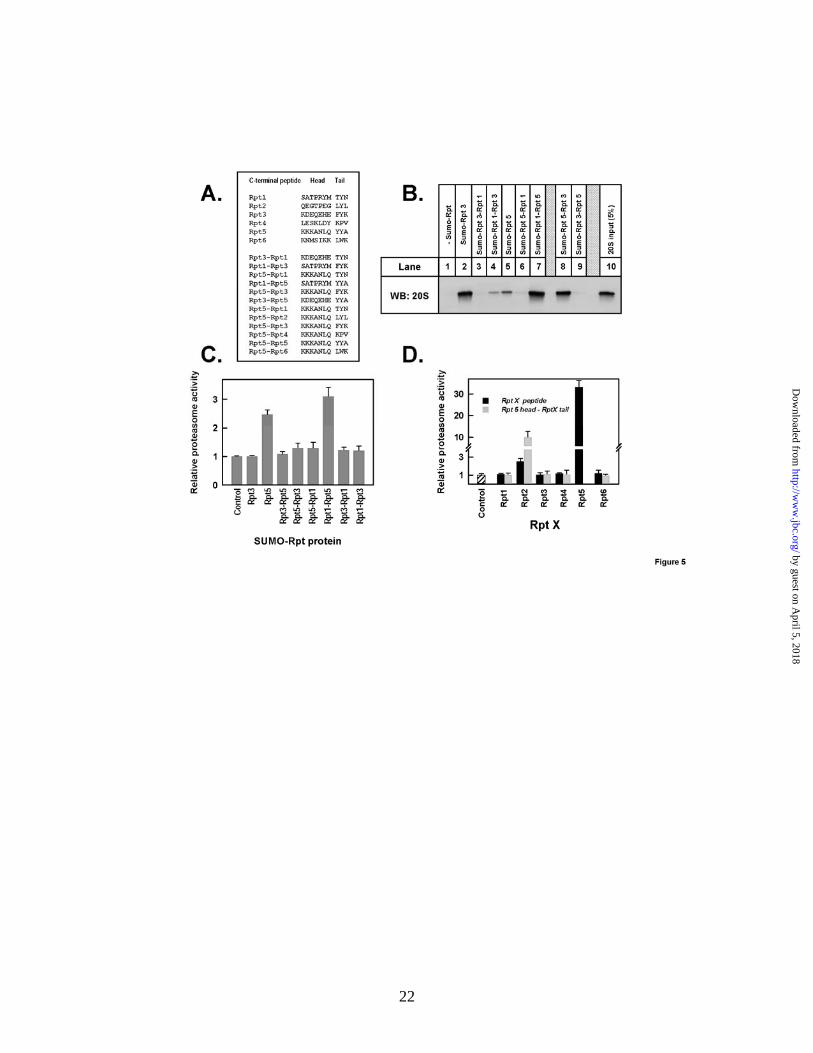
activate the 20S proteasome. We previously dis-covered that isolated peptides corresponding to the C-termini of PA700 subunits Rpt2 and Rpt5, but not those corresponding to the C-termini of the other four Rpt subunits of this regulatory complex, stimulated 20S proteasome-catalyzed hydrolysis of model substrates by a mechanism that involves enhanced gating of the substrate entry channel (34). The differential effects of the various Rpt C-terminal peptides could reflect their differential binding to the proteasome or their differential abil-ity to promote gate opening and substrate passage after binding. To distinguish between these possi-bilities, we directly tested the relative binding of the Rpt peptides to the proteasome. We expressed and purified recombinant proteins in which the C-terminal 10 residues of each Rpt subunit were ap-pended to the C-terminus of His-tagged SUMO-1, a protein that otherwise does not interact detect-ably with the proteasome. After incubation, 20S proteasome bound by these fusion proteins was isolated by pull-down assays on Ni-beads and de-tected by western blotting. As expected, the 20S proteasome bound to SUMO proteins containing the C-terminus of Rpt2 and of Rpt5 (Figure 1A, lanes 3 and 6). Surprisingly, however, 20S protea-some also bound to SUMO containing the C-terminus of Rpt3 (Figure 1A, lane 4), a subunit whose C-terminus does not enhance proteasome activity. The Rpt3-containing SUMO protein con-sistently pulled down more 20S proteasome than did the Rpt2 and Rpt5-containing proteins, sug-gesting that it bound with greater affinity to the proteasome than did the Rpt2- and Rpt5-containing proteins. The proteasome failed to bind detectably to SUMO-proteins with C-termini of the remaining non-activating Rpt subunits (Rpt1, Rpt4, and Rpt6). Binding of the 20S proteasome to SUMO-Rpt3 was blocked by excess free Rpt3 C-terminal peptide, but not by excess free Rpt5 C-terminal peptide (Figure 1B, lanes 2-4) nor by ex-cess free Rpt2 C-terminal peptide (Figure 1C). Likewise, the Rpt3 peptide did not block binding of the SUMO-Rpt5 to the proteasome (Figure 1B, lanes 5 and 6). These results indicate that binding
of the Rpt3 peptide was specific and likely oc-curred at a site unique from those bound by Rpt2 and Rpt5. As with the binding of Rpt2 and Rpt5, binding of the SUMO-Rpt3 protein depended on the presence of the last three residues. Thus, pro-teins lacking the last two or three residues had no detectable proteasome binding, while a protein lacking the only the last residue displayed detect-able but greatly diminished binding (Figure 1D). Binding of SUMO-Rpt3 to the 20S proteasome was demonstrated independently in a gel-shift as-say (Figure 1E). Thus, the SUMO-Rpt3 fusion protein with an intact C-terminal peptide, but not that without the last three residues, retarded the migration of 20S proteasome during native gel electrophoresis (Figure 1E, lanes 1-4). Similar results were obtained with SUMO-Rpt5 proteins (Figure 1E, lanes 5-7). In-gel proteasome activity assays also reflected the differential ability of the C-termini of Rpt 5 and Rpt 3 to activate protea-some function. Collectively, these results confirm the differential ability of the C-termini of different Rpt subunits to bind to the proteasome and iden-tify Rpt3 as a PA700 subunit that binds to the pro-teasome but does not directly activate proteasome hydrolysis of model peptide substrates.
The C-terminus of Rpt3 binds to a dedicated
site on the 20S proteasome. Previously, we showed that activating peptides from the C-termini of Rpt2 and Rpt5 chemically crosslink to distinct, dedicated, and identifiable subunits of the 20S pro-teasome (34). Consistent with the results presented above, these findings indicate that the fixed-order heterohexameric ring of Rpt subunits of PA700 binds to the fixed-order heteroheptameric ring of 20S α subunits with an invariant inter-ring subunit registration. This model predicts that Rpt3 also should bind at a unique and dedicated site on the α ring of the 20S proteasome and crosslink to a spe-cific subunit. To test this hypothesis we applied the same general chemical crosslinking strategy to Rpt3 employed previously for Rpt2 and Rpt5. We synthesized Dopa-containing peptides correspond-ing to the C-terminus of Rpt3 that either contained or lacked the last three residues and included ei-ther biotin or fluorescein for detection of crosslinked products. Crosslinking of the intact C-terminal peptide with 20S proteasome produced one major product, which was similar by each de-tection method after SDS-PAGE (Figures 2A and
by guest on April 5, 2018
http://ww
w.jbc.org/
Dow
nloaded from
6
2B). In some experiments a second band, whose intensity varied among independent crosslinking reactions, also was detected. No crosslinked prod-uct was detected with an Rpt3 peptide lacking the last three amino acids, indicating that the crosslinking was specific for conditions required for peptide binding. We exploited respective characteristics of the fluorescein and biotin tags of Rpt peptides for independent identification of the Rpt crosslinked subunit of 20S proteasome. For ease of detection of the crosslinked product, we continued subsequent analysis with the fluo-rescein-tagged peptide. For more facile enrichment of the crosslinked product, we exploited the biotin moiety. With the fluorescein-labeled peptide, a single crosslinked product also was detected by two-dimensional gel electrophoresis; in some ex-periments, such as that shown in Figure 2C, the product appeared as two or three closely separated spots. However, the subunit complexity of 20S proteasome from the unenriched sample and low protein content on these gels prevented us from further identification of the crosslinked product at this stage. Therefore, we subjected crosslinked 20S proteasome to reverse-phase HPLC to enrich and purify the modified subunit. SDS-PAGE of gradient fractions showed that this method sepa-rated most proteasome subunits from one another and from the major fluorescently-labeled band (Figure 3A). We subjected this band to two-dimensional PAGE, which like the unenriched sample, usually appeared as two or three closely-separated spots, whose position did not correspond to that of any unmodified proteasome subunit. The fluorescent spot was extracted, digested with trypsin and subjected to mass spectrometry, which identified peptides of only one 20S proteasome subunit, α1 (PSMA6), in repeated independent experiments (Supplemental Data, Table 1); in one experiment peptides of the α7 (PSMA3) subunit was detected in addition to α1. No proteasome peptides were identified when an equivalent area of the gel was analyzed from a 20S proteasome sample subjected to identical treatment with an Rpt3 peptide lacking the last three residues (data not shown). We attempted to confirm the identity of α1 as the crosslinked protein by western blot-ting, but the only available antibodies were insuf-ficiently sensitive to detect the low protein content at the position corresponding to the fluorescent
spot. No antibody to other 20S proteasome sub-units produced a detectable signal at this position (data not shown). Non-fluorescent α1 subunit was identified by western blotting at its expected posi-tion in the gel; it was similar for the intact and truncated crosslinking peptide, and presumably represents the non-cross-linked portion of the α1 protein.
To confirm the identification of α1 as the crosslinked product of the Rpt3 peptide, we util-ized the biotin-containing peptide to enrich the resulting cross-linked product on monomeric avidin beads under denaturing conditions, as de-scribed under Experimental Procedures. In con-trast to the analysis described above, this enrich-ment method permitted isolation of sufficient product for subsequent analysis by western blot-ting after one and two-dimensional PAGE. As shown in Figure 3E, crosslinking with the intact peptide, but not with the C-terminally truncated peptide, produced a biotin-labeled band that mi-grated indistinguishably from a modified α1 band, detected by western blotting on SDS-PAGE. Likewise, after two-dimensional PAGE, western blotting for the α1 subunit revealed modified spots in positions similar to those observed for the fluo-rescently-crosslinked protein in samples crosslinked with the intact peptide but not with the truncated peptide. Moreover, biotin was detected only in the modified spots, which coincided pre-cisely with those detected by the anti-α1 antibody (Figure 3F). Control experiments with antibodies against several other α subunits failed to detect modified proteins as a consequence of crosslinking in western blots of either the one- or two-dimensional gels (Figure 3E and data not shown). The spots detected coincidently by IRDye strepta-vidin (for biotin) and the α1 antibody were ex-tracted, digested with trypsin and subjected to mass spectrometry. As with the analogous ex-periment described above, peptides of the α1 sub-unit were selectively identified (Supplemental Data, Table 1). Collectively, these results indicate that α1 is the likely crosslinked product of the Rpt3 C-terminal peptide. This subunit is distinct from subunits identified previously as crosslinked products of C-terminal peptides of Rpt2 (α7) and Rpt5 (α4). These results support the general model for a fixed and distinct registration between
by guest on April 5, 2018
http://ww
w.jbc.org/
Dow
nloaded from
7
subunits of the interacting heteromeric Rpt sub-units and α-subunit rings (see Discussion).
Binding of Rpt3 does not affect proteasome
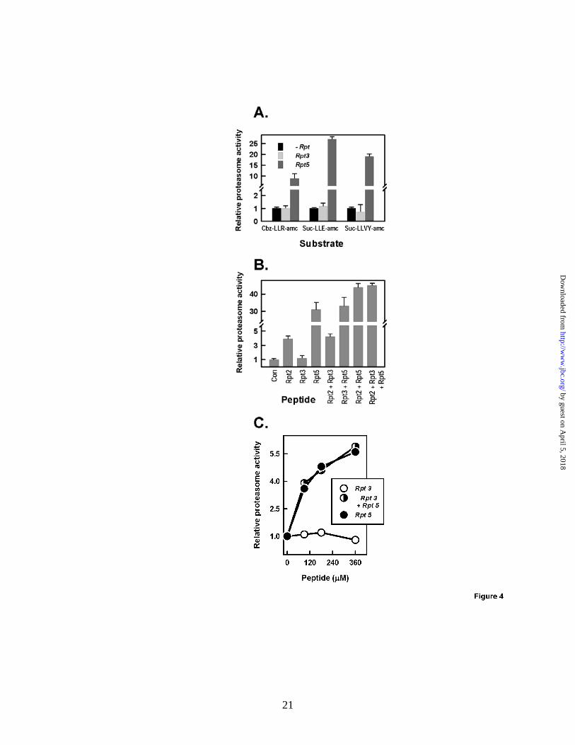
activation by Rpt2 or Rpt5. C-terminal peptides of Rpt2 and Rpt5 activate substrate hydrolysis by the 20S proteasome and their effects are either ad-ditive (with short peptide substrates) or synergistic (with longer protein substrates). Such results are consistent with the binding of these peptides to distinct sites on the proteasome and support a model in which the substrate assess pore can be gated to variable degrees by multiple independent binding events (40). Therefore, we tested whether Rpt3 could modulate the gating effects of Rpt2 and/or Rpt5, despite its inability to induce gating independently. Rpt3 had no proteasome activating activity by itself or in combination with Rpt2 and/or Rpt5, regardless of the substrate tested (Figure 4). Thus, binding of Rpt3 C-terminal pep-tide neither opens the substrate access pore di-rectly nor modulates the effect of other Rpt C-terminal peptides that do so. These results, how-ever, monitor the relative roles of physically sepa-rated binding molecules and may not reflect the roles and effects of these peptides when they func-tion in the context of an intact PA700 complex (see Discussion).
Features of proteasome binding and activa-
tion are determined by both the Hb-Y-X motifs and adjacent residues of the Rpt C-termini. Pre-vious work by us and others has established and emphasized the important role of the last three amino acids of C-terminal Rpt peptides in protea-some binding and activation by Rpt2 and Rpt5. These residues (LYL and YYA for Rpt2 and Rpt5, respectively) conform to a motif of Hb-Y-X. This motif also is present in Rpt3 (FYK) and, in an im-perfect form, in Rpt1 (TYN). Thus, various Hb-Y-X motif-containing peptides display distinct func-tional properties with respect to proteasome bind-ing and activation. Although such disparity likely reflects differences among the Hb-Y-X motifs and their cognate binding sites on the α ring of the 20S proteasome, features of Rpt C-terminal peptides other than the Hb-Y-X motif may provide addi-tional determinants of proteasome binding and/or activation. To explore this possibility, we synthe-sized “chimeric” peptides containing the Hb-Y-X motif (hereafter denoted as the “tail”) of given Rpt
subunits and the adjacent N-terminal seven resi-dues (hereafter denoted as the “head”) of other Rpt subunits. We also produced recombinant fusion proteins of SUMO and the corresponding chimeric peptides. We selected for this analysis examples of C-terminal peptides that: i) both bind to and activate the proteasome (e.g., Rpt5); ii) bind to but do not activate the proteasome (e.g., Rpt3), and; iii) neither bind to nor activate the proteasome (e.g., Rpt1). First, we determined the ability of various SUMO-Rpt chimeric peptide fusion pro-teins to bind to the 20S proteasome in pull-down assays analogous to those used with their wild-type counterparts (Figure 5). Neither SUMO-Rpt3 head-Rpt1 tail (Panel B, lane 3) nor SUMO-Rpt5 head-Rpt1 tail (Panel B, lane 6) displayed appre-ciable proteasome binding, thereby supporting a critical role of the Hb-Y-X motif of wild-type Rpt3 and Rpt5 C-terminal peptides for their re-spective proteasome binding. Surprisingly, how-ever, chimeric peptides consisting of an Rpt3 or Rpt5 tail with an Rpt1 head displayed proteasome binding properties suggestive of an important in-fluence of the Rpt1 head. Thus, the Rpt1 head reduced the proteasome binding expected of the Rpt3 tail (compare lanes 2 and 4), but increased the proteasome binding expected of the Rpt5 tail (compare lanes 5 and 7). The influence of head region on proteasome binding also was demon-strated by the lack of binding of the chimeric pep-tide consisting of an Rpt3 head and an Rpt 5 tail (i.e., a peptide containing both a head and a tail of binding peptides, compare lanes 4 and 8). In con-trast, a peptide consisting of an Rpt5 head and an Rpt3 tail featured a proteasome binding affinity similar to that of Rpt3. These results further high-light the obligatory role of specific Hb-Y-X motifs for proteasome binding, but demonstrate the influ-ence of additional elements of the C-terminus on this process.
To explore the relationship between binding of the Hb-Y-X motif and proteasome activation, we compared the effects of the various chimeric pep-tide SUMO-fusion proteins on proteasome activa-tion to those of their wild-type counterparts and to the binding features of these chimeric peptides. As expected, no non-binding chimeric peptide acti-vated 20S proteasome catalysis. Moreover, chi-meric peptides containing both a head and a tail of non-activating Rpt subunits (e.g. Rpt1 and Rpt3), regardless of their binding capacity, did not acti-
by guest on April 5, 2018
http://ww
w.jbc.org/
Dow
nloaded from
8
vate the proteasome. Instead, proteasome activa-tion by chimeric proteins required the tail (Hb-Y-X motif) of a normally activating Rpt peptide. Thus, neither SUMO-Rpt5 head-Rpt1 tail, nor SUMO-Rpt5 head-Rpt3 tail activated the protea-some, although each could bind. In contrast, SUMO-Rpt1head-Rpt5 tail, activated the protea-some to a greater extent than did SUMO-Rpt5, an effect that mirrored the relative proteasome bind-ing of these proteins. These results indicate that proteasome binding is influenced by features of both the Hb-Y-X motif and the adjacent residues of specific Rpt subunits, but that proteasome acti-vation is restricted to the binding of a normally activating Hb-Y-X motif (e.g. Rpt5). To test this further, we synthesized a series of peptides con-sisting of an Rpt5 head and the tail of each of the six Rpt subunits. Only chimeric peptides contain-ing the activating Hb-Y-X tails Rpt2 and Rpt5 stimulated 20S proteasome activity (Figure 5D). Interestingly, the Rpt5 head-Rpt2 tail peptide stimulated the proteasome to a greater extent than the wild-type Rpt2 peptide, but to a lesser extent that the wild type Rpt5 (Figure 5D). These results provide additional evidence for the influence of the head region on the function of gating-competent Hb-Y-X motifs.
Rpt3 C-terminal peptide attenuates 26S pro-
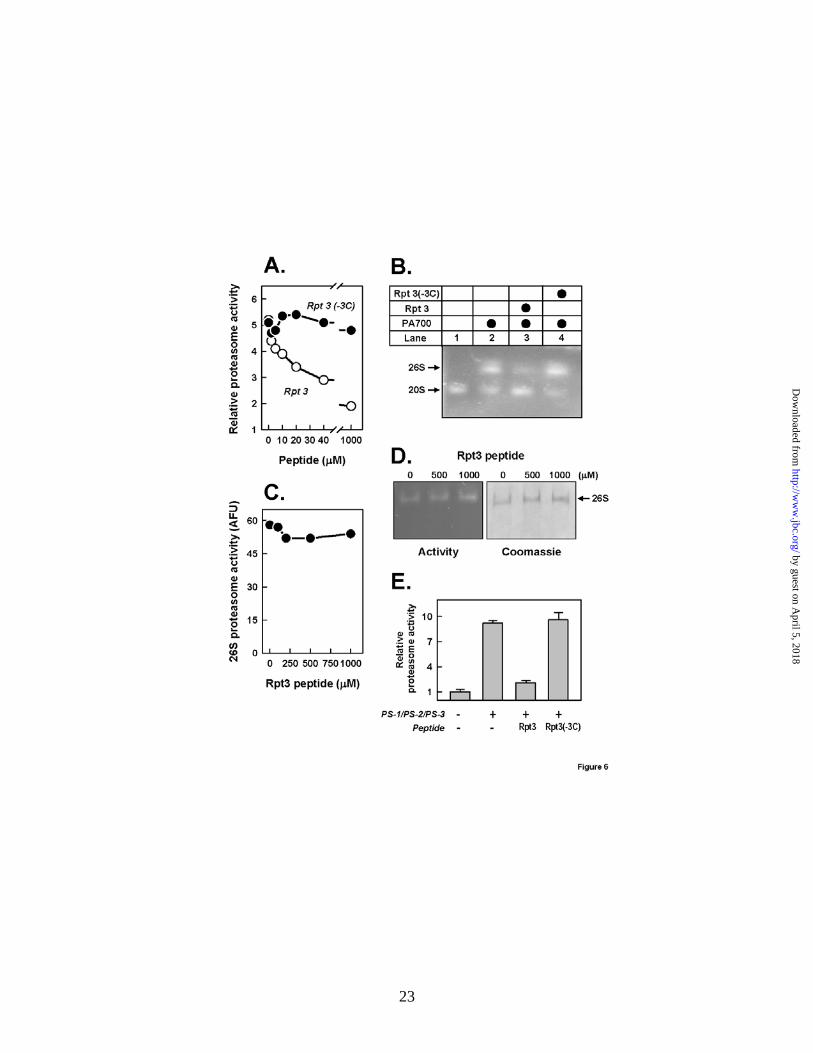
teasome assembly in vitro. The data presented above identify the C-terminus of Rpt3 as an im-portant binding element of intact PA700 to the proteasome. To test the role of Rpt3 in binding of intact PA700 to the 20S proteasome, we examined the effect of a C-terminal peptide of Rpt3 on the ATP-dependent in vitro reconstitution of 26S pro-teasome from purified PA700 and 20S protea-some. Both Rpt3 peptide (Figure 6A) and the SUMO-Rpt3 fusion protein (data not shown) in-hibited the PA700-dependent activation of the 20S proteasome, an indirect monitor of 26S protea-some assembly. The inhibitory effect was de-pendent on peptide concentration, and required the last three residues. Inhibition of assembly of acti-vated 26S proteasome activation by the intact Rpt3 C-terminal peptide also was demonstrated by na-tive PAGE (Figure 6B). The Rpt3 peptide had no effect on the activity of intact purified 26S protea-some, indicating that the peptide did not exert its effect in the assembly assay by inhibiting the ac-tivity of assembled 26S proteasome or by promot-
ing 26S proteasome disassembly (Figures 6C and 6D). These results suggest that the isolated Rpt3 peptide functions as a dominant-negative inhibitor of 26S proteasome assembly by competitively blocking binding of intact PA700 to the 20S pro-teasome. Remarkably, this effect is manifested despite the presence of at least two other PA700 subunits (Rpt2 and Rpt5) with the capacity to bind 20S proteasome (see Discussion).
Previously, we showed that 26S proteasome also could be assembled in vitro from 20S protea-some and three subcomplexes that collectively form intact PA700 (35). The C-terminal Rpt3 pep-tide but not the peptide lacking the last three resi-dues blocked assembly of 26S proteasome from these PA700 subassemblies (Figure 6E).
The C-terminus of Rpt3 is essential for as-
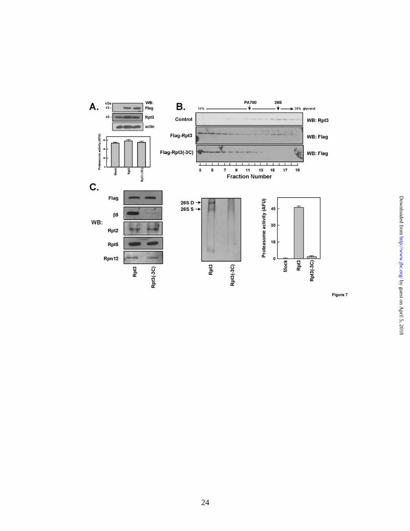
sembly of 26S proteasome in intact cells. To evaluate the relative role and importance of the C-terminus of Rpt3 to 26S proteasome assembly in intact cells, we transfected HEK293 cells with ex-pression vectors for either Flag-tagged wild-type Rpt3 or Flag-tagged Rpt3 lacking the last three C-terminal residues. We analyzed cells in which expression of these proteins was approximately equal to one another and equal to or less than that of endogenous Rpt3 (Figure 7A). The two Rpt3-expressing cell types were indistinguishable from one another and from non-transfected HEK293 cells by general morphological features and by rates of growth (data not shown). They also had similar overall proteasome activity (Figure 7A). In non-transfected control cells, endogenous Rpt3 displayed a tri-modal distribution when soluble extracts were subjected to glycerol density gradi-ent centrifugation. Most of the Rpt3 protein sedi-mented in fractions characteristic of the 26S pro-teasome. Smaller amounts were found in slower-sedimenting fractions corresponding to free PA700 and other lower molecular weight complexes (Fig-ure 7B, upper panel). Flag-tagged wild-type Rpt3 displayed a distribution pattern that was qualita-tively similar to that of endogenous Rpt3 although proportionally more exogenous protein was dis-tributed to the slowest sedimenting complexes (Figure 7B, middle panel). The reasons for and significance of this quantitative distinction are un-clear. Nevertheless, an appreciable portion of ex-pressed wild-type Flag-Rpt3 was assembled nor-mally into 26S proteasome as judged by its sedi-
by guest on April 5, 2018
http://ww
w.jbc.org/
Dow
nloaded from
9
mentation position in the glycerol gradient and by anti-flag immunoprecipitation of proteins with structural and functional features of 26S protea-some (Figure 7B and C, see below). In contrast, little or no detectable flag-tagged Rpt3 protein lacking C-terminal residues was present in gradi-ent fractions corresponding to the 26S proteasome and instead accumulated in slower-sedimenting fractions corresponding to those characteristic of PA700 and smaller subcomplexes (Figure 7B, lower panel). Moreover, although immunoprecipi-tation was equally efficient for the wild-type and mutant Rpt3 proteins, the resulting immunopre-cipitates differed significantly in other features. For example, Flag-immunoprecipitation from ex-tracts of cells expressing wild-type Rpt3 isolated a protein with features characteristic of intact 26S proteasome as judged by western blotting of repre-sentative component subunits (Figure 7C, left panel and data not shown), migration on native PAGE (Figure 7C, middle panel), and proteasome activity (Figure 7C, middle and right panels). In contrast, Flag-immunoprecipitation from extracts of cells expressing mutant Rpt3 isolated a protein with subunits characteristic of PA700 but without 20S proteasome subunits and proteasome activity. Collectively, these results show that lack of an intact C-terminus prevented Rpt3 from incorpora-tion into 26S proteasome, and that the contribu-tions of intact binding elements of Rpt2 and Rpt5 were not sufficient to overcome this deficiency. These results are consistent with the ability of iso-lated C-terminal Rpt3 peptide to attenuate binding of intact PA700 to the proteasome, and highlight an important role of Rpt3 binding in 26S protea-some assembly.
DISCUSSION
The results presented here reveal new details about structural and functional interactions be-tween the 20S proteasome and PA700, two multi-subunit subcomplexes that compose the 26S pro-teasome. PA700 and 20S proteasome bind at an axial interface of two different heteromeric rings. PA700 contributes a heterohexameric ring of AAA subunits (Rpt1-6) whereas the 20S proteasome contributes a heteroheptameric ring of α-type sub-units (α1-7). Thus, the interaction between sub-complexes of the eukaryotic 26S proteasome has greater structural complexity than that between
subcomplexes of archaeal proteasome complexes featuring interacting rings of homomeric proteins. Previous work has established an important role for the C-termini of certain Rpt subunits for bind-ing of PA700 to the proteasome and consequent proteasome gating (33;34). However, the relative roles of the different Rpt subunits in these proc-esses remain uncertain. The clearest examples of Rpt C-termini that interact directly with the pro-teasome are those of Rpt2 and Rpt5. Each features an Hb-Y-X motif that binds to pockets between specific adjacent α subunits of the 20S proteasome ring. Although the structure of intact 26S protea-some has not been solved at atomic resolution, possible explanations for the non-equivalent roles of the C-termini of different Rpt subunits in pro-teasome binding and activation have been pro-vided by structural studies of heterologous, artifi-cially engineered, and simpler archaeal model sys-tems (18;19;31;32;41). For example, the signifi-cance of the Hb-Y-X motif has been illustrated by showing the atomic details of how these residues from PAN and certain Rpt subunits interact with proteasome subunits to promote gate opening (19;31;32). Despite the considerable insight gained by these studies, a comprehensive molecular un-derstanding of the relative structure-function rela-tionships of the Rpt subunits for proteasome bind-ing and activation of authentic 26S proteasome remain uncertain. Nevertheless, the non-equivalent capacity of different Rpt subunits to bind to and activate the proteasome must depend on both the features of both individual Rpt C-terminal residues and the specific α subunits that create their respec-tive binding pockets. Thus, whereas Rpt2 and Rpt5 bind to gating-competent sites on the proteasome, Rpt3 likely binds to a site that cannot directly promote gating. The lack of proteasome activation by an isolated peptide, however, does not exclude its role in proteasome gating when it is part of the intact PA700 complex.
The current data provide initial information about the influence of residues upstream of the Hb-Y-X motif in proteasome binding and activa-tion. Goldberg and colleagues previously estab-lished a seven-residue minimum length require-ment for proteasome binding and activation of fea-tures of the Hb-Y-X motif peptide of PAN (33). Multiple substitutions for residues N-terminal to the Hb-Y-X motif had little effect on binding and activation, suggesting that they did not make iden-
by guest on April 5, 2018
http://ww
w.jbc.org/
Dow
nloaded from
10
tity-specific contributions to these processes. In contrast, the results presented here show that the identity of residues adjacent to the Hb-Y-X motif of given Rpt peptides can have appreciable influ-ence on proteasome binding and/or activation. Although an Hb-Y-X motif was always necessary for binding, alterations to adjacent residues could either diminish or enhance the apparent affinity of this effect. Likewise, residues adjacent to the Hb-Y-X motif had significant effects on proteasome activation. It is unclear from the current data, whether these various effects reflect general struc-tural features of the substituted peptides or features specific for the normal function of given Rpt sub-units. Information about the exact binding sites of chimeric peptides and their relationship to the normal binding sites of each component will be required for complete interpretation of these data.
The identification of α1 as the proteasome subunit to which Rpt3 specifically crosslinks ex-tends our previous results that identified α7 and α4 as the crosslinked products of Rpt2 and Rpt5 peptides, respectively (34). These collective re-sults support other data indicating that different Rpt C-termini bind to different and dedicated sites on the 20S proteasome. However, these crosslinked products do not necessarily represent the subunits to which their respective Hb-Y-X- motif residues directly bind because the crosslink-ing peptides’ reactive Dopa residue is located up to ten amino acids away from this site. Thus, it is not certain that these data can be used to fix the registration of interacting 20S proteasome and PA700 rings, each of which is composed of sub-units with invariant order (12;42). Our attempts to crosslink Rpt peptides in which the Dopa residue was located closer to the Hb-Y-X motif were un-successful. This could have many causes but may reflect the importance of the identity of residues adjacent to the Hb-Y-X motif for proper binding.
The current results demonstrate that the iso-lated C-terminal peptide of Rpt3 peptide blocks the in vitro assembly of 26S proteasome from in-tact PA700 and 20S proteasome. This effect most likely results from competition of the Rpt3 peptide with the C-terminus of the intact Rpt3 subunits in PA700 and indicates that binding contributions of Rpt2 and Rpt5 in intact PA700 are not sufficient to overcome this inhibition. Thus, diminished bind-ing of only one of several competent binding ele-ments of intact PA700 can severely impair overall
26S proteasome assembly. Although our current studies have focused on Rpt3, it is likely that analogous results would be achieved by interfer-ence with the binding of Rpt2 and Rpt 5. In fact, results compatible with this prediction have been obtained in previous independent studies in which enzymatic modification of the C-terminus of either Rpt2 or Rpt5 was sufficient to inhibit 26S protea-some assembly in vitro (34).
Additional evidence for a critical role of the Hb-Y-X motif of Rpt3 in 26S proteasome assem-bly was obtained in intact cells. Unlike wild-type Rpt3, mutant Rpt3 lacking this motif was excluded from 26S proteasome. Consistent with the bio-chemical data noted above, this result indicates that other binding-competent Rpt subunits (i.e., Rpt 2 and Rpt 5) are unable to overcome the bind-ing deficiency of mutant Rpt3 with respect to its incorporation into 26S proteasome. We suspect that Hb-Y-X deletion mutants of Rpt2 and Rpt5 will be similarly defective in their cellular incorpo-ration into 26S proteasome. Our results on the role of Rpt3 in 26S proteasome assembly and activa-tion appear to conflict with those of others ob-tained using a different experimental design and system. For example, a point mutation in the pe-nultimate tyrosine residue or the deletion of the C-terminal lysine residue of Rpt3 each diminished the activity but not the cellular assembly of yeast 26S proteasome (33). In a similar study, yeast ex-pressing Rpt3 lacking a single C-terminal residue showed reduced but not abolished assembly and activation of 26S proteasome (33;43). Reduced assembly of the single-residue deletion mirrors the effect observed for this same modification in our pull-down assays. In general, these more limited perturbations of the Hb-Y-X motif may produce less severe effects on these processes than does complete truncation.
In both previous and current work, we have established that 26S proteasome can be assembled in vitro by ATP-dependent reconstitution from purified 20S proteasome and PA700 (27;28). It is unclear, however, whether this process mimics the physiological pathway of 26S proteasome assem-bly. In fact, several recent reports provide evi-dence that intact PA700 may not be a direct inter-mediate of the cellular 26S proteasome assembly pathway, but rather that 26S proteasome is formed by sequential binding of multiple subassemblies of PA700 to 20S proteasome, which would serve as a
by guest on April 5, 2018
http://ww
w.jbc.org/
Dow
nloaded from
11
template for PA700 formation (43;44). Notably, three described subassemblies in these studies each contained two of the six different Rpt sub-units including one Hb-Y-X motif subunit (Rpt2, Rpt3, and Rpt5) and one non-binding subunit (Rpt1, Rpt6, and Rpt4), respectively (45;46;46-48). Independently, we purified three subassem-blies of PA700 that collectively account for all component PA700 subunits (35). Each had the same content of Rpt subunits found in several of the aforementioned cellular studies and two were identical in overall composition to cellular assem-bly intermediates found by others (46;49). Al-though we have not investigated the physiological significance of these subassemblies in detail, we note that they can be reconstituted in vitro into functional PA700 in the absence of 20S protea-some and into 26S proteasome in the presence of 20S proteasome. The former result indicates that the 20S proteasome is not an obligatory template for PA700 formation. Moreover, the cellular stud-ies described here indicate that the C-terminal mu-tant Rpt3 protein defective in 26S proteasome as-sembly accumulated as intact PA700. Thus, the
C-terminal mutation of Rpt3 prevented only as-sembly into 26S proteasome but not into intact PA700. Although this effect could reflect a direct decrease in binding affinity of the truncated pro-tein for the proteasome, it also could be mediated by indirect mechanisms. For example, recent work has identified multiple Rpt-binding proteins that serve as 26S proteasome assembly chaperones (45-47;50;51). Each of these chaperones binds to a unique Rpt subunit prior to 26S proteasome as-sembly but is released during assembly. One such protein, p28 (aka gankyrin or, in yeast, Nas6) binds to a C-terminal domain of Rpt3 (52). Thus, structural alterations of the Rpt3 C-terminus might block 26S proteasome assembly by attenuating an otherwise required dissociation of Nas6 from Rpt3. In fact, previous work in yeast has shown that Rpt3 lacking a single C-terminal residue failed to release Nas6, resulting in defective association with the 20S proteasome (43). Additional work will be required to determine the precise mecha-nism for the defective assembly of mammalian Rpt3 with a larger truncation studied here.
REFERENCES
1. Pickart, C. M. and Cohen, R. E. (2004) Nature Rev Mol Cell Biol 5, 177-187
2. Smith, D. M. and Goldberg, A. L. (2006) J Struct Biol 156, 72-83
3. Groll, M., Bochtler, M., Brandstetter, H., Clausen, T., and Huber, R. (2005) Chembiochem. 6, 222-256
4. Voges, D., Zwickl, P., and Baumeister, W. (1999) Ann Rev Biochem 68, 1015-1068
5. Bedford, L., Paine, S., Sheppard, P. W., Mayer, R. J., and Roelofs, J. (2010) Trends Cell Biol. 20, 391-401
6. Baumeister, W., Walz, J., Zühl, F., and Seemüller, E. (1998) Cell 92, 367-380
7. Bochtler, M., Ditzel, L., Groll, M., Hartmann, C., and Huber, R. (1999) Ann.Rev.Biophys.Biomol.Struct. 28, 295-317
8. Groll, M. and Clausen, T. (2003) Curr Opin Struc Biol 13, 665-673
9. Groll, M., Ditzel, L., Lowe, J., Stock, D., Bochtler, M., Bartunik, H. D., and Huber, R. (1997) Nature 386, 463-471
by guest on April 5, 2018
http://ww
w.jbc.org/
Dow
nloaded from
12
10. Gillette, T. G. and DeMartino, G. N. (2007) Cell 129, 659-662
11. Glickman, M. H., Rubin, D. M., Coux, O., Wefes, I., Pfeifer, G., Cjeka, Z., Baumeister, W., Fried, V., and Finley, D. (1998) Cell 94, 615-623
12. Tomko, R. J., Jr., Funakoshi, M., Schneider, K., Wang, J., and Hochstrasser, M. (2010) Mol.Cell 38, 393-403
13. Nickell, S., Beck, F., Scheres, S. H., Korinek, A., Forster, F., Lasker, K., Mihalache, O., Sun, N., Nagy, I., Sali, A., Plitzko, J. M., Carazo, J. M., Mann, M., and Baumeister, W. (2009) Proc.Natl.Acad.Sci.U.S.A 106, 11943-11947
14. Nickell, S., Michalache, O., Beck, F., Hegerl, R., Korinek, A., and Baumeister, W. (2007) Biochem Biophys.Res Commun. 353, 115-120
15. Groll, M., Bajorek, M., Kohler, A., Moroder, L., Rubin, D. M., Huber, R., Glickman, M. H., and Finley, D. (2001) Nat.Struct.Biol. 11, 1062-1067
16. da Fonseca, P. C. and Morris, E. P. (2008) J.Biol.Chem. 283, 23305-23314
17. Köhler, A., Cascio, P., Leggett, D. S., Woo, K. M., Goldberg, A. L., and Finley, D. (2001) Mol.Cell 7, 1143-1152
18. Forster, A., Masters, E. I., Whitby, F. G., Robinson, H., and Hill, C. P. (2005) Mol Cell 18, 589-599
19. Stadtmueller, B. M., Ferrell, K., Whitby, F. G., Heroux, A., Robinson, H., Myszka, D. G., and Hill, C. P. (2010) J.Biol.Chem. 285, 13-17
20. Liu, C.-W., Corboy, M. J., DeMartino, G. N., and Thomas, P. J. (2003) Science 299, 408-411
21. Liu, CW., Li, X., Thompson, D., Wooding, K., Chang, T., Tang, Z., Yu, H., Thomas, P. J., and DeMartino, G. N. (2006) Mol Cell 24, 39-50
22. Smith, D. M., Kafri, G., Cheng, Y., Ng, D., Wala, T., and Goldberg, A. L. (2005) Mol Cell 20, 687-698
23. Finley, D. (2009) Annu.Rev.Biochem. 78, 477-513
24. Thrower, J. S., Hoffman, L., Rechsteiner, M., and Pickart, C. M. (2001) EMBO J. 19, 94-102
25. Lee, C., Schwartz, M. P., Prakash, S., Iwakura, M., and Matouschek, A. (2001) Mol.Cell 7, 627-737
26. Yao, T. and Cohen, R. E. (2002) Nature 419, 403-407
27. Adams, G. M., Crotchett, B., Slaughter, C. A., DeMartino, G. N., and Gogol, E. P. (1998) Biochemistery 37, 12927-12932
by guest on April 5, 2018
http://ww
w.jbc.org/
Dow
nloaded from
13
28. DeMartino, G. N., Moomaw, C. R., Zagnitko, O. P., Proske, R. J., Ma, C.-P., Afendis, S. J., Swaffield, J. C., and Slaughter, C. A. (1994) J.Biol.Chem. 269, 20878-20884
29. Zwickl, P., Ng, D., Woo, K. M., Klenk, H. P., and Goldberg, A. L. (1999) J.Biol.Chem. 274, 26008-26014
30. Pühler, G., Weinkauf, S., Bachmann, L., Müller, S., Engel, A., Hegerl, R., and Baumeister, W. (1992) EMBO J 11, 1607-1616
31. Rabl, J., Smith, D. M., Yu, Y., Chang, S. C., Goldberg, A. L., and Cheng, Y. (2008) Mol.Cell 30, 360-368
32. Yu, Y., Smith, D. M., Kim, H. M., Rodriguez, V., Goldberg, A. L., and Cheng, Y. (2010) EMBO J. 29, 692-702
33. Smith, D. M., Chang, S. C., Park, S., Finley, D., Cheng, Y., and Goldberg, A. L. (2007) Mol.Cell 27, 731-744
34. Gillette, T. G., Kumar, B., Thompson, D., Slaughter, C. A., and DeMartino, G. N. (2008) J.Biol.Chem. 283, 31813-31822
35. Thompson, D., Hakala, K., and DeMartino, G. N. (2009) J.Biol.Chem. 284, 24891-24903
36. DeMartino, G. N. (2005) Methods Enzymol. 398, 295-306
37. McGuire, M. J., McCullough, M. L., Croall, D. E., and DeMartino, G. N. (1989) Bio-chim.Biophys.Acta 995, 181-186
38. Elsasser, S., Schmidt, M., and Finley, D. (2005) Meth Enzymol 398, 353-363
39. Koulich, E., Li, X., and DeMartino, G. N. (2008) Mol.Biol.Cell 19, 1072-1082
40. Li, X. and DeMartino, G. N. (2009) Biochem.J. 421, 397-404
41. Sadre-Bazzaz, K., Whitby, F. G., Robinson, H., Formosa, T., and Hill, C. P. (2010) Mol.Cell 37, 728-735
42. Forster, F., Lasker, K., Nickell, S., Sali, A., and Baumeister, W. (2010) Mol.Cell Proteomics.
43. Park, S., Roelofs, J., Kim, W., Robert, J., Schmidt, M., Gygi, S. P., and Finley, D. (2009) Nature 459, 866-870
44. Hendil, K. B., Kriegenburg, F., Tanaka, K., Murata, S., Lauridsen, A. M., Johnsen, A. H., and Hartmann-Petersen, R. (2009) J.Mol.Biol. 394, 320-328
45. Kaneko, T., Hamazaki, J., Iemura, S., Sasaki, K., Furuyama, K., Natsume, T., Tanaka, K., and Murata, S. (2009) Cell 137, 914-925
46. Funakoshi, M., Tomko, R. J., Jr., Kobayashi, H., and Hochstrasser, M. (2009) Cell 137, 887-899
by guest on April 5, 2018
http://ww
w.jbc.org/
Dow
nloaded from
14
47. Saeki, Y., Toh, E., Kudo, T., Kawamura, H., and Tanaka, K. (2009) Cell 137, 900-913
48. Le Tallec, B., Barrault, M. B., Guerois, R., Carre, T., and Peyroche, A. (2009) Mol.Cell 33, 389-399
49. Isono, E., Nishihara, K., Saeki, Y., Yashiroda, H., Kamata, N., Ge, L., Ueda, T., Kikuchi, Y., Tanaka, K., Nakano, A., and Toh-e A (2007) Mol.Biol.Cell 18, 569-580
50. Roelofs, J., Park, S., Haas, W., Tian, G., McAllister, F. E., Huo, Y., Lee, B. H., Zhang, F., Shi, Y., Gygi, S. P., and Finley, D. (2009) Nature 459, 861-865
51. Funakoshi, M., Tomko, R. J., Jr., Kobayashi, H., and Hochstrasser, M. (2009) Cell 137, 887-899
52. Nakamura, Y., Nakano, K., Umehara, T., Kimura, M., Hayashizaki, Y., Tanaka, A., Horikoshi, M., Padmanabhan, B., and Yokoyama, S. (2007) Structure. 15, 179-189
FOOTNOTES Abbreviations used: AAA, ATPases Associated with a variety of cellular Activities; AMC, 7-amino-4-methylcourmarin (AMC); AFU, arbitrary fluorescent units; SD, standard deviation.; IEF, isoelectric focusing.
ACKNOWLEDGEMENTS We thank Dr. Thomas Gillette for assistance with the Li-Cor imaging system and the Protein Core Facility at UT Southwestern for assistance with mass spectrometry. This work was sup-ported by grants from the National Institutes of Health (R01 DK46181) and the Welch Founda-tion (I-500) to GND.
by guest on April 5, 2018
http://ww
w.jbc.org/
Dow
nloaded from
15
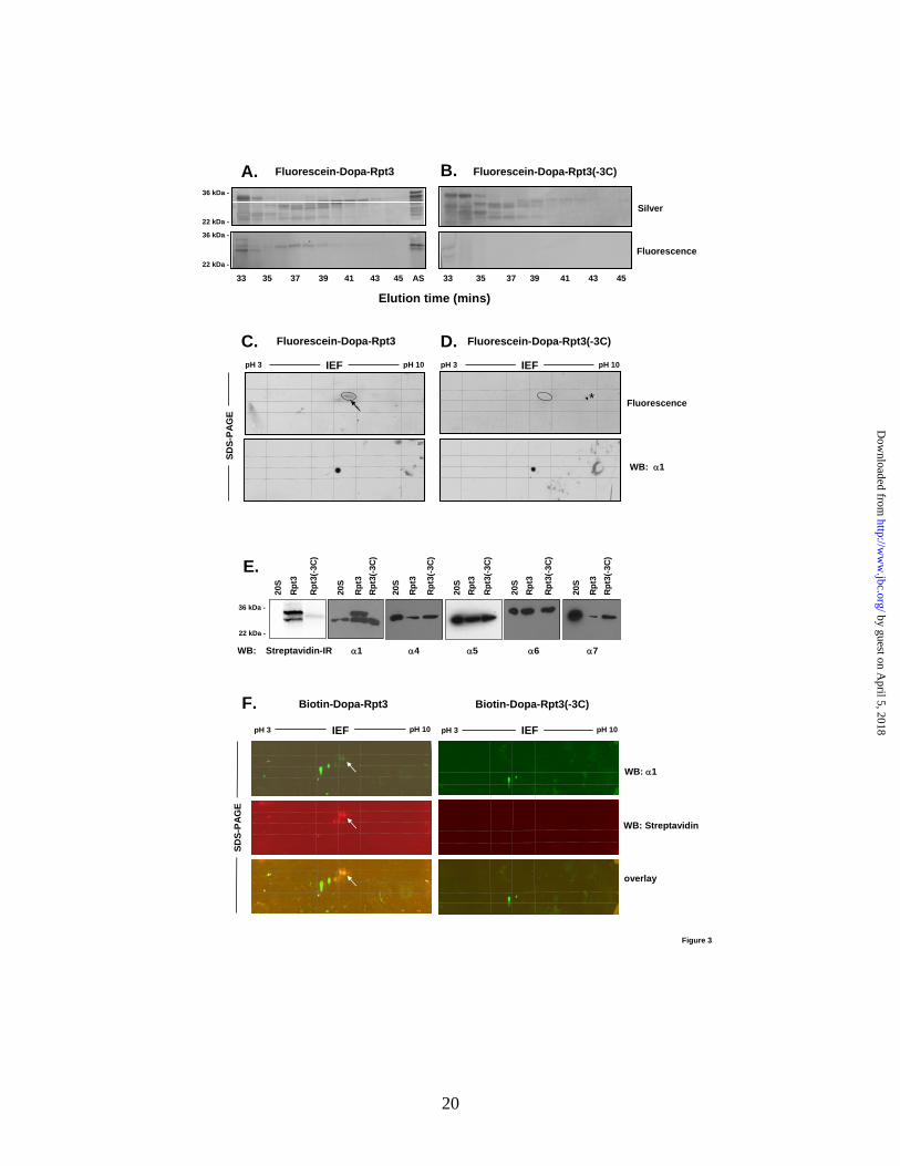
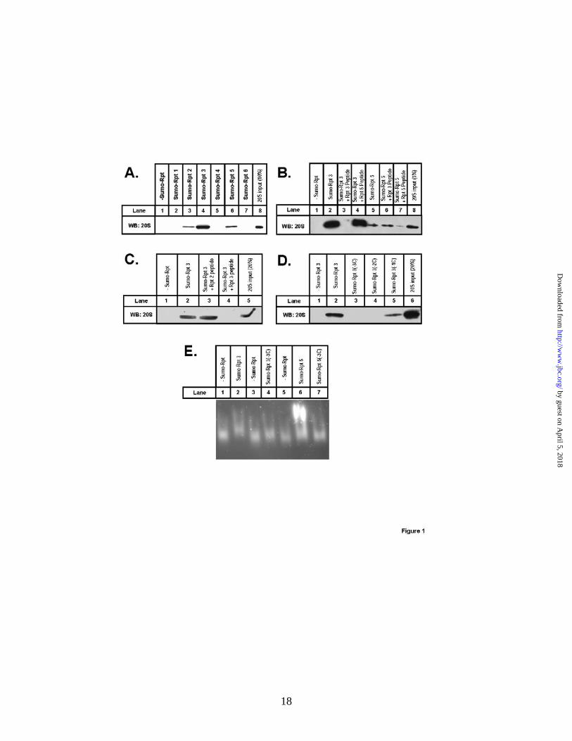
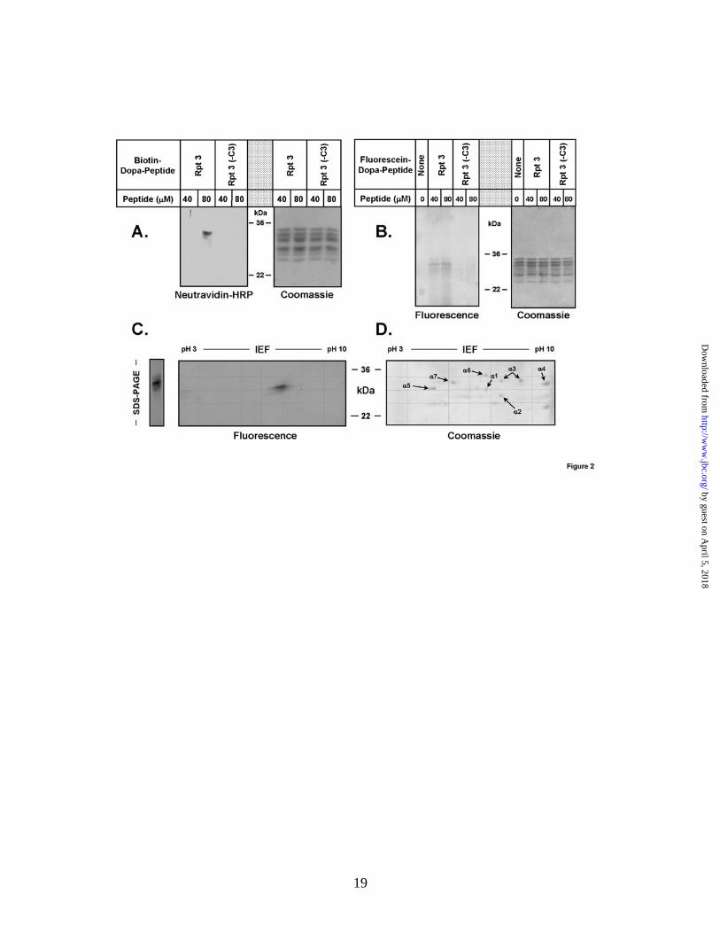
FIGURE LEGENDS Figure 1. The C-terminus of Rpt3 binds to the 20S proteasome. Fusion proteins of SUMO and the C-termini of the indicated Rpt proteins were expressed, purified, and used for pull-down as-says of purified 20S proteasome as described under Experimental Procedures. Panel A. Pull-down assays with the indicated His-tagged SUMO-Rpt peptide fusion peptides (lanes 2-7) were performed as described under Experimental Procedures. Lane 1 shows assay with 20S protea-some but no SUMO-Rpt protein. Relative intensities of the bands in lanes 1-8, as determined by densitometry are: 0, 0, 1.0, 4.32, 0, 1.49, 0, and 0.96, respectively. Panel B. Pull-down assays of 20S proteasome with indicated SUMO-Rpt peptide fusion proteins were conducted in the absence (lanes 2 and 5) or presence (lanes 3, 4, 6, and 7) of indicated Rpt C-terminal peptides. 20S pro-teasome, SUMO-Rpt protein, and Rpt peptides were present in relative concentrations of 240 nM, 100 μM, and 1 mM, respectively. Relative intensities of the bands in lanes 1-8, as determined by densitometry are: 0, 1.0, 0.01, 0.80, 0.16, 0.19, 0.03, and 0.30, respectively. Panel C. Pull-down assays of 20S proteasome with the indicated SUMO-Rpt peptide fusion proteins and Rpt C-terminal peptides as in Panel B. Relative intensities of the bands in lanes 1-5, as determined by densitometry are: 0, 1.0, 1.1, 0.01, and 1.06, respectively. Panel D. Pull-down assays were con-ducted with the indicated His-tagged SUMO-Rpt3 C-terminal peptide fusion proteins. -3C, -2C, and -1C denote deletions of the last 3, 2 and 1 C-terminal Rpt3 residues, respectively. Relative intensities of the bands in lanes 1-6, as determined by densitometry are: 0, 1.0, 0.01, 0, 0.44, and 1.93, respectively. Panel E. Indicated SUMO-Rpt C-terminal peptide fusion proteins (300 μM) were preincubated with 650 nM 20S proteasome for 15 mins at 37oC, subjected to na-tive PAGE, and visualized after overlay of Suc-LLVY-AMC fluorescent peptide substrate. Simi-lar results for experiments in all panels were obtained in at least four separate experiments. Figure 2. Dopa-Rpt3 C-terminal peptide cross-links to a specific α subunit of 20S proteasome. Indicated concentrations of biotin-Dopa-Rpt3 or biotin-Dopa-Rpt3(-3C) (Panel A) or fluorescein-Dopa-Rpt3 or fluorescien-Dopa-Rpt3(-3C) (Panel B) peptides were used for crosslinking assays with 20S proteasome as described in “Experimental Procedures.” Proteasome subunits were separated by SDS-PAGE gel and detected by Coomassie blue staining, as indicated, western blotting with α-neutravidin (Panel A) or fluorescence scanning (Panel B). Panel C. Crosslinking was conducted as above with fluorescein-Dopa-Rpt3. The sample was divided and subjected to either SDS-PAGE (left) or 2-dimensional PAGE (right), as described under Experimental Proce-dures. Crosslinked product was detected by fluorescence scanning. Panel D. 20S proteasome was subjected to two-dimensional PAGE and stained with Coomassie blue. Similar results for experiments in all panels were obtained in at least four separate experiments. Figure 3. Identification of the 20S proteasome subunit crosslinked to the C-terminal pep-tide of Rpt3. Chemical crosslinking of 20S proteasome with fluorescein-Dopa-Rpt3 (Panel A) or fluorescein-Dopa-Rpt3(-3C) (Panel B) and subsequent HPLC purification were conducted as de-scribed under “Experimental Procedures.” HPLC fractions were subjected to SDS-PAGE and either stained for protein with silver (upper) or scanned for fluorescence (lower). “AS” shows sample applied to the column. Fractions containing the major fluorescent (36-39 mins, left) and equivalent fractions from unlabeled control samples (right) were pooled, concentrated and sub-jected to two-dimensional PAGE, Panel C and Panel D, respectively. The area of the gel contain-ing the fluorescent spot (denoted by oval, upper left), and the corresponding area of the non-fluorescent control gel (upper right), were excised and processed for mass spectrometric identifi-cation of proteins, as described under Experimental Procedures. Gels were western blotted for proteasome subunit α1 (lower right and left). Grids indicate relative registration of gels based on the position of common markers. The spot denoted by the asterisk (*) is an imaging artifact
by guest on April 5, 2018
http://ww
w.jbc.org/
Dow
nloaded from
16
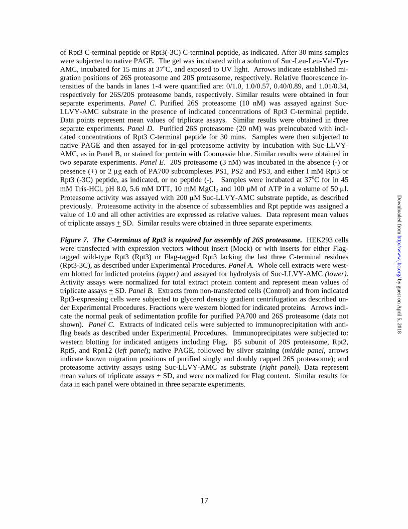
unique to this individual experiment. Similar results for experiments in all panels were obtained in at least four separate experiments. Panels E and F. Chemical crosslinking of 20S was per-formed with biotin-Dopa-Rpt3 or biotin-Dopa-Rpt3(-3C) and the samples were enriched on monoavidin beads as described under “Experimental Procedures.” Enriched samples were sub-jected to western blotting after SDS-PAGE (Panel E) or two-dimensional PAGE (Panel F) using either Streptavidin-IR or antibodies against individual 20S proteasome subunits as indicated. Un-treated 20S proteasome (20S) was used as a control for each blot (Panel E). Two dimensional gels of indicated samples were blotted for both the α1 subunit (Panel F, upper, green channel) and biotin (Panel F, middle, red channel). Panel F, lower, shows merged image (yellow) for the two blots. Arrows point to cross-linked products. Figure 4. The C-terminus of Rpt3 does not influence 20S proteasome activation. Panel A. 20S proteasome (12 nM) activity against the indicated peptide substrates was determined in the pres-ence and absence of peptides corresponding to the last ten residues of Rpt3 (400 μM) or Rpt5 (200 μM). Panel B. 20S proteasome (12 nM) activity against Suc-LLVY-AMC substrate was determined in the absence (Con) and presence of the indicated C-terminal Rpt peptides (400 μM). Panel C. 20S proteasome (30 nM) activity against [methyl-14C]-casein substrate was determined in the presence of the indicated Rpt peptides. For each experiment activity in the absence of Rpt peptides was ascribed a value of 1.0 and other activities are expressed as relative values. Data represent mean values + SD of triplicate assays. Similar results were obtained in at least four separate experiments. Figure 5. Structural determinants of Rpt C-terminal peptides on proteasome binding and acti-vation. Panel A. Structures of C-terminal Rpt peptides used for proteasome binding and activa-tion assays. Panel B. His-tagged SUMO proteins containing the C-terminal fusions of the indi-cated peptides were expressed, purified, and used for pull-down assays with 20S proteasome, as described under Experimental Procedures. Lane 1 shows assay containing 20S proteasome but no SUMO-Rpt protein. Relative intensities of the bands in lanes 1-10, as determined by densitometry are: 0, 1.0, 0.01, 0.13, 0.29, 0.02, 1.14, 0.89, 0.01, and 0.88, respectively. Panel C. 20S protea-some activity against Suc-LLVY-AMC substrate was measured in the presence of indicated SUMO-Rpt C-terminal peptide fusion proteins (100 μM). Proteasome activity in the absence of SUMO-Rpt protein was assigned a value of 1.0 and all other activities are expressed as relative values. Data represent mean values of triplicate assays + SD. Similar results were obtained in at least three separate experiments. Panel D. Peptides were synthesized corresponding to either the C-terminal ten residues of each indicated Rpt subunit (RptX peptide, black bars), or the C-terminal three residues of each indicated Rpt subunit (RptX) and the adjacent seven residues of Rpt5 (Rpt5 head, RptX tail, gray bars). 20S proteasome (12 nM) activity was assayed in the ab-sence (Control) or presence of these peptides (400 μM) using Suc-LLVY-AMC substrate. Activ-ity in the absence of Rpt peptides was assigned a value of 1.0 and all other activities are ex-pressed as relative values. Data represent mean values of triplicate assays + SD. Similar results were obtained in at least four separate experiments. Figure 6. The C-terminal peptide of Rpt3 inhibits 26S proteasome assembly and activation in vitro. Panels A and B. In vitro 26S proteasome assembly and activation from purified 20S pro-teasome and PA700 was conducted as described under Experimental Procedures. Panel A. 20S proteasome (15 nM) and PA700 (75 nM) were preincubated in the presence or absence of indi-cated Rpt3 C-terminal peptide or Rpt3(-3C) C-terminal peptide. After 30 mins proteasome activ-ity was measured using Suc-LLVY-AMC substrate. 20S proteasome activity in the absence of PA700 was assigned a value of 1.0 and all other activities are expressed as relative values. Panel B. 20S proteasome (75 nM) and PA700 (200 nM) were preincubated in the presence or absence
by guest on April 5, 2018
http://ww
w.jbc.org/
Dow
nloaded from
17
of Rpt3 C-terminal peptide or Rpt3(-3C) C-terminal peptide, as indicated. After 30 mins samples were subjected to native PAGE. The gel was incubated with a solution of Suc-Leu-Leu-Val-Tyr-AMC, incubated for 15 mins at 37oC, and exposed to UV light. Arrows indicate established mi-gration positions of 26S proteasome and 20S proteasome, respectively. Relative fluorescence in-tensities of the bands in lanes 1-4 were quantified are: 0/1.0, 1.0/0.57, 0.40/0.89, and 1.01/0.34, respectively for 26S/20S proteasome bands, respectively. Similar results were obtained in four separate experiments. Panel C. Purified 26S proteasome (10 nM) was assayed against Suc-LLVY-AMC substrate in the presence of indicated concentrations of Rpt3 C-terminal peptide. Data points represent mean values of triplicate assays. Similar results were obtained in three separate experiments. Panel D. Purified 26S proteasome (20 nM) was preincubated with indi-cated concentrations of Rpt3 C-terminal peptide for 30 mins. Samples were then subjected to native PAGE and then assayed for in-gel proteasome activity by incubation with Suc-LLVY-AMC, as in Panel B, or stained for protein with Coomassie blue. Similar results were obtained in two separate experiments. Panel E. 20S proteasome (3 nM) was incubated in the absence (-) or presence (+) or 2 μg each of PA700 subcomplexes PS1, PS2 and PS3, and either I mM Rpt3 or Rpt3 (-3C) peptide, as indicated, or no peptide (-). Samples were incubated at 37oC for in 45 mM Tris-HCl, pH 8.0, 5.6 mM DTT, 10 mM MgCl2 and 100 µM of ATP in a volume of 50 μl. Proteasome activity was assayed with 200 μΜ Suc-LLVY-AMC substrate peptide, as described previously. Proteasome activity in the absence of subassemblies and Rpt peptide was assigned a value of 1.0 and all other activities are expressed as relative values. Data represent mean values of triplicate assays + SD. Similar results were obtained in three separate experiments. Figure 7. The C-terminus of Rpt3 is required for assembly of 26S proteasome. HEK293 cells were transfected with expression vectors without insert (Mock) or with inserts for either Flag-tagged wild-type Rpt3 (Rpt3) or Flag-tagged Rpt3 lacking the last three C-terminal residues (Rpt3-3C), as described under Experimental Procedures. Panel A. Whole cell extracts were west-ern blotted for indicted proteins (upper) and assayed for hydrolysis of Suc-LLVY-AMC (lower). Activity assays were normalized for total extract protein content and represent mean values of triplicate assays + SD. Panel B. Extracts from non-transfected cells (Control) and from indicated Rpt3-expressing cells were subjected to glycerol density gradient centrifugation as described un-der Experimental Procedures. Fractions were western blotted for indicated proteins. Arrows indi-cate the normal peak of sedimentation profile for purified PA700 and 26S proteasome (data not shown). Panel C. Extracts of indicated cells were subjected to immunoprecipitation with anti-flag beads as described under Experimental Procedures. Immunoprecipitates were subjected to: western blotting for indicated antigens including Flag, β5 subunit of 20S proteasome, Rpt2, Rpt5, and Rpn12 (left panel); native PAGE, followed by silver staining (middle panel, arrows indicate known migration positions of purified singly and doubly capped 26S proteasome); and proteasome activity assays using Suc-LLVY-AMC as substrate (right panel). Data represent mean values of triplicate assays + SD, and were normalized for Flag content. Similar results for data in each panel were obtained in three separate experiments.
by guest on April 5, 2018
http://ww
w.jbc.org/
Dow
nloaded from
20
Fluorescence
pH 3 pH 10IEF
36 kDa -
22 kDa -
36 kDa -
22 kDa -
Fluorescein-Dopa-Rpt3A. B. Fluorescein-Dopa-Rpt3(-3C)
33 35 37 39 41 43 45 AS 33 35 37 39 41 43 45
Elution time (mins)
Silver
D.pH 3 pH 10IEF
WB: α1
Fluorescein-Dopa-Rpt3C.
Fluorescence
SDS-
PAG
E
Fluorescein-Dopa-Rpt3(-3C)
*
α1
Rpt
3
Streptavidin-IR
36 kDa -
22 kDa -
α4 α5 α6 α7
Rpt
3(-3
C)
20S
Rpt
3 R
pt3(
-3C
)
20S
Rpt
3
Rpt
3(-3
C)
20S
Rpt
3
Rpt
3(-3
C)
20S
Rpt
3
Rpt
3(-3
C)
20S
Rpt
3 R
pt3(
-3C
)
20S
WB:
E.
WB: α1
WB: Streptavidin
overlay
Biotin-Dopa-Rpt3(-3C)
pH 3 pH 10IEF
Biotin-Dopa-Rpt3
pH 3 pH 10IEF
SDS-
PAG
E
F.
Figure 3
by guest on April 5, 2018
http://ww
w.jbc.org/
Dow
nloaded from
Brajesh Kumar, Young Chan Kim and George N. DeMartinoessential for 26S proteasome assembly but not for activation
The C-terminus of RPT3, an ATPase subunit of PA700 (19S) regultory complex, is
published online October 11, 2010J. Biol. Chem.
10.1074/jbc.M110.153627Access the most updated version of this article at doi:
Alerts:
When a correction for this article is posted•
When this article is cited•
to choose from all of JBC's e-mail alertsClick here
Supplemental material:
http://www.jbc.org/content/suppl/2010/10/08/M110.153627.DC1
by guest on April 5, 2018
http://ww
w.jbc.org/
Dow
nloaded from


![[r] Gams M. Computational Analysis of Human Thinking Processes (YSRI,2004)(T)(19s)](https://img.pdfslide.us/doc/110x75/577cdd341a28ab9e78ac7649/r-gams-m-computational-analysis-of-human-thinking-processes-ysri2004t19s.jpg)