Embed Size (px)
Citation preview
doi:10.1182/blood-2006-12-064683Prepublished online April 23, 2007;2007 110: 1251-1261
Sigaux and Jean SoulierDelattre, Alain Aurias, Thierry Leblanc, Hervé Dombret, Alan M. Gewirtz, André Baruchel, FrançoisWillem A. Dik, Anton W. Langerak, Bertrand Montpellier, Bertrand Nadel, Pierre Walrafen, Olivier Emmanuelle Clappier, Wendy Cuccuini, Anna Kalota, Antoine Crinquette, Jean-Michel Cayuela, translocation defining a new T-ALL subtype in very young childrengenomic duplications in human T-cell acute leukemia (T-ALL), the
locus is involved in chromosomal translocation andC-MYBThe
http://bloodjournal.hematologylibrary.org/content/110/4/1251.full.htmlUpdated information and services can be found at:
(795 articles)Oncogenes and Tumor Suppressors � (4217 articles)Neoplasia �
(3667 articles)Clinical Trials and Observations �Articles on similar topics can be found in the following Blood collections
http://bloodjournal.hematologylibrary.org/site/misc/rights.xhtml#repub_requestsInformation about reproducing this article in parts or in its entirety may be found online at:
http://bloodjournal.hematologylibrary.org/site/misc/rights.xhtml#reprintsInformation about ordering reprints may be found online at:
http://bloodjournal.hematologylibrary.org/site/subscriptions/index.xhtmlInformation about subscriptions and ASH membership may be found online at:
Copyright 2011 by The American Society of Hematology; all rights reserved.Washington DC 20036.by the American Society of Hematology, 2021 L St, NW, Suite 900, Blood (print ISSN 0006-4971, online ISSN 1528-0020), is published weekly
only.For personal use at PENN STATE UNIVERSITY on February 23, 2013. bloodjournal.hematologylibrary.orgFrom
NEOPLASIA
The C-MYB locus is involved in chromosomal translocation and genomicduplications in human T-cell acute leukemia (T-ALL), the translocationdefining a new T-ALL subtype in very young childrenEmmanuelle Clappier,1-3 Wendy Cuccuini,1,2 Anna Kalota,4 Antoine Crinquette,1 Jean-Michel Cayuela,1,2 Willem A. Dik,5
Anton W. Langerak,5 Bertrand Montpellier,6 Bertrand Nadel,6 Pierre Walrafen,7 Olivier Delattre,8 Alain Aurias,8
Thierry Leblanc,9 Herve Dombret,10 Alan M. Gewirtz,4 Andre Baruchel,10 Francois Sigaux,1,2 and Jean Soulier1,2
1Genome Rearrangements and Cancer Group, Institut National de la Sante et de la Recherche Medicale (INSERM) U728 and Institut Universitaired’Hematologie, Paris 7 University, Hopital Saint-Louis, Paris, France; 2Hematology Laboratory, Assistance Publique Hopitaux de Paris (APHP), HopitalSaint-Louis, Paris, France; 3Laboratoire de Biochimie Genetique, APHP, Hopital Robert Debre, Paris, France; 4Division of Hematology/Oncology, Department ofMedicine, University of Pennsylvania, Philadelphia; 5Department of Immunology, Erasmus MC, University Medical Center Rotterdam, Rotterdam, theNetherlands; 6Centre d’Immunologie de Marseille-Luminy, Universite de la Mediterranee, Marseille, France; 7Genomic Vision, Pasteur Biotop, Paris, France;8INSERM U509, Institut Curie-Section Recherche, Paris, France; 9Pediatric Hematology Department, APHP, Hopital Saint-Louis, Paris, France; 10AdultHematology Department, APHP, Hopital Saint-Louis, Paris, France
The C-Myb transcription factor is essen-tial for hematopoiesis, including in theT-cell lineage. The C-Myb locus is a com-mon site of retroviral insertional mutagen-esis, however no recurrent genomic in-volvement has been reported in humanmalignancies. Here, we identified 2 typesof genomic alterations involving the C-MYB locus at 6q23 in human T-cell acuteleukemia (T-ALL). First, we found a recip-rocal translocation, t(6;7)(q23;q34), thatjuxtaposed the TCRB and C-MYB loci(n � 6 cases). Second, a genome-wide
copy-number analysis by array-basedcomparative genomic hybridization (array-CGH) identified short somatic duplica-tions that include C-MYB (MYBdup, n � 13cases of 84 T-ALL, 15%). Expression anal-ysis, including allele-specific approaches,showed stronger C-MYB expression inthe MYB-rearranged cases compared withother T-ALLs, and a dramatically skewedC-MYB allele expression in the TCRB-MYB cases, which suggests that a trans-location-driven deregulated expressionmay overcome a cellular attempt to down-
regulate C-MYB. Strikingly, profiling ofthe T-ALLs by clinical, genomic, and large-scale gene expression analyses showsthat the TCRB-MYB translocation definesa new T-ALL subtype associated with avery young age for T-cell leukemia (me-dian, 2.2 years) and with a proliferation/mitosis expression signature. By con-trast, the MYBdup alteration was associatedwith the previously defined T-ALL sub-types. (Blood. 2007;110:1251-1261)
© 2007 by The American Society of Hematology
Introduction
C-Myb is a leucine zipper transcription factor, the expression ofwhich is associated with immature and proliferative cellular stagesand turned off during the maturation of the hematopoietic lin-eage.1,2 Silencing strategies in mice have shown that c-Myb plays amajor role in hematopoiesis, including lineage commitment, prolif-eration, and differentiation.3-6 In the T-cell lineage, c-Myb isinvolved at several key steps throughout the maturation process.7-11
The c-Myb gene was first identified as the cellular homologue ofthe transforming v-Myb gene of 2 avian retroviruses that induceleukemia, AMV and E26.12-15 Transgenic expression of v-Myb,which is a truncated and mutated form of mammalian c-Myb,induces lymphoid or myeloid tumors in mice.16,17 The murinec-Myb locus is also a common site of retroviral insertion inlymphoid and myeloid leukemia.18-23 In humans, the C-MYB geneis located at chromosomal band 6q23.3, and interestingly thechromosome 6q is frequently involved in chromosomal abnormali-ties in human cancer, including hematologic malignancies.24 Inaddition, C-MYB is frequently expressed in human cancer.14,25-27
However, despite intensive studies in a large range of humanneoplasias including leukemia, no clear recurrent involvement of
the C-MYB locus in genomic abnormalities has been reported todate.23,28-31
T-cell acute lymphoblastic leukemias (T-ALLs) are highlymalignant tumors that derive from T-cell progenitors.32-34 Immuno-phenotypic and gene expression analyses of leukemic cells haverevealed heterogeneity that partially reflects distinct stages of T-cellmaturation arrest.35-38 Correlations between oncogene expression,immunophenotype, and large-scale expression profiles have al-lowed the definition of distinct oncogenic T-ALL subtypes.37,38
Genetic studies have pointed out an increasing number of onco-genes in T-ALLs, further demonstrating the complexity of T-ALLoncogenesis and the requirement for several cooperative oncogenicevents.33,34,38 T-ALL oncogenes include aberrantly activated tran-scription factor, namely bHLH (TAL1, TAL2, LYL1, and BHLHB1),LIM-only genes (LMO1 and LMO2), homeobox genes (HOXA,TLX1/HOX11, TLX3/HOX11L2, NKX2-5), and the CALM-AF10and MLL fusion genes.33,34,38-42 In addition, the tumor suppressorlocus CDKN2A/p16/ARF is inactivated in most T-ALL cases,43,44
and NOTCH1 is activated by mutations in half the cases.45,46 TheCyclin D2 gene and the NUP214-ABL1 fusion can also be involved
Submitted December 26, 2006; accepted April 19, 2007. Prepublishedonline as Blood First Edition Paper, April 23, 2007; DOI 10.1182/blood-2006-12-064683.
The online version of this article contains a data supplement.
The publication costs of this article were defrayed in part by page chargepayment. Therefore, and solely to indicate this fact, this article is herebymarked ‘‘advertisement’’ in accordance with 18 USC section 1734.
© 2007 by The American Society of Hematology
1251BLOOD, 15 AUGUST 2007 � VOLUME 110, NUMBER 4
only.For personal use at PENN STATE UNIVERSITY on February 23, 2013. bloodjournal.hematologylibrary.orgFrom
in multistep oncogenesis.47,48 Notably, most T-ALL oncogeneswere identified initially by their involvement in recurrent genomicabnormalities. It is expected that the identification of geneticlesions underlying each subtype of T-ALL will help to adapttherapy, including the development of specific agents againstoncogenic pathways—so-called targeted therapy.49
Using combined cytogenetic, genomic, and molecular tools, wedemonstrate for the first time a clear recurrent genomic involve-ment of the C-MYB gene locus at 6q23.3 in a human cancer, namelyT-ALL, by 2 distinct types of somatic alteration: TCRB-relatedtranslocation and cryptic duplication of a short genome region.Expression analysis suggested that a deregulated C-MYB expres-sion may be oncogenic in T-ALL. Moreover, the t(6;7) TCRB-MYBtranslocation defines a new T-ALL subtype that is associated with avery young age and a proliferation/mitosis expression signature.
Patients, materials, and methods
Patient samples and molecular annotations
A series of 92 T-ALL patients (56 children, 36 adults) were diagnosed andtreated at Saint-Louis Hospital, Paris, France. The patients’ age at diagnosisranged from 13 months to 66 years (median age, 15 years). Informedconsent was obtained from the patients or relatives in accordance with theDeclaration of Helsinki. The study was approved by the Hopital Saint-Louisand Institut Universitaire d’Hematologie Institutional Review Board. FourT-cell lines, CCRF-CEM, HSB-2, Jurkat, and MOLT-4, were included inthe analysis. This series was previously characterized for oncogeneexpression and classified for oncogenic groups according to combinedimmunophenotypic data, oncogene expression, and global gene expressionanalysis using Affymetrix (Santa Clara, CA) data.38 Three additional T-ALLcases were added to this series, one from Saint-Louis Hospital (TL93) and2 that were analyzed by W.A.D. and A.W.L. (T142), and B.M. and B.N.(UPN5846). Large-scale expression data were obtained for case TL93 andwere used after normalization using previously described methods38 for anew global analysis. The array-based comparative genomic hybridization(array-CGH) study was performed on 84 samples with available material ofthe 92 initial cases (80 T-ALL cases and the 4 T-cell lines), as part of theCIT National French program. Constitutional genomic DNA was obtainedfrom follow-up samples of 6 patients in complete remission; these DNAsamples were used as paired controls in array-CGH experiments to rule outcopy-number polymorphisms. Additional T-ALL oncogenic annotationswere obtained as follows: CDKN2A/p16/ARF genomic data using array-CGH analysis and/or locus-specific fluorescent in situ hybridization (FISH),and NOTCH1 mutations by sequencing polymerase chain reaction (PCR)–amplified genomic fragments of exons 26, 27, and 34 (encoding theheterodimerization domain [HD]), and the PEST domain.45
Cytogenetic and molecular analyses
Interphasic fluorescence in situ hybridization (FISH) analyses were per-formed on cryopreserved leukemic cells. All FISH probes were preparedfrom bacterial artificial chromosome (BAC) or P1-derived artificial chromo-some (PAC) clones obtained from the BAC/PAC Resource Center atChildren’s Hospital Oakland Research Institute Oakland, CA (http://bacpac.chori.org). Whole chromosome painting FISH was performed usingWCP6 and WCP7 from Abbott Laboratories (http://www.vysis.com). TheTCRB-flanking FISH probes have been described previously.38 The C-MYBlocus FISH probes were RP11–845K5, RP11–184J4, RP11–104D9, RP11–141K5, RP11–55H4, and RP11–166A21. For molecular combing analysis(fiber-FISH), DNA was combed using the Molecular Combing System(Genomic Vision, Paris, France) as described.50 All FISH micrographimages were performed with an upright epifluorescence microscope (LeicaDM6000 B LT, Rueil-Malmaison, France) equipped with appropriate filterblocks for fluorescence analysis (fluoroscein isothiocyanate [FITC], TexasRed, and 4�,6-diamidino-2-phenylindole [DAPI]). The images were cap-
tured through a PlanApo 63�/1.32 oil immersion objective (0.132 mm/pixel; Leica) with a CCD camera (3 � 1⁄2�) JAI M300 (http://www.jai.com)and Isis FISH analysis software (Metasystems, Altlussheim, Germany).Images shown in the figures are at original magnification, �630. Southernblot analysis for the C-MYB locus was performed using EcoRI, HindIII,and XbaI digestions and a panel of PCR-amplified probes from BACRP3–388E23 DNA. Molecular cloning of the TCRB translocation break-point sequences was performed using ligation-mediated PCR methods aspreviously reported.38
Genome wide array-CGH analysis
A 4K BAC-PAC array-CGH (Curie Institute–French Ligue Contre leCancer, Paris, France) was used on 84 T-ALL samples. This array contained3922 BAC and PAC DNAs, as previously described.51 This array wasdesigned to regularly cover the genome and also included additional clonesfor putative oncogenes such as C-MYB, or for regions known to becommonly rearranged in cancer. Hybridization, scanning, normalization,and data analysis were performed using standard procedures.51
High-density 244K oligonucleotide arrays (Agilent Technologies, http://www.agilent.com) were used to map the C-MYB duplication precisely, andto confirm somatic gain in paired tumor and constitutional genomic DNAfrom the same patients in cases TL29 and TL59.
Genomic databases and bioinformatics tools
The following databases and tools were used: the UCSC Genome Browser(http://genome.ucsc.edu), Ensembl (http://www.ensembl.org/index.html),the Immunogenetics (IMGT) repertoire for immunoglobulins and T-cellreceptors (http://imgt.cines.fr/textes/IMGTrepertoire), the NCBI blast tools(http://ncbi.nlm.nih.gov/blast), the Mitelman Database of ChromosomeAberrations in Cancer (http://cgap.nci.nih.gov/Chromosomes/Mitelman),the Database of Genomic Variants (http://projects.tcag.ca/variation), theMouse Retrovirus Tagged Cancer Gene Database (http://rtcgd.ncifcrf.gov),the Entrez databases (http://www.ncbi.nlm.nih.gov/), and the Gene Ontol-ogy project database (http://www.geneontology.org).
Array-CGH data were analyzed using the Curie Institute VAMP tools52
(http://www.curie.fr/recherche/themes/detail_equipe.cfm/lang/_gb/id_equipe/303.htm) and the CGH Analytics 3.2 software (Agilent Technol-ogies, http://www.agilent.com).
Large-scale gene expression data were normalized, and the analysis wasperformed using dChip (http://www.dchip.org), as previously described.38
C-MYB expression and sequence analyses
RNA samples were reverse transcribed and cDNAs were analyzed forC-MYB transcripts by real-time quantitative PCR (RQ-PCR) using Taqmanmethods (Applied Biosystems, http://www.appliedbiosystems.com/). Re-sults were normalized on expression of the housekeeping gene TBP,according to the formula: level of C-MYB � 2[CtTBP�CtMYB]. RQ-PCRsystem for analysis of MYB expression was as follows: MYB-F: 5�-CTATTACCACATTTCTGAAGCACAAAA-3�, MYB-R: 5�-GCTGAGG-GACATTGACTATATTTACATG-3�, and MYB probe: 5�-6-FAM-CTC-CAGTCATGTTCCATACCCTGTAGCGTT-TAMRA-3�; TBP housekeepinggene: TBP-F: 5�-CACGAACCACGGCACTGATT-3�, TBP-R: 5�-TTTTCT-TGCTGCCAGTCTGGAC-3�, and TBP probe: 5�-6-FAM-TGTGCACAG-GAGCCAAGAGTGAAGA-TAMRA-3�. Additional PCR systems for theanalysis of alternative transcripts and differential use of promoters areshown in the Figure S4 (available on the Blood website; see the SupplementalMaterials link at the top of the online article). In selected cases, the entire C-MYBopen reading frame was sequenced on both strands from leukemic cDNA afterPCR of cDNAfrom leukemic cells; primers sequences are available in Figure S4.A polymorphic poly-T (T8/9) microsatellite found in the 3�-UTR regionwas used for analysis of C-MYB allelic expression. Fragment size analysisof a fluorescent-labeled PCR fragment using Genescan methods (AppliedBiosystems) allowed discrimination of the T8/T9 alleles. PCR primerswere as follows: S217F-5�-6-FAM-AGGTAATGAATTGTAGCCAG-3� andS218R-5�-AAATACTGATCTGTTGGATCC-3�. Leukemic genomic DNAsamples were first analyzed to determine the heterozygous cases, and then
1252 CLAPPIER et al BLOOD, 15 AUGUST 2007 � VOLUME 110, NUMBER 4 only.For personal use at PENN STATE UNIVERSITY on February 23, 2013. bloodjournal.hematologylibrary.orgFrom
leukemic cDNAs from informative cases were analyzed to determineC-MYB allele expression.
Results
Identification of a recurrent translocation t(6;7)(q23;q34) inT-ALL involving the TCRB and C-MYB loci
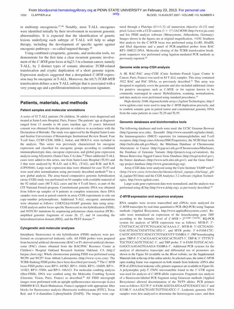
In the course of conventional cytogenetic analysis at diagnosis ofacute leukemia patients in Saint-Louis Hospital, we identified achromosomal translocation t(6;7)(q23;q34) in 2 pediatric T-ALLcases (TL34 and TL92, Figure 1A,B). Dual-color FISH usingTCRB flanking probes demonstrated involvement of the TCRBlocus at 7q34 in both cases (Figure 1B). By using inverse PCR fromthe TCRB sequence, we amplified and characterized the break-points’ derivative sequences in case TL34 (Figure 1C). Thebreakpoint on chromosome 6q23.3 was mapped in the vicinity ofthe C-MYB oncogene. Probes were derived from the C-MYB locusand demonstrated in FISH and Southern blot experiments that thesame locus was involved in case TL92 (Figure 1D). The t(6;7)(q23;q34) involves telomeric regions in both chromosomes 6 and 7,hence we assumed that this translocation could have been missed insystematic T-ALL karyotype series. We therefore used C-MYBflanking FISH probes that allowed us to identify 2 additional caseswith translocations involving the C-MYB and TCRB loci (TL33 andTL93, Figure 1E) from a series of 84 T-ALL (80 T-ALL patients
and 4 T-cell lines). Finally, 2 isolated cases with a TCRB-MYBtranslocation (UPN5846 and T142) were detected from indepen-dent T-ALL series by systematic molecular analysis of oncogenicTCRB genes rearrangements, as performed by B.M. and B.N., andW.A.D. and A.W.L. These cases were added to the present study,giving a total of 6 TCRB-MYB cases, including 3 cases fullycharacterized at the molecular level on both derivative chromo-somes (Figure 1C; Figure S1). Sequence analysis of the TCRBderivative sequences was suggestive of a specific mechanism ofV(D)J-mediated translocation typically seen at the TCRB lo-cus.53,54 These translocations were reciprocal and balanced, and ledto the juxtaposition of the C-MYB proto-oncogene near to theTCRB regulatory sequence (Figure 1F), which suggested deregu-lated expression.
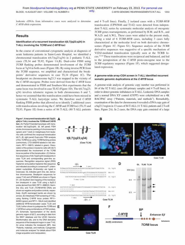
A genome-wide array-CGH screen in T-ALL identified recurrentsomatic genomic duplications at the C-MYB locus
A genome-wide analysis of genomic copy number was performed in84 of the 92 T-ALL cases (80 primary samples and 4 T-cell lines), inorder to detect genomic imbalances in T-ALL. Leukemic DNA samplesand a normal DNA XY control (GYPT) were cohybridized on a 4KBAC/PAC array (“Patients, materials, and methods”). Remarkably,examination of the data for chromosome 6 revealed a DNAcopy gain ofa 6q23.3 region in 13 cases of 84 T-ALL (11 T-ALL patients and 2 T-celllines, Figure 2A). In 2 cases, the DNA copy gain consisted of a large
Figure 1.Arecurrent translocation t(6;7)(q23;q34) in T-ALL involves the TCRB and C-MYBloci. (A) Partial R-banded karyotype with trans-location t(6;7)(q23;q34). (B, left panel) FISHwhole chromosome painting of chromosomes 6(green) and 7 (red) on metaphases from leuke-mic cells confirmed the reciprocal translocationt(6;7). (B, right panel) Dual-color FISH analysisof the TCRB locus using TCRB-flanking probes(centromeric, CTD-3092H9 labeled in red; telo-meric, RP11–168I15, labeled in green). Disso-ciation of the probes in leukemic cells with t(6;7)demonstrated the involvement of the TCRBlocus as partner of the translocation. (C) Deriva-tive sequences of t(6;7)(q23;q34) breakpoints incase TL34 and corresponding germ-line se-quences. Recognition sequence signal (RSS)heptamer and putative heptamer-like sequenceare indicated according to consensus53; untem-plated nucleotides (N-diversity) are typed inlowercase. GL indicates germ line; der, deriva-tive chromosomes. Breakpoint sequences forcases T142 and UPN5846 are shown in FigureS1. (D) Southern blot mapping of the transloca-tion breakpoint in case TL92 using a 6q23.3probe derived from BAC RP11–388E23. Germ-line (GL) and TL34 (TCRB-MYB) DNAs areshown as negative and positive controls, respec-tively. EcoRI rearranged bands are shown byarrows. (E) Interphasic FISH screening of T-ALLusing flanking C-MYB locus probes RP11–845K5 (green) and RP11–184J4 (red) identifiedadditional MYB-translocated cases TL33 andTL93, further shown to juxtapose the TCRB andC-MYB loci using combinations of FISH probes.(F) Schematic representation of the der(6)genomic region of t(6;7), according to data fromthe IMGT database and the UCSC GenomeBioinformatics site, and to the DNA derivativesequence of the breakpoint region in caseT142.An arrow indicates the breakpoint (BP). See“Patients, materials, and methods; Cytogeneticand molecular analyses” for details about FISHimage acquisition and manipulation.
C-MYB GENOMIC REARRANGEMENTS IN T-ALL 1253BLOOD, 15 AUGUST 2007 � VOLUME 110, NUMBER 4 only.For personal use at PENN STATE UNIVERSITY on February 23, 2013. bloodjournal.hematologylibrary.orgFrom
chromosomal gain (6.0 Mb in TL01, and 22.4 Mb in TL76), while in theother cases the amplified region was restricted to a region less than 2 Mbincluding the C-MYB locus. The MOLT4 cell line showed a morecomplex profile with a gain of a large region of chromosome 6q in apseudotetraploid karyotype reinforced by higher level of amplificationin a region less than 2 Mb at the C-MYB locus (Figure 2A top panel).
We then investigated whether the short DNA copy gain of theC-MYB locus was a somatic rearrangement that originated inleukemic cells, or whether it could be a constitutional large-sizecopy-number variation (CNV) as was recently reported in thehuman genome.55-57 Notably, no CNV was reported in this region inthe Database of Genomic Variants (Figure S2). Then we comparedour data to those from 2 independent array-CGH studies on breast
cancer samples that were performed using the same array and thesame control DNA (GYPT, a unique XY healthy subject whoseDNA was used in several studies); no recurrent genomic imbalancein the C-MYB locus was observed in these other studies, whichsuggested that there was no frequent CNV in this region and thatthe C-MYB gain was associated with T-ALL (data not shown).Finally, we were able to demonstrate definitively the somatic originof the C-MYB gain by cohybridizing on the same array pairedleukemic and constitutional DNAs from the same patient in2 T-ALL cases with available constitutional material (Figure 2B).
Thereafter, we defined a minimal region of DNA copy gainusing high-density 244K oligonucleotide array-CGHs (Agilent) toa consistent region of length approximately 230 kb including the
Figure 2. Identification of recurrent somatic genomic duplications at the C-MYB locus in T-ALL. (A) Global representation of large-scale analysis of genomic copy numberusing a 4K array-CGH in MOLT4 cell line (upper panel) and TL63 primary T-ALLcase (lower panel). DNAcopy gain at the C-MYB locus at 6q23 is indicated, as well as the DNAcopy loss atthe CDKN2A/p16/ARF locus at 9p21 as an example. (B) Compared analysis of array hybridization using as normal DNAeither a healthy subject DNA(control) or germ-line DNA(GL) of thesame patient as DNAreference. This analysis allowed us to distinguish copy-number variations (CNVs) and somatic genomic imbalances.As an example, a CNV at 5p15 was resolved inthe TL26 and TL29 cases (a gain was observed when the leukemic DNA was cohybridized with an unrelated control DNA, but it disappeared using paired leukemic and GL controls).Asomatic loss of CDKN2A/p16/ARF was evidenced in both the TL26 and TL29 cases, because the imbalance persisted after cohybridization with paired control; similarly the somatic gainat the 6q23/C-MYB locus was confirmed in case TL29. For each BAC/PAC of the array, gains were represented in red; losses, in green; and balanced signals, in yellow. (C) Copy-numberanalysis using a very high-density oligonucleotide array (Agilent), focused on the C-MYB region, enabled the minimal region of genomic gain to be mapped to approximately 230 kb.(C, right panel) A 6-Mb–sized copy gain region was found in case TL01; (left panel) a short minimal genomic gain including the C-MYB gene was evidenced in case TL29 (array-CGHperformed with paired leukemic and GL DNAs). The genomic region of gain of chromosome 6 is magnified, with the horizontal cursor (blue) pointing out the C-MYB gene (shown in red inthe right panel). The so-called “moving average” ratio between leukemic and GL DNAappears as a blue line in case TL01, and a brown line in case TL29. Two copies (alleles) of the locusappear as a moving average close to 0, whereas a DNAcopy gain of an allele shifts the line close to ratio � 0.5 along the corresponding genomic region. (D) Interphasic C-MYB FISH usingthe RP1–32B1 (green) and RP3–388E23 (red) probes in the MYBdup cases showed no extra signal (except in the complex MOLT4 pseudotetraploid cell line, not shown), which suggestslocal duplication. (E) Molecular combing analysis using C-MYB locus probes RP11–55H4 (red) and RP11–166A21 (green) demonstrated a local duplication (bottom); the normal allele isshown on the top. See “Patients, materials, and methods; Cytogenetic and molecular analyses” for details about FISH image acquisition and manipulation.
1254 CLAPPIER et al BLOOD, 15 AUGUST 2007 � VOLUME 110, NUMBER 4 only.For personal use at PENN STATE UNIVERSITY on February 23, 2013. bloodjournal.hematologylibrary.orgFrom
C-MYB gene (Figure 2C). A panel of probes covering the C-MYBlocus detected no extra signal in metaphases or interphase nuclei incases with C-MYB copy gain (Figure 2D), which suggests a localduplication. That was demonstrated by molecular combing analysisshowing a direct tandem duplication of the C-MYB locus (Figure 2E).Cases with C-MYB locus duplication are further referred to in the studyreported herein as MYBdup cases.
Genomic location of the C-MYB locus rearrangements inhuman and mouse T-cell leukemias
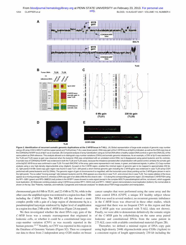
In the present study 2 types of recurrent somatic rearrangementwere identified in T-ALL, namely reciprocal chromosomaltranslocation TCRB-MYB, and short genomic duplicationsMYBdup, which target a unique 6q23.3 region that includes theC-MYB proto-oncogene. Molecular mapping of the 6 t(6;7)(q23;q34) chromosomal breakpoints, which included completebreakpoint sequencing in 3 cases (TL34, T142, and UPN5846),showed 2 discrete breakpoint clusters at 6q23.3 (Figure 3 toppanel): one located 5 kb telomeric, 3� of the C-MYB gene(4 cases: TL33, TL93, T142, and UPN5846), and the other 50 kbmore telomeric (2 cases: TL34 and TL92). In all cases, thetranslocation placed the C-MYB proto-oncogene in the vicinityof the TCRB regulatory sequence, which suggests that abnormal
regulation of C-MYB expression could confer oncogenic proper-ties. The 6q23 genomic duplication was mapped to a shortregion of approximately 230 kb that encompassed the entireC-MYB gene in all duplicated cases (Figure 3 top panel), whichreinforced the view of a targeting of this gene by oncogenicsomatic events in T-ALL. Another gene, known as AHI1, waslocated in the vicinity of the t(6;7) breakpoints and wasdisrupted in 2 of 6 cases with the t(6;7), but not in the other4 cases. It was also partially included in the minimal region ofduplication.
Reminiscent of the 6q23 rearrangements that we identified inhuman T-ALL, the C-Myb locus is known to be a frequentinsertion site in retrovirally induced leukemias. The lower panelof Figure 3 shows the insertion sites in murine T-cell leukemiaas reported in the Retrovirus Tagged Cancer Gene Data-base.19,21-23 These sites are distributed around the c-Myb gene,within a region that extends from approximately 100 kbupstream to 150 kb downstream of the gene. Importantly,insertion sites flank both sides of c-Myb, which suggests thatthis gene is an oncogenic target in this chromosomal region.
Therefore the combined genomic data from human (presentstudy) and murine T-cell leukemias (from previous reports) demon-strate that this locus is recurrently targeted in T-cell oncogenesis
Figure 3. Genomic rearrangements targetthe C-MYB locus in both human and mouseT-cell leukemias. Representation of the C-MYB locus in both the human and mousegenomes according to the UCSC database;note that the orientation of the mouse locus wasinverted in this figure in order to maintain theorientation of the human locus and facilitatecomparison. The ALDH8A1, HSB1L, C-MYB,and AHI1 genes are shown according to anno-tations in UCSC. The genomic rearrangementswe described in this work in human T-ALL areindicated: breakpoints (vertical black arrows) ofthe 6 TCRB-MYB cases, and duplicatedgenomic regions (horizontal black bars) in the13 T-ALL cases (TL01, TL29, TL38, TL40,TL49, TL59, TL61, TL63, TL66, TL76, TL77,CCRF-CEM, and MOLT4). The darker horizon-tal bottom line refers to a MYBdup in addition to alarger 6q gain in MOLT4, leading to extra copiesof the locus. Two FISH probes that were usedfor breakpoint mapping are shown. On themouse genome (bottom panel), all the reportedretroviral insertion sites of T-cell leukemias areindicated (vertical black arrows), based on datafrom the Retrovirus Tagged Cancer GeneDatabase.
C-MYB GENOMIC REARRANGEMENTS IN T-ALL 1255BLOOD, 15 AUGUST 2007 � VOLUME 110, NUMBER 4 only.For personal use at PENN STATE UNIVERSITY on February 23, 2013. bloodjournal.hematologylibrary.orgFrom
and point to the C-MYB gene as a strong candidate oncogene inthese T-ALL cases.
C-MYB expression analysis in human T-ALLs
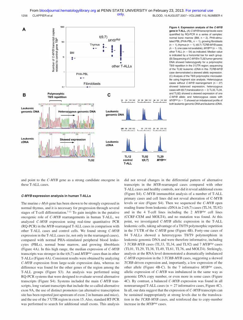
The murine c-Myb gene has been shown to be strongly expressed innormal thymus, and it is necessary for progression through severalstages of T-cell differentiation.7-11 To gain insights in the putativeoncogenic role of C-MYB rearrangements in human T-ALL, weanalyzed C-MYB expression using real-time quantitative PCR(RQ-PCR) in the MYB-rearranged T-ALL cases in comparison withother T-ALL cases and control cells. We found strong C-MYBexpression in the T-ALL cases (ie, not only in the rearranged cases),compared with normal PHA-stimulated peripheral blood leuko-cytes (PBLs), normal bone marrow, and growing fibroblasts(Figure 4A). In this high range, the median expression of C-MYBtranscripts was stronger in the t(6;7) and MYBdup cases than in otherT-ALLs (Figure 4A). Consistent results were obtained by analyzingC-MYB expression from large-scale expression data, whereas nodifference was found for the other genes of the region among theT-ALL groups (Figure S3). An analysis was performed usingRQ-PCR systems that were designed to evaluate several alternativetranscripts (Figure S4). Systems included the main C-MYB tran-scripts, long variant transcripts that include the so-called alternativeexon 9A, the use of distinct promoters (an alternative transcriptionsite has been reported just upstream of exon 2 in humans and mice),and the use of the 3�UTR region in exon 15. Also, standard RT-PCRwas performed to search for additional small exons. This analysis
did not reveal changes in the differential pattern of alternativetranscripts in the MYB-rearranged cases compared with otherT-ALL cases and healthy controls, nor did it reveal additional exons(Figure S4). C-MYB immunoblot analysis of a number of T-ALLprimary cases and cell lines did not reveal aberration of C-MYBlevels or size (Figure S4). Then we sequenced the C-MYB openreading frame from leukemic cDNA in 2 t(6;7) cases (TL34, TL92)and in the 4 T-cell lines including the 2 MYBdup cell lines(CCRF-CEM and MOLT4), and no mutation was found. At thispoint, we investigated C-MYB allelic expression in the T-ALLleukemic cells, taking advantage of a T8/T9 polymorphic repetitionin the 3�UTR of the C-MYB gene (Figure 4B). Forty-one cases of84 T-ALLs showed a heterozygous T8/T9 polymorphism inleukemic genomic DNA and were therefore informative, including3 TCRB-MYB cases (TL33, TL34, and TL92) and 7 MYBdup cases(TL01, TL29, TL38, TL49, TL61, TL76, and MOLT4). Strikingly,analysis at the RNA level demonstrated a dramatically imbalancedC-MYB expression in the 3 TCRB-MYB cases, suggesting a skewedTCRB-driven expression and, importantly, a low expression of theother allele (Figure 4B-C). In the 7 informative MYBdup cases,allelic expression of C-MYB was imbalanced in the same way asgenomic DNA copy number, or even more in some cases (Figure4C). By contrast, a balanced C-MYB expression was found in allnonrearranged T-ALL cases (n � 27 informative cases, Figure 4C).In all, our data suggest that the expression of C-MYB transcripts canbe sustained inappropriately at strong levels due to the transloca-tion in the TCRB-MYB cases, and reinforced due to copy-numberincrease in the MYBdup cases.
Figure 4. Expression analysis of the C-MYBgene in T-ALL. (A) C-MYB transcript levels werequantified by RQ-PCR in a series of samples:normal bone marrow (BM, n � 2), PHA-stimu-lated PBL (PHA-PBL, n � 1), growing fibroblasts(n � 1), thymus (n � 1), t(6;7) TCRB-MYB cases(n � 5, one case not available), MYBdup (n � 13),other T-ALL (n � 54) as indicated. Median valueis indicated by a horizontal bar for each group.(B) Sequencing of C-MYB inTL92 tumor genomicDNA showed heterozygosity for a polymorphicT8/9 repetition in the 3�UTR region; sequencingof the TL92 leukemic cDNA in this TCRB-MYBcase demonstrated a skewed allelic expression.(C) Analysis of the T8/9 polymorphic microsatel-lite using fragment size analysis. Heterozygouscases without C-MYB rearrangement (n � 27)showed balanced expression; heterozygouscaseswith t(6;7) translocation (n � 3;TL33,TL34,and TL92) showed a skewed expression of oneC-MYB allele; and heterozygous cases withMYBdup (n � 7) showed an imbalanced profile ofboth leukemic genomic DNAand leukemic cDNA.
1256 CLAPPIER et al BLOOD, 15 AUGUST 2007 � VOLUME 110, NUMBER 4 only.For personal use at PENN STATE UNIVERSITY on February 23, 2013. bloodjournal.hematologylibrary.orgFrom
The TCRB-MYB translocation defines a new distinct T-ALLoncogenic subtype, associated with very young age and aproliferation/mitosis signature by microarray large-scaleexpression analysis, whereas the MYBdup is foundin other T-ALL subtypes
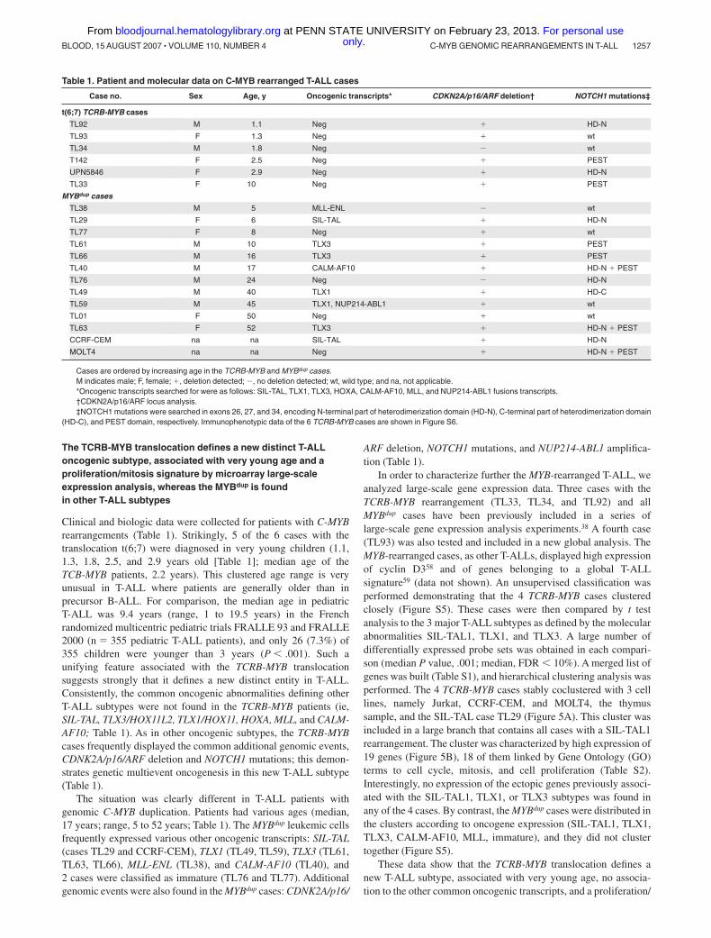
Clinical and biologic data were collected for patients with C-MYBrearrangements (Table 1). Strikingly, 5 of the 6 cases with thetranslocation t(6;7) were diagnosed in very young children (1.1,1.3, 1.8, 2.5, and 2.9 years old [Table 1]; median age of theTCB-MYB patients, 2.2 years). This clustered age range is veryunusual in T-ALL where patients are generally older than inprecursor B-ALL. For comparison, the median age in pediatricT-ALL was 9.4 years (range, 1 to 19.5 years) in the Frenchrandomized multicentric pediatric trials FRALLE 93 and FRALLE2000 (n � 355 pediatric T-ALL patients), and only 26 (7.3%) of355 children were younger than 3 years (P � .001). Such aunifying feature associated with the TCRB-MYB translocationsuggests strongly that it defines a new distinct entity in T-ALL.Consistently, the common oncogenic abnormalities defining otherT-ALL subtypes were not found in the TCRB-MYB patients (ie,SIL-TAL, TLX3/HOX11L2, TLX1/HOX11, HOXA, MLL, and CALM-AF10; Table 1). As in other oncogenic subtypes, the TCRB-MYBcases frequently displayed the common additional genomic events,CDNK2A/p16/ARF deletion and NOTCH1 mutations; this demon-strates genetic multievent oncogenesis in this new T-ALL subtype(Table 1).
The situation was clearly different in T-ALL patients withgenomic C-MYB duplication. Patients had various ages (median,17 years; range, 5 to 52 years; Table 1). The MYBdup leukemic cellsfrequently expressed various other oncogenic transcripts: SIL-TAL(cases TL29 and CCRF-CEM), TLX1 (TL49, TL59), TLX3 (TL61,TL63, TL66), MLL-ENL (TL38), and CALM-AF10 (TL40), and2 cases were classified as immature (TL76 and TL77). Additionalgenomic events were also found in the MYBdup cases: CDNK2A/p16/
ARF deletion, NOTCH1 mutations, and NUP214-ABL1 amplifica-tion (Table 1).
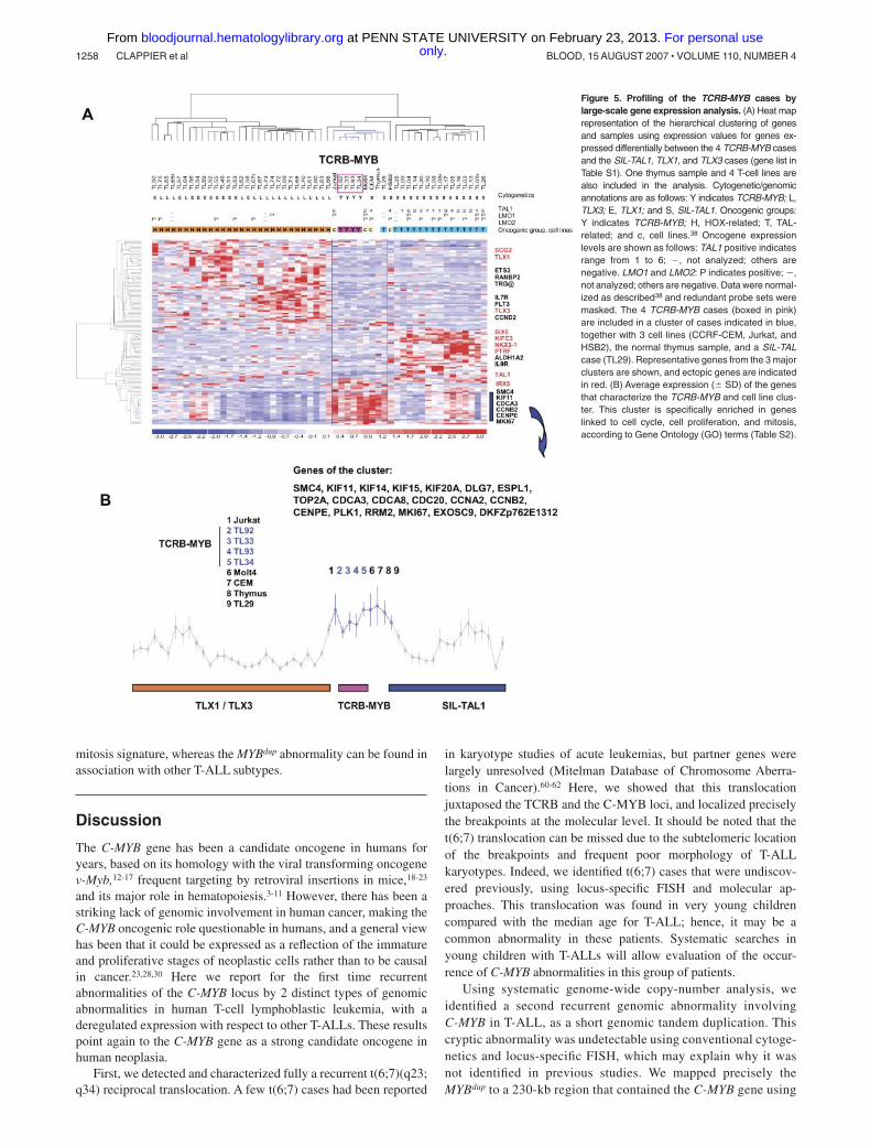
In order to characterize further the MYB-rearranged T-ALL, weanalyzed large-scale gene expression data. Three cases with theTCRB-MYB rearrangement (TL33, TL34, and TL92) and allMYBdup cases have been previously included in a series oflarge-scale gene expression analysis experiments.38 A fourth case(TL93) was also tested and included in a new global analysis. TheMYB-rearranged cases, as other T-ALLs, displayed high expressionof cyclin D358 and of genes belonging to a global T-ALLsignature59 (data not shown). An unsupervised classification wasperformed demonstrating that the 4 TCRB-MYB cases clusteredclosely (Figure S5). These cases were then compared by t testanalysis to the 3 major T-ALL subtypes as defined by the molecularabnormalities SIL-TAL1, TLX1, and TLX3. A large number ofdifferentially expressed probe sets was obtained in each compari-son (median P value, .001; median, FDR � 10%). A merged list ofgenes was built (Table S1), and hierarchical clustering analysis wasperformed. The 4 TCRB-MYB cases stably coclustered with 3 celllines, namely Jurkat, CCRF-CEM, and MOLT4, the thymussample, and the SIL-TAL case TL29 (Figure 5A). This cluster wasincluded in a large branch that contains all cases with a SIL-TAL1rearrangement. The cluster was characterized by high expression of19 genes (Figure 5B), 18 of them linked by Gene Ontology (GO)terms to cell cycle, mitosis, and cell proliferation (Table S2).Interestingly, no expression of the ectopic genes previously associ-ated with the SIL-TAL1, TLX1, or TLX3 subtypes was found inany of the 4 cases. By contrast, the MYBdup cases were distributed inthe clusters according to oncogene expression (SIL-TAL1, TLX1,TLX3, CALM-AF10, MLL, immature), and they did not clustertogether (Figure S5).
These data show that the TCRB-MYB translocation defines anew T-ALL subtype, associated with very young age, no associa-tion to the other common oncogenic transcripts, and a proliferation/
Table 1. Patient and molecular data on C-MYB rearranged T-ALL cases
Case no. Sex Age, y Oncogenic transcripts* CDKN2A/p16/ARF deletion† NOTCH1 mutations‡
t(6;7) TCRB-MYB cases
TL92 M 1.1 Neg � HD-N
TL93 F 1.3 Neg � wt
TL34 M 1.8 Neg � wt
T142 F 2.5 Neg � PEST
UPN5846 F 2.9 Neg � HD-N
TL33 F 10 Neg � PEST
MYBdup cases
TL38 M 5 MLL-ENL � wt
TL29 F 6 SIL-TAL � HD-N
TL77 F 8 Neg � wt
TL61 M 10 TLX3 � PEST
TL66 M 16 TLX3 � PEST
TL40 M 17 CALM-AF10 � HD-N � PEST
TL76 M 24 Neg � HD-N
TL49 M 40 TLX1 � HD-C
TL59 M 45 TLX1, NUP214-ABL1 � wt
TL01 F 50 Neg � wt
TL63 F 52 TLX3 � HD-N � PEST
CCRF-CEM na na SIL-TAL � HD-N
MOLT4 na na Neg � HD-N � PEST
Cases are ordered by increasing age in the TCRB-MYB and MYBdup cases.M indicates male; F, female; �, deletion detected; �, no deletion detected; wt, wild type; and na, not applicable.*Oncogenic transcripts searched for were as follows: SIL-TAL, TLX1, TLX3, HOXA, CALM-AF10, MLL, and NUP214-ABL1 fusions transcripts.†CDKN2A/p16/ARF locus analysis.‡NOTCH1 mutations were searched in exons 26, 27, and 34, encoding N-terminal part of heterodimerization domain (HD-N), C-terminal part of heterodimerization domain
(HD-C), and PEST domain, respectively. Immunophenotypic data of the 6 TCRB-MYB cases are shown in Figure S6.
C-MYB GENOMIC REARRANGEMENTS IN T-ALL 1257BLOOD, 15 AUGUST 2007 � VOLUME 110, NUMBER 4 only.For personal use at PENN STATE UNIVERSITY on February 23, 2013. bloodjournal.hematologylibrary.orgFrom
mitosis signature, whereas the MYBdup abnormality can be found inassociation with other T-ALL subtypes.
Discussion
The C-MYB gene has been a candidate oncogene in humans foryears, based on its homology with the viral transforming oncogenev-Myb,12-17 frequent targeting by retroviral insertions in mice,18-23
and its major role in hematopoiesis.3-11 However, there has been astriking lack of genomic involvement in human cancer, making theC-MYB oncogenic role questionable in humans, and a general viewhas been that it could be expressed as a reflection of the immatureand proliferative stages of neoplastic cells rather than to be causalin cancer.23,28,30 Here we report for the first time recurrentabnormalities of the C-MYB locus by 2 distinct types of genomicabnormalities in human T-cell lymphoblastic leukemia, with aderegulated expression with respect to other T-ALLs. These resultspoint again to the C-MYB gene as a strong candidate oncogene inhuman neoplasia.
First, we detected and characterized fully a recurrent t(6;7)(q23;q34) reciprocal translocation. A few t(6;7) cases had been reported
in karyotype studies of acute leukemias, but partner genes werelargely unresolved (Mitelman Database of Chromosome Aberra-tions in Cancer).60-62 Here, we showed that this translocationjuxtaposed the TCRB and the C-MYB loci, and localized preciselythe breakpoints at the molecular level. It should be noted that thet(6;7) translocation can be missed due to the subtelomeric locationof the breakpoints and frequent poor morphology of T-ALLkaryotypes. Indeed, we identified t(6;7) cases that were undiscov-ered previously, using locus-specific FISH and molecular ap-proaches. This translocation was found in very young childrencompared with the median age for T-ALL; hence, it may be acommon abnormality in these patients. Systematic searches inyoung children with T-ALLs will allow evaluation of the occur-rence of C-MYB abnormalities in this group of patients.
Using systematic genome-wide copy-number analysis, weidentified a second recurrent genomic abnormality involvingC-MYB in T-ALL, as a short genomic tandem duplication. Thiscryptic abnormality was undetectable using conventional cytoge-netics and locus-specific FISH, which may explain why it wasnot identified in previous studies. We mapped precisely theMYBdup to a 230-kb region that contained the C-MYB gene using
Figure 5. Profiling of the TCRB-MYB cases bylarge-scale gene expression analysis. (A) Heat maprepresentation of the hierarchical clustering of genesand samples using expression values for genes ex-pressed differentially between the 4 TCRB-MYB casesand the SIL-TAL1, TLX1, and TLX3 cases (gene list inTable S1). One thymus sample and 4 T-cell lines arealso included in the analysis. Cytogenetic/genomicannotations are as follows: Y indicates TCRB-MYB; L,TLX3; E, TLX1; and S, SIL-TAL1. Oncogenic groups:Y indicates TCRB-MYB; H, HOX-related; T, TAL-related; and c, cell lines.38 Oncogene expressionlevels are shown as follows: TAL1 positive indicatesrange from 1 to 6; �, not analyzed; others arenegative. LMO1 and LMO2: P indicates positive; �,not analyzed; others are negative. Data were normal-ized as described38 and redundant probe sets weremasked. The 4 TCRB-MYB cases (boxed in pink)are included in a cluster of cases indicated in blue,together with 3 cell lines (CCRF-CEM, Jurkat, andHSB2), the normal thymus sample, and a SIL-TALcase (TL29). Representative genes from the 3 majorclusters are shown, and ectopic genes are indicatedin red. (B) Average expression (� SD) of the genesthat characterize the TCRB-MYB and cell line clus-ter. This cluster is specifically enriched in geneslinked to cell cycle, cell proliferation, and mitosis,according to Gene Ontology (GO) terms (Table S2).
1258 CLAPPIER et al BLOOD, 15 AUGUST 2007 � VOLUME 110, NUMBER 4 only.For personal use at PENN STATE UNIVERSITY on February 23, 2013. bloodjournal.hematologylibrary.orgFrom
high-density oligonucleotide array-CGH. Local tandem duplica-tion was demonstrated using molecular combing. These datahighlight the strength of high-density array-CGH to identifycryptic copy-number abnormalities in leukemia, as was shownrecently for the short LMO2 activating deletions in T-ALL.59
Importantly, we demonstrated that the duplication was somaticand not due to one of the constitutional copy-number variations(CNVs) that was recently discovered to be relatively common inthe human genome.55-57 For this purpose, the availability ofsamples from the same patients in remission, which allowedpaired constitutional and tumor analysis, was invaluable (Figure2B). Interestingly, the MYBdup abnormality was frequently foundin association with the known classifying oncogene transcripts(SIL-TAL, CALM-AF10, MLL-ENL, TLX1, TLX3),37,38 whichsuggests that MYBdup could be an additional oncogenic event.
In contrast with the MYBdup abnormality, clinical and bio-logic data suggest that the TCRB-MYB translocation defines anew T-ALL subtype. Strikingly, patients with this translocationwere very young (no adult; median age, 2.2 years) comparedwith the age range of T-ALL patients. Moreover, the ectopictranscripts that have been associated previously with theprincipal T-ALL subtypes37,38 were not found in these cases,which supports further the view that this rearrangement isassociated with distinct oncogenic pathways. Coclustering ofthese cases with T-cell lines and normal thymus sample in asubgroup that is characterized by a high expression of cell cycle,cell proliferation, and mitosis genes suggests a specific relatedbiology (Figure 5, Figure S5, and Table S2).
The c-Myb locus is known as a frequent insertion site inretrovirus-mediated oncogenesis in animals.19,21-23 A detailed mapof the mouse insertion sites based on the RTCG database andcomparison with human data shows that the c-Myb insertionsmimic the genomic abnormalities here described in human leuke-mia (Figure 3), and provides new validation of the power of theseexperimental approaches in animals to pinpoint human oncogenes.In all, genomic data in mice and humans involve a genomic regionencompassing the c-Myb gene, with retroviral sites at both sides ofthe c-Myb gene in mice, which suggests that this gene is a majoroncogenic target in this chromosomal region. Three other genes,namely AHI1 (Abelson Helper Integration), ALDH8A1 (aldehydedehydrogenase 8 family, member A1), and HBS1L (HBS1-like,S cerevisiae), are located by this genomic region. The AHI1 genewas disrupted in 2 of 6 t(6;7) T-ALL cases. AHI1 is a WD40 andSH3-containing protein that has a complex pattern of isoforms.63 Itwas found to be mutated in a congenital brain malformationsyndrome known as Joubert syndrome,64 and was investigated as apotential oncogene at retroviral insertions Ahi1 in Abl� mice, andin human leukemic cell lines and Ph� samples.63,65 Although wefound no deregulation of AHI-1 gene expression, or of the othergenes of the region, in the t(6;7) or in the MYBdup T-ALL cases(Figure S3), the possibility remains that AHI-1 gene could play arole as an oncogenic cofactor, in a “1 hit, 2 targets” genomic event.Comprehensive c-Myb and Ahi1 deregulation in cellular andanimal models should be useful to investigate this issue.
The molecular characterization of the 6 TCRB-MYB cases thatwas performed in the present work showed that this translocationjuxtaposed the C enhancer in the vicinity of C-MYB andsuggested transcriptional deregulation. In MYBdup cases, copy-number gain could be associated with reinforced C-MYB expres-sion. Notably, the transcriptional consequences of the C-MYBgenomic abnormalities appear unusual considering classical mod-els of clearly ectopic oncogene expression, as seen in TLX1 and
TLX3 cases for instance. Considering that C-MYB is expressed athigh levels in normal thymus, it is likely that it is a strong andsustained, rather than ectopic, expression that may be oncogenic. Ithas been previously reported in mice that c-Myb levels are tightlyregulated throughout hematopoietic differentiation, including dur-ing T-cell differentiation, with brief up and down level changes thatregulate transition between differentiation stages and a finaldown-regulation.4,6-11 Abrogation of subtle levels of C-MYB into astable massive expression due to TCRB regulatory sequences islikely to disturb progression through T-cell differentiation. Thisview is strongly supported by our finding that C-MYB expressionwas skewed massively toward single allele expression in theTCRB-MYB translocated cases, which suggests a cellular attempt todown-regulate C-MYB in these cases (Figure 4C). In the MYBdup
cases, an increase of C-MYB expression related to copy-numbergain may also reinforce oncogenic pathways. For both types ofabnormality, in vitro and in vivo models should be useful toinvestigate the role of c-Myb in T-cell oncogenesis.
A number of published reports have shown that C-MYBknockdown by antisense inhibit the in vitro growth of leukemic cells,including T-cell lines.66-68 Although these experiments have to beconfirmed in primary T-ALL samples using new silencing strategies,they suggest that C-MYB expression is necessary for the growth ofleukemic T cells. Interestingly, C-MYB expression was recently in-volved in an oncogenic MLL 3 HOXA 3 C-MYB transcriptionalpathway, which suggests that indirect biallelic stimulation of C-MYBcould also be involved in oncogenic pathways.69
In conclusion, we report for the first time a recurrent somaticinvolvement of the C-MYB locus in human leukemia due to2 distinct genomic events, TCRB-associated translocation andcryptic duplication of a short genome region, and leading toderegulated C-MYB expression. Moreover, a new T-ALL clinicobio-logic entity has been here identified that associates a very youngage, the t(6;7) TCRB-MYB translocation, no association with othercommon oncogenic transcripts, and a proliferation/mitosis expres-sion signature. These results suggest that C-MYB could play anoncogenic role in T-ALL, and point to this gene as a potential targetfor therapeutic intervention in human malignancies.
Acknowledgments
J.S. and F.S. were supported by INSERM and Paris 7 University,the Canceropole d’Ile-de-France, and a grant from the LigueNationale Contre le Cancer (Programme Cartes d’Identite desTumeurs, CIT). The construction of the 4K array-CGH at the CurieInstitute by A.A. and O.D. was supported by grants from the CIT(Ligue Nationale Contre le Cancer). W.C. was supported by a grantMedaille d’or de l’Internat CHU de Reims. W.A.D. and A.W.L.were supported by a grant from the Dutch Cancer Society (EMC2002-2707). A.K. is supported by a grant from the NIH to A.M.G.(RO1 CA101859). A.M.G. is a Distinguished Clinical Scientist ofthe Doris Duke Charitable Foundation (DDCF).
We thank Lucie Hernandez, Claire Pichereau, Nathalie Rodri-guez, Xavier Fund, Laurence Grollet, Yannick Fourne, CharlesDecraenes, Marie-Francoise Auclerc, Jean-Pierre Kerckaert, VahidAsnafi, Elizabeth Macintyre, and Helene Cave for helpful contribu-tions. We are grateful to Anne Janin for support. We also thankJean-Christophe Bories and Didier Auboeuf for critical reading ofthe paper and helpful comments.
C-MYB GENOMIC REARRANGEMENTS IN T-ALL 1259BLOOD, 15 AUGUST 2007 � VOLUME 110, NUMBER 4 only.For personal use at PENN STATE UNIVERSITY on February 23, 2013. bloodjournal.hematologylibrary.orgFrom
Authorship
Contribution: E.C. performed experiments, analyzed the data, andwrote the paper; W.C. performed the array-CGH experiments; A.K.and A.C. performed experiments; J.-M.C. contributed to themolecular annotations of T-ALL cases; W.A.D., A.W.L., B.M., andB.N. characterized additional t(6;7) T-ALL cases; P.W. performedmolecular combing experiments; O.D. and A.A. developed the 4Karray; H.D., T.L., and A.B. managed the patients and conducted the
French national trials GRAALL and FRALLE; A.M.G. contributedto data analysis; F.S. led the T-ALL project at Saint-Louis Hospitaland performed the gene expression profiling analysis; J.S. designedthe study, analyzed the data, and wrote the paper.
Conflict-of-interest disclosure: The authors declare no compet-ing financial interests.
Correspondence: Jean Soulier, Genome Rearrangements andCancer Group, INSERM U728, Institut Universitaire d’Hema-tologie, Hopital Saint-Louis, 1 Avenue Claude Vellefaux 75010Paris, France; e-mail: [email protected].
References
1. Weston K. Myb proteins in life, death and differ-entiation. Curr Opin Genet Dev. 1998;8:76-81.
2. Oh IH, Reddy EP. The myb gene family in cellgrowth, differentiation and apoptosis. Oncogene.1999;18:3017-3033.
3. Mucenski ML, McLain K, Kier AB, et al. A func-tional c-myb gene is required for normal murinefetal hepatic hematopoiesis. Cell. 1991;65:677-689.
4. Emambokus N, Vegiopoulos A, Harman B, Jen-kinson E, Anderson G, Frampton J. Progressionthrough key stages of haemopoiesis is dependenton distinct threshold levels of c-Myb. EMBO J.2003;22:4478-4488.
5. Sandberg ML, Sutton SE, Pletcher MT, et al. c-Myb and p300 regulate hematopoietic stem cellproliferation and differentiation. Dev Cell. 2005;8:153-166.
6. Sakamoto H, Dai G, Tsujino K, et al. Proper levelsof c-Myb are discretely defined at distinct steps ofhematopoietic cell development. Blood. 2006;108:896-903.
7. Badiani P, Corbella P, Kioussis D, Marvel J,Weston K. Dominant interfering alleles define arole for c-Myb in T-cell development. Genes Dev.1994;8:770-782.
8. Allen RD III, Bender TP, Siu G. c-Myb is essentialfor early T cell development. Genes Dev. 1999;13:1073-1078.
9. Pearson R, Weston K. c-Myb regulates the prolif-eration of immature thymocytes following beta-selection. EMBO J. 2000;19:6112-6120.
10. Bender TP, Kremer CS, Kraus M, Buch T, Rajew-sky K. Critical functions for c-Myb at three check-points during thymocyte development. Nat Immu-nol. 2004;5:721-729.
11. Lieu YK, Kumar A, Pajerowski AG, Rogers TJ,Reddy EP. Requirement of c-myb in T cell devel-opment and in mature T cell function. Proc NatlAcad Sci U S A. 2004;101:14853-14858.
12. Duesberg PH, Bister K, Moscovici C. Geneticstructure of avian myeloblastosis virus, releasedfrom transformed myeloblasts as a defective virusparticle. Proc Natl Acad Sci U S A. 1980;77:5120-5124.
13. Souza LM, Strommer JN, Hillyard RL, KomaromyMC, Baluda MA. Cellular sequences are presentin the presumptive avian myeloblastosis virus ge-nome. Proc Natl Acad Sci U S A. 1980;77:5177-5181.
14. Baluda MA, Reddy EP. Anatomy of an integratedavian myeloblastosis provirus: structure and func-tion. Oncogene. 1994;9:2761-2774.
15. Lipsick JS, Wang DM. Transformation by v-Myb.Oncogene. 1999;18:3047-3055.
16. Badiani PA, Kioussis D, Swirsky DM, Lampert IA,Weston K. T-cell lymphomas in v-Myb transgenicmice. Oncogene. 1996;13:2205-2212.
17. Davies J, Badiani P, Weston K. Cooperation ofMyb and Myc proteins in T cell lymphomagen-esis. Oncogene. 1999;18:3643-3647.
18. Shen-Ong GL, Potter M, Mushinski JF, Lavu S,Reddy EP. Activation of the c-myb locus by viral
insertional mutagenesis in plasmacytoid lympho-sarcomas. Science. 1984;226:1077-1080.
19. Hwang HC, Martins CP, Bronkhorst Y, et al. Iden-tification of oncogenes collaborating with p27Kip1loss by insertional mutagenesis and high-throughput insertion site analysis. Proc Natl AcadSci U S A. 2002;99:11293-11298.
20. Kanter MR, Smith RE, Hayward WS. Rapid in-duction of B-cell lymphomas: insertional activa-tion of c-myb by avian leukosis virus. J Virol.1988;62:1423-1432.
21. Kim R, Trubetskoy A, Suzuki T, Jenkins NA,Copeland NG, Lenz J. Genome-based identifica-tion of cancer genes by proviral tagging in mouseretrovirus-induced T-cell lymphomas. J Virol.2003;77:2056-2062.
22. Lund AH, Turner G, Trubetskoy A, et al. Genome-wide retroviral insertional tagging of genes in-volved in cancer in Cdkn2a-deficient mice. NatGenet. 2002;32:160-165.
23. Wolff L. Myb-induced transformation. Crit RevOncog. 1996;7:245-260.
24. Harper ME, Franchini G, Love J, Simon MI, GalloRC, Wong-Staal F. Chromosomal sublocalizationof human c-myb and c-fes cellular onc genes.Nature. 1983;304:169-171.
25. Slamon DJ, Boone TC, Murdock DC, et al. Stud-ies of the human c-myb gene and its product inhuman acute leukemias. Science. 1986;233:347-351.
26. Barletta C, Pelicci PG, Kenyon LC, Smith SD,Dalla-Favera R. Relationship between the c-myblocus and the 6q-chromosomal aberration in leu-kemias and lymphomas. Science. 1987;235:1064-1067.
27. Siegert W, Beutler C, Langmach K, Keitel C,Schmidt CA. Differential expression of the onco-proteins c-myc and c-myb in human lymphoprolif-erative disorders. Eur J Cancer. 1990;26:733-737.
28. Ganter B, Lipsick JS. Myb and oncogenesis. AdvCancer Res. 1999;76:21-60.
29. Introna M, Golay J. How can oncogenic transcrip-tion factors cause cancer: a critical review of themyb story. Leukemia. 1999;13:1301-1306.
30. Weston K. Reassessing the role of C-MYB in tu-morigenesis. Oncogene. 1999;18:3034-3038.
31. Lutwyche JK, Keough RA, Hughes TP, Gonda TJ.Mutation screening of the c-MYB negative regula-tory domain in acute and chronic myeloid leukae-mia. Br J Haematol. 2001;114:632-634.
32. Pui CH, Relling MV, Downing JR. Acute lympho-blastic leukemia. N Engl J Med. 2004;350:1535-1548.
33. Armstrong SA, Look AT. Molecular genetics ofacute lymphoblastic leukemia. J Clin Oncol.2005;23:6306-6315.
34. Graux C, Cools J, Michaux L, Vandenberghe P,Hagemeijer A. Cytogenetics and molecular ge-netics of T-cell acute lymphoblastic leukemia:from thymocyte to lymphoblast. Leukemia. 2006;20:1496-1510.
35. Breit TM, Wolvers-Tettero IL, van Dongen JJ.Phenotypic and genotypic characteristics of hu-
man early T-cell differentiation: the T-cell acutelymphoblastic leukaemia model. Res Immunol.1994;145:139-143; discussion: 1994;145:155-158.
36. Asnafi V, Beldjord K, Boulanger E, et al. Analysisof TCR, pT alpha, and RAG-1 in T-acute lympho-blastic leukemias improves understanding ofearly human T-lymphoid lineage commitment.Blood. 2003;101:2693-2703.
37. Ferrando AA, Neuberg DS, Staunton J, et al.Gene expression signatures define novel onco-genic pathways in T cell acute lymphoblastic leu-kemia. Cancer Cell. 2002;1:75-87.
38. Soulier J, Clappier E, Cayuela JM, et al. HOXAgenes are included in genetic and biologic net-works defining human acute T-cell leukemia (T-ALL). Blood. 2005;106:274-286.
39. Asnafi V, Radford-Weiss I, Dastugue N, et al.CALM-AF10 is a common fusion transcript in T-ALL and is specific to the TCRgammadelta lin-eage. Blood. 2003;102:1000-1006.
40. Ferrando AA, Armstrong SA, Neuberg DS, et al.Gene expression signatures in MLL-rearrangedT-lineage and B-precursor acute leukemias:dominance of HOX dysregulation. Blood. 2003;102:262-268.
41. Nagel S, Kaufmann M, Drexler HG, MacLeod RA.The cardiac homeobox gene NKX2–5 is deregu-lated by juxtaposition with BCL11B in pediatricT-ALL cell lines via a novel t(5;14)(q35.1;q32.2).Cancer Res. 2003;63:5329-5334.
42. Przybylski GK, Dik WA, Grabarczyk P, et al. Theeffect of a novel recombination between the ho-meobox gene NKX2–5 and the TRD locus in T-cell acute lymphoblastic leukemia on activation ofthe NKX2–5 gene. Haematologica. 2006;91:317-321.
43. Cayuela JM, Madani A, Sanhes L, Stern MH, Si-gaux F. Multiple tumor-suppressor gene 1 inacti-vation is the most frequent genetic alteration inT-cell acute lymphoblastic leukemia. Blood. 1996;87:2180-2186.
44. Gardie B, Cayuela JM, Martini S, Sigaux F.Genomic alterations of the p19ARF encoding ex-ons in T-cell acute lymphoblastic leukemia.Blood. 1998;91:1016-1020.
45. Weng AP, Ferrando AA, Lee W, et al. Activatingmutations of NOTCH1 in human T cell acute lym-phoblastic leukemia. Science. 2004;306:269-271.
46. Grabher C, von Boehmer H, Look AT. Notch 1activation in the molecular pathogenesis of T-cellacute lymphoblastic leukaemia. Nat Rev Cancer.2006;6:347-359.
47. Clappier E, Cuccuini W, Cayuela JM, et al. CyclinD2 dysregulation by chromosomal translocationsto TCR loci in T-cell acute lymphoblastic leuke-mias. Leukemia. 2006;20:82-86.
48. Graux C, Cools J, Melotte C, et al. Fusion ofNUP214 to ABL1 on amplified episomes in T-cellacute lymphoblastic leukemia. Nat Genet. 2004;36:1084-1089.
49. Downing JR, Mullighan CG. Tumor-specific ge-netic lesions and their influence on therapy in pe-diatric acute lymphoblastic leukemia. HematologyAm Soc Hematol Educ Program. 2006;118-122.
1260 CLAPPIER et al BLOOD, 15 AUGUST 2007 � VOLUME 110, NUMBER 4 only.For personal use at PENN STATE UNIVERSITY on February 23, 2013. bloodjournal.hematologylibrary.orgFrom
50. Lebofsky R, Bensimon A. Single DNA moleculeanalysis: applications of molecular combing. BriefFunct Genomic Proteomic. 2003;1:385-396.
51. Idbaih A, Marie Y, Pierron G, et al. Two types ofchromosome 1p losses with opposite significancein gliomas. Ann Neurol. 2005;58:483-487.
52. La Rosa P, Viara E, Hupe P, et al. VAMP: visual-ization and analysis of array-CGH, transcriptomeand other molecular profiles. Bioinformatics.2006;22:2066-2073.
53. Marculescu R, Vanura K, Montpellier B, et al. Re-combinase, chromosomal translocations and lym-phoid neoplasia: targeting mistakes and repairfailures. DNA Repair (Amst). 2006;5:1246-1258.
54. Marculescu R, Vanura K, Le T, Simon P, Jager U,Nadel B. Distinct t(7;9)(q34;q32) breakpoints inhealthy individuals and individuals with T-ALL.Nat Genet. 2003;33:342-344.
55. Sebat J, Lakshmi B, Troge J, et al. Large-scalecopy number polymorphism in the human ge-nome. Science. 2004;305:525-528.
56. Iafrate AJ, Feuk L, Rivera MN, et al. Detection oflarge-scale variation in the human genome. NatGenet. 2004;36:949-951.
57. Redon R, Ishikawa S, Fitch KR, et al. Globalvariation in copy number in the human genome.Nature. 2006;444:444-454.
58. Ross ME, Zhou X, Song G, et al. Classification ofpediatric acute lymphoblastic leukemia by geneexpression profiling. Blood. 2003;102:2951-2959.
59. Van Vlierberghe P, van Grotel M, Beverloo HB, etal. The cryptic chromosomal deletiondel(11)(p12p13) as a new activation mechanismof LMO2 in pediatric T-cell acute lymphoblasticleukemia. Blood. 2006;108:3520-3529.
60. Chan LC, Ha SY, Ching LM, et al. Cytogeneticsand immunophenotypes of childhood acute lym-phoblastic leukemia in Hong Kong. Cancer GenetCytogenet. 1994;76:118-124.
61. Heerema NA, Sather HN, Sensel MG, et al. Fre-quency and clinical significance of cytogeneticabnormalities in pediatric T-lineage acute lympho-blastic leukemia: a report from the Children’sCancer Group. J Clin Oncol. 1998;16:1270-1278.
62. Sinclair P, Harrison CJ, Jarosova M, Foroni L.Analysis of balanced rearrangements of chromo-some 6 in acute leukemia: clustered breakpointsin q22-q23 and possible involvement of c-MYB ina new recurrent translocation, t(6;7)(q23;q32through 36). Haematologica. 2005;90:602-611.
63. Jiang X, Hanna Z, Kaouass M, Girard L, JolicoeurP. Ahi-1, a novel gene encoding a modular proteinwith WD40-repeat and SH3 domains, is targetedby the Ahi-1 and Mis-2 provirus integrations. J Vi-rol. 2002;76:9046-9059.
64. Ferland RJ, Eyaid W, Collura RV, et al. Abnormalcerebellar development and axonal decussationdue to mutations in AHI1 in Joubert syndrome.Nat Genet. 2004;36:1008-1013.
65. Jiang X, Zhao Y, Chan WY, et al. Deregulated ex-pression in Ph� human leukemias of AHI-1, agene activated by insertional mutagenesis inmouse models of leukemia. Blood. 2004;103:3897-3904.
66. Gewirtz AM, Anfossi G, Venturelli D, Valpreda S,Sims R, Calabretta B. G1/S transition in normalhuman T-lymphocytes requires the nuclear pro-tein encoded by c-myb. Science. 1989;245:180-183.
67. Calabretta B, Sims RB, Valtieri M, et al. Normaland leukemic hematopoietic cells manifest differ-ential sensitivity to inhibitory effects of c-myb anti-sense oligodeoxynucleotides: an in vitro studyrelevant to bone marrow purging. Proc Natl AcadSci U S A. 1991;88:2351-2355.
68. Gewirtz AM. Myb targeted therapeutics for thetreatment of human malignancies. Oncogene.1999;18:3056-3062.
69. Hess JL, Bittner CB, Zeisig DT, et al. c-Myb is anessential downstream target for homeobox-medi-ated transformation of hematopoietic cells. Blood.2006;108:297-304.
C-MYB GENOMIC REARRANGEMENTS IN T-ALL 1261BLOOD, 15 AUGUST 2007 � VOLUME 110, NUMBER 4 only.For personal use at PENN STATE UNIVERSITY on February 23, 2013. bloodjournal.hematologylibrary.orgFrom