Embed Size (px)
Citation preview

The Brain in Schizotypal Personality Disorder: A Review of Structural MRI and CT Findings
CitationDickey, Chandlee C., Robert W. McCarley, and Martha E. Shenton. 2002. “The Brain in Schizotypal Personality Disorder: A Review of Structural MRI and CT Findings.” Harvard Review of Psychiatry 10 (1) (January): 1–15. doi:10.1080/10673220216201.
Published Versiondoi:http:10.1080/10673220216201
Permanent linkhttp://nrs.harvard.edu/urn-3:HUL.InstRepos:28520171
Terms of UseThis article was downloaded from Harvard University’s DASH repository, and is made available under the terms and conditions applicable to Other Posted Material, as set forth at http://nrs.harvard.edu/urn-3:HUL.InstRepos:dash.current.terms-of-use#LAA
Share Your StoryThe Harvard community has made this article openly available.Please share how this access benefits you. Submit a story .
Accessibility
The Brain in Schizotypal Personality Disorder: A Review ofStructural MRI and CT Findings
Chandlee C. Dickey, MD, Robert W. McCarley, MD, and Martha E. Shenton, MDFrom the Clinical Neuroscience Division, Laboratory of Neuroscience, Department of Psychiatry,VA Boston Healthcare System, Boston, Mass. (Drs. Dickey, McCarley, and Shenton); the Divisionof Behavioral Neurology, Departments of Neurology and Psychiatry (Dr. Dickey), and the SurgicalPlanning Laboratory, MRI Division, Department of Radiology (Dr. Shenton), Brigham and Women’sHospital, Boston, Mass; and the Department of Psychiatry, Harvard Medical School, Boston, Mass.(Drs. Dickey, McCarley, and Shenton).
AbstractStudies of schizotypal personality disorder (SPD) are important because the condition is geneticallyrelated to schizophrenia and because data accumulating to confirm its biological underpinnings arechallenging some traditional views about the nature of personality disorders. This review of 17structural imaging studies in SPD indicates that individuals with this disorder show brainabnormalities in the superior temporal gyrus, parahippocampus, temporal horn region of the lateralventricles, corpus callosum, thalamus, and septum pellucidum, as well as in total cerebrospinal fluidvolume, similar to those seen in persons with schizophrenia. Differences between SPD andschizophrenia include lack of abnormalities in the medial temporal lobes and lateral ventricles inSPD. Whether the normal volume, and possibly normal functioning, of the medial temporal lobes inindividuals with SPD may help to suppress psychosis in this disorder remains an intriguing but stillunresolved question. Such speculation must be tempered due to a paucity of studies, and additionalwork is needed to confirm these preliminary findings. The imaging findings do suggest, however,that SPD probably represents a milder form of disease along the schizophrenia continuum. Withfurther clarification of the neuroanatomy of SPD, researchers may be able to identify whichneuroanatomical abnormalities are associated with the frank psychosis seen in schizophrenia.
Although schizophrenia was once considered the “graveyard of neuropathologists,”1 recentneuroimaging techniques have radically changed this view. Early studies using computerizedtomography (CT) were pivotal in demonstrating ventricular abnormalities in the disorder butdid not provide the resolution required to document alterations in regions with unclearboundaries such as the amygdala and various thalamic nuclei. With the advent of magneticresonance imaging (MRI), these latter brain regions of interest have been evaluated inschizophrenia and found to be abnormal. In recent comprehensive reviews2,3 of MRI-documented morphological brain abnormalities in schizophrenia, most brain regions studiedshowed neuroanatomical alteration compared with the same regions in healthy controls.Nonetheless, a convergence of findings suggested that the major locus for brain abnormalitieswas the temporal lobe; fewer studies reported abnormalities in the lateral ventricles, prefrontalcortex, inferior parietal cortex, basal ganglia, thalamus, corpus callosum, or septumpellucidum.2,3 Note that, although many regions are involved in schizophrenia, they do notappear to be equally affected, and the temporal lobe regions are the most severely altered. (For
© 2002 President and Fellows of Harvard CollegeReprint requests: Martha E. Shenton, PhD, VA Boston Healthcare System, Psychiatry 116A, 940 Belmont St., Brockton, MA 02401([email protected]).
NIH Public AccessAuthor ManuscriptHarv Rev Psychiatry. Author manuscript; available in PMC 2010 April 13.
Published in final edited form as:Harv Rev Psychiatry. 2002 ; 10(1): 1–15.
NIH
-PA Author Manuscript
NIH
-PA Author Manuscript
NIH
-PA Author Manuscript

a recent review of MRI findings in schizophrenia and a discussion comparing the various brainregions, see Shenton and colleagues.2)
In many cases, however, these MRI findings are difficult to interpret, given the possibleconfounding effects of the chronicity of the psychotic illness and the medications used to treatit. Although the definition of a personality disorder4 requires that a person experience distress,the stress of chronic psychosis as seen in schizophrenia is arguably more relentless. McEwenand Margarinos5 have demonstrated that increased stress-induced adrenal cortisol release,along with excitatory amino acids, may result in atrophy of the hippocampal CA3 region. Suchatrophy may help to explain some of the medial temporal lobe findings in schizophrenia (seesection on temporal lobe structures, below).
Medications can also affect brain morphology. Chakos and colleagues6,7 compared the volumeof the basal ganglia in patients taking traditional and atypical antipsychotics and found that thetraditional antipsychotics increased caudate volume more than did the atypical medications.Other possible effects of medication on brain morphology have been reported for superiortemporal gyrus volume.8 In addition, a recent animal model9 demonstrated increased volumeand glial density in the prefrontal cortex with chronic exposure to conventional neuroleptics.The effect of anticholinergics, benzodiazepines, and anticonvulsants on specific brain regionshas been less extensively examined.
One way to avoid the possible confounding effects of medication is to study patients during afirst episode of schizophrenia, before they are treated with medications,10–15 as well as toinvestigate at-risk populations,16 including first-degree relatives of individuals withschizophrenia.17–23 An alternative approach is to study other populations presumed* to havesimilar genetic vulnerability, such as patients with schizotypal personality disorder (SPD). Ourreview will focus on CT and MRI structural imaging studies of persons with SPD.
SPD is characterized by difficulties with social interaction and language, together with oddbehavior and magical thinking. Because individuals with this disorder are not consideredpsychotic, they have generally not been prescribed medications. Nonetheless, persons withSPD and those with schizophrenia have a similar genetic predisposition, as suggested bymultiple family studies24,27–31 reporting that 6–7% of individuals diagnosed withschizophrenia have a first-degree relative with SPD. Similarly, first-degree relatives of personswith SPD have a 6.9% chance of developing schizophrenia.27
In an early epidemiological study conducted in Denmark, Kety and colleagues29 found thatthe data supported the notion of a commonality between schizophrenia and schizophrenia-likedisorders, and they grouped these conditions into the “schizophrenia spectrum disorders.” Thiswork was followed by Kendler and colleagues’ Roscommon County family studies,27,30 whichfurther supported the spectrum concept and encouraged the use of other research tools to definethe phenotypic similarities between SPD and schizophrenia.
Other methodologies such as neurochemical analyses, behavioral studies, andneuropsychological and evoked-potential measures have also shown abnormalities in SPD thatare similar to what has been demonstrated in schizophrenia.32 These include elevatedhomovanillic acid levels,33,34 aberrant eye-tracking,35–38 reduced prepulse inhibition,39
cognitive deficits,40–43 and electrophysiological abnormalities.44–48 One hypothesis thatattempts to incorporate findings from these various methodologies has been proposed by Siever(personal communication), who stated that the relative sparing in terms of symptoms and
*”Presumed,” since the underlying defective gene-gene interactions in schizophrenia have yet to be elucidated, although populationstudies have supported the contention that schizophrenia and SPD share a common genetic diathesis (see below).24–28
Dickey et al. Page 2
Harv Rev Psychiatry. Author manuscript; available in PMC 2010 April 13.
NIH
-PA Author Manuscript
NIH
-PA Author Manuscript
NIH
-PA Author Manuscript
biological abnormalities in SPD compared with schizophrenia may be due to the fact thathypodopaminergic function emanates from the basal ganglia and extends to the frontal lobes.These projections may be “neuroprotective” to other regions such as the frontal lobes.32,49Structural MRI studies of the basal ganglia and frontal lobes as well as functional studiesexamining dopaminergic function are needed to test this hypothesis further.
Another impetus for studying SPD, in addition to the disorder’s close genetic and biologicalties with schizophrenia, is the importance of such research for the conceptualization ofpersonality disorders. More specifically, personality disorders have traditionally not beenthought to have a neurological basis. Now a wealth of data from multiple sources is radicallychallenging this view (see the studies cited in the previous paragraph). Moreover, with theneuroanatomical basis of SPD becoming more clearly established, investigations of thebiological underpinnings of SPD may be a useful model to apply to other personality disorders.
The critical question that we ask, and seek to answer, in this review is: Do the imaging datasupport the notion that SPD is a less severe version of schizophrenia, or is it a distinct disorder?If the former, might we expect that persons with SPD will have fewer neuroanatomicalabnormalities, and therefore less-severe clinical symptoms, than do individuals withschizophrenia? If the data support the idea that SPD is a less penetrant form of schizophrenia,then the next question concerns what abnormalities are present in schizophrenia but absent inSPD. Answers to this last question need to be examined in future studies and may help to directattention to strategies for preventing the development of schizophrenia.
We performed a Medline search in February 2001 for English-language articles including thekey words schizotypal personality disorder, schizophrenia, relatives, computerizedtomography, and magnetic resonance imaging. We found and reviewed 17 studies. We beganwith investigations in which subjects met full DSM criteria for SPD, then continued withstudies in which subjects had some of the features of SPD but did not meet the full criteria,reports of children with symptoms consistent with SPD, and finally other studies (i.e., reportsof persons with SPD and schizophrenia analyzed together, or of individuals with SPD whohave family members with schizophrenia). This organization reflects the different strategiesused by researchers to enlist subjects with SPD for their studies. Such strategies includerecruiting families of probands with schizophrenia, recruiting patients from clinics, recruitingcommunity dwellers by means of newspaper advertisements, and recruiting college studentswho score high on scales of psychopathology thought to tap cognitive manifestations of SPD.Diagnostic criteria have also differed and range from meeting five out of the nine requiredDSM-IV criteria, to having some features of schizotypy derived by diagnostic impressionduring clinical interview, to scoring high on scales of psychopathology.
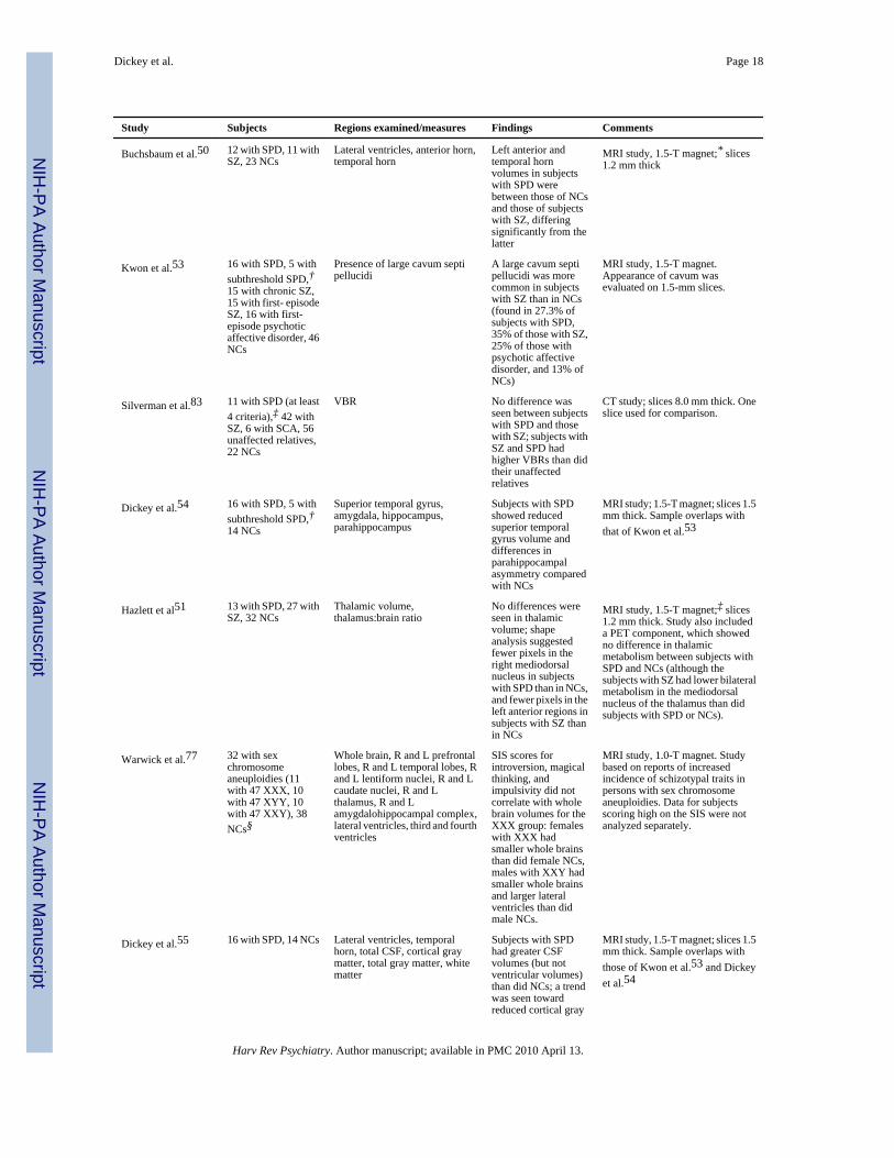
We included all 17 studies in our review, even though some included very few patients withSPD or SPD-like pathology. Table 1 provides a summary of these studies.
OVERVIEWTwo important changes have occurred over time in brain morphology studies of individualswith SPD. First, CT techniques have gradually given way to MRI, which has allowed theinvestigation of more regions and with finer neuroanatomical resolution (includingdifferentiation between gray and white matter). Second, researchers have gone from examiningsubjects with some features of schizotypy to studying persons determined throughsemistructured interviews to meet full DSM criteria for SPD. This change can been seen inTable 1, where it is clear that the majority of recent studies use MRI and involve subjectsmeeting full criteria for SPD.
Dickey et al. Page 3
Harv Rev Psychiatry. Author manuscript; available in PMC 2010 April 13.
NIH
-PA Author Manuscript
NIH
-PA Author Manuscript
NIH
-PA Author Manuscript
All eight of the MRI studies that analyzed the data for subjects with full criteria separately haveemanated from two centers, Mt. Sinai School of Medicine49–52 and Harvard Medical School.53–55 (Note that in 1992 and 1994 researchers from the University of Pennsylvania56,57 usedCT to examine a cohort of subjects who met full criteria for SPD, considered under the categoryof subjects at high risk for schizophrenia [all had mothers with the disorder].) This illustratesnot only the difficulty in recruiting this important subject population but also the fact thatdifferent laboratories employ different approaches for understanding the intrinsicmorphological abnormalities of the brain found in the schizophrenia spectrum disorders.Importantly, however, nine of the 17 studies were published since 1998, suggesting a markedincrease in interest in this topic.
STUDIES OF SUBJECTS WHO MEET FULL CRITERIA FOR A DIAGNOSIS OFSPD
Two laboratories investigating SPD have used individuals who meet full DSM criteria for thediagnosis of SPD. The two laboratories have employed distinctly different recruitmentprocedures, however. The first laboratory, at Mt. Sinai School of Medicine, has recruited itssubjects from local inpatient and outpatient units. Some of these individuals have receivedmedications, including neuroleptics. Our laboratory at Harvard Medical School and theVeterans Affairs Boston Healthcare System, by contrast, has recruited subjects from thecommunity by means of newspaper ads and posted fliers so as to avoid the potentialconfounding effects of medication. The use of such disparate approaches may have resulted inthe sampling of quite different populations. This fact, plus differences between the clinicalassessment protocols in the two laboratories, makes direct comparisons between the studypopulations difficult. The Mt. Sinai cohort, for example, may include subjects with either more-serious symptoms or a greater proportion of positive symptoms, leading them to attend a clinicand be prescribed neuroleptic medications; our cohort may include subjects with a greaterproportion of negative symptoms, or with fewer or more-attenuated symptoms. All of this isconjecture, however, since neither group has reported measures of positive and negativesymptoms. In addition, neither group of researchers has discussed the potential issue of highAxis I and Axis II comorbidity, which has been described by McGlashan.58 This may be animportant focus for future work on the biological basis of SPD. These different approachesmay be complementary in that they may help to elucidate how clinical features affect brainmorphology. Note that, as with other studies included in this review, the number of subjectsstudied in these laboratories is limited, and within a laboratory, samples have partiallyoverlapped. This reflects the difficulties inherent in recruiting subjects with SPD. However,since researchers are just beginning to understand the neuroanatomy of SPD, extensive studyof various brain regions in a limited number of subjects may be a prudent approach.
Below, we review findings from Mt. Sinai on the thalamus and corpus callosum. We thenreview findings from our laboratory on cerebrospinal fluid (CSF), gray and white matter,temporal lobe structures, and the cavum septi pellucidi, and finally the findings from bothlaboratories on the lateral ventricles.
Mount Sinai Group: Clinic-Based StudiesThalamus—The thalamus is the major relay station of the brain; it consists of multiple nucleiand their connections to cortical regions (i.e., mediodorsal nuclei with the prefrontal cortex,and anterior and midline nuclei with limbic and paralimbic structures59). Due to theseinterconnections, the thalamus is considered by some to be key to the understanding ofschizophrenia.60
Dickey et al. Page 4
Harv Rev Psychiatry. Author manuscript; available in PMC 2010 April 13.
NIH
-PA Author Manuscript
NIH
-PA Author Manuscript
NIH
-PA Author Manuscript
The first study of thalamic volume in subjects with SPD, conducted by Hazlett and colleagues,51 showed no differences in thalamic volume or thalamus:brain ratio between patients withSPD and controls but did show differences in shape. Patients with SPD had fewer pixels in theright mediodorsal nucleus and patients with schizophrenia had fewer pixels in the left anteriorregion than did controls. In a second component of the study, the investigators determined withpositron emission tomography that patients with schizophrenia had diminished metabolism inthe mediodorsal nucleus bilaterally compared with SPD patients and comparison subjects.
To refine these findings further, Byne and colleagues52 examined the pulvinar and mediodorsalnuclei of the thalamus in a subset of the subjects. They reported that, compared with controls,both the patients with SPD and those with schizophrenia had reduced pulvinar nuclei, but thepatients with schizophrenia had the additional abnormality of reduced mediodorsal nuclei.Various subdivisions of the pulvinar are involved in relaying sensory inputs to primary visualand auditory sensory areas,59 to the prefrontal cortex,52 and to the temporoparietal heteromodalassociation cortex.52,61 There are a few reports of damage to this region resulting in languagedisturbances.51,60 Thus, these nuclei may be critically involved in the processing andintegration of visual and auditory information, and damage could hypothetically result inmisperceptions.
Taken together, these results suggest that frontolimbic/thalamic connectivity may be differentin SPD than in schizophrenia, and this may, in part, contribute to the differences in the clinicalsymptoms in the two disorders. Such a possibility is particularly interesting, given the currentinterest in thalamic connections and, as proposed by Andreasen and others,60 their possiblecentral role in the production of these conditions.
Corpus callosum—The corpus callosum is the major interhemispheric fiber pathway. Oneof the theories of the etiology of schizophrenia62 involves a failure of interhemisphericcommunication. As a result, the corpus callosum has been the subject of 27 investigations; 17of these have reported abnormalities.2 In the only study to examine corpus area and shape inSPD, Downhill and colleagues49 reported that the genu of the corpus was larger in patientswith SPD than in those with schizophrenia or control subjects, whereas the posterior corpuswas largest in controls, second largest in patients with SPD, and smallest in patients withschizophrenia. (The difference in the latter measure between SPD patients and schizophreniapatients was not statistically significant, however.) Furthermore, these investigators found thatthe shape differences were consistent with the differences in corpus area. They concluded thatthese area and shape abnormalities of the corpus may lead to poor interhemisphericconnectivity and could be responsible for the improper reality testing found in theschizophrenia spectrum disorders.
Our Laboratory: Community-Based StudiesCSF, gray, and white matter—In many studies of patients with schizophrenia, thereappears to be an abundance of CSF, whether measured in the ventricles, in the sulci, or as totalCSF volume.2,3 In our sample of individuals with SPD, we demonstrated increased CSFvolume that was not attributable to lateral ventricle enlargement. We also examined total graymatter volume and found no difference between persons with SPD and normal controls.However, when the cortical gray matter was more carefully delineated with the elimination ofthe subcortical structures and the cerebellum, we found a trend toward reduced cortical graymatter in persons with SPD compared with controls.55 We found no difference in white matterbetween the two groups.
Temporal lobe structures—Interest in temporal lobe structures in schizophrenia stemsfrom the critical role of these structures in language and auditory processing and the observation
Dickey et al. Page 5
Harv Rev Psychiatry. Author manuscript; available in PMC 2010 April 13.
NIH
-PA Author Manuscript
NIH
-PA Author Manuscript
NIH
-PA Author Manuscript
that language abnormalities and auditory hallucinations are among the hallmarks of thisdisorder. Of note, many independent research laboratories investigating schizophrenia havereported abnormalities in temporal lobe structures,2,3,63–67 including the superior temporalgyrus (STG), parahippocampal gyrus, and amygdala-hippocampal complex. The volumes inthese regions have also been correlated with cognitive and clinical symptoms including formalthought disorder and auditory hallucinations, as well as with verbal memory problems.62,68The amygdala, more specifically, may be involved in the attaching of emotional relevance,particularly to visual stimuli including emotional facial expressions;69 in general arousal andother basic functions including sleep, feeding, and sexual activities;68 and in memory.68
Our own laboratory70 has reported reductions in gray matter volume in the STG, amygdala,hippocampus, and parahippocampal gyrus in persons with schizophrenia, and we haveextended this work to patients with first-episode psychosis71 and individuals with SPD. Weapplied the methodology of our previous studies in schizophrenia to a group of individualswith SPD recruited from the community by means of newspaper advertisements. We predictedthat we would see similar, but more-attenuated, volume reduction in the subjects with SPD.We found such subjects to have selective reduction of the left STG gray matter andparahippocampal asymmetry.54 In an attempt to refine the STG results, we examined two ofits main components, Heschl’s gyrus and the planum temporale,72 and found the former to bereduced. In addition, we found that subjects with SPD exhibited formal thought disorder.54This was intriguing, since reduced STG gray matter is one of the most robust findings inschizophrenia (all of the 12 studies examining this found volume reduction2), andparahippocampal asymmetry has been shown postmortem to be abnormal in persons withschizophrenia.73 This study demonstrating partial—but not complete—replication suggestedthat perhaps, at least in this region, there is a relationship between volume affected and clinicalseverity.
Cavum septi pellucidi—The septum pellucidum is a membrane formed in utero by twoleaflets that fuse secondary to pressure of the growing hippocampus and corpus callosum.Space remaining when the closure is incomplete is termed “cavum septi pellucidi”; such aspace is seen in 15% of healthy controls52 at 6 months.53 The presence of a large cavum septipellucidi has been noted in schizophrenia (11 out of 12 studies reported abnormalities).2 Onestudy53 has examined this neurodevelopmental abnormality in patients with SPD, and it founda prevalence of 27%. These data suggest that SPD and schizophrenia probably have aneurodevelopmental component to their etiology.
Clinic- and Community-Based Studies: Lateral Ventricle FindingsThe two laboratories have each examined the lateral ventricles in patients with SPD. Theyproduced slightly different results in the anterior and temporal horns, possibly due to differentdemographic variables.
Historically, enlarged lateral ventricles have been one of the most common findings in theschizophrenia literature: 78% of the 55 MRI2 studies (as well as 75% of the CT studies2)examining this region showed larger lateral ventricles in persons with schizophrenia than incontrols. However, neither the Mt. Sinai group (first with CT74 and then with MRI50) nor ourgroup55 has found a statistically significant difference in total lateral ventricle volume betweenindividuals with SPD and controls. Thus, in this region there appears to be a difference betweenschizophrenia and SPD: persons with SPD are less affected than are those with schizophrenia.
Subtle differences may exist between persons with SPD and healthy controls in particularregions of the lateral ventricles, however. In an evaluation of clinic-based SPD patients at Mt.Sinai, Buchsbaum and coworkers50 reported that the left anterior and temporal horns in theseindividuals were larger than those in age- and sex-matched normal controls but significantly
Dickey et al. Page 6
Harv Rev Psychiatry. Author manuscript; available in PMC 2010 April 13.
NIH
-PA Author Manuscript
NIH
-PA Author Manuscript
NIH
-PA Author Manuscript

smaller than those in patients with chronic schizophrenia. This contrasts with what ourlaboratory has shown in our community-based sample, in which we reported no difference.55 Therefore, although both groups report no statistically significant difference betweenpersons with SPD and controls, the Mt. Sinai study included the additional feature of comparingsuch volumes with those of schizophrenia patients and demonstrated a continuum among thethree groups on this measure.
These two studies were similar in that they both involved subjects meeting full criteria for SPD,but they differed in demographic variables. Left- and right-handed males and females wereincluded in Buchsbaum and colleagues’ investigation,50 whereas only right-handed males wereincluded in Dickey and coworkers’ study.55 Perhaps the greatest difference in the samples,however, results from the method of recruitment—clinic versus community. Subjects in aclinic-based sample may have more-severe SPD symptoms than do those in a community-based one; they may also have fewer negative symptoms such as social anxiety. The issue ofhigh Axis I and Axis II comorbidity, which has been described in SPD58 but is not addressedin these publications, may also be important in deciphering the findings. In addition,pharmacological treatment of SPD patients could be playing a role in clinic-based samples.These variables may be key in understanding the subtle differences in the findings concerningthe anterior and temporal horns.
In summary, these two MRI studies of subjects who met full criteria for SPD did not showenlarged lateral ventricles. This may suggest that in individuals with pure SPD this region isspared the abnormalities typically seen in persons with schizophrenia. Subsequent studies toexamine the lateral ventricles either have analyzed SPD patients together with schizophreniapatients or have not used subjects clearly diagnosed with SPD (see below). One tentativeconclusion, therefore, is that enlargement of the lateral ventricles is not a feature of SPD, andthe presence of enlarged ventricles in schizophrenia may be a morphological index of clinicalseverity.
OTHER STUDIESStudies of Subjects with Schizotypal Features Who Meet Some but Not All Criteria for aDiagnosis of SPD: Frontotemporal Area
One approach to understanding the schizophrenia spectrum disorders is to study individualswho do not meet criteria for a particular disorder but who nonetheless have some of the featuresof that disorder. This approach is best exemplified by Raine and coworkers,75 who examined17 subjects who scored high on scales of schizotypal features but were not assessed using DSMcriteria. These individuals were employees of local hospitals and other work settings. Excludedfrom the pool of perspective subjects were physicians and other workers expected to have highsocial class and a high level of education. In this study high degrees of schizotypy were foundto be significantly associated with reduced left prefrontal area and with left and rightprefrontal:temporal area ratios. The prefrontal cortex is involved in impulse inhibition,assessing the behavioral relevance of stimuli, using working memory while shifting sets,making judgments, and planning. It has vast interconnections with most other sections of thecortex and can influence the activation or de-activation of those areas.59 Unfortunately, theimaging protocol was performed on a machine with low magnetic field strength (0.15 T, asopposed to the 1.5 T often used), and only one slice was used to determine prefrontal andtemporal areas for each subject. Nonetheless, this early study suggested that persons with someschizotypal features may have aberrations in the prefrontal and temporal cortices— areas thathave been shown to be abnormal in individuals with schizophrenia.
An excess of schizotypal traits in subjects with a sex chromosome aneuploidy (SCA) wasdocumented in a recent thesis.76 To follow up on this observation, Warwick and
Dickey et al. Page 7
Harv Rev Psychiatry. Author manuscript; available in PMC 2010 April 13.
NIH
-PA Author Manuscript
NIH
-PA Author Manuscript
NIH
-PA Author Manuscript
colleagues77 studied individuals with SCA and some features of schizotypy. Using MRI toexamine multiple brain regions including the prefrontal cortex, they detected no abnormalities.Unfortunately, data for the subjects with SCA and many features of schizotypy were notanalyzed separately from data for those with SCA alone.
Given the paucity of prefrontal studies examining subjects who have been clearly diagnosedwith SPD, no firm conclusions can be drawn for this brain region.
A Study of Children at Risk for Developing SPD or Schizophrenia: Amygdala, TemporalCortex, and Corpus Callosum
In the only relevant study of children, Hendren and colleagues78 reported that 8- to 12-year-olds with symptoms of either SPD or schizophrenia showed reduced amygdala and temporalcortex volumes and reduced corpus callosum area, similar to what has been shown inschizophrenia.2,3 The authors suggested that the occurrence of abnormalities at a young age isthe result of genetic or environmental events occurring in utero and altering neurodevelopment;they did not explore other possible etiologies, such as postnatal stress. Hendren and coworkersdid not demonstrate enlarged lateral ventricles, as has been shown in subjects meeting fullcriteria for SPD.50,55 Instead, they hypothesized that enlarged ventricular volume mayrepresent disease progression in schizophrenia, a speculation shared by others,79,80 but becausethe study was cross sectional, their data did not address that issue directly. Due to thesubjects’young age, the investigators were unable to make definitive distinctions between SPDand schizophrenia, so subject groups were not analyzed separately. As suggested by the authors,it will be interesting to follow these children and retrospectively review their scans to determinewhether the children who subsequently developed SPD had quantitatively fewer abnormalitiesthan did those who subsequently developed schizophrenia.
A Study Analyzing Patients with SPD and Those with Schizophrenia Together: VentriclesIn a hospital-based study of patients with SPD or schizophrenia who also had prodromalsymptoms of obsessive-compulsive disorder (OCD), persons with nonpsychotic OCD, andnormal controls, Kurokawa and coworkers81 examined MRIs to determine whether thepresence of enlarged ventricles might promote the early detection of SPD or schizophrenia inpersons who early in the course of the illness show symptoms of OCD. They found that thepatients who had developed SPD or schizophrenia had larger ventricles than did those withOCD alone. They concluded that patients with OCD symptoms and enlarged ventricles on MRImay be at risk for later developing one of the schizophrenia spectrum disorders. They did notanalyze data for the SPD patients separately, however, probably due to the small sample size(n = 4). Conclusions about ventricular size in SPD cannot be drawn from this study, since thesubjects with schizophrenia may have been driving the findings.
Studies of Patients with SPD Who Have First-Degree Relatives with Schizophrenia:Ventricles
The Mt. Sinai group, in search of genetic markers common to schizophrenia spectrumdisorders, has studied family pedigrees of probands with schizophrenia. Within these families,some members have been affected by SPD. Shihabuddin and colleagues82 studied a familywith the linkage marker for such disorders on the short arm of chromosome 5(5p14.1–13.1).Eleven family members (of whom three had schizophrenia and two had SPD) underwent CTto determine whether there was a relationship between the presence of the marker and brainabnormalities. All of the affected members and one unaffected member carried the markerallele. These six individuals had enlarged lateral ventricles and an enlarged ventricle:brain ratio(VBR), whereas the remaining unaffected members did not.
Dickey et al. Page 8
Harv Rev Psychiatry. Author manuscript; available in PMC 2010 April 13.
NIH
-PA Author Manuscript
NIH
-PA Author Manuscript
NIH
-PA Author Manuscript

Silverman and colleagues83 have shown increased VBR in persons with SPD or schizophreniain a larger group of families. In this study, however, the researchers lumped the individualswith SPD together with family members who had four of nine criteria for the disorder but notfull-blown SPD. How the persons with full criteria differed from those with partial criteria wasnot detailed. Nonetheless, the investigation suggested a relationship between the geneticloading and brain structure.
Prior to the publication of DSM-III, persons with features consistent with SPD (for example,subtle thought disorder, social isolation, magical thinking) were classified as having borderlineschizophrenia.84 Today, many of these subjects would be reclassified by DSM-IV criteria ashaving SPD. In an early CT study of offspring of mothers with schizophrenia, Schulsinger andcoworkers84 found that the offspring with schizophrenia had enlarged third ventricles and anincreased VBR, whereas the mentally healthy offspring had normal ventricles, and those withborderline schizophrenia had the smallest ventricles. This is the only study in our review thatreported smaller ventricles in subjects with an SPD-like disorder than in controls. In addition,the offspring with schizophrenia had experienced more obstetrical complications than hadeither of the other two groups. These authors84 suggested the “diathesis-stress” model forinterpreting the data—that is, that “schizophrenia is the result of deleterious environmentalinfluences acting on a genetic predisposition.”
In a second CT study of offspring of mothers with schizophrenia, Cannon and colleagues57found that the offspring suffering from SPD or schizophrenia had enlarged sulci, but only thosewith schizophrenia had enlarged ventricles. (The study also included offspring of unaffectedmothers. Some of these offspring had psychiatric disorders, and some did not. The fouroffspring with SPD who had unaffected mothers were not analyzed separately.) Theseresearchers concluded that the offspring with the more-severe disorder, schizophrenia, hadmore morphological abnormalities (sulcal and ventricular enlargement), whereas those withSPD had sulcal enlargement alone. Previously these researchers56 had demonstrated thatoffspring of mothers with schizophrenia (healthy, with SPD, or with schizophrenia) hadenlarged third ventricles, but unfortunately they did not perform analyses comparing the threegroups.
DISCUSSIONIt is difficult to reach conclusions from such a limited number of studies composed of smallsample sizes and subjects with disparate characteristics. Nonetheless, from studies withsubjects who meet full criteria for SPD, it appears that individuals with SPD may have reducedgray matter of the superior temporal gyrus, asymmetry of the parahippocampus, abnormalitiesin thalamic shape and pulvinar volume, larger sulci, abnormalities in the shape of the corpuscallosum, and a high prevalence of large cavum septi pellucidi. Each of these potentialabnormalities has been well documented in schizophrenia.2,3 In most of the studies, however,the subjects with SPD do not have all of the abnormalities that might be present inschizophrenia. For example, persons with SPD have reduced superior temporal gyrus volumesand parahippocampal asymmetry but not the frank parahippocampal volume differences ordifferences in other medial temporal lobe structures such as the hippocampus and amygdalathat are found in schizophrenia (see above). Also, from the studies done to date, lateralventricles appear to be normal.
The specific pathogenesis of these morphometric alterations in SPD cannot be determined fromthe studies reviewed. For example, one cannot deduce whether the abnormalities are a directresult of neurodevelopmental genetic programing, whether they are a result of distalabnormalities causing deafferentation, or whether they represent a decrease in interneurons.
Dickey et al. Page 9
Harv Rev Psychiatry. Author manuscript; available in PMC 2010 April 13.
NIH
-PA Author Manuscript
NIH
-PA Author Manuscript
NIH
-PA Author Manuscript

Byne and colleagues,52 in their discussion of thalamic abnormalities, proposed the possibilityof different etiologies for different nuclei within the thalamus.
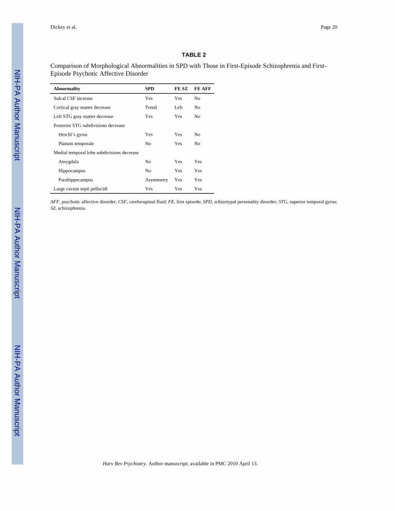
To determine whether the additional regions affected in schizophrenia are critical for theproduction of frank psychosis (i.e., whether they are also present in affective disorder withpsychosis) or are inherent to schizophrenia, data from our laboratory comparing affectivedisorder with psychosis is informative. Table 2, a comparison among SPD, first-episodeschizophrenia, and first-episode psychotic affective disorder,71 shows that the development ofpsychosis may require abnormalities in the medial temporal lobe structures. This is in contrastto the involvement of the superior temporal gyrus, the increase in sulcal CSF, and the decreasein cortical gray matter found in both SPD and schizophrenia, but not in first-episode psychosis.These patterns of abnormalities suggest that the superior temporal gyrus may be critical in theschizophrenia spectrum disorders.
To return to our earlier question of whether SPD should be considered a distinct disorder or asubset of schizophrenia, we believe that the CT and MRI data produced so far cannot fullyseparate the two conditions. To date, there has been no definitive report of an SPD abnormalitythat has not been shown in schizophrenia.† In fact, one of the strategies of SPD research is toexamine brain regions that have been found to be abnormal in schizophrenia to determinewhether they may represent abnormalities fundamental to the schizophrenia spectrum disordersor to psychosis. Once other brain regions are examined, additional abnormalities may be found.However, based on the available literature, one can conclude that not all the abnormalitiesfound in schizophrenia are found in SPD. For example, medial temporal lobe structures arenormal in SPD.54 Therefore, SPD may be considered to represent an attenuated form ofschizophrenia.
The implication of these findings is that individuals with SPD are comparatively spared insome brain regions while those with schizophrenia are relatively afflicted, despite possiblysimilar genetic diatheses. It may be, however, that this genetic continuum can result in somesubjects having more of a critical genetic load and others having less. Additionally, subtle inutero differences such as lower incidence of exposure to influenza virus—or less stress-inducedsteroid release—in persons with SPD than in those with schizophrenia may account for thedifferences in the development of the two disorders. Finally, as these individuals age, repeatedenvironmental stressors may have an additional impact on the progression of both SPD andschizophrenia. How sparing or affliction occurs is critical to determine in future studies.
We believe that SPD represents part of the continuum of clinical symptoms observed in theschizophrenia spectrum and involves some of the same morphological abnormalities.Conservatively speaking, however, the available CT and MRI data cannot rule out thepossibility that SPD is a distinct disorder, although this possibility seems unlikely. One primarystrategy of SPD research is to examine brain regions that have been found to be abnormal inschizophrenia to determine whether or not they are also observed in SPD or are a nonspecificconcomitant of psychosis. One example of the separation between SPD and psychosis isdemonstrated by the finding that medial temporal lobe structures (the amygdala andhippocampus) are abnormal in first-episode schizophrenia and in first-episode psychosis71 butnot in SPD.54
†There are two possible exceptions. Downhill and colleagues49 reported corpus callosum shape differences in certain regions in personswith SPD. The corpus has been shown to be abnormal in schizophrenia,2 although perhaps in slightly different regions. Also, Hazlettand colleagues51 found fewer pixels in the region of the right mediodorsal nucleus in patients with SPD than in those with schizophreniaor normal controls.
Dickey et al. Page 10
Harv Rev Psychiatry. Author manuscript; available in PMC 2010 April 13.
NIH
-PA Author Manuscript
NIH
-PA Author Manuscript
NIH
-PA Author Manuscript

FUTURE DIRECTIONSClearly more studies are necessary to investigate the neuroanatomy of SPD. Persons who meetfull criteria for SPD need to be evaluated, and this population, although difficult to tap, isimportant to our understanding of which brain abnormalities are inherent to the schizophreniaspectrum and which are due to the ravages of schizophrenia and its treatments—or perhaps toother prenatal or environmental stresses. The relationship among genetic load, pre- andpostnatal environmental factors, and morphological abnormalities in the development ofschizophrenia is far from clear. Nonetheless, understanding the interaction of these factors iscritical for understanding the pathogenesis of schizophrenia and potential avenues forintervention.
This review focused on CT and structural MRI findings in SPD. Morphological studies candescribe volumes, shapes, and anatomical patterns but cannot address the critical question ofthe functional capacity of the structures. To date, no functional MRI (fMRI) studies of SPDhave been published. As more laboratories move toward fMRI and begin to elucidate thefunctional anatomy of this disorder, it will be important to discover how individuals with SPDdiffer from those with schizophrenia in the realms of attention, language processing, andemotional processing/expression, where some of the core abnormalities in the schizophreniaspectrum are seen. This complementary coupling of morphometric and functional studies canthen begin to address the relationship between anatomy and clinical phenomena. For example,if one believes that magical ideation and certain delusions represent clinical phenomena alonga continuum of severity, then fMRI experiments involving subjects with SPD or schizophreniawho are experiencing magical ideation or delusions, respectively, may help to sort out theanatomy involved in those two phenomena. Such an experiment may point to specific areas offunctional impairment in the brains of these individuals. However, interpreting findings of areduced fMRI signal is difficult without knowing underlying structural volumes. In addition,understanding how persons with a schizophrenia spectrum disorder process information maybe invaluable for family members coping with these devastating conditions.
AcknowledgmentsSupported, in part, by a VA Career Development Award (Dr. Dickey); by grants from the National Institute of MentalHealth, including MH 01110 (Dr. Shenton), MH 50740 (Dr. Shenton), MH 52807 (Dr. McCarley), and MH40799 (Dr.McCarley); and by VA Merit Awards (Drs. Shenton and McCarley).
We wish to thank Drs. Ferenc Jolesz and Ron Kikinis, of the Brigham and Women’s Hospital Surgical PlanningLaboratory, for their continuing support of our neuroimaging efforts; Marie Fairbanks, for her administrative support;and Sarah Toner, for her literature search and for her assistance in producing the manuscript.
REFERENCES1. Plum F. Prospects for research on schizophrenia, 3: Neurophysiology: neuropathological findings.
Neurosci Res Program Bull 1972;10:384–388. [PubMed: 4663816]2. Shenton ME, Dickey CC, Frumin M, McCarley RW. A review of MRI findings in schizophrenia.
Schizophr Res 2001;49:1–52. [PubMed: 11343862]3. McCarley RW, Wible CG, Frumin M, Hirayasu Y, Levitt JJ, Fischer IA, et al. MRI anatomy of
schizophrenia. Biol Psychiatry 1999;45:1099–1119. [PubMed: 10331102]4. American Psychiatric Association. Diagnostic and statistical manual of mental disorders. 4th ed..
Washington, DC: American Psychiatric Association; 1994.5. McEwen BS, Margarinos AM. Stress effects on morphology and function of the hippocampus. Ann
NY Acad Sci 1997;821:271–284. [PubMed: 9238211]6. Chakos MH, Lieberman JA, Bilder RM, Borenstein M, Lerner G, Bogerts B, et al. Increase in caudate
nuclei volumes of first-episode schizophrenic patients taking antipsychotic drugs. Am J Psychiatry1994;151:1430–1436. [PubMed: 7916539]
Dickey et al. Page 11
Harv Rev Psychiatry. Author manuscript; available in PMC 2010 April 13.
NIH
-PA Author Manuscript
NIH
-PA Author Manuscript
NIH
-PA Author Manuscript

7. Chakos MH, Lieberman JA, Alvir J, Bilder R, Ashtari M. Caudate nuclei volumes in schizophrenicpatients treated with typical antipsychotics or clozapine [Letter]. Lancet 1995;345:456–457. [PubMed:7853978]
8. Keshavan MS, Haas GL, Kahn CE, Aguilar E, Dick EL, Schooler NR, et al. Superior temporal gyrusand the course of early schizophrenia: progressive, static, or reversible? J Psychiatr Res 1998;32:161–167. [PubMed: 9793869]
9. Selemon LD, Lidow MS, Goldman-Rakic PS. Increased volume and glial density in primate prefrontalcortex associated with chronic antipsychotic drug exposure. Biol Psychiatry 1999;46:161–172.[PubMed: 10418690]
10. Hirayasu Y, Shenton ME, Salisbury DF, Kwon JS, Wible CG, Fischer IA, et al. Subgenual cingulatecortex volume in first-episode psychosis. Am J Psychiatry 1999;156:1091–1093. [PubMed:10401458]
11. Bilder RM, Wu H, Bogerts B, Degreef G, Ashtari M, Alvir JM, et al. Absence of regional hemisphericvolume asymmetries in first-episode schizophrenia. Am J Psychiatry 1994;151:1437–1447.[PubMed: 8092337]
12. DeLisi LE, Hoff AL, Neale C, Kushner M. Asymmetries in the superior temporal lobe in male andfemale first-episode schizophrenic patients: measures of the planum temporale and superior temporalgyrus by MRI. Schizophr Res 1994;12:19–28. [PubMed: 8018582]
13. DeLisi LE, Hoff AL, Schwartz JE, Shields GW, Halthore SN, Gupta SM, et al. Brain morphology infirst-episode schizophrenic-like psychotic patients: a quantitative magnetic resonance imaging study.Biol Psychiatry 1991;29:159–175. [PubMed: 1995085]
14. Gur RE, Maany V, Mozley PD, Swanson C, Bilker W, Gur RC. Subcortical MRI volumes inneuroleptic-naive and treated patients with schizophrenia. Am J Psychiatry 1998;155:1711–1717.[PubMed: 9842780]
15. Nopoulos P, Torres I, Flaum M, Andreasen NC, Ehrhardt JC, Yuh WT. Brain morphology in first-episode schizophrenia. Am J Psychiatry 1995;152:1721–1723. [PubMed: 8526236]
16. Lawrie SM, Whalley H, Kestelman JN, Abukmeil SS, Byrne M, Hodges A, et al. Magnetic resonanceimaging of brain in people at high risk of developing schizophrenia. Lancet 1999;353:30–33.[PubMed: 10023948]
17. Frangou S, Sharma T, Sigmundsson T, Barta P, Pearlson G, Murray RM. The Maudsley Family Study,4: Normal planum temporale asymmetry in familial schizophrenia: a volumetric MRI study. Br JPsychiatry 1997;170:328–333. [PubMed: 9246250]
18. Staal WG, Hulshoff Pol HE, Schnack HG, Hoogendoorn ML, Jellema K, Kahn RS. Structural brainabnormalities in patients with schizophrenia and their healthy siblings. Am J Psychiatry2000;157:416–421. [PubMed: 10698818]
19. Cannon TD, Van Erp TGM, Huttunen M, Lönnqvist J, Salonen O, Valanne L, et al. Regional graymatter, white matter, and cerebrospinal fluid distributions in schizophrenic patients, their siblings,and controls. Arch Gen Psychiatry 1998;55:1084–1091. [PubMed: 9862551]
20. Seidman LJ, Faraone SV, Goldstein JM, Goodman JM, Kremen WS, Matsuda G, et al. Reducedsubcortical brain volumes in nonpsychotic siblings of schizophrenic patients: a pilot magneticresonance imaging study. Am J Med Genet 1997;74:507–514. [PubMed: 9342202]
21. Filbey FM, Holcomb J, Nair TR, Christensen JD, Garver DL. Negative symptoms of familialschizophrenia breed true in unstable (vs stable) cerebral-ventricle pedigrees. Schizophr Res1999;35:15–23. [PubMed: 9988837]
22. Sharma T, Lancaster E, Sigmundsson T, Lewis S, Takei N, Gurling H, et al. Lack of normal patternof cerebral asymmetry in familial schizophrenic patients and their relatives—the Maudsley FamilyStudy. Schizophr Res 1999;40:111–120. [PubMed: 10593451]
23. Chua SE, Sharma T, Takei N, Murray RM, Woodruff PW. A magnetic resonance imaging study ofcorpus callosum size in familial schizophrenic subjects, their relatives, and normal controls.Schizophr Res 2000;41:397–403. [PubMed: 10728717]
24. Kendler KS, Walsh D. Schizotypal personality disorder in parents and the risk for schizophrenia insiblings. Schizophr Bull 1995;21:47–52. [PubMed: 7770740]
25. Kendler KS, Neale MC, Walsh D. Evaluating the spectrum concept of schizophrenia in theRoscommon Family Study. Am J Psychiatry 1995;152:749–754. [PubMed: 7726315]
Dickey et al. Page 12
Harv Rev Psychiatry. Author manuscript; available in PMC 2010 April 13.
NIH
-PA Author Manuscript
NIH
-PA Author Manuscript
NIH
-PA Author Manuscript
26. Kendler KS, McGuire M, Gruenberg AM, Walsh D. Schizotypal symptoms and signs in theRoscommon Family Study: their factor structure and familial relationship with psychotic andaffective disorders. Arch Gen Psychiatry 1995;52:296–303. [PubMed: 7702446]
27. Kendler KS, McGuire M, Gruenberg AM, O’Hare A, Spellman M, Walsh D. The Roscommon FamilyStudy, I: Methods, diagnosis of probands, and risk of schizophrenia in relatives. Arch Gen Psychiatry1993;50:527–540. [PubMed: 8317947]
28. Tsuang MT, Stone WS, Faraone SV. Schizophrenia: a review of genetic studies. Harvard RevPsychiatry 1999;7:185–207.
29. Kety, SS.; Rosenthal, D.; Wender, PH.; Schulsinger, F. The types and prevalence of mental illnessin the biological and adoptive families of adopted schizophrenics; Presented at the Second ResearchConference of the Foundations’ Fund for Research in Psychiatry, Dorado, Puerto Rico; 1967 June/July.
30. Kendler KS, McGuire M, Gruenberg AM, O’Hare A, Spellman M, Walsh D. The Roscommon FamilyStudy, III: Schizophrenia-related personality disorders in relatives. Arch Gen Psychiatry1993;50:781–788. [PubMed: 8215802]
31. Siever LJ, Silverman JM, Horvath TB, Klar H, Coccaro E, Keefe RS, et al. Increased morbid risk forschizophrenia-related disorders in relatives of schizotypal personality disordered patients. Arch GenPsychiatry 1990;47:634–640. [PubMed: 2360857]
32. Siever LJ. Biologic factors in schizotypal personal disorders. Acta Psychiatr Scand Suppl1994;384:45–50. [PubMed: 7879643]
33. Siever LJ, Amin F, Coccaro EF, Trestman R, Silverman J, Horvath TB, et al. CSF homovanillic acidin schizotypal personality disorder. Am J Psychiatry 1993;150:149–151. [PubMed: 8417559]
34. Siever LJ, Amin F, Coccaro EF, Bernstein D, Kavoussi RJ, Kalus O, et al. Plasma homovanillic acidin schizotypal personality disorder. Am J Psychiatry 1991;148:1246–1248. [PubMed: 1883008]
35. Siever LJ, Keefe R, Bernstein DP, Coccaro EF, Klar HM, Zemishlany Z, et al. Eye trackingimpairment in clinically identified patients with schizotypal personality disorder. Am J Psychiatry1990;147:740–745. [PubMed: 2343917]
36. Siever LJ, Friedman L, Moskowitz J, Mitropoulou V, Keefe R, Roitman SL, et al. Eye movementimpairment and schizotypal psychopathology. Am J Psychiatry 1994;151:1209–1215. [PubMed:8037257]
37. Clementz BA, Reid SA, McDowell JE, Cadenhead KS. Abnormality of smooth pursuit eye movementinitiation: specificity to the schizophrenia spectrum? Psychophysiology 1995;32:130–134. [PubMed:7630977]
38. Thaker GK, Cassady S, Adami H, Moran M, Ross DE. Eye movements in spectrum personalitydisorders: comparison of community subjects and relatives of schizophrenic patients. Am JPsychiatry 1996;153:362–368. [PubMed: 8610823]
39. Cadenhead KS, Geyer MA, Braff DL. Impaired startle prepulse inhibition and habituation in patientswith schizotypal personality disorder. Am J Psychiatry 1993;150:1862–1867. [PubMed: 8238643]
40. Trestman RL, Keefe RS, Mitropoulou V, Harvey PD, DeVegvar ML, Lees-Roitman S, et al. Cognitivefunction and biological correlates of cognitive performance in schizotypal personality disorder.Psychiatry Res 1995;59:127–136. [PubMed: 8771227]
41. Kinney DK, Holzman PS, Jacobsen B, Jansson L, Faber B, Hildebrand W, et al. Thought disorder inschizophrenic and control adoptees and their relatives. Arch Gen Psychiatry 1997;54:475–479.[PubMed: 9152101]
42. Voglmaier M, Seidman L, Salisbury D, McCarley R. Neuropsychological dysfunction in schizotypalpersonality disorder: a profile analysis. Biol Psychiatry 1997;41:530–540. [PubMed: 9046985]
43. Voglmaier MM, Seidman LJ, Niznikiewicz MA, Dickey CC, Shenton ME, McCarley RW. Verbaland nonverbal neuropsychological test performance in subjects with schizotypal personality disorder.Am J Psychiatry 2000;157:787–793. [PubMed: 10784473]
44. Salisbury DF, Voglmaier MM, Seidman LJ, McCarley RW. Topographic abnormalities of P3 inschizotypal personality disorder. Biol Psychiatry 1996;40:165–172. [PubMed: 8830949]
45. Niznikiewicz MA, Voglmaier M, Shenton ME, Seidman LJ, Dickey CC, Rhoads R, et al.Electrophysiological correlates of language processing in schizotypal personality disorder. Am JPsychiatry 1999;156:1052–1058. [PubMed: 10401451]
Dickey et al. Page 13
Harv Rev Psychiatry. Author manuscript; available in PMC 2010 April 13.
NIH
-PA Author Manuscript
NIH
-PA Author Manuscript
NIH
-PA Author Manuscript
46. Niznikiewicz MA, O’Donnell BF, Nestor PG, Smith L, Law S, Karapelou M, et al. ERP assessmentof visual and auditory language processing in schizophrenia. J Abnorm Psychol 1997;106:85–94.[PubMed: 9103720]
47. Cadenhead KS, Light GA, Geyer MA, Braff DL. Sensory gating deficits assessed by the P50 event-related potential in subjects with schizotypal personality disorder. Am J Psychiatry 2000;157:55–59.[PubMed: 10618013]
48. Trestman RL, Horvath T, Kalus O, Peterson AE, Coccaro E, Mitropoulou V, et al. Event-relatedpotentials in schizotypal personality disorder. J Neuropsychiatry Clin Neurosci 1996;8:33–40.[PubMed: 8845699]
49. Downhill JE Jr, Buchsbaum MS, Wei T, Spiegel-Cohen J, Hazlett EA, Haznedar MM, et al. Shapeand size of the corpus callosum in schizophrenia and schizotypal personality disorder. Schizophr Res2000;42:193–208. [PubMed: 10785578]
50. Buchsbaum MS, Yang S, Hazlett E, Siegel BV Jr, Germans M, Haznedar M, et al. Ventricular volumeand asymmetry in schizotypal personality disorder and schizophrenia assessed with magneticresonance imaging. Schizophr Res 1997;27:45–53. [PubMed: 9373894]
51. Hazlett EA, Buchsbaum MS, Byne W, Wei TC, Spiegel-Cohen J, Geneve C, et al. Three-dimensionalanalysis with MRI and PET of the size, shape, and function of the thalamus in the schizophreniaspectrum. Am J Psychiatry 1999;156:1190–1199. [PubMed: 10450259]
52. Byne W, Buchsbaum MS, Kemether E, Hazlett EA, Shinwari A, Mitropoulou V, et al. Magneticresonance imaging of the thalamic mediodorsal nucleus and pulvinar in schizophrenia andschizotypal personality disorder. Arch Gen Psychiatry 2001;58:133–140. [PubMed: 11177115]
53. Kwon JS, Shenton ME, Hirayasu Y, Salisbury DF, Fischer IA, Dickey CC, et al. MRI study of cavumsepti pellucidi in schizophrenia, affective disorder, and schizotypal personality disorder. Am JPsychiatry 1998;155:509–515. [PubMed: 9545997]
54. Dickey CC, McCarley RW, Voglmaier MM, Niznikiewicz MA, Seidman LJ, Hirayasu Y, et al.Schizotypal personality disorder and MRI abnormalities of temporal lobe gray matter. Biol Psychiatry1999;45:1393–1402. [PubMed: 10356620]
55. Dickey CC, Shenton ME, Hirayasu Y, Fischer I, Voglmaier MM, Niznikiewicz MA, et al. Large CSFvolume not attributable to ventricular volume in schizotypal personality disorder. Am J Psychiatry2000;157:48–54. [PubMed: 10618012]
56. Cannon TD, Raine A, Herman TM, Mednick SA, Schulsinger F, Moore M. Third ventricleenlargement and lower heart rate levels in a high-risk sample. Psychophysiology 1992;29:294–301.[PubMed: 1626039]
57. Cannon TD, Mednick SA, Parnas J, Schulsinger F, Praestholm J, Vestergaard A. Developmental brainabnormalities in the offspring of schizophrenic mothers, II: Structural brain characteristics ofschizophrenia and schizotypal personality disorder. Arch Gen Psychiatry 1994;51:955–962.[PubMed: 7979884]
58. McGlashan TH. Schizotypal personality disorder: Chestnut Lodge follow-up study, VI: Long-termfollow-up perspectives. Arch Gen Psychiatry 1986;43:329–334. [PubMed: 3954556]
59. Mesulam, MM., editor. Principles of behavioral and cognitive neurology. 2nd ed. Oxford, England:Oxford University Press; 2000.
60. Andreasen NC, Arndt S, Swayze V II, Cizadlo T, Flaum M, O’Leary D, et al. Thalamic abnormalitiesin schizophrenia visualized through magnetic resonance image averaging. Science 1994;266:294–298. [PubMed: 7939669]
61. Nolte, J. The human brain: an introduction to its functional anatomy. 4th ed. St. Louis: Mosby; 1999.62. Gruzelier, JH. Hemispheric imbalance: syndromes of schizophrenia, premorbid personality, and
neurodevelopmental influences. In: Steinhauer, S.; Gruzelier, J.; Zubin, J., editors. Handbook ofschizophrenia, vol 5: Neuropsychology, psychophysiology, and information processing. New York:Elsevier; 1991. p. 599-650.
63. Barta PE, Pearlson GD, Powers RE, Richards SS, Tune LE. Auditory hallucinations and smallersuperior temporal gyral volume in schizophrenia. Am J Psychiatry 1990;147:1457–1462. [PubMed:2221156]
Dickey et al. Page 14
Harv Rev Psychiatry. Author manuscript; available in PMC 2010 April 13.
NIH
-PA Author Manuscript
NIH
-PA Author Manuscript
NIH
-PA Author Manuscript

64. Marsh L, Harris D, Lim KO, Beal M, Hoff AL, Minn K, et al. Structural magnetic resonance imagingabnormalities in men with severe chronic schizophrenia and an early age at clinical onset. Arch GenPsychiatry 1997;54:1104–1112. [PubMed: 9400346]
65. Menon RR, Barta PE, Aylward EH, Richards SS, Vaughn DD, Tien AY, et al. Posterior superiortemporal gyrus in schizophrenia: grey matter changes and clinical correlates. Schizophr Res1995;16:127–135. [PubMed: 7577766]
66. Vita A, Dieci M, Giobbio GM, Caputo A, Ghiringhelli L, Comazzi M, et al. Language and thoughtdisorder in schizophrenia: brain morphological correlates. Schizophr Res 1995;15:243–251.[PubMed: 7632621]
67. Nestor PG, Shenton ME, McCarley RW, Haimson J, Smith RS, O’Donnell B, et al.Neuropsychological correlates of MRI temporal lobe abnormalities in schizophrenia. Am JPsychiatry 1993;150:1849–1855. [PubMed: 8238641]
68. Sarter M, Markowitsch HJ. The amygdala’s role in human mnemonic processing. Cortex 1985;21:7–24. [PubMed: 3886288]
69. Whalen PJ, Rauch SL, Etcoff NL, McInerney SC, Lee MB, Jenike MA. Masked presentations ofemotional facial expressions modulate amygdala activity without explicit knowledge. J Neurosci1998;18:411–418. [PubMed: 9412517]
70. Shenton ME, Kikinis R, Jolesz FA, Pollak SD, LeMay M, Wible CG, et al. Abnormalities of the lefttemporal lobe and thought disorder in schizophrenia: a quantitative magnetic resonance imagingstudy. N Engl J Med 1992;327:604–612. [PubMed: 1640954]
71. Hirayasu Y, Shenton ME, Salisbury DF, Dickey CC, Fischer IA, Mazzoni P, et al. Lower left temporallobe MRI volumes in patients with first-episode schizophrenia compared with psychotic patients withfirst-episode affective disorder and normal subjects. Am J Psychiatry 1998;155:1384–1391.[PubMed: 9766770]
72. Dickey CC, Shenton ME, Faraone S, Niznikiewicz MA, Voglmaier MM, Seidman LJ, et al. Reducedleft Heschl’s gyrus volume in schizotypal personality disorder [Abstract]. Biol Psychiatry 2000;4713-4S.
73. McDonald B, Highley JR, Walker MA, Herron BM, Cooper SJ, Esiri MM, et al. Anomalousasymmetry of fusiform and parahippocampal gyrus gray matter in schizophrenia: a postmortem study.Am J Psychiatry 2000;157:40–47. [PubMed: 10618011]
74. Siever LJ, Rotter M, Losonczy M, Guo SL, Mitropoulou V, Trestman R, et al. Lateral ventricularenlargement in schizotypal personality disorder. Psychiatry Res 1995;57:109–118. [PubMed:7480378]
75. Raine A, Sheard C, Reynolds GP, Lencz T. Pre-frontal structural and functional deficits associatedwith individual differences in schizotypal personality. Schizophr Res 1992;7:237–247. [PubMed:1390403]
76. Gotz, M. The psychiatric consequences of sex chromosome abnormalities: a cohort study[Unpublished master’s dissertation]. Edinburgh: University of Edinburgh; 1996.
77. Warwick MM, Doody GA, Lawrie SM, Kestelman JN, Best JJ, Johnstone EC. Volumetric magneticresonance imaging study of the brain in subjects with sex chromosome aneuploidies. J NeurolNeurosurg Psychiatry 1999;66:628–632. [PubMed: 10209175]
78. Hendren RL, Hodde-Vargas J, Yeo RA, Vargas LA, Brooks WM, Ford C. Neuropsychophysiologicalstudy of children at risk for schizophrenia: a preliminary report. J Am Acad Child Adolesc Psychiatry1995;34:1284–1291. [PubMed: 7592265]
79. DeLisi LE, Sakuma M, Ge S, Kushner M. Association of brain structural change with heterogeneouscourse of schizophrenia from early childhood through five years subsequent to a first hospitalization.Psychiatry Res 1998;84:75–88. [PubMed: 10710165]
80. Nair TR, Christensen JD, Kingsbury SJ, Kumar NG, Terry WM, Garver DL. Progression ofcerebroventricular enlargement and the subtyping of schizophrenia. Psychiatry Res 1997;74:141–150. [PubMed: 9255859]
81. Kurokawa K, Nakamura K, Sumiyoshi T, Hagino H, Yotsutsuji T, Yamashita I, et al. Ventricularenlargement in schizophrenia spectrum patients with prodromal symptoms of obsessive-compulsivedisorder. Psychiatry Res 2000;99:83–91. [PubMed: 10963984]
Dickey et al. Page 15
Harv Rev Psychiatry. Author manuscript; available in PMC 2010 April 13.
NIH
-PA Author Manuscript
NIH
-PA Author Manuscript
NIH
-PA Author Manuscript

82. Shihabuddin L, Silverman JM, Buchsbaum MS, Siever LJ, Luu C, Germans MK, et al. Ventricularenlargement associated with linkage marker for schizophrenia-related disorders in one pedigree. MolPsychiatry 1996;1:215–222. [PubMed: 9118345]
83. Silverman JM, Smith CJ, Guo SL, Mohs RC, Siever LJ, Davis KL. Lateral ventricular enlargementin schizophrenic probands and their siblings with schizophrenia-related disorders. Biol Psychiatry1998;43:97–106. [PubMed: 9474442]
84. Schulsinger F, Parnas J, Petersen ET, Schulsinger H, Teasdale TW, Mednick SA, et al. Cerebralventricular size in the offspring of schizophrenic mothers: a preliminary study. Arch Gen Psychiatry1984;41:602–606. [PubMed: 6732420]
Dickey et al. Page 16
Harv Rev Psychiatry. Author manuscript; available in PMC 2010 April 13.
NIH
-PA Author Manuscript
NIH
-PA Author Manuscript
NIH
-PA Author Manuscript

NIH
-PA Author Manuscript
NIH
-PA Author Manuscript
NIH
-PA Author Manuscript
Dickey et al. Page 17
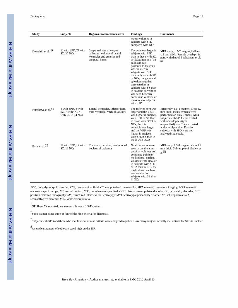
TABLE 1
Morphological Studies of SPD
Study Subjects Regions examined/measures Findings Comments
Schulsinger et al.84 33 offspring ofmothers with SZ (11with borderline SZ[probably SPD], 8with SZ, 14 mentallyhealthy)
Lateral ventricle, third ventricle,VBR
Subjects with SZ hadlargest ventricles,followed by mentallyhealthy subjects;those with borderlineSZ had the smallest
CT study; each measurementperformed on 1 slice. Subjects withborderline SZ had fewer obstetricalcomplications than did those withSZ.
Cannon et al.57 34 offspring ofmothers with SZ (12with SPD, 7 with SZ,15 unaffected)
Maximum width of sylvianfissure, width of anteriorinterhemi spheric fissure, meanwidth of 3 broadest cortical sulci,width of third ventricle, area oflateral ventricle, VBR,qualitative assessment ofcerebellar vermis
Enlarged thirdventricles correlatedwith lower heart rate18 y previously
CT study; each measurementperformed on 1 slice, sometimesusing calipers. Sample overlapswith Schulsinger et al.84 Authorsalso measured heart rate onenrollment in the study, 18 ypreviously. The 3 groups were notcompared on CT measures.
Raine et al.75 17 with schizotypalfeatures, but not SPD
Prefrontal cortex, temporal lobe High scores onschizotypy scalescorrelated withreduced left prefrontalarea and with high leftand rightprefrontal:temporallobe area ratios
MRI study, 0.15-T magnet; 12slices, 10 mm thick, with singleslices used for measures ofprefrontal and temporal cortex
Cannon et al.57 126 offspring ofmothers with SZ (31with SPD, 17 withSZ, 33 with anotherdiagnosis, 45 with nodiagnosis) and 77offspring of NCmothers (1 with SZ, 4with SPD, 26 withanother diagnosis, 46with no diagnosis)
Ventricles and sulci, VBR,sulcus:brain ratio, temporal andfrontal fissures, lateral ventricles
Offspring of motherswith SZ who had SZor SPD showed largersulci than did anyother group; offspringof mothers with SZwho had SZ had largerventricles than didany other group
CT study; 13 slices, 8 mm thick.The sample overlaps in part withthose of Schulsinger et al.84 andCannon et al.56
Hendren et al.78 25 children aged 8–12 y (12 withsymptoms of SPD orearly-onset SZ, 13NCs
Amygdala, hippocampus, part oftemporal cortex (slices on whichamygdala and hippocampuscould be visualized), corpuscallosum area, cerebellum area,frontal lobe area (on midsagittalslice), lateral ventricle, thirdventricle, temporal horn
Children withsymptoms of SPD orSZ had reducedamygdala andtemporal cortexvolumes, callosal area
MRI study, 1.5-T magnet; areameasures were performed on 1slice. MRS data were also reportedfor 9 subjects and 8 controls. Poorcomplex verbal memory was seenin children with symptoms of SPDor SZ.
Siever et al.74 36 with SPD, 23 withother personalitydisorders, 133 withSZ, 42 NCs
VBR, frontal and posterior hornsof lateral ventricle, thirdventricle
VBRs of subjects withSPD were higher thanthose of subjects withother personalitydisorders and wereintermediate between(but not significantlydifferent from) VBRsof NCs and those ofsubjects with SZ
CT study (2 different scannersused); slices 0.8–1.0 cm thick,depending on scanner. Some of thesubjects with SPD had beenexposed to neuroleptics.
Shihabuddin et al.82 11 individuals from asingle pedigree (2with SPD, 3 with SZ,6 unaffected)
Lateral ventricles, VBR, anteriorhorn, temporal horn, measure ofsulcal CSF by brain region
Affected subjects plus1 unaffected subject(all of whom carried agenetic marker forschizophrenia) hadlarger ventricles, ahigher VBR, andlarger CSF volumes infrontal and parietalregions than did the 5subjects not carryingthe marker
CT study; slices 8 mm thick. Datafor subjects with SPD were notanalyzed separately.
Harv Rev Psychiatry. Author manuscript; available in PMC 2010 April 13.
NIH
-PA Author Manuscript
NIH
-PA Author Manuscript
NIH
-PA Author Manuscript
Dickey et al. Page 18
Study Subjects Regions examined/measures Findings Comments
Buchsbaum et al.50 12 with SPD, 11 withSZ, 23 NCs
Lateral ventricles, anterior horn,temporal horn
Left anterior andtemporal hornvolumes in subjectswith SPD werebetween those of NCsand those of subjectswith SZ, differingsignificantly from thelatter
MRI study, 1.5-T magnet;* slices1.2 mm thick
Kwon et al.53 16 with SPD, 5 withsubthreshold SPD,†15 with chronic SZ,15 with first- episodeSZ, 16 with first-episode psychoticaffective disorder, 46NCs
Presence of large cavum septipellucidi
A large cavum septipellucidi was morecommon in subjectswith SZ than in NCs(found in 27.3% ofsubjects with SPD,35% of those with SZ,25% of those withpsychotic affectivedisorder, and 13% ofNCs)
MRI study, 1.5-T magnet.Appearance of cavum wasevaluated on 1.5-mm slices.
Silverman et al.83 11 with SPD (at least4 criteria),‡ 42 withSZ, 6 with SCA, 56unaffected relatives,22 NCs
VBR No difference wasseen between subjectswith SPD and thosewith SZ; subjects withSZ and SPD hadhigher VBRs than didtheir unaffectedrelatives
CT study; slices 8.0 mm thick. Oneslice used for comparison.
Dickey et al.54 16 with SPD, 5 withsubthreshold SPD,†14 NCs
Superior temporal gyrus,amygdala, hippocampus,parahippocampus
Subjects with SPDshowed reducedsuperior temporalgyrus volume anddifferences inparahippocampalasymmetry comparedwith NCs
MRI study; 1.5-T magnet; slices 1.5mm thick. Sample overlaps withthat of Kwon et al.53
Hazlett et al51 13 with SPD, 27 withSZ, 32 NCs
Thalamic volume,thalamus:brain ratio
No differences wereseen in thalamicvolume; shapeanalysis suggestedfewer pixels in theright mediodorsalnucleus in subjectswith SPD than in NCs,and fewer pixels in theleft anterior regions insubjects with SZ thanin NCs
MRI study, 1.5-T magnet;‡ slices1.2 mm thick. Study also includeda PET component, which showedno difference in thalamicmetabolism between subjects withSPD and NCs (although thesubjects with SZ had lower bilateralmetabolism in the mediodorsalnucleus of the thalamus than didsubjects with SPD or NCs).
Warwick et al.77 32 with sexchromosomeaneuploidies (11with 47 XXX, 10with 47 XYY, 10with 47 XXY), 38NCs§
Whole brain, R and L prefrontallobes, R and L temporal lobes, Rand L lentiform nuclei, R and Lcaudate nuclei, R and Lthalamus, R and Lamygdalohippocampal complex,lateral ventricles, third and fourthventricles
SIS scores forintroversion, magicalthinking, andimpulsivity did notcorrelate with wholebrain volumes for theXXX group: femaleswith XXX hadsmaller whole brainsthan did female NCs,males with XXY hadsmaller whole brainsand larger lateralventricles than didmale NCs.
MRI study, 1.0-T magnet. Studybased on reports of increasedincidence of schizotypal traits inpersons with sex chromosomeaneuploidies. Data for subjectsscoring high on the SIS were notanalyzed separately.
Dickey et al.55 16 with SPD, 14 NCs Lateral ventricles, temporalhorn, total CSF, cortical graymatter, total gray matter, whitematter
Subjects with SPDhad greater CSFvolumes (but notventricular volumes)than did NCs; a trendwas seen towardreduced cortical gray
MRI study, 1.5-T magnet; slices 1.5mm thick. Sample overlaps withthose of Kwon et al.53 and Dickeyet al.54
Harv Rev Psychiatry. Author manuscript; available in PMC 2010 April 13.
NIH
-PA Author Manuscript
NIH
-PA Author Manuscript
NIH
-PA Author Manuscript
Dickey et al. Page 19
Study Subjects Regions examined/measures Findings Commentsmatter volumes insubjects with SPDcompared with NCs
Downhill et al.49 13 with SPD, 27 withSZ, 30 NCs
Shape and size of corpuscallosum; volume of lateralventricles and anterior andtemporal horns
The genu was larger insubjects with SPDthan in those with SZor NCs; a region of thecallosum justposterior to the genuwas smaller insubjects with SPDthan in those with SZor NCs; the genu andsplenium togetherwere smaller insubjects with SZ thanin NCs; no correlationwas seen betweencorpus and ventricularmeasures in subjectswith SPD
MRI study, 1.5-T magnet;‡ slices1.2 mm thick. Sample overlaps, inpart, with that of Buchsbaum et al.50
Kurokawa et al.81 4 with SPD, 4 withSZ, 7 with OCD, 1with BDD, 14 NCs
Lateral ventricles, inferior horn,third ventricle, VBR on 3 slices
The inferior horn waslarger and the VBRwas higher in subjectswith SPD or SZ thanin those with OCD orNCs; the thirdventricle was largerand the VBR washigher in subjectswith SPD/SZ than inthose with OCD
MRI study, 1.5-T magnet; slices 1.0mm thick; measurements wereperformed on only 3 slices. All 4subjects with SPD were treatedwith neuroleptics (typeunspecified), and 2 were treatedwith clomipramine. Data forsubjects with SPD were notanalyzed separately.
Byne et al.52 12 with SPD, 12 withSZ, 12 NCs
Thalamus, pulvinar, mediodorsalnucleus of thalamus
No differences wereseen in the thalamus;pulvinar volumes andcombined pulvinar/mediodorsal nucleusvolumes were smallerin subjects with SPDor SZ than in NCs; themediodorsal nucleuswas smaller insubjects with SZ thanin NCs
MRI study; 1.5-T magnet; slices 1.2mm thick. Subsample of Hazlett etal.51
BDD, body dysmorphic disorder; CSF, cerebrospinal fluid; CT, computerized tomography; MRI, magnetic resonance imaging; MRS, magneticresonance spectroscopy; NC, normal control; NOS, not otherwise specified; OCD, obsessive-compulsive disorder; PD, personality disorder; PET,positron emission tomography; SIS, Structured Interview for Schizotypy; SPD, schizotypal personality disorder; SZ, schizophrenia; SZA,schizoaffective disorder; VBR, ventricle:brain ratio.
*GE Signa 5X reported; we assume this was a 1.5-T system.
†Subjects met either three or four of the nine criteria for diagnosis.
‡Subjects with SPD and those who met four out of nine criteria were analyzed together. How many subjects actually met criteria for SPD is unclear.
§An unclear number of subjects scored high on the SIS.
Harv Rev Psychiatry. Author manuscript; available in PMC 2010 April 13.
NIH
-PA Author Manuscript
NIH
-PA Author Manuscript
NIH
-PA Author Manuscript
Dickey et al. Page 20
TABLE 2
Comparison of Morphological Abnormalities in SPD with Those in First-Episode Schizophrenia and First-Episode Psychotic Affective Disorder
Abnormality SPD FE SZ FE AFF
Sulcal CSF increase Yes Yes No
Cortical gray matter decrease Trend Left No
Left STG gray matter decrease Yes Yes No
Posterior STG subdivisions decrease
Heschl’s gyrus Yes Yes No
Planum temporale No Yes No
Medial temporal lobe subdivisions decrease
Amygdala No Yes Yes
Hippocampus No Yes Yes
Parahippocampus Asymmetry Yes Yes
Large cavum septi pellucidi Yes Yes Yes
AFF, psychotic affective disorder; CSF, cerebrospinal fluid; FE, first episode; SPD, schizotypal personality disorder; STG, superior temporal gyrus;SZ, schizophrenia.
Harv Rev Psychiatry. Author manuscript; available in PMC 2010 April 13.