Embed Size (px)
Citation preview
THE BONE CHANGES OCCURRING INRENAL AND COELIAC INFANTILISM,AND THEIR RELATIONSHIP TO RICKETS
U PART I. RENAL RICKETS. ROYAL So5BY MEDJC;NE
LEONARD G. PARSONS, M.D., F.R.C.P., WITHORAWiPhysician to the Children's Hospital, Birmingham.
The occurrence of bone deformities associated with the presence of albuminin the urine was first observed by R. C. Lucas (1) who in the year 1883 publisheda paper entitled " A Form of Late Rickets associated with Albuminuria."A perusal of this paper, however, shows that the cases described were notexamples of renal infantilism. The interest of British paediatricians in renalinfantilism was first aroused by Morley Fletcher (2) who in the year 1911 showedbefore the Children's Section of the Royal Society of Medicine, a typical exampleof this condition occurring in a child who had developed genu valgum whenfive years of age. In a paper written by Miller and myself (3) in 1912 theoccurrence of genu valgum in several of the cases was commented on. Naish (4)-writing upon this subject in the same year said: " The presence of ricketsusually of late origin in as many as five out of the eight cases is a strikingphenomenon," but the credit for emphasising the importance and strikingcharacter of the bone changes is clearly due to Barber (5) who has published aseries of papers on renal dwarfism, and to whose description of the bone changesthere is, from a purely clinical standpoint, little to add. Barber regards thebone changes as typical of late rickets, but has not attempted any explanationof the reason why this should occur. Paterson (6) writing in 1920 discusseswhether the bone changes should be regarded as those of rickets or not, andinfers that in his view the condition is not rickets, but " would suggest thatthese bony changes are nutritional in origin." Shipley, Park, McCollum and8immonds (7 recording their observations on one case, suggest that the conditionmnay be a true renal rickets, whereas Lathrop (8) from the same medical school,,in a paper recently published, states that although the clinical appearances aredifficult to differentiate from rickets, the radiogram shows nothing suggestiveof that disease, and the blood calcium and phosphorus are distinctly outsidethe rachitic zone.
I have recently had the opportunity of investigating somewhat fully theclinical characteristics, and the chemical and radiographic findings of fivehitherto unpublished cases of renal infantilism with bone changes. Thesecases form the basis of the present communication in which an attempt is madeto classifv the bone changes and to explain their pathogenesis.
A6
on 3 June 2018 by guest. Protected by copyright.
http://adc.bmj.com
/A
rch Dis C
hild: first published as 10.1136/adc.2.7.1 on 1 January 1927. Dow
nloaded from

ARCHIVES OF I)ISEASE IN C'HILDHOOD
CLINICAL 1)ESCRIPTION.The clinical characteristics of renal infantilism,i. or (lwarfism. are now so
well known-i that, in a paper which concerns itself chiefly w%ith the bone deformi-ties, a lengthy description of the disease is not called for. It mav, however, beadvanitageouis to recall the salient points in its symptomatology. The childrenare always stuinte(l in growth and there is a delay in the appearance of secondarysex characteristics. Apart from the retardation of dlevelopment the mostcharacteristic symptoms are polydipsia and polyuria. often of extrenie degreean(l associated with nioctuirnal incontinence. The facies is frequently a tvpicalone, being sallow in colouir and characteristically Mwrinkled. Trhe uirine has a lowspecific gravity and tusutally contains a faint haze of albumin. Cardiovaseularchanges mav be, btut tusually are not, present. The age at the oiiset of thesyinptomiis varies. in quite a considerable proportion the symptoms appearto have beeil present from birth, whereas in. others the child was apparentlvnlormal for the first few years of life. The disease is a fatal one, death occurringfrom uraemia, usually in the second decade of life.
The autopsv findings have in the great majority of cases shown very smallkidneys the result of severe chronie interstitial nephritis? but cases are on recordassociated with pyelitis and apparently an ascending infection of the urinarytract, congenital cystic kidnevs and bilateral renal calculi. The end result is,however, identical in all cases, i.e., an almost complete destruction of the renalparenchyma.
Those cases in w\hich boneedeformities occuir are now usuially desigmiatedIrenal rickets." Genii valguin, w%hich mav (levelop very rapidly, is the
comnmoi)est manifestatioin of renal as of coeliac rickets. Enlargemnent of theepiphyses of the wrists and ankles may occur with or without genii v%algguin.In some instances bowing of the legs occurs, instead of gentu valgum, and inothers again the pictuire is that of severe rickets with well marked rosary.Harrison's sulcus, pigeon chest, enlargenment of epiphyses, genu valgum, etc.
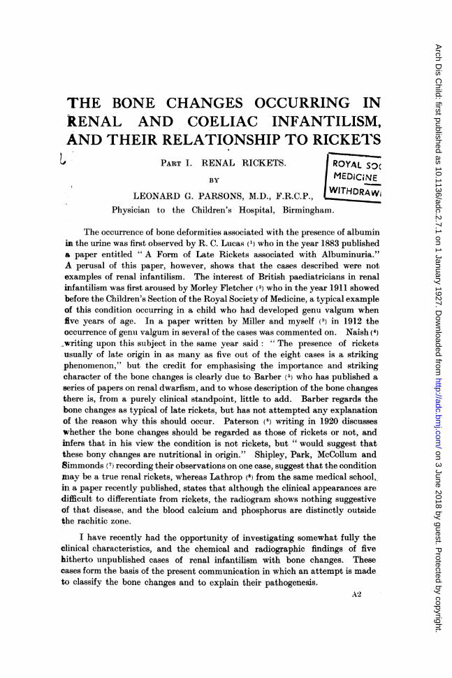
The most striking deformnities that I have seeni are those present in H.T.,of whonm a photograph is shown (Fig. 1). Trhis box is seventeen years of age,has a very marked rosary, a deformed chest, enlargement of the epiphyses, andextraordinary bending of the femora. tibive and fibulae. In some cases there(loes not appear to be any bending of the shafts of the long bones, but theepiphyses are enlarged, and there is obvious bending or displacement of theetnlarged epiphvseal en(d of the bone, the explanation of which will appear lateroil ill this paper.
Trbe ages of the children at the onset of the borie chainges varies considerably.Barber states that the uisual time for genii valgum to develop is between eleveniand fourteen years of age. The earliest age at which I have seen bone changesis sixteen months. Paterson has described a case at twenty-one months, andalso one in which. accordinig to the parents' history. the deformities were presentat birth (9). On the other hand there are cases oIn record in which the bone(leformities have niot been recognised until the age of seventeen years. In m,ycases the onset has been earlier. all having occurred before the age of ten years,the usual time of onset being from five to seven ,years.
on 3 June 2018 by guest. Protected by copyright.
http://adc.bmj.com
/A
rch Dis C
hild: first published as 10.1136/adc.2.7.1 on 1 January 1927. Dow
nloaded from
BONE CHANGES IN RENAL AND COELIAC INFANTILISMl
I
Fig. 1.-Photograph of H.T., aged 16 years, showing the severe nature of the deformitieswhich may occur in renal rickets.
3
:, a,:.
!,Fo,iooo"r::: :.
:i
on 3 June 2018 by guest. Protected by copyright.
http://adc.bmj.com
/A
rch Dis C
hild: first published as 10.1136/adc.2.7.1 on 1 January 1927. Dow
nloaded from
ARCHIVES OF DISEASE IN CHILDHOOD
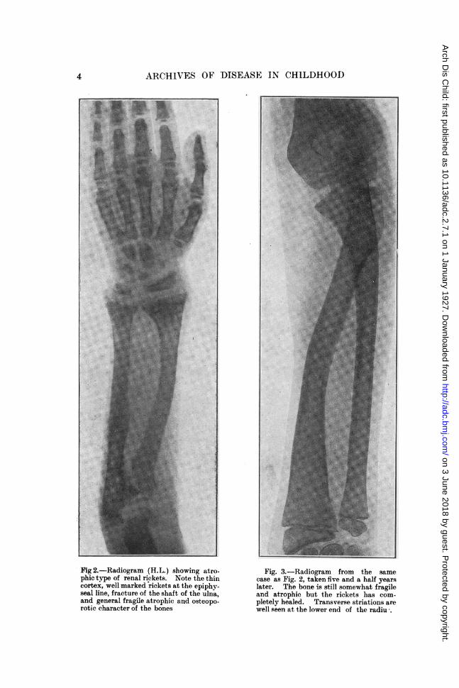
Fig 2.-Radiogram (H.L.) showing atro-phic type of renal rickets. Note the thincortex, well marked rickets at the epiphy-seal line, fracture of the shaft of the ulna,and general fragile atrophic and osteopo-rotic character of the bones
Fig. 3.-Radiogram from the samecase as Fig. 2, taken five and a half yearslater. The bone is still somewhat fragileand atrophic but the rickets has com-pletely healed. Transverse striations arewell seen at the lower end of the radiu.
4
on 3 June 2018 by guest. Protected by copyright.
http://adc.bmj.com
/A
rch Dis C
hild: first published as 10.1136/adc.2.7.1 on 1 January 1927. Dow
nloaded from

BONE CHANGES IN RENAL ANI) COELJAC INFANTILISM
RADIOGRAPHIC MANIFESTATIONS.A study of radiograms of renal rickets shows that the bone changes fall
into three well defined groups, which I propose to call1. The atrophic tvpe.2. The florid tvpe.3. The woollv, stippled, or honeycomb type.
1. The Atrophic Type.In this group the radiogram (Fig. 2) presents a picture strikingly similar
to that seen in coeliac rickets of a moderate degree. The whole bone presentsa fragile, atrophic, osteoporotic appearance. Near the epiphyseal end of thediaphysis there may be seen one or two straight lines of cancellous tissue, runningparallel to the epiphyseal line. The cortex is also thin and atrophic. A fracture,or fractures of the shaft may be present, but these are not so commonly seenas in coeliac disease, and finally there is well marked rickets at the epiphvsealline.
2. The Florid Type.The appearances in this group are those characteristic of florid rickets.
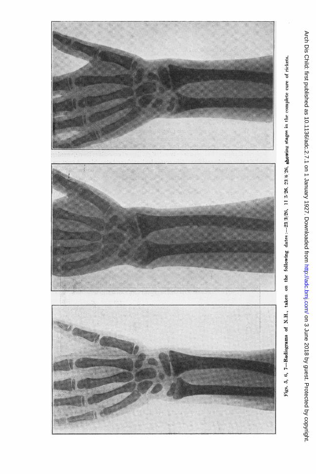
The shaft may perhaps be a little more fragile than that of a normal child of thesame development, buit not sufficiently so to call for comment, whilst the changesso characteristic of the atrophic type are absent. (Figs. 4, 5, 6, 9, 10.)
Fig. 4.-Radiogram of N.H., taken Jain. 30th, 1926, showing florid type of renal rickets.
3. The Woolly, Stippled, or Honeycomb Type.This is the most extraordinary manifestation of renal rickets. It is quite
unlike the picture usually associated with rickets, and yet is so characteristicthat a radiographer who has had experience of these cases can make the diagnosisof renal rickets from the radiogram. (Figs. 11, 12, 14, 15.) In this type the
on 3 June 2018 by guest. Protected by copyright.
http://adc.bmj.com
/A
rch Dis C
hild: first published as 10.1136/adc.2.7.1 on 1 January 1927. Dow
nloaded from
144ce
00
M.
Od
CZ
l)
to
4-
0
4)1
c:
es
Cl-*1*. .
"3
.0
4)
r4
I
1
on 3 June 2018 by guest. Protected by copyright.
http://adc.bmj.com
/A
rch Dis C
hild: first published as 10.1136/adc.2.7.1 on 1 January 1927. Dow
nloaded from

BON'E CHANG'ES IN RENAL AND (OELIAAC INFANTILISM 7
Fi-. S. Itadiogramn ot N.H., takeii oni 30/8/26, shiowing stage in thJe complete(lire (f riekets.
epiphyseal enl(l of the shaft of the bone is swollen, forming a large metaphvsis.This portioni of the bonee presenits a curiouis appearance Mwhich sometimessuggests a honeycomb, at other times shows marked stippling, whilst at othersit has a woolly appearanice, the bone looking moth-eaten as if the shaft werebeinig eaten away subperiostallv and giving at first sight the suggestion of osteo-myelitis or syphilitic disease. The metaphysis extends over a much greaterarea than in ordiinary rickets. Associated with these appearances is a degreeof osteoporosis throuighout the shaft of the bone,but the cortex does not appearthin as in the atrophic type. The condition of the shaft in the atrophic type isone which suggests that the bone has itever been anvthiing else but atrophic,whereas in the woolly type it is conceivable that osteoporosis has occurred in abone which at some previous time might have approached the normal.
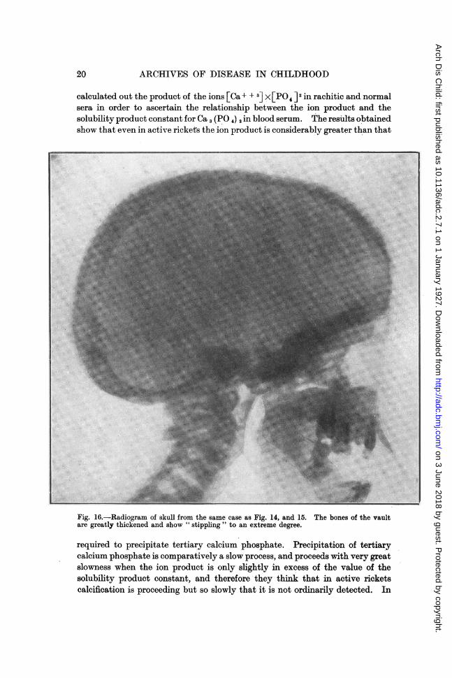
The bones of the vault of the skull sometimes show changes and in the caseof H.T., these are most marked. (Fig. 16). These bones are greatly thickenedand show extreme stippling presenting an appearance in the radiogram quiteunlike anything with which I am acquainted with the exception of Paget'sdisease. Indeed my colleaguie, Dr. Teall reported on this case as follows :-the bone change in the skuill is, from the radiographic point of view, identical
with that of Paget's disease."The changes do not ocetr to the same (legree in all the long bones, e.g.,
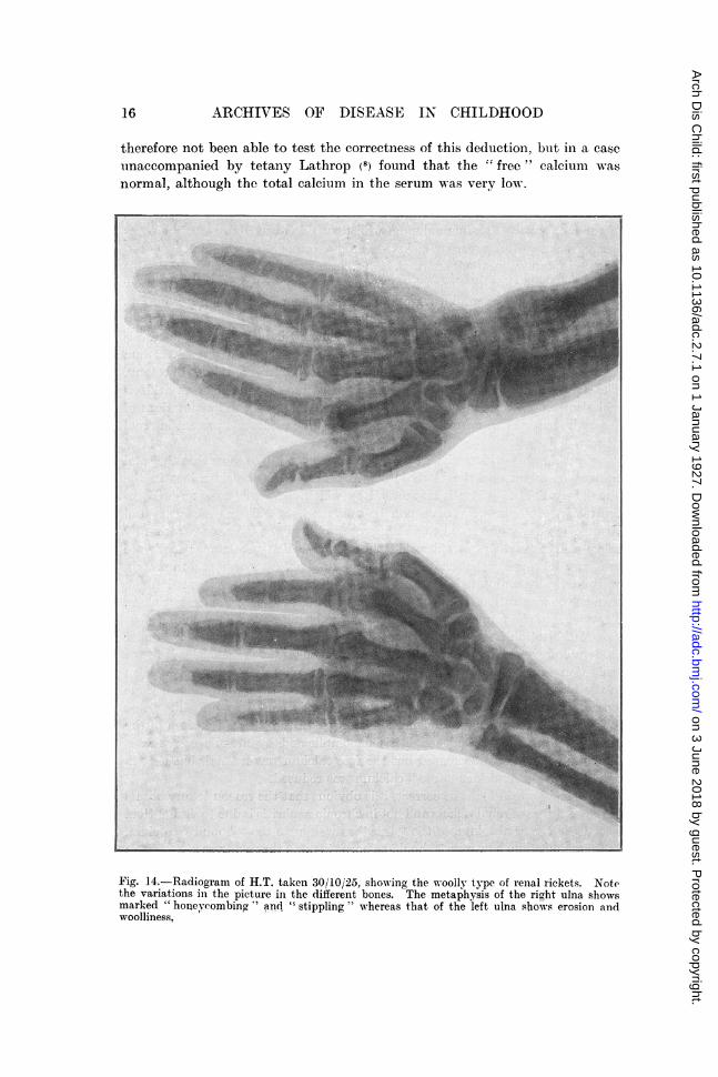
the radiuis and iulna of one arm mav show changes muich more marked than thoseof the other sidle. Thuis the mnetaphysis on one side may be larger and showmore stippling, whereas on the other, it may show a more erode(d appearancean(d not so mutchi enlargement. (Fig. 14).
l7
on 3 June 2018 by guest. Protected by copyright.
http://adc.bmj.com
/A
rch Dis C
hild: first published as 10.1136/adc.2.7.1 on 1 January 1927. Dow
nloaded from
All(HIrfV'ES OF D)1SEASE IN (CHILDHOOD
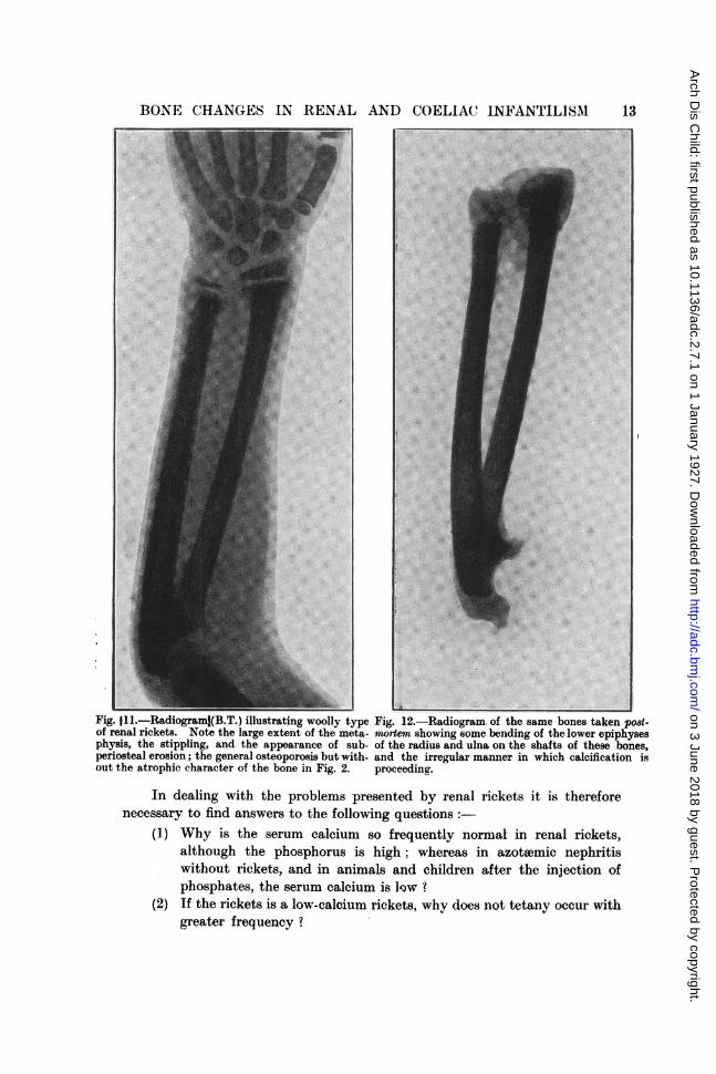
I have recently had the opportunity of examining the bone changes in acase (B.T.) the antemortem radiograms (Fig. 11) of which showed the woollytype of bone deformity. Such an examination together with radiograms of thebones taken after death (Fig. 12) showed that the woolliness or stippling was dueto irregular calcification and the presence of islets of cartilage deep down in themetaphysis. The proliferative cartilage was much more irregular and brokenlip than in ordinary rickets. The metaphysis was deeper, and although itshowed broadening or splaying out, the degree of broadening was not so greatas in uincomplicated rickets. As a result of the presence of these islets ofcartilage deep in the metaphysis (Fig. 13), the whole metaphysis was bent onthe diaphysis and this, together with bending of the shaft as a result of osteo-porosis in the neighbourhood of the wrists and ankles, resulted in a deformitvat these points which gave the epiphvsis an appearance of being larger than itwas in reality.
In the course of a paper based on three cases which conforam to my woollvtype, Paterson (6) describes the occurrence of fractures on the diaphyseal sideof the epiphyseal line, and suggests that the stippled " appearance is due tothe throwing ouit of calliis in an attempt to repair such fractures. Whilst theoccurrence of fractures in this situation is obviously possible owing to thesoftness of the tissue, yet up to the present time I have not observed them inany of my cases and the stippling is due, as already explained, to the irregularityof calcification in the metaphysis. In a description of a case of renal infantilismwith bone changes of the woolly type, Shipley (7) pointed out that thechanges in the bones revealed bv X-ray and histology were most extensiveand differed from the changes ordinarily found in rickets. "The proliferativecartilage was most irregular and the calcification defective. In the deepmetaphysis were large islands of cartilage bordered on one or more sides by densecalcium deposits. These islands of cartilage gave to the metaphysis as seen inthe Rdentgenogram a honeycomb appearance. The trabecuhe were thin,and the osteoi(d borders comparatively speaking narrow. Surrounding thetrabeculse aiid islands of cartilage, and lying between them, was a loose fibroustissue. Obviouslyfor a long time the pathological condition in the bone had beenin a state of fluix between healing and relapse." They have suggested thepossibility that this condition may have " its origin in the extreme functionaldisability of the kidney properly to excrete phosphate," and that if this sug-gestion " should prove to be correct a true renal rickets exists, and rickets undercertain conditions, may have an endogenous origin." I believe this suggestionto be correct. As evidence against the rachitic origin of the bone deformityPaterson mentions the absence of bowing of the bones in one of his cases, a childaged twenty months, but as I have already pointed out,extreme bending of thebones (toes occur in the woolly type. He also thinks that the histologicalchanges in the bone are not those of rickets, but his description of these changesis almost identical with that I have quote(d from the paper by Shipley and hisco-workers. These investigators, whose experience of experimental rickets isuinrivalled, accept the view that the changes are rachitic in nature and thatthey " may belong to the low calcium form of the disease."
X
on 3 June 2018 by guest. Protected by copyright.
http://adc.bmj.com
/A
rch Dis C
hild: first published as 10.1136/adc.2.7.1 on 1 January 1927. Dow
nloaded from
BONE CHAN(hU,I8 IN RENAL ANI) (COELlAC INPANTILISm 9.
Sections of the bone in thecase (B.T.) to which I havejust referred shows a conditionsimilar to that described byPaterson and bv Shipley. Ihave referred these sections toProfessor Hasuwell Wilson, Pro-fessor of Pathology in theUniversity of Birmingham,who considers that althoughthe changes present differ fromthose ordinarily seen in ric-kets, yet they are definitelyrachitic in natuire.
Discussion. There appearsno explanation why one caseshouild show florid rickets,another atrophic, and vetanother the woolly type,though the last is, I think,the most serious form of thedisease. Again, although thereseems no reason whv the floridshould not change into theatrophic type, yet in myexperience all three typesalways breed true, and thereis never any changing fromone type to another. nor doesone long bone show onetype of change, and anotherbone in the same child achange of the other types.The cases reported by Barberappear to have been instancesof florid rickets, and he doesnot seem to be familiar withthe atrophic or woolly types.
From a study of my casestwo striking, if apparentlycontradictory results, have
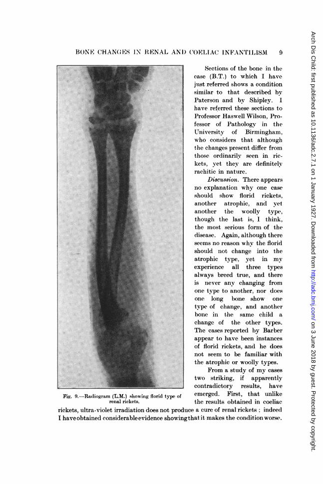
Fig. 9.-Radiogram (L.M.) showing florid type of emerged. First, that unlikerenal rickets. the results obtained in coeliac
rickets, ultra-violet irradiation does not produce a cure of renal rickets: indeedI haveobtained considerableevidence showingthat it makes the condition worse.
L "
.- .:"
on 3 June 2018 by guest. Protected by copyright.
http://adc.bmj.com
/A
rch Dis C
hild: first published as 10.1136/adc.2.7.1 on 1 January 1927. Dow
nloaded from
ARCH IVES OF DISEASE I-N CHILDIJOGI)
Secondlv, that the atrophic and florid types of renal rickets may show completerecovery. Recovery in the atrophic type results in the production of a bone,whose radiogram resembles that seen in mil(d coeliac disease before the productionof rickets, i.e., a somewhat fragile and osteoporotic bone with a thin cortex,showing one or more transverse lines, in the neighbourhood of, and runningparallel to the epiphvseal line, the latter being perfectly sharp and straighlt.The reproduced radiograms of H.L. (Figs. 2 and 3) show the atrophic type ofremial rickets, before and after cure, the first radiogram being taken fouir yearsago when he was twelve years of age the second recentlv. He is now sixteenyears of age andI his condition is astonishingly good. Recovery in the floridtype is well shown in the series of radiograms of N.H., a girl aged ten years.(Figs. 4, 5, 6, 7, 8.) 1 have not seen any case in which ossificationi is complete,since none of my cases have lived beyond seventeen years, but in Barber's lastpaper there are radiograms of a girl aged twenty who had been the subject offlorid rickets, buit in whom the epiphyses have joined up so that ossification iscomplete, anid as far as can be judged from the reproduction of the radiogramsthe bone is normal. From the quotation given above from Shipley's paper, itis obvious that even in the woolly tvpe, attempts at healing take place fromtime to time.
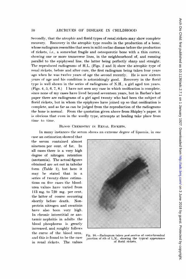
BLOOI) CHEMISSTRY IN RENAL RICKETS.
In many instances the sertum showis an extreme (legree of lip.-emia, in onecase an estimation showed thatthe sertum containe(l almost [ :1nineteen per cent. of fat. Inall cases there is a very high(legree of nitrogen retention(azotoemia). The actuial figuresobtained are set ouit in tabuilarform (Table 1), but here itinav be stated that in aseries of twenty-three estima-tionis oin five cases the bloo(d-urea values have varie(d from113 mg. to 720 mg. per cent,the latter of course occuirringshortlv before (leath. Non-p)rotein nitrogen andl creatininhave also beeni very high.In chronic interstitial or azo-thmic nephritis in a(lullts thel)lood phosphoruis is greatlyrincreasedl aind roughlv followsthe cuirve of the bloodI urea,
Fi&. i10.--Radiogram takeln post-mnortem of costochlrondtandltl thi.s i.s foundl to b)e the? ca.se junci:tion of ril) of L.M., showing the tvpical appeararin renal rickets. The values of florid rickets.
ralnee
Ilo
on 3 June 2018 by guest. Protected by copyright.
http://adc.bmj.com
/A
rch Dis C
hild: first published as 10.1136/adc.2.7.1 on 1 January 1927. Dow
nloaded from

BONE CHANGES IN RENAL AND COELIAC INFANTILISM 11
TABLE I.C
o. I0n
CoV
N.H. ? aged 26/2126 11710- years.
8/4/26 180
16/4/26 138-3
191/5/26 150
29/6/26 137 78-0 1-5 485
15/9/26 226-5 115-4 1-3 548
1/11/26 178-6
17/11/26 111-5 79-0 1-63 574
A.N. & aged 13/4/2616 months.
17/4/26
23/6/26 109-2 54-1 1-27 554
1/7/26 117-4 55 1-5 594
25/9/26 191-6 96-8 1-7 601
28/101/26 173-3 85-7
23/11/26 208-3 115-2
B.T. & aged10 years.
14/5/26
19/5/26
27/5/26
339
407-5
720-0
H.T. c aged 21/9/26 136-516 years.
11/9/26 135
26/9/26 113-6
19'10!26 112-9
10/1!27 134-7
14/1/27 134-7
L.M. (3 aged 30/11/26 673.816 years.
II
This table gives the details ofmilligrammes per cent.
370
110
105
59.4
93-8
1-5
1-4 643
706-9
7-73 545
Q Co
10-0 13-9*
11-7 6-0
9-0 6-1
11-1 5-5
10-3 6-5
10-09 5-58
9-12 5-23
8-6 7-3
9-0
7-7 7-1
Q4)
Florid rickets.
Rickets healing.
Rickets healed, recentlyrecovered from an at-tack of vomiting(? urwmia).
Woolly type rickets.Serum contained 18-97% of fat.
9-,
9-1
9-
8-2
7-3
7.3
2-0 561 12-7
14-3
10-6
13-7
8-5
1-9 561 10-0 4-7
1-4 10-4 4-1
1-4 14-6 6-35
: 13-0 10-07
1-856597 12-7
5.9 425-7 3-95 16-3
Woolly type rickets.Uremia.Bloodtakena few hoursbefore death,
Woolly type of rickets.rreviousiy treatec nyultra violet light, fallin phosphorus on cessa-tion of this treatment.
nH 7.16 . nrtroidut of
[Ca++]3 X [PO 4-'is 23.40.
Florid rickets ureemia-death 2/12/26.
blood chemistry findings, the values are all expressed as
1-6
on 3 June 2018 by guest. Protected by copyright.
http://adc.bmj.com
/A
rch Dis C
hild: first published as 10.1136/adc.2.7.1 on 1 January 1927. Dow
nloaded from

AR( HIVTES OF DISEASE IN CHILDHOOD
for serumii phosphorus (normal, 3 mg. per cent.) have varied between 4-1 mg.and 16-13 mng. per cent., w-hereas those for serum calcium (normal, 9-10mg. percent.) have varied from 3-95 mg. to 14-6 mg. per cent. The blood calcium of3-9Imug. per cent. occurred shortly before death, an(d is by far the lowestfigure obtained, the next lowest figure being 7-3 ing. per cent. I w-ould,however, emphasise the fact that in only six out of twenty-Qne estiimiations wasthe calcium under 9'0 mug. per cent., but that in every patient the calcium w-aslow, when compared with the amount of blood phosphorus.
Further evidence in favour of the view that in renal rickets the serumcalcium functions as low calcium, although the actual figures may bc normalor even higher than normal, is found in the fact that some of these cases arecomplicated by tetany. Tetany does not occur with such frequency as in coeliacrickets, but nevertheless it is a well recognised complication of renal rickets.
Now the combination of tetany with rickets is always associated with a lowserum calcium, and the onset of tetany is dependent not only on the totalamount of calciuiii but also on the amount of calcium ions. The work of Loeb,Matthews and others (10) sho-s that the excitability of the neuro-muscular
Na vt Kmechanism varies directly with the ratio -+ Mg and that increase(l irritabilitycan theoretically occur, either from a decrease in calcium, or an increase insodium ions. In infantile tetany the increased irritability is due to a decreasein calcium ioIns for the magnesium, sodium, and potassium in the serum areessentially normal (Kramer, Howland, and Tisdall (11).)
The problem of the occurrence of tetany in renal rickets is, how-ever, evenimore complicated because the following additional data have to be considered.The injection of phosphates into animals and children has been found to raisethe blood phosphorus, lower the blood calcium, and in some cases producetetany. Binger (12) found that the injection of normal or alkaline phosphatesinto dogs produced all these results, but acid phosphates on the other hand,whilst lowering blood calcium and raising blood phosphorus, did not producetetany. Klercker and Odin ( 13) administered acid and normal phosphatesto children, with resulting lowering of calcium increase of phosphorus, and anincrease of electrical excitability. In those cases in which latent tetany waspresent the administration of these salts produced acute tetany. In chronicazotaemic nephritis there is a retention of phosphorus with the production ofa high blood phosphorus, but the blood calcium is low. Thus in a recentinvestigation, Boyd, Courtney, and MacLachlan (14) have found consistentlylow calcium values in such cases, and De Wesselow (15) has shown that the higherthe retention of phosphorus in nephritis, the lower is the calcium in the serum.Although this inverse relationship does not hold in renal rickets, yet it seemsvery clear that the mere presence of a high blood phosphorus means a lowcalcium, and the consequent production of tetany unless some mechanism isbrought into play to prevent its occurrence.
I-')
on 3 June 2018 by guest. Protected by copyright.
http://adc.bmj.com
/A
rch Dis C
hild: first published as 10.1136/adc.2.7.1 on 1 January 1927. Dow
nloaded from
BONE CHANGES IN RENAL AND COELIA(' INFANTIL1SMI 13
Fig. t11.-Radiograml(B.T.) illustrating woolly type Fig. 12.-Radiogram. of the same bones taken pO8t-of renal rickets. Note the large extent of the meta- mortem showing some bending of the lower epiphysesphysis, the stippling, and the appearance of sub- of the radius and ulna on the shafts of these bones,periosteal erosion; the general osteoporosis but with- and the irregular manner in which calcification isout the atrophic character of the bone in Fig. 2. proceeding.
In dealing with the problems presented by renal rickets it is thereforenecessary to find answers to the following questions:-
(1) Why is the serum calcium so frequently normal in renal rickets,although the phosphorus is high; whereas in azotsemic nephritiswithout rickets, and in animals and children after the injection ofphosphates, the serum calcium is ow?
(2) If the rickets is a low-calcium rickets, why does not tetany occur withgreater frequency?
- I
I on 3 June 2018 by guest. Protected by copyright.
http://adc.bmj.com
/A
rch Dis C
hild: first published as 10.1136/adc.2.7.1 on 1 January 1927. Dow
nloaded from

ARCHIVES OFIDISEASE IN CHILDHOOI)
(3) Why (toes tetany occulr in this form of nephritis andc not in otherform of chronic nephritis
(4) What is the evidenee that the bonie deformiiities are of rachitic originand why does cure sometimes occur ?
We have seen that, contrary to what usually ol)tains in azotiemic nephritisand after the injections of phosphates, in renal rickets with high phosphorusthe serunm calcium may be normal or even higher than normal. It seemsconceivable that the serum calcium is maintaiined at this level to preventthe occurrence of tetany, wkhich is stuch a verv seriouis complication. andl towhich the young and growing child is much more prone than the adult. Nowthe rise in serum calciuim p)roduced by the injection of parathyroi(d hormone illtetaniy parathyropriva and in normal aninmals is dute to washing out of calciuifrom the bones (Green-ald and Gross (l6)). It is therefore not improbable thatin renal rickets there is a washing out of calcium from the bonles with resultingosteoporosis and rickets and the maintenance of the blood calcium at such alevel that tetany shall not occulr.
Epiphysis
CartilageEpiphyseal disc of
Cartilage
Rachitic MNletaphysis
Fig. 1 3.-Drawing of the antero-posterior section of the lower end of the radius, seen in the twopreceding radiograms (Figs. 11 & 12) showing the depth of the rachitic metaphysis, the presenceof islets of cartilage deep in the metaphysis, and the bending of metaphysis and epiphvsis on theshaft of the radius.
14
on 3 June 2018 by guest. Protected by copyright.
http://adc.bmj.com
/A
rch Dis C
hild: first published as 10.1136/adc.2.7.1 on 1 January 1927. Dow
nloaded from

BONE CHANGES IN RENAL AND COELIAC INFANTILISM 15
Recent work by Ross and Scriver (17) has demonstrated the possibility ofsuch mobilisation of calcium from the bones. These workers have shown thatin cases of infantile tetany, ammonium chloride, by virtue of its acid producingeffect, causes a mobilisation of calcium from the tissues and presumably fromthe bones. According to Kramer (18) their results furnish an explanationof the fact that ammonium chloride, whilst capable of restoring calcium to itsnormal value in those cases of rickets in animals and children in which the cal-cium is low and phosphorus normal, is yet, contrarv to expectation, not capableof producing healing of the rachitic process. The observations, indeed, showthat such treatment would aggravate rather than ameliorate the condition.
De Wesselow and others have shown that in severe nephritis, there isusually a diminution in the bicarbonate content of the plasma and an acidosisand that these favour the ionisation of "the diminished amount of calciumpresent 16),and so probably prevent the more frequent occurrenee of tetany.The observations of Ross and Scriver show that the occurrence of an acidosiswill also mobilise calcium ions from the calcium reservoirs,and estimations ofthe hydrogen-ion concentration show that a definite acidosis is present in renalrickets.
Considerable evidence has recently been forthcoming to show that the totalcontent of calcium in the blood is not an index of the calcium ions in thecirculatory fluids. There is no method by which the actual calcium-ion con-centration of serum or plasma can be measured, but Pincus, Peterson andKramer (220) have attempted to obtain some index of the ion concentration byusing the method of ultraffitration through collodion sacs under moderatepressure. Assuming that thI calcium which does not filter is bound to proteinand practically un-ionised, and that all the calcium that does ifiter is ionised,then the concentration of calcium in the ifitrate is a measure of ionised calcium.They refer to the former as " bound " and the latter as " free " calcium.
An objection has been urged against the method of ultraffitration undermoderate pressure, that removal of the calcium ions by such means leads to theionisation of the previously un-ionised calcium phosphate held in solutionin the plasma, but this obviously does not invalidate the assumption of theseworkers that the calcium which does not filter is bound to protein. Theyfound that in tetany there is a marked decrease in the " free " calcium in theserum, and that in chronic nephritis uncomplicated by uraemic convulsionsthe total calcium was decreased but the free calcium was normal, but that inone case with convulsions " free " calcium was reduced.
If the view of Pincus be correct, it is obvious that the reason tetany occursin the nephritis of renal rickets and not in chronic nephritis is due to diminution,either relative or absolute, in the " free " as well as in the " bound " calcium.In this connection, however, it should be borne in mind that a degree of azot*emiacomparable with that occurring in renal rickets,while not infrequent in adults,is of extreme rarity in childhood apart from renal rickets, and that the childis more liable to show tetany than the adult. I have not had the opportunityof estimating the calcium in the case of renal rickets with tetany and have
on 3 June 2018 by guest. Protected by copyright.
http://adc.bmj.com
/A
rch Dis C
hild: first published as 10.1136/adc.2.7.1 on 1 January 1927. Dow
nloaded from
ARCHIVES OF DISEASE IN CHILDHOOD
therefore not been able to test the correctness of this deduction, but in a caseuinaccompanied by tetany Lathrop (8) found that the " free " calcium wasnormal, although the total calcium in the serum was very low-.
Fig. 14.-Radiogram of H.T. taken 30/10/25, show:ing the woolly type of renal rickets. Notethe variations in the picture in the different bones. The metaphysis of the riaht ulna, shlowsmarked "hoiieveombin" and "stippling;" whereas that of the left ulna shows erosion andwoolliness. o i an "tn
16
on 3 June 2018 by guest. Protected by copyright.
http://adc.bmj.com
/A
rch Dis C
hild: first published as 10.1136/adc.2.7.1 on 1 January 1927. Dow
nloaded from
BONE CHANGES IN RENAL AND COELIAC INFANTILISM 17
WHAT IS THE EVIDENCE THAT THE BoNE DEFORMITIES ARE TRUF, RICKETS ?
The evidence, so far presented, that the condition is a truly rachitic one inthe atrophic and florid types is based mainly on the results of X-ray examinationsof the bones which show appearances at the epiphyseal line indistinguishable,from those of rickets. The metaphysis is on the whole somewhat deeper than
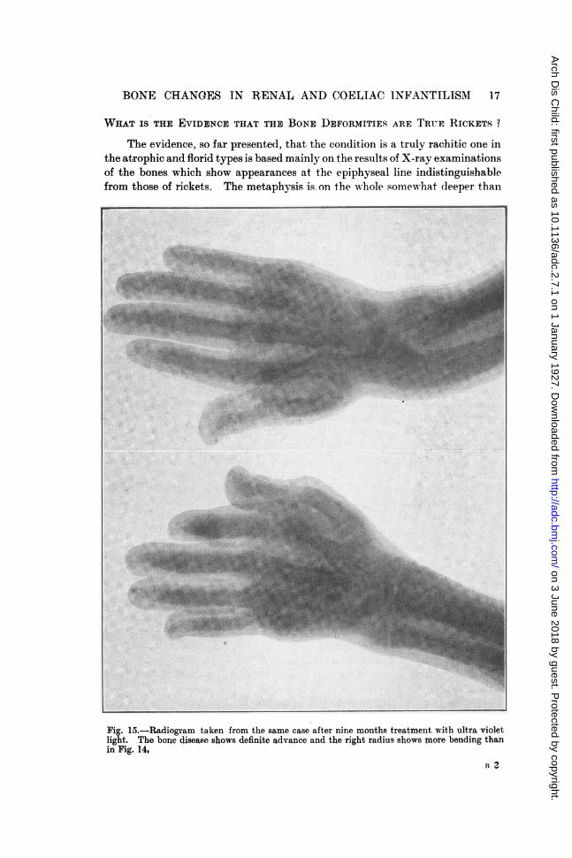
Fig. 15.-Radiogram taken from the same case after nine months treatment with ultra violetlight. The bone disease shows definite advance and the right radius shows more bending thanin Fig. 14,
on 3 June 2018 by guest. Protected by copyright.
http://adc.bmj.com
/A
rch Dis C
hild: first published as 10.1136/adc.2.7.1 on 1 January 1927. Dow
nloaded from
ARCHIVES OF DISEASE IN CHILDHOOD
in ordinary low-phosphorus rickets, but the rachitic nature of the change wouldbe admitted by anyone conversant with the X-ray findings in rickets. Histolo-gical evidence proving that the florid type of deformity is rachitic in nature hasalso been obtained in one of my cases (L.M.) This boy who showed markedinfantilism, genu valgum, enlargement of the epiphyses at the wrists, a definiterosary, and rickets of the florid type in the radiogram, developed a fatal attackof urwsmia shortly after admission to hospital. (Figs. 9 and 10). The autopsywas performed by Professor Haswell Wilson who reported on the histologicalexamination of the bones as follows :-" Sections were prepared from a costo-chrondral junction, and from the lower epiphyses of the right radius. Thechanges seen in these two situations are essentially of the same nature. Theline of ossification is broad and irregular and very little calcification is presentin the matrix of the cartilage. The columns of cartilage cells are distorted andbroken up by irregular masses of vascular osteoid tissue. Much fibrous tissueis seen between the trabeculae of osteoid tissue and of the spongy bone beyondit, and in this a few detached islands of cartilage are lying. The histologicalpicture is indistinguishable from that of ordinary rickets.'
With regard to the woolly type, the histological evidence already detailedand the radiograms show that the similarity between this type and experimentallow-calcium rickets cannot be doubted. The results of blood chemistry alsoshow that in all types when bone deformities are present, the calcium can beregarded as functioning as a low blood calcium.
There are two chief theories on the mechanism of the deposition of calciumphosphate as bone; that of Howland and that of Robison, the former. beingperhaps the more usually held. According to Howland, the rachitic bone tissueremains uncalcified just so long as calcium and phosphorus are supplied to itin such low concentrations that precipitation of tertiary (or normal) calciumphosphate cannot take place. The whole process can indeed be predictedwith mathematical accuracy. Normally the serum and circulatory fluids aresaturated solutions in which the calcium phosphate of bone is in equilibrium withthe dissolved salt, and this in turn with calcium and phosphate ions, and thismay be represented as follows
[Ca++]3 + [POs12 + Ca3(PO4)2 _ Ca3(PO4)2Ionised, In solution unionised. Solid in bone.
An increase of Ca + + or PO-4 causes the reaction to proceed to the rightand results in deposit of bone, whereas a decrease in concentration wouldresult in some of the solid calcium phosphate in bone going into solution, thereaction proceeding to the left. Now according to mass actiola law, atequilibrium the product of the concentration on one side divided by the productof concentration on the other side is constant at constant temperatures andtherefore we have
[Ca++]3 X [PO=]2 = K [Ca3 (PO4)2].Tertiary calcium phosphate is a sparingly soluble salt and in such
[Ca++]3 x [PO4]2 = K. This K is called the solubility-product constantand whenever the product of the concentration of calcium and phosphate
18
on 3 June 2018 by guest. Protected by copyright.
http://adc.bmj.com
/A
rch Dis C
hild: first published as 10.1136/adc.2.7.1 on 1 January 1927. Dow
nloaded from

BONE CHANGES IN RENAL AND COELIAC INFANTILISM 19
ions becomes greater than the solubility product solid calcium phosphate willbe deposited.
The occurrence of carbonic dioxide in considerable concentrations in theserum has, however, an influence on tertiary calcium phosphate which may beexpressed thus:-
Ca8(PO4)2 +2H2CO3-__ 2 Ca HPO4+Ca(HCO3)2. [Ca++]2+
[HPO-] 2+[Ca++] 2+[HCO3 12.If the CO 2 tension of this solution is reduced, the reaction proceeds to the
left and calcium phosphate is deposited. Howland suggests that becausecartilage and the trabeculae of bone are inactive tissues, it is to be expected thatthe CO 2tension would be low in them, and it is in them that precipitation occursif the solubility product be exceeded. The truth of the foregoing generalprinciples can, he states, be proved by clinical observations, using as factorsfor a rough product the total concentrations of calcium and phosphorus inserum, each being expressed in milligrams per 100 c.c. This product in thenormal child is between fifty and sixty. "When the product is below thirtvrickets is invariably present. When it is above forty either demonstrable healingis taking place, or there has never been any rickets. With products betweenthirty and forty rickets is usually present (21)."
Robison on the other hand, does not agree that the deposition of calciumphosphate is purely a physico-chemical process, such as is outlined above.His hypothesis may briefly be summarised as follows (Kay (22)) "the osteo-blasts and the hypertrophic cartilage cells of the young animal secrete a veryactive phosphatase which, by hydrolysing the phosphoric esters of the blood,brings about a local increase of PO" concentration. The solubility productof tertiary calcium phosphate is thereby exceeded and the deposition of thissalt occurs in the ossifying zone."
It is of interest to note that Howland (23) in his last paper on thissubject, written in collaboration with Shipley and Kramer, published shortlybefore his death, appeared to regard the physico-chemical theory as not thecomplete explanation since he concludes: "the process is clearly not oneof simple precipitation, it depends upon the activity of living tissue. Itcannot occur unless the concentration of calcium and phosphorus inthe serum and presumably in the fluid bathing the cells, exceeds a certainminimum value."
Now it is.clear that in all my cases on most occasions the product Ca x Pis greatly in excess of fifty, and for this reason Lathrop will not accept the viewthat the bone deformities of renal infantilism are true rickets. The co-existenceof bone c-hanges-which in my view are undoubtedly rickets-with a Ca x Pproduct of over fifty may be difficult to explain, but the occurrence cannotbe denied, and therefore some explanation must be sought.
A series of papers by Holt, La Mer and Chown(24) furnishes some interestingpoints in this connection. They have found that the blood serum is normallysupersaturated with tertiary calcium phosphate and have devised a methodby which the calcium and phosphate ions can be calculated. They have also
on 3 June 2018 by guest. Protected by copyright.
http://adc.bmj.com
/A
rch Dis C
hild: first published as 10.1136/adc.2.7.1 on 1 January 1927. Dow
nloaded from
ARCHIVES OF DISEASE IN CHILDHOOD
calculated out the product of the ions [Ca + + 3] X [PO4 ]2 in rachitic and normalsera in order to ascertain the relationship between the ion product and thesolubility product constant for Ca,3 (PO4) in blood serum. The results obtainedshow that even in active rickets the ion product is considerably greater than that
Fig. 16.-Radiogram of skull from the same case as Fig. 14, and 15. The bones of the vaultare greatly thickened and show " stippling " to an extreme degree.
required to precipitate tertiary calcium phosphate. Precipitation of tertiarycalcium phosphate is comparatively a slow process, and proceeds with very greatslowness when the ion product is only slightly in excess of the value of thesolubility product constant, and therefore they think that in active ricketscalcification is proceeding but so slowly that it is not ordinarily detected. In
20
on 3 June 2018 by guest. Protected by copyright.
http://adc.bmj.com
/A
rch Dis C
hild: first published as 10.1136/adc.2.7.1 on 1 January 1927. Dow
nloaded from

BONE CHANGES IN RENAL AND COELIAC INFANTILISM 21
support of the latter view there is given a personal communication from Shipleyto the effect that rats fed on a diet rich in phosphorus but poor in calcium(McCollum's diet 3143), showed during the first six weeks a broad rachiticmetaphysis free of calcium, but as the condition became chronic the metaphysisnot infrequently became peppered throughout with fine deposits of lime salts.This description appears to correspond very closely with the "stippling"which occurs in some cases of the woolly type of renal rickets. These workerswould define rickets " not as a state in which the concentrations of calcium andphosphate are so low that Ca,3 (PO 4) 2 cannot be precipitated, but as a state inwhich, as the result of lowered ion concentration, Ca.3 (PO4) 2 is deposited soslowly that new bone production exceeds it in rapidity, and consequently un-calcified bone or osteoid tissue is produced." Such a conception explains thefact that in adult life after growth has finished, the concentrations of calciumand phosphorus are similar to those found in active rickets in infants.
With regard to the Ca x P product, they say that whilst a high product isusually associated with a high ion product and vice versa, yet it is only a roughguide to the true ion concentration and not as good as the ion product forseveral reasons, the chief of which is that it does not give enough weightto calcium concentrations. The suggestion is made that the ion product shouldbe used instead of the Ca X P product, and that it should be expressed as thenegative logarithm of molecular concentration (p product) as is so usually donewith hydrogen ions (pH). If this be done, the p product for active rickets isusually greater than 24-10, whereas when the disease is healing or absent,it is usually less than 24*10. Thep product for the solubility-constant of tertiarycalcium phosphate in blood serum is 26-0.
For the purpose of comparison with these results the p product has beencalculated in all my cases, and in every instance the result is less than 24-10,i.e., rickets should either be healed or absent. The calculations have been madeon the assumption that the pH of the blood serum in each ease was 7 35, butwe know that in chronic nephritis an acidosis occurs, and Holt has shown that'small variations in pH produce considerable changes in that fraction of thetotal phosphorus ionised as PO 4. The pH of the blood has beenestimated in only one of my cases. The method used was that'described by Drucker and Cullen (19) and gave a pH of 7-16.When the correction was made for this factor a higher p productwas obtained but it did not exceed 24-10, being in fact 23-40. Theinfluence of acidosis is, however, well demonstrated if the p product of the casedescribed by Lathrop (8) to which reference has already been made, be calculated.The Ca x P product in this case was 43*7, a result which made him regard thebone changes as non-rachitic. ThepH of the blood was 6-98 and the p productat that pH is 24-30, a figure which is quite compatible with active rickets.
The importance of acidosis has also been emphasised by Freudenberg andGyorgy (25) who have expressed the relationship between the calcium bicarbonateand phosphate ions, and the hydrogen ion concentration of the blood by the-following formula:
on 3 June 2018 by guest. Protected by copyright.
http://adc.bmj.com
/A
rch Dis C
hild: first published as 10.1136/adc.2.7.1 on 1 January 1927. Dow
nloaded from
ARCIIIIVES OF DISEASE IN CHILDHOOD
[Ca++] [HCO] [H PO-] -K[H-]
or, in words, the concentration of calcium ions decreases as the bicarbonateand phosphate ions increase, and increases as the hydrogen ion concentrationincreases; i.e., as there is a shift of the reaction to the acid side, and vice ver8a,Now, as we have seen, in renal infantilism there is an increase in phosphate ion,sometimes a diminution in carbon dioxide, usually an acidosis and not infre-quently a normal calcium. This must mean, if the formula be correct, that theincrease in phosphate ion is balanced by the degree of acidosis.
Finally, I would again draw attention to the similarity between the effectsproduced by the injection of parathyroid hormone and the findings -in renalrickets. In the former the calcium and phosphorus in the blood are bothraised above normal, i.e., the blood previously supersaturated becomes evenmore supersaturated, so that it is clearly possible under such conditions toobtain a Ca x P product that exceeds fifty and to obtain this increased calciumfrom the body reservoirs (of Greenwald and Gross(16)).
Admitting, therefore, that it is possible under certain conditions, e.g.,acidosis, after administration of parathyroid hormone, for the factor Ca X Pto exceed fifty, the occurrence of rickets in renal infantilism becomes easy ofexplanation, because the calcium relative to the phosphorus is low and we thushave one of the conditions laid down by Howland under which rickets willoccur. Further evidence in favour of this view is found in a case of floridrickets (N.H.) in which cure occurred. (Figs. 4, 5, 6, 7, 8.) When admittedinto hospital in January, 1926, this child was severely ill with symptoms ofuraemia. Her condition improved and in August the rickets had completelyhealed. As the bone healed the blood phosphorus fell, whereas the calciumremained at about the same figure until when the cure was complete thephosphorus was only slightly above the normal figure. I do not think thatany explanation other than alteration in the relative proportions of concen-tration of the calcium and phosphorus can explain such a cure.
I would, therefore, submit that the clinical, radiographic, histological, andchemical evidence presented, show that the bone deformities are rachitic innature.
Earlier in this paper I have made the statement that I have not been ableto produce a cure in renal rickets by ultra-violet irradiation, whereas in coeliacrickets such treatment results in cure. The explanation of these results is pro-bably to be found in the fact that ultra-violet light raises the blood phosphorusby increasing phosphorus absorption, and therefore treatment by this methodmakes the disparity between the calcium and phosphorus even more marked.Indeed, I think the tendency is very definitely to make the disease worse. Ifshe radiograms of the case H.T. be examined it is obvious that the diseaseis more marked after nine months irradiation (Fig. 15) than before this treat.ment was started (Fig. 14). During that period the bending of the lowerlimbs also increased greatly. It may be objected that this is only evidencethat the disease has progressed in spite of, and not because of, the treatment.
22
on 3 June 2018 by guest. Protected by copyright.
http://adc.bmj.com
/A
rch Dis C
hild: first published as 10.1136/adc.2.7.1 on 1 January 1927. Dow
nloaded from
BONE CHANGES 1N RENAL AND COELIAC INFANTILISM
If the treatment were not to blame it is difficult to explain the rapid drop inblood phosphorus from 8-5 mg. to 4-7 mg. per cent. which occurred in twentydays after this treatment was stopped, and which was unaccompanied by any fallin blood urea or non-protein nitrogen. During the same period the bloodcalcium also fell, but not to a degree at all comparable with the fall in phosphorus,and this also may quite probably have been due to the cessation of ultra-violetray treatment.
Complete cure does. however, occur, as we have seen, both in the floridand atrophic types, and even in the woolly type there is evidence of attemptsat cure. The mechanism differs somewhat according to the age of the child.Those rare cases of renal rickets which survive to sixteen years and over,havereached for them almost the limit of the growth period, and therefore the callto mobilise calcium to prevent tetany becomes less insistent and there is alsoless required to ossify cartilage. This is particularly the case when the epiphyseshave united with the shafts of the bones, and the growth period has ended.In children suffering from renal rickets the blood urea is always high, but itvaries considerably, and at times for some unexplained reason the blood urearises even higher. At the same time the child shows signs of ursemia, e.g.,vomiting, headache, drowsiness, etc., or the blood urea remains at a higher levelfor some time without such marked signs of uremia, but with some considerableincrease of the general symptoms. After a time a degree of recovery usuallyoccurs, the blood urea falls and the child returns to its normal condition. Inprecisely the same way and correlated with the rise in blood urea, the bloodphosphorus rises. As the child recovers the phosphorus is better excretedand the blood phosphorus, as in the case already quoted, may reach a normalfigure and the calcium phosphorus ratio thus become normal. Also owing tothe fact that the phosphorus is nearer the normal level, the blood calcium wouldtend to become normal, and therefore there would not be any need to mobilisecalcium from the bones to prevent tetany. In either case healing of ricketswill occur. Such an explanation accounts for cure, for relapse, and also forthe state of " flux between healing and repair " referred to by Shipley.
The factors necessary for the prevention of rickets are usually stated tobe an adequate supply of calcium, phosphorus, and Vitamin D in the dietary.Renal rickets develops and persists in spite of an adequate supply of all thesefactors, and hence the suggestion of Shipley that under certain conditions ri9ketsmay have an endogenous origin appears to be incontrovertible. In view of astatement made by Ashcroft that " sufficient evidence has been found towarrant the suggestion that the disease may be due to a fibrosis of the suprarenalgland "(26) I would add that in the case of L.M. the suprarenals were large andshowed a normal appearance under the microscope. The suggestion of Ash-croft appears to be based on very slender evidence, especially as Swingle(27)has recently shown that whilst total removal of the adrenals of cats results in arapid rise of blood phosphorus, blood urea and non-protein nitrogen, thesechanges do not occur as long as any adrenal cortex remains.
23
on 3 June 2018 by guest. Protected by copyright.
http://adc.bmj.com
/A
rch Dis C
hild: first published as 10.1136/adc.2.7.1 on 1 January 1927. Dow
nloaded from
ARCHIVES OF DISEASE IN CHILDHOOD
The answers put forward to the questions asked may therefore be sum-marised thus:-
The bone deformities associated with renal infantilism are those of true low-calcium rickets, and have their origin in the inability of the kidney properlytoexcrete phosphate. As a result of this inability on the part of the kidney toexcrete phosphorus the blood calcium becomes relatively low and rickets occurs.During those periods in which the kidney is functioning better and excretingmore phosphorus the calcium-phosphorus ratio becomes relatively more normaland healing may take place.
The blood calcium is maintained at a higher figure than is usual in severenephritis with phosphate retention, by the mobilisation of calcium from thebones, and this is another factor in producing rickets. When the phosphateis better excreted the serum calcium automatically tends to rise to a higherlevel (see Freudenberg's and Gyorgy's formula) and therefore the necessity forobtaining calcium from the bones is lessened and healing may occur.
The mobilisation of calcium from the bones may be considered as a protec-tive mechanism for the prevention of tetany and therefore be, in part, theexplanation of its infrequent occurrence.
Another factor in the prevention of tetany is the occurrence of acidosis,for this also mobilises calcium from the bones and favours its ionisation.
Tetany sometimes occurs in renal rickets because the " free " calcium isreduced relatively and may indeed be reduced absolutely; whereas in chronicnephritis the free calcium is normal, the reduction in calcium being due todiminution in the amount of " bound " calcium.
CONCLUSIONS.1. The bone deformities of renal infantilism are those of true low-calcium
rickets. The radiograms show that a case of renal infantilism may developbone changes of one of three types
(a) atrophic rickets.(b) florid rickets.(c) woolly rickets.
2. The primary cause of renal rickets is the inability of the kidney properlyto excrete phosphate, i.e., a true endogenous rickets exists.
3. The blood shows marked lipaemia, nitrogenous and phosphate retention,and an acidosis.
4. Blood calcium, whilst low compared with blood phosphorus, fre-quently shows normal values, in contra-distinction to what usually obtains inazotaemic nephritis and after the injection of phosphate. The suggestion ismade that this is due to mobilisation of calcium from the bones as a preventativemechanism against tetany.
5. Healing of rickets may occur in those periods in which phosphorusexcretion improves materially, or when the growth period is over.
24
on 3 June 2018 by guest. Protected by copyright.
http://adc.bmj.com
/A
rch Dis C
hild: first published as 10.1136/adc.2.7.1 on 1 January 1927. Dow
nloaded from
BONE CHANGES IN RENAL AND COELIAC INFANTILISM 25
6. Treatment of renal rickets by ultra-violet irradiation is contraindicated,and evidence is produced to show that such treatment aggravates the bonechanges.
I have great pleasure in expressing my thanks to nmy cclleagues, Dr. C. G.Teall, radiographer, Dr. E. M. Hickmans, bioche:nist to the Children'sHospital, Birmingham, and Professor Haswell Wilson for their valuable helpin the preparation of this paper, and to the Medical Research Council fordefraying a part of the expenses incurred.
REFERENCES.1. Lucas, R. C., Lancet, 1883, I, 993.2. Fletcher, H. M., Proc. R. Soc. Med., 1911, IV (Sec. Child. Dis.), 95.3. Miller, R., and Parsons, L. G., Brit. J. Child. Di8., 1912, IX, 289.4. Naish, A. E., lbid, 337.5. Barber, H., Brit. Med. J., 1913, II, 1204. Latncet, 1918, I, 142. Lancet, 1920, I, 18.
Quart. J. Med., Oxf., 1921, XIV, 205. Guy's Hosp. Rep., 1922, LXXII, 62 and 1926,LXXVI, 307.
6. Paterson, D., Proc. R. Soc. Med., 1920, VIII. (Sec. Child. Dis.), 107.7. Shipley, P. G., Park, E. A., McCollum, E. V. and Simmonds, N., Amer. J. Dis. Child.
Chic., 1922, XXIII, 91.8. Lathrop, F. W., Arch. Int. Med., Chic., 1926, XXXVIII, 612.9. Paterson, D., Brit. J. Child. Dis., 1921, XVIII, 186.
10. Loeb, M., quoted, Progress. Med., Philad., 1924, II, 516.11. Kramer, B., Tisdall, F. F., and Howland, J., Amer. J. Dis. Child., Chic., 1921, XXII, 431.12. Binger, C'., quoted, Mledicine, Balt., 1926. V, 1.13. Klercker, K. O., and Odin, M., Acta Paed., Upps., 1925, V, 79.14. Boyd, G. L., Courtney, A. M., and MacLachlan, I. F., Amer. J. Dis. Child., C'ic., 1926,
XXXII, 29.15. De Wesselow, 0. L. V., Quart. J. Med., Oxf., 1923, XVI, 341.16. Greenwald, I., and Gross, J., J. Biol. Chem., N.Y., 1926, LXVIII, 325.17. Ross, S. G., and Scriver, J. B., Amer. J. Di8. Child., Chic., 1926, XXXII, 637.18. Kramer, B., Ibid., 638.19. Drucker, P., and Cullen, G. E., J. Biol. Chen., N.Y., 1925, LXIV, 221.20. Pincus, J. B., Peterson, H. A., and Kramer, B., Ibid., 1926, LXVIII, 601.21. Howland, ,J., Medicine, Balt., 1923, II, 350.22. Kay, H. M., Brit., J. Kxper. Path., 1926, Vll, 177.23. Shipley, P. G., Kramer, B., and Howland, J., Biochemn, J. Camb., 1.926, XX, 388.24. Holt, L. E., La Mer, V. K., and Chown, H. B., J. Biol. Chew7., N.Y., 1925, LXIV, 509, 568,
579.25. Freudenberg, and Gydrgy, P., quoted Arch. Int. Med., Chic., 1926, XXXVITT, 502.26. Aschroft, G. V., J. Bone and Joint Surg., Boston, 1926, VIII, 279.27. Swingle, W. W., quioted Lancet, 1927, I, 154.
on 3 June 2018 by guest. Protected by copyright.
http://adc.bmj.com
/A
rch Dis C
hild: first published as 10.1136/adc.2.7.1 on 1 January 1927. Dow
nloaded from


![Bone Loss Induced by 1,25(OH)2D 1Haiyun Chen, Xiaoqing Hu ... · absorption, renal tubular calcium reabsorption, and calcium mobilization from bone [1, 2]. Vitamin D deficiency is](https://img.pdfslide.us/doc/110x75/604a467f60a6c778704fd6d6/bone-loss-induced-by-125oh2d-1haiyun-chen-xiaoqing-hu-absorption-renal.jpg)