Embed Size (px)
Citation preview

JOURNAL OF BACTERIOLOGY, Dec. 1987, p. 5648-5652 Vol. 169, No. 120021-9193/87/125648-05$02.00/0Copyright C 1987, American Society for Microbiology
The Blue Copper Protein Gene of Alcaligenes faecalis S-6 DirectsSecretion of Blue Copper Protein from Escherichia coli Cells
KEIICHI YAMAMOTO,* TAKESHI UOZUMI, AND TERUHIKO BEPPU
Department ofAgricultural Chemistry, The University of Tokyo, Bunkyo-ku, Tokyo 113, Japan
Received 3 June 1987/Accepted 28 August 1987
The gene encoding a blue copper protein (a member of the pseudoazurins) of 123 amino acid residues,containing a single type I Cu2+ ion, was cloned from Akcaligenes faecalis S-6. The nucleotide sequence of thecoding region, as well as the 5'- and 3'-flanking regions, was determined. The deduced amino acid sequenceafter Glu-24 coincided with the reported sequence of the blue protein, and its NH2-terminal sequence of 23residues resembled a typical signal peptide. The cloned gene was expressed under the control of the tacpromoter in Escherichia coli, and the correctly processed blue protein was secreted into the periplasm. The blueprotein produced in E. coli possessed the activity to transfer electrons to the copper-containing nitrite reductaseof A. faecalis S-6 in vitro.
Blue copper-containing proteins with relatively low mo-lecular weights have been found in various bacteria. Theseproteins contain a single type I Cu2" ion, giving an intenseblue color due to strong absorption at around 600 nm in theoxidized state (1). These proteins isolated from Pseudomo-nas aeruginosa (9) and Alcaligenes denitrificans (18) arecalled azurins, whereas other blue copper proteins, such asamicyanin from Pseudomonas strain AM1 (3) and pseudo-azurin from Achromobacter cycloclastes (2), possess mark-edly different amino acid sequences. Although these proteinsseem to be involved in the electron transport systems ofbacterial cells, their physiological roles have remainedmostly unknown.A blue protein with a Mr of 12,000 from a denitrifying
bacterium, Alcaligenes faecalis S-6, has been noteworthysince its role as an electron carrier for the denitrifyingenzyme has been identified (14). This blue protein is easilyreduced with ascorbate and other reducing agents, and thereduced protein transfers electrons to a copper-containingnitrite reductase of the same organism, catalyzing reductionof NO2- to NO under anaerobic conditions in vitro. Whenthe reduced protein and the nitrite reductase are incubated inthe presence of air, the enzyme catalyzes the reduction ofmolecular oxygen to H202, which causes suicidal inactiva-tion of the enzyme itself. Thus, the blue protein seems toplay a dual physiological role not only as an essentialelectron carrier but also as a regulatory factor of the anaer-obic nitrate respiration system. The 123-amino-acid se-quence of the blue protein (8) shows close homology withpseudoazurins from Achromobacter cycloclastes (65%amino acid identities) (2). Linkage of pseudoazurin withcopper-containing nitrite reductase has also been reportedwith Achromobacter cycloclastes (16). The present paperdeals with the cloning and sequencing of the A. faecalis S-6blue protein gene, along with its expression in Escherichiacoli. The results revealed that the blue protein is synthesizedwith an NH2-terminal signal sequence and secreted into theperiplasm in E. coli.
* Corresponding author.
MATERIALS AND METHODS
Bacterial strains and plasmids. Alcaligenes faecalis S-6was used as the source of the blue protein gene for cloning,and E. coli HB101 (hsd2O recA13 ara proA2 lacYl gal2KrpsL20 xyl mtl supE), E. coli C600 (hsdR hsdM leu thr thisupE), and E. coli JM105 [A(lac pro) thi rpsL endA sbcB15hsdR4 F' traD36 proAB lacIq AlacZM15] were used as hosts.The plasmid pYEJ001 (Pharmacia P-L Biochemicals, Mil-waukee, Wis.) was used as a cloning vector, and plasmidspBR322, pUC18, and pDR540 (Pharmacia P-L Biochemi-cals) (6) were used as expression vectors.
Media. A. faecalis was cultured in NBAN (nutrient broth-acetate-nitrate) medium anaerobically at 30°C. E. coli strainswere cultured in L broth aerobically at 37°C. The concen-tration of ampicillin used for selection was 50 pgIml. Toexpress the cloned blue gene under the control of the tacpromoter, 1 mM isopropyl-p-D-thiogalactoside (IPTG) wasadded.Enzymes and chemicals. Nitrite reductase and blue protein
were prepared from A. faecalis S-6 as described previously(13, 14). Restriction enzymes, the large (Klenow) fragmentof DNA polymerase I, T4 DNA ligase, T4 polynucleotidekinase, bacterial alkaline phosphatase, and a DNA sequenc-ing kit for the dideoxy method were purchased from TakaraShuzo Co., Ltd. (Kyoto, Japan). An in vitro packaging kitwas obtained from Amersham International. The labeledcompounds, [y-32P]ATP and [oa-32P]dCTP, were obtainedfrom New England Nuclear Corp. (Boston, Mass.). DEAE-Toyopearl 650M and CM-Toyopearl 650M were obtainedfrom Toyo Soda Mfg. Co.
Synthesis of the oligonucleotide probes. Three kinds ofmixed oligonucleotide probes were synthesized by using aBeckman DNA synthesizer (system 1 plus) and purified bypreparative polyacrylamide gel electrophoresis. The synthe-sized probes were end-labeled with T4 polynucleotide kinaseand [y_-32P]ATP.
Construction of a genomic library for A. faecalis. Chromo-somal DNA of A. faecalis S-6 prepared by the method ofSaito and Miura (21) was partially digested with Sau3A1 togive fragments larger than 30 kilobases (kb). The fragments
5648
Dow
nloa
ded
from
http
s://j
ourn
als.
asm
.org
/jour
nal/j
b on
02
Janu
ary
2022
by
109.
87.1
99.9
4.

BLUE COPPER PROTEIN GENE OF A. FAECALIS 5649
Amino acid
mRNA
Probe 17A
Amino acid
mRNA
Probe 2OB-1
Amino acid
mRNA
Probe 2OB-2
6His
51CAUC
3'GTAG
Met Leu Asn Lys
AUG CUN AAU AAAC G
TAC GAN TTA TTTG C
45Lys Asp Met Ile Pro Glu
AAA GAU AUG AUUG C C
3TTT CTA TAC TAAC G G
80His Tyr
CCN GAA GG3
GGN CTT CC5
Ala Met Gly Met
5 CAU UAU GCN AUG GGU AUGC C C
3 GTA ATA CGN TAC CCA TACG G G
FIG. 1. Mixed oligonucleotide probes used and their correspond-ing amino acid and mRNA sequences in the blue protein (7). Theamino acid numbers from the NH2 terminus of the blue protein are
indicated by italic type.
were treated with bacterial alkaline phosphatase and thenligated to the left and right arms of cosmid pJB8 DNAprepared as described by Ish-Horowicz et al. (12). Theligated DNA was packaged in bacteriophage particles byusing an in vitro packaging kit and used to infect E. coliHB101. A total of approximately 6,000 Ampr colonies was
obtained from 5 pug of DNA.Cloning of the blue protein gene. The genomic library ofA.
faecalis S-6 in E. coli was screened by colony hybridizationusing the synthetic oligonucleotide probes according to themethod of Wallace et al. (24). The temperatures for thehybridization and washing were 43°C and room temperature,respectively. Small-scale preparation of recombinant plas-mid DNAs from transformants was performed by the boilingmethod as described by Holmes and Quigley (7). Transfor-mation of E. coli with plasmid DNA was performed by themethod of Norgard et al. (17).
Southern blot analysis. Transfer of DNA fragments fromagarose gel to nitrocellulose paper was performed as de-scribed by Southern (23).DNA sequencing. Specific restriction fragments of the
cloned DNA were ligated into the appropriate M13 vectormpl8 or mpl9 and sequenced by the chain-terminatingdideoxy method (22).
Fractionation of extracellular, periplasmic, and cytoplasmicproteins in E. coli. Fractionation was performed by themethod of Cornelis et al. (5). Cells of E. coli and A. faecalisS-6 were harvested at the early stationary phase and washedtwice with 10 mM Tris hydrochloride buffer (pH 7.5) con-
taining 25% sucrose. The washed cells were suspended inthe same buffer containing 25% sucrose and 1 mM EDTAand incubated with shaking for 10 min at room temperature.After centrifugation at 7,000 x g for 10 min, the cells were
quickly and vigorously suspended in ice-cold water. Thesuspension was further shaken for 10 min at 4°C and centri-fuged at 9,000 x g for 10 min. The pelleted cells were
suspended in 10 mM Tris hydrochloride buffer (pH 7.5) anddisrupted by sonication. The extracellular enzyme fractionwas defined as the sum of the activities in the culture
supernatant, the two washes, and the supernatant afterEDTA treatment. The periplasmic fraction was defined as
the activity found in the supernatant after treatment withcold water. The cytoplasmic fraction was defined as theactivity found in the supernatant after sonication.
Determination of the NH2-terminal amino acid sequence ofthe blue protein. The blue protein produced in E. coli was
purified (14). The NH2-terminal amino acids of the proteinwere determined in a Beckman 890D protein-peptide se-
quencer. Approximately 20 nmol of the protein was sub-jected to 10 cycles of automated Edman degradation, and thephenylthiohydantoin derivatives of the released amino acidswere analyzed with a high-performance liquid chromato-graph equipped with a Shodex protein WS-803 column(Showa Denko, Tokyo, Japan).
Assay of enzyme activities. To measure the degree ofinactivation of nitrite reductase by the blue protein, 1,400 Uof the nitrite reductase was incubated with various amountsof the blue protein in 200 RI of 20 mM sodium phosphatebuffer (pH 7.0) containing 1 mM ascorbate at 30°C for 30min. Residual enzyme activity was measured according tothe method of Kakutani et al. (14). Activity of P-lactamasewas measured by iodometry (20).
RESULTSCloning of the blue protein gene. For identification of the
blue protein gene, we synthesized three kinds of 17- and20-mer mixed oligonucleotide probes, 17A, 20B-1, and 20B-2, corresponding to the hexa- and heptapeptides in theamino acid sequence of the blue protein (Fig. 1). The cosmidlibrary of the A. faecalis genes in E. coli was screened withthe labeled synthetic probes. Fourteen colonies which hy-bridized with all three probes were detected among 6,000Ampr colonies. A large plasmid containing a 40-kb Sau3A1fragment in pJB8 was recovered from one of the colonies anddesignated pAB1. On the other hand, A. faecalis chromo-somal DNA was digested with various restriction enzymesand analyzed by Southern blot hybridization with the syn-thetic probes. A 4.8-kb EcoRI fragment was found to hybrid-ize with all three probes. Therefore, pAB1 was digested withEcoRI, the 4.2- to 5-kb fragments were isolated electro-
lY a
0 >
0I
I)0.4
4.8kb
1-I 0
Id U
cn
I
II
0.2 0.4 0.6 kb
IISphI
PstI
Sau3AI L j j
TaqI
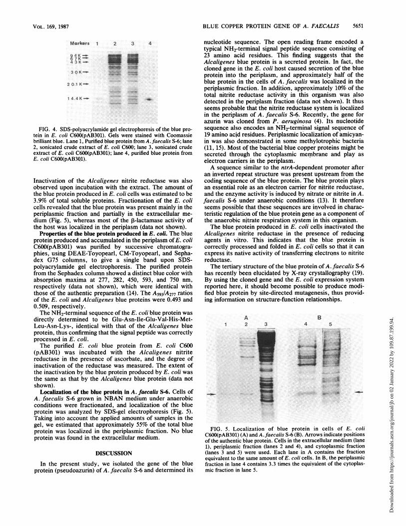
FIG. 2. Restriction map of the insert DNA in pAB101 andsequencing strategy. The hatched area shows the open readingframe.
VOL. 169, 1987
Dow
nloa
ded
from
http
s://j
ourn
als.
asm
.org
/jour
nal/j
b on
02
Janu
ary
2022
by
109.
87.1
99.9
4.

5650 YAMAMOTO ET AL.
GCATGCAGGCTTGTCATGTCGCGCCTAGGCTGGCCGGAGGCTGCGGCAAAAGGGCTGGCGGGCATATCGA ~CTGGTGGA T 90
g AAAATCAAACAUTTTTATTCTGAGCGTCATTATCATUAATAACTTCACCTGTTTUATCCAGATCAAAGAGUTTGGCAGGCGACAGGTC 180- - { - ~ - - - -
TAAACCCCGTTACGTGGCGTGTTGAGGCCGAGACGGCAGGTGCGCCAATCGTTGGAGATCAUGACAAAATGCUTAACATCGCGATCAAAT 270S D MetArgAsnileAlaIleLysPhe
TTGCTGCCGCAGGCATCCTCGCCATGCTGGCTGCCCCCGCTCTTGCCGAAAATATCGAAGTTCATATGCTCAACAAGGGCGCCGAGGGCG 360AlaAlaAlaGlyIleLeuAlaMetLeuAlaAlaProAlaLeuAlaGluAsnileGluValHisMetLeuAsnLysGlyAlaGluGlyAla
6 10
CCATGGTTTTCGAGCCTGCCTATATCAAGGCCAATCCCGGCGACACGGTCACCTTTATTCCGGTGGACAAAGGACATAATGTCGAATCCA 450MetValPheGluProAlaTyrI leLysAlaAsnProGlyAspThrValThrPhelleProValAspLysGlyHisAsnValGluSerlle
TCAAGGACATGATCCCTGAAGGCGCCGAAAAGTTCAAAAGCAAGATCAACGAGAACTATGTGCTGACGGTTACCCAGCCCGGCGCATATC 540LysAspMetIleProGluGlyAlaGluLysPheLysSerLysIleAsnGluAsnTyrValLeuThrValThrGlnProGlyAlaTYrLeu45 51
TGGTAAAGTGCACACCGCATTATGCCATGGGTATGATCGCGCTCATCUCTGTCGGTGACAGCCCGGCCAATCTCGACCAGATCGTTTCGU 630ValLysCysThrProHisTyrAlatletGlyMetIleAlaLeuIleAlaValGlyAspSerProAlaAsnLeuAsp(lnlleValSerAla
80 86
CCAAGAAGCCGAAGATTGTTCAGGAGCGGCTGGAAAAGGTCATCGCCAGCGCCAAATAAGAGCGCCAAATAAGATTGACCGAAAACTCTC 720LYsLysProLysIleValGlnGluArgLeuGluLysValIleAlaSerAlaLysTRM
GATGAGCCGAACTTGAACCGGCTTCATUACGAGGACATCATGACCAGACAGCCAGGCCTGCAG 783FIG. 3. Nucleotide and deduced amino acid sequences of the blue protein gene. The overlined sequences correspond to the probes used
for cloning. The underlined sequence is the determined NH2-terminal sequence of the blue protein produced in E. coli. SD, Shine-Dalganosequence. An inverted repeat structure is indicated by the facing arrows. The regions which show a high degree of homology with thentrA-dependent promoters are boxed. Arrowhead Cleavage site of the signal peptide as determined with the purified enzyme.
phoretically, and these were subcloned into the EcoRI site ofpYEJOO1. A recombinant plasmid, pAB101, was recoveredfrom a colony detected by colony hybridization with thesynthetic probes.DNA sequencing of the cloned blue protein gene. The 4.8-kb
EcoRI insert of pABlOlwas digested with various restrictionenzymes, and the resulting fragments, separated by agarosegel electrophoresis, were examined with the labeled probes.The SphI-PstI fragment of about 780 base pairs (bp) was
found to hybridize with the three probes.This fragment was cloned into phage M13 mpl8 and
sequenced according to the strategy shown in Fig. 2. Asingle open reading frame of 438 bp was found to encode a
polypeptide of 146 amino acid residues (Fig. 3). The aminoacid sequence after Glu-24, deduced from the nucleotidesequence, was completely identical with that of the blueprotein previously reported (8). The NH2-terminal sequenceof the open reading frame from Met-1 to Ala-23 was similarto a typical signal sequence containing a basic NH2-terminalsegment, followed by a stretch of hydrophobic residues.Two possible ribosome binding sequences were shown to
be present 4 and 10 bp upstream from the initiation codon.Considering the space to the initiation codon, the latterGGAGA sequence may be functional. No sequence homol-ogous with the typical E. coli consensus promoter was foundin the upstream region. However, nucleotides 71 to 78 and 87to 91 were found to show a high degree of homology with thepromoters of genes whose transcription requires the ntrAgene product (10), except that the space between the twosequences is 8 bp, which is longer than the 4 bp in the usualntrA-dependent promoters. An inverted repeat structure
with AG = -10.2 kcal/mol was found between the putativentrA-dependent promoter and the ribosome-binding se-quence.
Expression of the blue protein gene in E. coli. The 783-bpSphI-PstI fragment containing the coding sequence for theblue protein was inserted between the SphI and PstI restric-tion sites on pUC19 downstream from the lac promoter withthe correct orientation. The resulting expression plasmidpAB201 was introduced into E. coli JM105, and thetransformant was grown in the presence of IPTG. A veryfaint protein band corresponding to the blue protein wasobserved in the cell extract by sodium dodecyl sulfate(SDS)-gel electrophoresis, although no activity of the blueprotein in inactivating Alcaligenes nitrite reductase wasdetected in either the cell extract or the culture medium. Itseemed probable that expression of the blue protein genefrom the lac promoter was inhibited by the inverted repeatDNA sequence upstream of the coding sequence describedabove. Therefore, the SphI-PstI fragment cloned into'theM13 mpl8 phage DNA was recovered by double digestionwith HindIII and SalI, and the inverted repeat sequence wasremoved by partial digestion with Sau3A1. The resulting550-bp Sau3A1-SalI fragment was ligated to the large PstI-Sall fragment of pBR322 and a small PstI-BamHI fragmentof pDR540 containing the tac promoter (6). The constructedplasmid, pAB301, carrying the blue protein coding sequencedownstream from the tac promoter, was introduced into E.coli C600, and the transformant was grown aerobically in Lbroth containing IPTG at 37°C. A protein comigrating withthe authentic blue protein from A. faecalis S-6 was detectedin the cell extract by SDS-gel electrophoresis (Fig. 4).
J. BACTERIOL.
Dow
nloa
ded
from
http
s://j
ourn
als.
asm
.org
/jour
nal/j
b on
02
Janu
ary
2022
by
109.
87.1
99.9
4.
BLUE COPPER PROTEIN GENE OF A. FAECALIS
Markers 1 2 3 4
9 4 K --
4 3 K -
3 OK-
2 0.1 K-
14.4K-:
FIG. 4. SDS-polyacrylamide gel electrophoresis of the blue pro-tein in E. coli C600(pAB301). Gels were stained with Coomassiebrilliant blue. Lane 1, Pijrified blue protein from A. faecalis S-6; lane2, sonicated crude extract of E. coli C600; lane 3, sonicated crudeextract of E. coli C600(pAB301); lane 4, purified blue protein fromE. coli C600(pAB3Q1).
Inactivation of the Alcaligenes nitrite reductase was alsoobserved upon incubation with the extract. The amount ofthe blue protein produced in E. coli cells was estimated to be3.9% of total soluble proteins. Fractionation of the E. colicells revealed that the blue protein was present mainly in theperiplasmic fraction and partially in the extracellular me-dium (Fig. 5), whereas most of the P-lactamase activity ofthe host was localized in the periplasm (data not shown).
Properties of the blue protein produced in E. coli. The blueprotein produced and accumnulated in the periplasm ofE. coliC600(pAB301) was purified by successive chromatogra-phies, using DEAE-Toyopearl, CM-Toyopearl, and Sepha-dex G75 columns, to give a single band upon SDS-polyacrylamide gel electrophoresis. The purified proteinfrom the Sephade2 column showed a distinct blue color withabsorption maxima at 277, 282, 450, 593, and 750 nm,respectively (data not shown), which were identical withthose of the authentic preparation (14). The A593/A277 ratiosof the E. coli and Alcaligenes blue proteins were 0.493 and0.509, respectively.The NH2-terminal sequence of the E. coli blue protein was
directly determined to be Glu-Asn-Ile-Glu-Val-His-Met-Leu-Asn-Lys-, identical with that of the Alcaligenes blueprotein, thus confirming that the signal peptide was correctlyprocessed in E. coli.The purified E. coli blue protein from E. coli C600
(pAB301) was incubated with the Alcaligenes nitritereductase in the presence of ascorbate, and the degree ofinactivation of the reductase was measured. The extent ofthe inactivation by the blue protein produced by E. coli wasthe same as that by the Alcaligenes blue protein (data notshown).
Localization of the blue proteir in A. faecalis S-6. Cells ofA. faecalis S-6 grown in NBAN medium under anaerobicconditions were fractionated, and localization of the blueprotein was analyzed by SDS-gel electrophoresis (Fig. 5).Taking into account the applied amounts of samples in thegel, we estimated that approximately 55% of the total blueprotein was localized in the periplasmic fraction. No blueprotein was found in the extracellular medium.
DISCUSSION
In the present study, we isolated the gene of the blueprotein (pseudoazurin) of A. faecalis S-6 and determined its
nucleotide sequence. The open reading frame encoded atypical NH2-terminal signal peptide sequence consisting of23 amino acid residues. This finding suggests that theAlcaligenes blue protein is a secreted protein. In fact, thecloned gene in the E. coli host caused secretion of the blueprotein into the periplasm, and approximately half of theblue protein in the cells of A. faecalis was localized in theperiplasmic fraction. In addition, approximately 10% of thetotal nitrite reductase activity in this organism was alsodetected in the periplasm fraction (data not shown). It thusseems probable that the nitrite reductase system is localizedin the periplasm of A. faecalis S-6. Recently, the gene forazurin was cloned from P. aeruginosa (4). Its nucleotidesequence also encodes an NH2-terminal signal sequence of19 amino acid residues. Periplasmic localization of amicyan-in was also demonstrated in some methylotrophic bacteria(11, 15). Most of the bacterial blue copper proteins might besecreted through the cytoplasmic membrane and play aselectron carriers in the periplasm.A sequence similar to the ntrA-dependent promoter after
an inverted repeat structure was present upstream from thecoding sequence of the blue protein. The blue protein playsan essential role as an electron carrier for nitrite reductase,and the enzyme activityis induced by nitrate or nitrite in A.faecalis S-6 under anaerobic conditions (13).' it thereforeseems possible that these sequences are involved in charac-teristic regulation of the blue protein gene as a co,mponent ofthe anaerobic nitrate respiration system in this organism.The blue protein produced in E. coli cells inactivated the
Alcaligenes nitrite reductase in the presence of reducingagents in vitro. This indicates that the blue protein iscorrectly processed and folded in E.' coli cells so that it canexpress its native activity of transferring electrons to nitritereductase.The tertiary structure of the blue protein of A. faecalis S-6
has recently been elucidated by X-ray crystallography (19).By using the cloned gene and the E. coli expression systemreported here, it should become possible to produce modi-fied blue protein by site-directed mutagenesis, thus provid-ing information on structure-function relationships.
A1 2
B4 5
| t Y
jVri .,;t.Io;eij,t ll
L ..s_g * _L iF>Rs
-:qv .;.. .. tiv, *
s*2Y .*
3
_
=ES___r:
FIG. 5. Localization of blue protein in cells of E. coliC600(pAB301) (A) and A. faecalis S-6 (B). Arrows indicate positionsof the authentic blue protein. Cells in the extracellular medium (lane1), periplasmic fraction (lanes 2 and 4), and cytoplasmic fraction(lanes 3 and 5) were used. Each lane in A contains the fractionequivalent to the same amount of E. coli cells. In B, the periplasmicfraction in lane 4 contains 3.3 times the equiyalent of the cytoplas-mic fraction in lane 5.
5651VOL. 169, 1987
i--ii
!1117i.
Dow
nloa
ded
from
http
s://j
ourn
als.
asm
.org
/jour
nal/j
b on
02
Janu
ary
2022
by
109.
87.1
99.9
4.

5652 YAMAMOTO ET AL.
LITERATURE CITED1. Adman, E. T. 1985. Structure and function of small blue copper
proteins, p. 1-42. In P. M. Harrison (ed.), Metalloproteins:metal proteins with redox roles, part 1. Verlag Chemie Interna-tional, Inc., Deerfield Beach, Fla.
2. Ambler, R. P. 1977. The evolution of metalloenzymes, metal-loproteins and related materials, p. 100-118. In G. S. Hunt(ed.), Proceedings of a Symposium of the Inorganic Biochem-istry Discussion Group of the Chemical Society, University ofSussex. Symposium Press, London.
3. Ambler, R. P., and J. Tobari. 1985. The primary structures ofPseudomonas AM1 amicyanin and pseudoazurin. Biochem. J.232:451-457.
4. Canters, G. W. 1987. The azurin gene from Pseudomonasaeruginosa codes for a pre-protein with a signal peptide. FEBSLett. 212:168-172.
5. Cornelis, P., C. Digneffe, and K. Willemot. 1982. Cloning andexpression of a Bacillus coagulans amylase gene in Escherichiacoli. Mol. Gen. Genet. 186:507-511.
6. de Boer, H. A., L. J. Comstock, and M. Vasser. 1983. The tacpromoter: a functional hybrid derived from the trp and lacpromoters. Biochemistry 80:21-25.
7. Holmes, D. S., and M. Quigley. 1981. A rapid boiling method forthe preparation of bacterial plasmids. Anal. Biochem. 144:193-197.
8. Hormel, S., E. Adman, K. A. Walsh, T. Beppu, and K. Titani.1986. The amino acid sequence of the blue copper protein of
Alcaligenes faecalis. FEBS Lett. 197:301-304.9. Horio, T. 1985. Terminal oxidation system in bacteria. I. Puri-
fication of cytochromes from Pseudomonas aeruginosa. J.Biochem. 45:195-205.
10. Hunt, T. P., and B. Magasanik. 1985. Transcription of ginA bypurified Escherichia coli components: core RNA polymeraseand the products of glnF, glnG and glnL. Proc. Nati. Acad. Sci.USA 82:8453-8457.
11. Hlusain, M., and V. L. Davidson. 1985. An inducible periplasmicblue copper protein from Paracoccus denitrificans. J. Biol.Chem. 260:14626-14629.
12. Ish-Horowicz, D., and J. F. Burke. 1981. Rapid and efficientcosmid cloning. Nucleic Acids Res. 9:2989-2998.
13. Kakutani, T., H. Watanabe, K. Arima, and T. Beppu. 1981.
Purification and properties of a copper-containing nitritereductase from a denitrifying bacterium, Alcaligenes strain S-6.J. Biochem. 89:453-461.
14. Kakutani, T., H. Watanabe, K. Arima, and T. Beppu. 1981. Ablue protein as an inactivating factor for nitrite reductase fromAlcaligenes faecalis strain S-6. J. Biochem. 89:463-472.
15. Lawton, S. A., and C. Anthony. 1985. The role of cytochromesand blue copper proteins in the oxidation of methanol andmethylamine in organism 4025, an obligate methylotroph. J.Gen. Microbiol. 131:2165-2171.
16. Liu, M.-Y., M.-C. Liu, W. J. Payne, and J. Legall. 1986.Properties and electron transfer specificity of copper proteinsfrom the denitrifier "Achromobacter cycloclastes". J. Bacte-riol. 166:604-608.
17. Norgard, M. V., K. Keen, and J. J. Monahan. 1978. Factorsaffecting the transformation of Escherichia coli strain X1776 bypBR322 plasmid DNA. Gene 3:279-292.
18. Norris, G. E., B. F. Anderson, E. N. Baker, and S. V. Rumball.1979. Purification and preliminary crystallographic studies onazurin and cytochrome c' from Alcaligenes denitrificans andAlcaligenes sp. NCIB 11015. J. Mol. Biol. 135:309-312.
19. Petratos, K., D. W. Banner, T. Beppu, K. S. Wilson, and D.Tsernoglou. 1987. The crystal structure of pseudoazurin fromAlcaligenes faecalis S-6 determined at 2.9 A resolution. FEBSLett. 218:209-214.
20. Ross, G. W., and C. H. O'Callaghan. 1975. P-Lactamase assays.Methods Enzymol. 43:69-85.
21. Saito, H., and K. Miura. 1963. Preparation of transformingdeoxyribonucleic acid by phenol treatment. Biochim. Biophys.Acta 72:619-629.
22. Sanger, F., S. Nicklen, and A. R. Coulson. 1977. DNA sequenc-ing with chain-terminating inhibitors. Proc. Natl. Acad. Sci.USA 74:5463-5467.
23. Southern, E. M. 1975. Detection of specific sequences amongDNA fragments separated by gel electrophoresis. J. Mol. Biol.98:503-517.
24. Wallace, R. B., M. J. Johnson, T. Hirose, T. Miyake, E. H.Kawashima, and K. Itakura. 1981. The use of synthetic oligo-nucleotides as hybridization probes. II. Hybridization of oligo-nucleotides of mixed sequence to rabbit P-globin DNA. NucleicAcids Res. 9:879-894.
J. BACTERIOL.
Dow
nloa
ded
from
http
s://j
ourn
als.
asm
.org
/jour
nal/j
b on
02
Janu
ary
2022
by
109.
87.1
99.9
4.


![Doc1 - CDC...126 Gr. A Streptococci erythromycin 2001 2003 [24] 53% faecalis erythromycin chloram- phenicol ciprofloxacin gentamicin avoparcin ' E. faecalis vancomycin](https://img.pdfslide.us/doc/110x75/6118145c1932226e937f5e05/doc1-cdc-126-gr-a-streptococci-erythromycin-2001-2003-24-53-faecalis-erythromycin.jpg)