Embed Size (px)
Citation preview
THE BIOLOGICAL SIGNIFICANCE OF THETEGUMENT IN DIGENETIC TREMATODES
by K. E. Dixon
ABSTRACT
The structure of the tegument of digenetic trematodes is adapted to serve two primary functions, absorption and protection, and represents a compromise between the demands of the two roles. I t is suggested that the covering layer of cytoplasm serves as a n absorptive surface for most or even all of the parenchyma1 cells that are able to establish intermittent connections with it for the purpose of inward and outward transfer of substances. Because the surface must therefore be a delicate structure and hence susceptible to damage, the ability of the parenchymal cells, protected beneath the basal lamina and muscle cells, to regenerate the surface layer of cytoplasm, represents an important protective adapta- tion. These suggestions extend our understanding of the structural and functional contribution of the surface cytoplasm of the tegument to the organism. They also have implications for our view of the method of formation of the tegument and for the nomenclature of the tegumental structures.
INTRODUCTION
The surface structures of digenetic trematodes have received a great deal of attention, particularly since electron microscopy became more readily available to parasitologists fifteen to twenty years ago. No doubt one of the major reasons for this concentrated study is that the surface can be identified unequivocally, unlike many of the internal structures, which, particularly in the early years, were often difficult to assign with any confidence to an organ system. Notwithstanding these valid technical reasons, recognition of the nutritional contribution of the surface to the parasite was also an important factor. This interest had its genesis in part in and received much of its momen- tum from the studies of Clark Read and his associates (see Pappas and Read, 1975), directed towards elucidating the functional aspects of molecular trans-
K. E. Dixon is Senior Lecturer in Biology at the Flinders Univers~ty of South Australia.
69
70 RICE UNIVERSITY STUDIES
port across the surface membranes of cestodes and, more recently, trematodes. The structure of the surface of digenetic trematodes remained enigmatic
until the careful electron microscopic studies of Threadgold (1963) clarified its unique syncytial organization and so confirmed and extended the earlier light microscope observations of Hein (1904). Threadgold introduced the term tegument to refer to the syncytium, which he defined as consisting of a continuous outer layer of cytoplasm connected to nucleated cell bodies lying beneath the basal lamina and the muscle layers. Lee (1966) has suggested epidermis as a simpler alternative but Hockley (1973) has continued to use "tegument" in preference to "epidermis" because he believes the latter does not indicate the unusual structure of the tegument, a view with which I agree (see also Lumsden, 1975). Furthermore, since there is no dermis, "epidermis" is not appropriate. In this article, the term tegument will be used as originally defined by Threadgold (1963).
Since Threadgold's description of the tegument of adult Fasciola hepatica appeared, many other species have been studied (reviewed in Lee, 1966 and 1972; Hockley, 1973; Lumsden, 1975). There have been few attempts, however, to integrate the structural information with the functional require- ments of the parasite. This article takes an ontogenetic approach by examin- ing the functional basis for the cercarial surface structures from which the adult tegument is derived. It is not a review but an attempt to provide an integration of some information already available.
I t will be argued that the primary function of the tegument is absorptive and its structure is highly dynamic, in keeping with this function. It is pro- posed that the surface layer of cytoplasm serves most or even all of the parenchymal cells that are able to make intermittent connections with it. By this means a continuous cytoplasmic pathway is provided throughout the animal. Of equal importance, the structure of the tegument ensures that damage to the delicate absorptive surfaces remains localized and the surface layer of cytoplasm can be regenerated from the parenchymal cells protected beneath the basal lamina and muscle cells.
THE ADULT TEGUMENT
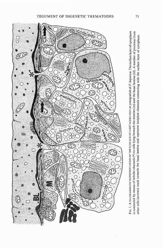
In all adult digenetic trematodes, the tegument consists of two regions (figure I). At the surface, a layer of cytoplasm rests on a basal lamina, which overlies the major muscle layers. Internal to the basal lamina and the muscle cells, in the parenchyma, lie nucleated cell bodies, which are connected to the surface layer of cytoplasm by narrow tubular extensions passing between blocks of muscle cells and through the basal lamina. Lee (1966 and 1972), Lyons (1973), Hockley (1973), and Lumsden (1975) have reviewed, compre- hensively and in detail, the published descriptions of the tegument of adult digenetic trematodes.
TEGUMENT O F DIGENETIC TREMATODES 7 1
72 RICE UNIVERSITY STUDIES
The adult fluke develops from the cercaria by a process which in some species (e.g., F. hepatica, reviewed by Dixon, 1968) is sudden and dramatic enough to be called a metamorphosis. The transition also resembles meta- morphosis because it involves in part the replacement of cercarial investments with the tegument of the adult.
T H E CERCARlAL TEGUMENT
The exact nature of the cercarial investments and the mechanisms by which they are formed and lost differ in different species. In all species in which early stages of development have been closely studied, the germ ball from which the cercaria develops is surrounded by a thin, nucleated cyto- plasmic layer, called a "primitive epithelium" by Dubois (1929) and an "embryonic epithelium" by Dixon and Mercer (1967). I will continue to use the latter term here because "primitive" has little if any meaning in this context, whereas the epithelium is indisputably embryonic (but see Rifkin, 1970; Belton and Belton, 1971). Eventually, a second layer of cells is formed between the embryonic epithelium and the muscle cells and it is from this layer that the cercarial tegument ultimately forms, finally developinginto the adult tegument.
In some species, e.g., Notocotylus attenuatus (Southgate, 1971) and Schistosoma mansoni (Hockley, 1972), the cercarial tegument is formed at an early stage of development of the cercaria, but in other species, e.g., F. hepatica (Dixon and Mercer, I967), it forms during transformation to the metacercarial stage. In other species still, e.g., Cloacitrema narrabeenensis (Dixon, 1970; Dixon and Colton, 1975), thecercarial tegument is laid down at intermediate stages of development. No matter when the cercarial tegument appears, however, its formation precedes, usually only briefly, the degenera- tion and loss of the embryonic epithelium.
Apparently the cercarial tegument can be formed in either of two ways, depending on the species, In the first of these, outer cells of the embryo form a single layer around the developing organism, later fusing into a syncytium, presumably by the breaking down of the lateral membranes. In some species in which the cercarial tegument is formed in this way, the nuclei degenerate completely, as convincingIy shown by Matricon-Gondran (1 97 1) in Cercaria pectinata, and by Hockley (1972) in S. mansoni, and the parenchymal cells eventually establish connections with the surface layer of cytoplasm which remains. In some other species, e.g., Acanthoparyphiunz spinulosurn (Bills and Martin, 1966) and C. narrabeenensis (Dixon and Colton, 1975), the nuclei apparently do not degenerate but sink down into the sub-tegumental parenchyma, retaining their connection with the surface layer of cytoplasm. The alternative method of formation of the cercarial tegument relies solely on the upflow of cytoplasm from parenchymally situated cells, a process
TEGUMENT OF DIGENETIC TREMATODES 73
which forms the layer of cytoplasm at the surface, e.g., F. hepatica (Dixon, 1968).
Questions as to the functional basis for this structural heterogeneity naturally follow.
THE FUNCTION OF THE CERCARIAL INVESTMENTS
The surface structures of cercariae, bo th embryonic epithelium and cercarial tegument, have a number of functions to perform: structural, nuttitional, and protective, and in formation of the metacercarial cyst wall.
Structural,functions. The embryonic epithelium is formed very early in development when the embryo consists of only a few cells. Although similar structures are not formed during embryonic development in the Turbellaria (Skaer, 1973), the formation of a similar envelope during early development of the oncosphere of the cestodes Eubothrium rugosum and Diphyllobo- thrium latttm has been described by Schaunnsfand (1826), cited in Rybicka (1966). One of the functions of this structure is presumably to enclose the cells of the embryo, acting in an analogous way t o a n egg envelope.
Absorptive .functions. Since cercariae grow within the redia or daughter sporocyst, they must acquire the necessary substances for growth. The requirements and mechanisms for the nutrition of intra-redial cercariae have received scant attention. The conclusion that the cercariae must take up substances from the fluid in the redial lumen has been generally accepted although the mechanism of uptake has not been studied. The conclusion seems inescapable, though, that absorption takes place a t the cercarial surface, since it is doubtful that the intestinal caeca are functional. I n the cercaria of F. hepatica, for example, the gut is not patent but consists of a number of isolated cavities in which a secretion of the ceIls lining the cavities is stored. I t can be speculated that this secretion is responsible for digestion of the ventral plug region of the metacercarial cyst wall during excystment, thus permitting the metacercaria to escape from the cyst wall (Dixon, 1966). The gut in this condition could not function as an organ of absorption within the redia, however. Observations of the undeveloped nature of the caecal epithelium of the newly excysted metacercaria are consistent with this reasoning (Bennett, 1975). K4ie (1971a), in an ultrastructural study of the caecal epithelium of the intra-redial cercaria of Neophasis lageniformis, has also suggested that the caeca in this species are not functional.
On the other hand, positive evidence that the cercarial surface is absorptive is not conclusive. All the descriptions of the embryonic epithelium suggest that it has no specialized organelles or structure t o enhance its ability t o take up substances from the redial lumen. For example, microvilli or similar devices for increasing the surface area for absorption have not been described. O n the contrary, the cytoplasm of the embryonic epithelium appears empty
74 RICE UNIVERSITY STUDIES
except for a few scattered mitochondria, suggesting that these cells are not metabolically active. It therefore seems likely that while an embryonic epithe- lium persists, the cercaria has to rely on diffusion of growth substances across the surface, with the diffusion gradient perhaps maintained by active processes at the surface of the inner mass of embryonic cells.
The cells that will ultimately form the cercarial tegument have general ultrastructural characteristics which would enable them t o take on an absorptive role although they are not specialized for this function. Some cytochemical studies have demonstrated the presence of enzymes (mainly phosphatases) often associated with absorption, e.g., Dixon (1970) and Dixon, Wetherall, and Colton (1975) in C. narrabeenensis, K$ie (1971 b) in Zoogonoides viviparus, and Krupa and Bogitsh (1972) in S. mansoni. Direct experimental evidence that the cercariae are capable of absorbing substances is still largely lacking. Dixon (1970) and Dixon et a1. (1975) have shown that the cercariae of C. narrabeenensis take up horse-radish peroxidase and 3~-glucose. Notwithstanding the pa;city of actual data, the logicaljustifica- tion for concluding that the cercarial surface is absorptive is compelling.
The absorptive role of the tegument of the cercaria is ideally suited to animals that have no circulatory system and instead have to rely on diffusion of substances from celI to cell. Gallagher and Threadgold (1967) have sug- gested that the ramifying parenchymal cells function as a cellular equivalent of a circulatory system. The attractions of this suggestion are enhanced by the recognition that the structure of the tegument provides a continuous cytoplasmic pathway from the exterior to the interior of the body. In this light, the absorptive capacity of the tegument could be utilized more effi- ciently if it provided a common absorptive surface for most or all of the parenchymal cells, For this to be feasible, the parenchymal cells would have to be able t o establish connections with the surface cytoplasm of the tegument, and these cells would then be indistinguishable cytologically from the sub- muscular cell bodies normally considered as contributing to the tegument. If this view is correct, it further suggests that the tegument is a dynamic and highly active structure, and not a static one as might be inferred from the electron microscope data.
Similar but less explicit suggestions have been made by Lee (1972) and Hockley (1973). Lee has written: "sunken cells are not permanently in contact with the epidermis but. . . they migrate from within the parenchyma to make contact with the epidermis and then perhaps lose contact once their function at the epidermis is complete." Hockley (1973) comments that the subtegu- mental cells of S. mansonicercariae may be only temporarily connected to the tegument. The view expressed here extends the suggestions of Lee and Hockley further by proposing essentially that many, if not all, of the paren- chymal cells establish intermittent connections frequently with the surface cytoplasm.
TEGUMENT O F DIGENETIC TREMATODES 75
All the observations of metacercarial cyst formation, a process in which the surface structures play an important part (discussed further below), are consistent with the suggestion that many, if not in some species most, of the parenchymal (cystogenic) cells are able to establish connections with the surface at the time of encystment. Furthermore, the failure to observe con- nections between the surface cytoplasm and the parenchymal cells in F. hepatica (Bjorkman and Thorsell, 1964) and in S, mansoni cercariae (Hockley, 1972) is evidence in favor of this hypothesis. Additional support comes from reports that the number of connections increases markedly during development of the schistosomulum (Hockley, 1972).
If parenchymal cells are able to establish connections with the cytoplasm intermittently, then an efficient mechanism exists for the regulation of the flow of materials, in both directions, between the interior and the exterior of the animal.
Protective functions. The surface structures of cercariae must also provide protection against the host defenses and the vicissitudes of the external environment. This function is greatly complicated by the fact that thesurface is still a major or even the sole organ of absorption and hence is very suscep- tible to damage. Clegg (1972) has drawn attention to the unsuitability of the surface for protection of the organism. If the surface was composed of a normal cellular epithelium, then damage resulting from injury could only be repaired by the proliferation of remaining undamaged epithelial cells. On the other hand, if the absorptive surface was a tegument, the damage could be restricted to the outer layer of cytoplasm, while the cell bodies remained protected beneath the basal lamina and able to regenerate the surface layer at the appropriate time.
There have been few studies of the regeneration of the tegument in the adult and none at all in the cercaria. Hockley (1972) has reviewed the reports of changes in the tegument of adult flukes following exposure to drugs. NO conclusions useful for this article can be reached, however, because damage could be due either to the direct action of the drug on the surface or to more general systemic effects which result in degeneration of the surface.
The tegument is subjected to partial and in some cases complete breakdown during encystment and formation of the metacercarial stage. Therefore, a consideration of the role of the tegument in encystment is relevant.
Encystment functions. The possible evolutionary relationships of meta- cercarial cyst walls have been recently discussed (Dixon, 1975) on the basis of the degree of protection from the rigors of the environment afforded by cyst walls of different complexities. The postulated sequence (which does not imply a single line of evolution) begins in a simple, single-layered cyst wall, usually composed of carbohydrate-protein complexes (see Dixon, 1975, for examples). An intermediate, more resistant stage has at least two layers of material, formed by the addition of a layer of resistant protein, usually
76 RICE UNIVERSITY STUDIES
stabilized by disulphide bonds, to the simple cyst wall. The ultimate in protection is reached in those species that encyst in the open, when another layer of protein-a tanned protein-is added and this additional layer is incorporated into an outer cyst wall.
In the formation of the two simpler types of cyst walls, the surface layer of cytoplasm of the tegument has to receive the cystogenous materials from the cells in which they were synthesized, and then secrete them at the appropriate time to the exterior. Transfer of the cystogenous materials is achieved when the parenchymal cells establish connections with the surface cytoplasm. Secretion can be accomplished by a normal apocrine process involving little or n o damage to the tegument.
The formation of the most complex cyst walls does, however, involve damage to the surface. In some species, e.g., C. narrabeenensis (Dixon and Colton, 1975), the cystogenous precursors of the outer cyst wall are trans- ferred to and secreted from the cercarial tegument. In other species, where apparently the volume of cystogenouk material is greater, e.g., F. hepatica (Mercer and Dixon, 1967) and Parorchis acanthus (Rees, 1967; Cable and Schutte, 19731, the precursor granules of the outer cyst wall are transferred to the embryonic epithelium, which is then shed. In both species, the basal lamina therefore forms, temporarily, the surface of the transforming meta- cercaria, until upwelling of cytoplasm, principally or perhaps exclusively from the parenchymal cells synthesizing the keratin scrolls, forms a surface layer of cytoplasm. The establishment of a coherent surface is essential for the secretion and unrolling of the individual keratin scrolls (Dixon, 1968).
The regeneration of the metacercarial surface during encystment therefore illustrates the advantages of the tegumental organization for repair of the surface if it is damaged.
CONCLUSIONS
The surface coverings of digenetic trematodes, because they represent the parasitelhost or parasitelenvironment interface, are clearly not evolution- arily conservative structures. On the contrary, they have become adapted to serving a number of functions which must all contribute to the overall economy of the organism.
The preceding analysis implies that the surface layer of cytoplasm of the tegument is primarily adapted as an absorptive organ serving most or all of the parenchyma1 cells that make more or less frequent but intermittent connections with it. Temporary connections provide a means of regulating the flow of absorbed materials inwards and the flow of secretory materials outwards. Since to fulfill this role the surface is inevitably a delicate structure and therefore susceptible to damage, the ability of the parenchymal cells, protected beneath the basal lamina and muscle cells, to regenerate the surface
TEGUMENT OF DIGENETIC TREMATODES 77
layer of cytoplasm is clearly an important adaptation. Viewed in this light, arguments as to whether the tegument is formed by the upwelling of cyto- plasm from parenchymally situated cells or by the degeneration of nuclei in surface cells and the subsequent establishment of connections with paren- chymally situated cells become in part misleading and are largely irrelevant to an understanding of the functions of the tegument. The hypothesis also has implications for the nomenclature of these surfaces. The term tegument, as noted earlier, includes both the surface layer of cytoplasm and the nucleated parenchymal cell bodies. If the hypothesis advanced here is correct, however, there is no exclusive association between cell "bodies" (a term which is also no longer appropriate) and the anucleate surface cytoplasm, which should therefore be considered as a separate structure.
Although this analysis has been confined almost exclusively to cercariae and sub-adults, it is obvious that the inferences apply equally to the functions of the tegument in a11 other stages. The hypothesis is of such a general nature that it can probably be applied profitably to groups other than the Digenea which exhibit this type of structure.
ACKNOWLEDGMENTS
Thanks are due to Dr. J. C. Pearson, Dr C. P. Read, and Professor J. D. Smyth for their crlticisrn of the ideas on wh~ch t h ~ s article is based.
REFERENCES CITED
Belton, J . C. and C. M. Belton 1971 Freeze-etch and cytochemical studies of the integument of larval
Acanthatrium oregonense (Trematoda). Journal of Parasitology 57:252-260.
Bennett, C . E. 1975 Fasciola hepatica: development of caecal epithelium during migra-
tion in the mouse. Experimental Parasitology 37:426-441.
Bils, R. R. and W. E. Martin 1966 Fine structure and development of the trematode integument.
Transactions of the American Microscopical Society 8578-88,
Bjorkman, N. and W. Thorsell I964 On the fine structure and resorptive function of the cuticle of the
liver fluke Fasciola hepatica L. Experimental Cell Research 33:3 19-329.
78 RICE UNIVERSITY STUDIES
Cable, R. M. and M. H. Schutte 1973 Comparative fine structure and origin of the metacercarial cyst
in two philophthalmid trematodes, Parorchis acanthus (Nicoll, 1906) and Philophthalmus megalurus (Cort, 19 14). Journal of Parasitology 59: 103 1-1040.
Clegg, J. A. 1972 The schistosome surface in relation to parasitism. In Functional
Aspects of Parasite Surfaces. A. E. R. Taylor and R. Muller, eds. Oxford: Blackwell. Pp. 23-40.
Dixon, K. E. 1966 The physiology of excystment of the metacercaria of Fasciola
hepatica L. Parasitology 56:431-456.
1968 Encystment of the cercaria of Fasciola hepatica. Wiadomosci Parazytologiczne 14:689-701:
1970 Absorption by developing cercariae of Cloacitrema narrabeenensis (Philophthalmidae). JournaI of Parasitology 56 (Section II):416- 417.
1975 The structure and composition of the cyst wall of the metacercaria of Cloacitrema narrabeenensis (Philophthalmidae). International Journal for Parasitology 5: 1 13-1 18.
Dixon, K. E. and M. Colton 1975 The formation of the cyst wall of the metacercaria of Cloacitrema
narrabeenensis. In preparation.
Dixon, K. E. and E. H. Mercer 1967 The fine structure of the cystogenic cells of the cercaria of Fasciola
hepatica L. Zeitschrift fur Zellforschung und Mikroskopische Anatomie 77:33 1-344.
Dixon, K. E., M. B. Wetherall, and M. Colton 1975 The body wall of the redia of Cloacitrema narrabeenensis
(Philophthalmidae) and its role in nutrition of the cercaria. In preparation.
Dubois, G. 1929 Les cercaires de la region de Neuchitel. Bulletin de la SociktC
Neuchiteloise des Sciences Naturelles 53:l-177.
Gallagher, S. S. E. and L. T. Threadgold 1967 Electron microscope studies of Fasciola hepatica. 11. The inter-
relationship of the parenchyma with other organ systems. Para- sitology 57:627-632.
TEGUMENT OF DIGENETIC TREMATODES 79
Hein, W. 1904 Zur epithelfrage der Trematoden. Zeitschrift fiir Wissenschaften
Zoologische 77:400-438.
HockIey, D. J. 1972 Schistosoma mansoni: the development of the cercarial tegument.
Parasitology 64:245-252.
1973 Ultrastructure of the tegument of Schistosoma. Advances in Parasitology 11:233-305.
Kbie, M. 1971a On the histochemistry and ultrastructure of the redia of Neophasis
lageniformis (Lebour, 1910) (Trematoda, Acanthocolpidae). Ophelia 9: 113-143.
1971b 011 the histochemistry and ultrastructure of the tegument and associated structures of the cercaria of Zoogonoides viviparus in the first intermediate host. Ophelia 9165-206.
Krupa, P. and B. Bogitsh 1972 Ultrastructural phosphohydrolase activities in Schistosoma
mansoni sporocysts and cercariae. Journal of ParasitoIogy 58:495-5 14.
Lee, D. L. 1966 The structure and composition of the helminth cuticle. Advances
in Parasitology 4: 187-254.
1972 The structure of the helminth cuticle. Advances in Parasitology 10:347-379.
Lumsden, R. D. 1975 Surface ultrastructure and cytochemistry of parasite helminths.
Experimental Parasitology 37:267-339.
Lyons, K. 1973 The epidermis and sense organs of the Monogenea and some
related groups. Advances in Parasitology 11:193-232.
Matricon-Gondran, M. 1971 Origine et differenciation du tegument d'un Trematode Digenetique:
ktude ~Itrastructurale chez Cercaria pectinata (larve de Baccigkr baccige'r, Fellodistomatides). Zeitschrift fiir Zellforschung und Mikroskopische Anatomie 120:488-524.
Mercer, E. H. and K. E. Dixon 1967 The fine structure of the cystogenic cells of the cercaria of Fasciola

RICE UNIVERSITY STUDIES
hepatica L. Zeitschrift fiir Zellforschung und Mikroskopische Anatomie 77:33 1-344.
Pappas, P. W. and C. P. Read I975 Membrane transport in helminth parasites: a review. Experimental
Parasitology 37:469-530.
Rees, G . 1967 The histochemistry of the cystogenous gland cells and cyst wall of
Parorchis acanthus Nicoll, and some details of the morphology and fine structure of the cercaria. Parasitology 5797-1 10.
Rifkin, E. 1970 An ultrastructural study of the interaction between the sporocysts
and the developing cercaria of Schistosoma mansoni. Journal of Parasitology 56 (Section II):284.
Rybicka, K. 1966 Embryogenesis in Cestodes. Advances in Parasitology 4:107-186.
Skaer, R. J. 1973 Planarians. In Experimental Embryology of Marine and Fresh-
water Invertebrates. G. Reverberi, ed. Amsterdam: North- Holland. Pp. 104-125.
Southgate, V. T. 1971 Observations on the fine structure of the cercaria of Notocotylus
attenuatus and formation of the cyst wall of the metacercaria. Zeitschrift fiir Zellforschung und Mikroskopische Anatomie 120:420-449.
Threadgold, L. T. 1963 The tegument and associated structures of FascioIa hepatica.
Quarterly Journal of Microscopical Science 104:505-512.