Embed Size (px)
Citation preview
T H E BIOLOGICAL AND ANTINEOPLASTIC PROPERTIES OF THREE TRIAZINE DERIVATIVES
BENJAMIN GOLDBEKG, PH.D., and EMANUEL B. SCHOENBACH, M.D.
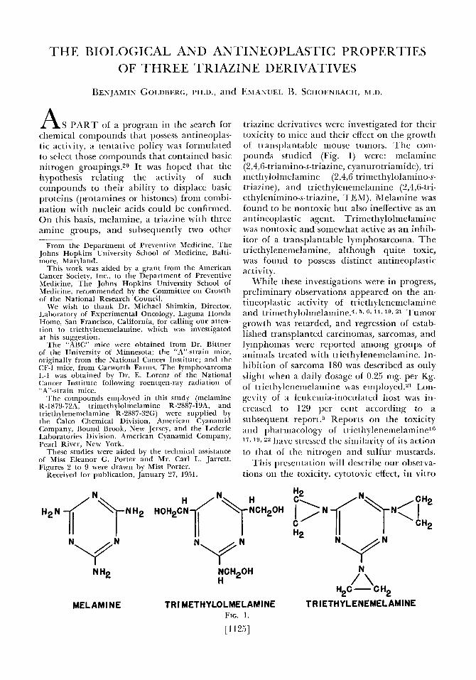
A S PART of a program in the search for c:hernical compounds that possess antineoplas- tic: activity, a tentative policy was formulated to select those compounds that contained basic nitrogen groupings.20 I t was hoped that the hypothesis relating the activity of such compounds to their ability to displace basic proteins (protamines or histones) from combi- nation with nucleic acids could be confirmed. On this basis, melamine, a triazine with three amine groups, and subsequently two other
From the Department of Preventive Medicine, T h e Johns Hopkins University School of Medicine, Balti- more, Maryland.
This work was aided by a grant from the American Cancer Society, Inc., to the Department of Preventive Medicine, The Johns Hopkins University School of Medicine, recommended by the Committee on Growth of the National Research Council.
We wish to thank Dr. Michael Shimkin, Director, Laboratory of Experimental Oncology, Laguna Honda Home, San Francisco, California, for calling our atten- tion to triethylenemelamine, which was investigated at his suggestion.
‘The “ABC” mice were obtained from Dr. Bittner of the University 01 Minnesota; the “A”-strain mice, originally from the National Cancer Institute; and the CF-l mice, from Carworth Farms. The lymphosarcoma I,-1 was obtained by Dr. E. Lorenz of the National Cancer Institute following roentgen-ray radiation of “A”-strain mice.
The compounds employed in this study (melamine K-1879.724, trimethylolmelamine R-2887-19A, and triethylenemelamine R-2887-32G) were supplied by the Calco Chemical Division, American Cyanamid Company, Bound Brook, New Jersey, and the Lederle Laboratories Division, American Cyanamid Company, I’carl River, New York.
These studies were aided by the technical assistance of Miss Eleanor G. Porter and Mr. Carl L. Jarrett. Figures 2 to 9 were drawn by Miss Porter.
Received for publication, January 27, 1951.
triazine derivatives were investigated for their toxicity to mice and their effect on the growth of transplantable mouse tumors. T h e coni- pounds studied (Fig. 1) were: melamine (2,4,6-triamino-s-triazine, cyanurotriamide), tri- inetliylolmelainine (2,4,6-trimethylolaniino-s- triazine), and triethylenemelamine (2,4,6-tri- ethylenirnino-s-triazine, TEM). Melamine was lound to be nontoxic but also ineffective as an antineoplastic agent. Trimethylolmelarnine was nontoxic and somewhat active as an inhib- itor of a transplantable lymphosarcoma. T h e triethylenemelamine, although quite toxic, was found to possess distinct antineoplastic activity.
While these investigations were in progress, preliminary observations appeared on the an- tineoplastic activity of triethylenemelamine and triniethylolmelamine.4~ b , & 1% 1% 21 Tumor growth was retarded, and regression of estab- lished transplanted carcinomas, sarcomas, and lymphomas were reported among groups of animals treated with triethylenemelamine. In- hibition of sarcoma 180 was described as only slight when a daily dosage of 0.25 mg. per Kg. of triethylenemelamine was employed.21 Lon- gevity of a leukemia-inoculated host was in- creased to 129 per cent according to a subsequent report.5 Reports on the toxicity and pharmacology of triethylenenielamine16 1 7 , l ~ 22 have stressed the similarity of its action to that of the nitrogen and sulfur mustards.
This presentation will describe our observa- tions on the toxicity, cytotoxic effect, in vitro
NCH20H H /”\
H2C - C H2 MELAMINE TRI METHYLOLMELAMINE T R IETHYLENEMELAMINE
FIG. 1.
[1125]
11261 CANCER September 1951
metabolic studies, and antineoplastic activity of these three triazine derivatives.
MATERIALS AND METHODS
The following strains of mice were em- ployed in these experiments: “ABC” male and female mice; “A”-strain female mice; and CF-1 male and female mice. The mice weighed 20 to 30 gm. They were housed in wire cages in groups of five to eight. Their diet consisted of dog pellets (Purina Dog Chow) and tap water ad libitum. The tumors employed in these experiments were the sarcoma 180 and lyni- phosarcoma L-I. Sarcoma 180 was inoculated by implanting fragments subcutaneously, bi- laterally, with a trocar in the axillary region. Results were evaluated by weighing the dis- sected tumors on a torsion balance at the end of the experiment. The lymphosarcoma was implanted intramuscularly in the right hind leg of “ABC” and “A”-strain mice. The cri- teria employed to evaluate the effect on this tumor were estimation, with calipers, of tumor size in three planes and host longevity, as compared to groups of mice receiving the vehicle alone. Blood for hematological exam- ination was obtained from the tail by exposing and lacerating the lateral tail veins. Observa- tions were made on marrow lrom the sternum and the femur. Both imprints and smears were employed. The marrow was sometimes mixed with human or rabbit serum for the prepara- tion of smears. Smears or imprints were stained with Wright’s stain; the Feulgen nu- clear stain, employing decolorized basic fuchsin, counterstained with fast green; or a polychrome stain after 95 per cent alcohol- ether (1:l) fixation. Material for smears from the inguinal lymph nodes and the spleen was obtained and treated in similar fashion. Tis- sues for histological examination were fixed in 10 per cent commercial formalin and sec- tioned at 6 to 10 p. They were stained with hematoxylin and eosin, the Feulgen tech- nique, or a polychrome stain.
The oxygen uptake in air of tissue or tumor preparations was ascertained, employing the standard Warburg manometric procedure. A Kinger-phosphate solution (pH 7.4) contain- ing 0.61 per cent calcium chloridels and glucose (0.2 per cent) was used. All tissue preparations and solutions were chilled in an ice-water bath prior to test, which was con- ducted at 37” C.
Melamine and trimethylolmelamine were administered as suspensions in 5 or 10 per cent acacia. Triethylenemelamine was dissolved in 0.9 per cent saline; although it was apparently quite soluble in the saline solution, gelatinous particles, even at concentrations as low as 0.1 per cent, were always evident. It is not known whether these represent an impurity, reaction product, or polymer formation.
TOXICITY
Melamine. Because of the insolubility of melamine in saline in concentrations greater than 25 mg. per ml., dosages exceeding 250 mg. per Kg. were suspended in 10 per cent gum acacia. CF-1 male mice were used for the acute toxicity tests on this compound. I t was found to be well tolerated. All animals sur- vived, without evidence of weight loss, when a single dose of 1000 mg. per Kg. was adminis- tered subcutaneously or intraperitoneally; 5000 mg. per Kg. administered per 0s by gav- age was similarly well tolerated. Approxi- mately half the animals died when 5000 mg. per Kg. was administered as a single dose sub- cutaneously or intraperitoneally. Deaths oc- curred within twenty-four to forty-eight hours. The only manifest toxic signs noted prior to death were general sluggishness, delayed reac- tion to noxious stimuli, and slow, deep respi- ration. The chronic toxicity was low, and 625 mg. per Kg. per day for a period of ten days was not lethal and did not affect the weight of animals adversely.
Gross pathological examination of mice that had succumbed to, or were moribund from, lethal doses of melamine showed very small pale spleens, very small hemorrhagic lymph nodes, hemorrhagic adrenals, shrunken thy- mus, and distended stomach filled with a non- viscous clear liquid. Search of injected areas showed considerable amounts of compound forty-eight hours after injection, but at longer intervals (eight to ten days) no trace of the compound was found. The toxic manifesta- tions described were not found among survi- vors that had received the same dosage and that were sacrificed after a ten-day period of observation.
Trimethylolmelanzine. CF-1 male mice were employed to assay the toxicity of trimeth- ylolmelamine. This compound was quite in- soluble in water and was administered, after trituration, as a suspension in 5 or 10 per cent
gum acacia. An odor of formaldehyde was evident in the dry crystalline state and in solu- tion. A strong positive reaction for aldehyde was obtained with decolorized bd\ic fuchsin. A single dose of 5000 mg. per Kg. administered by gavage, 3000 mg. per Kg. injected subcu- taneously, or 2000 nig. per Kg. injected in traperitoneally were without apparent ill effect.
‘I’rimethylolmelamine, dissolwd in 0.2 N hydrochloric acid and adjusted to approxi- mately pH 6 by the addition of sodium bicar- bonate, was administered intravenously to CF-1 male mice daily for eight days. No dedths or weight loss occurred even when a daily dos- age o f 100 rng. per Kg. was attained. Erythro- cyte and leukocyte counts, as well as dif- ferential examination of stained blood films, of these mice showed no deviation from the normal after two, four, or eight daily intra- venou, injections at this dosage level.
Mice were injected intraperitoneally with 500 or 1000 mg. per Kg. of trimethylolniel- aniine daily for ten days, using a saline suspen- sion of the compound. The animals that received the higher dose suffered a low mor- tality (about 20 per cent) and some weight loss (about 10 per cent). Those receiving the lower dosage did not die, and no weight loss was manifest. The only gross pathological findings noted in these animals were a small spleen and thymus at a dosage of 1000 mg. per Kg. No gross abnormalities were noted in mice treated with 500 mg. per Kg. Cell counts of the x enous blood were performed five and nine days after the last injection. Whereas the white-cell counts of the treated animals were consistently lower than those of the controls, the differ- ences were not great, and, in view of the wide variation encountered in venous white-cell counts in mice, no special significance was at- tac hed to the results obtained. The red-cell ( aunt\ and differential leukocyte examina- tions were essentially unaltered, and no evi- dence of anemia was noted in treated animals.
Triethylenemelunazne. Preliminary toxicity tests indicated that triethylenemelaniine was equally toxic when administered intrale- nously, intraperi toneally, or subcutaneously. The time that elapsed before toxic manifesta- tions became evident was approximately equal for each of the three routes. Only the acute subcutaneous LD,, was therefore ascertained. A single injection of 1 mg. per Kg. resulted in
BIOLOGICAL AND ANTICANCER PROPERTIES OF THREE TRIAZINES - Goldlrrrg & L % h O C n b U C h [1127
no mortality or evidence ok toxicity. When dose5 of 2 or 3 mg. per Kg. were injected, 20 per cent and 60 per cent rerpectively of the animals died during the ensuing ten-day pe- riod. Alinost no mice survived an injection of 4 mg. per Kg. or more. T h e estimated acute subcutaneous LD,, for the triethylenemel- amine was 3 ing. per Kg. The interval between administration and death from triethylene- melamine toxicity varied with the dosage eni- ployed. With an individual dose of 50 nig. per Kg., or greater, death occurred within twenty- four hours. When very large doses were ad- ministered (1000 mg. per Kg.) death occurred within thirty minutes, preceded by marked salivation and tearing. Other manifestations preceding death with such supralethal doses were convulsions, tremoi s (especially of the head), and paralysis of the hind legs. With smaller doses death was delayed. At, or above, the LD,, (3 to 10 iiig. per Kg.), death occurred after an interval of four to ten days. The animals appeared listless, cold, hunched, and with rough coat. Diarrhea appeared approx- imately twenty-four hours prior to death. No paralysis, convulsions, hemorrhagic phenoni- ena, or salibation was present.
Triethylenemelaniine appeared to possess a considerable chronic toxicity, so that the pa- renteral daily dosage that could be tolerated was very much less than indicated by the acute LD5,. Normal CF-I mice survived a series of fourteen daily doses of 0.5 mg. per Kg. but showed evidence of considerable systeniic tox- icity, marked leukopenia, and weight loss ( 1 0 per cent). None of the mice could tolerate 1 mg. per Kg. daily for more than five injections or two daily doses of 2 mg. per Kg.
“A”-strain mice received 0.5 mg. per Kg. of triethylenemelamine daily, and deaths were hrst noted after thirteen injections. All mice died after seventeen injections at this dosage; 0.25 mg. per Kg. daily resulted in only one death after seventeen injections, but marked weight loss (20 per cent) was observed. Daily injections at this level were continued until the animals had received twenty-one doses. More deaths occurred, and the survivors looked quite unhealthy. When 0.1 nig. per Kg. was injected daily, the drug appeared to be tolerated, but the treated animals did not gain in weight when compared to the untreated controls. When tumor-bearing animals were treated, the drug appeared to be somewhat more toxic, and mice did not appear to be
CANCER SeptPmber 1951
(For captions see opposite page.)
able to tolerate more than five to seven daily doses of 0.5 mg. per Kg. or eight daily do5es 01 0.9 nig. per Kg. For assa) of tlie antineo- pla5tic effect, indi\itlual doses of 0.2 or 0.3 nig. per Kg. were employed, and thew were admin- ktered at intervals greater than one day, as noted in the individual experiments. The weight loss obserbed in mice receiving triethyl- enemelanline was usually noted after seventy- two hollr5. T h e maximum weight loss occurred on the fourth to sixth day following a single nonlethdl dose of- 2 mg. per Kp. arid rarely exceeded 20 per cent ot the body weight of mice that survil ed.
Triethylenemelamine was less toxic when administered b y galage. Almost all mice that rewiled a single dose of 15 or 20 mg. per Kg. per 0 s succumbed after ninety-six hours. When a dose oT 10 mg. per Kg. was administered by galage, only 20 per cent of the mice suc- cumbed. Normal and tumor-bearing mice
KIOLOGICAL AND ANTICANCER PROPERTIES OF THREE TRIA7INES . Goldbeig & Schoenbaclz [I129
could tolerate fourteen daily doses of 2 nig. per Kg. without lethal effect. Marked weight loss occurred afler three closes o f 3 ing. per Kg. daily, and draths began to occur after four daily doses. With eleven doses of 3 mg. per Kg. the final mortality was 30 per cent.
T h e gross pathological observations in mice that received lethal amounts of triethylene- melamine were marked diminution in the siLe of the thymus, spleen, arid lynipli nodes, hemorrhagic and enlarged adrenals, and a hemorrhagic marrow. Although Thicrsch and Philips noted diarrhea as a prominent toxic symptom, we have noted formed stools long after manifestations of a pronounced toxic state were evident. Diarrhea appeared as a terminal syniptoin, and usually the stomach, small intestine, and cecum were considerably dilated and filled with a nonviscous fluid. Autopsy showed that the villi of the small in- testine usually became hyperemic, and, in the
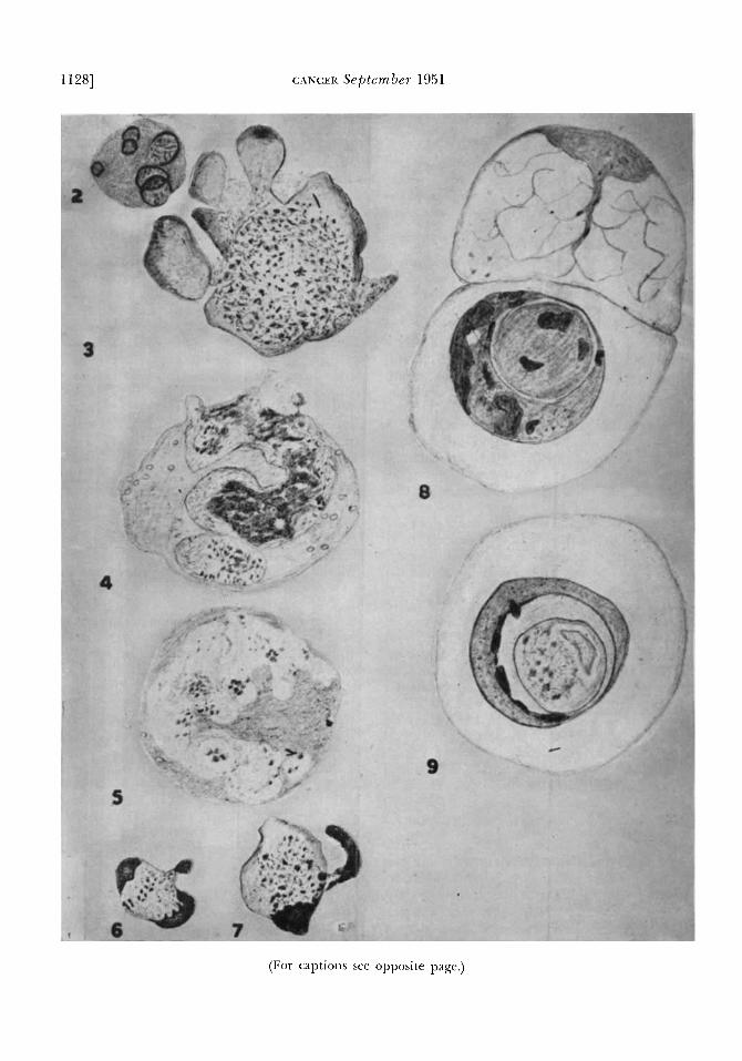
Figures 2 to 9 show the nuclear alterations noted after the administration of triethylene- melamine. All figures are drawn freehand with the use of the oil-immersion lens. The cells were observed in sections or smears that had been stained with the Fenlgen tecli- nique and counterstained with fast green. These represent photographic reproductions of colored drawings. Figures 2 to 7 were smear preparations, from the sternal or femoral marrow of mice, obtained thirteen or twenty-four hours after subcutaneous injection of 10 mg. per Kg. of triethylenemelamine. Figures 8 and 9 were sections of the small in- testine.
FIG. 2. Cell showing six nuclear fragments, each of which has reticular structure similar to that seen in the normal nucleus.
FIG. 3. The cell appears to be disintegrating. Some peripheral, fragmented, cytoplasmic masses are seen. Feulgen-positive chromatin appears as numerous dark granules scattered within the nucleus. The outline of the nucleus is indistinct.
FIG. 4. Cell with fragmented nucleus undergoing karyolysis. The residual Feulgen- positive material is represented as dark granules and as diffuse, heavily shaded material.
FIG. ti. Cell with elongated, irregularly shaped nucleus. The chromatin is almost com- pletely absent. Only a small amount of Feulgen-positive material remains, and this is shown as dark granules.
FIG. 6. Nucleus showing three niasses of Fenlgen-positive material, one portion of which is probably being extruded.
FIG. 7. Cell showing two segregated masses of Feulgen-positive nuclear material, one almost completely extruded from the cell. The granular condition of the remaining Feulgen-posit ive material is represented.
Fic. 8. Two adjacent nuclei from epithelium of a crypt of Lieberkuhn. One nucleus shows a faintly staining reticulum with a small amount of Feulgen-positive material. Its neighbor contains a complex, shrunken, pyknotic mass consisting of a central body with one envelope. The Feulgen-positive areas are shown as large, irregular, dark masses in each portion of this shrunken area.
FIG. 9. A degenerate nucleus seen in the epithelium lining a crypt of Lieberkuhn. An enclosed, complex, shrunken, pyknotic mass with a central body and surrounding envelope is represented. The Feulgen-positive material appears as dark bodies of varying lorm arid size on the inner aspect of the outer envelope and as granular, darkened areas within the central body.
11301 CANCER September 1951
terminal state, atrophy of the villi with slough was present. T h e colon showed only ii hyper- cmia in animal, that were in d terminal state.
T h e sternal ni‘irrow, normally pale pink in color, was dark red aiter the adniinistra- tion of triethyleiienielarnine. Examination ot smears and imprints, as well as histological sections, showed that the altered appearance of the marrow was due to an extreme change in its cellular content. T h e nornial comple- ment ol myeloid and erythroid heinatopoietic elements had disappeared, and only mature erythrocytes and the stronia o f reticular cells remained.
‘The changes as just described were readily observed in mice injected subcutaneously with 10 mg. per Kg. of triethylenemelamine. T h e bone marrow was studied in animals rcceiving this dosage because of the ease with which these alterations could be observed. T h e mice were sacrificed at various interkals during the initial twenty-four-hour period and thereafter at twenty-four-hour intervals. Some karyo- klastic effects were noted as early as two and three hours after drug administration, and at twelve t o thirteen hours nuclear changes were generally evident. Twenty-four hours follow- ing the injection, the manifestations of nil- clear degeneration were either at their height or completed, arid marked depletion of the bone marrow was present.
T h e number of mitoses observed in the bone marrow was reduced following injection of the triethylenenielamine. However, isolated mitoses were observed sixteen to eighteen hours following injection of 10 mg. per Kg. Preliminary observations, i n which sections and smears from onion-root tips were studied, indicated that the mitotic rate was essentially zero within twelve hours after exposure to concentrations of 0.05 or 0.1 mg. per ml. T h e nuclei were all in a resting phase except for a few in late prophase.
Alterations in the structure, form, and behavior of the nuclei were by far the most striking effects observed following the admin- istration of triethylenemelaniine. T h e nucleus appeared to undergo fragmentation into large and small particles. T h e larger fragments were usually circular and pyknotic, and two to six were present in a cell. Some appeared to re- semble a normal nucleus with a fine, reticular network of Feulgen-positive material (Fig. 2). I n other cells, a fine dustlike appearance of
these Feulgen-staining particles was noted that sugge5tetl an “explosi\e” disintegration o f the nucleus (Fig. 3).
Although pyknosis and karyorrhexis of the marrow-cell nuclei were the most prominent alterations noted following the injection of triethylenemelamine, nuclear fragmentation without loss of reticular nuclear structure, as previously noted, and karyolysis were also fre- qucntly encountered (Figs. 4, 5). I n cells un- dergoing karyolysis, the nuclear membrane became indistinct, aiid the nuclear contents appeared to traverse its limits. T h e basophilic material in the nucleus, which was also Feul- gen positive, became disorganized, with the forination of granules. T h e latter appeared to move iron1 the nucleus into the cytoplasm, aiid some were observed tree ot the cell. This basophilic grdnular substance could occasion- ally be seen about the peripheral border of the cell. These and other alterations (Figs. 6, 7) were interpreted as the extrusion of con- siderable portions of the nucleus from the cell. Representation of this phenomenon is pre- sented in Figs. 4 to 7. T h e extracellular nu- clear fragments stained more intensely with thc Feulgen reagent than the Feulgen-positive niateridl norninlly present in the nucleus, and it appeared as if condensation of the Feulgen- positive nuclear material had occurred. Re- gions of the cell, distinct from the cytoplasm but with no basophilic material, were encoun- tered on occasion. These “ghost” areas were interpreted to be nuclei from which the baso- philic contents had been removed by the proc- esses just described.
I n the bone marrow, the decreasing order of sensitivity to triethylenemelamine appeared to be as follows: immature erythroid cells, im- mature myeloid cells, mature myeloid cells, and erythrocytes. T h e erythrocytes, as far as could be judged, were not affected by this agent. Pol) morphonuclear leukocytes and meg- akaryocytes were among the hematopoietic elements in the bone marrow last to undergo degeneration. Pyknotic fragmentation ap- peared most frequently in the cells of the ery- throid series, while karyolysis, when it occurred, was usually seen in the cells of the myeloid series.
T h e lymph nodes and spleen also showed striking alterations following the use of tri- ethylenemelaniine. T h e cells in these organs showed evidence of pyknosis and fragmenta- tion of the nuclei as early as two hours after
BIOLOGICAL Aivn ANTICANCER PROPERTIES OF THREE TRIAZINES . Goldbeyg 6 Sclioenbnch [1131
the administration of triethylenemelamine, especially in the germinative areas of the lymphatic nodules. Scattered niitoses were observed up to six hours after administration of 10 mg. per Kg. In the lymph nodes the in- tensity of thcse changes became pronounced and was accompanied by enormous amounts of nuclear debris after twenty-four hours. The snialler size 01 the spleen was apparently due to marked depletion of lymph nodules and ectopic rnyelogenous areas. While the deple- tion in the lymph nodules of the spleen was almost invariably observed, the region of the red pulp was seen occasionally to undergo no appreciable decrease in size but became hcm- orrhagic. T h e depletion of heinatopoietic elements of the spleen cxposed the stromal cells. I n the spleen, as in the bone marrow, the ~~olymorphoriuclear leukocytes, as well as megakaryocytes, appeared more resistant than the other cells of the hematopoietic series.
The intestinal epithelial cells in the crypts of 1,ieberkuhn showed a decrease in number of mitoses, together with swelling o f the nu- clci, which stained much less intensely than normal. In the crypts, some cells showed a large clear area containing a pyknotic mass of basophilic material. These spheres of pyknotic
eopoo
5 1000
0 A A W 0
I00
7
40 TINE IN HOURS
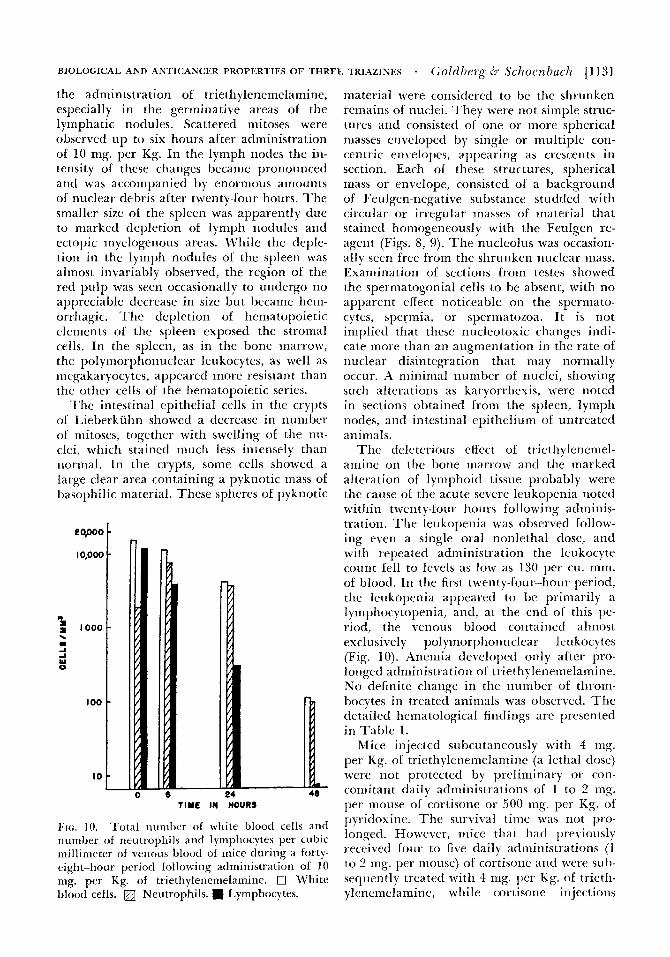
FIG. 10. ‘Total nunher of white blood cells and number of neutrophilr and lymphocytes per cubic millimeter of venous blood of mice during a forty- eight-hour period following adniinistration of 10 mg. per Kg. of triethylenenielamine. 0 White blood cells. Neutrophils. w Lymphocytes.
material were considered to be the shrunken remains of nuclei. They were not simple struc- tures and consisted of one or inore spherical masses enveloped by single or multiple con- centric en\ elopes, appearing a5 crescents in section. Each ot these structures, spherical mass or envelope, consisted of a background of Feulgen-negative substance studded with circular or irregular masses of material that stained homogeneously with the Feulgen re- agent (Figs. 8, 9). T h e nucleolus was occasion- ally seen free from the shrunken nuclear mass. Examination of sections from testes showed the spermatogonial cells to be absent, with no apparent effect noticeable on the spermato- cytes, spermia, or spermato7oa. It is not implied that these nucleotoxic changes indi- cate more than an augmentation in thc rate of nuclear disintegration that may normally occur. A minimal number of nuclei, showing such alterations as karyorrhexis, were noted in sections obtained from the spleen, lymph nodes, and intestinal epithelium of untreated animals.
T h c deleterious effect of triethylenemel- amirie on thc bone marrow and the marked alteration o f lymphoid tissue probably were the cause 01 the acute selere leukopenia noted within twenty-lour hours following adminis- tration. The leukopenia was observed follow- ing even a single oral nonlethal dose, and with repeated administration the leukocyte count fell to levels as low as 130 per cu. mm. of blood. In the first twenty-four-hour period, the leukopenia appeared to be primarily a lymphocytopenia, and, at the end of this pc- riod, the venous blood contained almost exclusi\ ely polyniorphoiiuclear leukocytes (Fig. 10). Anemia deleloped only after pro- longed administration ot trieth) lenenielamine. No definite change in the number of throm- bocytes in treated animals was obscrbed. The detailed heinatological findings are presented in Table I .
Mice injected subcutaneously with 4 mg. per Kg. of triethylenemelamine (a lethal dosc) were not protected by preliminary or con- comitant daily administrations of 1 to 2 mg. per mouse of cortisone or 500 mg. per Kg. of p)i idoxine. T h e surtival time was not pro- longed. However, mice that had p e l iously receibed four to fiqe daily atlministiations (1 to 2 nig. per mouse) of cortisone and were sub- sequently treated with 4 mg per Kg. of trieth- ylenemclamine, while cortisone injections
11321 CANCER September 1951
TABLE 1
SUMMARY OF HEMATOLOGICAL DATA FOLLOWING ADMINISTRATION O F TRIETHYLENE-
Dosage, mg./Kg. Route
10.0 Oral. 5.0 Oral 5.0 Oral
Controls Oral 2 Oral
Controls Oral 0.5 Subcu.
Controls Subcu. 10 Subcu. 10 Subcu. 10 Subcu.
Controls -
0.25 Subcu. 0.1 Subcu.
Controls - -
No. of daily doses
1 1 8 8
13 13
14 14
1 1 1
21 21
-
-
No. of mice
MELAMINE TO CF-1 MICE - ____
Days since treat.
4 4 2 5
5 5
4 4
4 3 3 6
3 5 4
15 15
3 3
1 1
1 1
1 2
2 2
K
-
-
Differential, % R.B.C., - millions W.B.C. Poly. Lym. Eos. Platelets
~ -~
9.9 11.0 10.7
9.0
11.4 10.7
7.2 11.3 - - - -
7.5 10.8 11.7
were continued, died before the control ani- mals that had received the same dose of tri- ethylenemelamine alone. Furthermore, mice so treated with cortisone succumbed even to sub- lethal doses of triethylenenielamine (2 mg. per Kg.). These relationships are being investi- gated.
ANTINEOPLA~I IC STUDIES
Melamine was administered subcutaneously, at a dosage of 500 mg. per Kg., into the p 0 5 -
terior dorsal region of mice bearing sarcoma 180. Observation was terminated when the tumor was nine days old, atter a total of seven injections. No effect of the compound on neo- plastic growth was noted, and the average tumor weight from control mice was 153 * 14.3
3,300 6,600
700 13,100
1,300 5,700
800 15,100
12,700 4,250
120 15,700
1,000 6,4on
13,900
37 63 33 67 15 85 37 63
20 80 20 80
15 84 45 55
67 32 93 7 94 6 13 85
16 84 14 85 13 86
0 0 0 0 0 0 1 0 1 0 0 2
1 1
-
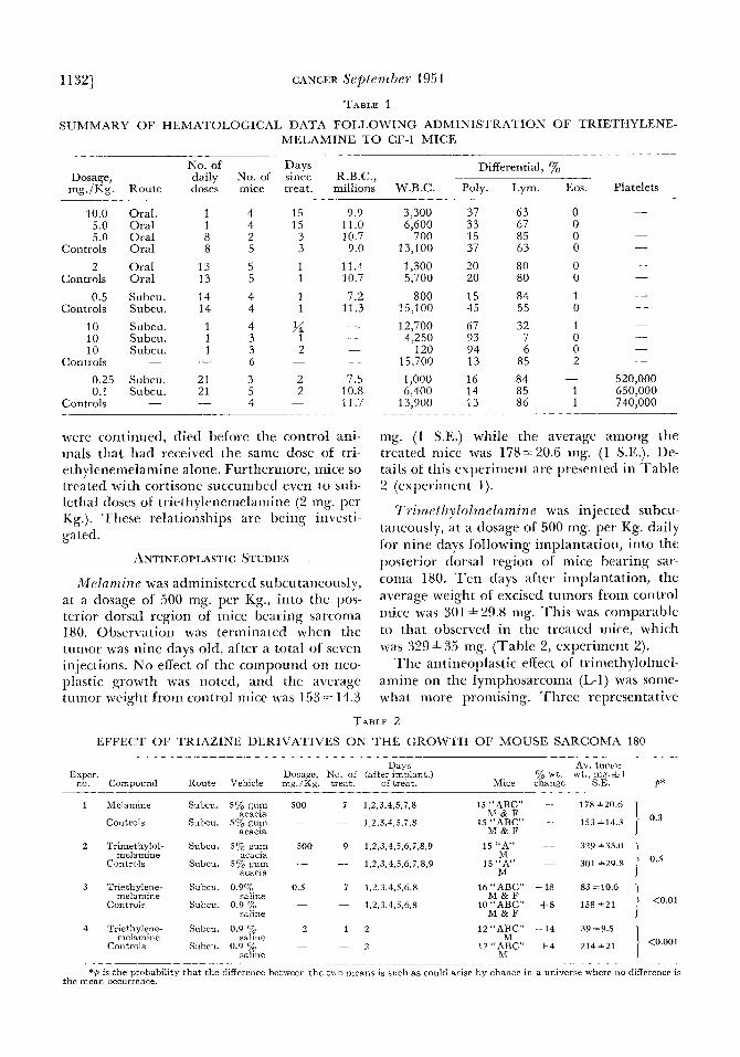
mg. (1 S.E.) while the average among the treated mice was 178A20.6 mg. (1 S.E.). De- tails of this experiment are presented in Table 2 (experiment 1).
T~tnz~ thy lo lme lamzne was injected subcu- taneously, a t a dosage of 500 mg. per Kg. daily for nine days following implantation, into the posterior dorsal region of mice bearing sar- coma 180. T e n days after implantation, the average weight of excised tumors from control mice was 301 h29.8 mg. 'This was comparable to that observed in the treated mice, which was 329
T h e antineoplastic effect of trimethylolmel- amine on the lymphosarcoma (L-I) was some- what more promising. 'Three representative
35 mg. (Table 2, experiment 2).
TABLE 2
EFFECT OF TRIAZINE DERIVATIVES ON THE GROWTH OF MOUSE SARCOMA 180
Days Av. tumor Exper. Dosage, No. of (after implant.) yo wt. wt., mg.&l
no. Compound Route Vehicle mg./Kg. treat. of treat. Mice change S.E. P*
1 Melamine Subcu. Svo gum 500 7 1,2.3,4.5,7,8 15 "ABC" - 178 +20.6 acacia M & F
acacia M & F Controls Subcu. 5% gum - - 1,2,3,4,5.7,8 15 "ABC" -
2 Trimethylol- Subcu. 5% gum 500 9 1.2,3.4,5,6,7.8.9 15"A" - 329+35.0 1 301 *29.8 i M
acacia M 15"A" -
melamine acacia 1,2,3,4,5,6.7,8,9 Controls Subcu. 5% gu,m - -
<0.01 3 Triethylene- Subcu. 0.9% 0.5 7 1,2,3,4.5.6,8 16 "ABC" - 18 83 *10.6
melamine saline M & F
saline M & F 10"ABC" +8 15X+21 1,2.3,4,5,6,8 Controls Subcu. 0.9 yo - -
2 i 12"ABC" - I 4 39*9.5 4 Triethylene- Subcu. 0.9 'r, 1 2 melamine saline M
Controls Subcu. 0.9 yo ._ - 12"ABC" +4 214*21 <O'oO1 saline M
~- ~ ____ ~ ~
*O is the probability that the difference between the two means is such as could arise by chance in a universe where no difference is the mean occurrence.
TA
BL
E
3
EF
FE
CT
OF
TR
IAZ
INE
DE
RIV
AT
IVE
S O
N T
HE
GR
OW
TH
OF
LY
MP
HO
SA
RC
OM
A L
-1 A
ND
HO
ST
SU
RV
IVA
L
Exp
er.
no.
Com
poun
d
1 T
rim
ethy
lolm
elam
ine
Con
trol
s
2 T
rim
ethy
lolm
elam
ine
Con
trol
s
3 T
rim
ethy
lolm
elam
ine
Con
trol
s
4 T
riet
hyle
nem
elam
ine
Con
trol
s
5 T
riet
hyle
nem
elam
ine
Con
trol
s
6 T
riet
hyle
nem
elam
ine
Con
trol
s
7A
Tri
ethy
lene
mel
amin
e
7B
Tri
ethy
lene
mel
amin
e C
ontr
ols
Dos
age
mg.
/Kg.
N
o. o
f tr
eat.
R
oute
V
ehic
le
Day
s (a
fter
impl
ant.
) of
trea
t.
Mic
e A
v. t
umor
dia
met
er, m
m.*
Sur
v. t
ime,
9
days
*
2
1 S.
E.
fit
(16t
h da
y)
(226
day
) 50
0 11
S
ubcu
. 10
% g
um
9.10
,11.
12,1
3,14
,15,
16,1
7,18
,20
13 "
AB
C"
11
12
-
-
Sub
cu.
10%
gym
9,
10,1
1,12
,13,
14,1
5,16
,17,
18,2
0 13
"A
BC
" 16
2
2
500
18
Ora
l 5%
gum
1,
2,4.
6,8.
11,1
3,14
,16,
18,2
0,22
, 12
"A
" 9
15
acac
ia
M
acac
ia
M
(13t
h da
y)
(22d
day
)
acac
ia
24.2
5.27
,29.
3 1.4
1 F
acac
ia
24,2
5.27
,29,
31,4
1 F
acac
ia
20,2
1,22
.23,
24,2
5 M
acac
ia
20,2
1,22
,23.
24,2
5 M
-
-
Ora
l 5%
gum
1,
2,4,
6,8.
11,1
3,14
,16,
18,2
0,22
, 11
"A
" 13
19
(14t
h da
y)
(19t
h da
y)
(23d
day
) (2
8th
day)
50
0 17
S
ubcu
. 5%
gum
1,
2,10
,11,
12.1
4.15
,16,
17,1
8,19
, 11
"AB
C"
10
7 10
13
38
+2.
4
-
-
Sub
cu.
5% g
ym
1,2,
10,1
1,12
,14,
15,1
6,17
,18,
19,
13"A
BC
" 16
19
22
e!
30*1
.9
U
(14t
h da
y)
(21s
t day
) (2
7th
day)
33
*2.
4 >
0.3
2 0.
3 12
S
ubcu
. 0.
9% s
alin
e 1.
2,3,
4,7,
8,9,
10,1
7,22
,24
19"A
BC
" 5
13
20
E -
-
Sub
cu.
0.9%
sal
ine
1,2,
3,4,
7,8,
9,10
,17,
22,2
4 1R
"AB
C"
15
26
31
30*1
.0
m F
e z K
<0.
001
*
2
M
M
F F 0.
2 12
S
ubcu
. 0.
9% s
alin
e 1,
2,3,
4.7,
8,9,
10,1
7,22
,24
19 "
AB
C'
8 20
25
30
+2.
7
R
rL
(app
rox.
)
h
i (1
3th
day)
(2
0th
day)
(3
4th
day)
(4
0th
day)
I
1.5
2 S
ubcu
. 0.
9% s
alin
e 6,
17,2
0,30
,34,
37,4
0,44
,53
10 "A
BC
" 5
6 12
17
4
6+
2,8
24 +
0.8
-
-
7 -
Sub
cu.
0.9%
sal
ine
6,17
,20,
30,3
4.37
,40,
44,5
3 10
"AB
C"
17
26
1 1.
0 -
(17t
h da
y)
(25t
h da
y)
(31s
t da
y)
3 12
O
ral
0.97
0 sa
line
1.
2,3,
4,8,
12,1
6,21
,25,
28,3
1,35
15
"A
BC
" 6
8 15
40
*3.9
0.
001
2 12
O
ral
0.9%
sal
ine
1.2,
3,4,
8,12
.16,
21.2
5,28
,31,
35
15 "
AB
C"
8 13
19
41
+2.
4 <
0.00
1 F
F F
-
-
Ora
l 0.
9% s
alin
e 1.
2.3,
4,8,
12,1
6,21
,25,
28,3
1,35
15
"A
BC
" 16
24
-
26 *
0.8
R c
R
(10t
h da
y)
(13t
h da
y)
(21s
t - da
y)
(31s
t -
day)
-
G 1
1
S
ubcu
. 0.
9% s
alin
e -8
hr
s.
9 "A
" 9
13
1 1
S
ubcu
. 0.
9% s
alin
e -1
hr
. 16
"A"
7 11
1
1
Sub
cu.
0.9y0
sali
ne
+1
hr.
19
"A
" 5
7
c; cs 3
F
0
1 S
ubcu
. 0.
97c
sali
ne
-1
hr.,
l7,
24
16 "
A"
7 11
8
5 >
70+
6.7
a
-
-
-
F
F
-
-
-
2 F
<
.001
g
-
Sub
cu.
0.9%
sal
ine
-1
hr.,
17,
24
24
"AI
8 11
19
23
34
+1.
6 i
i
-
F
-i -
*A
dia
met
er o
f 4
or 5
mm
. si
gnif
ies
alm
ost c
ompl
ete
abse
nce
ot t
umor
. t P
is
the
proh
abil
ity
that
the
diff
eren
ce b
etw
een
the
two
mea
ns is
suc
h as
cou
ld a
rise
by
chan
ce in
a u
nive
rse
whe
re n
o di
ffer
ence
is t
he
mea
n oc
curr
ence
. - - I 0
s
1 1 341 CANCER September 1951
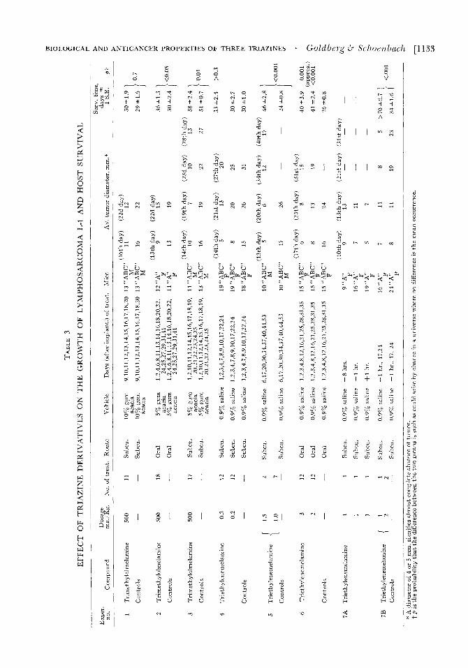
therapeutic trials are outlined in Table 3. In the first trial noted, a total of eleven injections of 500 mg. per Kg. each was administered on the ninth to twentieth day after implantation. The controls sunived an average period of 29 4 1.6 days after implantation of the tumors; and the experimental mice, 30.0 4 1.9 days. In another trial, the tumor-bearing mice received eighteen doses of 500 mg. per Kg. each on the first to twenty-fifth day alter tumor implanta- tion. The controls survived an average period ot 30A2.4 days; and the treated mice, an average period of 3 6 4 1.3 days. In the last experiment, seventeen injections of 500 mg. per Kg. were given on the first to twcnty-fifth day after implantation. The controls survived an average period ol 31 h0.7 days; and the treated mice, an average period of 38h2.4 days. Growth 01 thc local tumor was uniformly inhibited.
Trielliylenemelamine. The tumor inhibi- tion of triethylenemelamine was assayed by in- jection subcutaneously into mice bearing bilateral implants of sarcoma 180. The mice received seven daiiy injections of 0.5 mg. per Kg. beginning one day after implantation. Nine days after the tumor fragments had been implanted, the mice were sacrificed, arid the tumors were excised and weighed. Whereas the average weight of control tumors was 158421 mg., that of the experimental group was only 83 f 10.6 mg. (Table 2 , experiment 3).
Mice implanted with sarcoma 180 were treated with a single injection of triethylene- melaniine at a dosage of 2 mg. per Kg. subcu- taneously two days after implantation, in an effort to find a more favorable dosage regimen. Eight days later, the average weight of excised tumors from treated animals was only 39 9.5 mg., and the average weight of the controls was 214h.21 mg. These observations are pre- sented in Table 2 (experiment 4). Although the degree of weight loss among the treated animals was comparable in the last two experi- ments, the single injection of 2 mg. per Kg. appeared to bc far more effective than a num- ber of smaller doses.
Triethylenemelamine was administered to mice with large, established lymphosarcomas (L-1) approximately four days before the ex- pected death of the mice, in an effort to ascer- tain the effectiveness of this agent in produc- ing regression of an established tumor. The mice received one to twelve injections of 0.3
mg. per Kg. each over a period of twenty-three days. They survived one to twenty-four days following the initiation of therapy, and the average survival period was 14.0 days (a total average survival time of 40 days). The controls survived two to ten days, with an average sur- vival period of 4.5 days (total average of 30.5 days). The tumors aniong the treated animals appeared to have lost their firm texture and became noticeably sotter. Even more impres- sive results were obtained when a dose of 2 mg. per Kg. was injected subcutaneously into “ABC” mice bearing L-1 tumors (21 mm. aver- age diameter). Within four days, the average tumor si7e was reduced to approxiniately 6 mm. Eleven days after the administration of the drug, 95 per cent of the control mice had succumbed. All the treated animals were still alive, but the tuniorr among the majority of the mice had increased in ske. Continuation of this mode of drug administration failed to check the eventual growth of the tumor, which, as in other experiments, gradually ap- peared to gain an increased tolerance to the effects of triethylenemelamine.
In an initial therapeutic trial, using re- rently implanted tumors, seven injections of 0.5 mg. per Kg. of triethylenemelamine ad- ministered subcutaneously over a period of seventeen days (fourth, fifth, sixth, sevcnth, tenth, cleventh, and fourteenth day after im- plantation) almost completely prevented the growth of the L-1 tumor. With this dosage regimen considerable weight loss was encoun- tered, and shortly thereafter most of the ex- perimental animals succumbed to the toxic effects of the compound.
Because of the delayed toxicity of triethyl enemelamine, subsequent therapeutic trials were conducted in such a manner as to avoid the untoward manifestations. Administration of triethylenemelamine was discontinued when a distinct weight loss was noted among the groups of treated animals or when the mice appeared ill, as indicated by one or more of the following symptoms: sluggishness, hunched appearance, or rough coat. Therapy was resumed when the mice appeared in bet- ter condition. In the first such trial, the trieth- ylenemelaniine was administered twelve times subcutaneously at a dosage of either 0.3 or 0.2 mg. per Kg. during a period of thirty-nine days. Sixty “ABC” feniale mice inoculated with the L-1 lyinphosarcoma were employed. They were divided at random into three
groups of twenty each. Twenty served as con- trols, and twenty received individual doses of 0.3 or 0.2 mg. per Kg. The growth of the tu- mor was markedly inhibited, but the average survival time of the treated mice was only slightly prolonged (Table 3, experiment 4).
T h e effect of the triethylenemelamine on the lymphosarcoma appeared much more striking when the individual dosage, as well as the intervals between administration, was increased. Dosages of 1.5 to 1.0 mg. per Kg. of triethylenemelamine, spaced at intervals of three to thirteen days, were administered. This schedule permitted the lymphosarcoma-bear- ing mice to recover from the toxic effects. The details of this experiment are presented in Table 3 (experiment 5) . The tumors grew niore slowly, and the survival time of the treated mice was 46 f 2.8 days; among the con- trols the tumors were larger, and the survival time was 24A0.8 days. The final measure- ments of tumor diameters, among survivors, aleraged 17.2 mm. Here, again, there were indications that the tumor was gaining some tolerance to the growth-inhibiting properties noted earlier in the treatment.
Triethylenemelamine was found to be effec- tive when administered by gavage. The timing of doses was similar to that used in the preced- ing experiment. T h e mice were treated twelve times during a period of thirty-five days with either 2 or 3 mg. per Kg., and the results are presented in Table 3 (experiment 6). The average survival time for the control group was 26A0.8 days, while the mice receiving 2 ing. per Kg. lived, on the average, for 41 2.4 days, and those receiving 3 mg. per Kg. lived for an average of 40 f 3.9 days.
T h e observation that a single injection of a relatively large dose of triethylenenielaniine could noticeably affect the growth of the L-1 lymphosarcorna permitted further invcstiga- tions as to the mechanism of action of this drug. Sixty-nine female A mice were divided into four groups with respect to time of ad- ministration of a single therapeutic dose as related to the time of implantation of the tumor. One group received the agent eight hours before tumor implantation; another, one hour before; and a third, one hour after. The fourth group (controls) was implanted last, in order to obtiate the influence of un- iaborable environmental conditions on the minced tumor material. The results are sum- mariled in Table 3 (experiment 7A). Only one
BIOLOGICAL AND .4NTICANCER PROPERTIES OF THREE TRIAZINES . Goldberg dT Schoenbacli [ 1 1 35
injection of the triethylenemelamine was made. The dosage employed was 1 nig. per Kg. administered subcutaneously. It appeared that the action of triethylenernelamine occurred within an hour after injection. Cells inoculated one hour after triethylenemelaniine adminis- tration were unaffected, while thoye implanted one hour before treatment were inhibited. A direct action on the cells ot the tunior prob- ably occurred because the cells inoculated after the administration of triethyleneiiielamine were unaffected although exposed to the un- favorable host environment resulting from the host toxicity induced by the triethylenemela- mine.
Mice that had received triethylenemelamine one hour before tumor implantation, as noted in Table 3 (experiment 7A), received two additional subcutaneous injections with 2 ing. per Kg. on the seventeenth and twenty-fourth days after implantation (Table 3, experiment 7B). After the second injection, almost com- plete regression of the tumors occurred. Only one survivor of this group died with a tumor. Five mice died from the toxic effects of the therapeutic agent. T e n survivors, discarded on the eighty-seventh day after implantation, showed no evidence of tumor forty-four days after the longest-lived control had died with a large tumor.
It was possible completely to inhibit growth of the tumor in 90 per cent of the treated animals, for a period of more than three months, with an initial subcutaneous dose of triethylenemelamine (2 mg. per Kg.) given to mice at the time the lymphosarcoma was inoc- ulated. Prolongation of the time interval be- tween tumor implantation and treatment with triethylenenielainine resulted in a reduction in the proportion of animals in which the tumor growth could be suppressed. Thus, when seven hours elapsed between tumor im- plantation and institution of triethylenemel- amine therapy, only 30 per cent of the mice were free of tumors at the end of the three- month period. Although a single injection of 2 mg. per Kg. of triethylenemelamine was capable of producing very marked regression of established twenty-three-day-old lympho- sarcomas in all the treated mice, it was not found possible to prevent tumor growth or host death by continued administration at this dosage level. On continued treatment, the tu- mors appeared to escape from the growth- inhibitory properties of triethylenemelarnine.
11361 CANCER Srptenzber 1951
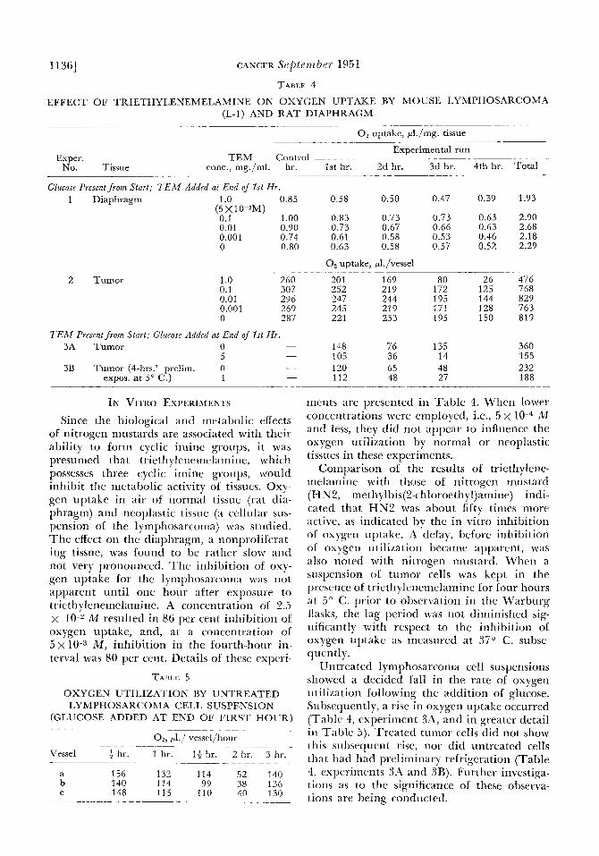
TABLE 4
EFFECT OF TRIETHYLENEMELAMINE ON OXYGEN UPTAKE BY MOUSE LYMPHOSARCOMA (L-I) AND RAT DIAPHRAGM
- ___ __ ~~~~~ ~~ ~ - - O2 uptake, pl./mg. tissue
Exper. No. Tissue
Experimental run TEM Control ~ ~ ~ _____ ~ _ _ _ _ _
conc., mg./ml. hr. 1st hr. 2d hr. 3d hr. 4th hr. Total
Glucose Present from Start; T E M Added at End of 1st Hr. 1 Diaphragm 1 .o 0.85 0.58 0.50 0.47 0.39 1.93
0.1 1 .oo 0.83 0.73 0.73 0.63 2.90 0.01 0.90 0.73 0.67 0.66 0.63 2.68 0.001 0.74 0.61 0.58 0.53 0.46 2.18 0 0.80 0.63 0.58 0.57 0.52 2.29
(5 x 1 0 - 3 ~ )
0, uptake, pl./vessel
2 Tumor 1 .o 260 0.1 307 0.01 296 0.001 269 0 287
'TEM Present from Start, Glucose Added at End of 1st Hr. - 3A Tumor 0
5 - 3B Tumor (4-hrs.' prelim. 0 -
- ___
expos. at 5" C.) 1 -
IN V I T R ~ EXPFRIMLNT~ Since the biological and metabolic effects
of nitrogen mustards are associated with their ability to form cyclic iniinc groups, it was presumed that triethylcnemelaniine, which possesses three cyclic irriiiie groups, would inhibit the metabolic activity ot tissues. Oxy- gen uptake in air of normal tissuc (rat dia- phragm) and ncoplastic tissue (a cellular sus- pension of the lymphosarconia) was studied. The effcct on the diaphragm, a nonpi olilcrat- ing tissue, was h i n d to be rather slow and not very pronounced. Thr. inhibition of oxy- gcn uptake for the lyrripliosarcorria was not apparent until onc hour after exposure to triethylencmelamine. A concentration of 2.5 x lo-" M resulted in 86 per cent inhibition of oxygen uptake, and, at a Concentration of 5 x 10⁻³ M , inhibition in the fourth-hour in- terval was 80 per cent. Details of thesc experi-
TABLE 5
OXYGEN UTILIZATION BY UNTREATED LYMPHOSARCOMA CELL SUSPENSION
(GLUCOSE ADDED AT END OF FIRST HOUR) ~~ ~~~~~
~
0 2 , p1 1 vrssel/hour ~ ~ ~~ ~~ ~~
Vessel f hr. 1 hr. 11. hr. 2 hr. 3 hr. -~ ~~ __ ~ ~ ~ ~ ~ _ _ _ _ _ ~
a 156 132 114 52 140 b 140 114 99 38 136 C 148 115 110 40 130
201 169 80 26 476 _. . ... ..
252 219 172 125 768 247 244 195 144 829 245 219 171 128 763 221 253 195 150 819
148 76 135 105 36 14 120 65 48 112 48 27
360 155 232 188
nicnts are presented in Table 4. When lower concentrations were employcd, i.e., 5 x M and less, they did not appear to influencc the oxygen titilkation by normal or neoplastic tissues in these experiments.
Comparison ol the results of triethylenc- roelaniinc with those of nitrogcn mustard (HNZ, methylbis(2-cli1oroethyl)amine) indi- cated that H N 2 was about fifty times more active, as indicatcd by the in vitro inhibition o f oxjgen uptake. A delay, before inhibition of oxygen utilization became apparcnt, was also rioted with nitrogen iuustard. Whcn a wspension ot turnor cells was kcpt in the p~ esence of ti icthylenerrielaminc lor four hours at 5" C. prior to obscrvation in the Warburg flasks, thc lag pcriod was not diminished sig- nificantly with respect to the inhibition of oxygen uptake as measured at 37" C. subse- quen tly.
Untreated lympliosarcoma cell suspensions showed a decided la11 in the rate of oxygen utilimtion following the addition of glucose. Subsequently, a rise in oxygen uptake occurred (Table 4, experiment X4, and in greater detail in Table 5) . Treated tunior cells did not show this subsequent rise, nor did untrcated cells that had liad preliminary refrigeration (Table 4, expcriiricnts 3.4 and SB). Furthcr investiga- tions a5 to the significance of these observa- tions are being conducted.
BIOLOGICAL AND ANTICANCER PROPERTIES OF THREE TRIAZINES . Goldberg kb S C h O e ? l b U C / l [1137
D~scussro~
Of the three triazine derivatives investi- gated, antineoplastic activity has been found in only two: triinethylolmelar~iine and trietli- ylenemelamine. Trinic.tliylolinelaiiiine pro- duced some inhibition of growth and a slight increase in survival time in the more respori- sive tumor, lyniphosarcoma L-1. Burchcnal et al.;? in screening thirty-nine triazines, reported no significant effect of trimethylolmelamine against transplanted mouse leukemia Ak 4. However, Rose et al. found that this com- pound inhibited the growth of Walker rat carcinoma 256 and two transplantable mouse tumors. They also noted that i t was toxic, producing a severe anemia in the rat. Some of the discrepancy in the results reported may be attributed to the instability oi the trimeth- ylolmelamine as well as to differences in mode of assay. T h e early samples employed in this report gave a strong odor and test for formal- dehyde, which was not as prominent subse- quently. In the mouse, no evidence of marked hematological, or other, toxicity was noted in our studies.
Triethylenemelamine has been found con- sistently to J>OSSCSS inhibitory activity on the growth of neoplastic tissue. The host weight loss (about 15 to 18 per cent) in experiments with sarcoma 180 made interpretation as to thc specificity of observed changes somewhat difficult. The observations, previously re- ported,12 employing sarcoma 180, indicated that weight loss alone, to the extent observed with triethylenciiielamine, might result in as much as 50 per cent inhibition of tumor growth. There seems to be some basis for a more specific antineoplastic effect of triethyl- enemelamine, inasmuch as greater than 50 per cent inhibition of tumor growth was observed, and other compounds that resulted in similar weight loss were not so effective in retarding tumor growth. Inhibition and complete sup- pression of newly implanted tumor, as well as inhibition of established, and in some in- stances complete regression of, tumors, for the experirncntal period (three months) were ob- served following the administration of trieth- ylencrrielaniinc to mice bearing lyrnphosar- coma 1,-1. In those instances in which one to three massive doses (1 t o 2 mg. per Kg.) of the agent (lid iiot completely suppress the tumor, the slower and eventual attainment of consid- erable tumor size indicated that tolerance to
this agent may develop. I n spite of the in- creased tolerance or, conversely, the decreased efficacy of the drug, successi~ e administrations continued to show some effect on the tumor, i.e., softer texture, inhibition ol growth, and, usually, appreciable regre5sion. Evidence ob- tained using different dosage regimens ap- peared to indicate that larger single doses at infrequent interkals were more effective than frequent smaller doses with respect to pro- longation of survival and local tumor inhibi- tion.
By administration of triethylenenielainine at various periods with reference to time of implantation, it was possible to divorce the effects of unfal orable host environment from those resulting from the direct action of the drug upon the tumor cells. Thus, the effect of the agent was greatest when given at, or after, tumor implantation. When the triethyl- enenielamine was administered subcutane- ously even one hour before tumor implanta- tion, almost no inhibitory effect was noted.
Burchenal et aL4 j , 6 reported that only triazine deri\ati\ es containing the ethyleni- mine ring possessed antineoplastic activity. I t apprars that, as in the case of the nitrogen mustards, the cyclic iniine group is associated with striking biological activity. It may be noted that, when a nitrogen mustard forms the imine ring, the nitrogen constituent is in the quaternary state and is part of an imonium ion, while the nitrogen of the cyclic iniines in triethylenemelnmine is in the tertiary state. .An equix alent amount of a bis(2-chloroethyl) aniine would yield only two thiids as ninny cjclic iniine groups as trietliylenemelaniine. HOM e l cr, one cannot predict that triethylene- melamine would be more potent, since other studies ha1 e indicated that the eflect of these groups per molecule is iiot necessarily adtli- ti\e.ll
Both experimental and clinical investiga- tions hale shown that nitrogen mustards act rapidly on exposed tissues. Bodenstein2, 3 has shown thdt embryonic amphibian tissues were affected within five minutes after exposure. El idence presented in this paper (Table 3, expel iinent 7 ) indicates that triethylenemela- mine had little effect upon tumors implanted one hour after treatment ol the host. Persotin1 obsei\ations with sill coma 180 had shown that, in l i tro, H N 2 in a concentidtion of 10 M almost abolished oxygen utilization after four- hours’ exposure. In our present report, a fifty-
11381 CANCER September 195 1
fold greater concentration of triethylenemela- mine was found necessary for similar inhibi- tion.
The action of triethylenemelamine on the cells of the bone marrow, lymphoid tissues, the intestinal epithelium, and spermatogonia characterize it as a karyoklastic or nucleotoxic agent similar to benzene,? ethyl arba am ate,^ arsenic,lO nitrogen and sulfur mustards,’“ hy- droquinone,lo roentgen rays,Y and radioactive agents.1 Dustin has indicated that these toxic substances affect all rapidly dividing tissues. The phenomena of pyknosis and karyorrhexis appear to be common to all these agents. Love- less and Revel1 have called these phenomena merely general toxic eifects, and they prefer to reserve the term “radiomimetic poisons” for those substances that act on resting nuclei and result in abnormal chromosome bcliavior after these initial general toxic effects have disappeared. These substances include the sulfur and nitrogen mustards, diepoxides, and protoanemonin. Presumably tricthylenemcla- mine may be grouped with these ~ubstances.’~
Bodenstein3 observed two types of nuclear fragmentation in embryonic amphibian-eye tissues. In only one type did enlargement of the nucleus take place prior to its subsequent disintegration. This latter phenomenon oc- curred in the epithelial cells of the glands of Lieberkuhn under the influence of triethylene- melamine. The similarity of manifestations from such a diversity of chemical and physical agents does not necessarily imply a common mechanism of action. Alteration in thc viabil- ity of the cell may result in a similar sequence of cellular and nuclear changes. Triethylene- melamine is a powerful alkylating agent, and it may be inferred that its action on the con- stituents of neoplastic tissue is through this mechanism. The observations on the antineo- plastic activity of triethylenemelamine re- ported at this time would indicate that this compound may influence the growth of certain neoplasms but only for a limited period. I t cannot be considered as a specific antineoplas- tic agent.
SUMMARY
1. The toxicity and antineoplastic activity of the following triazines in mice were inves-
tigated: melamine, trimethylolmelamine, and triethy lenemelamine.
2. Melamine and trimethylolmelamine were of a low order of toxicity. The LD,, for a single dose of triethylenemelamine was about 3 mg. per Kg., and the daily dosage tolerated was approximately one-twentieth of the LD,,.
3. Increased karyorrhexis and pyknosis were noted within two hours after the subcutaneous administration of triethylenemelamine and resulted in severe leukopenia and damage to proliferating cells and lymphoid tissues such as the bone marrow, intestinal epithelium, and spleen.
4. Neithcr melamine nor trimethylolmela- mine showed any activity against sarcoma 180. Trimethylolmelamine inhibited the growth of a lyniphosarcoma and increased the host- survival time slightly.
5. Triethylenemelaniine showed consider- able activity against both the mouse tumors employed. Administration of triethylenemel- amine in doses approximating the LD,, (1 to 2 mg. per Kg.) at selected intervals resulted in maximal suppression ok tumor implants, in- hibition of tumor growth, regression of cstab- lished tumors, and prolongation of host sur- vival.
6. Triethylenemelamine was found to be rapidly inactivated aiter administration. The effect following its injection could not be noted upon the lymphosarcoma (L-1) after onc hour. The action of the triethylenemela- mine upon the tumor could not be attributed to the unfavorable environment induced in the host, yince tumor implanted one hour after drug administration was not inhibited.
7. Oxygen utili~ation by tumor tissue was found to be inhibited by exposure to triethyl- enemelamine. A latent period was noted be- fore such inhibition became apparent. A greater concentration of triethylenemelamine was required to produce marked inhibition than had been previously observed in similar experiments with the nitrogen mustards.
8. The mechanism of tumor inhibition by triethylenemelamine did not appear to be specific but resembled that of other compounds that had been shown to possess nucleotoxic properties.
BIOLOGICAL AND ANTICANCER PROPERTIES OF THREE TRIAZINES . Goldbe7-g & S C ~ l O P n b U C ~ I [1139 REFERENCES
I . R L O ~ R I , rV., Ed.: Histopathology of Irradiation lrorn Internal and External Sources. New York. Rlc- (;raw-Hill Kook G o . 1948.
2. I%OI)ENSIXIN, I).: ’l’he effects of nitrogen mustard on enihryonic amphibian development. 1. Ectoderrnal effects. 7. Exper. ZOO^. 104: 311-341, 1947.
3. BODENSTEIN, D.: The effects of nitrogen mustard on emljryonic amphibian development. 11. The effects on e)e developnient. 7. Exper. Zool. 108: 93-125, 1948.
4. I%URCHENAI., 1. H.; CROSSLEY, M. L.; STOCK, C. C., and RHoAIjS, C. P.: The action of certain ethylenimine (aziridine) derivatives on monse leukemia. Arch. Riochrnt. 26: 321-323, 1950.
5. KUKCHENAL, J . H.; JOHNSTON, S. F.; CREMEK, M. A,: \ V l i i ~ ~ , L. E., and SI‘OCK, C. C.: Chemotherapy of leukemia. V. Effects of 2,4,6-trietliylenimino-s-tri- azine and related compounds on transplanted mouse leukemia. Proc. Soc. Exper. B i d . tr M r d . 74: 708-712, 1950.
6. KUKCHKNAL, J. H.; JOHNSTON, S. F.; STOCK, C. C.: CROSSLEY, M. L., and RHOADS, C. P.: The effect of 2,4,G-triethyleneimino-s-triazine and related compounds on transplanted mouse leukemia. [Ahstr.] Cancer Re- seurch 10: 208, 1950.
7. DIJSTIX. A. P.: Kernaroues sur les effets caryo- clakiues dn heniol. Compt . rend. Soc. de biol. 107: 1567-1569, 1931.
8. I)IISTIN, A. P., and GR~GOIRF., C.: ktude comparke de l’action des rayons X sur le thymus adulte et sur le thymus ernbryonnaire. Coiript. rend. SOC. de biol.
9. I)US.I.IN, P., JR.: The cytological action of ethyl- carbarnate (urethane) and other carbamic esters in normal and lenkaemic niice, and in rabbits. Brit . J. Cancer 1: 48-59, 1947.
10. DUSTIN, P., JR.: Some aspects of mitotic poison- ing. Nature, London 159: 794-797, 1947.
11. GOLDIN, .I.: NoF., H. A.: LANDING, B. H.; GOLD- HEKC, B., and FUCMANN, R.: Some relationships of structure to biological activity in the nitrogen mns- tards and related compounds. Actu, Union internat. contre cancer 6 : 501-516, 1949.
107: 1565-1567, 1931.
12. GOUIIN, A,; GOLDBEKC, B.; ORTECA, L. G.; FUC- hIANh’, R.; FAIJIAh’, F., and SCHOENBACH, E. B.: Effec-
tiveness of nitrogen niustards in retarding the growth of sarcoma 180 with reference to route of administra- tion, age of tumors, food intake, and weight loss of the mice. Cancer 2: 865-876, 1949.
13. KINDRLD, J. E.: Histologic changes occurring in the heniopoietic organs of albino rats after single injections of 2-chloroethyl vesicants; a quantitative study. Arch. Path. 43: 253-295, 1947.
14. LEWIS, M. R., and CROSSLEY, M. L.: Retardation of tninor growth in mice by oral administration of ethylenimine derivatives. Arch. Biochem. 26: 319-320, 1950.
15. LOVELESS, A,, and REVELL, S.: New evidence on the mode of action of mitotic poisons. Nature, London 164: 938-944, 1949.
16. PIIILIPS, F. S.: Recent contributions to the phar- macology of bis(2-haloethylj amines anti sulfides. J. Pharmacol. 6 Exper. Therap. 99 (l’t. 11): Pharmacol.
17. PHILIPS, F. S., and THIERSCH, J. B.: The nitrogen mustardlike actions of 2,4,G-tris (ethyleneiminoj-s-tri- azine and other bis(ethy1eneiminesj. J . Pliarniacol. & Exper. Therap. 100: 398-407, 1950.
18. POTTER, V. R., Ed.-in-Chief: Methods in Medical Research. Chicago. Year Book Publishers, Inc. 1948: Vol. I, p. 299.
19. ROSE, F. L.; HENDRY, J. A,, and WALroLE, A. L.: New c) totoxic agents with tumor-inhibitory activity. Nature, London 165: 993-996, 1950.
20. SCHOENBACH, F.. B., and GREENSPAN, E. M.: The pharmacology, mode of action and therapeutic poten- tialities of stilbamidine, pentamidine, propamidine and other aromatic diarnidines; a review. Medicine 27: 327-377, 1948.
21. SIJCI~JRA, K., and STOCK, C. C. Action of 3-bis @-chloroethyl) aminomethyl-4-niethoxy-1ncthyl-5-hy- droxy-6-methyl pyridine dihydrochloride, 2,4,G-tris (l-ariridyl)-s-triazine and 5-amino-7-hydroxy-1H-v-tria- 2010 ( d ) pyrimidine on carcinoma, sarcoma, osteogenic sarcoma, lymphosarcoma, and melanoma in animals. [Abstr.] Cancer Research 10: 244, 1950.
22. THIEKSCH, J. B., and PIIILIPS, F. S.: Some sub- stituted ethylenimines with actions like nitrogen mns- tards. [.4bstr.] Federation Proc. 9: 320, 1950.
Rev. 2: 281-323, 1950.


![Supporting Information Annulations Reactions in Onepot · 1 Supporting Information Synthesis of 1,2,4-Triazine Derivatives via [4+2] Domino Annulations Reactions in Onepot Dong Tang,a](https://img.pdfslide.us/doc/110x75/5f07e35c7e708231d41f41dd/supporting-information-annulations-reactions-in-1-supporting-information-synthesis.jpg)