Embed Size (px)
Citation preview
Georgia Southern University
Digital Commons@Georgia Southern
Electronic Theses and Dissertations Graduate Studies, Jack N. Averitt College of
Spring 2019
The Bioarchaeology of the Tugalo Site (9ST1): Diet, Disease, and Health of the Past Nompumelelo Beryl Hlophe
Follow this and additional works at: https://digitalcommons.georgiasouthern.edu/etd
Part of the American Material Culture Commons, Archaeological Anthropology Commons, Biological and Physical Anthropology Commons, Classical Archaeology and Art History Commons, Ethnic Studies Commons, Geography Commons, Indigenous Studies Commons, Museum Studies Commons, Other American Studies Commons, Other Classics Commons, Other History Commons, Other History of Art, Architecture, and Archaeology Commons, Other Philosophy Commons, and the Social and Cultural Anthropology Commons
Recommended Citation Hlophe, Nompumelelo Beryl, "The Bioarchaeology of the Tugalo Site (9ST1): Diet, Disease, and Health of the Past" (2019). Electronic Theses and Dissertations. 1941. https://digitalcommons.georgiasouthern.edu/etd/1941
This thesis (open access) is brought to you for free and open access by the Graduate Studies, Jack N. Averitt College of at Digital Commons@Georgia Southern. It has been accepted for inclusion in Electronic Theses and Dissertations by an authorized administrator of Digital Commons@Georgia Southern. For more information, please contact [email protected].
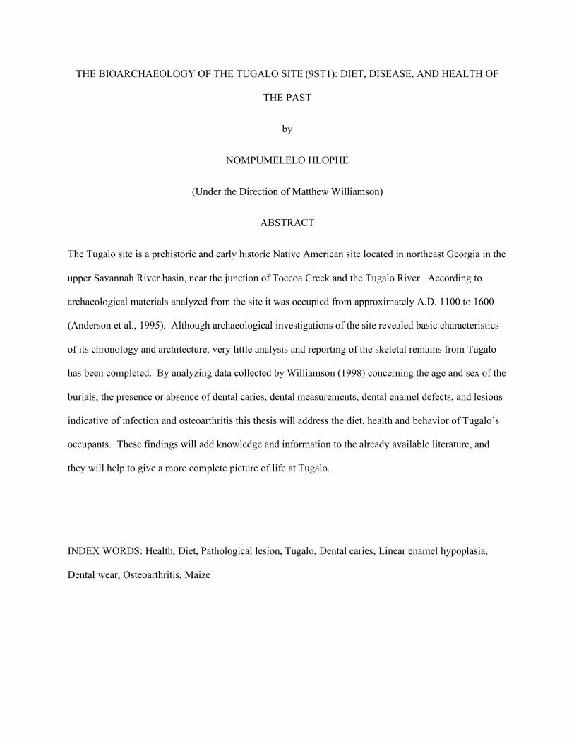
THE BIOARCHAEOLOGY OF THE TUGALO SITE (9ST1): DIET, DISEASE, AND HEALTH OF
THE PAST
by
NOMPUMELELO HLOPHE
(Under the Direction of Matthew Williamson)
ABSTRACT
The Tugalo site is a prehistoric and early historic Native American site located in northeast Georgia in the
upper Savannah River basin, near the junction of Toccoa Creek and the Tugalo River. According to
archaeological materials analyzed from the site it was occupied from approximately A.D. 1100 to 1600
(Anderson et al., 1995). Although archaeological investigations of the site revealed basic characteristics
of its chronology and architecture, very little analysis and reporting of the skeletal remains from Tugalo
has been completed. By analyzing data collected by Williamson (1998) concerning the age and sex of the
burials, the presence or absence of dental caries, dental measurements, dental enamel defects, and lesions
indicative of infection and osteoarthritis this thesis will address the diet, health and behavior of Tugalo’s
occupants. These findings will add knowledge and information to the already available literature, and
they will help to give a more complete picture of life at Tugalo.
INDEX WORDS: Health, Diet, Pathological lesion, Tugalo, Dental caries, Linear enamel hypoplasia,
Dental wear, Osteoarthritis, Maize
THE BIOARCHAEOLOGY OF THE TUGALO SITE (9ST1): DIET, DISEASE, AND HEALTH OF
THE PAST
by
NOMPUMELELO HLOPHE
B.A., University of South Africa, South Africa, 2015
A Thesis Submitted to the Graduate Faculty of Georgia Southern University in Partial Fulfillment of the
Requirements for the Degree
MASTER OF ARTS
STATESBORO, GEORGIA

© 2019
NOMPUMELELO HLOPHE
All Rights Reserved
1
THE BIOARCHAEOLOGY OF THE TUGALO SITE (9ST1): DIET, DISEASE, AND HEALTH OF THE PAST
by
NOMPUMELELO HLOPHE
Major Professor: Matthew A. Williamson
Committee: J. Matthew Compton
M. Jared Wood
Electronic Version Approved: May 2019
2
DEDICATION
To my mother and father
“I’m proud of my hard work. Working hard won’t always lead to the exact things we desire. There are
many things I’ve wanted that I haven’t always gotten. But, I have a great satisfaction in the blessings
from my mother and father, who instilled a great work ethic in me both personally and professionally.”
Tamron Hall
3
ACKNOWLEDGMENTS
I wish to thank my Master thesis committee members, Dr. Matthew Williamson (Chair), Dr.
Matthew Compton, and Dr. Jared Wood for their guidance and support. A big thank you goes to my chair
for allowing me to use his data, for his encouragement, his bioarchaeology, anatomy, and paleopathology
teachings, and his wonderful music. Without them, this thesis would have not been possible. I would
also like to thank my professors; Dr. Heidi Altman, Dr. Bridgman Sweeney, Dr. Ted Brimeyer, Dr.
Adrienne Cohen, Dr. Kevin Cook, Dr. Matthew Flynn, Dr. Ryan McNutt, Dr. Eric Silva, Dr. Jennifer
Sweeney Tookes, Dr. Matthew Taylor, and Dr. Marieke Van Willigen for their teachings and support. In
addition, I would like to thank Georgia Southern University for giving me a work space to complete my
analysis and writing.
Special thanks go to Dr. Lee Berger and Dr. Marina Elliott from the University of Witwatersrand,
South Africa. It is through them I was first introduced to the world of Biological Anthropology and
learned the importance of studying human skeletons in order to understand human lifeways. It is because
of their constant support and patience that I am able to gather and thoroughly evaluate human skeletal
materials.
A huge thank you goes to Lyda Hill, my sponsor, and her team for giving me this great
opportunity to follow my dreams. Thank you for believing in me and reminding me of the importance of
women in science.
I would like to thank the following Anthropology and Sociology, and Criminal Justice graduate
and undergraduate students from the Statesboro campus, present and past: Rhianna Bennet, Lindsey
Hinson, Scott Clark, Zack Dirnberger, Ahmauri Williams-Alford, Colin Partridge, Kendra Cooper, Ernest
Zittrouer, Megan Philips, Julia Thomas, Leigha Williams, Emily Jones, and Val Sheridan. You were not
only my cohorts, but you are also incredible friends. Thank you for always being willing to give a
helping hand when I needed it and the never-ending jokes that kept me sane. I will forever be grateful for
all the love and support from everyone.
4
To my family, thank you for the calls, constant love and support. To my siblings Nokubonga
Luthuli, Simo and Ongezwa Hlophe, my aunts and uncles Pamela Dube, Zethu and Joe Kapika, Lindiwe
and Sthembiso Nhlapo, and Ncamisile Hlophe who regularly showered me with love and support. To my
cousins Sibongile Nhlapo, Azande Dube, Sandile Nhlapo, Siphelele Nhlapo, Siyanda Nhlapo, Ayanda
Ngiba and Thobanjalo Shangase thank you for always making me laugh with your funny stories from
back home. To my friends in South Africa and the United States; Purva Mallya, Nomkhosi Khumalo,
Nosipho Madondo, Lindokuhle Ngwenya, Wandile Langazana, Gustavo Silveira, Giovana Macon,
Eugenia Tan, Sygan Fugeria, Kobih Nkrumah, Ken Tsheowang, Mengling Zhu, David Olatunde, Rakhi
Trivedi, Sharmita Porshia and Emily Petroff thank you for reminding me that behind every graduate
student is a group of good friends who constantly remind her there is a world outside the office/ lab.
Lastly, to my parents Zwelibanzi and Nomusa Hlophe, there are no words to express how grateful
I am for your constant love, encouragements, and prayers. With that I say “ngiyabonga ngothando
nesineke senu.”
5
TABLE OF CONTENTS
Page ACKNOWLEDGMENTS……………………………………………………………………………...3 LIST OF TABLES………………………………………………………………………………….......6 LIST OF FIGURES…………………………………………………………………………………….7 CHAPTER 1 INTRODUCTION…………………………………………………………………………...8 Understanding Bioarchaeology……………………………………………….............8 Examples of Bioarchaeological Studies……………………………………………....9 2 PURPOSE…………………………………………………………………………………..16 Archaeological Background of the Tugalo Site..……………………………………16 Cultural Context……………………………………………………………………...25 The Cherokee Life…………………………………………………………………...28 3 MATERIALS AND METHODS…………………………………………………………...32 Description of Skeletal and Dental Lesions Used in the Study………………….......34 Dental Caries…………………………………………………………………………34 Linear Enamel Hypoplasia……………………………………………………...........35 Periostitis…………………………………………………………………………......36 Osteoarthritis………………………………………………………………………....37 4 RESULTS………………………………………………………………………………......39
Specimen Description……………………………………………………………......39 5 DISCUSSION AND CONCLUSION……………………………………………………...45
Discussion……………………………………………………………………………45 Conclusion……………………………………………………………………….......51 Limitations…………………………………………………………………………...54 REFERENCES………………………………………………………………………………………...56 APPENDICES A Dental Lesions on Mandibular Permanent Dentition………………………………………. B Dental Lesions on Maxillary Permanent Dentition…………………………………………. C Dental Lesions on Mandibular Deciduous Dentition………………………………………... D Dental Lesions on Maxillary Deciduous Dentition…………………………………………. E Hypoplasia on Maxillary Permanent Dentition……………………………………………… F Hypoplasia on Mandibular Permanent Dentition…………………………………………….. G Pathological Lesions…………………………………………………………………………
H Georgia Southern University Laboratory Inventory Coding Format (From Williamson, 1998)……………………………………………………………………………….................82
3 6 7 8 8 9 16 16 25 28 32 34 34 35 36 37 39 39 45 45 51 54 56 66 70 74 76 77 79 80 81
6
LIST OF TABLES
Page
Table 1: Specimen List…………………………………………………………………………………..33
Table 2: Joint complexes observed for osteoarthritis (from Williamson, 1998)………………………...38
Table 3: Comparisons of lesion frequencies by sex……………………………………………………..42
Table 4: Comparison of lesion evidence by specimen number…………………………….....................43
Table 5: Comparison of lesion frequencies by specimen number……………………………………….44
Table 6: Comparisons of lesion frequencies of Mississippian Sites…………………………………….47
33
38
42
43
44
47
7
LIST OF FIGURES
Page
Figure 1: Map showing Tugalo Site……………………………………………………………...........19
Figure 2: Specimen Age Distribution………………………………………………………………....33
Figure 3: Dental Caries (Photo courtesy: Matthew Williamson)………………………………...........34
Figure 4: Linear Enamel Hypoplasia (Photo: Matthew Williamson by permission of San Diego Museum
of Man Pathology Collection)…………………………………………................................................35
Figure 5: Periostitis (Photo: Matthew Williamson by permission of San Diego Museum of Man Pathology
Collection)…………………..................................................................................................................37
Figure 6: Osteoarthritis (Photo courtesy: Matthew Williamson)………………………………...........51
19
33
34
35
37
51
8
CHAPTER 1
INTRODUCTION
Understanding Bioarchaeology
The term Bioarchaeology was first applied to the study of animal remains from archaeological
settings (Buikstra, 2006; Knüsel, 2010; Larsen and Walker, 2010). The focus later shifted when a
“bioarchaeological investigation” of human remains from the region of the lower Illinois River valley was
conducted to understand human lifeways (Buikstra, 1977). Human skeletal and dental tissues are known
to be sensitive to the environment because of factors that affect their growth and appearance (Larsen,
1987). Factors such as diet, disease, metabolic disruption, population mobility, physical exercise, and
work can leave lasting marks on skeletal and dental tissues. By analyzing human bones, it is possible to
get unique information on past diets and health related. Bioarchaeologists are interested in past diets
because they help us understand the general process of human adaptation. According to Bocquet-Appel
and Bar-Yosef (2008) and other researchers, studying human remains helps with understanding evolution,
the transition from foraging to farming, and for North America, the impact of European exploration and
colonization during the post-Columbian era. It also helps with identifying and documenting some of the
specific past diseases such as tuberculosis and syphilis to understand their etiology and evolution (Palfi et
al., 1999; Powell and Cook, 2005; Roberts and Buikstra, 2003; Roberts et al., 2002).
Larsen (2015) argues that knowing the context of a skeletal assemblage is as important as
analyzing the human remains. Context includes not only the location of deposition, but a host of other
archaeological and environmental factors, such as the kind of site, soil type, associated materials or
artifacts and related documents that may help interpret the burial and social circumstances, diet, climate
and living conditions of the population. As bioarchaeologists, it is important to be able to recognize
infectious diseases and their causes in skeletal material, in order to understand how health is experienced
by diverse groups. Human skeletal remains represent a once living population, but these remains can be
9
distorted archaeologically which may result in unfair judgment (biased information) about the populations
(Larsen, 1987). For example, during an excavation if a bone is accidently scratched or broken differently
from the other bones, and the excavator does not realize the damage happened during the excavation, it
can be misinterpreted as a cultural practice. Understanding the different health rates of society is essential
because although the same infectious disease can affect an entire population, each individual reacts
differently to it; sometimes one group can be prone to an infectious disease more than another group
because of the context of the situation. Hence, Larsen (2015) reminds us as researchers that when
analyzing skeletal remains we should not forget about the context.
Examples of Bioarchaeological Studies
Health and Disease Between Preagricultural and Agricultural Groups
Research conducted in the Southeastern United States has shown that when populations
transitioned from being foragers (A.D. 1150 and before) to maize using agriculture (A.D. 1150-1550),
there was a change in diet and health such as, increased reliance on maize as a dominant food source and
increased infection rates due to the adoption of larger sedentary residential units (Larsen, 1982). Before
the cultivation of maize, other native plants were being cultivated in the Southeast. Larsen (1982)
conducted a study aimed to determine if changes in health were associated with the transition to maize
agriculture where environmental and host resistance factors could be controlled. He analyzed human
skeletal remains from the Georgia coast for pathological skeletal and dental lesions such as periostitis and
dental caries (see Larsen, 1980 a,b; 1981b; 1982; 1983 a,b,c) and divided the materials into two groups
based on their subsistence patterns, (1) preagricultural, where subsistence practices relied on hunting,
gathering, fishing, and native crops, and (2) agricultural, where societies mastered the domestication of
plants into agriculture, along with hunting, gathering, and fishing.
Larsen chose dental caries (cavities in tooth enamel) to help distinguish the diet of the population
because dental caries are caused by consuming a lot of carbohydrates (Larsen, 2015). Organic acids are
10
produced by bacterial fermentation of these sugars and starches, which then erodes the dental enamel.
The frequency of dental caries will vary among individuals depending on the amount of carbohydrates
they have consumed. As Hara and Zero (2010:459) mention, our diet is the main contributor to the
development of caries. During this time period, Native Americans of the Southeast replaced much of
their diet with maize. Maize causes teeth to have cariogenic-related cavitation because maize is high in
sucrose, and unambiguous evidence shows that if an individual increases their oral ingestion of sucrose,
they will experience increased rates of cariogenic-related cavitation of their teeth (Rowe, 1975; Leverett,
1982). We see a global trend in increased caries prevalence from increased consumption of carbohydrates
due to the adoption of cereal grain or other starchy carbohydrate cultivar production and decreased
consumption of protein over time (Burt, 1993; Mayhall, 1970). As these studies indicate that there is a
relationship between maize and dental caries, this research aimed to determine whether the Tugalo
population experienced high rates of dental caries as a result of including maize as a major part of the diet
their diet.
Studies conducted on a wide variety of archaeological populations show that females have a
greater prevalence of dental caries than males. According to Larsen et al. (1991), adult females show
more carious teeth than male adults in the late prehistoric period of the Georgia Bight. The difference in
carious teeth prevalence suggests that maize consumption between males and females differed perhaps
due to gender roles. In his book, Larsen (2015) explains the sex difference in caries prevalence. The
conclusion for this difference is gathered from data collected on subsistence behavior in historical and
recent agriculturalists and foragers. For example, research conducted on southeastern North American
groups suggests that prehistoric women were often responsible for gathering plants and performing
agricultural activities such as planting, harvesting, and food preparation while men were more frequently
responsible for hunting as their primary subsistence task (Hudson, 1976; Swanton, 1942, 1946; Van
Doren, 1982). Research indicates that men and women ate more of the food closely associated with their
primary subsistence activities (Larsen, 2015).
11
Other researchers, such as Lukacs (2008), explain that we see a decline in women’s oral health
compared to men because research on cariogenesis shows that women’s higher caries rates are influenced
by changes in female hormones, and aversion to certain foods during pregnancy. With the adoption of
agriculture, we see increased sedentism and higher fertility rates (Lukacs, 2008). Laine and colleagues
argue that the increased fertility in prehistoric populations explains the higher frequency of female caries
seen in these groups because pregnancy influences the oral environment which results to more cariogenic
bacteria, decreased pH, and fluctuating of the altered buffet effect of saliva (Laine et al., 1988).
Understanding the different dietary, behavioral and environmental influences on a population is important
because it can help researchers determine what might have caused the variation of dental caries among
individuals or groups.
Dental caries not only tells us about diet and gender roles, but can also tell us about
socioeconomic status. For example, during the Edo-period Japanese, Yin-Shang-period (Shang dynasty)
Chinese, and Dynastic-era in Egypt, the high-status adults had a higher frequency of caries compared to
low-status adults (Leigh, 1934; Skashita et al., 1997; Suzuki et al., 1967). This suggests high-status diet
included soft and refined foods, which would result in the development of plaque which leads to caries.
On the contrary, in some populations from Medieval Europe; the elite had decreased carious prevalence
compared to the non-elite because they consumed more meat (Larsen, 2015). This is important because
not only in the United States do researchers notice an increase in dental caries with agricultural practices,
among some subgroups of elites, and a variation in dental caries by status, but globally as well,
suggesting an almost consistent behavior among agricultural populations. Depending on whether the
caries frequency at Tugalo is high or low, this can provide some explanation about the status of the adults
being analyzed.
In his study of the materials from the Georgia Coast, Larsen (1982) also recorded that the
agricultural group had an increased number of teeth affected by dental caries compared to the
preagricultural group. He observed that adult females had more caries in both preagricultural and
12
agricultural groups with all teeth being affected except the mandibular first incisor which had no evidence
of change. Larsen (2015) explains that the increase in dental caries is due to the shift in subsistence
technology and a change in diet. Sometimes changes affect specific individuals in the population, such as
the adult females having the highest rates of dental caries. This is important because although
preagricultural and agricultural groups performed different practices the majority of the time, both groups
had to make use of what was available to survive, reminding us once again that knowing the context is as
important as analyzing the human remains (Larsen, 2015).
Larsen also chose to investigate the frequency of periostitis. Periostitis (or periosteal reaction)
happens as a result of an inflammatory response in periosteum from a bacterial infection or traumatic
injury (Weston, 2012). Periosteal reactions are generally not specific to one single disease; however, their
documentation has been considered useful for accessing health levels in communities (Cohen and
Armelagos, 1984; Cohen and Crane-Kramer, 2007; Steckel and Rose, 2002). Typically, a population that
either engaged in part-time farming or full-time farming shows a higher frequency of periostitis and then
presumably, higher levels of infectious disease (Larsen, 2015). Since there was an increase in sedentism
and decline in nutritional quality in later prehistory (Larsen, 2015), chronic infectious disease seems to
have increased.
Sedentary populations may have had higher risks of infections because disease resistance was
decreasing, the quality of their living circumstances was poor, or the aggregated population made it easier
to transmit pathogens (Larsen, 2015). Archaeological studies indicate that during the late prehistoric
period (ca. A.D. 1000-A.D. 1650), there was an increased trade network and long-distance social contact.
These contacts might have introduced pathogens or diseases to sedentary populations (Larsen, 2015). In
his study, Larsen (1982) found that the adults in the agricultural group show a higher frequency of
periosteal reaction compared to the adults from the preagricultural group.
13
From Larsen’s (1982) comparisons of the transition from hunter-gatherers to agricultural
populations, he discerned the following: (1) increase in frequency of periosteal reaction, (2) increase in
frequency of dental caries, (3) decrease in frequency of degenerative joint disease; (1) health declined
during the transition as evidenced by the increase in periosteal reaction and dental caries; (2) functional
demand on the postcranial skeleton declined as indicated by the decreased frequency of degenerative joint
disease. He explained these trends happen because of the change in behavior and practices, related to the
transition from foraging to farming. People changed their diet from native plants to an increased reliance
on maize.
Larsen’s findings help us understand the transition from foraging to farming, and how it affected
the lives of different populations. It is an excellent example of bioarchaeological research by showing the
importance of using archaeological human remains to learn about past lifeways. The current study will
use Larsen’s investigation of Native American health on the Georgia Coast spanning the adoption of
maize agriculture as a model for interpreting Native American health at the Tugalo site.
Human History and Health Care
As biological anthropologists, we study present and past humans, artifacts, and cultural behaviors
in various forms to understand human evolution and behavior. Using bioarchaeology, researchers can
examine how communities in the past responded to severely sick and disabled individuals. Some past
societies cared for their severely diseased and disabled; they would use many of their resources to
accommodate the sick (Schrenk and Tilley, 2018). More than 4,000 years ago, in a hunter-gatherer
settlement in northern Vietnam, a young man survived for almost a decade while paralyzed from the waist
down along with limited use of his upper limbs (Schrenk and Tilley, 2018). The authors used a four-stage
procedure in this study: stage one described pathology; stage two assessed clinical and functional effects
and related disability; stage three created a basic model of care possibility given in response; and stage
four investigated the consequences of caregiving at an individual and community level. Caregivers and
14
care recipients need to circumnavigate a series of options to decide whether care is necessary for the
severely disabled or injured. Caregivers need to think about which care is possible, and whether they
should commit to providing resources or not. According to Schrenk and Tilley (2018), living with this
type of condition at any time in human history would have been a challenge, but in the Neolithic period of
Man Bac Vietnam, it would have been completely restricting. They do not know how exactly Man Bac
Burial 9 acquired the disability, but they presume he might have suffered from constant pain, long-term
immobility, and severe and life-threatening health challenges, such as dysfunction of a primary organ
system. Despite this major disability, Man Bac Burial 9 lived for a very long time. What does this tell us
about his community? Using the bioarchaeology of care approach, it shows that Man Bac burial 9 needed
a lot of the caregiver’s time. The community took the initiative to care for the sick, knowing it would be
a lot of work, showing that it was a supportive community.
Another example of healthcare in human history can be found in a study conducted using skeletal
materials of a young woman named Lesley by the researchers. She migrated to a trading village called
Tell Abraq in Dubai during the Bronze Age (2200 B.C. - A.D. 330) and acquired a debilitating disease
that resulted in permanent paralysis of her legs (Schrenk and Tilley, 2018). Using the four-stage
procedure Schrenk and Tilley (2018) determined the following: stage 1: she suffered from paralytic
poliomyelitis which is a highly contagious virus; stage 2: Lesley would have suffered from high fevers,
stiffness in the neck and limbs, headaches, vomiting, and muscle fatigue during the initial stage, within a
few days of contracting the disease she would have lost mobility in her legs and then her arms; stage 3:
the disease required two phases of health care: short-term care for when she contracted the virus and long-
term care following permanent paralysis; stage 4: looking at her lower limbs, researchers interpreted that
the virus paralyzed Lesley from the waist down and she would have needed continual help with hygiene,
and prevention of bedsores. Moreover, her upper limbs indicate continuous usage, likely dragging herself
around (Schrenk and Tilley, 2018). Caring for Lesley and her disability could have been strenuous for the
community and may have limited them from doing their daily activities. However, Lesley’s survival
15
indicates that her community or family were willing to support her where they could with what she
needed.
Larsen (1982), Schrenk and Tilley (2018) provide us with examples of bioarchaeological studies.
Their research work informs us about what a bioarchaeological study entails and how using available
contextual evidence can help us gather information about an individual or a population. Hence, I use
Larsen’s (1982), and Schrenk and Tilley’s (2018) research as examples to explain the different ways
bioarchaeological research can be conducted. Even though I will be using Larsen’s work as guidance to
help me interpret my results from Tugalo, I describe Schrenk and Tilley’s (2018) research because it
explains the importance of knowing various health problems within different groups and how those health
problems may affect a community. Which from their results it gives researchers an idea of what life
could have been like for those who had to care for the sick.
16
CHAPTER 2
PURPOSE
As previously discussed, a population’s health and lifeways can be determined by examining
pathological lesions because skeletal and dental tissues are known to be sensitive to disease and
nutritional stress (Larsen, 1987). Factors such as diets, disease, population size, mobility, and physical
exercise leave lasting marks on skeletal and dental tissues. Therefore, studying teeth and skeletal material
together is important for gathering information on the interaction between the environment and behavior
of the assessed group.
Between 1952 and 1957, a total of 19 individuals were excavated from the Native American site
of Tugalo (9ST1) in Stephens County Georgia (Sweeney and Huddleston, 2000). The limited
archaeological analysis suggests that the individuals represent burials possibly located near a
Mississippian period platform mound. Given the small sample size and uncertainty of the specific
periods of the burials, it is not possible to investigate large questions such as Larsen’s study of the
subsistence transition. However, using bioarchaeological analysis and Larsen’s investigation of Native
American health on the Georgia coast, I want to understand a few aspects of their lives such as behavioral
differences and overall health. Also, through comparisons of my results to other Mississippian and early
historic sites I hope to determine if their lesion frequencies are consistent with other sites from either of
those periods.
Archaeological Background of the Tugalo Site
Tugalo is a prehistoric and early historic Native American site located in northeast Georgia in the
upper Savannah River basin, near Toccoa Creek on the southwestern bank of the Tugalo River (Williams
and Branch, 1978; Williams, 2008). Historical and archaeological sources indicate that Tugalo was a
village with one platform mound, approximately 30 m in diameter and 4.3 m tall (Anderson et al., 1995;
17
Thomas, 1985; Wood, 2009). Available data indicates that the main part of Tugalo village, plus the
mound, covered a small area, only about 1-2 hectares in size (Williams, 2008). According to Caldwell
(1956a), the mound had four intact stages, and they were associated primarily with three archaeological
phases: Jarrett (ca. A.D. 1100-1200), Rembert (ca. A.D. 1350-1450), and Tugalo (ca. A.D. 1450-1600)
(Anderson et al., 1986). The four mound stages were “surmounted by an earth lodge or earth-embanked
structure” (Anderson et al., 1986:38). The structure of each mound stage measured 7.5-8.5m, and each
was built with individual set posts (Anderson et al., 1986). Each corner of the structure had a large post
in it. Each structure had an entrance passage located along the east wall near the northeast (Anderson et
al., 1986). The mound stages were relatively well preserved; each was topped by an earth embanked
structure that is interpreted as an elite residence (Wood, 2009). The mound stages showed the presence of
central hearths. According to Anderson et al. (1986), the last mound stage was undamaged and was
approximately 4.5 m high, and the base was 24 m square. Using the pottery sequence from the mound,
the primary occupation at Tugalo was date to the late Swift Creek through Historic Cherokee periods (ca.
A.D. 500-1700; Anderson, 1994, 1995).
Historically, Tugalo is known to be a Cherokee town situated in present Stephens County,
Georgia. An English group visited Tugalo around 1715, by 1716 the English were already trading with
the Cherokee. A trader was already settled at Tugalo during this time. According to one anonymous
report (1721:173), the population at Tugalo Town consisted of 208 people, almost equally divided
between men, women, and children (Williams and Branch, 1978). In 1721, as part of the ‘Varnod
census’, The Society of the Propagation of the Gospel in Foreign Parts published “the exact account of the
number and names of all towns belonging to the Cherokee nations, in which Tugalo is indicated to have
70 men, 66 women, and 60 children” (Marshall, 2009:8). Later, in 1725, Colonel George Chicken took a
journey into Cherokee Country and referred to Tugalo as “the most Ancient Town in these parts”, not
mentioning any previous local tradition (Mereness, 1916:145). Colonel George Chicken’s quote is
important in this context because not much is written about the occupation at Tugalo, hence, his quote

18
gives an archaeological and historical perspective of the site. When Colonel Georgia Chicken visited the
site in 1725, Tugalo was already observed as an ancient site by the locals (Williams and Branch, 1978).
19
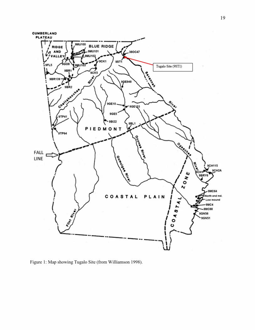
Figure 1: Map showing Tugalo Site (from Williamson 1998).
20
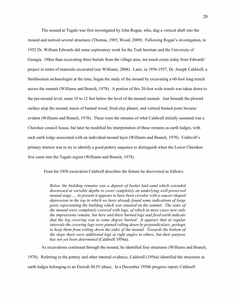
The mound at Tugalo was first investigated by John Rogan, who, dug a vertical shaft into the
mound and noticed several structures (Thomas, 1985; Wood, 2009). Following Rogan’s investigation, in
1952 Dr. William Edwards did some exploratory work for the Tsali Institute and the University of
Georgia. Other than excavating three burials from the village area, not much exists today from Edwards’
project in terms of materials excavated (see Williams, 2008). Later, in 1956-1957, Dr. Joseph Caldwell, a
Smithsonian archaeologist at the time, began the study of the mound by excavating a 60-foot long trench
across the summit (Williams and Branch, 1978). A portion of this 20-foot wide trench was taken down to
the pre-mound level, some 10 to 12 feet below the level of the mound summit. Just beneath the plowed
surface atop the mound, traces of burned wood, fired-clay plaster, and vertical burned posts became
evident (Williams and Branch, 1978). These were the remains of what Caldwell initially assumed was a
Cherokee council house, but later he modified his interpretation of these remains as earth lodges, with
each earth lodge associated with an individual mound layer (Williams and Branch, 1978). Caldwell’s
primary interest was to try to identify a good pottery sequence to distinguish when the Lower Cherokee
first came into the Tugalo region (Williams and Branch, 1978).
From his 1956 excavation Caldwell describes the feature he discovered as follows:
Below the building remains was a deposit of basket laid sand which extended downward at variable depths to cover completely an underlying well-preserved mound stage…. At present it appears to have been circular with a saucer-shaped depression in the top in which we have already found some indications of large posts representing the building which was situated on the summit. The sides of the mound were completely covered with logs, of which in most cases now only the impressions remain; but here and there burned logs and fired earth indicate that the log covering was to some degree burned. It appears that at regular intervals the covering logs were pinned rolling down by perpendiculars, perhaps to keep them from rolling down the sides of the mound. Towards the bottom of the slope there were additional logs at right angles to others, but their purpose has not yet been determined (Caldwell 1956a).
As excavations continued through the mound, he identified four structures (Williams and Branch,
1978). Referring to the pottery and other internal evidence, Caldwell (1956a) identified the structures as
earth lodges belonging to an Etowah III-IV phase. In a December 1956b progress report, Caldwell
21
(1956b) discussed the need for a complete pottery sequence at Tugalo to better understand the occupation
pattern. Continuing excavations at the site, Caldwell recognized ten distinct mound strata that might have
indicated continuous site occupation at Tugalo. He identified premound pottery as late middle period
Swift Creek mixed with Etowah I and II. The highest earth lodge had an ashbed layer located above it,
Caldwell wanted to call it Etowah V because it had a large sloppy diamond complicated stamped pottery
whose motifs looked more circular than rectilinear (Caldwell, 1956a; Williams and Branch, 1978).
Carbon dated Etowah ceramics seem to support Caldwell’s thesis that the Tugalo mound was utilized
until the late Etowah period (Williams and Branch, 1978).
Pottery discovered in the premound to the late Etowah stratum was identified as either Swift
Creek or Etowah and a small amount of the pottery was considered the so-called “Weird-and-wonderful
Woodstock” (early Woodstock; learned from personal communication with Caldwell by Williams and
Branch) (Williams and Branch, 1978). Unlike most Woodstock pottery that is sand tempered and
stamped with complicated decorations, the Woodstock pottery identified by Caldwell (1957) at Tugalo
had regional and temporal variations. However, the variation was poorly understood. Hence, he called it
“Weird-and-Wonderful- Woodstock” (Williams and Branch, 1978). In the earth lodge level of the
mound, pottery paste was uniform and had a fine temper treatment (Williams and Branch, 1978). Two
thin levels were distinguished in the Etowah V ashbed, it is in these two levels the first indication of
vessels with Lamar characteristics occurred (Williams and Branch, 1978). The next stratum in the mound
was a layer Caldwell called the “Brown Earth” layer (Williams and Branch, 1978). In this stratum,
Caldwell noticed more pottery rim experimentation, but only a small handful of incised sherds were
present (Williams and Branch, 1978). However, in this stratum, Etowah rim types and nested diamonds
had almost disappeared, but the same overstamped, roughened body treatment persisted (Williams and
Branch, 1978). Above the “Brown Earth” layer was the “Northeast Dump” (Caldwell, 1956a). In the
“Northeast Dump,” Caldwell distinguished a large, four-foot-thick mass of broken vessels and food bone
on the northeast slope of the mound. The pottery found in this stratum had all the attributes of Lamar:
22
applique, pinched rims, bold incision, and the cazuela vessel form (Caldwell, 1956a; Williams and
Branch, 1978). The lowest level of the pottery dump carbon dates to A.D. 1480 ± 65 (Williams and
Branch, 1978). According to Caldwell (1965a), Lamar ceramics continued into the upper levels of the
Historic period level. Although incising had drastically decreased there was still evidence of it (Williams
and Branch, 1978). Out of the entire sequence, curvilinear stamping exceeded rectilinear stamping for the
first time (Williams and Branch, 1978). Besides the sharp decrease in incising there were no obvious
differences in pottery between the lower level of the Northeast Dump and the Historic Cherokee
utilization level (Williams and Branch, 1978). The pottery from the dump represents the terminal style
found in the mound itself (Williams and Branch, 1978).
In his notes, Caldwell (1956a) indicates that he found some Savannah Check Stamped sherds in
the uppermost earth lodge and the Etowah ashbed layer in the mound (Williams and Branch, 1978). A
drastic change happened in the pottery, surface treatment and make-up of paste at Tugalo, in the break
between the Etowah ashbed dating to A.D. 1355 and the lower level of the Dump dating to A.D. 1480
(Williams and Branch, 1978). The temper and vessel shape of the pottery changed from very fine grit to
coarse grit. Etowah pottery types and possibly proto-Lamar pottery types occurred together in the thin
burned layers (Williams and Branch, 1978). In the higher level, Etowah types appear to have almost
disappeared, and the proto-Lamar persisted, however, there was no incising of any consequence
(Williams and Branch, 1978). Finally, there was evidence of a fully-fledged Lamar variety, which
extended into the historic Cherokee occupation (Williams and Branch, 1978). According to Williams
and Branch “It seems reasonable, then to conjecture that the Lower Cherokee whose discards formed the
historic stratum in the mound had occupied Tugalo at least since the third quarter of the 15th century”
(1978:37).
However, since there was no clearly-discerned hiatus at Tugalo, several possibilities are apparent
when Tugalo might have been occupied (Williams and Branch, 1978). Williams and Branch offer three
explanations for the abrupt transition in ceramic decoration and temper that Caldwell observed around
23
A.D. 1355. The first explanation is that the Cherokees were responsible for building the earth lodges and
later mound levels, but experienced a significant culture change and then began to decorate their pottery
in a much different style. Second, the Cherokees came from elsewhere and settled at Tugalo around A.D.
1355 either occupying an abandoned village or displacing the residents. Third, there only appears to be
an abrupt change in pottery style because of a lack of data.
Concerning the burial excavations conducted at Tugalo in 1956 to 1957, Caldwell wrote at least
two progress reports. In one undated progress report he writes:
Just outside the fences around the log-covered mound we have discovered several more human burials, loosely flexed and with no grave accompaniments. We are still exposing these, but the freezing weather makes it unlikely that any skeletal material can be saved. We now have nine graves immediately outside the fence, and very close together. We hope that within the log-covered mound some richer burials are to be discovered (Sweeney and Huddleston, 2000: 6).
In his second progress report which dates November 13- December 1, 1956, he writes:
Five burials have been discovered, all in poor condition. One was Cherokee, accompanied by a string of barrel shaped shell beads around the neck. Three burials, judging from the random sherds found in the graves fills, were probably Etowah IV. These were at varying degrees of flexion, and none had accompaniments. All of the sherds in the grave fill of the last burial, which was tightly flexed, were of the Swift Creek type. Probably, it is to this period that burial belonged. Again, there were no burial offerings (Caldwell, 1956b).
In his initial progress report, Caldwell (1956a) describes the feature of the Tugalo mound which
gives us an idea where the burials are situated. He explains that the sides of the Tugalo mound were
completely covered with logs arranged perpendicularly outside the fence lines. It is outside the fence line
around the log covered mound that several human burials, loosely flexed with no grave accompaniments
were discovered.
In 1957, Dr. A.R. Kelly and his students continued excavations at the site. Their excavations
focused mainly on the mound and the presumed village area (Anderson, 1994:206). He found six burials,
and each burial was associated with a separate feature (Sweeney and Huddleston, 2000). Out of the six
24
burials recorded by Kelly, four of them were identified during a NAGPRA-related (Brockington’s
physical) inspection (Sweeney and Huddleston, 2000).
As part of a NAGPRA inventory, Brockington and Associates, Inc., of Norcross, Georgia was
tasked with conducting an inventory of the materials from Tugalo, curated at the University of Georgia
(UGA) Riverbend Research Facility Laboratory of Archaeology (RRFLA) (Sweeney and Huddleston,
2000). This inventory identified a minimum of 19 individual human remains, 16 associated funerary
objects, and ten unassociated funerary objects from the site (Sweeney and Huddleston, 2000). However,
matching the human remains with burial numbers created in the field has proven difficult. Available data
is not organized correctly and cross-referencing between Brockington’s physical inspection and the
original field records was limited due to poor burial provenience (Sweeney and Huddleston, 2000).
According to the field, laboratory, and report documents curated at the University of Georgia, there was a
different numbering system used by each researcher for the burials at Tugalo. Caldwell recorded 14
burials located immediately outside the fence line, which he mentions in one of his progress reports
(Anderson, 1994). He numbered each burial independently (e.g., BU1). Kelly’s notes documented that
he recorded six burials and each burial had a separate feature. Kelly assigned numbers based on the
associated feature (e.g., F55) (Sweeney and Huddleston, 2000). Edwards excavated three burials from the
village area and did not assign any number to the burials. The inadequate provenience on the skeletal
data limited the ability of the Brockington’s researchers to directly relate the remains to any recorded
burials (Sweeney and Huddleston, 2000).
Dr. David Hally states the following in the UGA NAGPRA documentation:
Dating the burials is a problem due to the lack of field documentation. It is unlikely that any burials or unassociated funerary objects date to the Late Swift Creek component due to preservation conditions. The mound burials, associated funerary objects, and unassociated funerary objects could date to any of the prehistoric components- Jarrett, Rembert, and Tugalo- responsible for mound construction. It is also possible that some burials post-date mound construction and date to the early 18th century Estatoe component. Village burials could date to any of these four components. Since Jarrett, Rembert and Tugalo phases are all part of an archaeological sequence leading directly to Estatoe phase and the
25
18th century Lower Cherokee, these human remains, and funerary objects can be culturally identified with certainty with the following federally recognized tribes: Cherokee Nation of Oklahoma, Eastern Band of Cherokee Indians, and United Keetoowah Band of Cherokee Indians (Sweeney and Huddleston 2000:14).
On the other hand, according to Caldwell’s second progress report (1956b), one burial may also
be associated with the Swift Creek culture because of the sherds found in the burial fill. Also, there are at
least three burials that are associated with the Etowah IV phase because of the sherds found in the grave
fill (Sweeney and Huddleston, 2000). However, since the pottery sherd was part of the grave fill, there is
a possibility that the sherd was dug from the lower level stratum, and when the grave was being closed,
the pottery sherd got mixed with the burial materials.
Cultural Context
In the following section, I review the cultural and environmental context of the Tugalo site
beginning with the Woodland period (circa. 1200 B.C. to A.D. 1000) since the earliest ceramic found at
Tugalo is associated with the Late Swift Creek which dates to the Middle Woodland period (100 B.C. to
A.D. 500). People from the Woodland period practiced and followed the same hunting and gathering
ways as their ancestors. They occupied the coastlines and forests near rivers and lakes because the climate
was temperate, the areas were rich in game and was decent for survival (Bense, 2016; Norman, 2014;
Russo, 1994, 2010; Schwadron, 2010). Woodland populations, however, developed more refined ways of
doing things since they learned to exploit particular foods from their local regions more efficiently, they
cultivated and stored many plants (Bense, 2016; Hudson, 1976). During the Woodland period pottery
dispersed throughout the region, earth mounds were built and were used for burial and display, people
developed elaborate mortuary rituals, and there was an increase in long-distance trade (Anderson et al.,
2002; Bense, 2016; Stuiver et al., 1998). It is in this period we also see an extreme increase in sedentism
(Anderson et al., 2002).
Evidence of intensive cultivation of chenopod, sunflower, and gourd is first seen in the Late
Archaic period. Although the cultivation of these indigenous plants began prior to 1000 B.C. (e.g. Fritz,
26
1997), during the early Woodland period they were produced in quantity for the first time (Gremillion,
1998). Maize was not intensively cultivated until towards the end of the Woodland period (Anderson et
al., 2002). Due to the lack of stable carbon isotope signatures similar to those left by maize, it is
unknown whether Early or Middle Woodland societies depended heavily on these crops or not (Anderson
et al., 2002).
During the Middle Woodland period, mound building flourished for the first time in association
with the spread of the Hopewellian ceremonial complex (Bense, 2016). The mounds were built for
different purposes - some mounds for human burials (often local elite) and others as platforms for special
activities such as feasting or weddings (Bense, 2016). Woodland people had a complex mortuary ritual
centered upon earthen funerary monuments which provided for the distribution of wealth and the periodic
management of community labor; the widespread commitment to distinctive art styles; and a marked
increase in the extent, duration, and social import of trade and barter (Anderson et al., 2002; Jenkins,
1982). Outside of mound burials, most of the dead were interred in shallow graves (rectangular, oval,
circular holes in the ground) and were accompanied by modest grave goods, if any at all (Anderson et al.,
2002; Jenkins, 1982).
The beginning of the Mississippian tradition started taking shape between A.D. 700 and A.D. 900
along the middle course of the Mississippi River between present-day St. Louis, Missouri, and Vicksburg,
Mississippi (Kane and Keeton, 1993). The term Mississippian was first coined to describe the co-
occurrence of specific material culture traits such as “shell tempered pottery, wall-trench houses, and flat-
topped rectangular platform mounds” (Wood, 2009:1). However, after continuous study of Mississippian
populations, this term now includes structures of prehistoric economy and sociopolitical organization
(Hally, 1999; Milner and Schroeder, 1999; Steponaitis, 1986; Wood, 2009). Archaeologists found that in
the Mississippian tradition there are more large sites compared to any other previous tradition or time for
the region. Although there is variation among Mississippian sites, Mississippian sites also differ from
sites in earlier time periods by population size and social function. In a Mississippian population, society
27
is divided into elites and commoners, chiefs and society members (Wood, 2009). Unlike other prehistoric
and historic periods, the Mississippian period portrays one of the most complex and geographically
extensive prehistoric cultures in North America (Wood, 2009). In his dissertation Wood (2009) uses
Earle’s (1987) definition on Mississippian societies as chiefdoms. According to Earle (1987:288), a
chiefdom is “a society that has a sociopolitical organization, with a person/group of people that exercise
political and economic power over several village communities”. Chiefdoms are divided into three types;
(1) simple chiefdoms display one level of political controls (one administrative center) over the local
community, (2) complex chiefdoms have two or more levels of political control (multiple administrative
centers), and (3) paramount chiefdoms oversee several separate simple or complex chiefdoms and form a
loosely organized union ruled by one dominant chief (Anderson, 1994; Hudson, 1993; Hudson et al.,
1985; Wood, 2009).
A Mississippian society was not only divided into chiefdoms, but their populations were also
hierarchically structured; members either belonged to the elite or were commoners (Hally, 2008;
Steponaitis, 1986; Wood, 2009). Archaeological and historical evidence confirms that elites were treated
differently than commoners during their lives, especially in how they were buried (Wood, 2009). We see
evidence of this from remains and burial goods. Archaeological analysis proves that while they were
alive, elites may have had a better life than most commoners. They lived separately from everyone else,
were respected and had a better diet (Wood, 2009). Since elites were respected, they lived at the top of a
mound, or near the plaza, and also had access to exotic materials and finished goods (Steponaitis, 1986).
When a member of the elite group died, they were sometimes buried in earthen mounds with grave goods
(Anderson, 1994; Muller, 1986; Steponaitis, 1986). Symbols play an important role in Mississippian
Period ceremonialism (Wood, 2009). They are particularly important in burial rituals because they appear
in some graves of the members of society, which help archaeologists distinguish between the elite and
non-elite. Most individuals in the Mississippian tradition were buried in formally defined cemeteries,
28
which there are three main types; outlying community cemeteries, cemeteries of low-status individuals
that are located in regional centers and towns, and cemeteries for the elite (Milner et al., 1984).
During the Mississippian period (A.D. 900 to 1600) most societies transitioned to an agricultural
diet (Milner and Schroeder, 1999; Muller, 1986; Smith, 1986; Steponaitis, 1986). Although maize, beans,
and squash are most notable in these societies, they did not abandon native cultigens (e.g. Chenopodium,
sumpweed, sunflower, etc.), wild plants (nuts), or animals (fish, deer, turkey, turtles, waterfowl, etc.)
(Hally, 1999; Steponaitis, 1986).
The Cherokee Life
At the beginning of the eighteenth century, Cherokee settlements were divided into five
geographically distinctive areas that contained ten to twelve politically independent towns (Goodwin,
1977; Smith, 1979). According to Thorton (1990:13-46), before European contact, “the Cherokee
population totaled around 20,000 people, but due to epidemics and hostilities with Europeans and other
Native Americans, the number declined to 12,000 or less during the eighteenth century”. The total
number and locations of villages and populations also fluctuated (see Goodwin, 1977; Smith, 1979).
According to Gearing (1962) and Gilbert (1943), Cherokee social and political activities were organized
according to seven matrilineal clans, as well as a civil or ‘white’ division and a ‘red’, military division.
While the Cherokee relied mostly on hunting deer, bear, turkey, and a variety of smaller animals, they
also grew several varieties of corn, beans, and squash (Goodwin, 1977). In the late-eighteenth to early-
nineteenth century, Cherokee subsistence became more reliant on European-introduced animals (Bogan,
1982).
After the American Revolutionary War, neatly organized nucleated Cherokee towns changed into
dispersed communities that consisted of small log cabins with associated outbuildings and individual
family agricultural lands (Pillsbury, 1983; Riggs, 1996; Wilms, 1974). When Cherokee townhouses were
abandoned in the eighteenth century, their new settlements were established primarily in Northern
Georgia. In the nineteenth century, the Cherokee Nation was established in Georgia and later relocated to
29
the west through forced removal by the United States government in 1838 (Brown, 1938; Mooney, 1900).
Studies show that Cherokee ancestors occupied towns in the same regions as other cultural groups and
sometimes their towns were located where no Cherokee towns existed in the eighteenth century. It is
possible that eighteenth-century Cherokee towns are found in places that may have been settled by other
cultural groups in the sixteenth century (Schroedl, 2000).
Researchers started having an archaeological interest in the Cherokee in the late-nineteenth and
early-twentieth centuries when they attempted to identify mounds and archaeological remains as
representing the prehistoric Cherokee. The University of Georgia conducted investigations at the
Cherokee Lower Towns of Estatoe, Chauga, and Tugalo by focusing on the mounds of these sites. The
mounds produced evidence of eighteenth-century occupations (Kelly and DeBaillou, 1960; Kelly and
Neitzel, 1961; Williams and Branch, 1978). Studies done in Georgia, South Carolina, and North Carolina
began to form the core of a description of eighteenth-century Cherokee archaeological culture grounded
in ethnographic and ethnohistoric descriptions (Schroedl and McEwan, 2000). Cherokee archaeological
and ethnohistoric data are divided into seven periods covering A.D. 1540 to present. The periods include
the Spanish exploration (1540-1670), English contact (1671-1745), English colonial (1746-1775),
American Revolutionary War (1776-1794), American federal (1795-1818), Cherokee removal (1819-
1838), and Cherokee post-removal (1839- present) periods (Schroedl and McEwan, 2000). Within these
seven periods there is the Tugalo (1450-1600) and Estatoe (1700-1838) phases which account for the
Cherokee archaeological record in northwestern South Carolina and northeastern Georgia (Anderson,
1994; Hally, 1986).
Sites such as Chattooga, Estatoe, Tugalo, Chauga, and Coweeta Creek are among sites that can be
used to address the sixteenth-and seventeenth-century Cherokee record. These sites indicate that the use
of platform mounds probably declined or ceased by the end of the period, as did the use of village
palisades. According to Schroedl (2000), it is difficult to interpret the presence of Cherokee inhabitants at
Chauga, Estatoe and Tugalo, because there is either not a lot reported on these sites or evidence of
30
occupation for both Woodland and Mississippian period appear there as well. Archaeological materials
distinguished from either the mound or village areas date to either the Woodland or Mississippian period.
Anderson (1994) suggests that the Chattooga, Estatoe, Tugalo, Chauga, and Coweta Creek sites did not
get occupied by the Cherokee until the eighteenth century. At Chauga the mound structure is associated
with fences or walls, and it is possible there might have been a village palisade. However, it is currently
impossible to establish how these features related to the Cherokee occupation. At Chattooga, there is a
mound present, but there are townhouses, and some are superimposed. Studies reveal that the final
townhouse was destroyed in the mid-1730s, and other community structures could date to the mid-
seventeenth century or earlier. Thorton (1990) mentions that the Cherokee had established villages in all
the areas that they were known to occupy in succeeding decades prior to the American Revolutionary War
and after the smallpox epidemic in 1738. After the epidemic, the Cherokee population of about 20,000
reduced by nearly a half. It seems most Lower Towns, particularly in northwestern South Carolina and
northeastern Georgia, got abandoned because of population losses (Goodwin, 1977). Some villages were
reoccupied, but some, especially smaller villages were not. Remains of domestic pigs and peaches
indicate the use of introduced species. Although this might have caused dramatic alterations to Cherokee
subsistence during the late-eighteenth and early-nineteenth century, none of the archaeological or
ethnohistoric data suggest dramatic alterations to Cherokee subsistence (Goodwin, 1977; Walker, 1995).
After the American Revolutionary War, many Cherokee populations were depleted and so they
re-established seven Chickamauga towns in southern Tennessee, northern Alabama, and northern
Georgia. These eighteenth-century Cherokee village sites were occupied, and they had small populations
of either one or two families. Cherokee technological, economic, and subsistence patterns were altered by
the American Revolutionary War, but traditional patterns of social organization, belief systems, and the
occupation of nucleated towns were maintained despite demographic changes by epidemics and military
conflicts with the British (Hudson, 1976).
31
Understanding the cultural context of the Woodland, Mississippian and Historic Cherokee diet
and burial treatments will help us interpret the findings at Tugalo because archaeological evidence
suggests the occupation at Tugalo dates from the late Swift Creek through the Historic Cherokee period.
Therefore, knowing the cultural context of these time periods will help us distinguish the age (calendrical)
of the burials by evaluating their diet and burial treatment.
32
CHAPTER 3
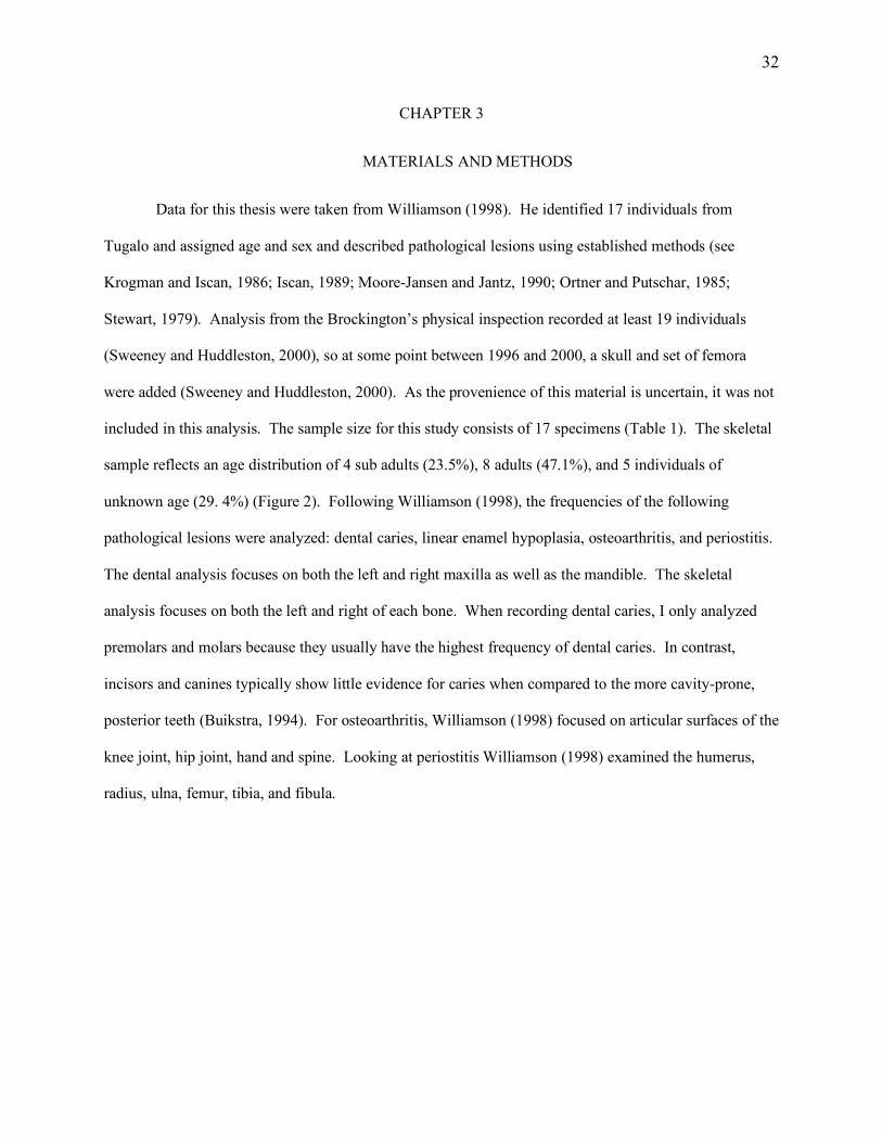
MATERIALS AND METHODS
Data for this thesis were taken from Williamson (1998). He identified 17 individuals from
Tugalo and assigned age and sex and described pathological lesions using established methods (see
Krogman and Iscan, 1986; Iscan, 1989; Moore-Jansen and Jantz, 1990; Ortner and Putschar, 1985;
Stewart, 1979). Analysis from the Brockington’s physical inspection recorded at least 19 individuals
(Sweeney and Huddleston, 2000), so at some point between 1996 and 2000, a skull and set of femora
were added (Sweeney and Huddleston, 2000). As the provenience of this material is uncertain, it was not
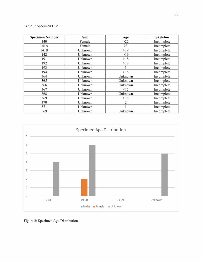
included in this analysis. The sample size for this study consists of 17 specimens (Table 1). The skeletal
sample reflects an age distribution of 4 sub adults (23.5%), 8 adults (47.1%), and 5 individuals of
unknown age (29. 4%) (Figure 2). Following Williamson (1998), the frequencies of the following
pathological lesions were analyzed: dental caries, linear enamel hypoplasia, osteoarthritis, and periostitis.
The dental analysis focuses on both the left and right maxilla as well as the mandible. The skeletal
analysis focuses on both the left and right of each bone. When recording dental caries, I only analyzed
premolars and molars because they usually have the highest frequency of dental caries. In contrast,
incisors and canines typically show little evidence for caries when compared to the more cavity-prone,
posterior teeth (Buikstra, 1994). For osteoarthritis, Williamson (1998) focused on articular surfaces of the
knee joint, hip joint, hand and spine. Looking at periostitis Williamson (1998) examined the humerus,
radius, ulna, femur, tibia, and fibula.
33
Table 1: Specimen List
Specimen Number Sex Age Skeleton 140 Female >22 Incomplete
141A Female 23 Incomplete 141B Unknown >19 Incomplete 142 Unknown >19 Incomplete 191 Unknown >18 Incomplete 192 Unknown >18 Incomplete 193 Unknown 1 Incomplete 194 Unknown >18 Incomplete 364 Unknown Unknown Incomplete 365 Unknown Unknown Incomplete 366 Unknown Unknown Incomplete 367 Unknown <15 Incomplete 368 Unknown Unknown Incomplete 369 Unknown >18 Incomplete 370 Unknown 2 Incomplete 371 Unknown 1 Incomplete 569 Unknown Unknown Incomplete
Figure 2: Specimen Age Distribution
0
1
2
3
4
5
6
7
0-18 19-30 31-39 Unknown
Specimen Age Distribution
Males Females Unknown
34
Description of Skeletal and Dental Lesions Used in the Study
Dental Caries
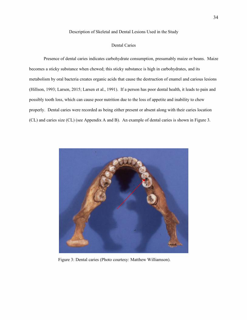
Presence of dental caries indicates carbohydrate consumption, presumably maize or beans. Maize
becomes a sticky substance when chewed; this sticky substance is high in carbohydrates, and its
metabolism by oral bacteria creates organic acids that cause the destruction of enamel and carious lesions
(Hillson, 1993; Larsen, 2015; Larsen et al., 1991). If a person has poor dental health, it leads to pain and
possibly tooth loss, which can cause poor nutrition due to the loss of appetite and inability to chew
properly. Dental caries were recorded as being either present or absent along with their caries location
(CL) and caries size (CL) (see Appendix A and B). An example of dental caries is shown in Figure 3.
Figure 3: Dental caries (Photo courtesy: Matthew Williamson).
35
Linear Enamel Hypoplasia
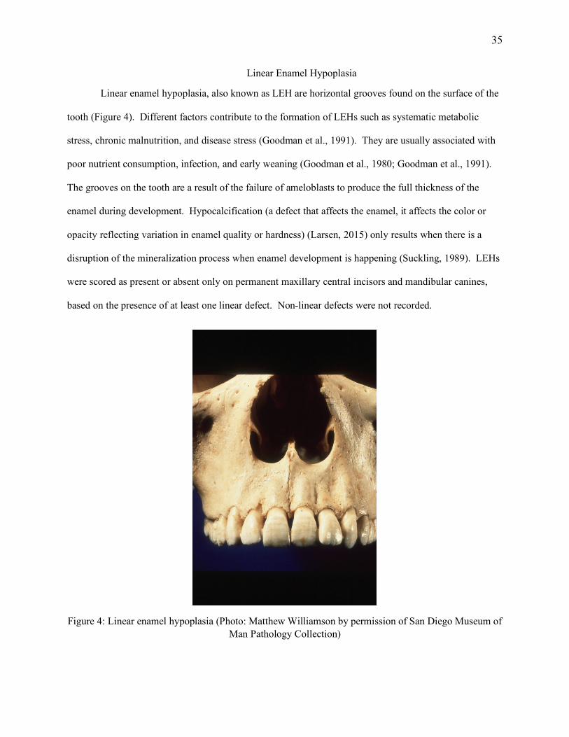
Linear enamel hypoplasia, also known as LEH are horizontal grooves found on the surface of the
tooth (Figure 4). Different factors contribute to the formation of LEHs such as systematic metabolic
stress, chronic malnutrition, and disease stress (Goodman et al., 1991). They are usually associated with
poor nutrient consumption, infection, and early weaning (Goodman et al., 1980; Goodman et al., 1991).
The grooves on the tooth are a result of the failure of ameloblasts to produce the full thickness of the
enamel during development. Hypocalcification (a defect that affects the enamel, it affects the color or
opacity reflecting variation in enamel quality or hardness) (Larsen, 2015) only results when there is a
disruption of the mineralization process when enamel development is happening (Suckling, 1989). LEHs
were scored as present or absent only on permanent maxillary central incisors and mandibular canines,
based on the presence of at least one linear defect. Non-linear defects were not recorded.
Figure 4: Linear enamel hypoplasia (Photo: Matthew Williamson by permission of San Diego Museum of Man Pathology Collection)
36
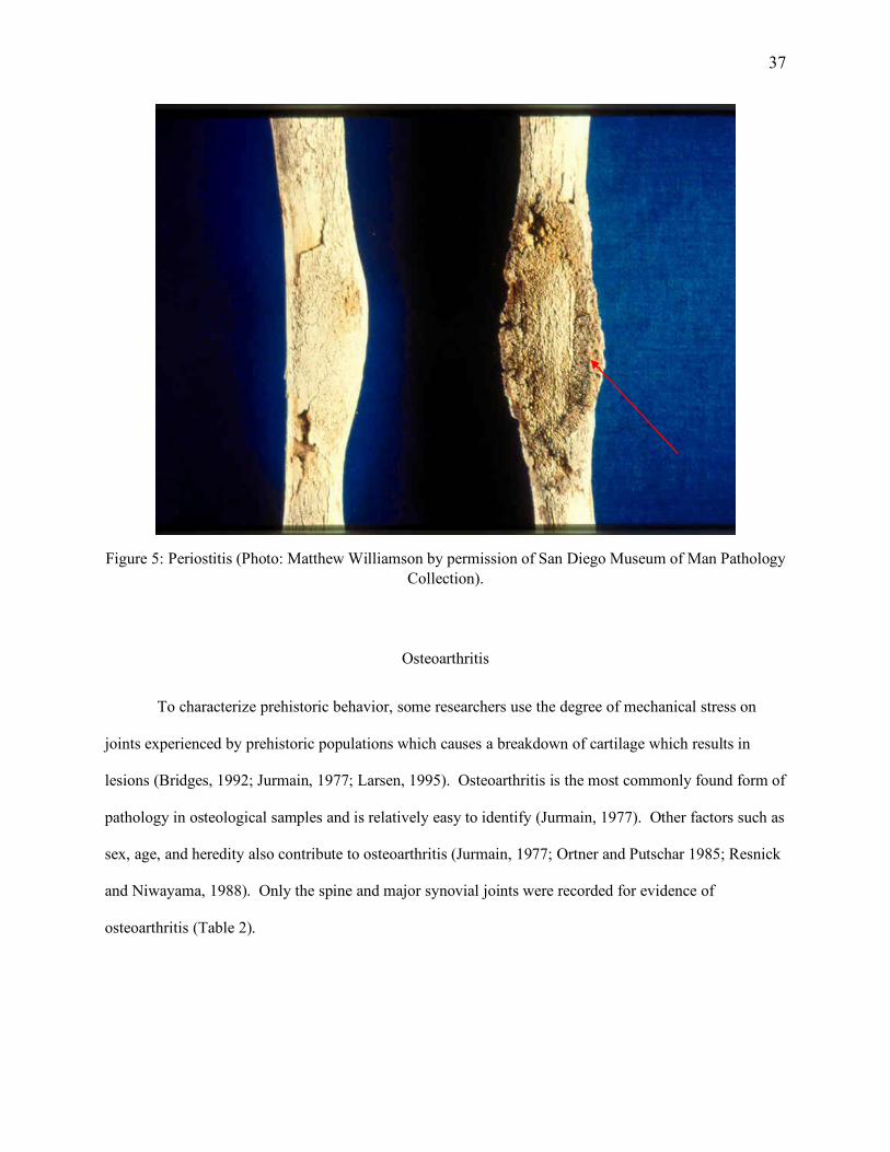
Periostitis
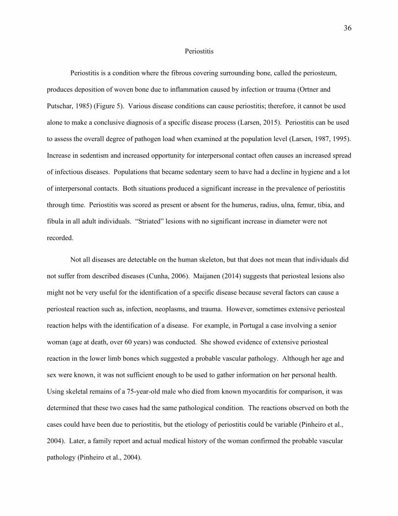
Periostitis is a condition where the fibrous covering surrounding bone, called the periosteum,
produces deposition of woven bone due to inflammation caused by infection or trauma (Ortner and
Putschar, 1985) (Figure 5). Various disease conditions can cause periostitis; therefore, it cannot be used
alone to make a conclusive diagnosis of a specific disease process (Larsen, 2015). Periostitis can be used
to assess the overall degree of pathogen load when examined at the population level (Larsen, 1987, 1995).
Increase in sedentism and increased opportunity for interpersonal contact often causes an increased spread
of infectious diseases. Populations that became sedentary seem to have had a decline in hygiene and a lot
of interpersonal contacts. Both situations produced a significant increase in the prevalence of periostitis
through time. Periostitis was scored as present or absent for the humerus, radius, ulna, femur, tibia, and
fibula in all adult individuals. “Striated” lesions with no significant increase in diameter were not
recorded.
Not all diseases are detectable on the human skeleton, but that does not mean that individuals did
not suffer from described diseases (Cunha, 2006). Maijanen (2014) suggests that periosteal lesions also
might not be very useful for the identification of a specific disease because several factors can cause a
periosteal reaction such as, infection, neoplasms, and trauma. However, sometimes extensive periosteal
reaction helps with the identification of a disease. For example, in Portugal a case involving a senior
woman (age at death, over 60 years) was conducted. She showed evidence of extensive periosteal
reaction in the lower limb bones which suggested a probable vascular pathology. Although her age and
sex were known, it was not sufficient enough to be used to gather information on her personal health.
Using skeletal remains of a 75-year-old male who died from known myocarditis for comparison, it was
determined that these two cases had the same pathological condition. The reactions observed on both the
cases could have been due to periostitis, but the etiology of periostitis could be variable (Pinheiro et al.,
2004). Later, a family report and actual medical history of the woman confirmed the probable vascular
pathology (Pinheiro et al., 2004).
37
Figure 5: Periostitis (Photo: Matthew Williamson by permission of San Diego Museum of Man Pathology Collection).
Osteoarthritis
To characterize prehistoric behavior, some researchers use the degree of mechanical stress on
joints experienced by prehistoric populations which causes a breakdown of cartilage which results in
lesions (Bridges, 1992; Jurmain, 1977; Larsen, 1995). Osteoarthritis is the most commonly found form of
pathology in osteological samples and is relatively easy to identify (Jurmain, 1977). Other factors such as
sex, age, and heredity also contribute to osteoarthritis (Jurmain, 1977; Ortner and Putschar 1985; Resnick
and Niwayama, 1988). Only the spine and major synovial joints were recorded for evidence of
osteoarthritis (Table 2).
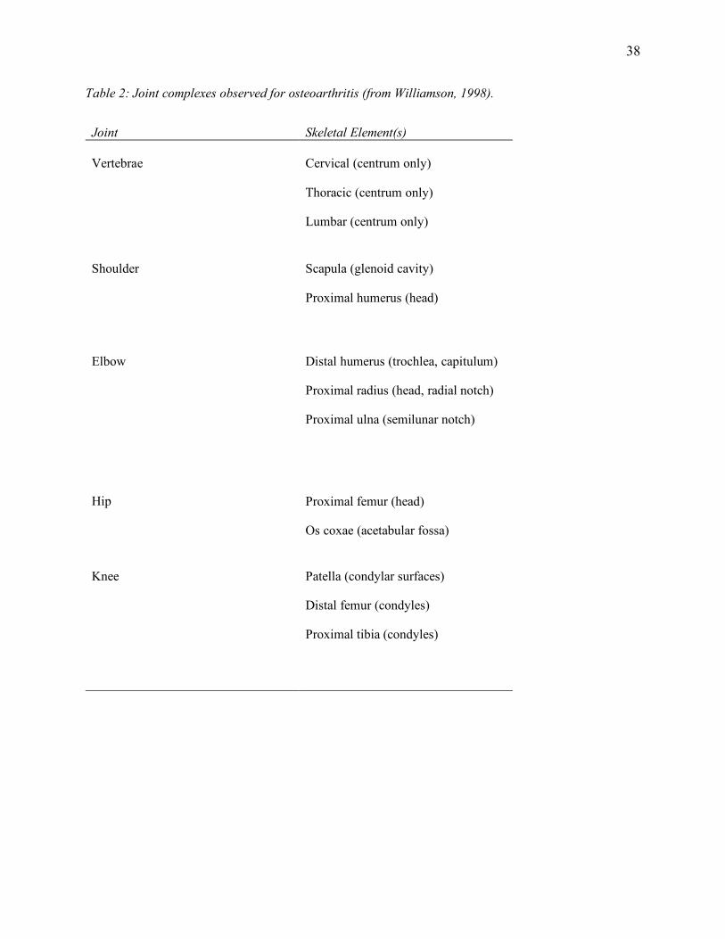
38
Table 2: Joint complexes observed for osteoarthritis (from Williamson, 1998).
Joint Skeletal Element(s) Vertebrae Cervical (centrum only)
Thoracic (centrum only)
Lumbar (centrum only)
Shoulder Scapula (glenoid cavity)
Proximal humerus (head)
Elbow Distal humerus (trochlea, capitulum)
Proximal radius (head, radial notch)
Proximal ulna (semilunar notch)
Hip Proximal femur (head)
Os coxae (acetabular fossa)
Knee Patella (condylar surfaces)
Distal femur (condyles)
Proximal tibia (condyles)
39
CHAPTER 4
RESULTS
Specimen Description
Skeletal data gathered from the Tugalo site were recorded under specimen number and not burial
number (see Table 1). In total, 17 specimens were analyzed for this study. Determination of sex was
only possible for two individuals, which are females. The other fifteen individuals could not be classified
due to incomplete skeletons or age. The age of specimens varies from infant to adults (approximately 1-
year-old to 23-year-old). Each specimen has different frequencies of pathological lesions and health
problems, some have none, and some have more than others (see Appendix A). Below are descriptions of
each specimen. Table 3 shows a comparison of lesion frequencies by sex which indicates the overall total
of skeletal and dental remains affected by lesion(s) for all the 17 specimens. Table 4 shows which
specimens show lesion(s) and the number of each tooth and bone affected. These have been marked with
the following symbol (✓). Specimens that show no evidence of lesion(s) have been marked with the
following symbol (-). For some specific conditions, some specimens could not be assessed due to the
absence of the necessary element to be observed. These have been marked with the following symbol (x).
Specimen 140
Specimen 140 is an adult (>22 years) female. She has a healed periosteal reaction on the
midshaft of her tibia. She does not have any evidence of dental caries, linear enamel hypoplasia and
osteoarthritis.
Specimen 141A
Specimen 141A is a 23-year-old female. No dental caries, linear enamel hypoplasia,
osteoarthritis, or periostitis could be observed due to the absence of specific elements to be observed.
40
Specimen 141B
Specimen 141B is an adult (>19 years) specimen of unknown sex. There is no evidence of
osteoarthritis and periostitis. Dental caries and linear enamel hypoplasia could not be observed due to the
absence of specific elements to be observed.
Specimen 142
Specimen 142 is an adult specimen (>19 years) of unknown sex. Skeletal analysis shows that
specimen 142 has an arthritic lesion on the anterior left temporomandibular joint (TMJ) fossa. There is
no evidence of dental caries and periostitis.
Specimen 191
Specimen 191 is an adult (>18 years). Sex could not be determined. Dental analysis shows that
this individual has a carious lesion on the second left mandibular molar. On the second right maxillary
molar, there is no evidence of a carious lesion. Evidence of linear enamel hypoplasia is present on the left
mandibular canine and first left maxillary incisor. Osteoarthritis and periostitis were not observed
because specific elements to be observed were absent.
Specimen 192
Specimen 192 is an adult (>18 years) of unknown sex. No osteoarthritis and periostitis were
observed due to the absence of specific elements to be observed. The specimen does not have dental
caries or linear enamel hypoplasia.
Specimen 193
Specimen 193 is a 1-year-old infant if unknown sex. Limited skeletal information is available,
but the specimen shows no signs of skeletal or dental pathological lesions. Although the first left
deciduous maxillary incisor is present, there is no evidence of linear enamel hypoplasia.
Specimen 194
Specimen 194 is an adult (>18 years) of unknown sex. Specimen 194 has a carious lesion on the
first left mandibular molar. Linear enamel hypoplasia is recorded on the right mandibular canine, first
41
right mandibular canine, first right maxillary incisor and first left maxillary incisor. No osteoarthritis and
periostitis were observed due to the absence of specific elements to be observed.
Specimen 364
The age and sex of this specimen could not be determined. Skeletal and dental lesions could not
be observed due to the absence of specific elements to be observed.
Specimen 365
The age and sex could not be determined. There is no evidence of dental caries, linear enamel
hypoplasia. Osteoarthritis and periostitis could not be observed because specific elements to be observed
are absent.
Specimen 366
The age and sex could not be determined. Only a right temporal bone is associated with
specimen 366. No dental or skeletal lesions could be observed.
Specimen 367
Specimen 367 is a juvenile and maybe 15-years-old or younger. Sex could not be determined.
Dental caries and hypoplasia could not be observed because specific elements to be observed are absent
and there is no evidence of osteoarthritis and periostitis.
Specimen 368
The age and sex could not be determined. No dental or skeletal lesions were observed.
Specimen 369
Specimen 369 is an adult specimen who is over 18-years-old. Sex could not be determined. The
specimen shows no signs of dental caries, but on the right mandibular canine, there is evidence of
hypoplasia. No other lesions were present.
Specimen 370
This specimen is a 2-year-old of unknown sex. There is no evidence of dental caries or
Osteoarthritis. No hypoplasia could be observed on either the mandible or maxillary dentition and
periostitis was observed either.
42
Specimen 371
Specimen 371 is a 1-year-old of unknown sex, with no evidence of dental caries. Hypoplasia,
osteoarthritis, and periostitis could not be observed due to the absence of specific elements to be
observed.
Specimen 569
The sex and age could not be determined. Specimen 569 has a flattened frontal and a small
button osteoma. Dental caries, hypoplasia, osteoarthritis, and periostitis were not observed due to the
absence of specific elements to be observed.
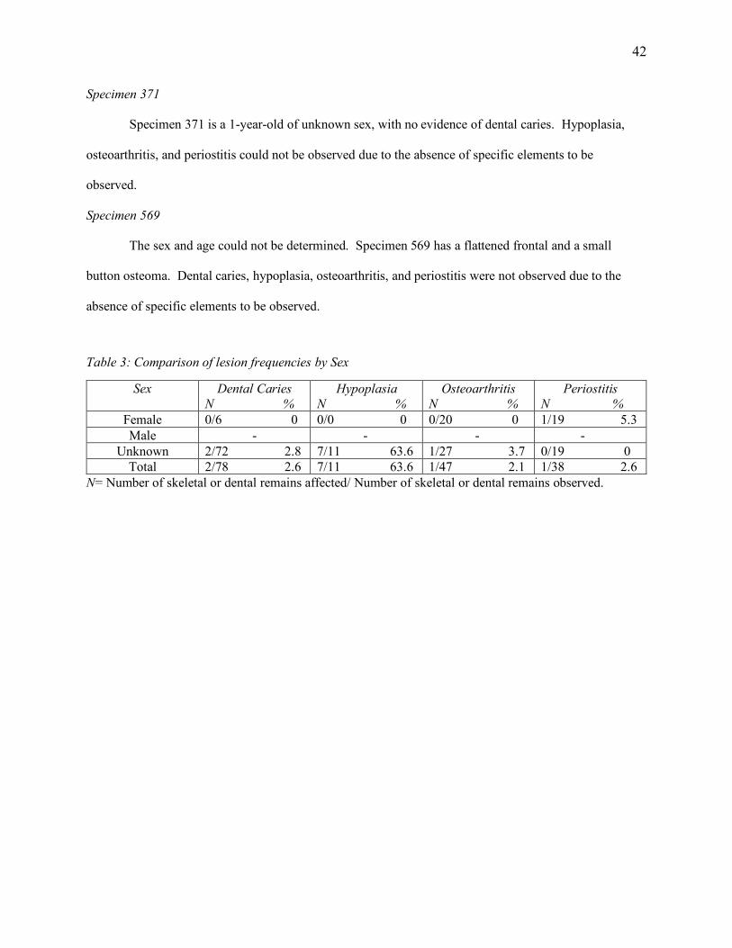
Table 3: Comparison of lesion frequencies by Sex
Sex Dental Caries N %
Hypoplasia N %
Osteoarthritis N %
Periostitis N %
Female 0/6 0 0/0 0 0/20 0 1/19 5.3 Male - - - -
Unknown 2/72 2.8 7/11 63.6 1/27 3.7 0/19 0 Total 2/78 2.6 7/11 63.6 1/47 2.1 1/38 2.6
N= Number of skeletal or dental remains affected/ Number of skeletal or dental remains observed.
43
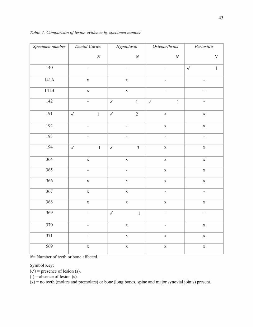
Table 4: Comparison of lesion evidence by specimen number
Specimen number Dental Caries
N
Hypoplasia
N
Osteoarthritis
N
Periostitis
N
140 - - - ✓ 1
141A x x - -
141B x x - -
142 - ✓ 1 ✓ 1 -
191 ✓ 1 ✓ 2 x x
192 - - x x
193 - - - -
194 ✓ 1 ✓ 3 x x
364 x x x x
365 - - x x
366 x x x x
367 x x - -
368 x x x x
369 - ✓ 1 - -
370 - x - x
371 - x x x
569 x x x x
N= Number of teeth or bone affected.
Symbol Key: (✓) = presence of lesion (s). (-) = absence of lesion (s). (x) = no teeth (molars and premolars) or bone (long bones, spine and major synovial joints) present.
44
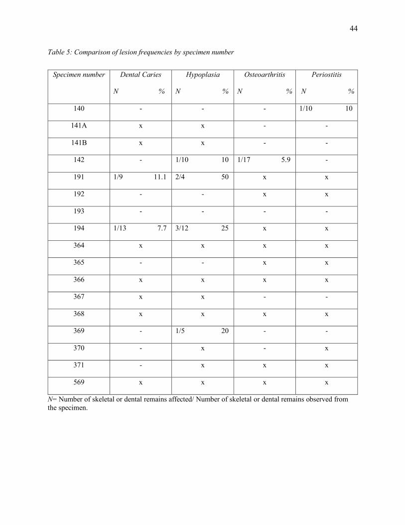
Table 5: Comparison of lesion frequencies by specimen number
Specimen number Dental Caries
N %
Hypoplasia
N %
Osteoarthritis
N %
Periostitis
N %
140 - - - 1/10 10
141A x x - -
141B x x - -
142 - 1/10 10 1/17 5.9 -
191 1/9 11.1 2/4 50 x x
192 - - x x
193 - - - -
194 1/13 7.7 3/12 25 x x
364 x x x x
365 - - x x
366 x x x x
367 x x - -
368 x x x x
369 - 1/5 20 - -
370 - x - x
371 - x x x
569 x x x x
N= Number of skeletal or dental remains affected/ Number of skeletal or dental remains observed from the specimen.
45
CHAPTER 5
DISCUSSION AND CONCLUSION
Discussion
Bioarchaeological interpretation of the health of the Tugalo sample, both as a collective and as
individuals, was based on the prevalence and distribution of pathologies as seen in Table 3, Table 4, and
Table 5. Out of 78 teeth evaluated only 2 (2.6%) showed evidence of caries, one in specimen 191, and
one in specimen 194. Compared to other specimens from the Tugalo sample, only specimen 191 and
specimen 194 show evidence of dental lesion, possibly suggesting poorer dental care or poor health.
Seven out of 11 (63.6%) teeth display linear enamel hypoplasia, one in specimen 142, two in specimen
191, three in specimen 194, and one in specimen 369. The high LEH frequency suggests that at one point
in their childhood, these individuals experienced a disruption in enamel development caused by poor
nutrition, infection or both, or early weaning (or a combination of factors) (Goodman et al., 1980).
Weaning is a process that infants undergo when they shift from drinking breast milk, which is a relatively
stable nutritious food source to solid foods, which are usually less stable, less digestible, and less
nutritious (Larsen, 2015). According to archaeologists, the weaning process relatively occurs after the
child turns one-years-old and is linked to hypoplasia (Coppa et al., 1995; Corruccini et al., 1985;
Lanphear, 1990; Lillie, 1996; Moggi-Cecchi et al., 1994; Ogilvie et al., 1989; Simpson et al., 1990;
Ubelaker, 1992b; Webb, 1995; and many more). However, there have been many discrepancies between
the age patterns of hypoplasia and weaning because weaning alone does not result in hypoplasia; other
factors such as illness, disease, and malnutrition may also contribute to the formation of hypoplasias
(Goodman & Rose, 1991; Hillson, 1996). Out of 38 bones evaluated only 1 (2.6) bone indicates
periostitis, which is found in specimen 140. Out of 47 bones evaluated only 1 (2.1%) bone from
specimen 142 shows evidence of osteoarthritis.
46
Even though specific dates are unknown, previous archaeological work suggests the burials at
Tugalo are Mississippian (Anderson, 1994). Consequently, I decided to compare lesion frequencies to
other Mississippian sites such as Moundville (by Powell, 1988) and Chauga (by Williamson, 1998)
(Table 6) in order to provide a context for the interpretation of my results. Moundville is a Mississippian
site located on the south bank of the Black Warrior River, situated thirteen miles southeast of Tuscaloosa
in west-central Alabama (Powell, 1988). I chose to include Moundville as a comparison because in her
study Powell (1988) conducts research to get a better understanding of the biological and social
dimensions of health at Moundville by evaluating skeletal metrics, development and diet-related features,
and skeletal evidence of iron-deficiency anemia, trauma and infectious disease experience. She analyzed
the remains by age, sex, and ranked status. For this study the category for ranked status is not included
due to insufficient information on burial provenience. The Chauga site (38OC47) is a Lower Cherokee
site located in western Oconee County, South Carolina, on the northern bank of the Tugaloo River
upstream of where the Tugaloo and Chauga Rivers meet (Williamson, 1998; Wood, 2009). Chauga data
were used because the site is located a few miles from the Tugalo site and was likely occupied during a
similar period as Tugalo and culturally affiliated. Ceramics discovered at the Chauga site indicate that the
main occupation at the site was during the early Mississippian Woodstock and Jarrett phases, and later
occupied during the late Mississippian and early historic Tugalo and Estatoe phases (Anderson, 1994;
Wood, 2009). The pottery sequence at the Tugalo site indicates the site was occupied during the Jarrett,
Rembert, and Tugalo phases which is around the same time Chauga was occupied. The likely cultural
affiliation between the Tugalo and Chauga sites might suggest similarities in terms of diet and overall
health. Although the sample size of Tugalo consists of 17 specimens, including infants and adults, when
comparing Tugalo data to Moundville and Chauga I focused on the adult specimens only because the
comparative sites mostly entails data from adults. Hence, the number of specimens shown in Table 6 for
Tugalo is 8 and not 17.
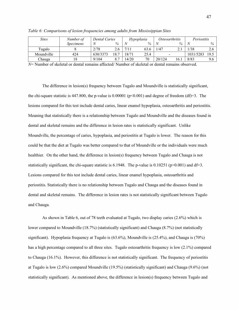
47
Table 6: Comparisons of lesion frequencies among adults from Mississippian Sites
Sites Number of Specimens
Dental Caries N %
Hypoplasia N %
Osteoarthritis N %
Periostitis N %
Tugalo 8 2/78 2.6 7/11 63.6 1/47 2.1 1/38 2.6 Moundville 424 630/3373 18.7 18/71 25.4 - 1031/5283 19.5
Chauga 18 9/104 8.7 14/20 70 20/124 16.1 8/83 9.6 N= Number of skeletal or dental remains affected/ Number of skeletal or dental remains observed.
The difference in lesion(s) frequency between Tugalo and Moundville is statistically significant,
the chi-square statistic is 447.800, the p-value is 0.00001 (p<0.001) and degree of freedom (df)=3. The
lesions compared for this test include dental caries, linear enamel hypoplasia, osteoarthritis and periostitis.
Meaning that statistically there is a relationship between Tugalo and Moundville and the diseases found in
dental and skeletal remains and the difference in lesion rates is statistically significant. Unlike
Moundville, the percentage of caries, hypoplasia, and periostitis at Tugalo is lower. The reason for this
could be that the diet at Tugalo was better compared to that of Moundville or the individuals were much
healthier. On the other hand, the difference in lesion(s) frequency between Tugalo and Chauga is not
statistically significant, the chi-square statistic is 6.1948. The p-value is 0.10251 (p>0.001) and df=3.
Lesions compared for this test include dental caries, linear enamel hypoplasia, osteoarthritis and
periostitis. Statistically there is no relationship between Tugalo and Chauga and the diseases found in
dental and skeletal remains. The difference in lesion rates is not statistically significant between Tugalo
and Chauga.
As shown in Table 6, out of 78 teeth evaluated at Tugalo, two display caries (2.6%) which is
lower compared to Moundville (18.7%) (statistically significant) and Chauga (8.7%) (not statistically
significant). Hypoplasia frequency at Tugalo is (63.6%), Moundville is (25.4%), and Chauga is (70%)
has a high percentage compared to all three sites. Tugalo osteoarthritis frequency is low (2.1%) compared
to Chauga (16.1%). However, this difference is not statistically significant. The frequency of periostitis
at Tugalo is low (2.6%) compared Moundville (19.5%) (statistically significant) and Chauga (9.6%) (not
statistically significant). As mentioned above, the difference in lesion(s) frequency between Tugalo and
48
Moundville is statistically significant, but the difference in lesion(s) frequency between Tugalo and
Chauga is not statistically significant. The lesion frequencies shown in Table 6 could be associated with
an individual or multiple individual, which can change how the rate of the frequency is perceived. Even
though my data show the lesion frequency of each individual (see Table 4), when comparing it to those of
Moundville and Chauga I interpreted the lesion frequencies as a collective because data from both
Moundville and Chauga is interpreted as a collective and does not show the lesions frequency of each
individual.
Despite the sample size variation between Tugalo and Chauga, the lesion frequency rate between
the two sites is not that much different, which emphasizes the likely cultural affiliation. Meaning Tugalo
and Chauga had a similar diet and suffered from similar health problems. On the other hand, the lesion
frequency rate of Moundville is much higher compared to Tugalo and Chauga and this is mostly due to
diet and individual healthcare. There are a few factors that could have contributed to the low frequency
of caries, osteoarthritis, and periostitis at Tugalo such as: (1) sample size is too small; (2) better diet and
health as a collective, which evidence of this could be the low caries, low osteoarthritis, and low
periostitis frequency.
Out of 17 specimens, specimen 142 has an arthritic lesion on the anterior TMJ fossa (Figure 6),
and specimen 569 has a flattened frontal and a small button osteoma. Although dental wear is not
discussed in this thesis, for specimen 142 I considered the dental wear to understand the presence of the
arthritic TMJ fossa. As we know, the main function of our teeth is to process food. We use the anterior
teeth to bite our food. We can also use the anterior teeth to process other materials that are not food
(Lozano et al. 2017). Dental wear is unavoidable and results from physiological and pathological factors
(Levartovsky et al., 2015). Tooth wear is rarely caused by one specific factor; any wear distinguished on
an individual could be a result of all possible combinations of etiological factors of that tooth.
Environmental factors, such as grit presence in unrefined diets and consistency of food, can affect the
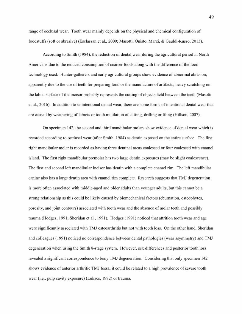
49
range of occlusal wear. Tooth wear mainly depends on the physical and chemical configuration of
foodstuffs (soft or abrasive) (Esclassan et al., 2009; Masotti, Onisto, Marzi, & Gauldi-Russo, 2013).
According to Smith (1984), the reduction of dental wear during the agricultural period in North
America is due to the reduced consumption of coarser foods along with the difference of the food
technology used. Hunter-gatherers and early agricultural groups show evidence of abnormal abrasion,
apparently due to the use of teeth for preparing food or the manufacture of artifacts; heavy scratching on
the labial surface of the incisor probably represents the cutting of objects held between the teeth (Masotti
et al., 2016). In addition to unintentional dental wear, there are some forms of intentional dental wear that
are caused by weathering of labrets or tooth mutilation of cutting, drilling or filing (Hillson, 2007).
On specimen 142, the second and third mandibular molars show evidence of dental wear which is
recorded according to occlusal wear (after Smith, 1984) as dentin exposed on the entire surface. The first
right mandibular molar is recorded as having three dentinal areas coalesced or four coalesced with enamel
island. The first right mandibular premolar has two large dentin exposures (may be slight coalescence).
The first and second left mandibular incisor has dentin with a complete enamel rim. The left mandibular
canine also has a large dentin area with enamel rim complete. Research suggests that TMJ degeneration
is more often associated with middle-aged and older adults than younger adults, but this cannot be a
strong relationship as this could be likely caused by biomechanical factors (eburnation, osteophytes,
porosity, and joint contours) associated with tooth wear and the absence of molar teeth and possibly
trauma (Hodges, 1991; Sheridan et al., 1991). Hodges (1991) noticed that attrition tooth wear and age
were significantly associated with TMJ osteoarthritis but not with tooth loss. On the other hand, Sheridan
and colleagues (1991) noticed no correspondence between dental pathologies (wear asymmetry) and TMJ
degeneration when using the Smith 8-stage system. However, sex differences and posterior tooth loss
revealed a significant correspondence to bony TMJ degeneration. Considering that only specimen 142
shows evidence of anterior arthritic TMJ fossa, it could be related to a high prevalence of severe tooth
wear (i.e., pulp cavity exposure) (Lukacs, 1992) or trauma.
50
Both Tugalo (2.1%) and Chauga (16.1%) show a low frequency of osteoarthritis, however,
Tugalo’s is much lower. It is possible that the low frequency at Tugalo is due to low mechanical stress,
and since the sample size consists of mostly younger and middle-aged adults, the low mechanical stress
could be related to behavior. Variation in mechanical stress is usually associated with the amount of
workload exerted by an individual (Larsen, 2015). As mentioned earlier, in Mississippian societies the
elites were treated differently to the commoners (Wood, 2009). Most elite adults were exposed to less
demanding labor and workload activities than non-elite adults (Larsen, 2015). Usually individuals from
the elite group demonstrate better health than members from the non-elite group (Farmer, 2003;
Strickland & Shetty, 1998).
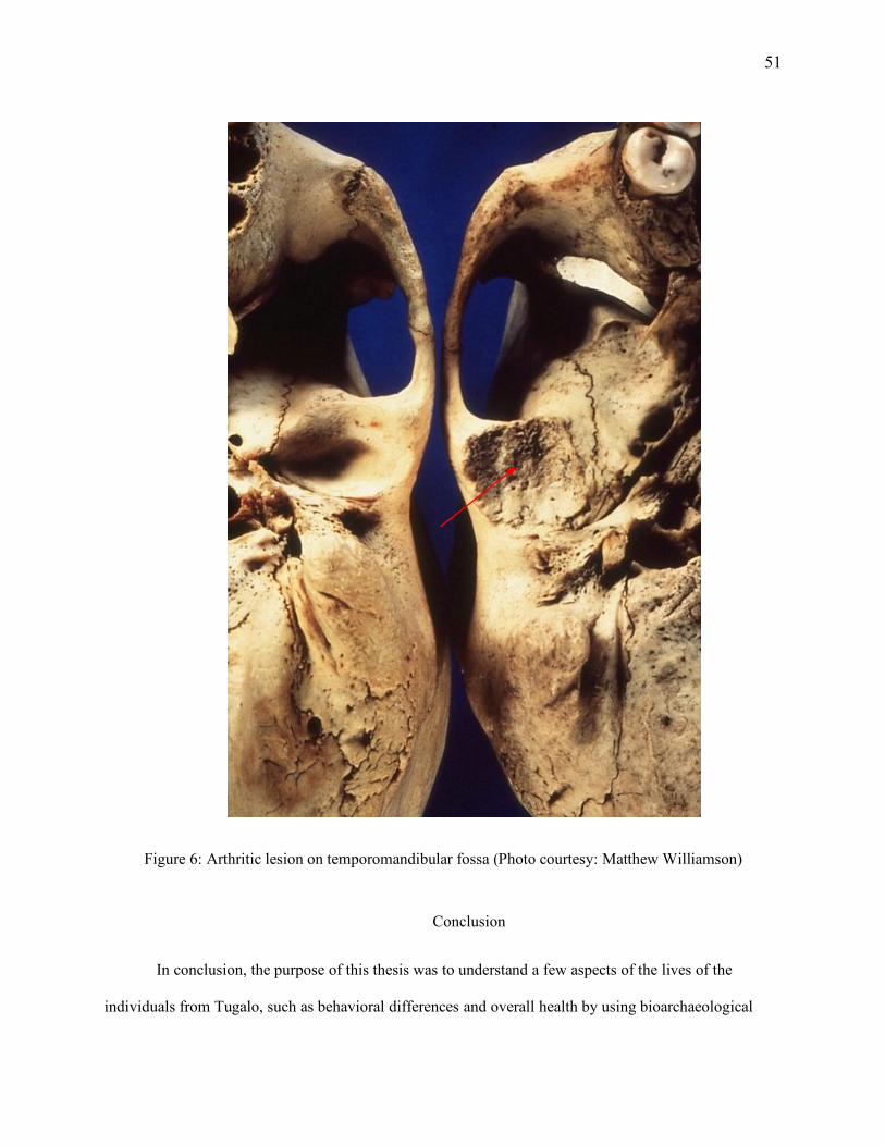
51
Figure 6: Arthritic lesion on temporomandibular fossa (Photo courtesy: Matthew Williamson)
Conclusion
In conclusion, the purpose of this thesis was to understand a few aspects of the lives of the
individuals from Tugalo, such as behavioral differences and overall health by using bioarchaeological
52
analysis. Analyzing data collected by Williamson (1998) concerning the age and sex of the burials, the
presence and absence of dental caries, dental enamel defects, and lesions indicative of infection and
osteoarthritis at Tugalo, helps to understand the behavioral differences of the individuals. Based on the
lesions observed, some individuals were healthier than others. For example, out of 17 specimens,
specimen 140 has one bone showing evidence of healed periostitis, specimen 142 has one tooth showing
evidence of hypoplasia and has an arthritic lesion, specimen 191 has one tooth showing evidence of
dental caries and two teeth showing evidence of hypoplasia, specimen 194 has one tooth showing
evidence of dental caries and three teeth showing evidence of hypoplasia, and specimen 369 has one tooth
showing evidence of hypoplasia. Even though some specimens did not have specific elements to be
observed, these four (142, 191, 194, and 369) specimens had poor dental care or were unhealthy
compared to the other specimens (see Table 5). Also, through comparisons of the Tugalo site results to
other Mississippian and early historic sites, I hoped to determine if their lesion frequencies are at least
consistent with other sites from either of those periods. Previous work done at Tugalo focused only on
understanding when the site was occupied and who occupied it. My study adds to this information by
explaining the lives of the people who occupied Tugalo.
Factors such as diet, disease, metabolic disruption, population mobility, physical exercise and
work leave lasting marks on skeletal and dental tissues. By analyzing human bones, it is possible to get
unique information for the reconstruction of past diets and health which was done with materials from
Tugalo. This research conducted a bioarchaeological study on 17 sets of burials was done, focusing on
dental caries, linear enamel hypoplasia, periostitis, and osteoarthritis. The frequencies of pathological
lesions and health problems vary for each specimen, but there is a high frequency of linear enamel
hypoplasia. Out of the 17 specimens, two individuals (specimen 142 and specimen 569) have distinct
pathologies. Specimen 142 has an arthritic lesion on the anterior left TMJ fossa, and specimen 569 has a
flattened frontal and a small button osteoma. What does this tell us about the population at Tugalo? For
this study, in-depth conclusions cannot be drawn because the sample size is too small.
53
Before analyzing each specimen, I anticipated finding a lot of dental caries considering Tugalo is
a prehistoric and early historic Native American site located in the Upper Savannah River basin in
Georgia. During this time, most prehistoric and early historic Native American groups had become
farmers; it is possible the people at Tugalo were agriculturalists and consumed a lot of maize. Instead, out
of the 17 specimens, only 2 (2.7%) teeth show evidence of dental caries. Why? Perhaps individually
these two specimens had poor dental care, but as a collective the dental care of the group was good, or
their diet was still low in carbohydrates and thus did not result in caries. Also, based on the overall lesion
frequency of the specimens, it could be suggested that this was a healthy non-elite group with one
(specimen 569) elite, as evidenced by a flattened frontal (see Table 4). It has been argued that
intentionally modifying heads could symbolize membership in the elite class. Meaning specimen 569
might have belonged in the elite class. In his progress report, Caldwell (1956a) gives us an idea of where
the burials might have been located. According to Caldwell (1956a), the burials were located outside the
fence line of the log covered mound in a flexed position and with no burial goods. Some research has
interpreted Mississippian sites as representing sociopolitical groups: (1) the elite group/member is
recognized by having certain artifacts, design motifs, exotic materials, and are usually buried in the
mounds; (2) a non-elite group/member is usually buried near the mound or village areas, with no burial
goods (Peebles, 1974). Considering the burial location, lesion frequency, the pathological lesions
associated with the specimens, and lack of field documentation, the assumption that the sample consists
of non-elites and possibly one elite is tentative.
I cannot declare whether the burials belong to the late Swift Creek, Mississippian, or the Historic
period because burial provenience is unknown. In his November 13- December 1, 1956 progress report,
Caldwell notes that the grave fill of one of the burials had a Swift Creek type pottery sherd, suggesting it
belonged with that burial. However, since the pottery sherd was part of the grave fill, there is a possibility
that the sherd was dug from the lower level stratum, and when the grave was being closed, the pottery
sherd got mixed with the burial materials. Nevertheless, using the information that Tugalo had one
54
platform mound, its primary occupation was probably during the Mississippian period. In the
Mississippian period, we see widespread adoption of intensive horticulture (Muller, 1986; Smith, 1986;
Steponaitis, 1986). Even though maize, beans, and squash are most notable in the Mississippian diet,
native cultigens, wild plants, and animals were not excluded from their diet. Archaeological evidence
suggests that Mississippian societies were hierarchically organized into at least two strata; commoner and
elite (Hally, 2008; Steponaitis, 1986). Although Tugalo, Moundville and Chauga are considered
Mississippian societies, different dietary, behavioral and environmental influences on the population
affects the lesion frequencies. Lesion frequencies are lower at Tugalo compared to Moundville but
closely resemble those from Chauga. Overall, this population shows evidence of low caries suggesting
better diet, high (63.6%) LEH suggesting early weaning, poor nutrition, or infection, low (2.6%)
periostitis which means better overall health, or the individuals died before any lesions could show on
their bones and low (2.1%) osteoarthritis meaning less physical work which could be related to status.
Limitations
Before conducting research, every scientist should consider the limitations of their study. By
doing this, we avoid biases, making mistakes, and having incomplete data. Researchers have conducted
excavations at Tugalo but most of the archaeological reports of the site either have no mention of burial
locations or the numbering system used for the burials is inconsistent. Since specific data of the burials
cannot be determined, this could be a problem. Archaeologists rely on burial materials to understand a
population and their environment. So, a restricted burial provenience can limit what we know about an
individual, their role in society and what we know about the population at Tugalo during occupancy.
Another limitation of the study is the absence of sex information. Although only 17 sets of
remains are used in this study, there are only two females and the rest the sex is unknown. In most
societies, gender is culturally determined, and so materials without assigned sex can limit what is known
about the society’s gender roles and class rankings. Due to the small sample size and poor burial
provenience, the results of this study may have been limited; therefore, there is still more work that still

55
needs to be done at Tugalo. In terms of future research, if money is not an issue and access to the remains
and site is possible, researchers should consider conducting radiocarbon dating, isotopic analysis, and
nitrogen analysis to help estimate the age (calendrical age) of the specimens who lived there or determine
the age at death, determine the types of food they ate (diet), and their environment.
When a living organism dies, it stops interacting with the environment, not affecting the C14 in
the remains. It takes thousands of years for the C14 to decay. Radiocarbon dating is a process that starts
with the analysis of C14 left in the remains. The amount of C14 found in a sample indicates the time
elapsed since the death of the organism (Bowman, 1990; Johnston, 201 5). Radiocarbon dating scores are
reported in uncalibrated years B.P. (Before Present). Calibration is done to turn B.P. years into calendar
years (Bowman, 1990; Johnston, 2015). Unlike radioactive isotopes (e.g. C14), stable isotopes do not
decay, which is a very important tool used to date archaeological finds. Since the nucleus does not decay,
the abundance of the stable isotopes stays the same. Although isotopes are everywhere, the balance of
each substance and eco-system is different (for example food, land and sea). Ever since we were born all
the food we eat and the water we drink has been incorporated into our body tissue and skeleton.
Measuring the ratios of the different isotopes in our bones and teeth can help trace back many things
about any individual such as the type of food they ate and the environment they grew up in (Müldner,
2009).
Conducting these different types of analysis cannot only help determine the age at death or give
us an estimate of the samples calendar age, but it can also help us determine the different food eaten by
the population (Larsen, 2015). If deeper analysis is conducted and consumption profiles are accurately
identified, it is possible to get the nutritional inferences and broader implications that can help researchers
understand past human adaptation in a broad biocultural context (Larsen, 2015).
56
REFERENCES
Anderson, David G. 1994 The Savannah River Chiefdoms: Political Change in the Late Prehistoric
Southeast. The University of Alabama Press, Tuscaloosa.
Anderson, David G., David J. Hally, and James L. Rudolph 1986 The Mississippian Occupation of the Savannah River Valley. Southeastern
Archaeology 5(1):32-51.
Anderson D.G., Stahle D.W., and Cleaveland, M.K. 1995 Paleoclimate and the potential food reserves of Mississippian societies: A case study
from the Savannah River Valley. American Antiquity, 60(2):258-286. Anderson, D. G., Kidder, T. R., Watson, P. J., Rafferty, J., Herbert, J. M., & Peacock, E.
2002 The Woodland Southeast. The University of Alabama Press, Tuscaloosa. Bense, Judith A. 2016 Archaeology of the Southeastern United States: Paleoindian to World War I. Routledge. Bocquet-Appel, J-P, Bar-Yosef O., editors.
2008 The Neolithic Demographic Transition and its Consequences. New York, NY: Springer. Bogan, A.E.
1982 Archaeological Evidence of Subsistence Patterns in the Little Tennessee River Valley. Tennessee Anthropologist 7(1):38-50.
Bowman, Sheridan
1990 Radiocarbon Dating: Interpreting the Past. University of California Press, Berkeley and Los Angeles.
Brown, J.P.
1938 Old Frontiers: The Story of the Cherokee Indians from Earliest Times to the Date of Their Removal to the West, 1838. Southern Publishers, Incorporated, Tennessee.
Buikstra, J. E.
1977 Biocultural Dimensions of Archaeological Study: A Regional Perspective. Biocultural Adaptation in Prehistoric America, 67-84.
1994 Standards for Data Collection from Human Skeletal Remains. Arkansas Archaeological
Survey Research series, 44.
2006 Preface. In: Buikstra JE, Beck LA, editors. Bioarchaeology: The Contextual Analysis of Human Remains. Amsterdam, Netherlands: Academic Elsevier; p. xvii-xx.
Burt, B.A.
1993 Relative Consumption of Sucrose and Other Sugars: Has It Been A Factor In Reduced Caries Experience? Caries Research 27(S1):56-63.
Caldwell, Joseph R.
1956a Tugalo Progress Report 1. University of Georgia Manuscript File, Number 188.
57
1956b Tugalo Progress Report 2. University of Georgia Manuscript File, Number 189. 1957 Survey and Excavations in the Allatoona Reservoir, Northern Georgia. Ms. File,
Department of Anthropology, University of Georgia.
Cohen, M.N., Armelagos, G.J., editors. 1984 Paleopathology at the Origins of Agriculture. Orlando, FL: Academic Press. (Reprinted,
University Press of Florida, Gainesville, FL, 2013). Cohen, M.N., Crane-Kramer, G.M.M., editors.
2007 Ancient Health: Skeletal Indicators of Agricultural and Economic Intensification. Gainesville, FL: University Press of Florida.
Coppa, A., Cucina, A., Chiarelli, B., Calderon, F.L., Mancinelli, D.
1995 Dental Anthropology and Paleoanthropology of the Precolumbian Populations of Hispaniola from the Third Millennium B.C. to the Spanish Conquest. Human Evolution 10:153-167.
Corruccini, R.S., Handler, J.S., Jacobi, K.P.
1985 Chronological Distribution of Enamel Hypoplasias and Weaning in a Caribbean Slave Population. Human Biology 57:699-711.
Cunha, B.A.
2006 The Atypical Pneumonias: Clinical Diagnosis and Importance. Clinical Microbiology and Infection. 12:12-24.
Earle, T. K. 1987 Chiefdoms in Archaeological and Ethnohistorical Perspective. Annual Review of
Anthropology, 16(1):279-308. Esclassan, R., A.M. Grimoud, M.P. Ruas, R. Donat, A. Sevin, F. Astie, S. Lucas, E. Crubezy
2009 Dental Caries, Tooth Wear and Diet in an Adult Medieval (12th-14th century) Population from Mediterranean France. Archives of Oral Biology, 54(3):287-297.
Farmer, P. 2003 Pathologies of Power: Health, Human Rights, and the New War on the Poor. Berkeley,
CA: University of California Press. Fritz, Gayle J.
1997 A Three-Thousand-Year-Old Cache of Crop Seeds from Marble Bluff, Arkansas. In People, Plants, and Landscapes: Studies in Paleoethnobotany, edited by Kristen J. Gremillion, pp. 42-62. Tuscaloosa: University of Alabama Press.
Gearing, F.O.
1962 Priests and Warriors: Structures for Cherokee Politics in the Eighteenth Century. American Anthropological Association Memoir, 93.
Gilbert Jr, W.H. 1943 The Eastern Cherokees; Anthropological Papers, No. 23. Smithsonian Institution Bureau
of American Ethnology Bulletin 133, 204. Goodman, A.H., Armelagos, G.J., Rose, J.C.
58
1980 Enamel Hypoplasia as Indicators of Stress in Three Prehistoric Populations from Illinois. Human Biology 52:515-528.
Goodman, A.H., Rose, J.C.
1991 Dental Enamel Hypoplasias as Indicators of Nutritional Status. In Kelley M.A., Larsen C.S, editors. Advances in Dental Anthropology. New York, NY: Wiley-Liss; pp. 279-293.
Goodman, A.H., Martinez, C., and A. Chavez
1991 Nutritional Supplementation and the Development of Linear Enamel Hypoplasias in Children from Tezonteopan, Mexico. Am. J. Clin. Nutr. 53:773-81.
Goodwin, G.C.
1977 Cherokees in Transition: A Study of Changing Culture and Environment Prior to 1775. [Chicago]: University of Chicago, Department of Geography.
Gremillion, Kristen J.
1998 Changing Roles of Wild and Cultivated Plant Resources among Early Farmers of Eastern Kentucky. Southeastern Archaeology 17:140-57.
Hally, David J. 1986 The Identification of Vessel Function: A Case Study from Northwest Georgia. American
Antiquity 51(2):267-295. 1999 The Settlement Pattern of Mississippian Chiefdoms in Northern Georgia. In Fifty
Years Since Viru: Recent Advances in Settlement Pattern Studies in the New World, edited by Brian Billman and Gary Feinman, pp. 187-211. Smithsonian Institution Press, Washington, D.C.
2008 King: The Social Archaeology of a Late Mississippian Town in Northwestern Georgia. University of Alabama Press, Tuscaloosa.
Hara, A.T., Zero D.T.
2010 The Caries Environment: Saliva, Pellicle, Diet, and Hard Tissue Ultrastructure. Dental Clinics of North America 54:455-467.
Hillson, S.W.
1993 Teeth. Cambridge, Cambridge University Press. 1996 Dental Anthropology. Cambridge, UK: Cambridge University Press.
2007 Dental pathology. Biological anthropology of the human skeleton, 299-340. Hodges, D.C.
1991 Temporomandibular Joint Osteoarthritis in a British Skeletal Population. American Journal of Physical Anthropology, 85(5):367-377.
Hudson, C. M. 1976 The Southeastern Indians. Knoxville: University of Tennessee Press.
Hudson, Charles, Marvin, Smith, David Hally, Richard Polhemus, and Chester DePratter
1985 Coosa: A Chiefdom in Sixteenth-Century Southeastern United States. American Antiquity 50:723-737.
59
Hudson, C. 1993 Reconstructing the de Soto Expedition Route West of the Mississippi River: Summary
and contents. The expedition of Hernando de Soto west of the Mississippi, 1541-1543. Iscan, M.Y.
1989 Age Markers in the Human Skeleton. Springfield, C.C. Thomas. Jenkins, N.J.
1982 Archaeology of the Gainesville Lake Area: Synthesis. University of Alabama Office of Archaeological Research Report of Investigations 23.
Johnston, Grahame 2015 Rescue Archaeology, Archaeology Expert, (accessed June 2018). Jurmain, R.D.
1977 Stress and the Etiology of Osteoarthritis. Am. J. Phys. Anthropol. 16:353-366. Kelly, Arthur R., and Clemens DeBaillou 1960 Excavation of the Presumptive Site of Estatoe. Southern Indian Studies 12:3-30. Kelly, Arthur R., and Robert S. Neitzel
1961 The Chauga Site in Oconee County, South Carolina. Laboratory of Archaeology Series, Report No.3. University of Georgia, Athens.
Knüsel, C.J. 2010 Bioarchaeology: A Synthetic Approach. Bulletins et Memoires de la Société
d’Anthropologie de Paris 22:62-73. Krogman, W.M. and Iscan, M.Y.
1986 The Human Skeleton in Forensic Medicine. Springfield, C. C. Thomas. Laine, M.A., Tenovuo, J., Lehtonen, O.P., et al.
1988 Pregnancy-Related Changes in Human Whole Saliva. Archives of Oral Biology 33:913-917.
Lanphear, K.M.
1990 Frequency and Distribution of Enamel Hypoplasias in a Historic Skeletal Sample. American Journal of Physical Anthropology 81:35-43.
Larsen, C.S.
1980a Dental caries: Experimental and Biocultural Evidence. In The Skeletal Biology if Aboriginal Populations in the Southeastern United States, edited by P. Willey and Fred H. Smith, pp. 75-80. Tennessee Anthropological Association Miscellaneous Paper No. 5.
1980b Human Skeletal and Dental Health Changes in the Prehistoric Georgia Coast. In Excursions in Southeastern Geology: The Archaeology-Geology of the Georgia Coast, edited by James D. Howard, Chester B. DePratter, and Robert W. Frey, pp. 192-201. Geological Society of America, Atlanta.
1981b Skeletal and Dental Adaptations to the Shift to Agriculture on the Georgia Coast.
Current Anthropology 22:422-423.
60
1982 The Anthropology of St. Catherines Island: 3. Prehistoric Human Biological Adaptation. Anthropological Papers of the American Museum of Natural History 57; pp. 157-276.
1983a Behavior of Temporal Changes in Cariogenesis. Journal of Archaeological Science
10(1):1-8. 1938b Subsistence Role Behavior and the Infections Disease Experience in the Prehistoric
Georgia Coast. American Journal of Physical Anthropology 60(2):216. 1983c Deciduous Tooth Size and Subsistence Change in Prehistoric Georgia Coast Population.
Current Anthropology 22:422-423.
1987 Bioarchaeological Interpretations of Subsistence Economy and Behavior from Human Skeletal Remains. Advances in Archaeological Method and Theory, Vol. 10, pp. 339-445.
1995 Biological Changes in Human Populations with Agriculture. Ann. Rev. Anthropol.
24:185-213. 2015 Bioarchaeology: Interpreting Behavior from the Human Skeleton (Vol. 69). Cambridge
University Press. Larsen, C.S., Shavit, R., and Griffin, M.C.
1991 Dental Caries Evidence for Dietary Change: An Archaeological Context. In Kelley M.A. and Larsen C.S., editors. Advances in Dental Anthropology. New York, Wiley-Liss, pp. 179-202.
Larsen, C.S. and Walker, P.L.
2010 Bioarchaeology: Health, Lifestyle, and Society in Recent Human Evolution. In: Larsen C.S., editor. A Companion to Biological Anthropology. Malden, MA: Wiley-Blackwell; pp. 379-394.
Leigh, R.W.
1934 Notes on the Somatology and Pathology of Ancient Egypt. University of California Publications in American Archaeology and Ethnology, 34(1).
Levartovsky, S., Matalon, S., Sarig, R., Baruch, O., & Winocur, E.
2015 The Association between Dental Wear and Reduced Vertical Dimension of the Face: A Morphologic Study on Human Skulls. Archives of oral biology, 60(1):174-180.
Leverett, Dennis H. 1982 Fluorides and the Changing Prevalence of Dental Caries. Science 217:26-30. Lillie, M.C. 1996 Mesolithic and Neolithic Populations of Ukraine: Indications of Diet from Dental Pathology.
Current Anthropology 37:135-142. Lozano, M., de Castro, J. B., Arsuaga, J. L., & Carbonell, E.
2017 Diachronic Analysis of Cultural Dental Wear at the Atapuerca Sites (Spain). Quaternary International, 433, 243-250.
Lukacs, John R.
61
1992 Dental Paleopathology and Agricultural Intensification in South Asia: New Evidence from Bronze Age Harappa. American Journal of Physical Anthropology 87, no. 2: 133–150. doi:10.1002/ajpa.1330870202.
2008 Fertility and Agriculture Accentuate Sex Differences in Dental Caries Rates. Current
Anthropology 49:901-914. Maijanen, H. M. K.
2014 The Prevalence of Pathological Conditions in a Modern Skeletal Collection–Implications for Forensic Anthropology.
Marshall 2009 The Estatoe Towns. Lamar Institute Publication 142:1-10. Masotti, S., Onisto, N., Marzi, M., & Gualdi-Russo, E.
2013 Dento-alveolar Features and Diet in an Etruscan Population (6th–3rd c. BC) from northeast Italy. Archives of oral biology, 58(4):416-426.
Masotti, S., Bogdanic, N., Arnaud, J., Cervellati, F., & Gualdi-Russo, E.
2017 Tooth Wear Pattern Analysis in a Sample of Italian Early Bronze Age population. Proposal of a 3-D Sampling Sequence. Archives of oral biology, 74, 37-45.
Mayhall, J.T.
1970 The Effect of Culture Change upon the Eskimo Dentition. Arctic Anthropology 7:117-121.
Mereness, Newton B. ed.
1916 Travels in the American colonies. Macmillan, New York. Milner, George R., T.E. Emerson, M.W. Mehrer, J.A. Williams, and D. Esarey
1984 Mississippian and Oneota Period. In American Bottom Archaeology, edited by C. Bareis and J. Porter, pp. 158-186. University of Illinois Press, Urbana.
Milner, George R., and Sissel Schroeder
1999 Mississippian Sociopolitical Systems. In Great Towns and Regional Polities in the Prehistoric American Southwest and Southeast, edited by J.E. Neitzel, pp. 95- 107. University of New Mexico Press, Albuquerque.
Moggi-Cecchi J., Pacciani E., Pinto-Cisternas J. 1994 Enamel Hypoplasia and Age at Weaning in 19th-Century Florence, Italy. American
Journal of Physical Anthropology 93:299-306. Mooney, J.
1900 James Mooney's History, Myths, and Sacred Formulas of the Cherokees: Containing the Full Texts of Myths of the Cherokee (1900) and The Sacred Formulas of the Cherokees (1891) as Published by the Bureau of American Ethnology: with a New Biographical Introduction, James Mooney and the Eastern Cherokees. Bright Mountain Books.
Moore-Jansen, P. and R.L. Jantz 1990 Data Collection Procedures for Forensic Skeletal Material. Report of Investigations No.
48, Second edition, Department of Anthropology, The University of Tennessee. Knoxville.
62
Muller, Jon 1986 Archaeology of the Lower Ohio River Valley. Academic Press, Inc. New
York. Müldner, Gundula 2009 Investigating Medieval Diet and Society by Stable Isotope Analysis of Human Bone. Norman, Sean Patrick,
2014 "Modeling the Relationship between Climate Change and Landscape Modification at the Crystal River Site (8CI1), Florida". Graduate Theses and Dissertations. http://scholarcommons.usf.edu/etd/5429
Ogilvie, M.D., Curran, B.K., Trinkaus, E.
1989 Incidence and Patterning of Dental Enamel Hypoplasia among the Neanderthals. American Journal of Physical Anthropology 79:25-41.
Ortner, D.J. and W.G.J. Putschar
1985 Identification of Pathological Conditions in Human Skeletal Remains. Smithsonian Contributions to Anthropology No. 28. Washington, D.C., Smithsonian Institution Press.
Palfi, G., Dutour, O., Deak, J., Hutus, I.
1999 Tuberculosis: Past and Present. Budapest, Hungary: Golden Book Publishers and Tuberculosis Foundation.
Pillsbury, R.C.
1983 The Europeanization of the Cherokee Settlement Landscape Prior to Removal: A Georgia Case Study. In Historical Archaeology of the Eastern United States, edited by Robert W. Neuman, pp. 56-69. Geoscience and Man 23. University of Louisiana State University, Baton Rouge.
Pinheiro, J., Cunha, E., Cordeiro, C., & Vieira, D. N.
2004 Bridging the Gap Between Forensic Anthropology and Osteoarchaeology—A Case of Vascular Pathology. International Journal of Osteoarchaeology, 14(2):137-144.
Powell, M.L.
1988 Status and Health in Prehistory: A Case Study of the Moundville Chiefdom. Smithsonian Institution Press, Washington, D.C.
Powell, M.L., Cook, D.C., editors
2005 The Myth of Syphilis: The Natural History of Treponematosis In North America. Gainesville, FL: University Press of Florida.
Resnick, D.M. and Niwayama
1988 Diagnosis of Bone and Joint Disorders. 2ndedition. Philadelphia, W.B. Saunders. Riggs, B. H.
1996 Removal Period Cherokee Households and Communities in Southwestern North Carolina (1835-1838). Bret H. Riggs.
Roberts, C.A., Buikstra, J.E.
63
2003 The Bioarchaeology of Tuberculosis. Gainesville, FL: University Press of Florida. Roberts, C.A., Lewis, M.E., Manchester, K., editors.
2002. The Past and Present of Leprosy: Archaeological, Historical, Paleopathological and Clinical Approaches. British Archaeological Reports, international Series, No. 1054.
Rowe, Nathaniel H.
1975 Dental caries. In Dimensions of Dental Hygiene (second ed.), edited by Pauline F. Steele, pp. 198-222. Lea & Febriger, Philadelphia.
Russo, Michael
1994 Why We Don't Believe in Archaic Ceremonial Mounds and Why We Should: The Case from Florida. Southeastern Archaeology 13(2):93-109.
2010 Shell rings and Other Settlement Features as Indicators of Cultural Continuity Between the Late Archaic and Woodland Periods of Coastal Florida. In Trend Tradition and Turmoil: What Happened to the Southeastern Archaic?: Proceedings of The Third Caldwell Conference St. Catherines Island, Georgia May 9-11, 2008, edited by David Hurst Thomas and Matthew C. Sanger, pp. 149-172. American Museum of Natural History, New York.
Schrenk, Alecia and Lorna Tilley
2018 Caring in Ancient Times. Anthropology-News. American Anthropological Association, pp. 3-5.
Schroedl, G.F.
2000 Cherokee Ethnohistory and Archaeology from 1540 to 1838. In Indians of the Greater Southeast: Historical Archaeology and Ethnohistory, edited by Bonnie G. McEwan, pp. 204-241. University Press Florida, Gainesville.
Schwadron, Margo
2010 Prehistoric Landscapes of Complexity: Archaic and Woodland Period Shell Works, Shell Rings, and Tree Islands of the Everglades, South Florida. In Trend Tradition and Turmoil: What Happened to the Southeastern Archaic?: Proceedings of the Third Caldwell Conference St. Catherines Island, Georgia May 9-11, 2008, edited by David Hurst Thomas and Matthew C. Sanger, pp. 113-146. American Museum of Natural History, New York.
Sheridan, S. G., Mittler, D. M., Van Gerven, D. P., & Covert, H. H.
1991 Biomechanical Association of Dental and Temporomandibular Pathology in a Medieval Nubian Population. American Journal of Physical Anthropology, 85(2):201-205.
Simpson, S.W., Hutchinson, D.L., Larsen, C.S.
1990 Coping with Stress: Tooth Size, Dental Defects, and Age-at-Death. In: Larsen CS, editor. The Archaeology of Mission Santa Catalina de Guale: 2. Biocultural Interpretations of a Population in Transition. Anthropological Papers of the American Museum of Natural History, No. 68; pp. 66-77.
Smith, B. A.
64
1979 Distribution of eighteenth-century Cherokee Settlements. The Cherokee Indian Nation: A Troubled History, 46-60.
Smith, B. D.
1986 The Archaeology of the Southeastern United States: From Dalton to de Soto, 10,500-500 B.P. In Advances in World Archaeology, Volume 5, edited by F. Wendorf and A.E. Close. Academic Press, New York.
Smith, B. H.
1984 Patterns of Molar Wear in Hunter–Gatherers and Agriculturalists. American Journal of Physical Anthropology, 63(1):39-56.
Society for the Promulgation of the Gospel
1721 A True and Exact Account of the Number and Names of All Towns Belonging to the Cherokee Nation, and the Number of Men, Women, and Children Inhabiting the Same, Taken Anno 1721. London Manuscript, Series B, Volume 4: 173ff.
Steckel, R. H., & Rose, J. C. (Eds.).
2002 The Backbone of History: Health and Nutrition in the Western Hemisphere (Vol. 2). Cambridge University Press.
Steponaitis, V.P.
1986 Prehistoric Archaeology in the Southeastern United States, 1970-1985. In Annual Review of Anthropology, Vol. 15 (1986), pp. 363-404.
Stewart, T.D.
1979 Essentials of Forensic Anthropology. Springfield, C.C. Thomas. Shetty, P. S., & Strickland, S. S. (Eds.).
1998 Human biology and social inequality. Cambridge University Press.
Stuiver, Minze, Paula J. Reimer, Edouard Bard, J. Warren Beck, G.S. Burr, Konrad A. Hughen, Bernd Kromer, Gerry McCormac, Johannes van der Plicht, and Marco Spurk.
1998 INTCAL98 Radiocarbon Age Calibration, 24,000-0 cal. B.P. Radiocarbon 40(3):1041-83.
Suckling, G.
1989 Development Defects of Enamel – Historical and Present-Day Perspectives of their Pathogenesis. Advances in Dental Research 3:87-94.
Suzuki, H., Yajima, K., Yamanobe, T., editors.
1967 Studies on the Graves, Coffin Contents and Skeletal Remains of the Tokugawa Shoguns and Their Families at the Zojoji Temple. Tokyo, Japan: Tokyo University Press.
Sweeney, Alex and Connie Huddleston
2000 Native and Graves Protection and Repatriation Act Preliminary Compliance Report for the Tugalo Site (9ST1) Hartwell Lake Georgia. Final Report. Brockington and Associates Inc., Norcross, Georgia. Gulf South Research Corporation Baton Rouge, Louisiana.
Thomas, C.
65
1985 Report on the Mound Explorations of the Bureau of Ethnology. Smithsonian Institute Press, Washington, D.C.
Thorton, R. 1990 The Cherokees: A Population History. Lincoln: University of Nebraska Press. Ubelaker, D.H.
1992b Enamel Hypoplasia in Ancient Ecuador. In: Goodman A.H., Capasso L.L., editors. Recent Contributions to the Study of Enamel Development Defects. Journal of Paleopathology, Monographic Publications, No. 2; pp. 207-217.
Walker, Renee B.
1995 Faunal Remains from the Chattooga Site (38Oc18), Oconee County, South Carolina. Ms. on file, Department of Anthropology, University of Tennessee, Knoxville.
Webb, S.
1995 Paleopathology of Aboriginal Australians: Health and Disease across a Hunter-Gatherer Continent. Cambridge, UK: Cambridge University Press.
Weston, D.A.
2012 Nonspecific infection in paleopathology: interpreting periosteal reactions. In: Grauer A, editor. A Companion to Paleopathology. Chichester, UK: Wiley-Blackwell; pp. 292-512.
Wilms, D. C.
1974 Cherokee Settlement Patterns in Nineteenth Century Georgia. Southeastern Geographer, 14(1):46-53.
Williams, M.
2008 Defining the Tugalo Village. Report 130, LAMAR Institute; Williamson, M.A. 1990. Regional variation.
Williams, Marshall W., and Carolyn “Liz” Branch 1978 The Tugalo Site, 9ST1. Early Georgia. Vol.6, No. 1-2, pp. 32-37. Williamson, Matthew A.
1998 Regional Variation in Health and Lifeways Among Late Prehistoric Georgia Agriculturalists. ETD Collection for Purdue University.
Wood, M. Jared. 2009 Mississippian Chiefdom Organization: A Case Study from the Savannah River
Valley (Doctoral dissertation, University of Georgia)
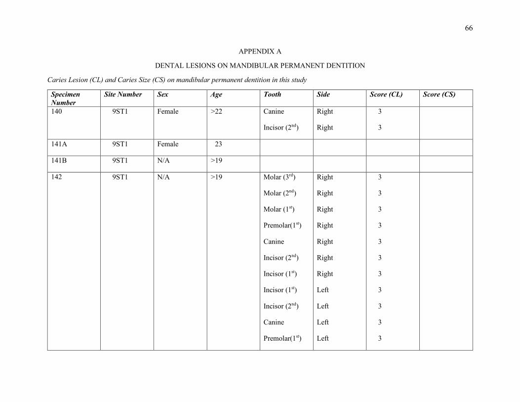
66
APPENDIX A
DENTAL LESIONS ON MANDIBULAR PERMANENT DENTITION
Caries Lesion (CL) and Caries Size (CS) on mandibular permanent dentition in this study
Specimen Number
Site Number Sex Age Tooth Side Score (CL) Score (CS)
140 9ST1 Female >22 Canine
Incisor (2nd)
Right
Right
3
3
141A 9ST1 Female 23
141B 9ST1 N/A >19
142 9ST1 N/A >19 Molar (3rd)
Molar (2nd)
Molar (1st)
Premolar(1st)
Canine
Incisor (2nd)
Incisor (1st)
Incisor (1st)
Incisor (2nd)
Canine
Premolar(1st)
Right
Right
Right
Right
Right
Right
Right
Left
Left
Left
Left
3
3
3
3
3
3
3
3
3
3
3
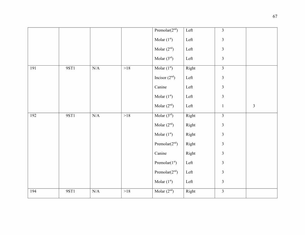
67
Premolar(2nd)
Molar (1st)
Molar (2nd)
Molar (3rd)
Left
Left
Left
Left
3
3
3
3
191 9ST1 N/A >18 Molar (1st)
Incisor (2nd)
Canine
Molar (1st)
Molar (2nd)
Right
Left
Left
Left
Left
3
3
3
3
1
3
192 9ST1 N/A >18 Molar (3rd)
Molar (2nd)
Molar (1st)
Premolar(2nd)
Canine
Premolar(1st)
Premolar(2nd)
Molar (1st)
Right
Right
Right
Right
Right
Left
Left
Left
3
3
3
3
3
3
3
3
194 9ST1 N/A >18 Molar (2nd) Right 3
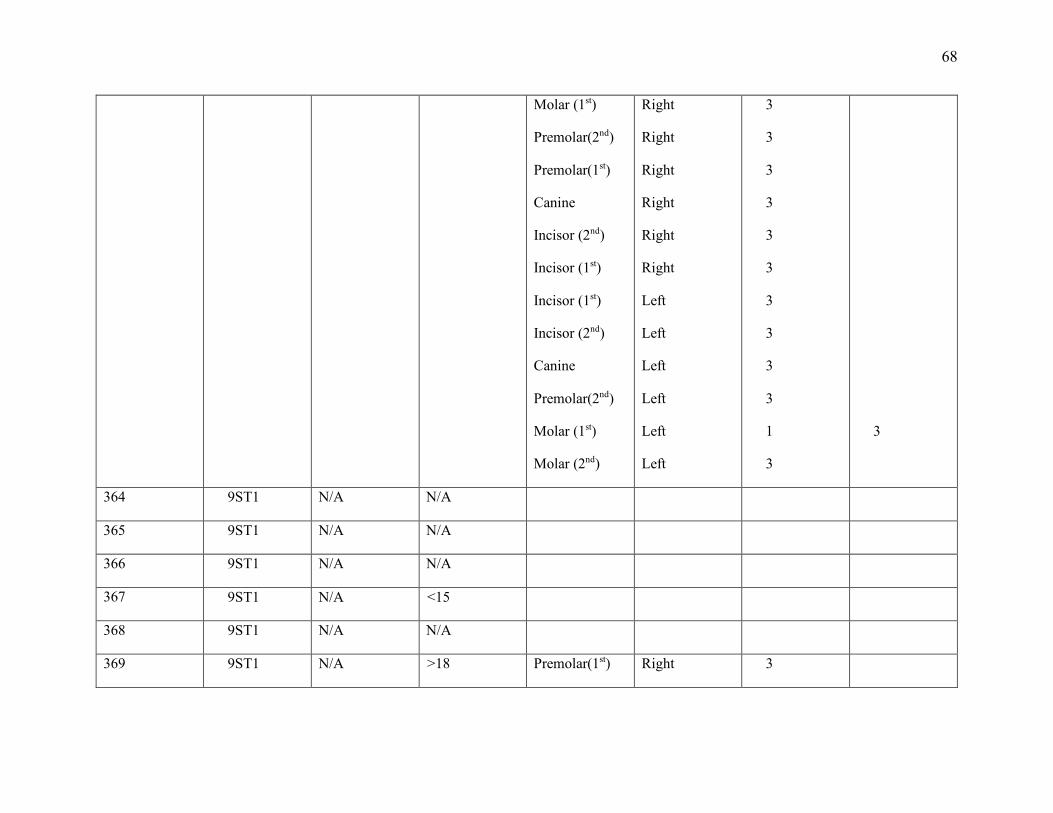
68
Molar (1st)
Premolar(2nd)
Premolar(1st)
Canine
Incisor (2nd)
Incisor (1st)
Incisor (1st)
Incisor (2nd)
Canine
Premolar(2nd)
Molar (1st)
Molar (2nd)
Right
Right
Right
Right
Right
Right
Left
Left
Left
Left
Left
Left
3
3
3
3
3
3
3
3
3
3
1
3
3
364 9ST1 N/A N/A
365 9ST1 N/A N/A
366 9ST1 N/A N/A
367 9ST1 N/A <15
368 9ST1 N/A N/A
369 9ST1 N/A >18 Premolar(1st) Right 3

69
Canine
Incisor (2nd)
Incisor (1st)
Incisor (1st)
Canine
Right
Right
Right
Left
Left
3
3
3
3
3
569 9ST1 N/A N/A
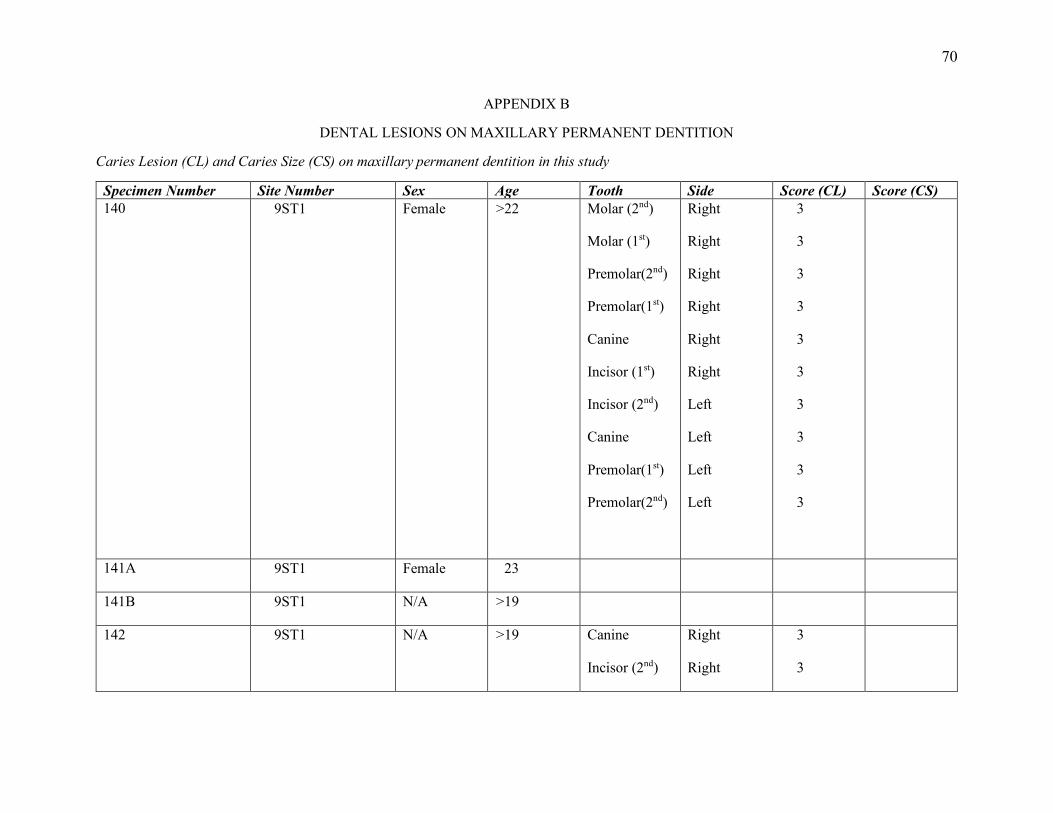
70
APPENDIX B
DENTAL LESIONS ON MAXILLARY PERMANENT DENTITION
Caries Lesion (CL) and Caries Size (CS) on maxillary permanent dentition in this study
Specimen Number Site Number Sex Age Tooth Side Score (CL) Score (CS) 140
9ST1 Female >22 Molar (2nd)
Molar (1st)
Premolar(2nd)
Premolar(1st)
Canine
Incisor (1st)
Incisor (2nd)
Canine
Premolar(1st)
Premolar(2nd)
Right
Right
Right
Right
Right
Right
Left
Left
Left
Left
3
3
3
3
3
3
3
3
3
3
141A 9ST1 Female 23
141B 9ST1 N/A >19
142 9ST1 N/A >19 Canine
Incisor (2nd)
Right
Right
3
3
71
Incisor (1st)
Canine
Premolar(1st)
Molar (2nd)
Molar (3rd)
Right
Left
Left
Left
Left
3
3
3
3
3
191 9ST1 N/A >18 Molar (3rd)
Molar (2nd)
Molar (1st)
Incisor (1st)
Incisor (1st)
Premolar(1st)
Premolar(2nd)
Molar (1st)
Right
Right
Right
Right
Left
Left
Left
Left
3
3
3
3
3
3
3
3
3
192 9ST1 N/A >18 Molar (3rd)
Molar (2nd)
Molar (1st)
Right
Right
Right
3
3
3
72
Premolar(2nd)
Premolar(1st)
Canine
Premolar(1st)
Premolar(2nd)
Molar (1st)
Molar (2nd)
Molar (3rd)
Right
Right
Left
Left
Left
Left
Left
Left
3
3
3
3
3
3
3
3
194 9ST1 N/A >18 Molar (3rd)
Molar (2nd)
Molar (1st)
Premolar(2nd)
Premolar(1st)
Canine
Incisor (2nd)
Incisor (1st)
Incisor (1st)
Incisor (2nd)
Canine
Right
Right
Right
Right
Right
Right
Right
Right
Left
Left
Left
3
3
3
3
3
3
3
3
3
3
3
73
Premolar(2nd) Left 3
364 9ST1 N/A N/A
365 9ST1 N/A N/A
366 9ST1 N/A N/A
367 9ST1 N/A <15
368 9ST1 N/A N/A Premolar(2nd)
Molar (2nd)
Left
Left
3
3
369 9ST1 N/A >18 Molar (3rd) Left 3
569 9ST1 N/A N/A
74
APPENDIX C
DENTAL LESIONS ON MANDIBULAR DECIDUOUS DENTITON
Caries Lesion (CL) and Caries Size (CS) on mandibular deciduous dentition in this study
Specimen Number Site Number Sex Age Tooth Side Score (CL) Score (CS) 193 9ST1 N/A 1 Molar(2nd)
Molar(1st)
Incisor(2nd)
Incisor(1st)
Incisor(1st)
Incisor(2nd)
Canine
Molar (1st)
Molar (2nd)
Right
Right
Right
Right
Left
Left
Left
Left
Left
3
3
3
3
3
3
3
3
3
370 9ST1 N/A 2 Molar (2nd)
Incisor (2)
Molar (1st)
Molar (2nd)
Right
Left
Left
Left
3
3
3
3
371 9ST1 N/A 1 Molar (2nd)
Molar (1st)
Right
Right
3
3

75
Incisor(2nd)
Canine
Right
Left
3
3
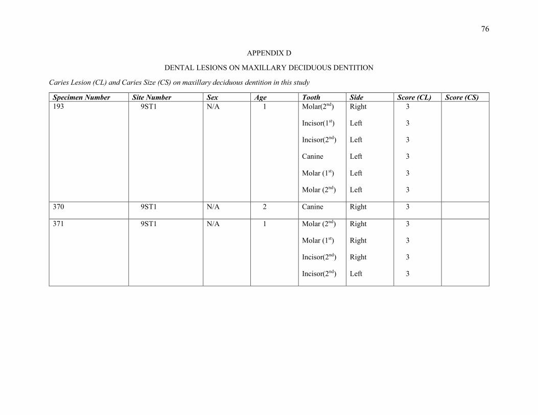
76
APPENDIX D
DENTAL LESIONS ON MAXILLARY DECIDUOUS DENTITION
Caries Lesion (CL) and Caries Size (CS) on maxillary deciduous dentition in this study
Specimen Number Site Number Sex Age Tooth Side Score (CL) Score (CS) 193 9ST1 N/A 1 Molar(2nd)
Incisor(1st)
Incisor(2nd)
Canine
Molar (1st)
Molar (2nd)
Right
Left
Left
Left
Left
Left
3
3
3
3
3
3
370 9ST1 N/A 2 Canine Right 3
371 9ST1 N/A 1 Molar (2nd)
Molar (1st)
Incisor(2nd)
Incisor(2nd)
Right
Right
Right
Left
3
3
3
3
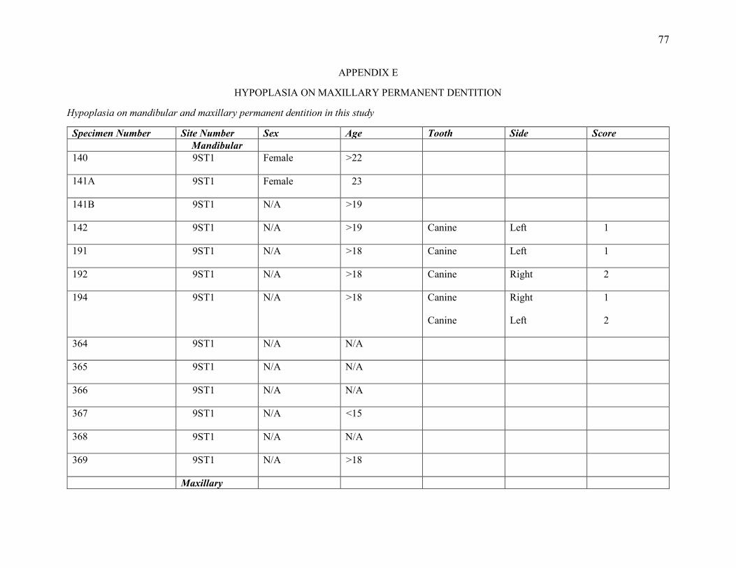
77
APPENDIX E
HYPOPLASIA ON MAXILLARY PERMANENT DENTITION
Hypoplasia on mandibular and maxillary permanent dentition in this study
Specimen Number Site Number Sex Age Tooth Side Score Mandibular 140 9ST1 Female >22
141A 9ST1 Female 23
141B 9ST1 N/A >19
142 9ST1 N/A >19 Canine Left 1
191 9ST1 N/A >18 Canine Left 1
192 9ST1 N/A >18 Canine Right 2
194 9ST1 N/A >18 Canine
Canine
Right
Left
1
2
364 9ST1 N/A N/A
365 9ST1 N/A N/A
366 9ST1 N/A N/A
367 9ST1 N/A <15
368 9ST1 N/A N/A
369 9ST1 N/A >18
Maxillary
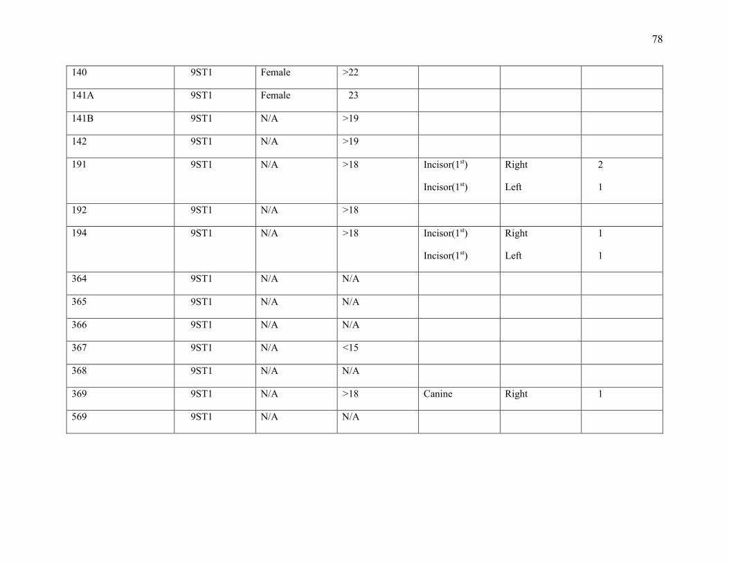
78
140 9ST1 Female >22
141A 9ST1 Female 23
141B 9ST1 N/A >19
142 9ST1 N/A >19
191 9ST1 N/A >18 Incisor(1st)
Incisor(1st)
Right
Left
2
1
192 9ST1 N/A >18
194 9ST1 N/A >18 Incisor(1st)
Incisor(1st)
Right
Left
1
1
364 9ST1 N/A N/A
365 9ST1 N/A N/A
366 9ST1 N/A N/A
367 9ST1 N/A <15
368 9ST1 N/A N/A
369 9ST1 N/A >18 Canine Right 1
569 9ST1 N/A N/A
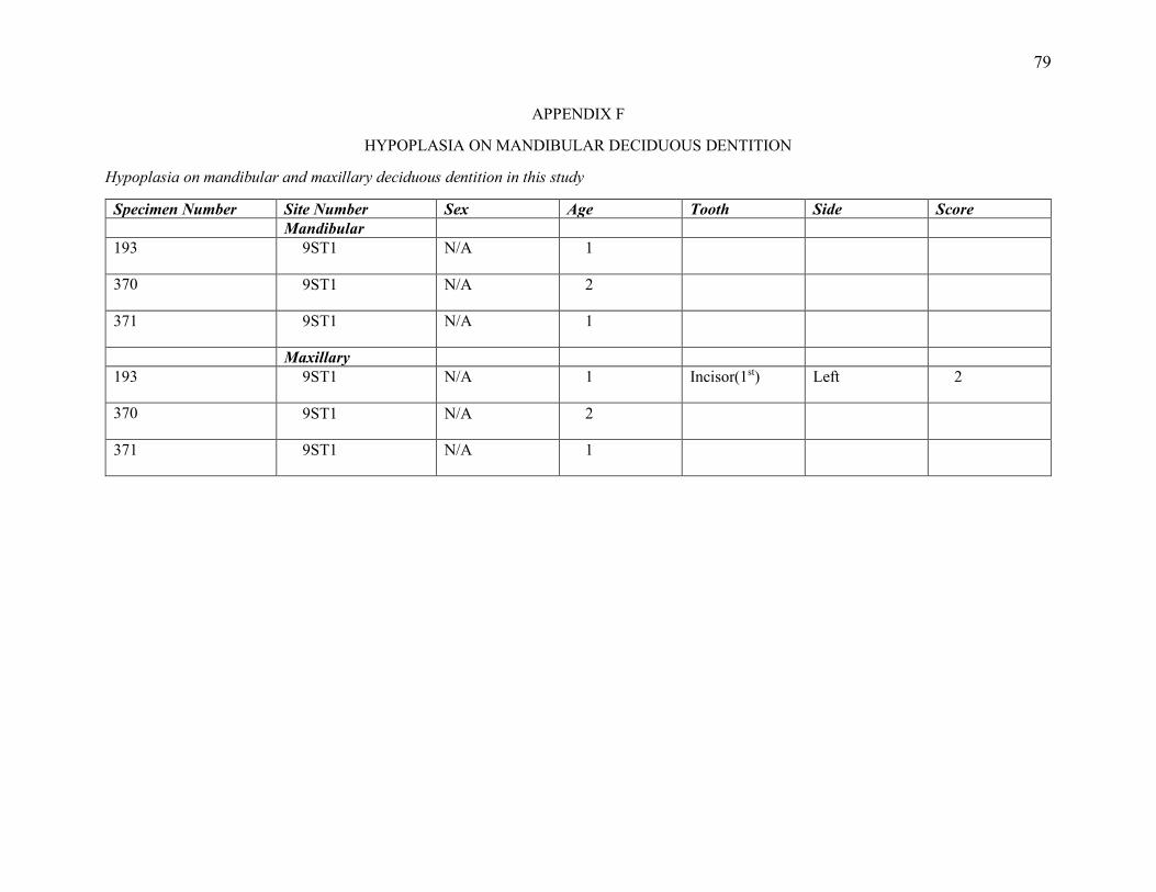
79
APPENDIX F
HYPOPLASIA ON MANDIBULAR DECIDUOUS DENTITION
Hypoplasia on mandibular and maxillary deciduous dentition in this study
Specimen Number Site Number Sex Age Tooth Side Score Mandibular 193 9ST1 N/A 1
370 9ST1 N/A 2
371 9ST1 N/A 1
Maxillary 193 9ST1 N/A 1 Incisor(1st) Left 2
370 9ST1 N/A 2
371 9ST1 N/A 1
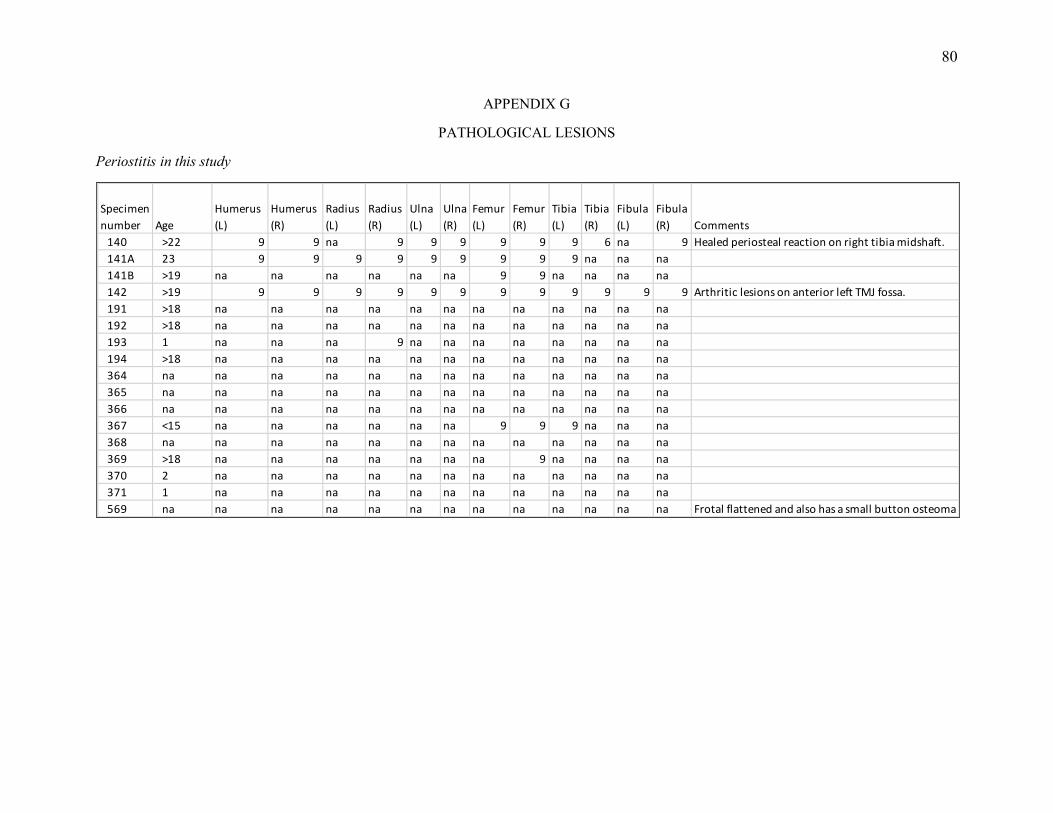
80
APPENDIX G
PATHOLOGICAL LESIONS
Periostitis in this study
Specimen number Age
Humerus (L)
Humerus (R)
Radius (L)
Radius (R)
Ulna (L)
Ulna (R)
Femur (L)
Femur (R)
Tibia (L)
Tibia (R)
Fibula (L)
Fibula (R) Comments
140 >22 9 9 na 9 9 9 9 9 9 6 na 9 Healed periosteal reaction on right tibia midshaft.141A 23 9 9 9 9 9 9 9 9 9 na na na141B >19 na na na na na na 9 9 na na na na142 >19 9 9 9 9 9 9 9 9 9 9 9 9 Arthritic lesions on anterior left TMJ fossa.191 >18 na na na na na na na na na na na na192 >18 na na na na na na na na na na na na193 1 na na na 9 na na na na na na na na194 >18 na na na na na na na na na na na na364 na na na na na na na na na na na na na365 na na na na na na na na na na na na na366 na na na na na na na na na na na na na367 <15 na na na na na na 9 9 9 na na na368 na na na na na na na na na na na na na369 >18 na na na na na na na 9 na na na na370 2 na na na na na na na na na na na na371 1 na na na na na na na na na na na na569 na na na na na na na na na na na na na Frotal flattened and also has a small button osteoma
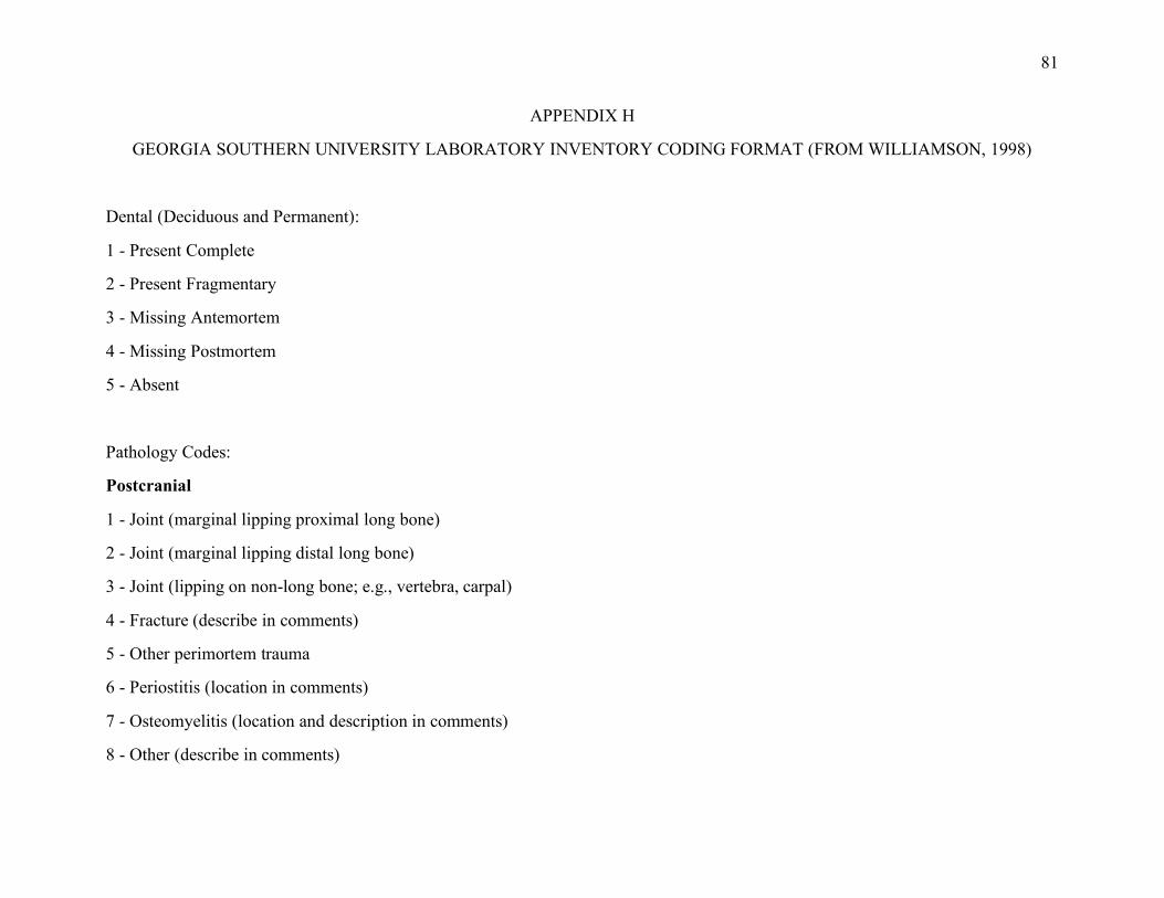
81
APPENDIX H
GEORGIA SOUTHERN UNIVERSITY LABORATORY INVENTORY CODING FORMAT (FROM WILLIAMSON, 1998)
Dental (Deciduous and Permanent):
1 - Present Complete
2 - Present Fragmentary
3 - Missing Antemortem
4 - Missing Postmortem
5 - Absent
Pathology Codes:
Postcranial
1 - Joint (marginal lipping proximal long bone)
2 - Joint (marginal lipping distal long bone)
3 - Joint (lipping on non-long bone; e.g., vertebra, carpal)
4 - Fracture (describe in comments)
5 - Other perimortem trauma
6 - Periostitis (location in comments)
7 - Osteomyelitis (location and description in comments)
8 - Other (describe in comments)

82
9 - No pathology noted
Dental Observations:
Caries, Size Hypoplasia
1 - Small 1 - Present
2 - Medium 2 - Absent
3 - Large