Embed Size (px)
Citation preview

J. Exp. Med.
The Rockefeller University Press • 0022-1007/2000/11/1223/13 $5.00Volume 192, Number 9, November 6, 2000 1223–1235http://www.jem.org/cgi/content/full/192/9/1223
1223
The Binding Site of Human Adenosine Deaminase for CD26/Dipeptidyl Peptidase IV: The Arg142Gln Mutation Impairs Binding to CD26 but Does Not Cause Immune Deficiency
By Eva Richard,
*
Francisco X. Arredondo-Vega,
*
Ines Santisteban,
*
Susan J. Kelly,
*
Dhavalkumar D. Patel,
*
‡
and Michael S. Hershfield
*
§
From the
*
Department of Medicine,
‡
Department of Immunology, and
§
Department of Biochemistry, Duke University Medical Center, Durham, North Carolina 27710
Abstract
Human, but not murine, adenosine deaminase (ADA) forms a complex with the cell mem-brane protein CD26/dipeptidyl peptidase IV. CD26-bound ADA has been postulated to regu-late extracellular adenosine levels and to modulate the costimulatory function of CD26 on Tlymphocytes. Absence of ADA–CD26 binding has been implicated in causing severe combinedimmunodeficiency due to ADA deficiency. Using human–mouse ADA hybrids and ADApoint mutants, we have localized the amino acids critical for CD26 binding to the helical seg-ment 126–143. Arg142 in human ADA and Gln142 in mouse ADA largely determine the ca-pacity to bind CD26. Recombinant human ADA bearing the R142Q mutation had normalcatalytic activity per molecule, but markedly impaired binding to a CD26
1
ADA-deficient hu-man T cell line. Reduced CD26 binding was also found with ADA from red cells and T cellsof a healthy individual whose only expressed ADA has the R142Q mutation. Conversely,ADA with the E217K active site mutation, the only ADA expressed by a severely immunode-ficient patient, showed normal CD26 binding. These findings argue that ADA binding toCD26 is not essential for immune function in humans.
Key words: adenosine deaminase deficiency • severe combined immunodeficiency • T lymphocyte • protein–protein interaction • adenosine deaminase complexing protein
Introduction
Adenosine deaminase (ADA)
1
deficiency in humans causesprofound lymphopenia, which is evident by midgestationand results in SCID during infancy (1, 2). Various effects
of adenosine (Ado) and 2
9
-deoxyadenosine (dAdo) havebeen implicated in pathogenesis (3). Skeletal, neurologic,and hepatic abnormalities that occur in some patients mayalso be due to the metabolic disorder, but these are of lessclinical relevance than the immunodeficiency. In contrastto the phenotype in humans, ADA knockout mice havenormal lymphoid development at birth and die perinatallyof hepatic and pulmonary injury (4, 5). Lymphopenia de-velops postnatally in strains genetically engineered to ex-press only placental ADA, but these animals die at a few
weeks of age from lung injury (4–6). The greater lym-phoid selectivity in humans than mice may reflect differ-ences in purine metabolism, tissue sensitivity to ADA sub-strates, or timing of development. In this context it isintriguing that human ADA, but not murine ADA, canform a complex with a multifunctional membrane proteinthat has a role in regulating thymocyte proliferation and Tcell activation.
The ADA of human erythrocytes behaves as a solublemonomer of
z
41 kD. Larger forms (
.
200 kD) found inextracts of other tissues of humans, rabbits, and cattle aredue to the binding of ADA to a homodimeric membraneglycoprotein with subunit
M
r
z
110 kD (7–9). This so-called “ADA complexing protein” (ADA-CP) was shown
to bind two ADA monomers with
K
A
of 4–20 nM (8, 10).ADA-CP occurs on secretory or absorptive surfaces of epi-thelia of liver, gut, kidney, and exocrine glands (11). Micelack “large” forms of ADA (12), but were found to possessa membrane protein with the tissue distribution and size ofADA-CP, which cross-reacted with Ab to human ADA-
Address correspondence to Michael S. Hershfield, Duke University Med-ical Center, Box 3049, Durham, NC 27710. Phone:
919-684-4184; Fax:919-684-4168; E-mail: [email protected]
1
Abbreviations used in this paper:
ADA, adenosine deaminase; ADA-CP,ADA complexing protein; Ado, adenosine; dAdo, 2
9
-deoxyadenosine;DPPIV, dipeptidyl peptidase IV; FPLC, fast protein liquid chromatog-raphy.
on April 14, 2018jem.rupress.org Downloaded from http://doi.org/10.1084/jem.192.9.1223Published Online: 30 October, 2000 | Supp Info:

1224
Binding Site of Adenosine Deaminase for CD26/Dipeptidyl Peptidase IV
CP, but bound neither murine nor human ADA (13). Un-like human and bovine ADA, murine ADA did not bind torabbit ADA-CP (10).
ADA-CP has been identified as a protein known both asCD26 and dipeptidyl peptidase IV (DPPIV; 14, 15). CD26was first defined as an antigen on activated human T lym-phocytes (16), and DPPIV as a widely distributed ectoen-zyme that cleaves peptides with Pro or Ala at position 2,including several hormones, neuropeptides, and cytokines(17, 18). CD26 and DPPIV cDNAs from human T cellsand intestine predict the same 766-residue, 88-kD poly-peptide (19, 20). This type II membrane protein has 6NH
2
-terminal cytoplasmic residues, a 34-residue mem-brane anchor, a large extracellular domain consisting of aglycosylated “stalk”, a cysteine-rich segment (residues 290–552), and a COOH-terminal region that bears the serineprotease (DPPIV) active site (residues 628–632 [21]). Resi-dues 294 and 340–343 of the cysteine-rich segment are es-sential for binding ADA (22, 23). In addition to ADA,CD26 has been reported to bind collagen, fibronectin, theCD45 tyrosine phosphatase, and the HIV-1 Tat protein(for reviews, see references 21 and 24).
Among lymphoid cells, CD26 is expressed on medullarythymocytes, blood T cells with a helper/memory pheno-type, activated B cells, and NK cells. On T cells, CD26 actsas a “co-stimulator” of antigen receptor–mediated activa-tion (24). CD26 ligation also stimulates the proliferation ofthymocytes and other hematopoietic cells in the mouse andrat (25, 26). It has been variously proposed that in humans,binding of ADA is important to the costimulatory functionof CD26, that such binding protects lymphocytes from ef-fects of extracellular Ado, and that immunodeficiency inpatients with ADA deficiency is due to the absence ofCD26-associated ADA (15, 23, 24, 27). We have definedthe amino acid residues of human ADA that are essentialfor CD26 binding, and investigated the effects on CD26binding of ADA mutations from individuals with ADA de-ficiency. Our findings are not consistent with some predic-tions about the nature of the CD26 binding site; neither dothey support the hypothesis that ADA–CD26 binding is es-sential for immune function in humans.
Materials and Methods
Materials
DEAE-Sepharose fast flow, Superose 12, and CNBr-activatedSepharose 4B were obtained from Amersham Pharmacia Biotech.ADA-Sepharose was prepared by coupling calf mucosal ADA(Sigma-Aldrich and Boehringer) to CNBr-activated Sepharose4B according to the manufacturer’s instructions. Plasmid pZC11and
Escherichia coli
SØ3834 were provided by Dr. Rod Kellems(University of Texas, Houston, TX). Goat anti–human ADA an-tiserum was provided by Dr. Dan Wiginton (University of Cin-cinnati, Cincinnati, OH). The mouse mAb 1C5 was preparedagainst ADA purified from human T cell leukemia cells (Hersh-field, M.S., unpublished details).
Lymphoid Cells and Cell Lines
Activated T lymphocytes were prepared by culturing bloodmononuclear cells with phytohemagglutinin and IL-2 as de-scribed (28), except that the cells were grown in AIM-V serum-free medium (Life Technologies). The HTLV-1–transformedAlNe cell line derived from an ADA-deficient patient (providedby Dr. Ken Weinberg, Los Angeles Children’s Hospital, Los An-geles, CA) was also maintained in AIM-V medium.
Enzyme Assays
DPPIV activity was determined using Gly-Pro-
p
-nitroanilidetosylate (Sigma-Aldrich) as substrate, monitoring A405 at 37
8
C(29). ADA activity was determined by monitoring the decrease inA263 at 37
8
C using as substrate 150
m
M Ado in 50 mM Tris-HCl, pH 7.4. Both the CD26 and ADA assays were performed in96-well microtiter plates (Costar UV plate; Corning Inc.) in reac-tion volumes of 0.2 ml, using a SpectraMax plus spectrophotom-eter (Molecular Devices). ADA was also assayed by a radiochem-ical-TLC method (28). Protein was determined using thebicinchoninic acid (BCA) method (Pierce Chemical Co.) withBSA as standard.
Rabbit Kidney CD26/DPPIV
CD26/DPPIV was purified from New Zealand white rabbitkidney homogenates by a published procedure involving DEAE-Sepharose and ADA-Sepharose 4B chromatography (30). Insome preparations, the ADA-Sepharose column was eluted with6 M urea in 0.01 M KPO
4
, pH 7.4, 0.1% Triton X-100, as de-scribed (31). Fractions with DPPIV activity were pooled, dia-lyzed against 0.01 M KPO
4
, pH 7.4, 0.1% Triton X-100, andstored at
2
20
8
C. The purified preparation had a DPPIV-specificactivity of 7,883
m
mol min
2
1
mg
2
1
. A partially purified prepara-tion used for screening ADA–CD26 binding had DPPIV activityof 6.5–7.2
m
mol min
2
1
mg
2
1
. ADA activity of these preparationswas
,
0.003
m
mol min
2
1
mg
2
1
.
Bovine ADA cDNA
Standard methods were used to amplify, clone, and sequencecDNA (32, 33). Degenerate reverse transcription PCR primerswere used to clone the ADA coding region from bovine thymusRNA: (
1
)5
9
ATCGAAGCTTCCATGGCCCAGACRCCCGC-MTTC, (
2
)5
9
GGCCATGGAGAGGTAGCCACGACACCTT-CACAGACA. Both strands were sequenced by the dideoxymethod (sequence data are available from EMBL/GenBank/DDBJunder accession no. AF280603). The expressed product of the bo-vine ADA cDNA was enzymatically active (Kelly, S.J., and M.S.Hershfield, unpublished data).
Construction of Human–Mouse ADA cDNA Hybrids and Point Mutants, and Expression of ADA for CD26 Binding Studies
cDNAs consisting of segments from both the human (34) andmouse (35) ADA coding regions were made by overlap extensionPCR (36; Tables I and II). The P126Q, R142Q, R149Q,A215T, and E217K ADA mutants have been identified in humansubjects (37–40); their expression in
E
.
coli
strain SØ3834 hasbeen reported (41). Other mutations (see Results) were intro-duced into the wild-type human or mouse ADA cDNAs byPCR mutagenesis essentially as described (37, 41). All final ADAcDNA PCR products were cloned into pBluescript II KS orpBluescript SK and fully sequenced using the ABI 377 PRISMDNA Sequencing Instrument (Applied Biosystems).
1225
Richard et al.
ADA cDNAs were ligated into the NcoI site of pZ (derivedfrom pZC11 from which wild-type human ADA cDNA hadbeen excised [42]). pZ/ADA plasmids were used to transform
E
.
coli
SØ3834, which has a deletion of the bacterial ADA gene (42).Single transformant colonies were grown at 37
8
C in Luria broth/carb/tet medium to constitutively express ADA, as described(41). Under these conditions, the yield of wild-type mouse ADAwas four- to sixfold higher than for human ADA, determinedboth by activity assay (Table III) and by Western blotting withcross-reacting goat anti–human ADA antiserum (not shown).Studies with human–mouse ADA hybrids (Table III) and resultsnot presented indicate that this difference in expression is relatedto the presence in human ADA of NH
2
-terminal codons used in-frequently in
E
.
coli
.To prepare ADA for CD26 binding studies, the cells from 100-
ml overnight cultures were sonicated in 3 ml of lysis buffer (41).
In some experiments the cells from 1-liter cultures were disruptedin 20 ml of lysis buffer using a Microfluidizer Model M-110L(Microfluidics, Inc.). After centrifugation (20,190
g
, 15 min, 4
8
C),supernatants were passed through 0.2
m
m filters and dialyzed at4
8
C against 0.01 M KPO
4
, pH 7.4. In addition to assaying ADAactivity, aliquots of these lysates (20-
m
g protein) were analyzed byWestern blotting using goat anti–human ADA antiserum, as de-scribed (41). Western blotting was also performed with the 1C5mouse mAb to human ADA (1:2,000 dilution of ammonium sul-fate–concentrated mouse ascites); a goat anti–mouse IgG–horse-radish peroxidase conjugate (Santa Cruz Biotechnology, Inc.) wasused as a second Ab, and was detected with the EnhancedChemiluminescence System (Amersham Pharmacia Biotech).
ADA-CD26 Binding
FPLC Screening.
In the standard assay, dialyzed (0.01 MKPO
4
, pH 7.4) lysates of pZ/ADA cDNA-transformed
E
.
coli
SØ3834 cells containing 100–250 nmol/min of ADA activity (di-luted as necessary with dialyzed lysate of untransformed SØ3834cells) were combined with 10–30 nmol/min (DPPIV activity) ofrabbit CD26 in 0.01 M KPO
4
, pH 7.4, 0.1% Triton X-100, totalvolume 150
m
l. After incubating for 2 h at 37
8
C, the entire mix-ture was injected onto a 1.5
3
30 cm Superose 12 column equili-brated with 50 mM Tris-HCl, pH 8.4 (fast protein liquid chro-matography [FPLC] system; Amersham Pharmacia Biotech). Thecolumn was eluted with this buffer at room temperature, flow rate0.5 ml/min. Fractions (0.5 ml) were assayed for ADA and DPPIVactivities, and the percentage of total ADA activity associated withthe peak of DPPIV activity (CD26) was calculated. In some ex-periments the column was equilibrated and eluted with 50 mMTris-HCl, pH 8.4, containing 150 mM NaCl. This shifted thepeak of free (41 kD) ADA activity from fraction 25 to fraction 27,but neither the DPPIV activity peak nor the amount of ADA ac-tivity associated with the DPPIV peak were altered.
Gel Mobility Shift Assay.
[
35
S]Met-labeled ADA was gener-ated in vitro from ADA cDNA constructs in pBluescript, usingthe TNT Coupled Wheat Germ Extract System (Promega) ac-cording to the manufacturer’s instructions (this system was usedin preference to rabbit reticulocyte extracts because it possessedvery low endogenous ADA-like activity). Translation productswere analyzed by SDS-PAGE and fluorography, and were alsoelectrophoresed on cellulose acetate and stained for ADA activityin situ, as described (28). Aliquots of the translation mixturescontaining equal amounts of
35
S-labeled products were then in-cubated (2 h, 37
8
C) with 0.7–2.2 nmol/min of rabbit CD26/DP-PIV in 0.01 M KPO
4
, pH 7.4, 0.1% Triton X-100, total volume20
m
l. These reaction mixtures were then electrophoresed on a3–15% gradient nondenaturing PAGE gel (pH 8.8, 30% acryl-amide, 0.8% bisacrylamide; running conditions 60 V/cm, 18 h,4
8
C [43]). [
35
S]ADA was located by fluorography.
Recombinant ADA Binding to AlNe T Cells
Aliquots (25
m
l) of pZ/ADA cDNA-transformed SØ3834 ly-sates containing 400 nmol/min of ADA activity were added to 2
3
10
7
AlNe T cells in 1 ml of serum-free growth medium and in-cubated 2 h at 37
8
C. The T cells were then washed three timeswith PBS, pH 7.4, and lysed by freezing and thawing (or by soni-cation, which gave identical results) in 150
m
l of 25 mM Tris-HCl, pH 7.5, 1 mM EDTA, 1 mM DTT, 0.1% Triton X-100.After centrifugation, the supernatant was analyzed as describedabove for the FPLC CD26 binding assay. ADA binding to intactAlNe cells was determined by flow cytometry using the 1C5 anti-ADA mAb (see below). For these studies, 10
6
AlNe cells were in-
Table I.
Human–Mouse ADA Hybrids
Construct
*
Templates(ADA cDNAs)
‡
Primers
‡
M1–81/H82–363 I: M wild-type 1, 2
II: H wild-type 3, 4
III: Products of I and II 1, 4
H1–81/M 82–125/H126–363 I: H 1–81/ M 82–352 5, 6
II: H wild-type 7, 4
III: Products of I and II 5, 4
H1–125/M126–143/H144–363 I: H wild-type 5, 8
II: H wild-type 9, 4
III: Products of I and II 5, 4
H1–143/M144–247/H248–363 I: H wild-type 5, 10
II: M 1–247/H 248–363 11, 4
III: Products of I and II 5, 4
H1–247/M248–352 I: H wild-type 5, 12
II: M wild-type 13, 14
III: Products of I and II 5, 14
H1–81/M82–352 I: H wild-type 5, 15
II: M wild-type 16, 14
III: Products of I and II 5, 14
M1–247/ 248–363 I: M wild-type 1, 17
II: H wild-type 18, 4
III: Products of I and II 1, 4
M1–125/H126–143/M144–352 I: M wild-type 1, 6
II: M wild-type 11, 14
III: Products of I and II 1, 14
*
H, human ADA; M, mouse ADA. Numbers indicate amino acid resi-dues.
‡
PCR reactions I and II generated DNA fragments with 45–84-bpcentral overlaps. These were gel purified, combined, annealed, and am-plified in PCR reaction III to yield the desired hybrid constructs (col-umn 1). ADA cDNA templates and the primer pairs for reactions I, II,and III are listed in columns 2 and 3 (primer sequences are in Table II).In addition to the templates and primers (10 pmol each), each 50 µlPCR reaction contained 4 dNTPs (0.2 mM each), 1
3
PCR buffer, 1.5mM MgCl
2
, and 2.5 U of AmpliTaq DNA polymerase (PerkinElmer).Thermal cycling was optimized for each reaction.

1226
Binding Site of Adenosine Deaminase for CD26/Dipeptidyl Peptidase IV
cubated for 60 min at 37
8
C in 1 ml of medium containing aliquotsof untransformed or ADA-expressing SØ3834 lysates. The cellswere then washed with cold PBS containing 1% BSA and 0.1%sodium azide (PBSW) and processed for flow cytometry.
Flow Cytometry
Surface antigens and bound ADA on AlNe T cells were de-tected with the following reagents: unconjugated anti-ADA mAb1C5 and control mAb P3; and fluorochrome-conjugated Ta1-RD1 (CD26), B1-RD1 (CD20), T11-RD1 (CD2), mouseIgG1-RD1, and either fluorescein- or PE-conjugated goat anti–mouse IgG1 provided by Beckman Coulter (Fullerton, CA). AllAb incubations were performed in PBSW at 4
8
C. After washingwith PBSW, cells were fixed with 0.4% paraformaldehyde, andanalyzed using an Epics Elite XL flow cytometer (BeckmanCoulter). Data were processed using the FACSConvert™ andCellQuest™ software programs (Becton Dickinson).
Results
Binding of Recombinant ADAs to Rabbit CD26.
Like theenzymes isolated from tissues (10), human and mouse ADAexpressed in
E
.
coli
SØ3834 differ markedly in ability to
Table II.
PCR Primers Used in Constructing Human–Mouse ADA Hybrids
Primer sequence
*
Location in ADA cDNA
‡
1
(
1
)CGCGCGAATTCGGGCACCATGGCCCAGACACCCGCATTC M1–21 (EcoRI/NcoI)2 (2)CATCTCTACAAACTCATAGGCGATCCTCTTGATGGCCTCTCTGCA Reverse of primer 33 (1)TGCAGAGAGGCCATCAAGAGGATCGCCTATGAGTTTGTAGAGATG M223–243/H244–2674 (2)GCGCAAGCTTCGGGCCATGGTCTTCAGAGGTTCTGCCCTGCAG H1073–1092 (HindIII/NcoI)5 (1)CGCGCGAATTCGGGCACCATGGCCCAGACGCCCGCCTTCGAC H1–24 (EcoRI/NcoI)6 (2)GTCTCGCTCCCCCTCCTGCAGGCCCTGGCCCACTAGGGCTACCACCT Reverse of primer 7
CGTCTGGGGTGACGTCCCCTTCAGTCTGGTTCCAGGG7 (1)CCCTGGAACCAGACTGAAGGGGACGTCACCCCAGACGAGGTGGTAGC M346–375/H376–429
CCTAGTGGGCCAGGGCCTGCAGGAGGGGGAGCGAGAC8 (2)TGCTTGCTCTCCCTCCTGCAGGCCCTGGTTCACAAGATCCACAACGT H346–375/M376–429
CATCAGGGGTGAGGTCCCCTTCAGCCTGGTTCCAGGG9 (1)CCTGATGACGTTGTGGATCTTGTGAACCAGGGCCTGCAGGAGGGAGA M376–429/H430–459
GCAAGCATTCGGGGTCAAGGCTCGGTCCATCCTGTGC10 (2)GCACAGAATGGACCGGACCTTGATGCCAAAGTCTCGCTCCCCCTCCT Reverse of primer 11
GCAGGCCCTGGCCCACTAGGGCTACCACCTCGTCTGG11 (1)CCAGACGAGGTGGTAGCCCTAGTGGGCCAGGGCCTGCAGGAGGGGGA H376–429/M430–459
GCGAGACTTTGGCATCAAGGTCCGGTCCATTCTGTGC12 (2)TTCTTTCAGTAGTCTGTTGTAGAGGGCCTGGTCTTCCAGGGTGTG Reverse of primer 1313 (1)CACACCCTGGAAGACCAGGCCCTCTACAACAGACTACTGAAAGAA H721–741/M742–76514 (2)GCGCGATATCCGGGCCATGGTCTCTATTGGTATTCTCTGTAGAGCC M1037–1059 (EcoRV/NcoI)15 (2)CATCTCCACAAACTCGTAGGCGATCCTTTTGATAGCCTCCCGGCA Reverse of primer 1616 (1)TGCCGGGAGGCTATCAAAAGGATCGCCTACGAGTTTGTGGAGATG H223–243/M 244–26717 (2)TTCCTGCCGCAGCCTGTTATAAAGAGCTTCATCCTCGATGGTGTG Reverse of primer 1818 (1)CACACCATCGAGGATGAAGCTCTTTATAACAGGCTGCGGCAGGAA M721–741/H742–765
*Cloning sites are underlined. The bold italic nucleotides are silent polymorphisms found in human ADA cDNA.‡The numbers indicate nucleotide position in human (H) or mouse (M) ADA cDNA (adenine of the initiation codon51).
Table III. ADA Activity of Human–Mouse Hybrids Expressed in E. coli SØ3834
ADA cDNAADA activity
(mean 6 SD)Wild-type
human ADA
mmol/min per mg protein %Human wt 3.3 6 0.7 100.0
Mouse wt 12.4 6 1.3 375.7
M1–81/H82–363 13.5 6 2.2 409.0
H1–81/M82–125/H126–363 4.3 6 0.4 130.3
H1–125/M126–143/H144–363(“m126–143”) 1.2 6 0.1 36.3
H1–143/M144–247/H248–363 2.9 6 0.3 87.8
H1–247/M248–352 2.0 6 0.2 60.6
H1–81/M82–352 1.8 6 0.7 54.5
M1–247/H248–363 12.3 6 3.8 372.7
M1–125/H126–143/M144–352 (“h126–143”) 13.1 6 3.6 396.9
wt, wild-type; H, human; M, mouse.
1227 Richard et al.
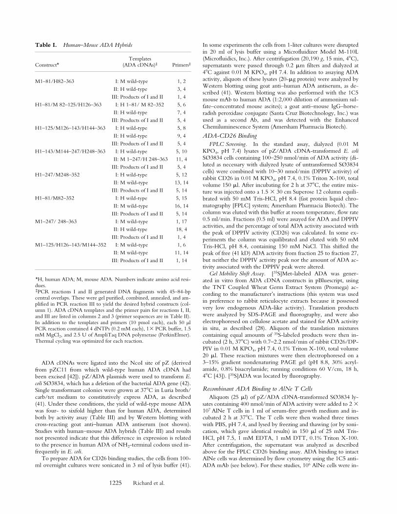
bind rabbit kidney CD26/DPPIV. When equal amounts ofADA activity were compared, z50% of human and ,1%of mouse ADA activity coeluted with DPPIV activity froma Superose 12 FPLC column (Fig. 1, A and B). The differ-ence in CD26 binding could also be demonstrated usingnondenaturing PAGE to assess the effect of rabbit CD26 on
the migration of 35S-labeled human and mouse ADA invitro translation products (Fig. 1 C). Addition of unlabeledhuman ADA expressed in E. coli SØ3834 (100 nmol/min)blocked the CD26-induced shift in migration of the human[35S]ADA translation product, whereas the same amount ofrecombinant mouse ADA had no effect (not shown).
Based on these results, we constructed human–mouseADA cDNA hybrids (Tables I–III) and used the above as-says to locate residues necessary for CD26 binding (analo-gous to human–rat CD26 “swap mutants” used to identifyCD26 residues necessary for ADA binding [22]). To refineour search, we also used point mutants found in ADA-defi-cient individuals, which we have previously expressed inSØ3834 (41). ADA mutants with sufficient catalytic activ-ity were screened by FPLC, although in some cases usingless ADA activity than in the “standard” assay (see Materialsand Methods). The PAGE assay was used for ADA mutantswith very low catalytic activity. Some mutants could notbe tested because their activity was too low for the FPLCassay, and their in vitro translation products did not enterthe nondenaturing gel (results not shown).
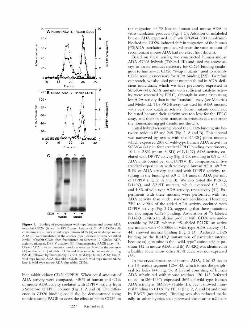
Initial hybrid screening placed the CD26 binding site be-tween residues 82 and 248 (Fig. 2, A and B). This intervalwas narrowed by results with the R142Q point mutant,which expressed 28% of wild-type human ADA activity inSØ3834 (41): in four standard FPLC binding experiments,10.4 6 2.9% (mean 6 SD) of R142Q ADA activity co-eluted with DPPIV activity (Fig. 2 C), resulting in 0.9 6 0.5ADA units bound per unit DPPIV. By comparison, in fivestandard experiments with wild-type human ADA, 48.7 65.1% of ADA activity coeluted with DPPIV activity, re-sulting in the binding of 5.9 6 1.4 units of ADA per unitof DPPIV (Fig. 2, A and B). We also tested the P126Q,R149Q, and A215T mutants, which expressed 0.3, 6.2,and 4.8% of wild-type ADA activity, respectively (41). Ex-periments with these mutants were performed with lessADA activity than under standard conditions. However,75% to .90% of the added ADA activity coeluted withDPPIV activity (Fig. 2 C), suggesting that these mutationsdid not impair CD26 binding. Association of 35S-labeledR142Q in vitro translation product with CD26 was unde-tectable by PAGE, whereas 35S-labeled E217K, an activesite mutant with ,0.005% of wild-type ADA activity (41,44), showed normal binding (Fig. 2 D). Reduced CD26binding by the R142Q mutant was of particular interestbecause (a) glutamine is the “wild-type” amino acid at po-sition 142 in mouse ADA, and (b) R142Q was identified ina healthy adult whose other ADA allele was not expressed(38).
In the crystal structure of murine ADA, Gln142 lies inthe 18-residue segment 126–143, which forms the periph-eral a2 helix (44; Fig. 3). A hybrid consisting of humanADA substituted with mouse residues 126–143 (referredto as “m126–143”) expressed 36% of wild-type humanADA activity in SØ3834 (Table III), but it showed mini-mal binding to CD26 by FPLC (Fig. 2, A and B) and noneby PAGE (not shown). Binding was also reduced mark-edly in other hybrids that possessed the murine a2 helix
Figure 1. Binding of recombinant wild-type human and mouse ADAto rabbit CD26. (A and B) FPLC assay. Lysates of E. coli SØ3834 cellscontaining equal units of wild-type human ADA (A) or wild-type mouseADA (B) were incubated in the absence (open circles) or presence (filledcircles) of rabbit CD26, then fractionated on Superose 12. Circles, ADAactivity; triangles, DPPIV activity. (C) Nondenaturing PAGE assay. 35S-labeled ADA in vitro translation products were incubated in the presence(1) or absence (2) of rabbit CD26 and then subjected to nondenaturingPAGE, followed by fluorography. Lane 1, wild-type human ADA; lane 2,wild-type human ADA plus rabbit CD26; lane 3, wild-type mouse ADA;lane 4, wild-type mouse ADA plus rabbit CD26.
1228 Binding Site of Adenosine Deaminase for CD26/Dipeptidyl Peptidase IV
residues (Fig. 2, A and B). Hybrids in which all other re-gions of human ADA, except for residues 126–143, werereplaced with their murine counterparts had substantialADA activity (Table III) and showed essentially normalCD26 binding (Fig. 2, A and B). Modifying mouse ADAby substituting human residues 126–143 for the corre-sponding mouse residues (“h126–143”), or by making theQ142R point mutation, conferred some ability to bindCD26, which could be appreciated by FPLC (Fig. 2, A–C).Together, these results show that the amino acids crit-ical for CD26 binding lie within segment 126–143 of hu-man ADA.
As bovine ADA also binds CD26, we amplified cDNAcarrying the ADA coding region from bovine thymusRNA. The predicted amino acid sequences of residues115–150 of human, bovine, and murine ADA are com-pared in Fig. 3. Within segment 126–143, human and bo-vine ADA differ at only 2 of 18 positions, whereas humanand murine ADA differ at 5, and murine and bovine ADAat 4 positions. The most notable differences are (a) residues141–143 of human and bovine ADA are all charged, Glu-Arg-Asp, whereas only Glu141 is charged in the mouse se-
quence, Glu-Gln-Ala; and (b) residue 131 in mouse ADAis Asp, whereas Ala and Ser are found in human and bovineADA, respectively. We prepared mutants of human ADAin which a2 helix residues from murine ADA replaced thecorresponding human residues. All were active when ex-pressed in E. coli SØ3834 (data not shown), but they fellinto two groups with respect to CD26 binding. The singlemutants E128D, A131D, G134N, and D143A all showedCD26 binding in the FPLC assay (Fig. 2 C). Binding bythe double mutants E128D/R142Q and R142Q/D143A,the triple mutant E128D/R142Q/D143A, and the qua-druple mutant E128D/A131D/R142Q/D143A was ineach case reduced, but comparable to that of R142Q alone(Fig. 2 C).
Binding of Recombinant ADAs to Human T Cell–associatedCD26. The AlNe HTLV-1 transformed T cell line is de-rived from an immunodeficient patient homozygous for amutation in ADA intron 10 that activates a cryptic 39 splicesite (45). ADA activity was unmeasurable in extracts ofAlNe cells when cultured in serum-free (ADA-free) me-dium, and ADA protein could not be detected by im-munoblotting with polyclonal Ab to human ADA (not
Figure 2. Binding of rabbit CD26 by human–mouse ADA hybrids and ADA point mutants. (Aand B) FPLC assay of human–mouse ADA hybrids.Hybrid structures are shown as bars in which whiteindicates human (h) and black indicates mouse (m)ADA amino acid residues (numbered beside bars).Gray bars in A and B show the percentage of inputADA activity associated with DPPIV activity (A),or the units of ADA bound per unit of DPPIV (B).Error bars (SD) shown for wild-type human ADA(A–C) and the R142Q mutant (C) are based onfive and four standard binding experiments (seeMaterials and Methods), respectively. (C) FPLC as-say of human (Hum) and mouse (Mus) ADA pointmutants. Black bars are mutants found in ADA-deficient individuals; gray bars are mutants preparedfor this study from human or mouse wild-type
cDNA, as indicated. Standard FPLC assay conditions were used for screening the hybrids and point mutants, except for the R149Q, A215T, P126Q mu-tants from ADA-deficient individuals. In those cases, a lower amount of ADA activity was used, accounting for a greater percentage of input ADA activ-ity becoming associated with CD26/DPPIV than was found with wild-type human ADA. (D) Nondenaturing PAGE assay. Lane 1, wild-type (WT) hu-man ADA; lane 2, wild-type human ADA plus rabbit CD26; lane 3, R142Q human ADA mutant; lane 4, R142Q plus rabbit CD26; lane 5, E217Khuman ADA mutant; lane 6, E217K plus rabbit CD26.

1229 Richard et al.
shown). AlNe cells express a substantial level of CD26 (seebelow). We compared the binding of several recombinantADAs to CD26 on AlNe cells (see Figs. 4–6).
Virtually no ADA activity was found in Superose 12–fractionated extracts of AlNe cells that had been incubatedwith a high concentration (400 nmol/min) of recombinantmurine ADA (Fig. 4 A), or with the m126–143 human–mouse hybrid (Fig. 4 B). After incubation with wild-typehuman ADA, 91% of cell-associated ADA activity coelutedwith DPPIV, resulting in a ratio of ADA to DPPIV activityin this peak of 3.7 (Fig. 4 A). With the R142Q mutant,z40% as much total ADA activity became cell associated aswas found with wild-type human ADA. However, only9% of the bound R142Q ADA activity coeluted with DP-PIV, resulting in a ratio of R142Q ADA to DPPIV activityof 0.1 (Fig. 4 B). Virtually identical results were obtainedfor wild-type human ADA and the R142Q mutant in asecond experiment using the same protocol. The R142QADA activity eluting as a 41-kD monomer may have re-sulted from unstable binding of mutant ADA to CD26 onAlNe cells, with subsequent dissociation in the extract orduring FPLC. This transient binding probably reflects the
high concentration of exogenous ADA activity used inthese experiments (see below).
We next examined binding of recombinant ADAs to thesurface of AlNe cells by flow cytometry, using mouse mAb1C5 to human ADA. Western blot experiments indicatedthat 1C5 recognizes a COOH-terminal epitope present inhuman but not in mouse ADA, and it reacts approximatelyas well with the R142Q and E217K mutants as with wild-type human ADA (Fig. 5). In these experiments (and inflow cytometry studies shown below in Fig. 6 D), equalunits of R142Q and wild-type ADA activity were com-pared (Fig. 5 A), whereas equal amounts of total SØ3834lysate protein were used to compare E217K and wild-typeADA (Fig. 5 B). Taken together with previous studies ofexpression (38, 41), these results indicate that the R142Qprotein has the same activity per molecule as wild-typeADA, but is somewhat less stable in cells, whereas theE217K protein is about as stable as the wild-type enzyme,but is catalytically inactive. The stability of E217K is un-usual because most ADA missense mutations identified inSCID patients result in unstable proteins that are difficult todetect with antisera to ADA, both in patient-derived celllines and when expressed in E. coli SØ3834 (41, 46).
In flow cytometry experiments, AlNe cells expressedhigh levels of CD26 (Fig. 6 A). They did not bind anti-ADA mAb 1C5 (Fig. 6 B). Incubating AlNe cells with re-combinant wild-type human ADA (followed by washing)induced 1C5 binding (Fig. 6, C and D), as did the R142Qand E217K mutants (Fig. 6 D). 1C5 binding increased withthe amount of ADA to which the cells were exposed (Fig.6 D). Based on mean fluorescence values, at the lowestconcentrations tested, E217K induced about half the 1C5binding induced by wild-type ADA, but equivalent bind-ing occurred at the higher end of the range (because ex-pression in SØ3834 is somewhat variable [41], the amountsof wild-type and E217K ADA protein may not have beenidentical). 1C5 binding was detected at all concentrationsof wild-type and E217K proteins tested, but was not mea-surable with R142Q ADA at 0.3 and 1.7 nmol/min per mlmedium. Wild-type ADA induced 7.5-fold more 1C5binding than R142Q at ADA activities of 8.3 and 42nmol/min, and fourfold greater binding at 209 nmol/min(very similar results were found in a second experiment notshown). ADA bound to CD26 is derived from an extracel-lular source (47). Serum ADA1 activity (the isozyme de-rived from the ADA gene) for 320 normal humans was re-ported to be 4.2 6 1.5 nmol/min per ml (48). Thus, underphysiological conditions, binding of circulating R142QADA to T cells via CD26 may be 10-fold lower thannormal.
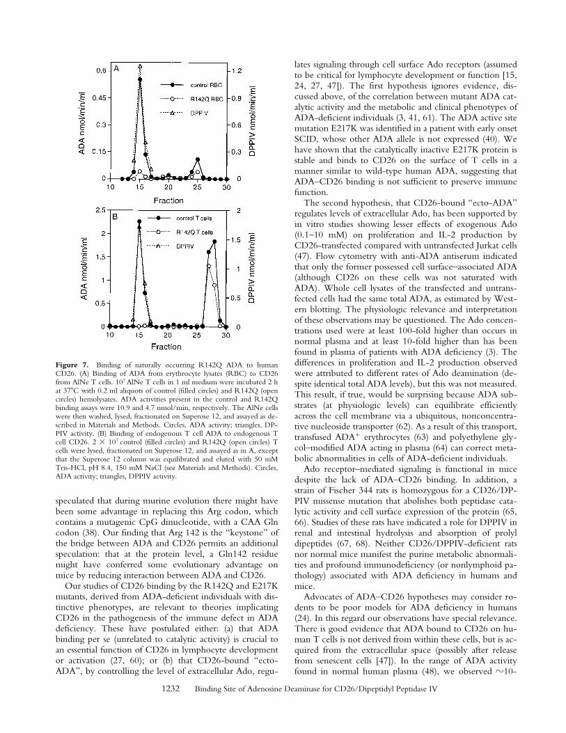
Binding of Naturally Occurring R142Q ADA to HumanCD26. Human ADA expressed in E. coli might interactdifferently with CD26 than would ADA produced in hu-man cells. To address this possibility, we studied the bind-ing of ADA in extracts of erythrocytes and cultured T cellsfrom the individual in whom the R142Q allele was identi-fied, whose second ADA allele carries a nonsense mutationin codon 3 (38). A limited quantity of R142Q cells was
Figure 3. Location of the a2 helix (CD26 binding site) in the murineADA crystal structure. The RasMol model is based on coordinates, as re-ported (reference 44). The a2 helix (residues 126–143) is shown in or-ange, with space-filling display of residues D131, Q142, and A143. Fororientation, the COOH-terminal helix (residues 337–351) is shown inblue. Bound inhibitor (rose) and the zinc ion (yellow) are space-fillingstructures at the active site. The panel below the model compares the par-tial amino acid sequences (residues 115–150) of human (HUM), murine(MUS), and bovine (BOV) ADA. Amino acids 126–143 of human ADA,green letters; amino acids D131, Q142, and A143 of murine ADA, redletters; other amino acids of bovine and murine ADA that differ from thehuman sequence, blue letters.

1230 Binding Site of Adenosine Deaminase for CD26/Dipeptidyl Peptidase IV
available for these studies, but the results support those ob-tained with the recombinant enzyme.
As a control for the erythrocyte studies, we used red cellsfrom an ADA heterozygote whose nonwild-type allele alsohas a nonsense mutation. ADA activities of the control andR142Q hemolysates was 18.8 and 5.5 nmol/h per mg pro-tein (normal: 63 6 41 nmol/h per mg), respectively. Aserythrocytes lack CD26, we examined CD26 binding byincubating aliquots of hemolyzed red cells with AlNe Tcells (ADA-deficient). After washing, Triton X-100 ex-tracts of the AlNe cells were analyzed by FPLC (Fig. 7 A).About 2.6-fold more control (wild-type) than R142QADA activity eluted as monomer, proportional to theamounts of ADA activity in the incubations. However,z14-fold more control (wild-type) than R142Q ADA ac-tivity coeluted with DPPIV activity.
For the T cell studies we assessed the interaction of en-dogenous ADA with endogenous CD26 (Fig. 7 B). Thecontrol T cells were obtained from an individual presumedto be homozygous for wild-type ADA. ADA activity of thecontrol and R142Q T cell extracts were 1,750 and 592nmol/h per mg protein, respectively; they expressed ap-proximately equal levels of CD26 by flow cytometry (not
shown). After FPLC fractionation of lysates of these cells,z1.6-fold more control (wild-type) than R142Q ADA ac-tivity eluted as monomer, whereas 45-fold more controlthan R142Q ADA activity coeluted with DPPIV activity,resulting in ratios of ADA units bound per unit CD26 of 1.8for the control and 0.03 for the R142Q T cells (Fig. 7 B).
DiscussionMurine ADA has a parallel a/b architecture with an
eight-stranded central b barrel and eight peripheral a he-lixes; the active site containing an essential Zn21 ion lies atthe COOH-terminal end of the b barrel (44; Fig. 3). Hu-man ADA is 83% identical in sequence and presumably hasthe same overall structure. Using a panel of recombinanthuman–mouse hybrids that retain catalytic activity, wehave identified the COOH end of the peripheral a2 helixas the primary determinant of the difference in the abilityof mouse and human ADA to bind CD26/DPPIV. Re-placing the entire 18-residue segment in human ADA withmurine residues 126–143 modestly reduced the expressionof ADA activity in E. coli, but abolished stable binding torabbit and human CD26. Both effects could largely beachieved by replacing Arg142 of human ADA with Gln,the residue found at this position in murine ADA. At theresolution of our screening assays, human to mouse substi-tutions at the other four nonidentical positions within thissegment did not reduce CD26 binding significantly, and invarious combinations with R142Q they had no greater ef-fect than the R142Q substitution alone. Making the con-verse Q142R substitution in mouse ADA, or introducingthe entire human 126–143 segment, conferred some abilityto bind rabbit CD26. No other regions of human ADAappear to be involved in CD26 binding, including the
Figure 4. Association of recombinant ADAs with CD26/DPPIV onthe AlNe human ADA-deficient T cell line. (A) Binding of wild-type hu-man ADA (hum; filled circles) and wild-type mouse ADA (mus; opencircles). (B) Binding of the m126–143 human–mouse ADA hybrid (filledcircles) and the R142Q human ADA mutant (open circles). Circles, ADAactivity; triangles, DPPIV activity.
Figure 5. Western blot of re-combinant ADAs with mAb1C5 (A–C) or polyclonal antise-rum (D). (A) Lanes 1 and 3,wild-type human ADA; lanes 2and 4, the R142Q mutant. Equalamounts of wild-type andR142Q ADA activity wereloaded: 209 nmol/min in lanes 1and 2, and 42 nmol/min in lanes3 and 4. (B) Lanes 1 and 3, wild-type human ADA; lanes 2 and 4,the E217K mutant. Equalamounts of wild-type andE217K extract protein wereloaded: 15 mg in lanes 1 and 2; 3mg in lanes 3 and 4 (for wild-
type ADA, this represented 209 and 42 nmol/min in lanes 1 and 3, re-spectively). (C) All lanes were loaded with 12.5 nmol/min of ADA activ-ity. Lane 1, wild-type human ADA; lane 2, wild-type mouse ADA; lane3, hybrid h1–337/m338–352 (amino acids 1–337 from human and 338–352 from mouse ADA sequences; for comparison with results shown inTable III, this hybrid expressed 2.8 mmol/min of ADA activity, or 85% ofwild-type human ADA); lane 4, hybrid m1–247/h248–363 (see TableIII). (D) As described for C, except that the blot was probed with a goatanti-ADA antiserum as described previously (41). WT, wild-type; Mut,mutant; H, human, M, mouse.

1231 Richard et al.
11 COOH-terminal residues, which are absent in mu-rine ADA.
Residues 294 (Leu) and 340–343 (Leu-Val-Ala-Arg) ofhuman CD26 have been identified as essential for bindingADA (22, 23). It was postulated that stable ADA–CD26binding is due to contacts between these CD26 residuesand a nonconserved hydrophobic surface of human ADA,possibly involving residues Leu346, Ala350, and Gly352,which are respectively Arg, Glu, and Gln in mouse ADA(23). This cannot be correct because the human–mouseADA hybrid h1–247/m248–352 binds CD26 (Fig. 2).Moreover, residues 141–143 are Glu-Arg-Asp in humanand bovine ADA, each of which binds CD26, and Glu-Gln-Ala in mouse ADA, which does not, suggesting thatcharged residues of ADA are critical for binding CD26.This type of contact between proteins, involving mainlyhydrophobic residues of one partner and charged residuesof the other, is not unique, and has recently been observedin the complex of hen egg white lysozyme with the an-tilysozyme mAb HyHEL-63 (49). Stable ADA–CD26binding may also arise from interactions of the critical hy-drophobic CD26 and charged ADA residues, not with eachother, but with other partners that will be identified whenthe 3D structures of human ADA and a CD26–ADA com-plex have been determined.
Significance of the R142Q Mutation. Most patients withADA deficiency have SCID, but 15–20% have a milderimmune deficit with a later clinical onset (37, 45, 50).Screening of populations and studies of the relatives of pa-tients with SCID has identified some healthy individuals,
most of African ancestry, with low or absent ADA activityin red cells, but 5–70% of normal activity in nucleated cells;this “partial ADA deficiency” has been shown to resultfrom several different missense mutations (37, 39, 51). Redcells of subjects with partial ADA deficiency show no (orminimal) accumulation of dAdo nucleotides (dAXP),whereas a massive increase occurs in patients with SCID,and a more modest elevation in those with milder immunedeficiency (3, 41, 45, 52). This correlation, and evidencethat dATP is toxic to lymphoid cells and can induce apop-tosis by p53- and caspase-dependent mechanism(s) (53–59),has implicated dAdo-induced dATP pool expansion as theprincipal cause of lymphopenia in ADA deficiency.
With this background, the finding of ,20% of normalred cell ADA activity in the healthy Somalian father of achild with SCID led to our discovery that he had transmit-ted an ADA allele with a codon 3 nonsense mutation to hisaffected daughter, and that his second ADA allele carriedthe R142Q mutation (38). We postulated that the R142Qmutation, which is distant from the active site, reduced cel-lular ADA activity by impairing enzyme folding or stability,but that residual activity due to the R142Q allele was suffi-cient to catabolize dAdo, preventing dATP accumulationand immune deficiency. Our present studies with the 1C5anti-ADA mAb show that the R142Q protein has a spe-cific activity per molecule close to that of wild-type humanADA. In our original report of the R142Q allele, we alsoidentified SCID patients who were homozygous for acodon 142 nonsense mutation (38). Both the R142Q andR142X mutations arose from a CGA (Arg) codon. We
Figure 6. Flow cytometry analysis of CD26 and ADAon the surface of AlNe cells. (A) CD26 expression. Shownare reactivity of AlNe cells with PE-conjugated anti-CD26mAb Ta1 (shaded histogram) and control mAb IgG1-PE(open histogram). (B and C) ADA. Anti-ADA mAb 1C5binding to AlNe cells that had been washed after incuba-tion with lysate of untransformed SØ3834 (B) or with ly-sate of SØ3834 expressing wild-type human ADA (400nmol/min per ml of medium) (C). (D) Binding of wild-type (wt) human ADA (circles), R142Q (triangles), andE217K (squares) ADA mutants. AlNe cells were incubatedwith 0.3, 1.7, 8.3, 42, 209, and 400 nmol/min per ml ofrecombinant wild-type and R142Q ADAs. The amountsof E217K-expressing SØ3834 lysate protein used (0.024–30 mg) were made equal to wild-type ADA lysate protein.After washing, cell surface–associated ADA was deter-mined by reactivity of mAb 1C5 and flow cytometry. Forclarity, the horizontal axis shows only units of ADA activ-ity. Data shown are from one of two experiments.
1232 Binding Site of Adenosine Deaminase for CD26/Dipeptidyl Peptidase IV
speculated that during murine evolution there might havebeen some advantage in replacing this Arg codon, whichcontains a mutagenic CpG dinucleotide, with a CAA Glncodon (38). Our finding that Arg 142 is the “keystone” ofthe bridge between ADA and CD26 permits an additionalspeculation: that at the protein level, a Gln142 residuemight have conferred some evolutionary advantage onmice by reducing interaction between ADA and CD26.
Our studies of CD26 binding by the R142Q and E217Kmutants, derived from ADA-deficient individuals with dis-tinctive phenotypes, are relevant to theories implicatingCD26 in the pathogenesis of the immune defect in ADAdeficiency. These have postulated either: (a) that ADAbinding per se (unrelated to catalytic activity) is crucial toan essential function of CD26 in lymphocyte developmentor activation (27, 60); or (b) that CD26-bound “ecto-ADA”, by controlling the level of extracellular Ado, regu-
lates signaling through cell surface Ado receptors (assumedto be critical for lymphocyte development or function [15,24, 27, 47]). The first hypothesis ignores evidence, dis-cussed above, of the correlation between mutant ADA cat-alytic activity and the metabolic and clinical phenotypes ofADA-deficient individuals (3, 41, 61). The ADA active sitemutation E217K was identified in a patient with early onsetSCID, whose other ADA allele is not expressed (40). Wehave shown that the catalytically inactive E217K protein isstable and binds to CD26 on the surface of T cells in amanner similar to wild-type human ADA, suggesting thatADA–CD26 binding is not sufficient to preserve immunefunction.
The second hypothesis, that CD26-bound “ecto-ADA”regulates levels of extracellular Ado, has been supported byin vitro studies showing lesser effects of exogenous Ado(0.1–10 mM) on proliferation and IL-2 production byCD26-transfected compared with untransfected Jurkat cells(47). Flow cytometry with anti-ADA antiserum indicatedthat only the former possessed cell surface–associated ADA(although CD26 on these cells was not saturated withADA). Whole cell lysates of the transfected and untrans-fected cells had the same total ADA, as estimated by West-ern blotting. The physiologic relevance and interpretationof these observations may be questioned. The Ado concen-trations used were at least 100-fold higher than occurs innormal plasma and at least 10-fold higher than has beenfound in plasma of patients with ADA deficiency (3). Thedifferences in proliferation and IL-2 production observedwere attributed to different rates of Ado deamination (de-spite identical total ADA levels), but this was not measured.This result, if true, would be surprising because ADA sub-strates (at physiologic levels) can equilibrate efficientlyacross the cell membrane via a ubiquitous, nonconcentra-tive nucleoside transporter (62). As a result of this transport,transfused ADA1 erythrocytes (63) and polyethylene gly-col–modified ADA acting in plasma (64) can correct meta-bolic abnormalities in cells of ADA-deficient individuals.
Ado receptor–mediated signaling is functional in micedespite the lack of ADA–CD26 binding. In addition, astrain of Fischer 344 rats is homozygous for a CD26/DP-PIV missense mutation that abolishes both peptidase cata-lytic activity and cell surface expression of the protein (65,66). Studies of these rats have indicated a role for DPPIV inrenal and intestinal hydrolysis and absorption of prolyldipeptides (67, 68). Neither CD26/DPPIV-deficient ratsnor normal mice manifest the purine metabolic abnormali-ties and profound immunodeficiency (or nonlymphoid pa-thology) associated with ADA deficiency in humans andmice.
Advocates of ADA–CD26 hypotheses may consider ro-dents to be poor models for ADA deficiency in humans(24). In this regard our observations have special relevance.There is good evidence that ADA bound to CD26 on hu-man T cells is not derived from within these cells, but is ac-quired from the extracellular space (possibly after releasefrom senescent cells [47]). In the range of ADA activityfound in normal human plasma (48), we observed z10-
Figure 7. Binding of naturally occurring R142Q ADA to humanCD26. (A) Binding of ADA from erythrocyte lysates (RBC) to CD26from AlNe T cells. 107 AlNe T cells in 1 ml medium were incubated 2 hat 378C with 0.2 ml aliquots of control (filled circles) and R142Q (opencircles) hemolysates. ADA activities present in the control and R142Qbinding assays were 10.9 and 4.7 nmol/min, respectively. The AlNe cellswere then washed, lysed, fractionated on Superose 12, and assayed as de-scribed in Materials and Methods. Circles, ADA activity; triangles, DP-PIV activity. (B) Binding of endogenous T cell ADA to endogenous Tcell CD26. 2 3 107 control (filled circles) and R142Q (open circles) Tcells were lysed, fractionated on Superose 12, and assayed as in A, exceptthat the Superose 12 column was equilibrated and eluted with 50 mMTris-HCl, pH 8.4, 150 mM NaCl (see Materials and Methods). Circles,ADA activity; triangles, DPPIV activity.

1233 Richard et al.
fold less binding of recombinant R142Q than wild-typehuman ADA to CD261 ADA2 AlNe T cells. The defect inCD26 binding was even greater with ADA from erythro-cytes and T cells obtained from the R142Q-expressingsubject (Fig. 7). It is reasonable to expect that his lympho-cytes and other tissues also have a markedly reduced levelof ADA-ligated CD26 in vivo. The existence of a healthyadult with defective ADA–CD26 binding suggests that in-teraction of these proteins is not essential for the develop-ment or maintenance of immune function in humans, as iscertainly the case in mice and perhaps some other species.
We wish to acknowledge Dr. Ann Mary Achyuthan for assayingADA-CP expression on AlNe T cells using Ab to human ADA-CPthat was kindly provided by Dr. William P. Schrader. Dr. PawanBali assisted in the development of assays for ADA and DPPIV, andEd Geisinger with preparing mutant ADA cDNAs. We wish tothank the carrier of the R142Q mutation and Dr. Chaim Roifmanfor providing material for study.
Supported by grants RO1 DK20902 (M.S. Hershfield) and RO1AI47604 (D.D. Patel) from the National Institutes of Health, andby a grant from Enzon, Inc. (M.S. Hershfield). E. Richard was sup-ported by Fellowship 98/9329 from Fondo de Investigación Sani-taria, Instituto de Salud Carlos III, Ministerio de Sanidad y Con-sumo, Spain.
Submitted: 30 June 2000Revised: 21 August 2000Accepted: 7 September 2000
References1. Giblett, E.R., J.E. Anderson, F. Cohen, B. Pollara, and H.J.
Meuwissen. 1972. Adenosine deaminase deficiency in twopatients with severely impaired cellular immunity. Lancet.2:1067–1069.
2. Hershfield, M.S. 2000. Immunodeficiency caused by defi-ciency of adenosine deaminase. Immunol. Allergy Clin. NorthAm. 20:161–175.
3. Hershfield, M.S., and B.S. Mitchell. 1995. Immunodefi-ciency diseases caused by adenosine deaminase deficiency andpurine nucleoside phosphorylase deficiency. 7th ed. In TheMetabolic and Molecular Bases of Inherited Disease. C.R.Scriver, A.L. Beaudet, W.S. Sly, and D. Valle, editors.McGraw-Hill Inc., New York. 1725–1768.
4. Wakamiya, M., M.R. Blackburn, R. Jurecic, M.J. McArthur,R.S. Geske, J. Cartwright, Jr., K. Mitani, S. Vaishnav, J.W.Belmont, R.E. Kellems, et al. 1995. Disruption of the aden-osine deaminase gene causes hepatocellular impairment andperinatal lethality in mice. Proc. Natl. Acad. Sci. USA. 92:3673–3677.
5. Migchielsen, A.A.J., M.L. Breuer, M.A. van Roon, H. te Ri-ele, C. Zurcher, F. Ossendorp, S. Toutain, M.S. Hershfield,A. Berns, and D. Valerio. 1995. Adenosine deaminase-defi-cient mice die perinatally and exhibit liver-cell degeneration,atelectasis and small intestinal cell death. Nat. Genet. 10:279–287.
6. Blackburn, M.R., S.K. Datta, and R.E. Kellems. 1998. Aden-osine deaminase-deficient mice generated using a two-stagegenetic engineering strategy exhibit a combined immunode-ficiency. J. Biol. Chem. 273:5093–5100.
7. Nishihara, H., S. Ishikawa, K. Shinkai, and H. Akedo. 1973.
Multiple forms of human adenosine deaminase. II. Isolationand properties of a conversion factor from human lung. Bio-chim. Biophys. Acta. 302:429–442.
8. Daddona, P.E., and W.N. Kelley. 1978. Human adenosinedeaminase binding protein. Assay, purification, and proper-ties. J. Biol. Chem. 253:4617–4623.
9. Andy, R.J., and R. Kornfeld. 1982. The adenosine deami-nase binding protein of human skin fibroblasts is located onthe cell surface. J. Biol. Chem. 257:7922–7925.
10. Schrader, W.P., C.A. West, A.D. Miczek, and E.K. Norton.1990. Characterization of the adenosine deaminase-adenosinedeaminase complexing protein binding reaction. J. Biol.Chem. 265:19312–19318.
11. Dinjens, W.N., J. ten Kate, E. van der Linden, J.T. Wijnen,P.M. Khan, and F.T. Bosman. 1989. Distribution of adeno-sine deaminase complexing protein (ADCP) in human tis-sues. J. Histochem. Cytochem. 37:1869–1875.
12. Trotta, P.P., M.P. Ahland, G.F. Brown, and M.E. Balis.1978. Studies on the effects of infusion of enzyme inhibitorson mouse adenosine deaminase. Mol. Pharmacol. 14:199–209.
13. Dinjens, W.N., J. ten Kate, J.T. Wijnen, E.P. van der Lin-den, C.J. Beek, M.H. Lenders, P.M. Khan, and F.T. Bos-man. 1989. Distribution of adenosine deaminase-complexingprotein in murine tissues. J. Biol. Chem. 264:19215–19220.
14. Morrison, M.E., S. Vijayasaradhi, D. Engelstein, A.P. Albino,and A.N. Houghton. 1993. A marker for neoplastic progres-sion of human melanocytes is a cell surface ectopeptidase. J.Exp. Med. 177:1135–1143.
15. Kameoka, J., T. Tanaka, Y. Nojima, S. Schlossman, and C.Morimoto. 1993. Direct association of adenosine deaminasewith a T cell activation antigen, CD26. Science. 261:466–469.
16. Fox, D.A., R.E. Hussey, K.A. Fitzgerald, O. Acuto, C.Poole, L. Palley, J.F. Daley, S.F. Schlossman, and E.L. Rein-hertz. 1984. TA1, a novel 105 KD human T cell activationantigen defined by a monoclonal antibody. J. Immunol. 133:1250–1256.
17. Hegen, M., G. Niedobitek, C.E. Klein, H. Stein, and B.Fleischer. 1990. The T cell triggering molecule Tp103 is as-sociated with dipeptidyl peptidase IV activity. J. Immunol.144:2908–2914.
18. Ulmer, A.J., T. Mattern, A.C. Feller, E. Heymann, and H.D.Flad. 1990. CD26 antigen is a surface dipeptidyl peptidase IV(DPPIV) as characterized by monoclonal antibodies cloneTII-19-4-7 and 4EL1C7. Scand. J. Immunol. 31:429–435.
19. Darmoul, D., M. Lacasa, L. Baricault, D. Marguet, C. Sapin,P. Trotot, A. Barbat, and G. Trugnan. 1992. Dipeptidyl pep-tidase IV (CD26) gene expression in enterocyte-like coloncancer cell lines HT-29 and Caco-2. Cloning of the com-plete human coding sequence and changes of dipeptidyl pep-tidase IV mRNA levels during cell differentiation. J. Biol.Chem. 267:4824–4833.
20. Tanaka, T., D. Camerini, B. Seed, Y. Torimoto, N.H. Dang,J. Kameoka, H.N. Dahlberg, S.F. Schlossman, and C.Morimoto. 1992. Cloning and functional expression of the Tcell activation antigen CD26. J. Immunol. 149:481–486.
21. Fleischer, B. 1994. CD26: a surface protease involved in T-cellactivation. Immunol. Today. 15:180–184.
22. Dong, R.P., K. Tachibana, M. Hegen, Y. Munakata, D.Cho, S.F. Schlossman, and C. Morimoto. 1997. Determina-tion of adenosine deaminase binding domain on CD26 andits immunoregulatory effect on T cell activation. J. Immunol.159:6070–6076.
23. Abbott, C.A., G.W. McCaughan, M.T. Levy, W.B. Church,

1234 Binding Site of Adenosine Deaminase for CD26/Dipeptidyl Peptidase IV
and M.D. Gorell. 1999. Binding to human dipeptidyl pepti-dase IV by adenosine deaminase and antibodies that inhibitligand binding involves overlapping, discontinuous sites on apredicted B propeller domain. Eur. J. Biochem. 266:798–810.
24. Morimoto, C., and S.F. Schlossman. 1998. The structure andfunction of CD26 in the T-cell immune response. Immunol.Rev. 161:55–70.
25. Vivier, I., D. Marguet, P. Naquet, J. Bonicel, D. Black, C.X.Li, A.M. Bernard, J.P. Gorvel, and M. Pierres. 1991. Evi-dence that thymocyte activating molecule is mouse CD26(dipeptidyl peptidase IV). J. Immunol. 147:447–454.
26. Bristol, L.A., K. Sakaguchi, E. Appella, D. Doyle, and L.Takacs. 1992. Thymocyte costimulating antigen is CD26(dipeptidyl peptidase IV). Costimulation of granulocyte, mac-rophage, and T cell lineage cell proliferation via CD26. J. Im-munol. 149:367–372.
27. Franco, R., A. Valenzuela, C. Lluis, and J. Blanco. 1998. En-zymatic and extraenzymatic role of ecto-adenosine deaminasein lymphocytes. Immunol. Rev. 161:27–42.
28. Arredondo-Vega, F.X., J. Kurtzberg, S. Chaffee, I. Santiste-ban, E. Reisner, M.S. Povey, and M.S. Hershfield. 1990.Paradoxical expression of adenosine deaminase in T cells cul-tured from a patient with adenosine deaminase deficiency andcombined immunodeficiency. J. Clin. Invest. 86:444–452.
29. Nagatsu, T., M. Hino, H. Fuyamada, T. Hayakawa, S.Sakakibara, Y. Nakagawa, and T. Takemoto. 1976. Newchromogenic substrates for X-prolyl dipeptidyl-aminopepti-dase. Anal. Biochem. 74:466–476.
30. De Meester, I., G. Vanhoof, A.M. Lambier, and S. Scharpé.1996. Use of immobilized adenosine deaminase (EC 3.5.4.4)for the rapid purification of native human CD26/dipeptidylpeptidase IV (EC 3.4.14.5). J. Immunol. Methods. 189:99–105.
31. Schrader, W.P., C.M. Harder, and D.K. Schrader. 1983.Adenosine deaminase complexing proteins of the rabbit.Comp. Biochem. Physiol. B. 75:119–126.
32. Erlich, H.A. 1989. PCR Technology. Principles and Appli-cations for DNA Amplificiation. Stockton Press, New York.246 pp.
33. Sambrook, J., E.F. Fritsch, and T. Maniatis. 1989. MolecularCloning. A Laboratory Manual. 2nd ed. Cold Spring HarborLaboratory, Cold Spring Harbor, NY.
34. Wiginton, D.A., G.S. Adrian, and J.J. Hutton. 1984. Se-quence of human adenosine deaminase cDNA including thecoding region and a small intron. Nucleic Acids Res. 12:2439–2446.
35. Yeung, C.-Y., D.E. Ignolia, D.B. Roth, C. Shoemaker,A.U. Al-Ubaidi, J.-Y. Yen, C. Ching, C. Bobonis, R.J.Kaufman, and R.E. Kellems. 1985. Identification of func-tional murine adenosine deaminase cDNA clones by comple-mentation in Escherichia coli. J. Biol. Chem. 260:10299–10307.
36. Ho, S.N., H.D. Hunt, R.M. Horton, J.K. Pullen, and L.R.Pease. 1989. Site-directed mutagenesis by overlap extensionusing the polymerase chain reaction. Gene. 77:51–59.
37. Ozsahin, H., F.X. Arredondo-Vega, I. Santisteban, H. Fuh-rer, P. Tuchschmid, W. Jochum, A. Aguzzi, H.M. Leder-man, A. Fleischman, J.A. Winkelstein, et al. 1997. Adenosinedeaminase deficiency in adults. Blood. 89:2849–2855.
38. Santisteban, I., F.X. Arredondo-Vega, S. Kelly, M. Loubser,N. Meydan, C. Roifman, P.L. Howell, T. Bowen, K.I.Weinberg, M.L. Schroeder, and M.S. Hershfield. 1995.Three new adenosine deaminase mutations that define asplicing enhancer and cause severe and partial phenotypes:implications for evolution of a CpG hotspot and expression
of a transduced ADA cDNA. Hum. Mol. Genet. 4:2081–2087.
39. Hirschhorn, R., S. Tzall, and A. Ellenbogen. 1990. Hot spotmutations in adenosine deaminase deficiency. Proc. Natl.Acad. Sci. USA. 87:6171–6175.
40. Hirschhorn, R., M.N. Nicknam, F. Eng, D.R. Yang, andW. Borkowsky. 1992. Novel deletion and a new missensemutation (Glu 217 Lys) at the catalytic site in two adenosinedeaminase alleles of a patient with neonatal onset adenosinedeaminase—severe combined immunodeficiency. J. Immunol.149:3107–3112.
41. Arredondo-Vega, F.X., I. Santisteban, S. Daniels, S. Toutain,and M.S. Hershfield. 1998. Adenosine deaminase deficiency:genotype-phenotype correlations based on expressed activityof 29 mutant alleles. Am. J. Hum. Genet. 63:1049–1059.
42. Chang, Z.Y., P. Nygaard, A.C. Chinault, and R.E. Kellems.1991. Deduced amino acid sequence of Escherichia coli aden-osine deaminase reveals evolutionarily conserved amino acidresidues: implications for catalytic function. Biochemistry. 30:2273–2280.
43. Bollag, D.M., M.D. Rozycki, and S.J. Edelstein. 1996. Pro-tein Methods. 2nd ed. Wiley-Liss, New York. 107–172.
44. Wilson, D.K., F.B. Rudolph, and F.A. Quiocho. 1991.Atomic structure of adenosine deaminase complexed with atransition-state analog: understanding catalysis and immuno-deficiency mutations. Science. 252:1278–1284.
45. Santisteban, I., F.X. Arredondo-Vega, S. Kelly, A. Mary, A.Fischer, D.S. Hummell, A. Lawton, R.U. Sorensen, E.R.Stiehm, L. Uribe, K. Weinberg, and M.S. Hershfield. 1993.Novel splicing, missense, and deletion mutations in 7 adeno-sine deaminase-deficient patients with late/delayed onset ofcombined immunodeficiency disease. Contribution of geno-type to phenotype. J. Clin. Invest. 92:2291–2302.
46. Wiginton, D.A., and J.J. Hutton. 1982. Immunoreactiveprotein in adenosine deaminase deficient human lymphoblastcell lines. J. Biol. Chem. 257:3211–3217.
47. Dong, R.P., J. Kameoka, M. Hegen, T. Tanaka, Y. Xu, S.F.Schlossman, and C. Morimoto. 1996. Characterization ofadenosine deaminase binding to human CD26 on T cells andits biologic role in immune response. J. Immunol. 156:1349–1355.
48. Muraoka, T., T. Katsuramaki, H. Shiraishi, and M.M.Yokoyama. 1990. Automated enzymatic measurement ofadenosine deaminase isoenzyme activities in serum. Anal.Biochem. 187:268–272.
49. Li, Y., H. Li, S.J. Smith-Gill, and R.A. Mariuzza. 2000.Three-dimensional structures of the free and antigen-boundFab from monoclonal antilysozyme antibody HyHEL-63.Biochemistry. 39:6296–6309.
50. Shovlin, C.L., H.A. Simmonds, L.D. Fairbanks, S.J. Dea-cock, J.M. Hughes, R.I. Lechler, A.D. Webster, X.M. Sun,J.C. Webb, and A.K. Soutar. 1994. Adult onset immunodefi-ciency caused by inherited adenosine deaminase deficiency. J.Immunol. 153:2331–2339.
51. Jenkins, T., A.R. Rabson, G.T. Nurse, and A.B. Lane. 1976.Deficiency of adenosine deaminase not associated with severecombined immunodeficiency. J. Pediatr. 89:732–736.
52. Hirschhorn, R. 1993. Overview of biochemical abnormali-ties and molecular genetics of adenosine deaminase defi-ciency. Pediatr. Res. 33(Suppl):S35–S41.
53. Ullman, B., L.J. Gudas, A. Cohen, and D.W. Martin, Jr.1978. Deoxyadenosine metabolism and cytotoxicity in cul-tured mouse T lymphoma cells: a model for immunodefi-

1235 Richard et al.
ciency disease. Cell. 14:365–375.54. Seto, S., C.J. Carrera, M. Kubota, D.B. Wasson, and D.A.
Carson. 1985. Mechanism of deoxyadenosine and 2-chloro-deoxyadenosine toxicity to nondividing human lymphocytes.J. Clin. Invest. 75:377–383.
55. Kizaki, H., H. Shimada, F. Ohsaka, and T. Sakurada. 1988.Adenosine, deoxyadenosine, and deoxyguanosine induceDNA cleavage in mouse thymocytes. J. Immunol. 141:1652–1657.
56. Benveniste, P., and A. Cohen. 1995. p53 expression is re-quired for thymocyte apoptosis induced by adenosine deami-nase deficiency. Proc. Natl. Acad. Sci. USA. 92:8373–8377.
57. Gao, X., T.B. Knudsen, M.M. Ibrahim, and S. Haldar. 1995.Bcl-2 relieves deoxyadenylate stress and suppresses apoptosisin pre-B leukemia cells. Cell Death Differ. 2:69–78.
58. Liu, X., C.N. Kim, J. Yang, R. Jemmerson, and X. Wang.1996. Induction of apoptotic program in cell-free extracts:requirement for dATP and cytochrome c. Cell. 86:147–157.
59. Li, P., D. Nijhawan, I. Budihardjo, S.M. Srinivasula, M. Ah-mad, E.S. Alnemri, and X. Wang. 1997. Cytochrome c anddATP-dependent formation of Apaf-1/caspase-9 complexinitiates an apoptotic protease cascade. Cell. 91:479–489.
60. Martin, M., J. Huguet, J.J. Centelles, and R. Franco. 1995.Expression of ecto-adenosine deaminase and CD26 in humanT cells triggered by the TCR-CD3 complex. J. Immunol.155:4630–4643.
61. Hirschhorn, R. 1995. Adenosine deaminase deficiency: mo-lecular basis and recent developments. Clin. Immunol. Immu-nopathol. 76:S219–S227.
62. Griffith, D.A., and S.M. Jarvis. 1996. Nucleoside and nucleo-base transport systems of mammalian cells. Biochim. Biophys.
Acta. 1286:153–181.63. Cohen, A., R. Hirschhorn, S.D. Horowitz, A. Rubinstein,
S.H. Polmar, R. Hong, and D.W. Martin, Jr. 1978. Deoxy-adenosine triphosphate as a potentially toxic metabolite inadenosine deaminase deficiency. Proc. Natl. Acad. Sci. USA.75:472–475.
64. Hershfield, M.S., R.H. Buckley, M.L. Greenberg, A.L. Mel-ton, R. Schiff, C. Hatem, J. Kurtzberg, M.L. Markert, R.H.Kobayashi, A.L. Kobayashi, and A. Abuchowski. 1987.Treatment of adenosine deaminase deficiency with polyeth-ylene glycol-modified adenosine deaminase. N. Engl. J. Med.316:589–596.
65. Tsuji, E., Y. Misumi, T. Fujiwara, N. Takami, S. Ogata, andY. Ikehara. 1992. An active-site mutation (Gly633→Arg) ofdipeptidyl peptidase IV causes its retention and rapid degrada-tion in the endoplasmic reticulum. Biochemistry. 31:11921–11927.
66. Erickson, R.H., Y. Suzuki, A. Sedlmayer, and Y.S. Kim.1992. Biosynthesis and degradation of altered immatureforms of intestinal dipeptidyl peptidase IV in a rat strain lack-ing the enzyme. J. Biol. Chem. 267:21623–21629.
67. Tiruppathi, C., Y. Miyamoto, V. Ganapathy, R.A. Roesel,G.M. Whitford, and F.H. Leibach. 1990. Hydrolysis andtransport of proline-containing peptides in renal brush-bor-der membrane vesicles from dipeptidyl peptidase IV-positiveand dipeptidyl peptidase IV-negative rat strains. J. Biol. Chem.265:1476–1483.
68. Tiruppathi, C., Y. Miyamoto, V. Ganapathy, and F.H. Lei-bach. 1993. Genetic evidence for the role of DPPIV in intes-tinal hydrolysis and assimilation of prolyl peptides. Am. J.Physiol. 265:G81–G89.