Embed Size (px)
Citation preview

1
THE ARIZONA
PHYSIOLOGICAL SOCIETY
November 1-2, 2013
Phoenix Biomedical Campus The University of Arizona College of Medicine-
Phoenix Sponsored by:
The American Physiological Society The University of Arizona College of Medicine Phoenix
Midwestern University Northern Arizona University
University of Arizona Department of Physiology-Tucson Fisher Scientific
Data Sciences International Kent Scientific Incorporation
Rainin Pipetting 360o
2
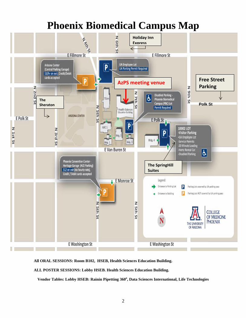
Phoenix Biomedical Campus Map
All ORAL SESSIONS: Room B102, HSEB, Health Sciences Education Building.
ALL POSTER SESSIONS: Lobby HSEB. Health Sciences Education Building.
Vendor Tables: Lobby HSEB: Rainin Pipetting 360o, Data Sciences International, Life Technologies
The Sheraton
The SpringHill Suites
Holiday Inn Express
Free Street Parking
AzPS meeting venue
Polk St
3
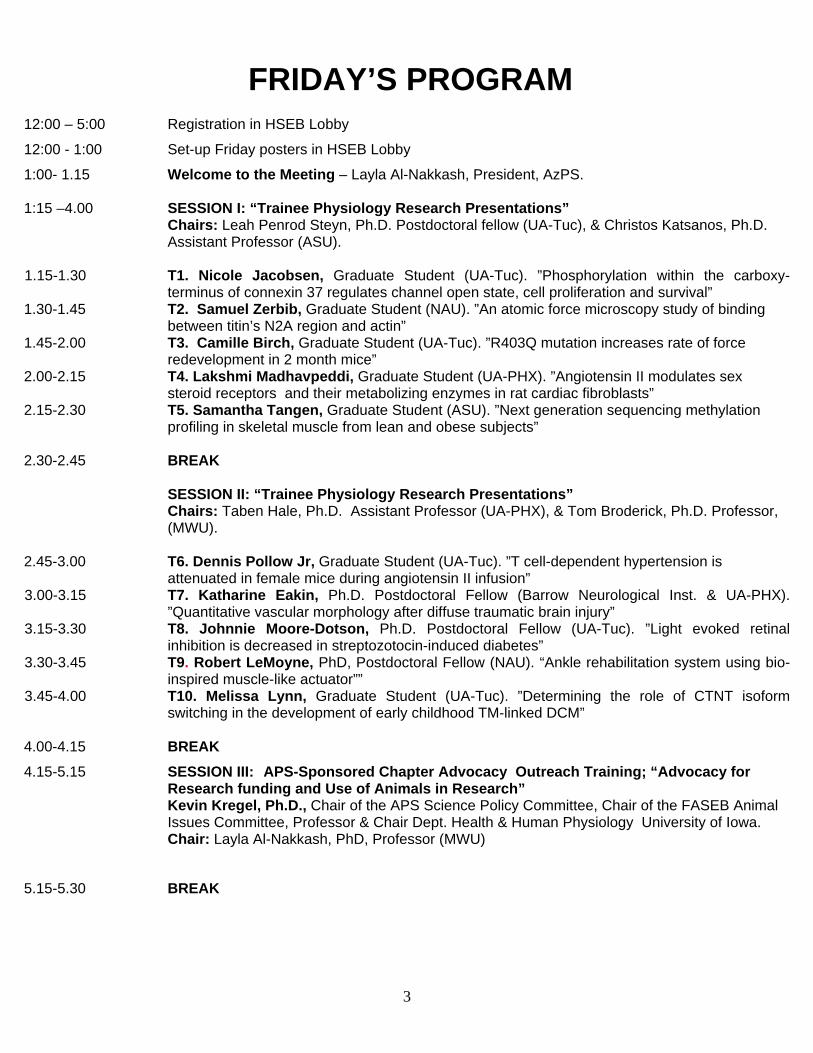
FRIDAY’S PROGRAM
12:00 – 5:00 Registration in HSEB Lobby
12:00 - 1:00 Set-up Friday posters in HSEB Lobby
1:00- 1.15 Welcome to the Meeting – Layla Al-Nakkash, President, AzPS. 1:15 –4.00 SESSION I: “Trainee Physiology Research Presentations”
Chairs: Leah Penrod Steyn, Ph.D. Postdoctoral fellow (UA-Tuc), & Christos Katsanos, Ph.D. Assistant Professor (ASU).
1.15-1.30 T1. Nicole Jacobsen, Graduate Student (UA-Tuc). ”Phosphorylation within the carboxy-terminus of connexin 37 regulates channel open state, cell proliferation and survival”
1.30-1.45 T2. Samuel Zerbib, Graduate Student (NAU). ”An atomic force microscopy study of binding between titin’s N2A region and actin”
1.45-2.00 T3. Camille Birch, Graduate Student (UA-Tuc). ”R403Q mutation increases rate of force redevelopment in 2 month mice”
2.00-2.15 T4. Lakshmi Madhavpeddi, Graduate Student (UA-PHX). ”Angiotensin II modulates sex steroid receptors and their metabolizing enzymes in rat cardiac fibroblasts”
2.15-2.30 T5. Samantha Tangen, Graduate Student (ASU). ”Next generation sequencing methylation profiling in skeletal muscle from lean and obese subjects”
2.30-2.45 BREAK SESSION II: “Trainee Physiology Research Presentations”
Chairs: Taben Hale, Ph.D. Assistant Professor (UA-PHX), & Tom Broderick, Ph.D. Professor, (MWU).
2.45-3.00 T6. Dennis Pollow Jr, Graduate Student (UA-Tuc). ”T cell-dependent hypertension is
attenuated in female mice during angiotensin II infusion” 3.00-3.15 T7. Katharine Eakin, Ph.D. Postdoctoral Fellow (Barrow Neurological Inst. & UA-PHX).
”Quantitative vascular morphology after diffuse traumatic brain injury” 3.15-3.30 T8. Johnnie Moore-Dotson, Ph.D. Postdoctoral Fellow (UA-Tuc). ”Light evoked retinal
inhibition is decreased in streptozotocin-induced diabetes” 3.30-3.45 T9. Robert LeMoyne, PhD, Postdoctoral Fellow (NAU). “Ankle rehabilitation system using bio-
inspired muscle-like actuator”” 3.45-4.00 T10. Melissa Lynn, Graduate Student (UA-Tuc). ”Determining the role of CTNT isoform
switching in the development of early childhood TM-linked DCM”
4.00-4.15 BREAK
4.15-5.15 SESSION III: APS-Sponsored Chapter Advocacy Outreach Training; “Advocacy for Research funding and Use of Animals in Research”
Kevin Kregel, Ph.D., Chair of the APS Science Policy Committee, Chair of the FASEB Animal Issues Committee, Professor & Chair Dept. Health & Human Physiology University of Iowa.
Chair: Layla Al-Nakkash, PhD, Professor (MWU)
5.15-5.30 BREAK

4
5.30-6.30 The AzPS Keynote Lecturer: Introduction: Dr. Ron Lynch, Ph.D., Professor (UA-Tuc)
Kim Barrett, Ph.D.
Professor of Medicine, Division of Gastroenterology, UCSD, & Current 86th American Physiological Society President.
“Physiological consequences of interactions with “good” and “bad” bacteria in the gut.”
6.30- 9.00 Chapter Reception & Buffet Dinner Begins
7.15 –7.45 Minute Poster Discussion: 31 minute posters.
Chairs: Scott Boitano, Ph.D. Associate Professor (UA-Tuc) & Johnnie Moore-Dotson, Ph.D.
Postdoctoral Fellow (UA-Tuc)
7.45 - 9.15 Posters in Session: HSEB Lobby, 31 posters.
5
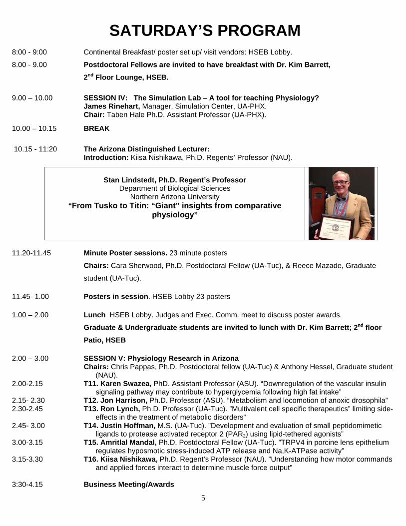
SATURDAY’S PROGRAM
8:00 - 9:00 Continental Breakfast/ poster set up/ visit vendors: HSEB Lobby.
8.00 - 9.00 Postdoctoral Fellows are invited to have breakfast with Dr. Kim Barrett,
2nd Floor Lounge, HSEB.
9.00 – 10.00 SESSION IV: The Simulation Lab – A tool for teaching Physiology? James Rinehart, Manager, Simulation Center, UA-PHX. Chair: Taben Hale Ph.D. Assistant Professor (UA-PHX).
10.00 – 10.15 BREAK
10.15 - 11:20 The Arizona Distinguished Lecturer: Introduction: Kiisa Nishikawa, Ph.D. Regents’ Professor (NAU).
Stan Lindstedt, Ph.D. Regent’s Professor
Department of Biological Sciences Northern Arizona University
“From Tusko to Titin: “Giant” insights from comparative physiology”
11.20-11.45 Minute Poster sessions. 23 minute posters
Chairs: Cara Sherwood, Ph.D. Postdoctoral Fellow (UA-Tuc), & Reece Mazade, Graduate
student (UA-Tuc).
11.45- 1.00 Posters in session. HSEB Lobby 23 posters
1.00 – 2.00 Lunch HSEB Lobby. Judges and Exec. Comm. meet to discuss poster awards.
Graduate & Undergraduate students are invited to lunch with Dr. Kim Barrett; 2nd floor
Patio, HSEB
2.00 – 3.00 SESSION V: Physiology Research in Arizona Chairs: Chris Pappas, Ph.D. Postdoctoral fellow (UA-Tuc) & Anthony Hessel, Graduate student
(NAU). 2.00-2.15 T11. Karen Swazea, PhD. Assistant Professor (ASU). “Downregulation of the vascular insulin
signaling pathway may contribute to hyperglycemia following high fat intake” 2.15- 2.30 T12. Jon Harrison, Ph.D. Professor (ASU). ”Metabolism and locomotion of anoxic drosophila” 2.30-2.45 T13. Ron Lynch, Ph.D. Professor (UA-Tuc). ”Multivalent cell specific therapeutics” limiting side-
effects in the treatment of metabolic disorders” 2.45- 3.00 T14. Justin Hoffman, M.S. (UA-Tuc). ”Development and evaluation of small peptidomimetic
ligands to protease activated receptor 2 (PAR2) using lipid-tethered agonists” 3.00-3.15 T15. Amritlal Mandal, Ph.D. Postdoctoral Fellow (UA-Tuc). ”TRPV4 in porcine lens epithelium
regulates hyposmotic stress-induced ATP release and Na,K-ATPase activity” 3.15-3.30 T16. Kiisa Nishikawa, Ph.D. Regent’s Professor (NAU). ”Understanding how motor commands
and applied forces interact to determine muscle force output” 3:30-4.15 Business Meeting/Awards

What Can DSI Telemetry do for Your Animal Studies?
Better Data. Better Science.
Become an advocate for animal welfare and better science.
Telemetry refines physiologic research techniques for better results
• Smaller implant size promotes better tolerance and allows the use of animals with unique physiologic characteristics• Collect high quality data without restraint in conscious animals• Continuous 24/7 physiologic monitoring
Telemetry reduces the number of animals, saving you money
• Record multiple physiologic parameters in a single animal• Detect negative impacts earlier• Consolidate study data• Optimize data analysis• Use animals as their own controls
Telemetry improves animal welfare • Large animal social housing capabilities• Reduces animal stress• Allows animal to exhibit more natural behaviors
DSI Surgical Services
DSI understands that surgical implantation takes time. DSI Surgical Services are available to help you maximize efficiency and meet your research goals. Our experienced surgical team is ready to assist with animal preparation and implantation.
DSI•11914thStreetNW•Suite100•St.Paul,MN55112•T:+1(651)481-7400•Tollfree:1(800)262-9687 F:+1(651)481-7417•www.datasci.com•[email protected]
Copyright©2013DataSciencesInternational
ContactDSItodaytolearnmoreabouton-siteopportunitiestoeducateVeterinaryandAnimalCarestaffon the benefits of DSI solutions for preclinical research!
HD-X11MouseImplant

Revised 102913: AzPS Nov 1-2, 2013
Friday Posters, Arranged by Lead Author (alphabetical) Lead Author Title REGULAR MEMBERS F2 Kathryn Corbell The influence of ovariectomy and genistein on estrogen
receptor content and activation in rat achilles tendon F3 Justin Hoffman Development and evaluation of small peptidomimetic
ligands to protease activated receptor 2 (PAR2) using lipid-tethered agonists
F4 Lana Leung Identifying the intracellular signaling pathways responsible for genistein- and estradiol-stimulated increases in basal jejunum ISC in female mice with and without endogenous estrogen
F5 Ron Lynch Multivalent cell specific therapeutics: limiting side -effects in the treatment of metabolic disorders
F6 Kiisa Nishikawa Understanding how motor commands and applied forces interact to determine muscle force output
F7 Mohammad Shahidullah Connexins form functional hemichannels in porcine ciliary epithelium
F8 Charles Tipton Historical: whats old is new again, exercise is medicine F9 Guojun Wei Dids inhibits Na,K-ATPase activity in porcine
nonpigmented ciliary epithelial cells by a SRC family kinase-dependent mechanism
Lead Author Title GRADUATE STUDENTS F10 Kameswari Ananthakrishnan Targeting glucagon like peptide-1 and α-2 adrenergic
receptor combination using glp-1/yohimbine to achieve β-cell specific targeting and therapy
F11 Samantha Behunin Phosphorylation Patterning Determined by AMP-Activated Kinase, the LKB1/MO25/STRAD Complex, and Protein Phosphatase 1 Alters Contractile Function in Cardiac Rat Trabeculae
F12 Camille Birch R403Q Mutation Increases the Rate of Force Redevelopment in 2 Month Mice
F13 Yang Gao Rapamycin, an inhibitor of mTOR signaling pathway, reverses lithium-induced cell proliferation in renal collecting ducts
F14 Miranda Good Hemichannel function is not sufficient for CX37-mediated growth suppression
F15 Jordan Harrison Diffuse brain injury does not affect chronic sleep patterns in the mouse
F16 Anthony Hessel Exploring the Winding Filament Hypothesis Using Transmission Electron Microscopy
F17 Michael Hicks Strain-Activated Fibroblasts Enhance Skeletal Myotube Contraction, and Increase Nicotinic Receptor Expression and Clustering

Revised 102913: AzPS Nov 1-2, 2013
F18 Richard Huynh Differences in collagen formation and anabolic signaling in tendon and skeletal muscle after chronic exercise training in the rat
F19 Nicole Jacobsen Phosphorylation within the carboxy-terminus of connexin 37 regulates channel open state, cell proliferation, and survival
F20 Katon Kras The proteolytic enzyme nagarse modifies parameters associated with mitochondrial function of mechanically-liberated mitochondria
F21 Sarah Lehman Effects of troponin T mutations on calcium handling of the cardiac thin filament
F22 Yulia Lipovka Estradiol activates ampk through interaction with estrogen receptor beta
F23 Melissa Lynn Determining the role of CTNT isoform switching in the development of early childhood TM-linked DCM
F24 Lakshmi Madhavpeddi Angiotensin II modulates sex steroid receptors and their metabolizing enzymes in rat cardiac fibroblasts
F25 Reece Mazade Light Adaptation Differentially Effects Spatial Inhibition to the Retinal OFF Pathway
F26 Dennis Pollow T cell-dependent hypertension is attenuated in female mice during angiotensin II infusion
F27 Gregory Powell The effects of developmental nicotine exposure on hypoglossal motoneuron morphology and electrophysiology
F28 Philip Sandoval Symmetry of Organic Cation Transport in MATE1 F29 Uzma Tahir How does the velocity-dependent behavior of muscles
Change with activation? F30 Samantha Tangen Next generation sequencing methylation profiling in
skeletal muscle from lean and obese subjects F31 Samuel Zerbib An atomic force microscopy study of binding between
titin’s N2A region and actin POST-DOC F32 Leah Steyn A Heterobivalent Ligand containing GLP-1 and
Yohimbine Specifically Targets Pancreatic β-cells In Vivo

Revised 102913: AzPS Nov 1-2, 2013
Saturday Posters, Arranged by Lead Author (alphabetical) Lead Author Title REGULAR MEMBERS S2 Tom Broderick #1 Effects of oxytocin on cardiovascular function in the
SHR rat using telemetry S3 Tom Broderick #2 Unexpected effects of voluntary running on mRNA
expression of natriuretic peptides in ob/ob mouse heart S4 David Carbone Prenatal dexamethasone exposure potentiates diet-
induced liver disease S5 Rayna Gonzales Vasoactive effcts of a novel endogenous androgen are
mediated by estrogen receptor activation in the rat mesenteric and cerebrovasculature
S6 Taben Hale Role of cardiac fibroblast in cardioprotective effects of prior transient ace inhibition
S7 Jon Harrison Metabolism and locomotion of anoxic drosophila S8 Karen Swazea (for Ricklefs) Downregulation of the vascular insulin signaling pathway
may contribute to hyperglycemia following high fat intake
Lead Author Title POST-DOCS S9 Katharine Eakin Quantitative vascular morphology after diffuse traumatic
brain injury in the rat S10 Robert LeMoyne Ankle rehabilitation system using bio-inspired muscle-
like actuator S11 Amritlal Mandal TRPV4 in porcine lens epithelium regulates hyposmotic
stress-induced ATP release and Na,K-ATPase activity S12 Johnnie Moore-Dotson Light evoked retinal inhibition is decreased in
streptozotocin-induced diabetes S13 Christopher Pappas Leiomodin 2 deficient mice display severe cardiac
dysfunction and juvenile lethality Lead Author Title UNDERGRADUATE
STUDENTS
S14 Mun Aw Na,K-ATPase α-1, NKCC2, and NHE3 Protein Expression in the Kangaroo Rat and Sprague-Dawley Rat Renal Outer Medulla
S15 Matthew Calhoun Variations in pancreatic regulation of glucose homeostasis in birds
S16 Madeline Espineira Basal/apical aquaporin 2 expression ratio in the renal collecting duct positively correlates with urine concentrating capacity
S17 Haley Masters Myostatin mRNA expression and plasma myostatin activation in the obese, insulin-resistant state – preliminary study
S18 Puneet Raman Lipopolysaccharide and interleuken-1 beta modulate erβ

Revised 102913: AzPS Nov 1-2, 2013
in rat mesenteric and pial arteries as well as human coronary artery vascular smooth muscle cells
S19 Farmin Samareh-Jahani Cognitive Dysfunction in Heart Failure and a Protective Role for Angiotensin (1-7).
S20 Anna Simperova Putative Protective Effects of Genistein in the Vasculature of Female ob/ob Mice
S21 Zach Fader Kinematic Differences Between Wildtype and Mutant Mouse mdm Genotypes During Walking and Jumping
S22 Diane Bejerano Insulin Signaling and Obesity: Role of Ceramide in the Development of Insulin Resistance in 3T3-L1 Adipocytes
S23 Jonathan Frischknecht The Effects of TNF-α and Ceramide in Insulin Signaling in C2C12 Myocytes
S24 Jason Greenlee Acetaminophen and exercise increase matrix metalloproteinase levels in achilles peritendinous tissue in humans
Revised 102913: AzPS Nov 1-2, 2013
Friday’s Abstracts (Alphabetical, by first author--poster board # shown)
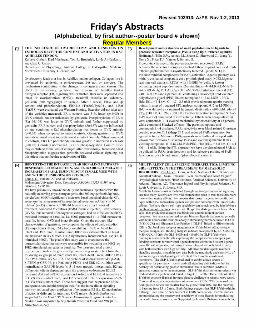
Regular Members F2 THE INFLUENCE OF OVARIECTOMY AND GENISTEIN ON
ESTROGEN RECEPTOR CONTENT AND ACTIVATION IN RAT ACHILLES TENDON. Kathryn Corbell, Karl Martineau, Tom L. Broderick, Layla Al-Nakkash, and Chad C. Carroll Department of Physiology, Arizona College of Osteopathic Medicine, Midwestern University, Glendale, AZ Ovariectomy leads to a loss in Achilles tendon collagen. Collagen loss is prevented by genistein, a phytoestrogen, but not by exercise. The mechanism contributing to the changes in collagen are not known. The effect of ovariectomy, genistein, and exercise on Achilles tendon estrogen receptor (ER) signaling was evaluated. Rats were separated into intact or ovariectomized (OVX), treadmill exercise or sedentary, genistein (300 mg/kg/day) or vehicle. After 6 weeks, ER- and - content and phosphorylation, ERK1/2 (Thr202/Tyr204), and c-Raf (Ser338) were evaluated via Western blotting. Exercise did not alter any of the variables measured. ER- content was 31% lower (p<0.05) in OVX animals but not influenced by genistein. Phosphorylation of ER- (Ser104/106) was lower in OVX animals and further suppressed by genistein. ER- content and phosphorylation (Ser187) was not influenced by any condition. c-Raf phosphorylation was lower in OVX animals (p<0.05) when compared to intact controls. Giving genistein to OVX animals returned c-Raf to levels found in intact controls. When compared to controls, ERK1/2 phosphorylation was greater in untreated OVX rats (p<0.05). Gensitein normalized ERK1/2 phosphorylation. Loss of ER- may contribute to the loss of collagen after ovariectomy. Increased c-Raf phosphorylation suggests that genistein activated cellular signaling, but this effect may not be due to activation of ERs.
F3 Development and evaluation of small peptidomimetic ligands to protease activated receptor 2 (PAR2) using lipid-tethered agonists Hoffman J., Tillu D.V., Asiedu M., Zhang Z., Sherwood C., Wang Y., Dong X., Price T.J., Vagner J, Boitano S. Proteolytic cleavage of the protease-activated receptor-2 (PAR2) activates the receptor through an attached tethered ligand. We used lipid-tethered peptidomimetics (synthetically tethered ligands; STL) to evaluate minimal components for PAR2 activation. Agonist potency was initially evaluated using an in vitro physiological assay (xCELLigence real time cell analysis; RTCA) with 16HBE14o- cells. A known activating parent peptidomimetic, 2-aminothiazol-4-yl-LIGRL-NH2 (2-at-LIGRL-NH2; RTCA EC50 = 310 nM; 95% Confidence Interval (CI): 240 – 400 nM) and a parent STL containing a hexadecyl lipid via three polyethylene glycol (PEG) linkers (compound 1: 2-at-LIGRL-PEG3-Hdc; EC50 = 1.4 nM; CI: 1.2 - 2.3 nM) provided potent agonist starting points. In a set of truncated STL analogs, compound 4 (2-at-LI-PEG3-Hdc) was defined as a minimal fragment, albeit with a ~200 fold reduced EC50 (310 nM; CI: 260 - 360 nM). Further truncation (compound 5: 2-at-L-PEG3-Hdc) eliminated in vitro activity. Effects were recapitulated in vivo; compounds 1 - 4 evoked mechanical hypersensitivity at 15 pmoles while compound 5 lacked efficacy. The parent compound and compounds 1 - 4 displayed PAR2 selectivity over Mas1 related G-protein coupled receptor C11 (MrgprC11) and required PAR2 expression for agonist activity. Minimum PAR2 agonists were further developed with known modifications 5-isoxazoyl (5-io) and cyclohexylalanine (Cha), yielding compound 11, 5-io-Cha-IGR-PEG2-Hdc (EC50 = 6.8 nM, CI: 4.1 nM - 11 nM). Using the STL approach we have developed novel SAR to be utilized for PAR2 drug discovery and for selective probing of PAR2 function across a broad range of physiological systems.
F4 IDENTIFYING THE INTRACELLULAR SIGNALING PATHWAYS RESPONSIBLE FOR GENISTEIN- AND ESTRADIOL-STIMULATED INCREASES IN BASAL JEJUNUM ISC IN FEMALE MICE WITH AND WITHOUT ENDOGENOUS ESTROGEN Leung, L., Bhakta, A., and Al-Nakkash, L. Midwestern University, Dept. Physiology, AZCOM, 19555 N. 59th Ave, Glendale, AZ.85308 We have previously shown that daily subcutaneous injections with the naturally occurring phytoestrogen genistein (600 mg genistein/kg body weight/day, 600G) significantly increases basal intestinal chloride, Cl-, secretion (Isc, a measure of transepithelial secretion, µA/cm2) by 70 µA/cm2 (n=15) in intact C57BL/6J female mice after 1 week of treatment, compared to controls (DMSO vehicle injected). Ovariectomy (OVX), thus removal of endogenous estrogen, had no effect on the 600G-mediated increase in basal Isc, i.e. 600G generated a 1.6-fold increase in basal Isc in both OVX and intact females. Given the estrogen-like characteristics of genistein, we compared the effects of daily estradiol (E2) injections (10 mg E2/kg body weight/day, 10E2) on basal Isc in intact and OVX mice. In intact mice, 10E2 was without effect on basal Isc, however, in OVX mice, 10E2 significantly increased basal Isc (i.e. it mimicked 600G). The goal of this study was to characterize the intracellular signaling pathways responsible for mediating the 600G- or 10E2-stimulated increases in basal Isc. We measured total protein expression in isolated segments of jejunum using western blot from the following six groups of mice: intact 0G, intact -600G, intact-10E2, OVX-0G, OVX-600G, OVX-10E2. The proteins of interest were: Akt, p-Akt, p-PTEN, p-GSK-3β, p-c-Raf, and p-PDK1, and ERK1/2. All blots were normalized to GAPDH levels (n=3-11 each group). Interestingly, E2 had differential effects dependent upon the presence endogenous E2; E2 increased Akt and p-PDK1expression 4.6-fold and 10.4-fold respectively in OVX versus intact mice, and E2 decreased ERK1/2 expression ~50% in OVX versus intact mice. These data suggest that the presence of the endogenous sex steroid estrogen modifies the intracellular signaling pathway activated upon application of exogenous E2 (i.e. E2 mechanism of action is different for intact and OVX mice). Ashesh Bhakta was supported by the MWU DO Summer Fellowship Program. Layla Al-Nakkash was supported by Soy Health Research Fund and NIH (R15-DK071625-01A2).
F5 MULTIVALENT CELL SPECIFIC THERAPEUTICS: LIMITING SIDE -EFFECTS IN THE TREATMENT OF METABOLIC DISORDERS. Ron Lynch1, Craig Weber1, Nathaniel Hart1, Kameswari Ananthakrishnan1, Sean Limesand3, W.K. Samson4 and Josef Vagner2
1Physiological Sciences, 2Bio5 Institute, 3Animal Sciences, University of Arizona, Tucson, AZ, 4Pharmaco-logical and Physiological Sciences, St. Louis University, St. Louis, MO. Metabolic Homeostasis is mediated through multi-organ endocrine signaling. Because many systems are involved, therapeutics used to treat these disorders have wide-ranging effects. We propose that ‘drugs’ targeted to specific cell types within the homeostatic system will provide outcomes with limited side-effects. We have shown cell-type specificity can be achieved by identifying a combination of receptors on a given cell type that distinguishes it from other cells, then producing an agent that binds this combination of surface receptors. We have synthesized several bivalent ligands that may target cells within the homeostatic axis; melanocyte stimulating hormone linked to CCK (MSH/CCK) and Glucagon Like Peptide 1 (GLP-1) linked to Glibenclamide (Glb, a Sulfonyl urea receptor antagonist), or Yohimbine ( 2 adrenergic receptor antagonist). Binding analyses indicate an apparent Kd of ~5 nM for MSH/CCK, ~10nM for GLP-1/Glb and ~10 pM for GLP-1/Yhb when binding is assessed with cells expressing the complementary receptor pair. Binding constants for individual ligand domains within the bivalent ligands were 100 nM or greater, indicating that each ligand will only bind to cells with both receptors with high affinity. All three bivalent agents maintain signaling capacity, though in each case both the magnitude and sensitivity of 2nd messenger and physiological effects differ from the constituent monomers. The GLP-1/Yhb is predicted to exhibit a high degree of specificity for pancreatic �-cells, and cell signaling data indicate that its potency for potentiating glucose stimulated insulin secretion (GSIS) is enhanced compared to the monomers. GLP-1/Yhb distribution in rodents was evaluated after injection, and found to target to �-cells. The effects of GLP-1/Yhb on glucose disposal during a glucose challenge in ratsalso were tested. Compared to equal concentrations of monomers, GLP-1/Yhb decreased the peak glucose concentration after load by greater than 20%, and the recovery to baseline from 15 to 5 min. Both findings suggest that GLP-1/Yhb exhibits a strong �-cell specific enhancement in GSIS potentiation. Current studies are investigating the potency and specificity of these ligands for modulating metabolic homeostasis in vivo. Supported by Juvenile Diabetes Research Fnd.
Revised 102913: AzPS Nov 1-2, 2013
F6 UNDERSTANDING HOW MOTOR COMMANDS AND APPLIED FORCES INTERACT TO DETERMINE MUSCLE FORCE OUTPUT Nishikawa, K; Monroy, J; Pace, C. Department Of Biological Sciences, Northern Arizona University The goal of predicting how muscle forces change during natural movements remains elusive. Muscle models perform poorly at predicting muscle force during stretch or shortening, as well as in doublet potentiation and work-loop experiments. Our goal is to explore the ability of the winding filament hypothesis to inform our understanding of muscle force output. We used the muscular dystrophy with myositis (mdm) mouse to test the hypothesis that titin contributes to active force in doublet potentiation and work loop experiments. We performed doublet potentiation, isovelocity stretch and shortening, and work-loop experiments in soleus muscles from wild type and mdm mice. Doublet potentiation was 20% lower in mdm than in wild type soleus. In work loop experiments, force increases steeply upon activation during stretch and a single added stimulus increases force during active stretch in wild type soleus. A doublet increases muscle work per cycle by 50%. Soleus muscles from mdm mice showed little increase in force upon activation during stretch, and the work per cycle was the same with and without the doublet. These results suggest that titin contributes to dynamic force output of active muscle, and demonstrate that the mdm mouse is an important model system for understanding how motor commands and applied forces interact to determine muscle force output. Supported by NSF IOS-1025806.
F7 CONNEXINS FORM FUNCTIONAL HEMICHANNELS IN PORCINE CILIARY EPITHELIUM Shahidullah M and Delamere NA, Department of Physiology, University of Arizona, 1501 N Campbell Avenue, Tucson, AZ, 85724, USA. Email: [email protected] To examine the existence of undocked connexons that may form functional hemichannels and permit exchange of substances between nonpigmented ciliary epithelium (NPE) and the aqueous humor. Intact porcine eyes were perfused via the ophthalmic artery and propidium iodide (PI) (MW 668) was added to the aqueous humor compartment as a tracer. At the end of the perfusion, thin sections of ciliary body were cut and PI detected by fluorescence microscopy. PI uptake and calcein efflux were also studied in isolated NPE cultured on 24 or 96 well-plates. In the intact eye preparation and perfusion of the anterior chamber with a calcium containing aqueous humor substitute caused little PI uptake by the NPE. In calcium-free solution, PI was avidly taken up by the NPE. PI entry into the NPE was inhibited by calcium and by the connexin antagonist 18α-glycyrrhetinic acid (18-AGA). Studies also were carried out with cultured porcine NPE. Under normal conditions, little PI entered the cultured cells but calcium-free medium stimulated PI accumulation and the entry was inhibited by 18-AGA. In cells loaded with calcein (MW 622), calcium-free solution stimulated calcein exit. 18-AGA partially suppressed calcein exit in calcium-free medium. Connexin 43 and connexin 50 proteins were detected by western blot analysis in both native and cultured NPE. In the intact eye, immunolocalization studies revealed connexin 50 at the basolateral, aqueous humor-facing, margin of the NPE. In contrast, connexin 43 was observed at the junction of the PE and NPE layer and on the basolateral membrane of PE. The results point to functional hemichannels at the NPE basolateral surface. It is feasible that hemichannels might contribute to the transfer of substances between the ciliary epithelium cytoplasm and aqueous humor. Grant: NIH Grant EY006915 Disclosure: No Commercial Relationship (N)
F8 HISTORICAL: WHATs OLD IS NEW AGAIN, EXERCISE IS MEDICINE Charles M. Tipton The University of Arizona In 2007, ACSM, AMA and the office of the Surgeon General launched a national initiative to mobilize physicians, health care professionals plus educators to promote exercise in their practice or activities to prevent, reduce, manage or treat diseases that impact health and the quality of life in humans. Two years later, Dr. Robert Salis, the “prime mover of the initiative”, urged practicing physicians to consider exercise to be an important vital sign when interacting with patients during office visits. Recently, ACSM issued a Position Stand that recommended healthy adults perform daily moderate exercise that totaled 150 min/wk. Although a timely and admirable initiative, it is not original because the concept has roots that began in antiquity (antiquity ended with the death of Galen in 210 A.D.). Specifically, the physician Susruta in India during 600 B.C.E. prescribed daily moderate exercise to his patients and followers for the purposes of promoting health, reducing corpulence, and for preventing diseases (including diabetes). He was followed by Hippocrates of Greece (480 – 370 B.C.E.) who was the first physician to provide a written exercise prescription for a patient suffering from consumption. A strong advocate of the humoral theory of disease, he advocated exercise because it would “purge” humors from the body. Lastly, Galen of Rome should be remembered because he prescribed exercise for subjects suffering from multiple diseases and because he influenced the practice of medicine, and the role of exercise in the practice of medicine until the 16th century.
F9 DIDS INHIBITS NA,K-ATPASE ACTIVITY IN PORCINE NONPIGMENTED CILIARY EPITHELIAL CELLS BY A SRC FAMILY KINASE-DEPENDENT MECHANISM Wei G, Shahidullah M and Delamere NA. Department of Physiology, University of Arizona, 1501 N Campbell Ave., Tucson, AZ 85724, USA. The anion transport inhibitor DIDS is known to reduce aqueous humor (AH) secretion but questions remain about anion-dependence of the effect. In some tissues, DIDS is reported to cause Na,K-ATPase inhibition. The purpose is examine the ability of DIDS to inhibit Na,K-ATPase activity in nonpigmented ciliary epithelium (NPE). Porcine NPE cells were cultured to confluence on permeable supports, treated with drugs by adding to both sides of the membrane, and then used for 86Rb uptake measurements or homogenized to measure Na,K-ATPase activity or to detect protein phosphorylation. DIDS inhibited ouabain-sensitive 86Rb uptake, activated Src family kinase (SFK) and caused a reduction of Na,K-ATPase activity. PP2, an SFK inhibitor, prevented the DIDS responses. In BCECF-loaded NPE, DIDS was found to reduce cytoplasmic pH (pHi). PP2-sensitive Na,K-ATPase activity inhibition, 86Rb uptake suppression and SFK activation were observed when a similar reduction of pHi imposed by low pH medium or an ammonium chloride withdrawal maneuver. PP2 and the ERK inhibitor U0126 prevented robust ERK1/2 activation observed in cells exposed to DIDS or subjected to pHi reduction but U0126 did not prevent SFK activation or the Na,K-ATPase activity response. The evidence points to an inhibitory influence of DIDS on NPE Na,K-ATPase activity by a mechanism that hinges upon Src family kinase (SFK) activation associated with a reduction of cytoplasmic pH. Funding: NIH Grant EY006915

Revised 102913: AzPS Nov 1-2, 2013
Graduate Students F10 TARGETING GLUCAGON LIKE PEPTIDE-1 AND α-2
ADRENERGIC RECEPTOR COMBINATION USING GLP-1/YOHIMBINE TO ACHIEVE β-CELL SPECIFIC TARGETING AND THERAPY Kameswari Ananthakrishnan1, Craig S. Weber1, Nathaniel Hart1, Josef Vagner3, Sean Limesand2 and Ronald M. Lynch1,3 Departments of Physiology1, Animal Sciences2 and the Bio5 Institute3, University of Arizona, Tucson, 85721 It has been shown through modeling and experimentation that the coupling of multiple receptor binding domains into a single molecule can enhance ligand binding affinity. Moreover, if the domains target different receptors, only cells that express that receptor combination will bind this agent with high specificity. β-cells, which are essential nutrient sensing cells, express a range of receptors, but as a combination, Glucagon Like Peptide-1 Receptor (GLP-1R) and α2Adrenergic Receptor (α2AR) are relatively unique. Furthermore, activation of GLP-1R and α2AR has inverse effects on β-cell signaling; GLP-1 activates cAMP production and subsequently potentiates Glucose Stimulated Insulin Secretion (GSIS), while α2AR agonists inhibit cAMP production and dampen GSIS. Hence, we propose that linking GLP-1 and an α2AR antagonist Yohimbine (Yhb) into a heterobivalent ligand (GLP-1/Yhb) would provide an agent with enhanced affinity, β-cell specificity and possibly unique therapeutic potential. Using microscopy, we established that Cy5 tagged GLP-1/Yhb bound to β-cells with high affinity at nM concentrations and was rapidly internalized. In cells where either GLP-1R or α2AR were knocked down (using siRNA), binding of GLP-1/Yhb was severely impaired (≤ half of control cells with both receptors), demonstrating specificity for cells with both receptors. Preliminary cAMP studies showed that over a range of concentrations, GLP-1/Yhb had similar activity as monomeric GLP-1 for stimulating cAMP production. Conversely, insulin secretion assays showed that at both 1nM and 100nM concentrations, GLP-1/Yhb potentiates GSIS significantly more than GLP-1 alone. Thus, though the cAMP activation is comparable to monomeric GLP-1, the divalent GLP-1/Yhb exhibits synergistic potentiation of GSIS indicating its therapeutic potential. Consequently, due to its specificity to β-cells and positive impact on β-cell function, GLP-1/Yhb could pave the way for successful β-cell specific targeting and therapy. Supported by: JDRF and Arizona Biomedical Research Commission
F11 Phosphorylation Patterning Determined by AMP-Activated Kinase, the LKB1/MO25/STRAD Complex, and Protein Phosphatase 1 Alters Contractile Function in Cardiac Rat Trabeculae Samantha M. Behunin, John P. Konhilas. Department of Physiology, College of Medicine, The University of Arizona, Tucson, Arizona 85721 Post-translational modifications (PTM) of myofilament proteins alter contractile function of the heart in healthy as well as diseased myocardium and the patterning of PTMs can influence cardiac disease progression. PTM of the thin filament regulatory protein cardiac troponin I (cTnI) is known to modify contractile properties, including steady-state Ca2+ sensitivity of force and crossbridge cycling rates. Accordingly, the purpose of this study was to determine the effect of cTnI PTM patterning on myofilament function. Therefore, I hypothesize that the impact of cTnI PTM on contractile function will depend on the relative phosphorylation levels. To do this, demembranated rat cardiac trabeculae from 2 month-old male Sprague-Dawley rats were treated with AMP activated kinase (AMPK) (0.005 U/ µL), Protein Phosphatase 1 (PP1) (1U/µL), and the upstream AMPK kinase, the LKB1/MO25/STRAD complex (0.2 mU/µL). Fibers that were incubated with activated AMPK displayed an increase in Ca2+ sensitivity compared to untreated control fibers (EC50 1.41±0.08 μM [n=2] vs. 2.52±0.43 μM [n=9] p<0.001). PP1 treatment, previously shown to decrease cTnI phosphorylation, tended to increase Ca2+ sensitivity compared to untreated fibers (EC50 2.31 μM [n=1] vs. 2.52±0.43 μM [n=9]). Interestingly, PP1 treatment also increased passive tension generation by 27% compared to control fibers (P<0.001 [n=3]). Surprisingly, the LKB1/MO25/STRAD complex decreased overall tension development (14.18±2.27 mN/mm2 [n=2] vs 37.03 ± 16.72 mN/mm2 [n=9] p=0.002), and desensitized the myofilament to Ca2+ (EC50 4.18 ±0.0001 µM [n=2] vs 2.52±0.43 μM [n=9] p<0.001). In conclusion, I have shown that the functional outcomes are determined by the differential PTM patterning of cTnI. Furthermore, we have identified the LKB1/MO25/STRAD complex as a potential novel regulator of myofilament function.
F12 R403Q Mutation Increases the Rate of Force Redevelopment in 2 Month Mice C.L. Birch1, J.P. Konhilas, PhD2. 1Department of Biomedical Engineering, University of Arizona, Molecular Cardiovascular Research Program, Sarver Heart Center , Tucson AZ 2Department of Physiology, University of Arizona, Molecular Cardiovascular Research Program, Sarver Heart Center , Tucson AZ Familial hypertrophic cardiomyopathy is a primary disease of the sarcomere. The R403Q mutation resides at the actin-interaction site on myosin and leads to progressive hypertrophic cardiomyopathy and ultimately ends with heart failure. Along with deteriorating cardiac function, these hearts experience an overall change in metabolic landscape, suggesting altered energetic function in hearts that express the R403Q mutation. We assessed the hypothesis that the R403Q mutation intrinsically increases the energetic cost of contraction. Therefore, the differences that arise in cross-bridge kinetics between wild-type (WT) and R403Q mice at 2 months of age were assessed. The rate of force redevelopment (ktr) in skinned cardiac tissue was measured following unloaded isotonic shortening and a rapid re-stretch to 15% of the original muscle length. This procedure was performed at a sarcomere length of 2.0m. Male R403Q mice display an increased rate of force redevelopment (49.89 s-1
± 8.13 n = 4) compared to WT counterparts (24.52 ± 4.29 n = 6) at maximal activation indicating an increase in total cross bridge cycling rate (p < 0.05). Additionally, there was no significant difference in Ca2+ sensitivity between male R403Q (n = 4) and WT counterparts (n = 2) which is consistent with previous findings. Further studies are being pursued to validate the relation between the R403Q mutation and Ca2+ sensitivity. In conclusion, the R403Q mutation increases the total cross bridge cycling rate which suggests a higher use of energy for force generation to occur. Although no overt pathology is observed at 2 months, the R403Q mutation causes an alteration in cross-bridge kinetics which may lead to downstream effects causing an overall change in the metabolic landscape.

Revised 102913: AzPS Nov 1-2, 2013
F13 RAPAMYCIN, AN INHIBITOR OF mTOR SIGNALING PATHWAY, REVERSES LITHIUM-INDUCED CELL PROLIFERATION IN RENAL COLLECTING DUCTS Yang Gao, Jill Romero-Aleshire, Qi Cai, Ted J. Price, Heddwen L. Brooks. University of Arizona, Tucson, AZ The most common side effect in patients undergoing lithium therapy is nephrogenic diabetes insipidus (NDI), in addition, lithium treatment can cause collecting duct cells to proliferate. The mammalian target of rapamycin (mTOR) signaling pathway is a key regulator of cell proliferation. We hypothesized that the mTOR signaling pathway may be playing a role in lithium-induced cell proliferation of renal collecting duct. We fed mice lithium for 14d; AQP2 protein expression was significantly decreased (16 ± 4.0% vs control 100 ± 8.8%) and proliferating cell nuclear antigen (PCNA) protein expression was significantly increased (172 ± 8.6% vs control 100 ± 1.4%) in renal inner medulla (IM). We demonstrate that p-mTOR (Ser 2448) was increased (154 ± 26.5%), as was phosphorylation of ribosomal S6 protein (p-rS6), a downstream component of mTOR pathway (404 ± 151.4%) in renal IM of lithium-treated mice. To test whether the inhibition of mTOR signaling pathway could reverse lithium-induced cell proliferation, we treated mice with Rapamycin (Rapa), an inhibitor of mTOR. Rapa reversed lithium-induced proliferation of IM collecting duct cells and decreased lithium-induced p-mTOR and p-rS6 levels. Rapa had no effect on the upstream components of mTOR; p-Akt and p-TSC2 remained elevated by lithium. In conclusion, the mTOR signaling pathway is involved in lithium-induced collecting duct cell proliferation.
F14 HEMICHANNEL FUNCTION IS NOT SUFFICIENT FOR CX37-MEDIATED GROWTH SUPPRESSION Good, ME; Ek-Vitorín, JF; Burt, JM University of Arizona, Tucson AZ 85724 Cx37-meidated growth suppression is channel dependent; however, it remains unclear if gap junction channel (GJC) or hemichannel (HC) function is necessary for this effect. Extracellular loop (E1 and E2) structure of connexins is integral to HC docking and GJC formation, but not to HC function. Mutation of any of the cysteines in E1 or E2 of Cx43 results in complete loss of Cx43 GJC function, but Cx43 with all E1 and E2 cysteines mutated retains HC function. Substitution of other residues suggested to mediate HC docking (e.g.: asparagine 55 and glutamine 58), also result in loss of GJC but not HC function. Since these and other residues are highly conserved across connexins, we hypothesized that their mutation in Cx37 would lead to a compromised GJC function but sustained HC function that suppress the proliferation of Rin cells. To explore the sufficiency of HC function for Cx37-mediated growth suppression, we constructed four Cx37 mutants: C61,65A; C54, 61, 65, 187, 192, 198A (C6A); Q58L; and N55I. Aside from forming functional GJCs, Cx37 wild type forms functional HCs that open more frequently at low than normal external calcium concentration, with transitions amplitudes between 115 – 500pS. GJC function was eliminated in all four Cx37 mutants, however, only 2 mutants, Cx37-N55I and Cx37-Q58L, retained HC function as determined by electrophysiology and dye uptake experiments. Contrary to our hypothesis, all mutants failed to suppress the growth of Rin cells. These data support our previous data indicating that Cx37-mediated growth suppression is channel-dependent and further indicate that HC function is NOT sufficient for this effect. Support: HL058732
F15 DIFFUSE BRAIN INJURY DOES NOT AFFECT CHRONIC SLEEP PATTERNS IN THE MOUSE Jordan L. Harrison1,2,3, Rachel K. Rowe1,2,5,7 Bruce F. O’Hara, Ph.D.6,7 and Jonathan Lifshitz, Ph.D.1,2,3,4
1 BARROW Neurological Institute at Phoenix Children’s Hospital, Phoenix, AZ 2 Department of Child Health, University of Arizona College of Medicine – Phoenix, AZ 3Interdisciplinary Graduate Program in Neuroscience, Arizona State University, Phoenix, AZ 4Phoenix Veteran Affairs Healthcare System, Phoenix, AZ 5Department of Anatomy & Neurobiology, College of Medicine, University of Kentucky, Lexington, KY, USA 6Department of Biology, College of Arts and Sciences, University of Kentucky, Lexington, KY, USA 7Spinal Cord and Brain Injury Research Center (SCoBIRC), College of Medicine, University of Kentucky Lexington, KY, USA This study was designed to test if our current model of diffuse brain injury produces chronic sleep disturbances similar to those reported by TBI patients. Adult male C57BL/6 mice were subjected to moderate midline fluid percussion brain injury (n=7; 1.4 atm; 6-10 min righting reflex time) or sham injury (n=5). Sleep-wake activity was measured post-injury using a non-invasive, piezoelectric cage system. Chronic sleep patterns were analysed weekly for increases or decreases in percent sleep (hypersomnia or insomnia) and changes in bout length (sleep fragmentation). During the first week after diffuse TBI, brain-injured mice exhibited increased mean percent sleep and mean bout length compared to sham-injured mice. Further analysis indicated the increase in mean percent sleep occurred primarily during the dark cycle. Injury-induced changes in sleep, however, did not extend beyond the first week post-injury and were not present in weeks 2-5 post-injury. Previously, we showed that the midline fluid percussion model used in this study immediately increased post-traumatic sleep. The current study extended the timeline of investigation to show that sleep disturbances extended into the first week post-injury, but did not develop into chronic sleep disturbances. However, the clinical prevalence of TBI-related sleep-wake disturbances warrants further experimental investigation.
F16 Exploring the Winding Filament Hypothesis Using Transmission Electron Microscopy Hessel, AL; Baker, E; Nishikawa, KC Over the past several years, our understanding of titin's contributions to passive and active muscle properties has exploded. Several models and theories have grown out of this work, most notably the winding filament hypothesis (WFH). This model fills existing muscle theory gaps while building on the sliding filament theory. In the WFH, the N2A region of titin binds to actin upon Ca2+ influx. The PEVK segment of titin (which lies next to the N2A region) winds on the thin filaments during force development because the cross-bridges not only translate but also rotate the thin filaments. We are working to test the hypothesis by measuring the passive and active "stretches" of titin's elastic elements, PEVK and tandem Ig domain regions. The winding filament hypothesis predicts that both the proximal tandem Ig and PEVK domains extend in sarcomeres from passively stretched muscles, but only PEVK will extend in actively stretched muscles due to N2A's interaction with the thin filaments. The present research will determine the location of the N2A epitope within the I-band and estimate force-length relationships for the proximal tandem Ig and PEVK segments in passive and activated soleus muscles from wild type and mdm mice. We believe that in mdm muscles, the proximal tandem Ig and PEVK domains should extend in sarcomeres from both passive and activated muscles. Wild-type and mdm muscles will differ in force-extension behavior of the proximal tandem Ig and PEVK domains of titin. Passive and activated muscles will differ in force-extension behavior of the proximal tandem Ig and PEVK domains. Following in the footsteps of previous work, we will use immuno-gold labeling with a polyclonal titin N2A antibody to locate the N2A epitope within the I-band and to estimate the force-length relationships of the proximal tandem Ig and PEVK segments. Prepared muscles, fixed at a range of lengths and forces from wild-type and mdm mice will be embeded in a porous plastic resin (LR White), sectioned at 40 nm parallel to the muscle fiber orientation and treated with a polyclonal N2A primary antibody and secondary antibody. This antibody is specific for the primary antibody and has been attached to a colloidal gold particle. Gold particles are visible on transmission electron micrographs, labeling the N2A region. This will allow us to calculate the distances between the antibody label, the edge of the A-band, and the center of the Z-line. From these data, the location of the N2A epitope, and force-extension curves for the proximal tandem Ig and PEVK regions of titin will be obtained. To date, we have developed and implemented a protocol for passive muscle. Active muscle protocols have been developed and are in their testing stages.

Revised 102913: AzPS Nov 1-2, 2013
F17 Strain-Activated Fibroblasts Enhance Skeletal Myotube Contraction, and Increase Nicotinic Receptor Expression and Clustering Michael R Hicks1,2, Thanh V. Cao1, Paul R. Standley1 1The University of Arizona, College of Medicine – Phoenix, Phoenix, AZ, 2 Arizona State University, School of Life Sciences, Tempe, AZ Introduction: Skeletal muscle performance, motor control, and recovery time are governed by multiple inputs from the biophysical environment. Fibroblasts embedded within fascia encasing skeletal muscle also responds to biomechanical stimuli by secreting cytokine. We hypothesize strained-activated fibroblasts regulate skeletal myotube functionality by increasing nicotinic receptor (nAChR) expression and clustering. Methods: To establish a coculture fibroblasts were seeded in Bioflex wells and myoblasts on non-deformable coverslips situated above Bioflex wells and orientated to allow paracrine crosstalk. Cyclic-short duration strain (CSDS), acyclic long-duration strain (ALDS), or combined strains (CSDS+ALDS) were applied to fibroblasts. 96hrs post-strain myotube contraction was induced by perfusion of ACh [10pM-1mM] and KCl [10mM]. Contractile half-maximum responses (ED50) were calculated; 105-140 myotubes/treatment, N=12. To myotube subsets, AChR expression was analyzed by Western Blot, N=7; or αBGT-labeled AChR macroclusters and microclusters quantified with CellProfiler; 108-120 myotubes/treatment, N=10. Agrin-treated myotubes and non-strained myoblasts in uniculture and coculture served as positive and negative controls, respectively. ANOVAs with Posthoc Tukey tests were used to determine significance, p<0.05. Results: CSDS and CSDS+ALDS-fibroblasts increased acetylcholine ED50 (2.67nM and 2.08nM) vs. uniculture (11.66nM). These values correlated to an increased AChR expression and microclustering by 2.11±0.43:9190±958 and 2.24±0.96:10670±1290, respectively vs. uniculture 1:5860±1073 (Fold:μm2/myotube; P<0.05). Non-strain and ALDS-fibroblast did not show a significant change for these outcomes; however, both increased nAChRs macroclustering (928±175 and 1478±190) vs. uniculture (438±141 μm2/myotube; P<0.05), similar to agrin-treatment (1769±356 μm2/myotube). CSDS and CSDS+ALDS-fibroblasts inhibited macroclustering formation suggesting a strain-induced nAChR remodeling. Conclusion: Fibroblasts are known mechanotransducers of paracrinemediators. These results indicate that mechanical strain applied to fibroblasts regulates muscle functionality. Fibroblasts constitute a novel cell type which modulates contraction of skeletal myotubes, AChR expression, and receptor aggregation.
F18 DIFFERENCES IN COLLAGEN FORMATION AND ANABOLIC SIGNALING IN TENDON AND SKELETAL MUSCLE AFTER CHRONIC EXERCISE TRAINING IN THE RAT Richard Huynh, Brent Volper, Katie Corbell, Karl Martineau, Tom L. Broderick, and Chad C. Carroll We have shown that exercise training increases Achilles tendon hydroxylyslpryridinoline (HP) cross-linking but not collagen content. Our goal was to determine if there are differences in how skeletal muscle and tendon regulate collagen and HP formation with chronic exercise. Potential differences in anabolic signaling (p70s6k and ERK1/2) were also investigated. Male Wistar rats (8-week-old) were divided into sedentary (S, n=15) or exercised (E, n=9) groups. Rats in the E group ran on a treadmill 5 days•wk-1 for 8 weeks (progression to 60 min•day-1, 20 m•min-1, and 8° incline). Using HPLC, the Achilles tendon and soleus were assayed for the collagen specific amino acid hydroxyproline (HYP) and HP. In contrast to tendon, soleus collagen was 2-fold greater in trained animals (S: 52±7, R: 104±27 g collagen•mg dry weight-1; p<0.05) but HP was substantially lower in trained animals (S: 821±158, R: 113±31 mmol•mol collagen; p<0.05). Phosphorylation of p70s6k
(Thr389) was 45% greater in the tendon of trained animals but unaltered in the soleus. In contrast, phosphorylation of ERK1/2 (Thr202/Tyr204) was 48% lower in the soleus of trained animals (p<0.05) but not altered in the tendon. Our findings suggest that tendon and skeletal muscle alterations in collagen and cross-linking content with exercise training are regulated in a differential manner. These differences may be attributed to variances in anabolic signaling.
F19 PHOSPHORYLATION WITHIN THE CARBOXY-TERMINUS OF CONNEXIN 37 REGULATES CHANNEL OPEN STATE, CELL PROLIFERATION, AND SURVIVAL NL Jacobsen, TK Nelson, and JM Burt University of Arizona, Tucson, AZ In rat insulinoma (Rin) cells, expression of connexin 37 (Cx37), but not Cx43, profoundly suppresses proliferation, in a channel- and carboxy-terminus (CT)- dependent manner. To determine if phosphorylation within the CT modulates channel behavior and proliferation of Rin cells, a series of mutations affecting the availability of putative phosphorylation sites in the CT (aa273-333) of Cx37 were generated. Conductance of Cx37 junctions was observed to steadily decline (run-down) in a manner consistent with dialysis of kinases or phosphatases necessary to stabilize channel activity. Pretreatment with PKC agonist (TPA) and phosphatase antagonist (Okadaic Acid) or with PKC antagonist (BIM) altered run-down behavior and the profile of observed channel events, with a higher incidence of high conductance events when phosphorylation was promoted and smaller events when dephosphorylation was promoted. Alanine substitution for serines 275, 302 and 328 in Cx37 (sites aligning with residues 255, 328-330, and 368 in Cx43, known targets for MAPK-, CKII- and PKC-dependent phosphorylation) promoted channel events of intermediary size (~275pS), but this Cx37-S3A mutant retained its anti-proliferative effect. The Cx37-S7A mutant, with alanine substitutions at four additional serines (285,319,321,325), was growth arrested as evidenced by no movement through the cell cycle. Also, Cx37-S7A channel events were similar to “dephosphorylated” Cx37. A phosphomimetic Cx37-S7D mutant, analogous to Cx37-S7A but with aspartic acid substitutions, appeared to induce cell death. These data indicate that phosphorylation at one or more sites within the CT regulates channel open state, as well as cell cycle progression and cell survival.
F20 THE PROTEOLYTIC ENZYME NAGARSE MODIFIES PARAMETERS ASSOCIATED WITH MITOCHONDRIAL FUNCTION OF MECHANICALLY-LIBERATED MITOCHONDRIA Kras. K., Willis, W., Katsanos C., Arizona State University and Mayo Clinic in Arizona, School of Life Sciences and Center for Metabolic and Vascular Biology, Scottsdale, AZ 85259, USA *Author of Correspondence ([email protected]) The protease Nagarse is traditionally used to liberate mitochondria from skeletal muscle. The present study investigated the effects of Nagarse on functional parameters of mechanically liberated mitochondria. Mitochondria from mouse (n = 7) gastrocnemius muscle (119-175 mg tissue mass) were isolated using a Potter-Elvhjem tissue homogenizer followed by differential centrifugation. After the first, slow speed (800g) centrifugation, the supernatant, which contains the mitochondria, was divided into two equal volumes. One volume was exposed to Nagarse, while the other was not, and then from each mitochondria were isolated using the usual process of differential centrifugation. Maximum ADP-stimulated (State 3) O2 consumption rates (Jo) were measured using polarography with malate + pyruvate + glutamate as substrates. State 3 Jo was higher in mitochondria prepared with Nagarse compared to without, 16.07+1.10 vs. 11.93±1.37 (nmol O2)(ml mito suspension)-1
min-1; P < 0.004. Nagarse treatment also decreased the protein concentration of the final mitochondrial suspension, 1.88±0.053 vs. 2.53±0.06 mg/(ml mito suspension); P<0.004. Thus, state 3 Jo expressed per mg of isolated protein (the mitochondrial specific activity) was further increased by Nagarse treatment, 442.2± 27.7 vs. 265.08±29.83 (nmol O2) min-1�mg-1; P<0.001. These findings suggest that Nagarse treatment: 1) possibly liberates a greater number of mitochondrial vesicles, and 2) hydrolyzes contaminating (non-mitochondrial) proteins that are otherwise included in the final mitochondrial pellet if Nagarse treatment is not used. We conclude that Nagarse treatment of mechanically homogenized skeletal muscle increases State 3 Jo, indicating that functional parameters of mitochondria treated with Nagarse are not directly comparable to those of mitochondria that have not undergone Nagarse treatment.
Revised 102913: AzPS Nov 1-2, 2013
F21 EFFECTS OF TROPONIN T MUTATIONS ON CALCIUM HANDLING OF THE CARDIAC THIN FILAMENT Sarah J. Lehman1, Edward P. Manning2, Steven D. Schwartz3, Jil C. Tardiff4
1Physiological Sciences, University of Arizona, Tucson, AZ, USA, 2Physiology and Biophysics, Einstein College of Medicine, Bronx, NY, USA, 3Chemistry and Biochemistry, University of Arizona, Tucson, AZ, USA, 4Cellular and Molecular Medicine, University of Arizona, Tucson, AZ USA. Cardiomyopathies are a leading cause of heart failure. This cardiac remodeling can be divided into two general groups: hypertrophic cardiomyopathy (HCM) and dilated cardiomyopathy (DCM). Numerous mutations within the cardiac thin filament have been associated with either HCM or DCM. The thin filament is composed of actin, tropomyosin, and the troponin complex (regulatory troponin T (cTnT), Ca2+ binding troponin C (cTnC), and inhibitory troponin (cTnI)). cTnT is the most commonly mutated region of the thin filament and many of these mutations disrupt the Ca2+ handling of the thin filament, independent of distance from the Ca2+ binding site II of cTnC. In order to interpret the molecular mechanisms of these thin filament mutations on Ca2+ handling, an understanding of the change in Ca2+ sensitivity and kinetics is necessary. To study the Ca2+ sensitivity, a steady state analysis of IAANS labeled reconstituted thin filament was performed in both the presence and absence of Ca2+. To further understand the pCa curves produced in this experiment, an analysis of the Ca2+ kinetics at Ca2+ binding site II in cTnC will be completed using stopped flow. This experimental approach will allow us to calculate the Ca2+ association and dissociation rates at site II of cTnC and the effect the various cTnT mutations have on the rate of thin filament Ca2+ exchange. Once the kinetics of the system are determined, this information will then be applied to the computational model of the thin filament. By obtaining the Ca2+ kinetics from an in vitro model and applying them to an in silico model, we hope to better predict the molecular mechanisms through which these cTnT mutations disrupt Ca2+ handling of the thin filament.
F22 ESTRADIOL ACTIVATES AMPK THROUGH INTERACTION WITH ESTROGEN RECEPTOR BETA Lipovka, Y., Konhilas, J. University of Arizona, Tucson AZ In industrialized countries, the prevalence of congestive heart failure (CHF) is increasing steadily and has become one of the leading causes of hospitalization. In addition, the risk of cardiovascular disease increases in post-menopausal women. Yet, the association between estrogen and the risk of CHF has not been adequately studied. Recently, AMP-kinase (AMPK) has emerged as prominent player in the development of cardiac hypertrophy and heart failure. Our on-going studies indicate that AMPK activation is deregulated during menopause, and that Estradiol has an upregulatory effect on the AMPK activation in several cell lines. In adition, Estradiol treatment of neonatal rat cardiomyocytes (NRCM) blocks the hypertrophic changes induced by phenylephrine treatment. Therefore, estradiol increase AMPK pathway activation which in turn attenuates phenylephrine induced increase in cardiomyocyte cell size. Our data also suggests that Estrogen Receptor Beta (ERβ) associates with AMPK and MO25, a component of the upstream AMPK activation complex in NRCM as well as in female mice hearts. Further studies are needed to establish whether the interaction is by direct binding, and if so, determine the ERβ binding sites. Also it is important to further explore the role of ERβ in myocyte hypertrophy.
F23 DETERMINING THE ROLE OF CTNT ISOFORM SWITCHING IN THE DEVELOPMENT OF EARLY CHILDHOOD TM-LINKED DCM Melissa L. Lynn, Lauren Tal Grinspan, J.P. Jin, Jil C. Tardiff University of Arizona, Tucson, AZ Recent studies have shown that sarcomeric protein mutations known to cause hypertrophic cardiomyopathy (HCM) can also be causative for dilated cardiomyopathy (DCM). In addition, the same phenotypic complexity that characterizes HCM is observed in DCM. For example, in a recent study of two unrelated multigenerational families with the tropomyosin (Tm) mutation D230N, a striking “bimodal” distribution of severity was observed. In these families, children (<1 year) with the mutation presented with a severe form of DCM that led to sudden, often fatal congestive heart failure while adults developed a mild to moderate cardiomyopathy in mid-life. To better understand the mechanism of this bimodal clinical phenotype, we began to investigate the potential modulating role of isoform switching by other sarcomeric components. We hypothesize that the age dependent remodeling seen in children with D230N Tm is a result of temporal isoform switches involving a closely linked Tm binding partner cardiac Troponin T (cTnT). Initial biophysical studies (circular dichroism and regulated in vitro motility, R-IVM) revealed that while D230N does not alter Tm’s thermal stability it does have a profound impact on myofilament activation. Both maximal velocity of filament sliding and calcium sensitivity were decreased. To study the effect of this change in Tm’s regulatory function in the context of cTnT isoform switching, R-IVM was employed and showed an additive decrease in Ca2+ sensitivity for the cTnT1(fetal)+D230N Tm filaments as compared to cTnT3(adult)+D230N. To extend these findings to the whole heart level we next generated a novel double transgenic murine model, utilizing a previously published heterozygous cTnT1 mouse crossed to heterozygous D230N Tm mice. Initial results from cTnT1+D230N mice showed a profound decrease in wall thickness and severe dilation when compared to either age matched non-transgenic mice or D230N Tm mice. This novel system suggests a unique, isoform-dependent, mechanism for the observed bimodal clinical expression of this severe pediatric cardiomyopathy.
F24 ANGIOTENSIN II MODULATES SEX STEROID RECEPTORS AND THEIR METABOLIZING ENZYMES IN RAT CARDIAC FIBROBLASTS. L Madhavpeddi, RJ Gonzales and TM Hale. Basic Medical Sciences Department, University of Arizona College of Medicine, Phoenix, AZ Gonadal sex steroids have been shown to influence angiotensin II (AngII)-induced cardiac remodeling; however, the importance of local sex steroid metabolism in this process is not well understood. Our preliminary data demonstrate that chronic AngII treatment increased aromatase levels, the enzyme that converts testosterone to 17β-estradiol, in cultured coronary vascular smooth muscle cells. Therefore, in this study we investigated the impact of AngII on sex steroid receptor and enzyme expression in primary rat cardiac fibroblasts. Given that sex steroids have been shown to upregulate their own receptor expression, we also tested the hypothesis that aromatase inhibition will alter the balance of local steroid metabolism thereby altering receptor and possibly enzyme levels in AngII-stimulated cardiac fibroblasts. Cardiac fibroblasts were isolated from adult male rats and treated at passage 1 in 2% charcoal-stripped FBS for 18 hours with AngII or vehicle (Veh), followed by testosterone (10nM; 6h) serving as the substrate for aromatase in the presence or absence of anastrozole (aromatase inhibitor; 100µM; 6.5h). Gene expression and protein levels of aromatase, 5α-reductase, androgen receptor (AR), and estrogen receptors (ERα, ERβ) were determined by qRT-PCR and western blot. Fibroblasts were characterized based on the expression of vimentin and collagen I. Cardiac fibroblasts expressed steroid receptors ERα, ERβ, and AR, as well as the metabolizing enzymes aromatase and 5α-reductase. AngII significantly reduced mRNA expression levels of ERβ, but did not alter aromatase levels. Additionally 5α-reductase, AR, and ERβ levels increased when anastrazole was administered in combination with testosterone. The present study demonstrates for the first time that cardiac fibroblasts express the enzymes and receptors necessary for local sex steroid metabolism and action. Enzymes and receptors levels are differentially impacted under pathophysiological conditions (i.e. AngII). Additionally, aromatase inhibition in the presence of testosterone (substrate for aromatase) alters steroid receptor and 5α-reductase (testosterone metabolizing enzyme) levels. Given the major role of the fibroblast in pathogenic cardiac remodeling, the relative balance of testosterone to estrogen action in these cells may play an important role in the development of AngII-mediated fibrosis.

Revised 102913: AzPS Nov 1-2, 2013
F25 Light Adaptation Differentially Effects Spatial Inhibition to the Retinal OFF Pathway Mazade, RE and Eggers, ED. University of Arizona, Tucson, AZ. Retinal OFF cone bipolar cells (OFF BC) bridge the rod and cone pathways by receiving both excitatory input from cones and inhibitory input via amacrine cells (ACs) activated by both pathways. While OFF BC inhibition is dominated by glycinergic input in the dark, there is a compensatory switch to larger GABAergic input in the light, preserving the inhibition in the dark-adapted state. However, it is unknown how this switch will affect the spatial inhibition to OFF BCs as it underlies a switch from morphologically small, narrow-field glycinergic to large, wide-field GABAergic ACs. Light-evoked inhibitory postsynaptic currents (L-IPSCs) were recorded at the reversal potential for cation currents from dark-adapted mouse OFF BCs, identified via fluorescent labeling. The magnitude of L-IPSCs was measured as charge transfer (Q) and peak amplitude (PA). A white OLED screen was used to set the background light and to generate 25 μm bars of light flashed for 500ms to map spatial inhibition. The average spatial distributions were compared with a Rank Sum test where significance was p < 0.05. Previous results suggested a potential widening of spatial inhibitory input due to a change from narrow-field glycinergic to wide-field GABAergic ACs. We found that under dark-adapted conditions, GABAergic spatial inhibition (n = 3) to OFF BCs was indeed wider than glycinergic input (n = 5) (p<0.05). However, we found that some OFF BCs’ spatial Q was significantly narrower in the light (n = 5, p<0.05) while the spatial PA of other OFF BCs was significantly wider (n = 4, p<0.05). These initial results suggest differential mechanisms contributing to the spatial inhibition with light adaptation. This indicates that factors in addition to the spatial extent of ACs are determining the spatial sensitivity of inhibition, such as inhibitory receptor properties and OFF BC subtype specific connections. Adjusting the spatial inhibitory surrounds affects the information that downstream ganglion cells receive which is likely to alter their spatial acuity to allow for the comparison of more distinct light stimuli, useful under light-adapted conditions for high resolution vision. This knowledge will be useful to apply to various retinal disease states as potential target sites for intervention or in the development of more accurate retinal prosthetic devices.
F26 T CELL-DEPENDENT HYPERTENSION IS ATTENUATED IN FEMALE MICE DURING ANGIOTENSIN II INFUSION. Pollow DP, Uhrlaub J, Nikolich-Zugich J, Hay M, Brooks HL. The University of Arizona, Tucson, AZ. Previous studies have provided extensive evidence that activation of the adaptive immune system is required for the development of Ang II-induced hypertension in male mice. Additionally, studies have shown that females are protected from Ang II-induced hypertension and that estrogen inhibits Ang II-induced inflammation and organ damage. The purpose of the present study was to determine if there are sex differences in the ability of the adaptive immune system to induce hypertension and alter T cell infiltration in response to Ang II infusion. Male (n=8) and female (n=8) Rag-1-/- mice, with a genetic deletion of the recombinase-activating gene, and lacking both T and B cells received adoptive transfer of male CD3+ T cells three weeks prior to 14 days Ang II infusion (490ng/kg/min). Blood pressure was monitored via non-invasive tail cuff. Control animals received either T cells only (no Ang II) or Ang II only (no T cells). In the absence of T cells, Ang II induced a similar increase in systolic blood pressure (SBP) in male and female mice (Δ10.9 vs. Δ14.1mmHg, p >0.05). However, following adoptive transfer of male CD3+ T cells, Ang II induced a significantly greater increase in SBP in males (Δ36.8mmHg, p <0.01), while SBP in female mice increased to a similar degree as in their Ang II-only controls (Δ16.9mmHg, p >0.05). Flow cytometric analysis of CD3+, CD8+, CD4+, and CD4+-Foxp3+ lymphocyte surface markers from Rag-1-/- males and females was performed on whole blood, splenic, renal and brain tissue. Absolute splenic CD3+ T cell count confirmed that both sexes had equal T cell engraftment. Flow cytometric analysis from the kidney revealed that females exhibited significantly less CD3+, CD8+, CD4+, and CD4+-Foxp3+ lymphocyte infiltration compared to males. These results suggest that during Ang II-infusion, intact female Rag-1-/- mice are protected from the hypertensive effects of CD3+ male T cells upon adoptive transfer compared to their male counterparts and that this protection may involve sex differences in T cell infiltration of the kidney.
F27 THE EFFECTS OF DEVELOPMENTAL NICOTINE EXPOSURE ON HYPOGLOSSAL MOTONEURON MORPHOLOGY AND ELECTROPHYSIOLOGY GL Powell1, RB Levine2,3, RF Fregosi2,3 Departments of Physiological Sciences, Physiology, and Neuroscience, University of Arizona, Tucson, Arizona 85721 Developmental nicotine exposure (DNE) has been previously shown to have significant effects on respiratory motoneuron electrophysiology and synaptic receptor expression. These effects could be potentially explained by changes in neuron morphology, particularly by changes in dendritic architecture. We have examined hypoglossal motoneurons (XII MNs) in neonatal rats, age 1 to 4 days, using slices from brainstem tissue and whole cell patch clamp techniques. Motoneurons were filled with the tracer neurobiotin (1% w/v) and processed using a goat anti-biotin primary antibody, a rabbit biotinylated anti-goat IgG secondary, and a nickel-cobalt DAB reaction bound to the avidin-biotin complex. Processed cells were then manually traced using the Neurolucida system. Electrophysiology data from each filled cell was also recorded during filling. Preliminary data indicate no significant differences in basic dendritic architecture such as the number of primary dendrites or terminals. However, nicotine exposed cells do appear to have a significantly larger dendritic surface area. Preliminary electrophysiological data indicate nicotine exposed cells have lower input resistance, hyperpolarized resting membrane potentials, and higher average cell capacitance, though both control and nicotine exposed measurements fit within the range of previous work for control cells.
F28 Symmetry of Organic Cation Transport in MATE1 Philip J. Sandoval and Stephen H. Wright, Department of Physiology, University of Arizona, Tucson AZ One of the primary functions of the kidneys is to clear the blood of toxic substances including xenobiotic compounds that are consumed in the diet as well as in prescription drugs. One class of “xenobiotic” includes molecules that are positively charged at physiological pH: “organic cations” (OCs). OC transporters expressed in the kidney are responsible for the clearance of OCs from the blood. The hallmark of these transporters is that they are capable of transporting a variety of OCs with different structures. The Multidrug and Toxin Extrusion Transporter 1 (MATE1) is an OC transporter expressed in the apical membrane of the proximal tubule and is thought to be involved in the efflux of OCs from the cell to the lumen of the nephron. The study of MATE1 has primarily been done by observing the influx of substrate into cultured cells that heterologously express the transporter. These studies must assume that the cytoplasmic and extracellular faces are symmetrical in their function. This need not be the case and, indeed, is not the case for other multidrug transporters. It is the goal of my research project to study drug interaction with MATE1 working in its more physiologically relevant efflux direction. I plan on accomplishing this by using isolated plasma membrane vesicles containing MATE1 that will be prepared from cultured CHO cells that stably express the transporter. Although the initial isolate will include vesicles in both right-side and inside-out orientations, I will take advantage of the fact the C-terminus of MATE1 is extracellular. I will separate the two orientations using an affinity column with an antibody to the C-terminus of MATE1 and compare the transport characteristics of the distinct populations of membrane vesicles. I predict based on previous studies with OC transporters that there will be an asymmetry for substrate between the cytosolic and extracellular faces of MATE1. PJS is funded by National Institute of Health Grant 5T32HL07249.

Revised 102913: AzPS Nov 1-2, 2013
F29 HOW DOES THE VELOCITY-DEPENDENT BEHAVIOR OF MUSCLES CHANGE WITH ACTIVATION? Tahir, UH; Monroy, JA; Nishikawa, KC Northern Arizona University; Flagstaff, AZ. [email protected] Despite the success of the sliding filament theory, many important properties of muscle remain unexplained. Surprisingly, the goal of predicting muscle force output during natural movements remains elusive, suggesting that the theory of muscle contraction is incomplete. Simple experiments using constant-velocity stretch and shortening of isolated muscles illustrate the non-linearity of muscle force output, which includes velocity- and history-dependent components. During constant velocity stretch, muscle force increases faster in the first 20 ms than during the next 50 ms of the stretch. There is a long-lasting increase in force (force enhancement) after stretch, and a long lasting decrease in force after shortening (force depression). In order to predict changes in muscle force during changes in length, we need to understand how the velocity-dependent behavior of muscle changes with activation. We investigated this using isovelocity stretch and shortening experiments in active and passive muscles. Soleus muscles from mouse was isolated and attached to a servomotor force lever. The muscles were then stretched or shortened through a range of initial lengths, velocities and activation levels. Activation of the muscle ranging from 0% to 100% was achieved by modulating the stimulation voltage and frequency. Preliminary results suggest that damping coefficients and the force-velocity relationship scale linearly from 0 – 100% activation. The results from these experiments have the potential to inform our understanding of muscle contraction and motor control, and to provide algorithms for controlling powered devices that, like muscles, will adapt instantaneously to changes in load. Supported by NSF IOS-1025806, IIP-1237878, and NSF BIOTEC 0742483.
F30 NEXT GENERATION SEQUENCING METHYLATION PROFILING IN SKELETAL MUSCLE FROM LEAN AND OBESE SUBJECTS Tangen, S.E. and Coletta, D.K. Arizona State University, School of Life Sciences PO Box 874501 Tempe, AZ 85287-4501 Obesity, characterized by alterations in metabolic function which results from an increase in excessive body fat, is not just a health risk but a multifactorial chronic disease, affecting about one-third of U.S. adults. It is well known that environmental and genetic factors contribute to the pathogenesis of obesity. However, the role of epigenetic factors on obesity is less understood. The overall aim of our study was to investigate the role of epigenetic processes, specifically DNA methylation, on molecular and clinical changes observed in obese insulin resistant (n=4; 2 male/2 female; BMI > 30 kg/m2) subjects compared with lean insulin sensitive (n=6; 3 male/3 female; BMI < 27 kg/m2) subjects. This study used next-generation sequencing reduced representation bisulfite treatment on DNA isolated from vastus lateralis muscle biopsies taken basally from all subjects enrolled in the study. From the sequencing, a list of 1935 significant methylated CpG sites were captured and were within 975 known genes (P ≤ 0.05). KEGG pathway analysis was performed on the resulting gene list, which revealed several pathways with significant enrichment including regulation of actin cytoskeleton, extracellular-matrix receptor interaction, Wnt signaling, and type 2 diabetes (all Benjamini-Hochberg P ≤ 0.05). Particular genes in those pathways revealed significant levels (P ≤ 0.05) of hypermethylation in the obese compared to the lean subjects including protein kinase C (PRKCE; +60%), laminin alpha 5 (LAMA5; +51%), and syndecan 3 (SDC3; +47%). Moreover, there were genes that revealed significant levels of hypomethylation in the obese compared to the lean subjects including cell division cycle 42 (CDC42; -60%), actinin alpha 4 (ACTN4; -46%), and vav 2 guanine nucleotide exchange factor (VAV2; -45%). These results provide evidence that obesity is associated with methylation changes in skeletal muscle DNA.
Post-Doc F31 AN ATOMIC FORCE MICROSCOPY STUDY OF BINDING
BETWEEN TITIN’S N2A REGION AND ACTIN S.J. Zerbib; Dr. K. Nishikawa; Dr. B. Nelson Northern Arizona University This study explored the use of atomic force microscopy (AFM) as a tool for the titin-actin interaction proposed in Nishikawa’s Winding Filament Hypothesis. The study was accomplished by AFM force spectroscopy in a fluid cell. Force-extension curves (n=100) were compared between four unique fluid environments. Intermolecular forces were examined from force-extension data on AFM between mouse N2A titin, bound to a gold coated AFM tip (k=0.08 N/m), and filamentous actin laced with tropomyosin, bound to an Eglass ™ surface. Ca2+-dependence was determined by comparison of curves at two concentrations of Ca2+. A concentration of .2 mM Ca2+ was used to simulate active muscle and a concentration of 1μM Ca2+ was used to simulate resting muscle. The main objective of this study was to determine whether or not titin’s N2A region bound actin in a Ca2+-dependent manner. It was determined from the force-extension data that the N2A region of titin in high .2mM Ca2+ had a greater mean number of snap-out events per curve (93% confidence), and higher mean maximum snap-out force (97% confidence), than in a low 1μM Ca2+ environment. These statistically significant differences were evidence that the N2A region of titin bound actin more frequently in .2mM Ca2+ than in low Ca2+. That greater likelihood of interaction between N2A titin and actin filaments suggests Ca2+-dependence to the binding of titin’s N2A region to actin filaments.
F32 A Heterobivalent Ligand containing GLP-1 and Yohimbine Specifically Targets Pancreatic β-cells In Vivo Leah V. Steyn, Kameswari Ananthakrishnan, Amy Kelly, Renata Patek,Josef Vagner, Ronald M. Lynch,and Sean W. Limesand The University of Arizona, Tucson, AZ 85719 Specific probes for β-cell imaging are required to measure β-cell mass in real time to evaluate the onset of diabetes and efficacy of interventions. However, the small volume occupied by islets of Langerhans within the abdomen requires targeting agents with unusually high specificity. Multivalent ligands provide specificity through cooperative binding of two or more receptor ligands that have been covalently linked. The aim of this study was to verify β-cell specificity of a heterobivalent ligand composed of GLP-1 and yohimbine (Yhb, ADRa2A antagonist). β-cell binding specificity was determined with either Cy5- or Indium-111 (In-111) labeled GLP-1/Yhb ligand. Cy5-labeled GLP-1/Yhb ligand was intravenously injected into the tail veins of rats to achieve a blood concentration of 250nmol/L. After 30 minutes, the pancreas was collected. The Cy5-labeled GLP-1/Yhb fluorescent intensities were ~2 fold higher in insulin positive areas compared to adjacent acinar tissue. In-111 labeled GLP-1/Yhb ligand was incubated with isolated rat islets or acinar tissue in the presence or absence of saturating unlabeled GLP-1/Yhb ligand. Islet incubations with In-111 labeled ligand had 2.5 to 3.8 fold greater signal than acinar tissue or islets incubated with saturating unlabeled GLP-1/Yhb ligand; demonstrating islet specificity. Biodistribution was determined using In-111 labeled GLP-1/Yhb ligand in normal and streptozotocin induced diabetic rats. Unlabeled competition identified specific binding in the pancreas only (0.184 ± 0.06 %IA / g of tissue vs. 0.034 ± 0.005 %IA / g of tissue). In diabetic rats with 91% less β-cell mass, In-111 labeled GLP-1/Yhb ligand binding in the pancreas was absent (0.035 ± 0.003 %IA/ g of tissue). Our findings indicate that the GLP-1/Yhb ligand exhibits high specificity for β-cells in vivo, therefore providing a basis for developing β-cell specific targeting agents. Supported by: Juvenile Diabetes Research Foundation and HBL Training Grant T32 HL007249. **If possible I would prefer to present on Friday

Revised 102913: AzPS Nov 1-2, 2013
Saturday’s Abstracts (Alphabetical, by first author--poster board # shown)
Regular Members
S2 EFFECTS OF OXYTOCIN ON CARDIOVASCULAR FUNCTION IN THE SHR RAT USING TELEMETRY. Broderick TL, Gaab K, Aliou Y, Lavoie J, Jankowski M, Gutkowska J. Department of Physiology, Laboratory of Diabetes and Exercise Metabolism, Midwestern University, Glendale, AZ Oxytocin (OT), traditionally associated with female reproductive function, also exerts effects on the cardio-renal axis, including diuresis and reduction in arterial blood pressure (MAP) and heart rate (HR). In this study, we performed a comprehensive assessment of the effects of OT on MAP and HR in male spontaneous hypertensive rats (SHR), with Sprague-Dawley rats (SD) serving as controls. MAP and HR, measured by telemetry, were recorded under the following conditions: (i) single intravenous (iv) injections of 0.1, 0.2 or 0.4 mg/kg of OT to assess the acute effects over a period of 90 minutes; (ii) single iv injections of 0.1, 0.2 or 0.4 mg/kg to determine the changes over 2 diurnal and nocturnal cycles; and (iii) chronic daily subcutaneous (sc) injections of 0.5 and 1.0 mg/kg over 5 days with measurements during the diurnal and nocturnal cycles and over 6 days following the last injection. Our results indicate that, compared to the saline infusions: (i) OT given by iv acutely increased MAP in SD and SHR rats, but returned MAP to baseline earlier during the 90 minute period in SHR rats. HR was temporally maintained lower in both strains before returning to baseline, (ii) OT decreased and increased in diurnal MAP and HR in all rats, but prevented these variations from occurring during the nocturnal cycles, (iii) sc injections of 1 mg/kg of OT over a period of 5 days lowered diurnal MAP and HR in SD and SHR rats, which persisted after the last daily injection. During nocturnal cycles, OT lowered MAP and HR, but the lowering effect of OT on MAP was greater in SHR rats. In conclusion, using the end time points as reference, our study indicates that a single iv injection of OT has no effect on MAP or HR after 90 minutes, but decreases diurnal and nocturnal MAP in the SHR rat after 2 days. Chronic sc injections of the highest concentration of OT result in a sustained decrease in diurnal MAP and HR in SHR and SD rats. However, a reduction in MAP with OT during nocturnal cycles was seen only in the SHR rat.
S3 UNEXPECTED EFFECTS OF VOLUNTARY RUNNING ON mRNA EXPRESSION OF NATRIURETIC PEPTIDES IN OB/OB MOUSE HEART. Broderick TL, Wang D, Jankowski M, Gutkowska J. Department of Physiology, Laboratory of Diabetes and Exercise Metabolism, Midwestern University, Glendale, AZ Regular exercise is generally recommended for the treatment of obesity and type 2 diabetes. Exercise reduces body weight, improves glycemic control and cardiovascular (CV) function. This study was designed to determine the impact of voluntary wheel running on the cardiac oxytocin (OT)-natriuretic peptide (NP) system and plasma CV risk factors in the ob/ob mouse, a model of insulin resistance coupled with severe obesity. Five-week-old male ob/ob mice and non-obese heterozygote control littermates were assigned to either a sedentary or running group. Voluntary running was performed using a wheel system for a period of 8 weeks. Compared to non-obese mice, daily running activity expressed in kilometers, was significantly lower in obese mice. In non-obese mice, running was not associated with any change in body weight and CV plasma markers, whereas in obese mice, running improved body weight, but exacerbated plasma glucose and triglyceride levels. OT receptor gene expression was decreased in hearts of ob/ob mice compared to non-obese mice, and expression of OT remained decreased after voluntary running in the ob/ob heart. Hearts from obese mice also expressed lower BNP mRNA, whereas no differences in A- and C-type NP were observed between non-obese and ob/ob mice. After exercise training, NP mRNA levels were unchanged in non-obese mice. In ob/ob mice, however, voluntary running was associated with a downregulation in the expression of all three NPs. Our results show that voluntary exercise running was decreased in the ob/ob mouse. Surprisingly, this was associated with a worsening of common CV plasma markers and reduced expression of peptides linked to the OT-NP system.
S4 PRENATAL DEXAMETHASONE EXPOSURE POTENTIATES DIET-INDUCED LIVER DISEASE D.L. Carbone and R.J. Handa Department of Basic Medical Sciences, The University of Arizona College of Medicine-Phoenix Synthetic glucocorticoids (sGC) are often used to promote fetal lung development in women at risk for preterm pregnancy. However, recent evidence suggests that exposure of the developing fetus to such compounds programs the offspring for metabolic disease in adulthood. To test this hypothesis, we have created a rodent model in which pregnant rats are treated with the sGC dexamethasone (DEX) during the final four days of gestation. This period models the late second or early third trimester in human pregnancies. Our fetal DEX paradigm resulted in intrauterine growth restriction (IUGR) of both male and female offspring, and reduced body size through the duration of the study. Because IUGR predicts a higher incidence of metabolic disease in adulthood, we weaned male and female offspring from control or DEX-treated dams onto a high-fat diet, which resulted in elevated liver pathology among the DEX-exposed group. This effect was particularly evident in the female offspring. Although mechanisms explaining our findings remain unclear, we have collected additional evidence suggesting that fetal DEX exposure alters growth hormone signaling and mitochondrial function, both of which are implicated in liver disease. These latter findings thus indicate logical directions for future research aimed at uncovering the mechanisms by which fetal sGC exposure programs adult disease. This work was supported by NIH NS039951 (RJH) and MH082679 (RJH).
S5 VASOACTIVE EFFCTS OF A NOVEL ENDOGENOUS ANDROGEN ARE MEDIATED BY ESTROGEN RECEPTOR ACTIVATION IN THE RAT MESENTERIC AND CEREBROVASCULATURE. Gonzales R, Ramirez A, Hale, T, and O’Connor D. Basic Medical Sciences Department, University of Arizona College of Medicine, Phoenix, AZ Androgens influence vascular reactivity involving both genomic and non-genomic mechanisms. The nongenomic vasorelaxant effects of androgens, such as testosterone and dihydrotestosterone (DHT), have been shown to be beneficial in a variety of vascular beds and species including humans. Furthermore, these nongenomic vasorelaxant actions have been shown to be independent of androgen receptor (AR) activation. However, the vascular properties and associated mechanisms for endogenous androgen metabolites downstream of these potent AR agonists have not been investigated. Additionally, the vasorelaxant effects of endogenous androgen metabolites in different vascular beds within the same species have not been addressed. Therefore, we studied the direct nongenomic vasorelaxing effects of the endogenous DHT metabolite, 5α- androstane 3β, 17β Diol (3�-diol), on pre-contracted mesenteric and basilar arterial rings isolated from male rats using standard isometric recordings via wire myography. Previous studies have demonstrated that the actions of 3�-diol are mediated by estrogen receptor (ER) beta. Thus, we confirmed the presence of ERbeta expression along with AR and ERalpha in both mesenteric and cerebral arteries using western blot and RT-PCR. In the contractile studies, 3�-diol reduced phenylephrine-induced contractions in mesenteric arterial rings in a concentration dependent manner (10-9 to 10-5 M). Similarly, serotonin-induced contractions in basilar arterial rings were also reduced by the addition of 3�-diol in a concentration dependent manner (10-9 to 10-5 M). The vasorelaxing efficacy for 3�-diol was greater in mesenteric arterial rings (Rmax = 77.34%±17.05) compared to basilar arterial rings (Rmax = 65.92% ±7.74). In both mesenteric and basilar arteries, non-selective ER inhibition with ICI 182, 780 and the selective ERbeta inhibition with 4-[2-phenyo5,7bis(trifluoromrthyl)pyrazolo(1,5�)pyrimidin-3-yl]phenol (PHTPP) attenuated 3�-diol relaxation compared to vehicle. Based on these findings it is concluded that 1) the endogenous androgen metabolite, 3�-diol, mediates direct vasorelaxation of arterial rings via ERbeta and 2) regional differences in vasorelaxation of 3�-diol exist between the peripheral and cerebral circulations. This work is funded by the generous support from the Sarver Heart Center (RJG).

Revised 102913: AzPS Nov 1-2, 2013
S6 ROLE OF CARDIAC FIBROBLAST IN CARDIOPROTECTIVE EFFECTS OF PRIOR TRANSIENT ACE INHIBITION KM D’Souza, LA Biwer, P Ramaiah, W Shahid, TM Hale, University of Arizona, College of Medicine – Phoenix Transient angiotensin converting enzyme (ACE) inhibition induces persistent changes that protect against future nitric oxide synthase inhibition (L-NAME)-induced cardiac dysfunction, fibrosis, and macrophage infiltration. The present study investigates whether the L-NAME induced cardiac pathology observed in vivo can be mechanistically linked to changes in cardiac fibroblasts. We evaluated changes in cardiac mass, chemokine production, macrophage infiltration, and cellular proliferation following 7 days of L-NAME (L) treatment in SHR that were previously treated with placebo (C+L) or enalapril (E+L) for 2wks followed by 2-wk washout. In separate rats, cardiac fibroblasts were isolated after 7 days of L-NAME (C+L, E+L) or placebo (Con) and cultured to passage 1 (n=10-12). Gene expression was measured by quantitative real-time PCR and chemokine production by ELISA. L-NAME equivalently increased systolic and diastolic pressure over 7 days regardless of pre-treatment. Prior enalapril induced a persistent 13% reduction in HW/BW. L-NAME increased heart mass in E+L (7%) but not C+L and induced microinfarcts and coronary artery injury in both treatment groups. In C+L, but not E+L there tended to be an increase in monocyte chemoattractant protein-1 (MCP-1) and granulocyte macrophage colony stimulating factor. By day 7 of L-NAME there was a marked increase in macrophages and proliferating cells in C+L, but not E+L left ventricles. In vitro, cardiac fibroblasts from C+L were hyperproliferative, demonstrated increased Collagen I gene expression, and showed increased monocyte chemoattractant protein-1 production. Prior enalapril produces anti-inflammatory effects that persist after cessation of treatment and protect against L-NAME induced pathology. The present findings demonstrate that L-NAME produces phenotypic changes in fibroblasts that persist in vitro that suggest that fibroblasts may participate in macrophage recruitment in addition to collagen production. However, fibroblasts isolated from SHR previously treated with enalapril display a different phenotype that more closely resembles cells from control hearts. Thus, it may be that the cardioprotection observed during and following cessation of ACE inhibition results, at least in part, from a modification of the cardiac fibroblast population.
S7 METABOLISM AND LOCOMOTION OF ANOXIC DROSOPHILA. V. Callier, S. Hand and J.F. Harrison. Arizona State University, Tempe and Louisiana State University, Baton Rouge. While many physiologists know that insects achieve the highest aerobic metabolic rates in the animal kingdom during flight, the capacity of insects to survive and sometimes perform in anoxia is less appreciated, even for our best-studied species. Here we studied the locomotion, heat production (micro-calorimetry), gas exchange and lactate production of larval and adult Drosophila melanogaster in normoxia and anoxia. In normoxia, adults have higher mass-specific rates of heat production and gas exchange. Third instar larvae have significant standing levels of lactate in normoxia, and their heat production is higher than predicted by their oxygen consumption rates, suggesting they utilize some anaerobic metabolism to fuel their high growth rates or dispose of excess carbon. Adults are very quickly paralyzed by anoxia, while larvae remarkably continue to exhibit escape locomotion for over 30 minutes, powered by high rates of lactate production. Both developmental stages produce lactate during anoxia, but larvae had very high lactate production rates (reaching concentrations over 70 mmol kg-1 after two hours of anoxia). When measured after 1-3 hours of anoxia, both larvae and suppress metabolic heat production to 3% of normoxic rates; in larvae, but not adults, this metabolic rate can be explained by lactate production. Nearly 100% of Drosophila from either developmental stage survive three hours of anoxia. Flexible use of anaerobic pathways allow Drosophila, and likely most insects, the capacity to survive flooding and other types of short-term asphyxiation. Supported partially by NSF IOS 1256745.
POST-DOCS
S8 Downregulation of the vascular insulin signaling pathway may contribute to hyperglycemia following high fat intake Ricklefs Ka, Simperova Ab, Reaven Pc, Sands Mc, Sweazea KLa,b aSchool of Nutrition and Health Promotion, bSchool of Life Sciences, Arizona State University; cCarl T. Hayden Veterans Affairs Medical Center, Phoenix, AZ The incidence of type 2 diabetes (T2D) worldwide has increased markedly and constitutes one of the major threats to global health. Insulin resistance is a major factor in the pathogenesis of T2D. High fat diets (HFD) are associated with increasing obesity as well as the development of mild hyperglycemia, insulin resistance, and impaired vasodilation, all major risk factors for T2D and cardiovascular disease. While insulin sensitivity has been studied extensively in skeletal muscle, the potential impacts of HFD and impaired insulin signaling in the vasculature is less known. Of particularly relevance, recent data indicate that impaired insulin-mediated vasodilation may lead to diminished blood flow and delivery of glucose for uptake by tissues. Thus, diets that lead to diminished vascular insulin signaling may contribute to reduced whole body glucose uptake. The purpose of this study was to compare the effects of 6-weeks of HFD (60% lard) or standard chow feeding on the insulin-mediated vasodilatory pathway (i.e., IRS-1, PI3K, Akt, and eNOS) in isolated aortas from male Sprague-Dawley rats. Western blot analyses indicate that protein expression of IRS-1, p-PI3K(p85), Akt, and p-Akt(t306) were not significantly affected by HFD. In contrast, inhibition of downstream intermediates p-IRS-1, p-PI3K(p55), and p-Akt(s473) were observed. Expression of p-Akt(s473) was modestly but not significantly (p=0.089) decreased whereas p-IRS-1 and p-PI3K(p55) expression was significantly diminished (p=0.016 and 0.045, respectively) in HFD aortas compared to chow fed controls. These results suggest that endothelial dysfunction and hyperglycemia observed in rats fed a HFD may be attributed in part to reduced expression of insulin signaling pathway intermediates.
S9 QUANTITATIVE VASCULAR MORPHOLOGY AFTER DIFFUSE TRAUMATIC BRAIN INJURY IN THE RAT Eakin, K.1,2, Adelson, P.D.,1,2,5 , Lifshitz, J.1-4
1Barrow Neurological Institute at Phoenix Children’s Hospital, Phoenix, AZ 2Department of Child Health, University of Arizona College of Medicine, Phoenix, AZ 3Phoenix VA Healthcare System, Phoenix, AZ 4Psychology Neuroscience Program, Arizona State University, Tempe, AZ 5School of Biological and Health Systems Engineering, Arizona State University, Tempe, AZ There is a dynamic relationship between diffuse traumatic brain injury (TBI) and changes to the neurovascular unit. The purpose of this study was to evaluate vascular changes during the first 7 days following diffuse TBI. We hypothesize that TBI will result in anomalous vascular length, diameter, and/or branching in the ventral posteromedial nucleus (VPM) of the thalamus and primary somatosensory barrel field (S1BF), regions known to be selectively vulnerable to TBI-induced pathology. On postinjury day (PID) 1, 2, or 7 following midline fluid percussion injury, rats were transcardially perfused with the vascular imaging reagent AltaBlu™ and sent to Numira Biosciences for high resolution vasculature imaging. The motor cortex (M2) region was also analyzed due to its location directly beneath the injury site. IgG immunostaining was used to assess blood brain barrier integrity. Vessel volume and surface area were significantly increased in the M2 region on PID1. IgG immunoreactivity was seen in S1BF across all time points and in the VPM on PID 7. The lack of overt IgG staining in M2 indicates that cerebrovascular autoregulation is primarily intact, suggesting morphological changes within the vascular network likely represent an acute response to the injury, rather than delayed or chronic pathological processes.

Revised 102913: AzPS Nov 1-2, 2013
S10 ANKLE REHABILITATION SYSTEM USING BIO-INSPIRED MUSCLE-LIKE ACTUATOR LeMoyne R., Hessel A., Tester J., Nishikawa K. Northern Arizona University, Flagstaff, Arizona The ankle foot complex provides major contribution during the stance phase of the gait cycle. Subphases of stance include controlled plantar flexion followed by controlled dorsiflexion, and later powered plantarflexion. These aspects of stance require sufficient range of motion, concentric, and eccentric contraction capabilities during both dorsiflexion and plantarflexion. In the case of brain injury, the neuromuscular control of the ankle foot complex may become impaired. A robust ankle-foot rehabilitation device is presented from an engineering conceptual design perspective for improving dorsiflexion and plantarflexion of the ankle in terms of range of motion, concentric, and eccentric contraction. The actuator for the rehabilitation device incorporates the winding filament model, which exemplifies the most representative simulation of muscle. The winding filament model better accounts for force enhancement and force depression than the Hill muscle model. The rehabilitation device enables three modes of restoring ankle function. The first mode improves range of motion by inducing a stretching load from the actuator. The second mode enables strengthening through concentric contraction based on a fractional proportion of maximum voluntary contraction for the actuator load. The third mode provides strengthening through eccentric contraction with an ordinal scale rate of perceived exertion to determine appropriate workload. An engineering conceptual design perspective for implementing the range of motion, concentric, and eccentric contraction for the ankle rehabilitation device that incorporates a winding filament model actuator will be presented. “This abstract contains proprietary information and is being submitted to The Arizona Physiological Society on a confidential basis. The information contained herein is to be kept confidential by The Arizona Physiological Society until publication, if applicable.”
S11 TRPV4 IN PORCINE LENS EPITHELIUM REGULATES HYPOSMOTIC STRESS-INDUCED ATP RELEASE AND Na,K-ATPase ACTIVITY Mandal A, Shahidullah M, and Delamere NA, Department. of Physiology, University of Arizona, 1501 N Campbell Avenue, Tucson, AZ, 85724, USA In several tissues TRPV4 channels are involved in the response to hyposmotic challenge. Our objective was to detect TRPV4 protein in porcine lens epithelium and its role in hyposmotic stress-induced ATP release. ATP was measured in the bathing medium by luciferin-luciferase assay. Propidium iodide uptake was measured by fluorimetry and protein by western blot analysis. Ouabain sensitive ATPase activity (Na,K-ATPase) was quantified by measuring inorganic phosphate using a colorimetric method. The Western blot analysis showed presence of TRPV4 in the lens epithelial homogenate. The TRPV4 antagonist RN 1734 (10µM) completely prevented ATP release by lenses exposed to hyposmotic (200 mOsm) solution. Lenses exposed to a TRPV4 agonist, GSK1016790A (GSK), or to hyposmotic solution displayed ATP release and an increased ability of propidium iodide (PI) (MW 668) to enter the epithelium. The increases in PI uptake and ATP release both were abolished by a mixture of agents that block connexin and pannexin hemichannels, 18α-glycyrrhetinic acid (AGA, 100µM) and probenecid (1mM). An increase in Na,K-ATPase activity was detected in the epithelium of lenses exposed to GSK in isosmotic solution and the response was prevented by AGA + probenecid. TRPV4 antagonists prevented the activation of Src family kinase and increase of Na,K-ATPase activity that occurred in the epithelium of lenses exposed to hyposmotic solution. The findings are consistent with hyposmotic shock-induced TRPV4 channel activation which triggers hemichannel-mediated ATP release. The results point to a pivotal role for TRPV4 activation as an initial event in a multi-step response of the lens to hyposmotic stress. Funding: NIH Grant EY009532.
S12 Light evoked retinal inhibition is decreased in streptozotocin-induced diabetes. J. Moore-Dotson, R.E. Mazade, A.S. Bernstein, M. Romero-Aleshire, H. Brooks and E.D. Eggers. Departments of Physiology and Biomedical Engineering, University of Arizona, Tucson, AZ. Diabetic retinopathy, a complication of diabetes clinically defined by abnormal vascular growth in the retina, is the leading cause of new cases of blindness reported among adults between in the United States. Although previous studies have focused primarily on hyperglycemic-induced changes to the retinal vasculature, recent studies have shown changes in retinal signaling prior to increased vascular growth. The purpose of this study is to determine whether light evoked retinal signalling of the inner retina is altered in early onset diabetes. We generated a mouse model of type 1 diabetes using streptozotocin, a toxin that targets pancreatic beta cells for cell death. Six weeks after inducing diabetes, we measured light evoked excitatory (L-EPSC) and inhibitory (L-IPSC) postsynaptic currents of rod bipolar cells that receive input from rod photoreceptors (excitatory) and amacrine cells (inhibitory). We found that the average peak amplitude and charge transfer of L-IPSCs in diabetic mice were significantly reduced compared to mice treated with vehicle control. However, there was no difference in the magnitude of L-EPSCs measured in diabetic mice. These results indicate that in early onset diabetes there is no change in the rod bipolar cell excitatory input from photorecptors, but there is a reduction in light induced inhibiton from amacrine cells. We also measured spontaneous (s) IPSCs mediated by the inhibitory neurotransmitter GABA to determine how diabetes affects inner retina inhibitory signaling. We found the frequency of GABAA and GABAC mediated sIPSCs was significantly increased in diabetic mice compared to control. Taken together these results suggest that in early onset diabetes amacrine cell mediated inhibition of rod bipolar cells in response to light is reduced as a result of decreased evoked GABA release.
S13 LEIOMODIN 2 DEFICIENT MICE DISPLAY SEVERE CARDIAC DYSFUNCTION AND JUVENILE LETHALITY Christopher T Pappas1, Christine Henderson1, Kirk Hutchinson2, Nima Jamilpour3, Pak Wong3, Henk Granzier2, and Carol C Gregorio1 1University of Arizona, Department of Cellular and Molecular Medicine, Sarver Heart Center, Tucson, AZ, USA 2University of Arizona, Department of Physiology, Tucson, AZ, USA 3University of Arizona, Department of Aerospace and Mechanical Engineering, Tucson, AZ, USA Striated muscle cells contain arrays of protein filaments that assemble into contractile units that are nearly crystalline in structure. Since contraction is predicated upon the overlap of thin and thick filaments, proper control of filament length is absolutely critical. However, how the cell is able to precisely regulate the assembly of these filaments at the level of single molecules is still largely unknown. We have implicated the actin-binding protein Leiomodin 2 (Lmod2) in this process by previously showing that it functions to elongate actin filaments in cardiomyocytes in culture. In order to determine the function of Lmod2 in vivo we generated a unique Lmod2 null (KO) mouse line and discovered that homozygous null animals die 2-3 weeks following birth. These mice display hearts with severe contractile dysfunction and ventricular chamber enlargement, consistent with dilated cardiomyopathy (DCM). Strikingly, Lmod2 KO hearts exhibit abnormally short thin filaments. Furthermore, cardiomyocytes isolated from the Lmod2 KO mice have reduced contractile force when plated on micropillar arrays. Together these data indicate Lmod2 functions as an actin elongation factor and is absolutely essential for proper cardiac function.

Revised 102913: AzPS Nov 1-2, 2013
UNDERGRADUATES & MEDICAL STUDENTS S14 Na,K-ATPase α-1, NKCC2, and NHE3 Protein Expression in the
Kangaroo Rat and Sprague-Dawley Rat Renal Outer Medulla Mun Aw and Thomas L Pannabecker, Department of Physiology, University of Arizona, Tucson. The kangaroo rat (Dipodomys merriami), a desert rodent, is able to concentrate its urine to more than 6,000 mosmol/kgH20 water, nearly twice that of the Sprague-Dawley rat. We hypothesize that active sodium reabsorption in the thick ascending limb (TAL) of the renal outer medulla of the kangaroo rat is greater than that of the Sprague-Dawley rat, and plays a critical role in generating a steeper corticopapillary osmotic gradient in the kangaroo rat. The outer medulla was dissected and prepared for semiquantitative western blotting. Using Western blotting, we measured a four-fold greater abundance of Na,K-ATPase α-1 subunit protein (n=6, p<0.0001), and nearly two-fold greater abundance of NKCC2 cotransporter and NHE3 protein expression in outer medullary homogenates of the kangaroo rat compared to the Sprague-Dawley rat (n=6, p<0.01). The volume of mitochondria per unit inner stripe TAL cell volume is identical in both species. Three-day water restriction did not substantially affect Na,K-ATPase α-1 subunit, NKCC2 and NHE3 expressions in Sprague-Dawley rats (n=6, p>0.1). We conclude that more abundant expression of proteins associated with active Na reabsorption by the TAL of the kangaroo rat compared with the Sprague-Dawley rat is associated with a greater urine concentrating ability of the kangaroo rat. In addition, the descending thin limbs and ascending thin limbs of the loops of Henle play an important role in the urine concentrating mechanism. Funding from APS, NSF IOS-0952885 and NIH DK08338.
S15 Variations in pancreatic regulation of glucose homeostasis in birds Calhoun M, McGraw K, Sweazea KL
Arizona State University, Tempe, AZ Plasma glucose concentrations in many avian species are naturally 1.5-2 times greater on average than mammals of similar body mass. However, mechanisms driving variations in plasma glucose concentrations across avian species are poorly understood. One possible explanation is that the relative amount of the pancreatic hormone insulin, which lowers blood glucose, and the counter regulatory hormone glucagon, differs across species. However, this is somewhat controversial as evidence suggests many avian species are resistant to the glucose-lowering effects of insulin. Therefore, the purpose of this pilot study was to correlate plasma glucose, tissue glycogen (storage form of glucose), as well as pancreatic insulin and glucagon concentrations across phylogenetically diverse species of birds (mourning dove, mallard duck, northern pintail duck, and Gambel’s quail). It was hypothesized that birds with higher plasma glucose would exhibit correspondingly higher glucagon and lower insulin concentrations in the pancreas. Plasma glucose was measured using a commercially available kit and glycogen concentrations in the pectoralis muscle and liver were quantified using the phenol-sulfuric acid technique according to previously published methods. Results from this study show that mourning doves have the highest concentrations of plasma glucose and liver glycogen whereas mallard ducks were found to have the lowest concentrations of glycogen in both tissues. Initial attempts to compare the relative concentrations of pancreatic insulin and glucagon using western blots were unsuccessful. Therefore, these hormones will be quantified by ELISA using commercially available kits. Results from these studies will help to elucidate whether the pancreas is responsible for regulating the wide diversity of plasma glucose concentrations measured for different avian species.
S16 BASAL/APICAL AQUAPORIN 2 EXPRESSION RATIO IN THE RENAL COLLECTING DUCT POSITIVELY CORRELATES WITH URINE CONCENTRATING CAPACITY Espineira M, Pannabecker T, The University of Arizona, Tucson, Arizona Our goal is to determine whether the water channel aquaporin 2 (AQP2) membrane expression patterns in the renal medullary collecting duct (CD) contribute to the ability of the kangaroo rat to produce more-highly-concentrated urine than that of the Sprague-Dawley rat. This is accomplished by comparing AQP2 basal and apical membrane protein expression levels of kangaroo rat with control and water-restricted Sprague Dawley rats. Inner medullary transverse sections at 1000 µm intervals throughout the corticopapillary axis were labeled for AQP2 protein expression with immunohistochemistry. The basal/apical AQP2 expression ratio throughout the trajectory from the inner/outer medullary border to the papilla tip is roughly two-fold greater in the CD of kangaroo rat (0.71, 1.23, 1.46, 1.18, and 1.40 at increasing 1000-µm interval depths) relative to control Sprague Dawley rats (0.46, 0.62, 0.65, 0.80, 0.88, and 1.08) and the water-restricted Sprague Dawley rats (0.41, 0.54, 0.54, 0.64, 0.54, and 0.77), respectively. Prior studies have shown that transepithelial water permeability of the inner two-thirds of Sprague-Dawley rat isolated perfused inner medullary CDs markedly exceeds that of the outer third without vasopressin. The high water permeable segment correlates with high basal/apical AQP2 expression ratios. A heightened basal/apical AQP2 expression ratio in kangaroo rat CD is consistent with prior studies indicating peritubular hypertonicity elevates CD water permeability and basal/apical AQP2 expression ratios. Funding from WAESO, APS, NSF IOS-0952885 and NIH DK08338.
S17 MYOSTATIN mRNA EXPRESSION AND PLASMA MYOSTATIN ACTIVATION IN THE OBESE, INSULIN-RESISTANT STATE – PRELIMINARY STUDY Masters, HM., Tran, L., Katsanos CS. School of Life Sciences and Center for Metabolic and Vascular Biology, Arizona State University and Mayo Clinic in Arizona, Scottsdale, AZ 85259
Obesity rates continue to rise in the United States and across western societies. The obese, insulin-resistant state is characterized by abnormal metabolic responses, including altered muscle protein metabolism. Myostatin stimulates degradation of muscle proteins and may be implicated in altering muscle protein metabolism in obesity. We sought to compare muscle mRNA expression as well as myostatin protein active form in plasma in obese compared to lean individuals. To this end, we measured myostatin mRNA expression in biopsy samples from vastus lateralis muscle and myostatin activation in plasma in obese, insulin-resistant subjects (BMI > 30; n=3) and compared these responses to those of lean, insulin-sensitive subjects (BMI < 30; n=3). Insulin sensitivity was determined from the plasma glucose and insulin responses during an oral glucose tolerance test (OGTT). Myostatin mRNA expression was quantified by qRT-PCR. Myostatin activation was determined as the percent active myostatin dimmers in plasma relative to its latent form in plasma, with both forms evaluated using western blot analyses. Myostatin mRNA expression was 2.4-fold higher (P<0.05) in the obese compared to the lean subjects, concomitant with an increase in plasma myostatin protein activation in the obese subjects (obese: 53.3±4.81%, lean: 34.19±7.66%; P = 0.05). Plasma myostatin protein activation was negatively correlated with insulin sensitivity (r = -0.94; P < 0.01), and mRNA expression was positively correlated with BMI (r = 0.86; P = 0.01). We provide evidence for the first time that myostatin mRNA expression in muscle and myosin activation in plasma are abnormal in the obese, insulin-resistant state. The data from this study implicate a role of myostatin in facilitating protein degradation in obesity. Grant Support: NIH Grant# R01 DK094062
Revised 102913: AzPS Nov 1-2, 2013
S18 LIPOPOLYSACCHARIDE AND INTERLEUKEN-1 BETA MODULATE ER� IN RAT MESENTERIC AND PIAL ARTERIES AS WELL AS HUMAN CORONARY ARTERY VASCULAR SMOOTH MUSCLE CELLS. Raman P, Ramirez A, O’Connor and Gonzales R. Basic Medical Sciences Department, University of Arizona College of Medicine, Phoenix, AZ Recent studies have shown that the sex steroid receptor, estrogen receptor beta (ERβ), plays a role in preserving vascular homeostasis. Yet the role of ERβ in pathophysiological conditions has not been elucidated. Vascular inflammation has been shown to play a key role in the occurrence of cardiovascular disease. Accordingly, we explored the impact of the cytokine interleukin-1 beta (IL1β) and lipopolysaccharide (LPS) on ERβ expression and levels in rat mesenteric arteries, rat pial arteries, and primary human coronary artery vascular smooth muscle (HCAVSM) cells. Three-month-old male Wistar rats were euthanized using isoflurane and whole brain was removed to obtain 2 MCA/rat. The mesenteric arcade from the small intestine was removed to obtain a collection of 2-3 mesenteric artery branches/rat. For the following experiments, pial and mesenteric artery segments were transferred to a stainless steel tissue bath (Danish Myograph Technology) pre-sterilized with ethanol solution and rinsed with sterile PSS. The tissue bath served as an ex vivo incubation chamber and arteries were gently secured using sterile nylon thread and bathed in fresh sterile PSS bubbled with a 21% O2, 5% CO2, N2 balance gas mixture. Following equilibration, arteries pial arteries were treated ex vivo with vehicle (10µL PSS; 3h) or IL1β (100ng/mL; 3h), while mesenteric arteries were treated with vehicle (PSS or saline), IL1β, or LPS (10ng/mL; 3h). Human coronary artery vascular smooth muscle (HCAVSM) cells were cultured to P6 and treated with vehicle (saline; 12h) or IL1β (5ng/mL; 6h, 9h, 12h). Protein levels of ERβ were determined by western blot. Rat pial arteries, mesenteric arteries, and HCAVSM cells expressed ERβ. IL1β increased ERβ protein levels in rat pial and mesenteric arteries, and LPS increased ERβ protein levels in mesenteric arteries. In HCAVSM cells, IL1β increased ERβ protein levels in the 6 hour, 9 hour, and 12 hour treatments, with the greatest change occurring in the 9 hour treatment. This study revealed that ERβ is upregulated in inflammatory conditions, demonstrated by treatment of HCAVSM cells and rat arteries with the vascular inflammatory stimuli cytokine (IL1β), and endotoxin (LPS). In conclusion, knowing that estrogen has vasculoprotective effects, it is likely that estrogen mediated protective effects in the vasculature may involve the sex steroid receptor subtype ER�. This work is funded by the generous support from the Sarver Heart Center (RJG).
S19 Cognitive Dysfunction in Heart Failure and a Protective Role for Angiotensin (1-7). M. Hay1,2, E. Constantopoulos1, A. R. Uprety2, F. Samareh- Jahani1, C. A. Barnes2,3,4 and J. Konhilas1. Dept. of Physiology1, Evelyn F. McKnight Brain Institute2, Dept. of Psychology3, Dept. of Neurology4. University of Arizona, Tucson, AZ. Patients with congestive heart failure (CHF) have a much higher probability of mortality if they are concomitantly diagnosed with cognitive decline and memory loss. The purpose of the present study was to develop a preclinical model of CHF induced cognitive dysfunction with the goal of developing novel cognitive protective therapies. A total of 26, male C57Bl/6J mice were randomly assigned to the sham group or CHF group. Myocardial infarction was induced by ligation of the left coronary artery. By week 4 and week 8 post MI, CHF mice (n=15) had approximately 50 and 70% decline in ejection fraction as measured by echocardiography. Mice were tested for changes in spatial learning memory via the Morris water task and object recognition via the standard spontaneous object recognition (SOR) task at 4 and 8 weeks post MI. At 8 weeks post MI, a subgroup of CHF mice (n=6) and shams (n=4) were given 50mcg/kg/hr Angiotensin (1-7) (Ang (1-7) or saline subcutaneously for 4 weeks and then cognitive function was retested. At both 4 (n=10) and 8 weeks (n=9) post MI, the mean performance of the CHF mice in the Morris water task was significantly worse than the performance of the sham controls (n=4). The corrected integrated path length (CIPL) at both 4 and 8 weeks post MI was significantly decreased on day 4 (-66.9 +1.6%, - 65.3+1.1% respectively, compared to sham. For the object recognition SOR test at 8 weeks post MI, when discrimination ratios were calculated from the time spent exploring the novel versus familiar object during the test phase, the CHF mice (n=5) had significantly lower discrimination ratios compared to the shams. Following 3 weeks treatment with systemic Ang (1-7), CHF mice (n=6) SOR discrimination ratios were similar to shams (n=3) (+0.43+0.1 vs +0.25+0.2) and significantly different to the CHF mice treated with saline (n=4, -.21+0.1, F (2,10) = 6.0, p=0.01, ANOVA). These results demonstrate that the preclinical mouse model of CHF exhibit both spatial memory and object recognition dysfunction and that this cognitive dysfunction is attenuated by treatment with systemic Ang (1-7). (JK R01HL098256, KO2HL105799, CB McKnight Brain Research Foundation).
S20 Putative Protective Effects of Genistein in the Vasculature of Female ob/ob Mice Simperova Aa, Ricklefs Kb, Al-Nakkash Lc, Sweazea KLa,b aSchool of Life Sciences, bSchool of Nutrition and Health Promotion, Arizona State University, Tempe, AZ; cDept of Physiology, Arizona College of Osteopathic Medicine, Midwestern University, Glendale, AZ Morbid obesity is associated with cardiovascular and metabolic disorders, such as diabetes and atherosclerosis. Endothelial dysfunction, an imbalance between contractile and relaxing endothelial factors, plays a central role in the pathogenesis of these diseases. Superoxide is a reactive oxygen species (ROS) that is elevated with obesity and causes endothelial dysfunction via scavenging of the endogenous vasodilator nitric oxide (NO) resulting in hypertension. The objective of this study was to characterize the effects of genistein, a naturally occurring isoflavonic phytoestrogen, on superoxide concentrations in the vasculature of obese (ob/ob) female mice, an animal model of type 2 diabetes. Genistein has documented antioxidant properties; however, it is not known whether this prevents oxidative stress in the vasculature. Genistein was hypothesized to reduce superoxide in blood vessels in female ob/ob mice. Isolated aortas and mesenteric arteries from untreated (standard rodent chow; 4 weeks) and genistein-treated (600mg genistein/kg rodent chow; 4 weeks) ob/ob mice were exposed to the superoxide indicator dihydroethidium (DHE) [2µL/mL HEPES buffer], embedded in OCT then frozen at -80°C. Frozen sections were collected onto glass microscope slides using a cryostat and examined by microfluorography. Contrary to the hypothesis, no significant differences in superoxide concentrations of untreated versus genistein-treated aortas (p=0.287) or mesenteric arteries (p=0.352) were observed. Therefore, genistein does not significantly affect superoxide concentrations within the vasculature of ob/ob female mice. To determine if genistein exerts other protective effects, studies will measure inflammatory markers in perivascular adipose tissue, which can act as a paracrine source of vascular inflammation and oxidative stress.
S21 Kinematic Differences Between Wildtype and Mutant Mouse mdm Genotypes During Walking and Jumping. Z Fader, Tahir U, M Isbell, Taylor-Burt K, CM Pace, KC Nishikawa Jumping and walking are locomotor behaviors used by many organisms, including mice that can help elucidate how muscles work. Walking and jumping animals are thought to utilize elastic components to store energy. The protein titin is known to contribute to the elastic properties of muscle. MDM mice have a deletion in the N2A region of their titin protein and exhibit different active and passive in vitro muscle properties compared to wildtype mice. MDM mice have also been shown to have differences in walking and lower jumping kinematics and performance than wild type mice, but the limb kinematics of walking and jumping are unknown. Therefore, the goal of this project is to determine whether MDM genotypes differ in their joint angles from wild type mice during walking and jumping. Homozygous mutant and wild type mice were filmed while walking and performing voluntary jumps using a high-speed digital imaging system. For each genotype limb markers were digitized so that metatarsal, ankle, and knee joint angles could be calculated. For both genotypes and both locomotor behaviors the ankle exhibited the largest excursions. However, for both locomotor behaviors, wildtype mice displayed a greater range of ankle movement. The range of movement in the knee joint was also larger during walking in wildtypes vs mutants. During jumping the range of movement of the knee joint didn't differ, but the absolute joint angles were very different. In summary, for both locomotor behaviors, a deletion in the titin gene resulted in differences in joint kinematics in order to successfully perform movement. By combining different kinds of whole animal locomotion studies, such as jumping and walking, with our muscle lever studies, we will broaden our understanding on how titin contributes to muscle function and movement.
Revised 102913: AzPS Nov 1-2, 2013
S22 Insulin Signaling and Obesity: Role of Ceramide in the Development of Insulin Resistance in 3T3-L1 Adipocytes. 1Bejerano D., 2Leung, L. and 2Vallejo J., 1AZCOM and 2Department of Physiology, Midwestern University, Glendale, AZ Epidemiological studies indicate that central obesity is the strongest risk factor for Type 2 Diabetes Mellitus (T2DM). An early defect occurring in the pathogenesis of T2DM is diminished insulin sensitivity in insulin-responsive tissues. A common feature of obese individuals is low-grade inflammation, mediated by cytokines such as tumor necrosis factor alpha (TNFα) secreted by macrophages in the adipose tissue. The increased secretion of TNFα contributes to an altered systemic as well as adipose tissue lipolysis, leading to elevated levels of circulating free fatty acids (FFA). Enhanced availability of FFA has been shown to increase the amounts of ectopic lipid stores in both adipose and non-adipose tissues, further enhancing the risk for T2DM.. Here we determined the effect of ceramide on the total protein expression and protein phosphorylation of key signaling molecules within the insulin signaling cascade in adipocytes. Using Western blot analysis we determined the total protein expression of Insulin Receptor β subunit (IR-β), phosphorylated Insulin Receptor β (p-IRβ), Protein Kinase B (PKB/Akt), and phosphorylated Protein Kinase B (p-Akt) in 3T3-L1 adipocytes after treatment with ceramide for 24 hours, 48 hours, and 72 hours, respectively. Our results indicate no significant changes in total protein expression of IR-β. However, total p-IRβ was significantly enhanced (p=0.0002) after treatment with ceramide for 72 hours followed by insulin treatment for 1 hour. Total protein expression of Akt was significantly increased (p=0.384) after ceramide treatment for 48 hours. Interestingly, p-Akt was not significantly altered in any of the treatment groups. These findings provide evidence for the ability of ceramide to interfere with insulin signaling and to alter protein expression. In addition these data suggests a possible mechanism for the effect of FFAs in the development of insulin resistance, therefore increasing the possibly of improved therapeutic treatments.
S23 The Effects of TNF-α and Ceramide in Insulin Signaling in C2C12 Myocytes 1Frischknecht J., 1Woolford, R., 2Leung, L. and 2Vallejo J., 1AZCOM and 2Department of Physiology, Midwestern University, Glendale, AZ Central obesity is the strongest risk factor for Type 2 Diabetes Mellitus (T2DM). A common feature of obese individuals is low-grade inflammation, mediated by cytokines such as tumor necrosis factor alpha (TNFα) secreted by macrophages in the adipose tissue. Both TNFα and non-esterified fatty acids (e.g. ceramide) have been proposed to be crucial factors in the development of the insulin-resistant state. The increased secretion of TNFα contributes to altered systemic as well as adipose tissue lipolysis, leading to elevated levels of circulating free fatty acids that might potentially increase sphingolipids and gangliosides. Since skeletal muscle is a key metabolic tissue, defects in insulin signaling in this tissue are central to the pathogenesis of T2DM. Here we determined the effect of TNFα and ceramide on the total protein expression and protein phosphorylation of key signaling molecules within the insulin signaling cascade in C2C12 myocytes. Using Western blot analysis we determined the total protein expression of Insulin Receptor β subunit (IR-β), phosphorylated Insulin Receptor β (p-IRβ), Protein Kinase B (PKB/Akt), and phosphorylated Akt (p-Akt) in C2C12 myocytes after treatment with TNFα or with ceramide for 24 hours, 48 hours, and 72 hours, respectively. Treatment with TNFα resulted in a significant enhancement in total protein expression of IR-β both after 48 hours (p=0.0099) and 72 hours (p=0.0055). However, total p-IRβ was significantly enhanced (p=0.0002) after treatment with TNFα for 48 hours followed by insulin treatment for 1 hour, but not observed after 72 hours of treatment. Interestingly, total expression of Akt and p-Akt was not significantly altered in any of the treatment groups. Treatment with ceramide resulted in no significant changes in total protein expression of IR-β. However, total p-IRβ was significantly enhanced (p=0.0251) after treatment with ceramide for 48 hours followed by insulin treatment for 1 hour. This significant enhancement in IR phosphorylation was not observed after 72 hours of treatment. Total protein expression of Akt was not significantly altered by ceramide treatment. Interestingly, p-Akt was significantly increased (p=0.0251) after treatment with ceramide for 48 hours followed by insulin treatment for 1 hour. This significant enhancement in AKT phosphorylation was not observed after 72 hours of treatment. These findings provide evidence of a differential potential for TNFα and ceramide to independently interfere with insulin signaling and to even alter protein expression.
S24 ACETAMINOPHEN AND EXERCISE INCREASE MATRIX METALLOPROTEINASE LEVELS IN ACHILLES PERITENDINOUS TISSUE IN HUMANS Jason Greenlee1, Mark Murphy1, Katie Corbell1, Henning Langberg2, Chad C. Carroll1
1Department of Physiology, Arizona College of Osteopathic Medicine,
Midwestern University, Glendale, Arizona 2CopenRehab, Section of Social Medicine, Department of Public Health, Faculty of Health Sciences, University of Copenhagen, Denmark The cyclooxygenase (COX) inhibitor acetaminophen (APAP) alters tendon mechanical and structural properties when consumed during exercise. The activity of tendon extracellular matrix (ECM) degrading enzymes, matrix metalloproteinases (MMP), and their inhibitors (TIMPs) are increased with exercise. TGF-β is also altered with exercise. In non-tendon tissue, MMPs are upregulated in the presence of COX-inhibitors, which could contribute to ECM remodeling. Using microdialysis and Western blotting, we evaluated the effects of APAP on MMP-2 and 9, TIMP-1, and TGF-β levels in human Achilles peritendinous tissue after 1-hour of treadmill exercise. Subjects were randomly assigned to a placebo (n=6, 261 y) or APAP (1000 mg, n=6, 251 y) group. Pro-MMP-9 levels increased with exercise only in the placebo group (p<0.05). Pro-MMP-9 levels were 2-fold higher at rest in the APAP group when compared to placebo (p<0.05) and equal to post-exercise values in the placebo group. A similar trend was noted for cleaved MMP-9 (p>0.05). Pro- and cleaved-MMP-2 tended to increase in the APAP group with exercise (p=0.11). TIMP-1 was not influenced by exercise but was ~2-fold greater in subjects given APAP. TGF-β levels were not altered by exercise or APAP. APAP increased MMP levels in the peritendinous space independent of exercise. Increased MMP levels may contribute to changes in tendon properties previously noted with chronic APAP use.