Embed Size (px)
Citation preview

Int. J. Electrochem. Sci., 8 (2013) 1573 - 1585
International Journal of
ELECTROCHEMICAL SCIENCE
www.electrochemsci.org
Electrochemical Determination of Enzymes Metabolizing
Ellipticine in Thyroid Cancer Cells - a Tool to Explain the
Mechanism of Ellipticine Toxicity to these Cells
Jitka Poljaková1, Tomáš Eckschlager
2, Jindřich Činátl
3, René Kizek
,4,5, Eva Frei
6, Marie Stiborová
1*
1 Department of Biochemistry, Faculty of Science, Charles University, Albertov 2030, CZ-128 40
Prague 2, Czech Republic, European Union
2 Department of Pediatric Hematology and Oncology, 2
nd Medical School, Charles University and
University Hospital Motol, V Uvalu 84, CZ-150 06 Prague 5, Czech Republic, European Union 3 Institute of Medical Virology, Frankfurt University Medical School, Paul Ehrlich Strasse 40, D-
60596 Frankfurt, Germany, European Union 4
Department of Chemistry and Biochemistry, Faculty of Agronomy, Mendel University in Brno,
Zemedelska 1, CZ-613 00 Brno, Czech Republic, European Union 5 Central European Institute of Technology, Brno University of Technology, Technicka 3058/10, CZ-
616 00 Brno, Czech Republic, European Union 6 Division of Preventive Oncology, National Center for Tumor Diseases, German Cancer Research
Center (DKFZ), Im Neuenheimer Feld 280, D-69 120 Heidelberg, Germany, European Union *E-mail: [email protected]
Received: 31 July 2012 / Accepted: 16 October 2012 / Published: 1 February 2013
The antineoplastic alkaloid ellipticine is a prodrug, the pharmacological efficiency of which is
dependent on its cytochrome P450 (CYP)- and/or peroxidase-mediated activation to species forming
DNA adducts in target tissues. Here, we found that this compound is cytotoxic to human BHT-101, B-
CPAP and 8505-C thyroid cancer cells and blocks one or more phases of cell cycle in these cancer
cells. Ellipticine toxicity to the thyroid cancer cells corresponded to levels of DNA adducts generated
by the CYP- and/or peroxidase-mediated ellipticine metabolites, 12-hydroxy- and 13-
hydroxyellipticine, in these cells. Cultivation of all tested cells under hypoxic conditions (1 % oxygen)
led to a decrease in ellipticine toxicity. Such a lower sensitivity of cells to ellipticine correlates with a
decrease in the formation of ellipticine-derived DNA adducts in these cells. Using Western blotting,
the expression of CYP1A1, 1B1, 3A4, thyroid peroxidase (TPO), cyclooxygenase-1 (COX-1) and
cytochrome b5, the enzymes that catalyze, and/or influence ellipticine metabolism, was investigated in
the cancer cells. Furthermore, the effects of ellipticine treatment on the expression levels of these
proteins in thyroid cancer cells were also examined. The results indicate that the highest expression
levels of cytochrome b5 together with CYP1A1 and 3A4 determine the highest DNA adduct formation
and cytotoxicity of ellipticine in B-CPAP cells. They also demonstrate that formation of covalent DNA
adducts by ellipticine is the predominant mechanism responsible for its cytotoxicity in studied cells.

Int. J. Electrochem. Sci., Vol. 8, 2013
1574
Keywords: Ellipticine; Thyroid Cancer Cells; Cytotoxicity; Cytochrome P450; Peroxidase; Protein
Expression; Western Blotting; DNA Adducts
1. INTRODUCTION
Ellipticine (5,11-dimethyl-6H-pyrido[4,3-b]carbazole) and its derivatives exhibit significant
antitumor and anti-HIV activities. Several mechanisms of ellipticine action have been elucidated and
indicated a rather complex mode of pharmacological action of this drug (for summary see [1-8]).
Ellipticine inhibits AKT kinase [9], restores function of the p53 mutant protein [10] and/or causes
uncoupling of mitochondrial oxidative phosphorylation [11]. Ellipticine is also a strong DNA
damaging agent acting as DNA intercalator, inhibitor of topoisomerase II and/or a compound
generating covalent DNA adducts after enzymatic activation with cytochrome P450 (CYP) and/or
peroxidases (for summary see [1-8]). DNA adduct formation by ellipticine is supposed to be
responsible for the specificity of this drug to some cancer types. Two major DNA adducts are
generated from the CYP- and/or peroxidase-mediated ellipticine metabolites 12/13-hydroxyellipticine
(metabolites M2 and M3 in Figure 1) that dissociate to ellipticine-12/13-ylium binding to DNA
[3,7,12-14] (Fig. 1). Of the CYP enzymes investigated, human CYP3A4 and rat CYP3A1 are most
effective in oxidizing ellipticine to these reactive metabolites, while the human CYP1A enzymes
preferentially form the detoxication metabolites, 9-hydroxyellipticine (metabolite M1 in Figure 1) and
7-hydroxyellipticine (metabolite M4 in Figure 1) [7,15].
Recently we have found that cytochrome b5 alters the ratio of ellipticine metabolites formed by
CYP1A1, 1A2 and 3A4. While the amounts of the detoxication metabolites (i.e. 7-hydroxy- and 9-
hydroxyellipticine) were either decreased or not changed with added cytochrome b5, the activation
metabolites, 12-hydroxy- and 13-hydroxyellipticine, increased considerably. The change in the
amounts of metabolites resulted in an increased formation of ellipticine-derived DNA adducts, one of
the DNA-damaging mechanisms of ellipticine’s antitumor action [13,15].
The same DNA adducts were also detected in several human cancer cells after exposure to
ellipticine such as breast adenocarcinoma MCF-7, the leukemias HL-60 and CCRF-CEM,
neuroblastomas IMR-32, UKF-NB-3, UKF-NB-4, and glioblastoma U87MG cells, and in rat
mammary adenocarcinoma in vivo (for a summary see [3,16-21]).
The aim of this work was to investigate if ellipticine is cytotoxic to another type of cancer cells,
thyroid cancer cell lines. Thyroid cancer accounts for more than 1 % of all malignant tumors and its
annual incidence was estimated to be more than 200 000 cases worldwide [22]. Most of these tumors
originate from thyroid follicular cells; they include well-differentiated papillary carcinoma and
follicular carcinoma, as well as poorly differentiated carcinoma and anaplastic carcinoma. A poorly
differentiated as well as anaplastic thyroid carcinomas may arise either de novo or from the pre-
existing papillary or follicular carcinoma. The majority of differentiated thyroid carcinomas progress
slowly, and, when detected at a early stage they are cured in majority of patients with surgery and
radioactive iodine therapy. Metastatic differentiated thyroid carcinoma that have become refractory to
radioactive iodine therapy has poor prognosis, especially anaplastic carcinoma metastasize in up to

Int. J. Electrochem. Sci., Vol. 8, 2013
1575
half of patients, leading again to poor prognosis. Results of conventional treatment modalities
(radiotherapy and/or chemotherapy) are not successful in those patients (5-year overall survival is 100
% in well differentiated, 70 % in poorly differentiated and 0 % in anaplastic thyroid carcinoma) [23].
Therefore, new therapeutic approaches are needed [24].
In this study, we investigated the effect of ellipticine on three different cell lines, the poorly
differentiated papillary thyroid cancer cells BHT-101, papillary thyroid cancer cells B-CPAP and
anaplastic thyroid cancer cells 8505-C. We also evaluated which of the mechanisms of ellipticine
action is responsible for its cytotoxicity to these cancer cells.
Figure 1. Metabolism of ellipticine by human CYPs and peroxidases showing the characterized
metabolites and those proposed to form DNA adducts. The compounds shown in brackets were
not detected under the experimental conditions and are the electrophilic metabolites postulated
as ultimate arylating species or the postulated N²-deoxyguanosine adducts. Adapted from [14]
Int. J. Electrochem. Sci., Vol. 8, 2013
1576
Because cytotoxicity of ellipticine in vitro and in vivo depends on amounts and activities of
enzymes that activate or detoxicate this drug, their expression levels in thyroid cancer cells were
determined by sodium dodecyl sulfate-polyacrylamide gel electrophoresis (SDS-PAGE) and Western
blot analyses with specific antibodies raised against such enzymes. In addition, DNA adduct formation
by ellipticine was investigated by the 32
P-postlabeling method [1-3,7,13]. Because metabolism of
ellipticine - both activation and detoxication - are oxidative processes, the influence of hypoxic culture
conditions on expression levels of metabolizing enzymes, upon cytotoxicity and on DNA adduct
formation was investigated. Hypoxic conditions may result in compensatory over expression of
oxidizing enzymes.
2. EXPERIMENTAL PART
2.1. Chemicals and material
Ellipticine was from Sigma Chemical Co. (St. Louis, MO, USA). Enzymes and chemicals for
the 32
P-postlabeling assay were obtained from sources described [1]. All these and other chemicals
used in the experiments were of analytical purity or better. 12-Hydroxy- and 13-hydroxyellipticine
were isolated from multiple HPLC runs of ethyl acetate extracts of incubations containing ellipticine
and human and/or rat hepatic microsomes as described [7].
2.2. Cell cultures
The human thyroid cancer cell lines BHT-101 and B-CPAP were purchased from the Leibniz
Institute DSMZ German Collection of Microorganisms and Cell Cultures (Braunschweigh, Germany)
and 8505-C cells from the European Collection of Cell Cultures (ECACC; Salisbury, UK). Cells were
grown at 37°C and 5 % CO2 in Iscove’s modified Dulbecco’s medium (IMDM) (KlinLab Ltd, Prague,
Czech Republic), supplemented with 10 % fetal calf serum, 2 mM L-glutamine, 100 units/ml of
penicillin and 100 µg/ml streptomycine (PAA Laboratories, Pasching, Austria). All cells were grown
at 37 °C, those under normoxic conditions in an atmosphere of ambient air with 5 % CO2 (74% N2,
20 % O2). For experiments with hypoxia a hypoxic chamber purchased from Billups-Rothenberg (Del
Mar, CA, USA) was prepared with an atmosphere containing 1 % O2, 5 % CO2, and 94 % N2.
2.3. MTT assay
The cytotoxicity of ellipticine to thyroid cancer cells in exponential growth was determined in a
96-well plate under the normoxic and hypoxic conditions (1 % oxygen). For a dose-response curve,
cells in exponential growth (104 cells per well) were seeded in a total volume of 100 l of medium.
Solution of ellipticine in dimethyl sulfoxide (DMSO) (1 l) in final concentrations of 0.02 - 50 M
was in wells excepting of controls. Cell viability was evaluated by MTT test as previously described
[25]. Briefly, after incubation (3 days) at 37oC in 5% CO2 the MTT solution (2 mg/ml PBS) was
added, the plates were incubated for 4 hours and cells lysed in solution containing 20 % of SDS and 50
% N,N-dimethylformamide pH 4.5. The absorbance at 570 nm was measured for each well by
multiwell ELISA reader Versamax (Molecular Devices, CA, USA). The mean absorbance of medium
controls was the background and was subtracted. The absorbance of control cells was taken as 100%
Int. J. Electrochem. Sci., Vol. 8, 2013
1577
viability and the values of treated cells were calculated as a percentage of control. Each value is the
average of 8 wells and standard deviation. The IC50 values (the half maximal ellipticine concentration
inhibiting viability of the cells) were calculated from at least 3 independent experiments using the
linear regression of the dose-log response curves by SOFTmaxPro.
2.4. Cell cycle analysis
To determine cell cycle distribution analysis, 5 x 10⁵ cells were plated in 60 mm dishes and
treated with ellipticine (0, 1 and 10 M) for 24 h. After treatment, the cells were collected by
trypsinization, cells were stained by DNA Prep Reagent Kit (Beckmann Coulter, Fullerton, CA, USA)
that contain permeabilisation reagent and propidium iodide solution with RNase, according to
manufacturer´s instructions, and analyzed by flow cytometry on a FACSCalibur cytometer (BD, San
Jose, CA, USA). The data were analyzed using ModFit LT software (Verity Software House, Topsham,
ME, USA).
2.5. Electrochemical estimation of contents of CYPs, peroxidases and cytochrome b5 in thyroid cancer
cell lines
To determine the expression of cytochrome b5, CYP1A1, 1B1 and 3A4, thyroid peroxidase
(TPO) and cyclooxygenase (COX)-1 proteins, cell pellets were resuspended in 25 mM Tris-HCl buffer
pH 7.6 containing 150 mM NaCl, 1% detergent NP-40 (Sigma, St. Louis, MO, USA), 1% sodium
deoxycholate, 0.1 % SDS and with solution of COMPLETE (protease inhibitor cocktail tablet, Roche,
Basel, Swizerland) at concentration described by provider. The samples were incubated for 60 min on
ice and centrifuged for 20 min at 14 000 g and 4°C. Supernatant was used for additional analysis.
Protein concentrations were assessed using the DC protein assay (Bio-Rad, Hercules, CA, USA) with
serum albumin as a standard and 10-75 g of extracted proteins were subjected to SDS-PAGE on a
11% gel for analysis of CYP1A1, 1B1 and 3A4, TPO and COX-1 protein expression, and a 17% gel
for analysis of cytochrome b5 protein expression [14,19,26,27]. After migration, proteins were
transferred to a polyvinylidene fluoride (PVDF) membrane and incubated with 5% non-fat milk to
block non-specific binding. The membranes were then exposed to specific rabbit polyclonal anti-
cytochrome b5 (1:750, Abcam, MA, USA), anti-CYP1A1 (1:1000, Millipore, MA, USA), anti-
CYP1B1 (1:500, Abcam, MA, USA), anti-CYP3A4 (1:5000, AbD Serotec, Oxford, UK), anti-COX-1
(1:1000, Abcam, MA, USA) antibodies and to specific mouse monoclonal anti-TPO (2.5 g/ml,
Abcam, MA, USA) antibody overnight at 4o
C. Membranes were washed with distilled water and
exposed to peroxidase-conjugated anti-IgG secondary antibodies (1:3000, Bio-Rad, Hercules, CA,
USA), and the antigen-antibody complex was visualized by enhanced chemiluminiscence’s detection
system according to the manufacturer’s instructions (Immun-Star HRP Substrate, Bio-Rad, Hercules,
CA, USA). X-Rays films were from MEDIX XBU (Foma, Hradec Králové, Czech Republic).
Antibody against actin (1:1000, Sigma, St. Louis, MO, USA) was used as loading control.
2.6. Treatment of thyroid cancer cells with ellipticine for DNA adduct analyses
Thyroid cancer cell lines were seeded 24 h prior to exposure at a density of 1 x 10⁵ cells/ml in
two 75 cm2 culture flasks in a total volume of 20 ml of IMDM and treated with 0, 1 or 10 M
Int. J. Electrochem. Sci., Vol. 8, 2013
1578
ellipticine. After 24 h the cells were harvested after trypsinizing by centrifugation at 2000 x g for 3 min
and two washing steps with 5 ml of PBS yielded a cell pellet, which was stored at -20ºC until DNA
isolation. DNA was isolated and labeled as described in the next section.
2.7. DNA isolation and 32
P-postlabeling of DNA adducts
DNA from thyroid cancer cells was isolated by the phenol-chloroform extraction as described
[14,19]. The nuclease P1 enrichment version of the 32
P-postlabeling methods was used for the
detection of ellipticine-derived DNA adducts as nuclease P1 enrichment was previously found to be
more suitable than butanol enrichment [1-4,12-15,19,21,28]. Calf thymus DNA incubated with 13-
hydroxy- and 12-hydroxyellipticine [7,12], and liver DNA of rats treated with ellipticine [29] were
used to compare DNA adduct spot patterns.
3. RESULTS AND DISCUSSION
3.1. Cytotoxicity of ellipticine to human thyroid cancer cell lines
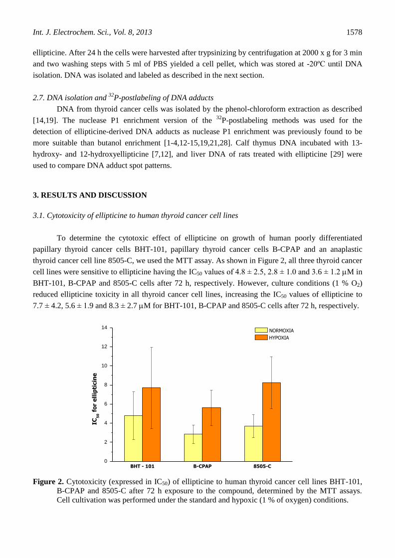
To determine the cytotoxic effect of ellipticine on growth of human poorly differentiated
papillary thyroid cancer cells BHT-101, papillary thyroid cancer cells B-CPAP and an anaplastic
thyroid cancer cell line 8505-C, we used the MTT assay. As shown in Figure 2, all three thyroid cancer
cell lines were sensitive to ellipticine having the IC50 values of 4.8 ± 2.5, 2.8 ± 1.0 and 3.6 ± 1.2 M in
BHT-101, B-CPAP and 8505-C cells after 72 h, respectively. However, culture conditions (1 % O2)
reduced ellipticine toxicity in all thyroid cancer cell lines, increasing the IC50 values of ellipticine to
7.7 ± 4.2, 5.6 ± 1.9 and 8.3 ± 2.7 M for BHT-101, B-CPAP and 8505-C cells after 72 h, respectively.
Figure 2. Cytotoxicity (expressed in IC50) of ellipticine to human thyroid cancer cell lines BHT-101,
B-CPAP and 8505-C after 72 h exposure to the compound, determined by the MTT assays.
Cell cultivation was performed under the standard and hypoxic (1 % of oxygen) conditions.
BHT - 101 B-CPAP 8505-C0
2
4
6
8
10
12
14
IC5
0 f
or
ell
ipti
cin
e
NORMOXIA
HYPOXIA

Int. J. Electrochem. Sci., Vol. 8, 2013
1579
As ellipticine acts, besides other mechanisms, via covalent modification of DNA which is
mediated by oxygen-dependent ellipticine bioactivation through CYPs, we speculate that a decrease in
ellipticine cytotoxicity under the hypoxic conditions might be caused by reduced CYP-catalyzed
bioactivation due to the oxygen deficiency. Therefore, formation of ellipticine-derived DNA adducts
and expressions levels of CYP enzymes in thyroid cancer cells were investigated (see 3.3. and 3.4.).
3.2. Ellipticine induced cell cycle arrest in human thyroid cancer cell lines
As many cancer drugs act by arresting cells in the cell cycle, we investigated the effect of
ellipticine treatment on the cell cycle distribution of thyroid cancer cells cultivated under the standard
(aerobic) and hypoxic conditions. Flow cytometric analysis was used for such a study (Fig. 3).
Compared to controls, treatment of cells with ellipticine for 24 h resulted in an appreciable
arrest of BHT-101, B-CPAP and 8505-C cells in S and/or G2/M phases of cell cycle with a
concomitant decrease in G0/G1 phase. Hypoxic conditions had no effect on the cell cycle distribution
nor influenced the effect of ellipticine exposure upon the cell cycle distribution. Treatment of BHT-
101 and B-CPAP thyroid cancer cells with 10 M ellipticine caused an arrest of around 40 % cells in S
phase of cell cycle. Even stronger effects of ellipticine on S and G2/M phases of the cell cycle were
found in the anaplastic thyroid cancer cells 8505-C (Fig. 3).
Figure 3. Effect of ellipticine treatment on cell cycle distribution in human thyroid cancer cells after
24 h ellipticine treatment under the standard or hypoxic cell culture cultivation conditions
assessed by flow cytometry.
Int. J. Electrochem. Sci., Vol. 8, 2013
1580
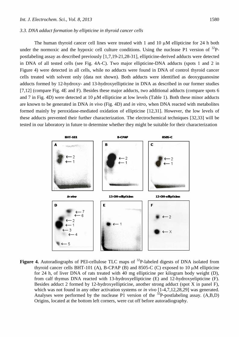
3.3. DNA adduct formation by ellipticine in thyroid cancer cells
The human thyroid cancer cell lines were treated with 1 and 10 M ellipticine for 24 h both
under the normoxic and the hypoxic cell culture conditions. Using the nuclease P1 version of 32
P-
postlabeling assay as described previously [1,7,19-21,28-31], ellipticine-derived adducts were detected
in DNA of all tested cells (see Fig. 4A-C). Two major ellipticine-DNA adducts (spots 1 and 2 in
Figure 4) were detected in all cells, while no adducts were found in DNA of control thyroid cancer
cells treated with solvent only (data not shown). Both adducts were identified as deoxyguanosine
adducts formed by 12-hydroxy- and 13-hydroxyellipticine in DNA as described in our former studies
[7,12] (compare Fig. 4E and F). Besides these major adducts, two additional adducts (compare spots 6
and 7 in Fig. 4D) were detected at 10 M ellipticine at low levels (Table 1). Both these minor adducts
are known to be generated in DNA in vivo (Fig. 4D) and in vitro, when DNA reacted with metabolites
formed mainly by peroxidase-mediated oxidation of ellipticine [12,31]. However, the low levels of
these adducts prevented their further characterization. The electrochemical techniques [32,33] will be
tested in our laboratory in future to determine whether they might be suitable for their characterization
Figure 4. Autoradiographs of PEI-cellulose TLC maps of 32
P-labeled digests of DNA isolated from
thyroid cancer cells BHT-101 (A), B-CPAP (B) and 8505-C (C) exposed to 10 M ellipticine
for 24 h, of liver DNA of rats treated with 40 mg ellipticine per kilogram body weight (D),
from calf thymus DNA reacted with 13-hydroxyellipticine (E) and 12-hydroxyelipticine (F).
Besides adduct 2 formed by 12-hydroxyellipticine, another strong adduct (spot X in panel F),
which was not found in any other activation systems or in vivo [1-4,7,12,28,29] was generated.
Analyses were performed by the nuclease P1 version of the 32
P-postlabeling assay. (A,B,D)
Origins, located at the bottom left corners, were cut off before autoradiography.

Int. J. Electrochem. Sci., Vol. 8, 2013
1581
Quantitative analyses showed that DNA adduct formation by ellipticine in human thyroid
cancer cell lines was dose-dependent with an over-proportional increase between 1 and 10 M
ellipticine, and influenced by the cell culture conditions (Table 1, Fig. 5). The highest amounts of
ellipticine-derived DNA adducts were found in B-CPAP cells, followed by BHT-101 and 8505-C cells
(Table 1, Fig. 5). A decrease in DNA adduct formation was found in thyroid cancer cell lines treated
with 10 M ellipticine cultured under hypoxic conditions (Table 1, Fig. 5). The decrease in DNA
adduct level correlated with a decrease in cytotoxicity of ellipticine to these cells (see Fig. 2). This was
not the case when thyroid cancer cells were treated with 1 M ellipticine under the lower oxygen
concentration; practically the same DNA adduct levels were found both under the standard and
hypoxic conditions. Therefore, low concentrations of oxygen seem to be sufficient for the oxidative
bioactivation of ellipticine at low concentrations (1 M).
Figure 5. DNA adduct formation by ellipticine in human thyroid cancer cell lines cultured under
normoxic and hypoxic conditions (total amounts of ellipticine-derived DNA adducts
expressed as RAL, relative adduct labeling). Thyroid cancer cells were exposed to ellipticine
for 24 h. DNA adduct formation was analyzed by the nuclease P1 version of the 32
P-
postlabeling assay [1]. Data are expressed as averages ± S.D. of three experiments.
BHT-101 B-CPAP 8505-C0.0
0.1
0.2
0.3
0.4
1
2
3
4
5
6
7
8
9
RA
L (
ad
du
cts
/ 1
07 n
uc
leo
tid
es)
1 M Elli
1 M Elli Hypoxia
10 M Elli
10 M Elli Hypoxia

Int. J. Electrochem. Sci., Vol. 8, 2013
1582
Table 1. Amounts of individual DNA adducts formed by ellipticine in human thyroid cancer cell lines
cultured under normoxic and hypoxic conditions
Cells Levels of DNA adducts (RAL x 10-7
)a
Adduct 1 Adduct 2 Adduct 6 Adduct 7 Total
BHT-101
Normoxia
1 M ellipticine 0.15 ± 0.03 0.13 ± 0.01 n.d. n.d. 0.28 ± 0.03
10 M ellipticine 3.9 ± 0.28 3.0 ± 0.50 0.1 ± 0.01 0.04 ± 0.01 7.04 ± 0.86
Hypoxia
1 M ellipticine 0.12 ± 0.07 0.10 ± 0.01 n.d. n.d. 0.22 ± 0.02
10 M ellipticine 2.35 ± 0.48 2.46 ± 0.17 0.05 ± 0.01 0.04 ± 0.01 4.9 ± 0.44
B-CPAP
Normoxia
1 M ellipticine 0.12 ± 0.01 0.14 ± 0.02 n.d. n.d. 0.26 ± 0.03
10 M ellipticine 3.33± 0.31 4.18 ± 0.42 0.09 ± 0.01 0.03 ± 0.01 7.63 ± 0.76
Hypoxia
1 M ellipticine 0.11 ± 0.06 0.15 ± 0.09 n.d. n.d. 0.26 ± 0.07
10 M ellipticine 1.67 ± 0.20 2.43 ± 0.22 0.05 ± 0.01 0.01 ± 0.01 4.16 ± 0.51
8505-C
Normoxia
1 M ellipticine 0.13 ± 0.05 0.08 ± 0.01 n.d. n.d. 0.21 ± 0.04
10 M ellipticine 2.22 ± 0.56 1.81 ± 0.28 0.05 ± 0.01 0.01 ± 0.01 4.09 ± 0.45
Hypoxia
1 M ellipticine 0.07 ± 0.01 0.16 ± 0.02 n.d. n.d. 0.23 ± 0.03
10 M ellipticine 1.39 ± 0.22 1.69 ± 0.20 0.06 ± 0.01 0.02 ± 0.02 3.16 ± 0.44
Thyroid cancer cells were exposed to ellipticine for 24 h. DNA adduct formation was analyzed by the
nuclease P1 version of the 32
P-postlabeling assay [1]. aRAL, relative adduct labeling; data are
expressed as averages ± S.D. of three experiments. N.d. - not detected (the detection limit of RAL was
1/1010
nucleotides).
3.4. Determination of CYP1A1, CYP1B1, CYP3A4, COX-1, TPO and cytochrome b5 protein levels in
thyroid cancer cells
Since a decrease in toxicity of ellipticine in the thyroid cancer cell lines was found under
hypoxic conditions that corresponded to a decrease in DNA adduct formation, the expression levels of
enzymes activating or detoxicating ellipticine (i.e. CYP1A1, 1B1, 3A4, COX-1 and TPO) were
analyzed by Western blotting. Furthermore, as cytochrome b5, a component of the CYP-dependent

Int. J. Electrochem. Sci., Vol. 8, 2013
1583
enzymatic system, influences ellipticine oxidation by the CYP1A1 and 3A4 enzymes [13,15],
expression levels of this protein were also evaluated.
As shown in Figure 6, expression patterns of metabolizing enzymes under different conditions
are quite complex. Except for the expression of CYP1A1, the levels of which were high in all cells
under all conditions, and TPO the levels of which were so low that the influence of culture conditions,
cell lineage or ellipticine exposure cannot be assessed, expression levels of all other enzymes were
different in the three cell lines. Most conspicuous was the near complete absence of cytochrome b5 in
8505-C cells.
B-CPAP cells seem to be the thyroid cell line most sensitive to ellipticine and are also the cells
with the highest ellipticine-DNA adduct levels. These cells also show consistent expression of
CYP3A4, the enzyme responsible for formation of the active intermediates 13-hydroxy- and 12-
hydroxyellipticine which lead to the two major ellipticine-derived DNA adducts [3,7,12,13,15]. The
line 8505-C showed the lowest DNA adduct levels, was a slightly less sensitive to ellipticine in the
cytotoxicity assay but the biggest effect of ellipticine on cell cycle distribution was observed, despite a
near complete lack of cytochrome b5 expression and a low CYP3A4 expression under normoxic
conditions. In the BHT-101 line cytotoxicity, DNA-adduct levels and expression of activating enzymes
such as the high levels of COX-1 are congruent. From the data on the three cell lines it seems as if
effects on cell cycle are not entirely dependent on activating enzymes while DNA adduct levels and
cytotoxicity as determined by the MTT assay are more dependent on ellipticine activation.
Figure 6. Immunoblots of cytochrome P450 (CYP) 1A1, 1B1, 3A4, cytochrome b5, cyclooxygenase-1
(COX-1) and thyroid peroxidase (TPO) in BHT-101 (A), B-CPAP (B) and 8505-C (C) thyroid
cancer cell lines. Actin was used as loading control. Control cells (CO) were either grown
under normoxic or hypoxic conditions and exposed to 1 µM (1E) or 10 µM (10E) ellipticine for
24 h, harvested and proteins of the cells were separated by gel electrophoresis (SDS-PAGE),
electro-blotted onto PVDF membranes and probed with the appropriate antibodies against the
individual proteins as described in the methods section.

Int. J. Electrochem. Sci., Vol. 8, 2013
1584
4. CONCLUSIONS
This study showed for the first time that ellipticine is toxic to BHT-101, B-CPAP and 8505-C
thyroid cancer cell lines and that this toxic effect corresponded to ellipticine-derived DNA adduct
formation in these cells. Our results indicate that expression of cytochrome b5, CYP1A1, 3A4 and
COX-1 influences the cytotoxicity and genotoxicity in the studied cell lines. Therefore, monitoring of
the expression levels of enzymes metabolizing the anticancer drug ellipticine (activation and
detoxication) by the Western blotting together with the 32
P-postlabeling technique utilized for
detection and quantitation of DNA adducts are appropriate tools for evaluating the mechanism(s) of
ellipticine toxicity in thyroid cancer cells. Future investigations will show whether ellipticine might be
useful for the treatment of anaplastic thyroid carcinoma.
ACKNOWLEDGEMENTS
The work has been supported by Grant Agency of the Czech Republic (grant P301/10/0356) and
Charles University in Prague (grant UNCE 204025/2012) and by the project for conceptual
development of research organization 00064203.
References
1. M. Stiborova, C. A. Bieler, M. Wiessler and E. Frei, Biochem. Pharmacol., 62 (2001) 1675.
2. M. Stiborova, M. Rupertova, H. H. Schmeiser and E. Frei, Biomed. Pap. Med. Fac. Univ. Palacky
Olomouc Czech Repub., 150 (2006) 13.
3. M. Stiborova, M. Rupertova and E. Frei, Biochim. Biophys. Acta, 1814 (2011) 175.
4. R. Kizek, V. Adam, J. Hrabeta, T. Eckschlager, S. Smutny, J. V. Burda, E. Frei and M. Stiborova,
Pharmacol. Ther., 133 (2012) 26.
5. C. Auclair, Arch. Biochem. Biophys., 259 (1987) 1.
6. N. C. Garbett and D. E. Graves, Curr. Med. Chem.: Anti-Cancer Agents, 4 (2004) 149.
7. M. Stiborova, J. Sejbal, L. Borek-Dohalska, D. Aimova, J. Poljakova, K. Forsterova, M.
Rupertova, J. Wiesner, J. Hudecek, M. Wiessler and E. Frei, Cancer Res., 64 (2004) 8374.
8. S. J. Froelich-Ammon, M. W. Patchan, N. Osheroff and R. B. Thompson, J. Biol. Chem., 270
(1995) 14998.
9. K. Fang, S. P. Chen, C. W. Lin, W. C. Cheng and H. T. Huang, Lung Cancer, 63 (2009) 227.
10. Y. Peng, C. Li, L. Chen, S. Sebti and J. Chen, Oncogene, 22 (2003) 4478.
11. M. A. Schwaller, B. Allard, E. Lescot and F. Moreau, J. Biol. Chem., 270 (1995) 22709.
12. M. Stiborova, J. Poljakova, H. Ryslava, M. Dracinsky, T. Eckschlager and E. Frei, Int. J. Cancer,
120 (2007) 243.
13. M. Stiborova, R. Indra, M. Moserova, V. Cerna, M. Rupertova, V. Martinek, T. Eckschlager, R.
Kizek and E. Frei, Chem. Res. Toxicol., 25 (2012) 1075.
14. J. Poljakova, J. Hrebackova, M. Dvorakova, M. Moserova, T. Eckschlager, J. Hrabeta, M.
Gottlicherova, B. Kopejtkova, E. Frei, R. Kizek and M. Stiborova, Neuro Endocrinol. Lett., 32
Suppl 1 (2011) 101.
15. V. Kotrbova, B. Mrazova, M. Moserova, V. Martinek, P. Hodek, J. Hudecek, E. Frei, and M.
Stiborova, Biochem. Pharmacol., 82 (2011) 669.
16. M. Stiborova, J. Poljakova, E. Martinkova, L. Borek-Dohalska, T. Eckschlager, R. Kizek and E.
Frei, Interdiscip. Toxicol., 4 (2011) 98.
17. L. Borek -Dohalska, E. Frei and M. Stiborova, Collect. Czech. Chem. Commun., 69 (2004) 603.

Int. J. Electrochem. Sci., Vol. 8, 2013
1585
18. J. Poljakova, E. Frei, J. E. Gomez, D. Aimova, T. Eckschlager, J. Hrabeta and M. Stiborova,
Cancer Lett., 252 (2007) 270.
19. J. Poljakova, T. Eckschlager, J. Hrabeta, J. Hrebackova, S. Smutny, E. Frei, V. Martinek, R. Kizek
and M. Stiborova, Biochem. Pharmacol., 77 (2009) 1466.
20. E. Martinkova, M. Dontenwill, E. Frei and M. Stiborova, Neuro Endocrinol. Lett., 30 Suppl 1
(2009) 60.
21. M. Stiborova, J. Poljakova, T. Eckschlager, R. Kizek and E. Frei, Biomed. Pap. Med. Fac. Univ.
Palacky Olomouc Czech Repub., 156 (2012) 115.
22. I. Landa and M. Robledo, J. Mol. Endocrinol., 47 (2011) R43.
23. K.N. Patel and A.R. Shaha, Cancer Control, 13 (2006) 119.
24. E. Kapiteijn, T.C. Schneider, H. Morreau, H. Gelderblom, J.W. Nortier and J.W. Smit, Ann.
Oncol., 23 (2012) 10.
25. J. Cinatl, Jr., J. Cinatl, P. H. Driever, R. Kotchetkov, P. Pouckova, B. Kornhuber and D. Schwabe,
Anticancer Drugs, 8 (1997) 958.
26. M. Stiborova, V. Martinek, H. Rydlova, P. Hodek and E. Frei, Cancer Res., 62 (2002) 5678.
27. M. Stiborova, V. Martinek, H. Rydlova, T. Koblas and P. Hodek, Cancer Lett., 220 (2005) 145.
28. M. Stiborova, M. Rupertova, D. Aimova, H. Ryslava and E. Frei, Toxicology, 236 (2007) 50.
29. M. Stiborova, A. Breuer, D. Aimova, M. Stiborova-Rupertova, M. Wiessler and E. Frei, Int. J.
Cancer, 107 (2003) 885.
30. M. Stiborova, M. Stiborova-Rupertova, L. Borek-Dohalska, M. Wiessler and E. Frei, Chem. Res.
Toxicol., 16 (2003) 38.
31. J. Poljakova, M. Dracinsky, E. Frei, J. Hudecek and M. Stiborova, Collect. Czech. Chem.
Commun., 71 (2006) 1169.
32. D. Hynek, L. Krejcova, O. Zitka, V. Adam, L. Trnkova, J. Sochor, M. Stiborova, T. Eckschlager,
J. Hubalek and R. Kizek, Int. J. Electrochem. Sci., 7 (2012) 34.
33. D. Dospivova, K. Smerkova, M. Ryvolova, D. Hynek, V. Adam, P. Kopel, M. Stiborova, T.
Eckschlager, J. Hubalek and R. Kizek, Int. J. Electrochem. Sci., 7 (2012) 3072
© 2013 by ESG (www.electrochemsci.org)


![Xenobiotic-Metabolizing Enzymes in Skeletal Muscle of ... · 232 Xenobiotic-Metabolizing Enzymes in Skeletal Muscle of Children and Adolescents . pounds [6]. Majority of CYP enzymes](https://img.pdfslide.us/doc/110x75/5ecdeb770de69043f505f631/xenobiotic-metabolizing-enzymes-in-skeletal-muscle-of-232-xenobiotic-metabolizing.jpg)