Embed Size (px)
Citation preview

ANTIMICROBIAL AGENTS AND CHEMOTHERAPY, July 2011, p. 3357–3362 Vol. 55, No. 70066-4804/11/$12.00 doi:10.1128/AAC.01483-10Copyright © 2011, American Society for Microbiology. All Rights Reserved.
The Antibiotic Dehydrophos Is Converted to a Toxic Pyruvate Analogby Peptide Bond Cleavage in Salmonella enterica�
Benjamin T. Circello,1 Charles G. Miller,1 Jin-Hee Lee,2 Wilfred A. van der Donk,2,3,4
and William W. Metcalf1,3*Department of Microbiology, University of Illinois at Urbana-Champaign, Urbana, Illinois 618011; Department of Chemistry,
University of Illinois at Urbana-Champaign, Urbana, Illinois 618012; Institute for Genomic Biology, University of Illinois atUrbana-Champaign, Urbana, Illinois 618013; and Howard Hughes Medical Institute, Urbana, Illinois 618014
Received 26 October 2010/Returned for modification 28 November 2010/Accepted 20 April 2011
The metabolic processing of dehydrophos, a broad-spectrum peptide antibiotic containing an unusualvinyl-phosphonate moiety, was examined by using a panel of Salmonella enterica mutants deficient in peptideuptake and catabolism. Dehydrophos bioactivity is lost in opp tpp double mutants, demonstrating a require-ment for uptake via nonspecific oligopeptide permeases. Dehydrophos bioactivity is also abolished in aquadruple Salmonella mutant lacking the genes encoding peptidases A, B, D, and N, showing that hydrolysisof the peptide bond is required for activity. 31P nuclear magnetic resonance spectroscopy was used to assessthe fate of dehydrophos following in vitro digestion of the antibiotic with purified PepA. The results suggest thatthe initial product of peptidase processing is 1-aminovinyl-phosphonate O-methyl ester. This phosphonateanalogue of dehydroalanine undergoes rearrangement to the more stable imine, followed by spontaneoushydrolysis to yield O-methyl-acetylphosphonate, a structural analogue of pyruvate. This compound is a knowninhibitor of pyruvate dehydrogenase and pyruvate oxidase and is probably the active species responsible fordehydrophos bioactivity.
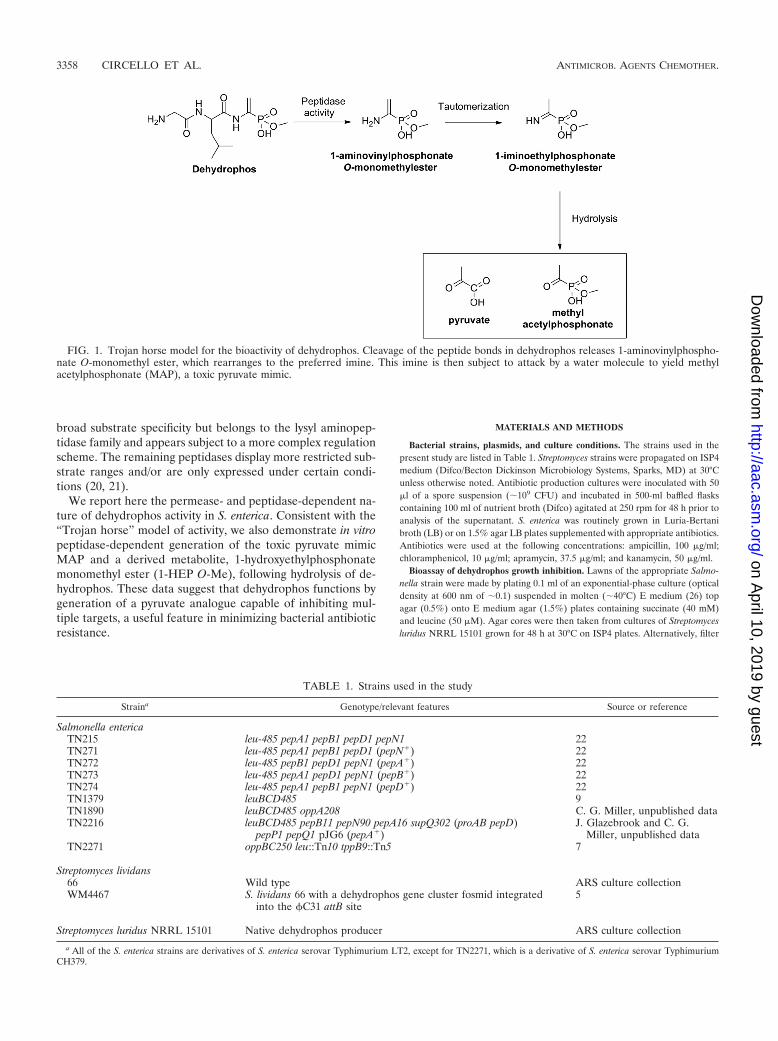
Dehydrophos (formerly A53868) is an unusual antibioticproduced by Streptomyces luridus. This compound has broadspectrum activity in vitro and is effective in vivo in Salmonellachallenged chickens (15). Dehydrophos consists of a glycine-leucine dipeptide linked to an O-methylated aminovinylphos-phonate (28) (Fig. 1). The cellular target of the compound hasyet to be established, and it is unknown whether it acts as atripeptide or whether it is metabolized by the target cell torelease a bioactive product. Structure-activity relationships ofseveral dehydrophos derivatives clearly revealed the impor-tance of the O-methyl and vinyl moieties but did not allowconclusions to be made regarding whether peptide bond cleav-age is required for bioactivity (16).
The coupling of phosphonic acids to amino acids has beenobserved in numerous bioactive natural products. In the caseof rhizocticin, plumbemycin, bialaphos, and phosalacine, theseamino acids convert a toxic phosphonate molecule into aninactive “Trojan horse” that is readily taken up by target or-ganisms. Once inside the cell, peptidase activity serves to re-lease the bioactive phosphonate moiety (6, 17). In other casesthe entire peptide is required for bioactivity. For example, theangiotensin converting enzyme inhibitor K-26 binds to its tar-get as a tripeptide consisting of Ile-Tyr coupled to (R)-1-ami-no-2-(4-hydroxyphenyl)-ethylphosphonic acid (AHEP) (2, 23).
Two mutually exclusive mechanisms for dehydrophos bioac-tivity can be envisioned based on whether the peptide bondsare cleaved or remain intact. In reporting the revised structure
of antibiotic, Whitteck et al. noted that cleavage of the peptidebond would produce an unstable enamine phosphonate, ex-pected to undergo tautomeric rearrangement followed byspontaneous hydrolysis (Fig. 1) (28). The expected product ofthis transformation would be methyl acetylphosphonate(MAP), a structural analog of pyruvate and a known inhibitorof both pyruvate oxidase and pyruvate dehydrogenase (24).Alternatively, the vinylphosphonate moiety of dehydrophoscould undergo a Michael-type addition to sulfhydryl-contain-ing target enzymes in a reaction analogous to that which occursduring the inactivation of cysteine dependent proteases byvinyl sulfones (25). This mechanism would require that thepeptide bond remain intact to prevent the rearrangement andhydrolysis reactions described above.
Both peptide transport and catabolism have been extensivelycharacterized in Salmonella enterica, providing a facile exper-imental system to distinguish between the two models for de-hydrophos action. S. enterica contains three distinct systems forpeptide uptake encoded the opp, tpp, and dpp operons, whichencode oligopeptide, tripeptide, and dipeptide permeases, re-spectively (10). The Opp permease is the primary peptideuptake system, transporting molecules containing up to 5amino acids (11–14). The Tpp permease is relatively specific,preferring hydrophobic residues and requiring anaerobic con-ditions for expression (7, 16), whereas the Dpp permeasetransports dipeptide substrates and acts as the signal receptorfor peptide chemotaxis (1). Mutant strains lacking all threepermeases are unable to assimilate most peptides (10). Pep-tides are converted to free amino acids in S. enterica via theaction of eight major peptidases, designated A, B, D, E, N, P,Q, and T, with overlapping specificities. Peptidases A, B, and Dpossess broad substrate specificities, with A and B belonging tothe leucyl aminopeptidase family (19, 21). Peptidase N also has
* Corresponding author. Mailing address: Department of Microbi-ology, University of Illinois at Urbana-Champaign, B103 CLSL, 601 S.Goodwin Ave., Urbana, IL 61801. Phone: (217) 244-1943. Fax: (217)244-6697. E-mail: [email protected].
� Published ahead of print on 2 May 2011.
3357
on April 10, 2019 by guest
http://aac.asm.org/
Dow
nloaded from
broad substrate specificity but belongs to the lysyl aminopep-tidase family and appears subject to a more complex regulationscheme. The remaining peptidases display more restricted sub-strate ranges and/or are only expressed under certain condi-tions (20, 21).
We report here the permease- and peptidase-dependent na-ture of dehydrophos activity in S. enterica. Consistent with the“Trojan horse” model of activity, we also demonstrate in vitropeptidase-dependent generation of the toxic pyruvate mimicMAP and a derived metabolite, 1-hydroxyethylphosphonatemonomethyl ester (1-HEP O-Me), following hydrolysis of de-hydrophos. These data suggest that dehydrophos functions bygeneration of a pyruvate analogue capable of inhibiting mul-tiple targets, a useful feature in minimizing bacterial antibioticresistance.
MATERIALS AND METHODS
Bacterial strains, plasmids, and culture conditions. The strains used in thepresent study are listed in Table 1. Streptomyces strains were propagated on ISP4medium (Difco/Becton Dickinson Microbiology Systems, Sparks, MD) at 30°Cunless otherwise noted. Antibiotic production cultures were inoculated with 50�l of a spore suspension (�109 CFU) and incubated in 500-ml baffled flaskscontaining 100 ml of nutrient broth (Difco) agitated at 250 rpm for 48 h prior toanalysis of the supernatant. S. enterica was routinely grown in Luria-Bertanibroth (LB) or on 1.5% agar LB plates supplemented with appropriate antibiotics.Antibiotics were used at the following concentrations: ampicillin, 100 �g/ml;chloramphenicol, 10 �g/ml; apramycin, 37.5 �g/ml; and kanamycin, 50 �g/ml.
Bioassay of dehydrophos growth inhibition. Lawns of the appropriate Salmo-nella strain were made by plating 0.1 ml of an exponential-phase culture (opticaldensity at 600 nm of �0.1) suspended in molten (�40°C) E medium (26) topagar (0.5%) onto E medium agar (1.5%) plates containing succinate (40 mM)and leucine (50 �M). Agar cores were then taken from cultures of Streptomycesluridus NRRL 15101 grown for 48 h at 30°C on ISP4 plates. Alternatively, filter
FIG. 1. Trojan horse model for the bioactivity of dehydrophos. Cleavage of the peptide bonds in dehydrophos releases 1-aminovinylphospho-nate O-monomethyl ester, which rearranges to the preferred imine. This imine is then subject to attack by a water molecule to yield methylacetylphosphonate (MAP), a toxic pyruvate mimic.
TABLE 1. Strains used in the study
Straina Genotype/relevant features Source or reference
Salmonella entericaTN215 leu-485 pepA1 pepB1 pepD1 pepN1 22TN271 leu-485 pepA1 pepB1 pepD1 (pepN�) 22TN272 leu-485 pepB1 pepD1 pepN1 (pepA�) 22TN273 leu-485 pepA1 pepD1 pepN1 (pepB�) 22TN274 leu-485 pepA1 pepB1 pepN1 (pepD�) 22TN1379 leuBCD485 9TN1890 leuBCD485 oppA208 C. G. Miller, unpublished dataTN2216 leuBCD485 pepB11 pepN90 pepA16 supQ302 (proAB pepD)
pepP1 pepQ1 pJG6 (pepA�)J. Glazebrook and C. G.
Miller, unpublished dataTN2271 oppBC250 leu::Tn10 tppB9::Tn5 7
Streptomyces lividans66 Wild type ARS culture collectionWM4467 S. lividans 66 with a dehydrophos gene cluster fosmid integrated
into the �C31 attB site5
Streptomyces luridus NRRL 15101 Native dehydrophos producer ARS culture collection
a All of the S. enterica strains are derivatives of S. enterica serovar Typhimurium LT2, except for TN2271, which is a derivative of S. enterica serovar TyphimuriumCH379.
3358 CIRCELLO ET AL. ANTIMICROB. AGENTS CHEMOTHER.
on April 10, 2019 by guest
http://aac.asm.org/
Dow
nloaded from

disks were spotted with 10 �l of an authentic standard dehydrophos (1 mg/ml).These cores and/or disks were then placed onto the freshly inoculated bioassayplates, incubated at 37°C for 12 h, and then scored for bioactivity.
Overexpression, purification, and activity assay of PepA. PepA was purified byusing a modified version of the Vogt purification of E. coli aminopeptidase I (27).A 500-ml culture of the peptidase A overproducer S. enterica TN2216 was grownfor 16 h at 37°C, and the cells were then harvested by centrifugation at 5,000 �g for 10 min. The cell pellet was resuspended in 20 ml of ice-cold buffer 1 (50 mMTris-HCl [pH 7.8], 200 mM KCl, 10 mM MgCl2, 100 mM EDTA, 100 mMdithiothreitol, and 5% glycerol) and disrupted by a single passage through achilled French press cell at 20,000 lb/in2. Solid debris was removed by centrifu-gation at 13,000 � g for 30 min at 4°C. The resuspended cells were then placedinto a 70°C water bath and continuously swirled until the temperature reached65°C. Precipitated proteins were removed by centrifugation at 13,000 � g for 15min at 4°C. The supernatant was dialyzed (Pierce, 10K MWCO) against 2 litersof 0.1 M phosphate buffer (pH 7.6) for 18 h at 4°C with one change of buffer.After dialysis, the extract was centrifuged again at 13,000 � g for 15 min at 4°Cto remove insoluble material. The supernatant was then dialyzed against buffer2 (20 mM Tris-HCl [pH 8.4], 10 mM KCl, 1 mM magnesium acetate, 0.1 mMEDTA) for 18 h. The precipitate that formed was collected and redissolved inbuffer 3 (20 mM KHCO3 [pH 10.3], 50 mM KCl, 1 mM magnesium acetate, 0.1mM EDTA). This precipitate failed to completely solubilize in buffer 3, leavingthe solution turbid. PepA purity was evaluated by visual inspection of an SDS-PAGE gel stained with Coomassie brilliant blue (estimated to be at least 95%pure). PepA action on dehydrophos was assayed at 37°C over 18 h by directaddition of dehydrophos (7 mM) to dissolved PepA. The reaction was monitoredby 31P nuclear magnetic resonance (NMR) spectroscopy.
31P NMR spectroscopy-based identification of dehydrophos metabolites andmonitoring of the hydrolysis reaction. Culture supernatant was passed through a0.2-�m-pore-size filter, concentrated 20-fold by evaporation, and D2O was addedto 25% as a lock solvent. 31P NMR spectra were acquired on a Varian UnityInova 600 spectrophotometer equipped with a 5-mm AutoTuneX probe at theVarian Oxford Instrument Center for Excellence at the University of IllinoisUrbana-Champaign. An external standard of 85% phosphoric acid was definedas 0 ppm. Experiments to confirm the identity of proposed metabolites wereroutinely performed by adding 1 to 5 mM standard to the sample and thenreacquiring the spectra.
RESULTS
Dehydrophos is transported into S. enterica via the Opp andTpp transporters. To determine which peptide transport sys-tems are involved in the uptake of dehydrophos, we tested theability of the antibiotic to inhibit the growth of S. entericapermease mutants. The mutants used are well-characterizedstrains that have been previously used to characterize peptideuptake in S. enterica (7). TN1379 encodes each of the knownpeptide permease systems, TN1890 contains both the Dpp andTpp permease systems (opp mutant) and TN2271 contains onlythe Dpp system (opp tpp mutant). As expected, dehydrophos
inhibits TN1379; however, it fails to inhibit TN2271 and showsonly a very small zone of inhibition with TN1890 (Fig. 2).These results indicate that the Opp peptide transporter is themajor transporter of dehydrophos, with the Tpp transporterplaying a relatively minor role. Dpp is unable to transportdehydrophos. This result also shows that a cytoplasmic targetor processing step is required for bioactivity.
Dehydrophos action requires peptidase activity. To testwhether peptide bond cleavage was required for dehydrophosactivity, we examined bioactivity against a series of character-ized S. enterica peptidase mutants (22). A quadruple mutantlacking PepA, PepD, PepB, and PepN was resistant to dehy-drophos, whereas the isogenic strain containing all of the miss-ing peptidases was sensitive to the antibiotic, indicating thatdehydrophos action is dependent on peptidase cleavage (Fig.3). Strains carrying any one of the deleted peptidases were alsosensitive to dehydrophos; however, the severity of the growthinhibition varied depending on the peptidase present.Strains carrying peptidase A or B displayed growth inhibi-tion similar to the wild type, whereas a strain carrying pep-tidase D had a slightly smaller zone of inhibition, and astrain carrying only peptidase N had a significantly smallerzone of inhibition (Fig. 4).
In vitro peptidase cleavage of dehydrophos by Salmonellapeptidase A results in the generation of methyl acetylphospho-nate. As described above, hydrolysis of the peptide bonds indehydrophos is expected to yield methyl acetylphosphonate viaa series of uncatalyzed chemical reactions. To test this predic-tion, we incubated synthetically prepared dehydrophos withpurified PepA and monitored the products of the cleavagereaction with phosphorus NMR spectroscopy (Fig. 5). Underthe conditions used, the reaction proceeded slowly, which al-lowed for the detection of discrete intermediates. After 1 h ofincubation, four separate peaks were observed. Based on aspiking experiment with a synthetic standard, the peak with achemical shift of 10.2 ppm corresponds to unaltered dehydro-phos. The peak at 10 ppm is presumed to be dehydrophoslacking the N-terminal glycine (desglycyldehydrophos); how-ever, this could not be rigorously established due to the lack ofan appropriate standard. After 3 h of incubation, the dehydro-phos peak decreased in intensity, while the putative desglycyl-
FIG. 2. The oligopeptide permease is required for dehydrophosactivity. Bioassays were performed as described by spotting syntheticdehydrophos onto a filter disc and assaying growth inhibition againstvarious Salmonella strains: strain TN1379 (center) contains the Opp,Dpp, and Tpp permease systems; strain TN2271 carries only the Dpppermeases (i.e., opp tpp mutant) (left); and strain TN1890 carries boththe Dpp and the Tpp oligopeptide permeases (i.e., opp mutant) (right).
FIG. 3. The antibiotic activity of dehydrophos is peptidase depen-dent. (a) Dehydrophos fails to inhibit S. enterica TN215 lacking pep-tidases A, B, D, and N when applied as either an agar core from thenative producer or as a pure synthetic compound. (b) In contrast, S.enterica TN1379 containing the full complement of peptidases exhibitsthe typically seen growth inhibition.
VOL. 55, 2011 ACTIVATION OF DEHYDROPHOS 3359
on April 10, 2019 by guest
http://aac.asm.org/
Dow
nloaded from

dehydrophos peak increased. After overnight incubation, thedesglycyldehydrophos peak disappeared, and the dehydrophospeak was greatly diminished. Two other peaks were seen in thespectra: one located at 6.2 ppm and the other located at 1 ppm.The peak at 6.2 ppm was transiently observed during the firsttwo time points but was not observed after the overnight in-cubation, whereas the peak at 1 ppm increased in intensity overtime and, after overnight incubation, represented the mostabundant phosphorus-containing species in the reaction mix-
ture. Spiking of the assay with a synthetic standard identifiedthe 1 ppm peak as MAP. The 6.2-ppm peak was not identified.
Dehydrophos bioactivity is not relieved by acetate. BecauseMAP is a known inhibitor of pyruvate dehydrogenase andpyruvate oxidase, we examined whether the bioactivity of de-hydrophos could be relieved by supplementation with acetate,which is required for growth of pdh pox double mutants (4). Tothis end, we performed a bioassay in the presence of dehydro-phos and acetate. The zone of inhibition remained unaltered,
FIG. 4. Reintroduction of any single peptidase allele is sufficient to restore dehydrophos bioactivity. Bioassay plates examining the effects ofreintroduction of either PepA (strain TN272 [b]), PepB (strain TN273 [c]), PepN (strain TN271 [d]), PepD (strain TN274 [e]), or to a quadruplemutant of the aforementioned peptidases (strain TN271 [a]).
FIG. 5. PepA catalyzed the degradation of dehydrophos. A time course following the fate of dehydrophos, monitored by 31P NMR, incubatedwith purified S. enterica PepA is shown. (a) After 1 h, a large peak representing unreacted dehydrophos (10.2 ppm) is present, along with asubstantial amount of the putative dipeptide (10 ppm). (b) After 3 h, the putative dipeptide peak dominates the spectrum, and the peak at 1 ppmhas increased in intensity. (c) After an overnight incubation, only two peaks remain: a large peak at 1 ppm and a peak representing unreacteddehydrophos at 10.2 ppm. (d) The addition of a synthetic standard identifies the peak at 1 ppm as methylacetylphosphonate (MAP).
3360 CIRCELLO ET AL. ANTIMICROB. AGENTS CHEMOTHER.
on April 10, 2019 by guest
http://aac.asm.org/
Dow
nloaded from

a finding consistent with the idea that dehydrophos has multi-ple, essential cellular targets (Fig. 6).
1-HEP O-Me is a product of dehydrophos breakdown. Earlyinvestigations into dehydrophos biosynthesis were confoundedby the presence of an unidentified phosphonate observed by31P NMR spectroscopy in the culture supernatants of nativeand heterologous dehydrophos producers (5). Our observationof MAP as the product of in vivo peptidase processing ofdehydrophos suggested a potential source and identity of thisunknown phosphonate. Spiking of samples containing the un-identified phosphonate with authentic standards showed thatthe unknown compound was not MAP (data not shown), lead-ing to the hypothesis that cells might detoxify MAP via reduc-tion to form 1-HEP O-Me, analogous to the thermodynami-cally favorable reduction of pyruvate to lactate by lactatedehydrogenase. To test this idea, we fed dehydrophos to Strep-tomyces lividans 66 and monitored the culture supernatant withphosphorus NMR spectroscopy. Over time, the phosphoruspeak associated with dehydrophos diminishes in intensity cor-responding with the appearance and growth of a second peak,which was confirmed to be 1-HEP O-Me by the addition of asynthetic standard (Fig. 7).
DISCUSSION
Many known phosphonate antibiotics utilize short peptidesas a “Trojan horse” to obtain access into the cellular environ-ment. The data presented here clearly indicate that dehydro-phos can be added to this list. The in vivo requirement for bothpeptide transport and hydrolysis, coupled with the results of invitro peptidase digestion, strongly suggest that the bioactiveagent derived from dehydrophos is MAP, a structural analog ofpyruvate. MAP is a known inhibitor of both pyruvate dehydro-
genase and pyruvate oxidase. With a Ki of 5 � 10�8 M, MAPbinds to pyruvate dehydrogenase 10,000 times better than thenatural substrate, pyruvate (24). MAP has relatively modest Ki
for pyruvate oxidase (1 mM, pH 7.0), which is a 10-fold loweraffinity than the enzyme has for its natural substrate pyruvate(8, 24). Mutants lacking both pyruvate dehydrogenase andpyruvate oxidase are incapable of growth unless the media aresupplemented with acetate, potentially explaining the mecha-nisms of growth inhibition by dehydrophos. Nevertheless, thesetwo enzymes cannot be the sole targets of dehydrophos be-cause supplementation of the media with acetate fails to re-lieve growth inhibition. This finding is not altogether surprisinggiven the number of essential cellular processes that involvepyruvate. Indeed, while MAP is best known as a potent pyru-
FIG. 6. Acetate fails to relieve antibiotic inhibition generated bydehydrophos. The central filter disc was spotted with synthetic dehy-drophos, while the outer disc contained 10 �l of a 40 mM acetatesolution.
FIG. 7. S. lividans 66 converts exogenous dehydrophos to 1-HEPO-Me. 31P NMR spectra demonstrate the fate of dehydrophos fed toS. lividans 66 over time. (a) Prior to inoculation, dehydrophos can beobserved at a chemical shift of 10.2 ppm. (b) After the inoculation ofS. lividans 66 and 48 h of growth, the peak corresponding to dehydro-phos has decreased in intensity, and a second peak has appeared at24.5 ppm. (c) The addition of a synthetic standard increased the in-tensity of the 24.5-ppm peak and confirmed its identity as 1-hydroxy-ethylphophonate O-methyl ester (1-HEP O-Me).
VOL. 55, 2011 ACTIVATION OF DEHYDROPHOS 3361
on April 10, 2019 by guest
http://aac.asm.org/
Dow
nloaded from

vate dehydrogenase inhibitor, it has also been shown to inhibitother pyruvate-utilizing enzymes (24).
Our model for in vivo production of MAP involves enzy-matic cleavage to produce an unstable compound that under-goes spontaneous chemical conversion to the pyruvate analog.Interestingly, our 31P NMR data show the transient accumu-lation of a phosphonate intermediate at 6.2 ppm that couldcorrespond to the unstable intermediate, which could be either1-aminovinylphosphonate O-methyl ester or 1-iminovinylphos-phonate O-methyl ester (Fig. 1). Although we could not rigor-ously establish the identity of this compound, we believe theintermediate is probably the latter compound because knownexamples of enamine/imine tautomerization occur rapidly andfavor the imine form (18). Regardless of which intermediateaccumulates, the observation of this transient peak in the 31PNMR spectrum raises interesting questions regarding themode of action of dehydrophos. It is possible that one of thetransient reactive intermediates produced during dehydrophoscatabolism might contribute to the toxicity of dehydrophos.Importantly, these data suggest that the peptide linkages havea dual role in the antibiotic activity of dehydrophos. Not onlydo they promote uptake of the inactive tripeptide, they alsostabilize an unstable phosphonate moiety by preventing theenamine/imine tautomerization of the vinyl amine functionalgroup.
Finally, it should be noted that our results suggest that thereis significant substrate specificity in both the uptake and pro-cessing of dehydrophos in S. enterica. These findings are rem-iniscent of the specificity seen in other phosphonate tripeptideantibiotics. For example, rhizocticin and plumbemycin containthe same bioactive component but differ in the amino acid sidechains. Interestingly, rhizocticin is an antifungal compoundwith little activity toward bacteria, whereas plumbemycin isantibacterial, with little activity toward fungi. It has been pro-posed that the differences in bioactivity are a reflection ofsubstrate specificity in either uptake or processing (3, 6). Thisobservation has significant implications in the developmentand design of new, more specific antibiotics. Given the variednature of oligopeptide uptake and cleavage, an intriguing ideawould be the attachment of small molecule inhibitors to mul-tiple peptide scaffolds. Applied appropriately, this could openthe door for the treatment of specific infections with specificantibiotics, while avoiding the deleterious effects on the naturalhost-associated microbial community.
ACKNOWLEDGMENTS
This study was supported by the National Institutes of Health (GMPO1 GM077596). B.T.C. was supported by a National Institutes ofHealth Chemistry-Biology Interface Training Program (GM070421).
The contents of this study are solely the responsibility of theauthors and do not necessarily represent the official views of theNIGMS or NIH.
REFERENCES
1. Abouhamad, W. N., M. Manson, M. M. Gibson, and C. F. Higgins. 1991.Peptide transport and chemotaxis in Escherichia coli and Salmonella typhi-
murium: characterization of the dipeptide permease (Dpp) and the dipep-tide-binding protein. Mol. Microbiol. 5:1035–1047.
2. Akif, M., et al. 2010. Crystal structure of a phosphonotripeptide K-26 incomplex with angiotensin converting enzyme homologue (AnCE) from Dro-sophila melanogaster. Biochem. Biophys. Res. Commun. 398:532–536.
3. Borisova, S. A., B. T. Circello, J. K. Zhang, W. A. van der Donk, and W. W.Metcalf. 2010. Biosynthesis of rhizocticins, antifungal phosphonate oligopep-tides produced by Bacillus subtilis ATCC 6633. Chem. Biol. 17:28–37.
4. Chang, Y. Y., and J. E. Cronan, Jr. 1983. Genetic and biochemical analysesof Escherichia coli strains having a mutation in the structural gene (poxB) forpyruvate oxidase. J. Bacteriol. 154:756–762.
5. Circello, B. T., A. C. Eliot, J. H. Lee, W. A. van der Donk, and W. W. Metcalf.2010. Molecular cloning and heterologous expression of the dehydrophosbiosynthetic gene cluster. Chem. Biol. 17:402–411.
6. Diddens, H., H. Zahner, E. Kraas, W. Gohring, and G. Jung. 1976. On thetransport of tripeptide antibiotics in bacteria. Eur. J. Biochem. 66:11–23.
7. Gibson, M. M., M. Price, and C. F. Higgins. 1984. Genetic characterizationand molecular cloning of the tripeptide permease (tpp) genes of Salmonellatyphimurium. J. Bacteriol. 160:122–130.
8. Hager, L. P. 1957. Trypsin activation of a ferricyanide-linked pyruvic acidoxidation. J. Biol. Chem. 229:251–263.
9. Hamilton, S., and C. G. Miller. 1992. Cloning and nucleotide sequence of theSalmonella typhimurium dcp gene encoding dipeptidyl carboxypeptidase. J.Bacteriol. 174:1626–1630.
10. Higgins, C. F., and M. M. Gibson. 1986. Peptide transport in bacteria.Methods Enzymol. 125:365–377.
11. Hiles, I. D., M. P. Gallagher, D. J. Jamieson, and C. F. Higgins. 1987.Molecular characterization of the oligopeptide permease of Salmonellatyphimurium. J. Mol. Biol. 195:125–142.
12. Hiles, I. D., L. M. Powell, and C. F. Higgins. 1987. Peptide transport inSalmonella typhimurium: molecular cloning and characterization of the oli-gopeptide permease genes. Mol. Gen. Genet. 206:101–109.
13. Hogarth, B. G., and C. F. Higgins. 1983. Genetic organization of the oligo-peptide permease (opp) locus of Salmonella typhimurium and Escherichiacoli. J. Bacteriol. 153:1548–1551.
14. Jamieson, D. J., and C. F. Higgins. 1984. Anaerobic and leucine-dependentexpression of a peptide transport gene in Salmonella typhimurium. J. Bacte-riol. 160:131–136.
15. Johnson, R. D., R. M. Kastner, S. H. Larsen, and E. E. Ose. 5 April 1984.Antibiotic A53868 and process for production thereof. U.S. patent 4,482,488.
16. Kuemin, M., and W. A. van der Donk. 2010. Structure-activity relationshipsof the phosphonate antibiotic dehydrophos. Chem. Commun. (Camb.) 46:7694–7696.
17. Laber, B., S. D. Lindell, and H. D. Pohlenz. 1994. Inactivation of Escherichiacoli threonine synthase by DL-Z-2-amino-5-phosphono-3-pentenoic acid.Arch. Microbiol. 161:400–403.
18. Lu, S. P., and A. H. Lewin. 1998. Enamine/imine tautomerism in �,�-unsat-urated-�-amino acids. Tetrahedron 54:15097–15104.
19. Mathew, Z., T. M. Knox, and C. G. Miller. 2000. Salmonella enterica serovarTyphimurium peptidase B is a leucyl aminopeptidase with specificity foracidic amino acids. J. Bacteriol. 182:3383–3393.
20. McHugh, G. L., and C. G. Miller. 1974. Isolation and characterization ofproline peptidase mutants of Salmonella typhimurium. J. Bacteriol. 120:364–371.
21. Miller, C. G. 1975. Peptidases and proteases of Escherichia coli and Salmo-nella typhimurium. Annu. Rev. Microbiol. 29:485–504.
22. Miller, C. G., and K. Mackinnon. 1974. Peptidase mutants of Salmonellatyphimurium. J. Bacteriol. 120:355–363.
23. Ntai, I., M. L. Manier, D. L. Hachey, and B. O. Bachmann. 2005. Biosyn-thetic origins of C-P bond containing tripeptide K-26. Organic Lett. 7:2763–2765.
24. O’Brien, T. A., R. Kluger, D. C. Pike, and R. B. Gennis. 1980. Phosphonateanalogues of pyruvate: probes of substrate binding to pyruvate oxidase andother thiamin pyrophosphate-dependent decarboxylases. Biochim. Biophys.Acta 613:10–17.
25. Rosenthal, P. J., et al. 1996. Antimalarial effects of vinyl sulfone cysteineproteinase inhibitors. Antimicrob. Agents Chemother. 40:1600–1603.
26. Vogel, H. J., and D. M. Bonner. 1956. Acetylornithinase of Escherichia coli:partial purification and some properties. J. Biol. Chem. 218:97–106.
27. Vogt, V. M. 1970. Purification and properties of an aminopeptidase fromEscherichia coli. J. Biol. Chem. 245:4760–4769.
28. Whitteck, J. T., et al. 2007. Reassignment of the structure of the antibioticA53868 reveals an unusual amino dehydrophosphonic acid. Angew Chem.Int. Ed. Engl. 46:9089–9092.
3362 CIRCELLO ET AL. ANTIMICROB. AGENTS CHEMOTHER.
on April 10, 2019 by guest
http://aac.asm.org/
Dow
nloaded from