Embed Size (px)
Citation preview
S4 A E S T H E T I C S U R G E R Y J O U R N A L ~ J A N U A R Y / F E B R U A R Y 2 0 0 6
S U P P L E M E N T
Facial aging reflects the dynamic, cumulative effects of time on the skin, soft tissues, and deep structural components of the
face, and is a complex synergy of skin textural changes and loss of facial volume. Many of the facial manifestations of aging
reflect the combined effects of gravity, progressive bone resorption, decreased tissue elasticity, and redistribution of subcuta-
neous fullness.
A convenient method for assessing the morphological effects of aging is to divide the face into the upper third (forehead and
brows), middle third (midface and nose), and lower third (chin, jawline, and neck). The midface is an important factor in
facial aesthetics because perceptions of facial attractiveness are largely founded on the synergy of the eyes, nose, lips, and
cheek bones (central facial triangle). For aesthetic purposes, this area should be considered from a 3-dimensional rather than a
2-dimensional perspective, and restoration of a youthful 3-dimensional facial topography should be regarded as the primary
goal in facial rejuvenation.
Recent years have seen a significant increase in the number of nonsurgical procedures performed for facial rejuvenation.
Patients seeking alternatives to surgical procedures include those who require restoration of lost facial volume, those who wish
to enhance normal facial features, and those who want to correct facial asymmetry. Important factors in selecting a nonsurgi-
cal treatment option include the advantages of an immediate cosmetic result and a short recovery time.
(Aesthetic Surg J 2006;26(suppl):S4-S9.)
The Anatomy of the Aging Face: Volume Lossand Changes in 3-Dimensional Topography
Sydney R. Coleman, MD; Rajiv Grover, BSc, MB BS, MD, FRCS (Plast)Dr. Coleman is Assistant Professor of Plastic Surgery in the Department of Surgery at New York University School of Medicine, New York, NY.
Dr. Grover is Consultant Plastic and Aesthetic Surgeon, King Edward VII Hospital, London, UK.
Aging of the human face is the result of both super-ficial textural wrinkling of the skin and changesin the 3-dimensional (3-D) topography of the
underlying structures. The skin, soft tissues (subcuta-neous fat, muscle, and fascia), and structural support(bone and teeth) are individually affected by the agingprocess, but they also act in dynamic unison to deter-mine the phenotypic presentation of the face throughoutlife. The major forces contributing to facial aging includegravity, skeletal remodeling, subcutaneous fat redistribu-tion and loss, hormonal imbalance, chronic solar expo-sure, and smoking. Other environmental factors that arepurported to affect facial appearance include mentalstress, diet, work habits, drug abuse, and disease.
Structural Components of Facial Aging
Skeletal structure
Aging of the craniofacial skeleton is not merely theresult of bone atrophy but is also due to a change in therelative dynamics of bone expansion and bone loss.1
There is an appreciable reduction in facial height, whichis mainly due to changes in the maxilla and mandible,and a modest increase in facial width and depth. Theorbits increase in size, whereas the maxilla decreases insize, compounding the inferior displacement of the malarfat pad and accentuation of the nasolabial fold.2
Maxillary resorption can also lead to a loss of support inthe upper lip, which contributes to perioral wrinkling. Inthe mandible, tooth loss causes marked resorption of thealveolar ridge, and the shape and projection of the chinalso change with age. There is a general coarsening ofmandibular bony protuberances at the points of insertionof masticatory muscles (eg, the gonial angle and inferioredge of the zygomatic eminence), and a general softeningelsewhere.
Subcutaneous fat distribution
The youthful face is characterized by a diffuse, bal-anced distribution of superficial and deep fat, which con-fers a well-rounded 3-D topography that is delineated bya series of arcs and convexities.3,4 Viewed frontally, the
S4-S9_YMAJ_Supp_Col286_CPRR.qxd 2/7/06 7:33 AM Page 4

A E S T H E T I C S U R G E R Y J O U R N A L ~ J a n u a r y / F e b r u a r y 2 0 0 6 S5The Anatomy of the Aging Face: Volume Loss andChanges in 3-Dimensional Topography
S U P P L E M E N T
primary arc of the jawline, convexities of the temples,and multiple smaller secondary arcs of the lips are evi-dent. In profile, 3 primary arcs are the most definitivefeatures of youth: the lateral cheek projection (the “ogee”curve), extending as an unbroken convex line from thelower eyelid to the cheek; the arc of the jawline, extend-ing from the lateral mandible to the mentum; and the arcof the forehead.5
Facial aging is associated with loss of soft tissue full-ness in certain areas (periorbital, forehead, malar, tem-poral, mandibular, mental, glabellar, and perioral sites)and persistence or hypertrophy of fat in others (sub-mental, lateral nasolabial fold and labiomental crease,jowls, infraorbital fat pouches, and malar fat pad).6,7
Magnetic resonance imaging (MRI) has been used toestablish that the percentage increase in fat in the upperthird relative to the remainder of the midface was sig-nificantly greater in old subjects (N = 10), compared toyoung subjects (N = 10).7 However, MRI scanning tech-nology requires subjects to adopt a prone position, thusthe direction of gravitational pull on the cheek massduring scanning is different from that exerted when anindividual is in the upright position. The hypertrophiccheek area would be expected to lie further down theface when the subjects were upright. Ideally, longitudi-nal imaging studies in the same patients over a lifetimewould further the understanding of the dynamics of fatdistribution in facial aging.
Figure 1. Coronal sections illustrating the loss of facial fullness that occurs with age. In the young face (left), the subcutaneous fat deposits (yellow)are masked by the fullness provided by extracellular colloidal fluids (purple). With advancing age, progressive loss of facial fullness causes the subcuta-neous fat deposits and underlying soft tissue and skeletal structures to become more evident (center); involutional exposure of fat deposits in the jowland chin gives the impression of descent (right). Reproduced with permission from Coleman, 2004.6
S4-S9_YMAJ_Supp_Col286_CPRR.qxd 2/7/06 7:33 AM Page 5
S6 A e s t h e t i c S u r g e r y J o u r n a l ~ J a n u a r y / F e b r u a r y 2 0 0 6 Volume 26, Number 1, Supplement
S U P P L E M E N T
In addition to redistribution of facial fat and con-comitant loss of surrounding fullness, the fat pocketsbecome more discernible as separate entities, as do manyof the underlying facial structures, such as the submaxil-lary glands and bony protuberances (Figure 1).6 Withage, the malar fat pad gradually slides forward anddown to bulge against the nasolabial crease, giving riseto the prominence of the nasal fold in the aged face.8
This redistribution and demarcation of fat gives thesenile face an unbalanced appearance. Skin wrinklingoccurs at some sites (periorbital and perioral areas) dueto the repeated underlying muscle action to give a pro-gression from dynamic rhytids initially to static rhytidsas the skin changes become permanent. Elsewhere (jowl,submental area, and nasolabial fold) sagging can occurdue to a relative excess of skin and/or lack of elasticrecoil as well as fat accumulation.6 Consequently, thedefining arcs and convexities of youth are disrupted.From the front, the jawline appears scalloped, the tem-poral, buccal, and suborbital areas are hollow, and thelips are straight and angular. In profile, the primary arcof the cheek is broken, the mandibular arc is replaced bya jowl line, and the forehead and brow lose their anteri-or projection.
Morphological Manifestations of Facial Aging
Many of the facial manifestations of aging are theresult of the combined effects of atrophy and loss of facialfullness, progressive bone resorption, decreased tissueelasticity, and gravity.6,9 The gradual loss of underlyingsoft tissue support and fullness is responsible for the soft-tissue descent and relative excess of facial skin associatedwith aging. A convenient method for assessing the mor-phological effects of aging is to divide the face into thirds.
Upper third (forehead and brows)
Progressive aging brings a loss of subcutaneous full-ness to the forehead, brow, temple, and upper eyelidareas, which accentuates the underlying anatomic struc-tures. The bony outline of the skull and supraorbital rimsbecome more evident, as do the muscles of the brow(notably corrugator and procerus muscles), and the tem-poral blood vessels assume an increasingly tortuousappearance.6 Loss of temporal support to the lateralbrow, coupled with loss of fullness in the upper eyelid,create the impression of brow ptosis,6 with the eyebrowseemingly descending to a position at or below the supe-rior orbital rim.10 Weakening of the orbital septum mayallow protrusion of intraorbital fat, thereby creating amore bony orbital anatomy.
In youth, the subcutaneous fullness of the forehead con-ceals the muscles of facial expression in this region. As thisfullness between the muscles and the skin disappears withage, the intrinsic tone of the glabellar, procerus, andfrontalis muscles gives rise to fixed wrinkles or folds. Thecombination of fixed glabellar frown lines, fixed transverseforehead furrows, temporal hollowing, a skeletonizedsupraorbital rim, and a relative excess of upper eyelid skinis responsible for creating the impression of upper facialaging. Different sides of the face can age differently, andsuch a phenomenon happens frequently. For example, in asingle subject, the upper eyelid skin may hang on one eye,and involute into the orbit of the other eye.
Middle third (midface)
In the midface, age-related loss of subcutaneous full-ness in the malar prominence and progressive buccal hol-lowing result in a less healthy facial proportion.Depletion of the infraorbital subcutaneous tissue accentu-ates the effect of intrinsic tone in the orbicularis oculimuscle on the overlying skin, giving rise to “crow’s feet”rhytids. As the overlying fullness dissipates with age, theinferior border of the orbicularis oculi muscle alsobecomes more evident and contributes to the develop-ment of the malar crescent over the zygomatic eminence(laterally) and the nasojugal fold (medially). Volume lossin the infraorbital area also leads to the emergence of for-merly concealed infraorbital fat pads (“palpebral bags”)and accentuation of the tear-trough depression, runningobliquely from the lateral nose at the level of the medialcanthus down to the anterior malar cheek below the mid-dle of the eyelid. Additionally, the minimal transversedepression that runs immediately below the eyelashesdeepens with age and extends downwards toward theinfraorbital rim.
Secondary to the loss of subcutaneous fullness, down-ward displacement of intraorbital fat over a weakenedorbital septum creates a deeper and wider orbit and dou-ble convex deformity of the lower eyelid.11 Loss of full-ness between the orbicularis oculi muscle and theoverlying skin of the lower eyelid brings these tissues intocloser proximity and confers a darker coloration to thethin infraorbital skin, resulting in a tired eye appearance.Dark coloration of the infraorbital skin may, however,also be attributable to dermal melanin deposition.12
Ptotic cheek fat descends to create the nasolabial fold,leaving behind a cheek concavity that is accentuated bydepletion of malar fullness.
Aging also alters the cartilaginous nasal skeleton andsoft tissue covering.13 Most of the loss of fullness occurs
S4-S9_YMAJ_Supp_Col286_CPRR.qxd 2/7/06 7:33 AM Page 6
A E S T H E T I C S U R G E R Y J O U R N A L ~ J a n u a r y / F e b r u a r y 2 0 0 6 S7The Anatomy of the Aging Face: Volume Loss andChanges in 3-Dimensional Topography
S U P P L E M E N T
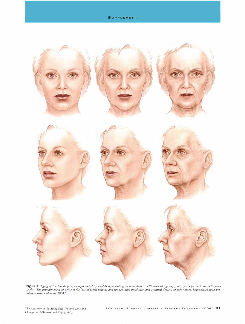
Figure 2. Aging of the female face, as represented by models representing an individual at ~20 years of age (left), ~50 years (center), and ~75 years(right). The primary event of aging is the loss of facial volume and the resulting involution and eventual descent of soft tissues. Reproduced with per-mission from Coleman, 2004.6
S4-S9_YMAJ_Supp_Col286_CPRR.qxd 2/7/06 7:33 AM Page 7
S8 A e s t h e t i c S u r g e r y J o u r n a l ~ J a n u a r y / F e b r u a r y 2 0 0 6 Volume 26, Number 1, Supplement
S U P P L E M E N T
in the glabella, nasion, and upper dorsum (cephalic nose).Most of the changes that occur with aging in the nosetake place in the glabella, nasion, and medial eyelid.Flattening of the medial forehead results in blunting ofthe nasofrontal angle, giving the illusion of increasednasal length. The attachments between the upper andlower lateral nasal cartilages weaken, causing progressiveptosis of the nasal tip. Pyriform remodeling affects thealar base and, in combination with upper maxillaryresorption, results in a narrowing of the nasolabial angleand further accentuation of nasal tip ptosis. Chin padptosis, which occurs secondary to mandibular boneresorption, further contributes to the appearance ofincreased nasal projection and length.
Lower third (chin, jawline, and neck)
With progressively increasing skin laxity, depletion ofmalar and perioral fat deposits, and resorption of alveo-lar bone, a relative excess of skin occurs in the aginglower face, leading to loss of definition of the jawline.Loss of masseteric ligament support allows descent offacial fat to the mandibular border, leading to the forma-tion of facial jowls.14 Upward retreat of the mandibularborder results in exposure of the submental contents,including the submaxillary gland. As the lateral projec-tion of mandibular fullness dissipates, the angle of themandible appears to merge from the buccal region intothe neck. In the chin, loss of lateral and inferior volumeresults in relative protrusion of the central chin, whereaslateral mental atrophy results in ptosis of the lateral chin,which can create the impression of chin widening whenviewed from the front.6
As the subcutaneous fullness of the mandible recedes,the fat of the jowl, which was previously concealed by the surrounding soft tissues, is revealed. Ptosis of the unsupported skin, chin pad, and facial portion of the platysma muscle, coupled with the downward pull of the platysma muscle, leads to the development ofthe characteristic jowled “turkey neck” deformity.Protrusion of the large submental fat pad either betweenthe two free borders of the platysma muscle or frombehind the complete submental platysma cover furtheradds to this effect. In addition, contraction of the platys-ma muscle, caused in part by the need to support thedeeper neck and floor of mouth structures, gives rise tovertical fibrous bands on the neck, whereas laxity in theoverlying skin can create horizontal rhytides. As aging progresses, the hyoid bone and larynx gradual-ly descend, resulting in loss or blunting of the cervico-mental angle.
Structural Rejuvenation of the Aging Face: TheNeed For Facial Rebalancing
Loss of facial fullness and the ensuing facial fat redis-tribution provides the visual clue to an individual’s age(Figure 2).6 Accordingly, the primary goal in any rejuve-nation procedure should be to restore the ample, bal-anced distribution of facial fullness that exemplifies theyouthful face.4 The fat compartmentalization of theaging face must be smoothed over, and the former pri-mary arcs and convexities of youth, rebuilt.
Most conventional face lift procedures incorporate lift-ing and tightening techniques to defy the facial soft-tissuedescent that results from atrophy and loss of skin elastici-ty. These procedures fail to address the issue of facialshape because they are incapable of reversing the facialsoft-tissue atrophy that occurs with aging. Rather, byexcising skin in areas most prone to atrophic sagging,they in effect “tailor” the skin to fit the shrunken frame-work. Present trends in facial rejuvenation are movingaway from conventional excision and suspension proce-dures and toward the use of conservative skin excision,deep fascial–SMAS manipulation, volume restoration,and modification of facial animation (via chemodenerva-tion). Dermal and subcutaneous fillers, used either aloneor as an adjunct to surgical and nonsurgical facial rejuve-nation techniques, are a logical and effective treatmentchoice for “lifting and filling” the facial soft tissues. Thefacial soft tissue remodelling that might be expected tooccur in response to the implant of volume replacerscould theoretically result in undesirable cosmetic changesand loss of effect over time. However, to date there is noclinical evidence to suggest that tissue remodelling has asignificant impact on the efficacy of volume-enhancementtechniques currently used in clinical practice. For thosesurgeons who do not wish to use fat transplants, newfiller materials for volume augmentation offer theprospect of possibly more predictable (albeit temporary)aesthetic outcomes.15 Moreover, volume augmentation ofareas of loss of facial fullness can be complemented bymicroliposuction of the hypertrophic fat pockets.16 In thisway, the primary goal in facial rejuvenation—restorationof a youthful 3-D facial topography—may be realized. ■
References1. Bartlett SP, Grossman R, Whitaker LA. Age-related changes of the
craniofacial skeleton: an anthropomorphic and histologic analysis.Plast Reconstr Surg 1992;90:592-00.
2. Pessa JE, Zadoo VP, Yuan C, Ayedelotte JD, Cuellar FJ, Cochran CS,et al. Concertina effect and facial aging: nonlinear aspects of youth-fulness and skeletal remodeling and why perhaps infants have jowls.Plast Reconstr Surg 1999;103:635-644.
S4-S9_YMAJ_Supp_Col286_CPRR.qxd 2/7/06 7:33 AM Page 8
A E S T H E T I C S U R G E R Y J O U R N A L ~ J a n u a r y / F e b r u a r y 2 0 0 6 S9The Anatomy of the Aging Face: Volume Loss andChanges in 3-Dimensional Topography
S U P P L E M E N T
3. Hamra ST. The role of orbital fat preservation in facial aestheticsurgery. Clin Plast Surg 1996;23:17-28
4. Donofrio LM. Fat distribution: a morphologic study of the aging face.Dermatol Surg 2000;26:1107-1112.
5. Little JW. Volumetric perceptions in midfacial aging with altered pri-orities for rejuvenation. Plast Reconstr Surg 2000;105:252-266.
6. Coleman SR. Structural Fat Grafting. St. Louis, MO: Quality MedicalPublishing; 2004.
7. Gosain AK, Klein MH, Sudhakar PV, Prost RW. A volumetric analysisof soft-tissue changes in the aging midface using high-resolutionMRI: implications for facial rejuvenation. Plast Reconstr Surg2005;115:1143-1152.
8. Owsley JQ. Elevation of the malar fat pad superficial to the orbicularisoculi muscle for correction of prominent nasolabial folds. Clin PlasticSurg 1995;22:279-293.
9. Zimbler MS, Kokoska MS, Thomas JR. Anatomy and pathophysiologyof facial aging. Facial Plast Surg North Am 2001;9:179-187.
10. Sherris DA, Larrabee WF. Anatomic considerations in rhytidectomy.Facial Plast Surg 1996;12:215-222.
11. Ellenbogen R, Youn A, Yamini D, Svehlak S. The volumetric face lift.Aesth Surg J 2004;24:514-522.
12. Lowe NJ, Wieder JM, Shorr N, Boxrud C, Saucer D, Chalet M.Infraorbital pigmented skin. Preliminary observations of laser therapy.Dermatol Surg 1995;21:767-770.
13. Guyuron B. The aging nose. Dermatol Clin 1997;15:659-664.
14. Özdemir R, Kilinc H, Ünlü E, Ca_ri Uysal A, Sensöz O, Nazmi Baran C.Anatomicohistologic study of the retaining ligaments of the face anduse in face lift: retaining ligament correction and SMAS placation.Plast Reconstr Surg 2002;110:1134-1147.
15. Rohrich RJ, Rios JL, Fagien S. Role of new fillers in facial rejuvena-tion: a cautious outlook. Plast Reconstr Surg 2003;112;1899-1902.
16. Coleman SR. Facial recontouring with lipostructure. Clin Plast Surg1997;24:347-367.
This supplement is funded through sponsorship by Q-Med.
Sydney Coleman receives royalties from Byron Medical andis an unpaid consultant for Medicis; he has no financialinterest in Q-Med.
Reprint requests: Sydney Coleman, MD, 44 Hudson Street, New York, NY10013.
Copyright © 2006 by The American Society for Aesthetic Plastic Surgery,Inc.
1090-820X/$32.00
doi:10.1016/j.asj.2005.09.012
S U P P L E M E N T
S4-S9_YMAJ_Supp_Col286_CPRR.qxd 2/7/06 7:33 AM Page 9