Embed Size (px)
Citation preview
Retrospective Theses and Dissertations Iowa State University Capstones, Theses andDissertations
1-1-1963
The age and manner of closure of various epiphysesand other centers of ossification in the front limb ofthe domestic horse (Equus caballus).Victor Shank MyersIowa State University
Follow this and additional works at: https://lib.dr.iastate.edu/rtd
This Thesis is brought to you for free and open access by the Iowa State University Capstones, Theses and Dissertations at Iowa State University DigitalRepository. It has been accepted for inclusion in Retrospective Theses and Dissertations by an authorized administrator of Iowa State University DigitalRepository. For more information, please contact [email protected].
Recommended CitationMyers, Victor Shank, "The age and manner of closure of various epiphyses and other centers of ossification in the front limb of thedomestic horse (Equus caballus)." (1963). Retrospective Theses and Dissertations. 18519.https://lib.dr.iastate.edu/rtd/18519
{, c I
THE AGE AND MANNER OF CLOSURE OF VARIOUS
EPIPHYSES AND OTHER CENTERS OF OSSIFICATION IN THE FRONT
LIMB OF THE DOMESTIC HORSE (EQUUS CABALLUS )
s-r-7~ · m??9a- by
(! • ? Victor Shank Myers, Jr.
A Thesi~ Submitted to the
Graduate Faculty in Partial Fulfillment of
The Requireme nts for the Degree of
MASTER OF SCIENCE
Major Subject: Veterinary Medicine and Surgery
Signatures have been redacted for privacy
Iowa State Unive r s ity Of Scie nce and Te chnolog y
Ames, Iowa 19 63
1192195
ii
TABLE OF CONTENTS
INTRODUCTION
REVIEW OF THE LITERATURE
METHODS AND MATERIALS
FINDINGS
Third Phalanx Distal Sesamoid Second Phalanx First Phalanx Proximal Sesamoids Third Metacarpal Bone Second and Fourth Metacarpal Carpal Bones Radius and Ulna Radius Ulna Humerus Humerus Scapula
DISCUSSION
SUMMARY AND CONCLUSIONS
ACKNOWLEDGMENTS
LITERATURE CITED
•
Bones
Page
1
3
15
27
27 27 27 32 38 38 39 39 40 49 50 55 56 59
60
66
69
70
1
INTRODUCTION
Abnormalities of the locomotor system are probably the
most important single group of disorders to affect the equine
species. Lameness is often the difference between a valuable
and a worthless horse. Veterinarians are constantly being
asked to treat lame horses. The first and often the most
important step in treating lameness is the establishment of an
accurate diagnosis. It is in this field of diagnosis of lame-
ness that radiology has become so useful.
The development and improvement of radiology and the
diagnostic radiograph have been followed by an increased de-
mand for radiographic service. Portable x-ray machines have
placed this importa nt diagnostic tool in the hands of many
veterinarians. The increased demand and the increased avail-
ability of the equipment have resulted in a previously infre-
quent procedure becoming almost routine.
After satisfactory radiographs are taken the ir value is
entirely dependent upon the ability of the practitioner to
properly interpret the information on the film. He must be
able to differentiate between normal epiphyseal lines and
fractures. He should know the normal ages for epiphyseal
closure in order to recognize incomplete or late epiphyseal
closures.
It becomes apparent that knowledge of the normal growth
and development of bone not only aids in the diagnosis of
2
lamenesses, but provides information to help guide the nutri-
tion, training and use of these horses. If it is possible to
make a correlation between the age of the animal and the time
of epiphyseal closure, it should be possible to more logically
control the use and training of horses.
I t is evident, then, that the full potential of a good
radiographic examination is not used unless the examiner has a
knowledge of the developmental changes expected between birth .
and maturity.
Since no English work on radiographic epiphyseal closure
times is available, it was concluded that a study such as this
would be beneficial to both the equine practitioner and t he re-
search worker interested in bone growth and development . With
the possible exception of a recent Russian study, all of the
radiographic studies of epiphyseal closure in the equine
species were made using radiographs of different horses of
different ages and did not follow one horse through its post-
natal development. These workers, thus, did not take full ad-
vantage of the radiographic method of studying bone develop-
ment .
It was therefore proposed that two animals, as closely
r e lated as possible, would be k e pt unde r carefully controlled
conditions to provide a uniform series of radiographs. These
findings should provide a b a sis for diagnostic use as well as
for subse quent research.
3
REVIEW OF THE LITERATURE
Although no attempt was made to conduct a complete re-
view of the literature on epiphyseal closure times in all
animals, the literature concerning some animals and some of
the more recent human literature was reviewed.
In man, Pyle was found to be co-author of several atlases
on bone development (40,20,27,39). Many recent studies of
epiphyseal closure in the dog were found (24,25,54,55,23,49,
47,2,36,6). The monkey was studied by several workers in-
cluding (63,65,34). Dawson (10,11) did the early work on the
rat skeleton, while Walker and Wirtschafter (64) wrote a
laboratory atlas on the development of the rat skeleton.
Other animals were studied in relationship to their epi-
physeal closure times (30,67,7,8,53,29,13).
Pryor {37,38) wrote that the bones of the human female
developed more rapidly than those of the human male. As the
children became older these differences became more marked.
Stevenson (57) and Todd and Todd (60) did not confirm these
results, but since then these findings were supported in man
(33,17,18,26,19,20). They were also supported by people work-
ing on other species (34,63,56). In the dog Hare (25) found
no sex influence, but he did find that there were differences
between breeds. Raker,l Mackay-Smith,2 and Buchanan3 stated 1Raker, c. W. Philadelphia, Pa. Breed differences in
maturity. Private communication. 1963. 2Mackay-Smith, M. P. Philadelphia, Pa. Breed dif-
ferences in maturity. Private communication. 1963. 3Bucharian, J. H. Ames, Iowa. Breed differences in
maturity. Private communication. 1963.
4
that the Arabian breed of horses matured relatively slowly
when compared to other equine breeds. Raker also believed
that the Standardbred horse matured more slowly than the
Thoroughbred.
The following discussion of the formation and growth of
bones was taken from these textbooks (22 ,62,5,35). Two types
of bone were described. Intramembranous bones formed in
fibrous or membranous areas did not have cartilage anlages .
Endochondral bones were formed in are as where cartilage
anlages had already been formed. It must be kept in mind,
however, that all bone laid down by the periosteum and endo-
steum was actually formed by intramembranous ossification.
Bone growth at epiphyseal lines was described as being endo-
chondral in type.
All the bones of the forelimb of the horse were found to
be of the endochondral type. A cartilaginous model of the
limb bones developed early in the gestation period from
embryonal mesenchymal cells. A mass of mesenchymal cells
formed in the area and these cells became differentiated into
chondroblasts. The chondroblasts then began to lay down
cartilage. Surrounding this cartilage, a perichondrium be-
came differentiated from the mesenchymal cells in that area.
The work of Ewart (15,16), Rosenberg (43), and Saarni
(46) showed that cartilaginous anlages of the limb bones of
the horse appeared during the first month of fetal life.
5
These cartilaginous anlages grew in both length and width by
interstitial growth of the chondrocytes. Interstitial growth
was described as growth from within by division and enlarge-
ment of the individual cartilage cells. Cartilage was also
described as having appositional growth or the addition of
new cartilage layers on the outside of the models. The inner
layer of the perichondrium formed this new cartilage on the
outside of the old.
The first center of ossification in the equine forelimb
appeared during the second month of fetal life (Schmidt, 48;
Saarni, 46; and Ewart, 15). The process began with the swel-
ling and maturation of the cartilage cells in the area. The
matrix around these cells gradually became thinner and
calcified. After these chondrocytes became surrounded by a
calcified matrix, they died. Following the death of these
cells there was erosion and dissolution of the calcified
matrix and the dead cells, leaving hollow spaces in the area.
During this same period there was an increase in the blood
supply to the perichondrium. This increased vascularity in
some way affected the inner layer of the perichondrium, so
that osteoblasts were formed instead of chondroblasts. This
influence of cell environment on osteogenesis was emphasized
by Bassett (3). According to Ham and Leeson (22), a center
of ossification was formed when osteoblasts and capillaries
had completed their invasion into the area of dead cartilage
6
cells . Patten (35) emphasized that cartilage was not con-
verted to bone, but rather cartilage was destroyed and bone
then formed in the same area. There was no transition of
cartilage to bone, but rather a replacement of destroyed
cartilage with bone.
It was found that all the diaphyseal centers of ossifica-
tion were present in t he equine forelimb before any of the
secondary centers of ossification appeared. Diaphyseal
ossification began with the scapula and seemed to proceed in
an orderly manner down the leg, except that the ossification
center of the third phalanx appeared before that of the second
phalanx. Schmidt (48) reported that the diaphyseal center
of ossification of the scapula appeared toward the end of the
second month of fetal life. Saarni (46) reported that all
the diaphyseal centers were present in an equine fetus about
130 days after conception. Other works consulted were
Kupfer (31), Ewart (15), and Tohara (61). Stoss (58) gave
age ranges for equine fetuses of varying crown rump lengths .
His work was consulted when crown rump lengths were given
without ages .
Although bone did not grow by interstitial growth, due to
its solid matrix, the osteogenic cells in the cambium layer
of the periosteum did cause the diaphyses to grow in width by
appositional growth . Appositional growth occurred when new
bone was laid down on the outside of bone already present.
7
The shafts of the long bones in the young were continual-
ly undergoing changes according to Enlow (14). The endosteum
and periosteum continually laid down intramernbranous bone and
resorbed bone from the diaphyseal shaft during early life.
Kupfer (31) stated that the first secondary center of
ossification to appear in the equine fetus was the coracoid
process of the scapula. According to Bloom and Bloom (4, pp.
508-509), the secondary or epiphyseal centers of ossification
ossified in a different manner from the diaphyseal centers.
Here the large mass of uncalcified cartilage is invaded centripetally by several thick cords of vascular mesenchyme. When the mesenchyme reaches the center, calcification starts in the surrounding cartilage matrix. Then erosion of this calcified cartilage by vascular mesenchyme begins and typical intracartilaginous ossification proceeds centri-fugally, with the result that a progressively larger cavity filled with bone marrow and small amounts of spongy bone develops in the secondary center of ossification. This process continues until only a thin rim of cartilage is left at the articular surface and along the epiphyseal line. The latter is removed when the epiphyseal and diaphyseal cavities fuse when growth of the bone ceases.
Kilpfer (31) listed the order of appearance of the
secondary centers of ossification of the equine forelimb as
follows: coracoid process, distal radial epiphysis, distal
epiphysis of the third metacarpal bone, distal epiphysis of
the humerus, distal epiphysis of the first phalanx, proximal
epiphysis of the first phalanx, proximal epiphysis of the
radius, proximal epiphysis of the humerus, epiphysis of the
major tuberosity of the humerus, epiphysis of the medial
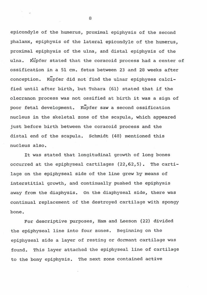
8
epicondyle of the humerus, proximal epiphysis of the second
phalanx, epiphysis of the lateral epicondyle of the humerus,
proximal epiphysis of the ulna, and distal epiphysis of the
ulna. Kupfer stated that the coracoid process had a center of
ossification in a 51 cm. fetus between 23 and 28 weeks after
conception. Kupfer did not find the ulnar epiphyses calci-
fied until after birth, but Tohara (61) stated t hat if the
olecranon process was not ossified at birth it was a sign of
poor fetal development. Kupfer saw a second ossification
nucleus in the skeletal zone of the scapula, which appeared
just before birth between the coracoid process and the
distal end of the scapula. Schmidt (48) mentioned this
nucleus also.
It was stated that longitudinal growth of long bones
occurred at the epiphyseal cartilages (22,62,5). The carti-
lage on the epiphyseal side of the line grew by means of
interstitial growth, and continually pushed the epiphysis
away from the diaphysis. on the diaphyseal side, there was
continual replacement of the destroyed cartilage with spongy
bone.
For descriptive purposes, Ham and Leeson (22) divided
the epiphyseal line into four zones. Beginning on the
epiphyseal side a layer of resting or dormant cartilage was
found . This layer attached the epiphyseal line of cartilage
to the bony epiphysis. The next zone contained active
9
growing chondrocytes, and mitotic figures were often seen in
this zone. These cells were arranged in columns stacked
longitudinally. The third zone was made up of more mature
cartilage cells still in the same longitudinal columns.
These were the cells from zone two after the epiphysis had
been pushed further away from them by means of interstitial
growth of the cartilage. The cells in zone three closest to
the diaphysis were the most mature. The fourth and last zone
was made up mostly of dead chondrocytes surrounded by a
calcified matrix. This layer of cells connected the epi-
physeal plate to the diaphysis. These were not four complete-
ly isolated layers, as they all ran together and one could
not determine exactly where one zone ended and the other
began. Under the fourth zone of the epiphyseal line
osteogenesis was taking place at the same speed as inter-
stitial growth took place in zones two and three. This
meant that in the young animal in which the long bones were
growing in length, two processes were always going on at the
same rate. One was the interstitial growth of cartilage in
zones two and three. The other was the calcification of the
cartilage matrix, the death of the chondrocytes, and the re-
placement of this dead cartilage by spongy bone. The part
of the diaphysis adjoining zone four of the epiphyseal
cartilage was called the metaphysis and it was made up of
newly formed spongy bone . Submetaphyseal bone was continually
10
undergoing resorption, replacement, and remodeling (Ingalls,
28) . The epiphysis grew only by appositional growth which
took place on all sides of the epiphysis, except along the
epiphyseal cartilage side, according to Siegling (SO) .
Growth of the long bones stopped when the cartilage cells
in zone two ceased to divide. When this happened the epi -
physeal line ossified from the diaphyseal side and no further
growth in length occurred at that particular epiphyseal line .
As soon as any part of the cartilage zone was ossified, the
bone could no longer grow in length at that epiphyseal line .
In reviewing the English literature on radiographic
epiphyseal closure times in the equine, no original work was
found . Adams (1) in his recent book on equine lamenesses
did not mention epiphyseal closure times . Several men
prominen~ in veterinary anatomy, veterinary radiology, and
equine medicine and surgery were consulted (Raker,l Manning,2
Riley,3 Carlson,4 Hare, 5 Smith,6 Mackay-Smith, 7 Banks,8
!Raker, c . w. Philadelphia, Pa . Epiphyseal closure times. Private communication. 1963.
2Manning, J. P . Urbana, Ill. Epiphyseal closure times. Private communication . 1963.
3Riley, w. F. East Lansing, Mich. Epiphyseal closure times. Private communication. 1963.
4carlson, w. D. Fort Collins, Colo. Epiphyseal closure times . Private communication . 1963.
Saare, w. c . D. Philadelphia, Pa . Epiphyseal closure times . Private communication . 1963.
6smith, R . N. Bristol, England. Epiphyseal closure times . Private communication. 1963.
?Mackay-Smith, M. P. Philadelphia, Pa. Epiphyseal closure times. Private communication. 1963.
8Banks, w. c. College Station, Texas . Epiphyseal clo-sure times. Private communication . 1963.
11
Worthman,l Morgan,2 Wheat,3) and none of these men knew of
any English work on the subject. Since the aforementioned
correspondences, Equine Medicine and Surgery (42) was pub-
lished. Rooney (42) in this book quoted the chart listed in
the German embryology book by Zietschmann and Krolling (66),
on ages of epiphyseal closure in the horse. Actually,
Zietschmann and Kr~lling derived their data from Lesbre
(32), who published his works in 1897. Thus the most recent
English referenc e on epiphyseal closure in the horse dated
back to the end of the 19th century. Sisson and Grossman
(52) gave closure times for some of the epiphyseal lines in
the horse, but these were the same ones that Sisson (51) gave
in 1910.
Lesbre (32) wrote on the sequence of skeletal ossifica-
tion in domestic mammals. The horse was included in this
study. For a sununary of Lesbre's work, see Table 1 following
the Discussion Section. Lesbre stated that he saw a definite
distal epiphyseal line for the second phalanx after birth.
This was an anatomical study of epiphyseal closure times .
In 1950 Tohara (61) published a radiographic study on
equine bone development. In the English summary, Tohara
lworthman, R. P. Pullman, Wash. Epiphyseal closure times. Private communication. 1963.
2Morgan, J. P. Fort Collins, Colo. Epiphseal closure times. Private conununication. 1963.
3Wheat, J. D. Davis, Calif. Epiphyseal closure times . Private conununication. 1963.
12
stated that the proximal end of the second phalanx fused in
the 7th to 8th months. The distal end of the first phalanx
was fused at birth, while the proximal epiphyseal line closed
in the 9th to 10th months after birth. The distal end of the
third metacarpal bone fused to the diaphysis in the 17th to
18th months. Tohara found no evidence of a proximal epi-
phy sis of the third phalanx, a distal epiphysis of the second
phalanx, or a proximal epiphyses of the metacarpal bones dur-
ing the gestation period. These centers were described by
Lesbre (32), Retterer (41), and Ewart (15), but were not seen
by Kupfer (31) in the horse. According to Ellenberger and
Baum (12), Kupfer (31) saw signs of a beginning proximal
epiphysis of the third metacarpal bone in a 74 cm. esel (ass)
fetus about fifty weeks after conception. Kupfer (31) did
not say he saw a definite ununited epiphysis, but only that
signs of the epiphysis were present in one esel fetus.
Struthers (59) did not find an epiphysis for either the
distal end of the second phalanx or the proximal end of the
third, but he did think that they were probably present in
younger fetuses. The age of the fetus Struthers described
was said to be one month prior to delivery. Saarni (46) said
the proximal epiphyses of the metacarpal bones were not
ossified in an 89 cm. fetus, while they were already fused to
the diaphysis in a 95 cm. fetus. He did not actually see an
ununited epiphysis, however, Saarni (46) also described a
13
distal epiphysis of the second phalanx.
In 1960 Schmidt (48 ) wrote on radiographic epiphyseal
closure times in the equine limbs. According to Schmidt,
the epiphyseal closure time of the proximal ends of the first
and second phalanges and the distal end of the third meta-
carpal bone was six months of age. The other centers below
the carpus were closed before birth, Schmidt said. The
distal radial and ulnar epiphyses fused together before four
months and these two then fused to the radial diaphysis by
three years of age, Schmidt stated. The exact time of
closure for the proximal epiphysis of the radius was not
given, but it was joined in a y ear old horse. The exact
closure times for the olecranon, proximal epiphysis of the
humerus, the major tuberosity of the humerus, and the tuber
scapulae were not de termined. The tuber scapulae was united
to the scapula by 18 months of age. The distal epiphysis of
the humerus and the epiphysis of the medial epicondyle were
fused in a year old horse, Schmidt stated.
Rozhdestvenskaya (45,44) published two papers in 1960 on
the bone formation of foals during the first year of life.
A German abstract of one of these articles gave no epiphyseal
closure times, however.
Rooney (42), Zietschmann and Krolling (66), Lesbre (32),
Sisson and Grossman (52), Kupfe r (31), Tohara (61), and
Schmidt (48) all gave epiphyseal closure times for some of
14
the bones of the front limbs of the horse. None of these
refere nces, however, followed a single animal from birth to
maturity as was done in this study.
15
METHODS AND MATERIALS
Two Arabian mares were chosen in January of 1960 to be-
gin this study of bone development in the horse . They were
very closely related and were bred to the same stallion.
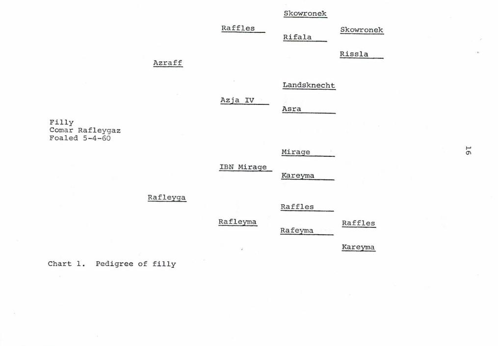
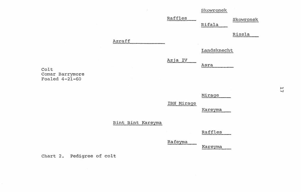
(See Charts 1 and 2 for pedigrees of the two experimental
horses) • Radiographs of both the mares and the stallion
showed no signs of hereditary bone defects. These horses
showed good bone development.
The mares were put in an isolation barn at Iowa State
University approximately six weeks before they were due to
foal . They were observed every 15 minutes, beginning two
weeks before they were due to f oal . This observation con-
tinued until both mares had foaled without complications .
Neither of the mares had retained fetal membranes . A motion
picture was taken of the colt's birth on April 21, 1960. The
filly was born on May 4, 1960 . No signs of infection were
seen in either the mares or the foals. Both mares delivered
healthy foals the next year.
The foals were kept in isolation after birth and were
in contact with no horses except their dams. They were
not ridden, but did receive controlled exercise in a
special exercise yard under the supervision of their at-
tendants. Both received yearly injections of equine en-
c ephal omyeli tis vaccine and tetanus toxoid. They were
treated for internal parasites eight time during the
first three years of life. It was thought that their
Skowronek
Raffles Skowronek Rifala
Riss la Azraf f
Landsknecht
Azja IV Asra
Filly Coma r Rafleygaz Foaled 5-4 - 60
t-' Mirage °' IBN Mirag:e
Kareyrna
Raf leysa Raffles
Raf leyrna Raffles Raf eyrna
Kareyrna
Chart 1. Pedigree of filly
Colt Comar Barrymore Foaled 4-21-60
Azraf f
Bint Bint Kareyma
Chart 2. Pedigree of colt
Raffles
Azja IV
IBN Mirage
Rafeyma
Skowronek
Skowronek Rifala
Riss la
Landsknecht
Asra
Mirage
Kareyma
Raffles
Kareyma
18
dams inocculated them with intestinal parasites.
As soon as the foals began to eat they received a 13.1
per cent digestable protein grain mixture to which was added
a vitamin mixture (Clovite1 ) at the rate of one tablespoon
per gallon of feed. A trace mineralized salt was also pro-
vided for the foals, and good alfalfa hay was available free
choice . Both foals were weaned on November 4, 1960, at
which time their grain ration was improved to a 13.2 per
cent digestable protein mixture. The alfalfa hay as well as
the vitamin and trace mineral supplements were continued . I n
the summer of 1962 the feed was reduced for both animals when
it was thought that they were overweight . Both animals were
in good flesh on their respective third birthdays .
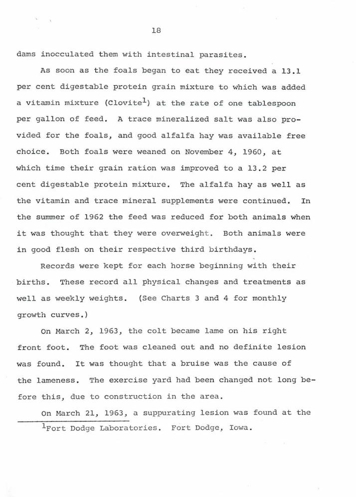
Records were kept for each horse beginning with their
births . These record all physical changes and treatments as
well as weekly weights .
growth curves.)
(See Charts 3 and 4 for monthly
on March 2, 1963, the colt became lame on his right
front foot . The foot was cleaned out and no definite lesion
was found . It was thought that a bruise was the cause of
the lameness . The exercise yard had been changed not long be-
fore this, due to construction in the area .
On March 21, 1963, a suppurating lesion was found at the
lFort Dodge Labora t ories . Fort Dodge, Iowa.
(/) a z :'.) 0 a.. 0 0 0 I
0
10001
900~ I
aool ,J 600
500
400
300
200
BI RTH 81 lbs
2
1 9
4 6
Chart 3, Gr~wth chart of f illy
GROWTH CHART OF FILLY
8 10 12 14 16 18 20 22 24 B IRTH T O 3 6 MONTHS OF AGE
26 2 8 30 32 34
3 YEARS 773 lbs.
36
i ' I l<
f 1000
900
800
700-
(/) 6 00 a z :::> 0 Cl..
0 0 0
5 00
6 400
300
200
20
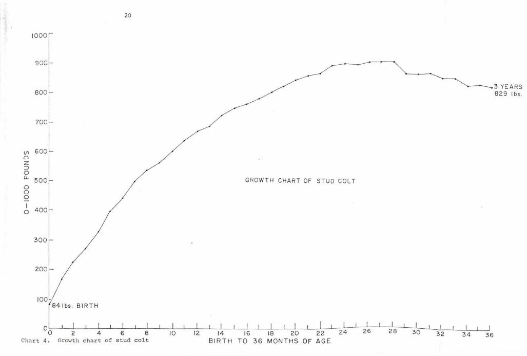
84 1bs . BIRTH
00 Char t 4.
2 4 6 8 Growth c hart of s t ud c o lt
GRO WT H CHART OF STUD COL T
10 12 14 16 18 20 22 24 26 BIRTH TO 36 MONTHS OF AGE
2 8 30 32
---.----3 YEARS 829 l bs.
34 36
21
posterior aspect of the medial collateral sulcus of the right
front foot. The frog was removed around this area and drain-
age was established. On April 21, 1963, the colt was almost
sound after he was shod with shoes and a leather pad. Some
loss of weight occurred during this lame period.
A Picker Meteor X-Ray Machine1 style FlO was used to
take the radiographs. The settings varied as the animals
grew and their bones became more dense. Par speed cassettes
were used in most cases. Lightning speed screens were used
to take the shoulder radiographs during the third year.
Kodak Medical X-Ray Film2 Blue Brand was used for all the
radiographs except one series, when Kodak Royal Blue Film2
was used. The time t emperature method of development was
used. The films were fixed for at least one half hour and
were then washed for at least one hour before being dried.
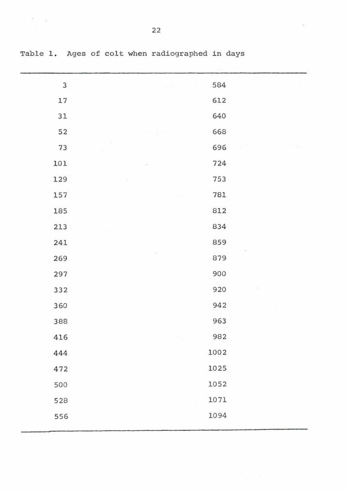
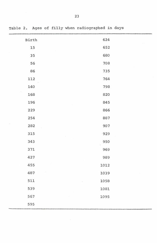
Three hundred and fifty-three radiographs were taken of
the front legs of the colt on 44 different occasions between
three days of age and three years of age. In the filly
radiographs were taken 43 times beginning with the day of
birth and ending on May 4, 1963, when she was three years
old. During this time a total of 342 radiographs were taken
of her front legs (Tables 1 and 2) •
1 Picker X-Ray Corporation. White Plains, New York. 2Eastman Kodak Company. Rochester, New York.
22
Table 1 . Ages of colt when radiographed in days
3 584
17 612
31 640
52 668
73 696
101 724
129 753
157 781
185 8 12
213 834
241 859
269 879
297 900
332 920
360 942
388 963
416 982
444 1002
472 1025
500 1052
5 28 1071
556 1094
23
Table 2. Ages of filly when radiographed in days
Birth 624
15 652
35 680
56 708
86 735
112 764
140 798
168 820
196 845
229 866
254 887
282 907
315 929
343 950
371 969
427 989
455 1012
487 1039
511 1058
539 1081
567 1095
595
24
The nomenclature used to describe the di fferent radio-
graphic views taken was the same as that described by Emmer-
son1 and Habel (21).
Anteroposterior (A.P.) radiographs of the front feet
were taken on a wooden block two inches by four inches by ten
inches . The foot was raised two inches from the ground sur -
face by the block . Lateromedial (L.M.) radiographs of the
front feet on the same block were taken. In the antero-
posterior view the cassette was behind the foot and as close
to it as possi ble, while in the lateromedial v i ew the cassette
was medial to the foot and as close to it as possible. Both
of these views included the distal third of the metacarpal
bones and the three phalanges. The anteroposterior and
l ateromedial radiographs of the metacarpal bones usually
i ncluded a part of or the whole of the carpus . Both latero-
medial and anteroposterior radiographs of the carpus were
taken i f they were not included on other radiographs . Both
lateromedial and anteroposterior radiographs of the radius
a n d ulna were t aken. These usually included a varying amount
of the carpus . In all of the above cases the horses were
standing with all four feet squarely under them . The pro-
cedures used for taking these radiographs were described by
Carlson (9) •
lEmmerson, M. A. Private communication .
Ames, Iowa . 1962 .
Radiographic nomenclature .
25
Mediolateral (M.L.) radiographs of the elbow joints were
taken. The procedure used called for one assistant to pull
the leg forward and upward, while a second assistant held the
cassette in place on the lateral side of the elbow joint. The
X-ray beam was then directed at the cassette from a point
just in front of the animal and on the opposite side. A
rnedio±ateral view of the shoulder joint was taken in much
the same manner. The cassette was centered over the shoulder
joint and the leg was extended forward and upward so as to
project the air filled trachea over the shoulder joint.
Radiographs were not taken of both front legs on any
one occasion. First the left leg was radiographed and then
on the next occasion the right leg was radiographed. It was
assumed that both legs ossified symmetrically as Tohara (61)
stated.
Radiographs of the shoulder were not taken between July
3, 1960, and November 26, 1961, on the colt and between June
29, 1960, and December 21, 1961, on the filly. The elbow
joints were not radiographed for the first two months after
birth. Part of the elbow joints were included on other
radiographs taken during this period. Supplementary radio-
graphs of the elbow joint were taken on two clinic cases at
birth, also.
In summary it should be said that these colts were raised
as ideally as was possible. They were fed a good ration at
26
all times and were kept in isolation to prevent infectious
diseases from interfering with their growth.
The drawings used in this work to represent epiphyseal
closures are not tracings. They were used to help illustrate
the age and manner of closure. Due to the angle, the radio-
graphs often showed more than one dark line in the epi-
physeal line area. In the drawings the dark lines were
represented as one line, which in fact they actually were .
The drawings are true to scale, but they have been reduced
in size.
The criteria used for evaluating complete epiphyseal
fusion was the same as that used by Smith (53) . Union was
considered complete as soon as the dark line of epiphyseal
cartilage had disappeared. A white line representing a zone
of increased density was present after the disappearance of
the dark line. This was called an epiphyseal scar by Smith .
27
FINDINGS
Third Phalanx
No ~adiographic signs of an epiphysis were seen in the
third phalanx of either foal. (Figures 1 and 2) .
Distal Sesamoid
This bone was ossified on the first radiographs taken of
both foals and seemed to originate from a single center of
ossification. (Figures 1 a n d 2).
Second Phalanx
Distal end
No radiographic signs of a distal epiphysis of the
second phalanx were seen in either the colt or the filly .
(Figures 1 and 2) •
Proximal end
Colt When the first radiogr a phs were taken at three
days of age the epiphyseal line between the proximal epiphysis
of the second phalanx and the diaphysis was a solid smooth
dark line . The epiphyseal line ran tra nsversely through the
long axis of the bone on both the a nteroposterior and the
lateromedial views. The first signs of ossification of this
epiphyseal line were seen when the colt was 129 days old . The
epiphyseal line had begun to disappear in the center of the
bone. This epiphyseal line fused in the center first and
then proceeded to ossify outwa rdly . At 185 days of age the
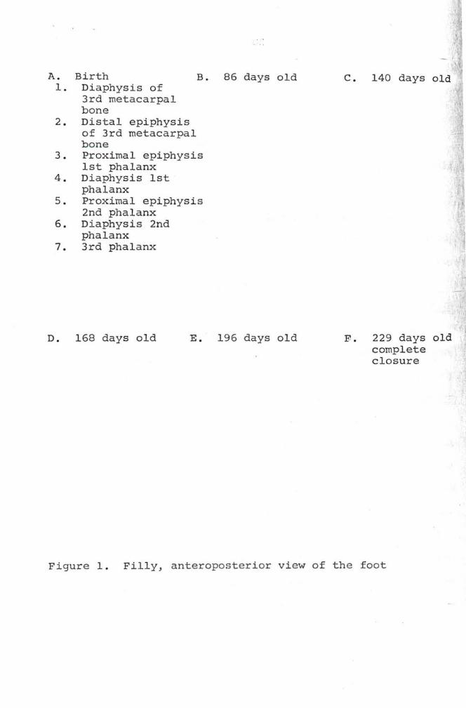
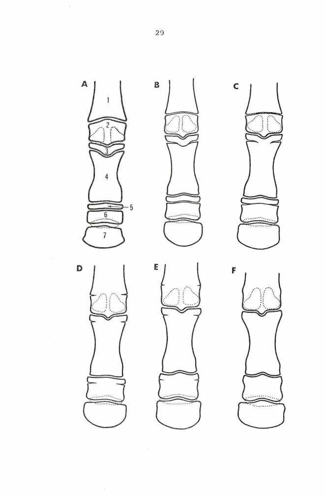
A. Birth B. 86 days old 1. Diaphysis of
3rd metacarpal bone
2. Distal epiphysis of 3rd metacarpal bone
3. Proximal epiphysis 1st phalanx
4 . Diaphysis 1st phalanx
5 . Proximal epiphysis 2nd phalanx
6. Diaphysis 2nd phalanx
7 . 3rd phalanx
D. 168 days old E . 196 days old
c . 140 days old
F. 229 days old complete closu re
Figure 1 . Filly, anteroposterior view of the fo ot
29
F
\I ~ \______)
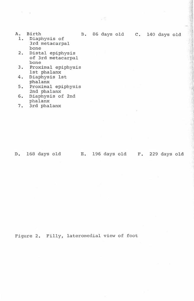
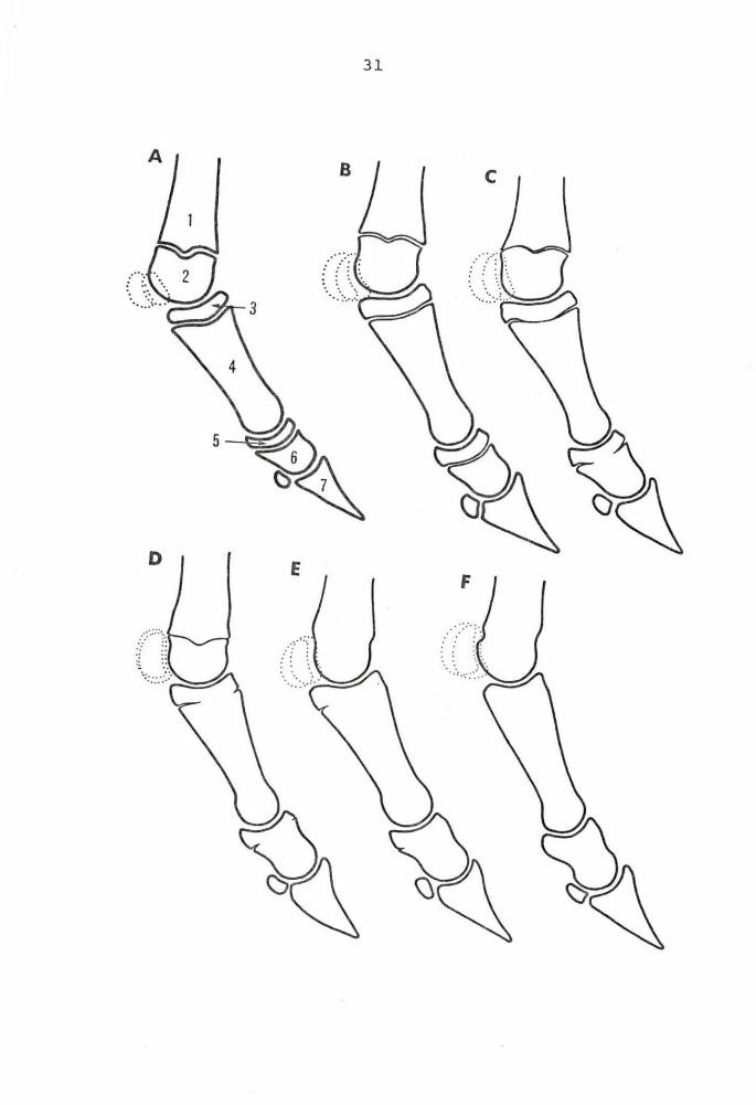
A. Birth B. 86 days old 1 . Diaphysis of
3rd metacarpal bone
2 . Distal epiphysis of 3rd metacarpal bone
3. Proximal epiphysis 1st phalanx
4 . Diaphysis 1st phalanx
5 . Proximal epiphysis 2nd phalanx
6 . Diaphysis of 2nd phalanx
7. 3rd phalanx
D. 168 days old E. 196 days old
c. 140 days old
F . 229 days old
Figure 2 . Filly, lateromedial view of foot
31
A
D E F
32
the proximal epiphysis was closed except on the posterior
surface of the lateromedial view and on the medial and
lateral surface of the anteroposterior view. By 213 days the
epiphyseal line was almost fused, but the ou tsi de edges of
the ante roposterior view were still slightly darke r than the
rest of the bone. After 241 days n o signs of the proximal
epiphyseal line of the s econd phalanx were seen on either the
anteroposterior or the l ateromedial views of the colt ' s foot .
(Figures 1 and 2) •
Filly The radiog raphic appearance of this epiphysea l
line at birth in the filly was similar to that of the colt ' s
at three days of a ge . In the filly the proximal epiphyseal
line of the second phala nx began to close in the center by
the 140th day after birth . After 1 96 days the lateromedia l
view showed only the posterior end of the epiphys eal line
open, while the anter oposterior view showed both the l ateral
and medial edges open. After 229 days the p roximal epiphy-
seal line of the second phalanx of the filly was completely
closed . (Figures 1 and 2) •
Distal e nd
Co lt
First Phalanx
Definite signs of a distal epiphyseal line we re
seen on the r adiographs taken when the colt was three days
old . on both the anteroposterior and the lateromedia l views ,
the cortex of the bone was fused, but a thin dark line was
33
still present at the center of the epiphyseal line. This
dark line was barely visible after 17 days and it had com-
pletely disappeared by the 31st day after birth. A zone of
increased density was still present in the area of epi-
physeal closure on the 31st day.
Filly The distal epiphyseal line of the first
phalanx was represented as a zone of increased density in the
filly at birth. No dark line was visible, but the zone of
increased density suggested that this epiphyseal line had
closed recently.
Proximal end
Colt At three days of age the proximal epiphysis of
the first phalanx was separated radiographically from the
diaphysis by a solid dark epiphyseal line. On the latero-
medial view this line had a slight distal convexity, while
on the anteroposterior view it was transverse to the long
axis of the bone on the lateral and medial sides and had a
definite distal depression in the center (Figures 1, 2, 3,
and 4) • It was in the area of this depression in the center
of the anteroposterior view where fusion first began. The
last sign of the epiphyseal line at the central depression
was gone by the 157th day. The late romedial view at 157 days
still showed an open epiphyseal line, but it was very narrow
and indistinct in the center. At 185 days and also at 213
days the lateromedial view showed only the posterior edge of
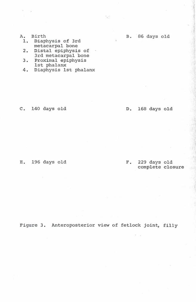
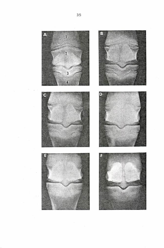
A. Birth 1. Diaphysis of 3rd
metacarpal bone 2. Distal epiphysis of
3rd metacarpal bone 3. Proximal epiphysis
1st phalanx 4 . Diaphysis 1st phalanx
c. 140 days old
E . 196 days old
B. 86 days old
D. 168 days old
F . 229 days old complete closure
Figure 3 . Anteroposterior view of fetlock join~ fi lly
35
A. Birth 1. Diaphysis o f 3rd
metacarpal bone 2 . Distal epiphysis of
3rd metacarpal bone 3. Proximal epiphysis
1st phalanx 4 . Diaphysis 1st phalanx
c. 140 days old
E. 196 days old
B. 86 days old
D. 168 days old
F. 229 days old
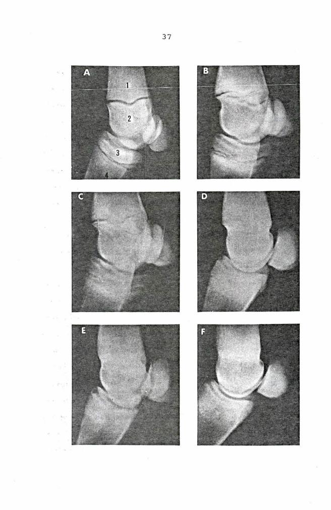
Figure 4. Lateromedial view of fetlock joint, filly
37
38
the epiphyseal line open. On the anteroposterior view at
these same ages the lateral and medial edges of the bone were
open. Complete closure of this epiphyseal line was found in
the colt after 269 days, but not after 241 days .
Proximal end
Filly The c entral depression on the anteroposterior
view began to close after 140 days in the filly. On the
lateromedial view at the same age the epiphyseal line was
very narrow in the center, but it remained open. After 168
days, both views showed the center of the bone united . On
the anteroposterior view after 196 days, both the lateral and
medial sides were open, but they were closed by 229 days . On
the lateromedial view, only the posterior edge was open at
196 days and it had closed by 229 days (Figures 1, 2, 3, and
4) •
Proximal Sesamoids
These bones were ossified in the first radiographs taken
of both foals and their growth after birth was limited to the
appositional type (Figures 1, 2, 3, and 4).
Third Metacarpal Bone
Distal end
Colt A slight proximal convexity of the distal epi-
physeal line of the third metacarpal bone was noted on the
anteroposterior view (Figures 1 and ·3 .1 ) . On the lateromedial
view the epiphyseal line had a central depression (Figures 2
39
and 4) . Schmidt (48) called the lateromedial view of this
epiphyseal line "butterfly wing shaped". Both of these views
showed a narrow, but solid dark line present after 157 days.
A rapid change then took place and by 185 days the line was
almost completely closed. A very indistinct line remained on
the anterior part of the lateromedial view and on the lateral
and medial edges of the anteroposterior view. This epiphy-
seal line was completely fused by the 213th day after parturi-
tion in the colt.
Distal end
Filly The distal epiphyseal line of t he third meta-
carpal bone of the filly had begun to close in the center on
both the anteroposterior and laterornedial views by the 168th
day . On the laterornedial view it was completely closed on
the 196th day, while the edges remained open at that age on
the anteroposterior view. Both views showed complete closure
by the 229th day (Figures 1, 2, 3, and 4) .
Proximal end
No radiographic signs of a proximal epiphysis of the
third metacarpal bone were seen in this study.
Second and Fou~th Metacarpal Bones
No radiographic evidence of a proximal or a distal epi-
physis was visible on these radiographs.
Carpal Bones
All of the carpal bones were calcified in both foals on
the first radiographs and they showed no evidence of any
40
secondary centers of ossification.
Radius and Ulna
Distal end
Colt The first radiographs of the colt showed the
distal epiphysis of the radius and the small wedge shaped
epiphysis that represented the distal end of the ulna separ-
ated by an epiphyseal line. On the anteroposterior view,
the ulnar epiphysis was situated laterally to the radial
epiphysis, while on the lateromedial view it lay posterior
and slightly distal to the radial epiphysis. On both views
the ulnar epiphysis overlapped the distal radial epiphysis
and the accessory carpal bone. After 101 days the proximal
end of the epiphyseal line between the radial and ulnar
epiphyses was fused on the anteroposterior view and the two
ends were fused on the lateromedial view. This epiphyseal
line was still open in the center on the 24lst day on the
anteroposterior view, but it was not visible on the later-
omedial view. By the 269th day no radiographic signs of this
epiphyseal line remained (Figures 5 and 6) •
The distal radial epiphysis began to fuse to the dia-
physis on the 584th day on the lateromedial view and on the
612th day on the anteroposte rior view. It then proceeded to
close from the center outward in a rapid manner. The later-
omedial view showed complete closure by the 696th day, while
the anteroposterior view did not show complete closure until
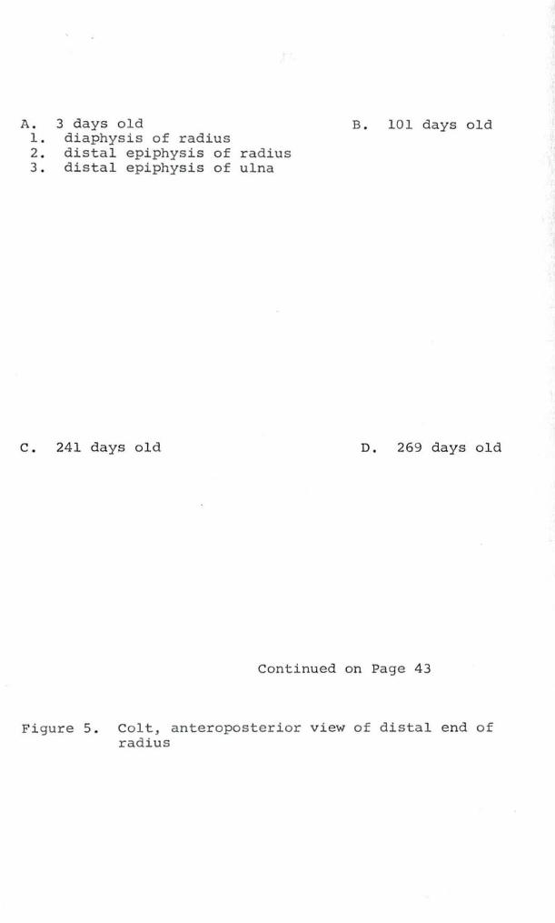
A. 3 days old B. 101 days old 1. diaphysis of radius 2 . distal epiphysis of radius 3 . distal epiphysis of ulna
c. 241 days old D. 269 days old
Continued on Page 43
Figure 5. Colt, anteroposterior view of distal end of radius
42
I
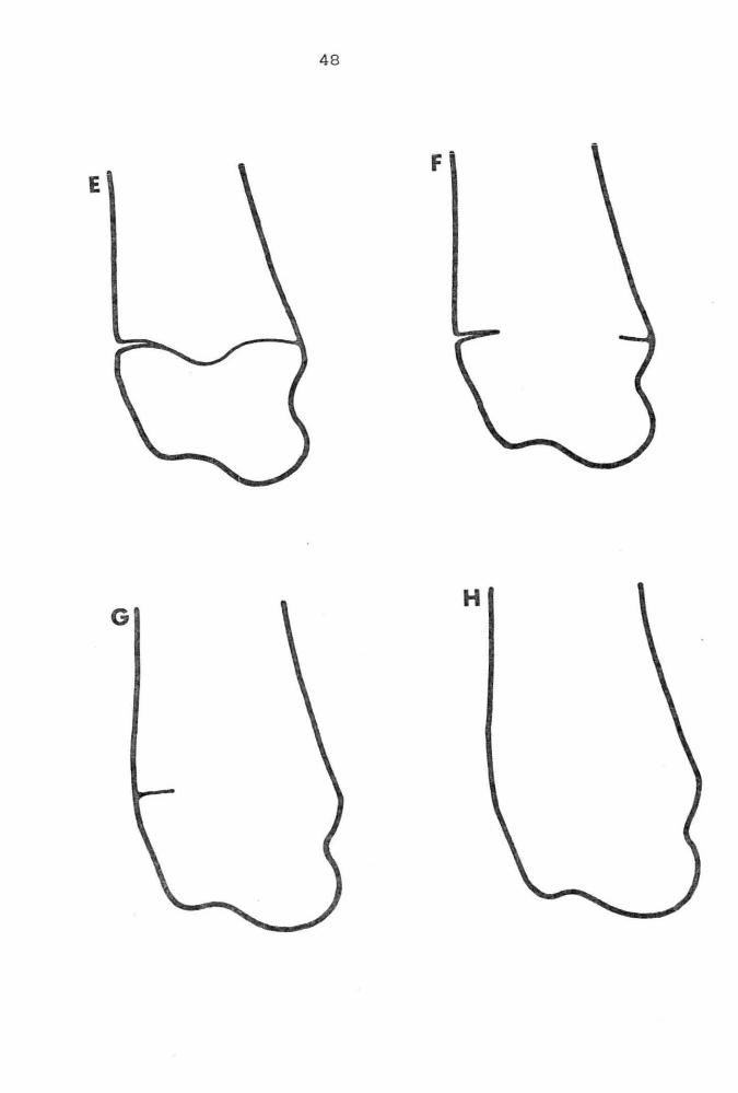
E . 584 days old F . 612 days old
G. 668 days old H. 724 days old
Figure 5 . (Continued)
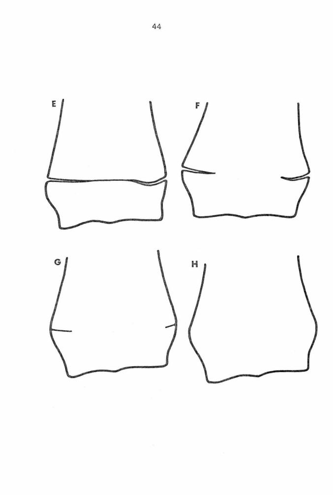
44
E
G
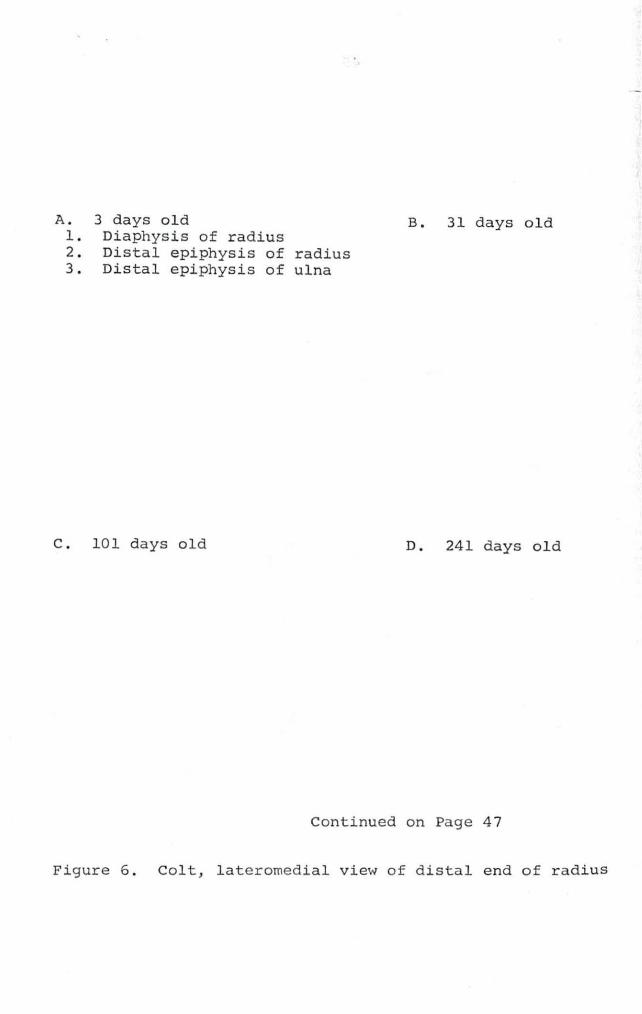
A. 3 days old 1 . Diaphysis of radius
B. 31 days old
2 . Distal epiphysis of radius 3. Distal epiphysis of ulna
C. 10 1 days old D. 241 days old
Continued on Page 4 7
Figure 6 . Colt, lateromedial view of distal end of radius
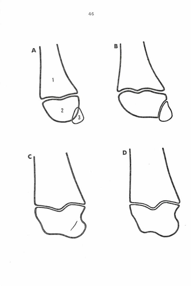
46
A
D
E. 556 days old F. 584 days old
G. 640 days old H. 696 days old
Figure 6 . (Continued)
48
F E
H G
49
the 724th day (Figures 5 and 6) .
Filly The wedge- shaped bone that represents the dis -
tal end of the ulna did not begin to unite to the distal
radial epiphysis until the 112th day. By this date the
proximal apex had fused to the distal radial epiphysis . Com-
plete closure of this epiphyseal line was noted on the 196th
day.
Narrowing of the distal radial epiphyseal line was evi-
dent by the 487th day, but complete closure of the center of
this line was not observed until the 624th day. Closure then
proceeded rapidly and no epiphyseal line was visible on the
708th day.
Radius
Proximal end
Colt The proximal epiphysea l line of the radius was
still a solid single dark line on the 24lst day after birth.
Be twe en the 269th day and the 388th day the p osterior seven-
eighths o f the epiphysea l line slowly united a nd on ly t he
a nterior one - e ighth wa s l eft op e n. By the 4 16th day comple te
epiphy sea l closure had taken place . The radial tuberosity,
which arose on the anterior e d ge of this epiphyseal line was
not completely smoothed anteriorly until the 900 th day.
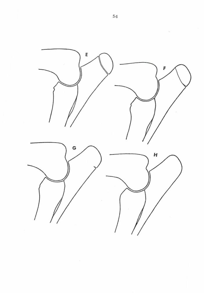
Filly Although v e ry narrow the proximal radial epi -
phys eal line was still c omple tely open on the 343rd day.
This line r emained ope n on the pos terior a nd ante rior e nds on
50
the 37lst day, but it was closed in the center. By the 427th
day, complete closure of this epiphyseal line had become
evident. The radial tuberosity did not become smooth an-
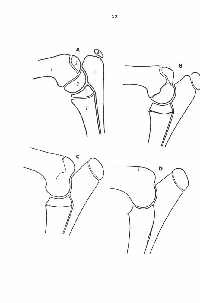
teriorly until the 907th day (Figure 7) •
Ulna
Proximal end
Colt The proximal ulnar epiphysis was seen on the
radiographs of the thorax of the colt when he was three days
old. It was a small center of ossification which was
situated proximally to and on the posterior edge of the ulnar
diaphysis. By the lOlst day this epiphysis had increased
markedly in size. Tne proximal ulnar epiphyseal line was
very narrow posteriorly on the 612th day, but it was still
completely open. On the 640th day the anterior three-fourths
of the line was closed, but the posterior quarter was open.
The posterior part of this line was not completely fused un-
til the 812th day.
Filly The proximal ulnar epiphysis was already os-
sified at birth in the filly . This epiphyseal line was still
completely open on the 624th day, but the anterior three fourths
was very narrow and irregular. Only a small area on the
posterior border remained open on the 680th day. Complete
closure did not take place until the 907th day (Figure 7) •
A. Birth (clinic case) B. 140 days old 1. Diaphysis of humerus 2 . Epiphysis of medial epicondyle 3 . Distal epiphysis of humerus 4. Proximal epiphysis of ulna 5 . Diaphysis of ulna 6 . Proximal epiphysis of radius 7. Diaphysis of radius
c. 343 days old D. 427 days old
Continued on Page 53
Figure 7. A. clinic case, B - H. filly, mediolateral of elbow joint
E . 487 days old F . 624 days old
G. 680 days old H. 90 7 days old
Figure 7 . (Continued}
54
Distal end
Colt
55
Humerus
The distal epiphysis and the epiphysis of the
medial epicondyle were completely ununited after 129 days in
the colt . These two secondary centers of ossification had
united with each other by the 185th day. They were both
separated from the diaphysis of the humerus at this a ge . On
the 24lst day the distal epiphysis had begun to fuse to the
diaphys is i n the cente r of the bone. After 332 days thi s
epiphyseal line was c ompletely closed, but the posterior half
of the epiphyseal line between the medial epicondyle and the
diaphysis remained ope n . This epiphyseal line was completely
c l osed on the 416th day .
Filly On the 196th day the epiphyseal line between
the dist al epiphysis and the diaphysis had fused in the cen-
t er , but the medial epicondyle had not fused to the distal
epiphysis . These two secondary centers of ossification were
united by the 254th day. By the 37lst day (about one year)
the dista l epiphyseal line was c omplet ely fused . The epi -
physeal line between the medial epicondy l e a nd the hume r a l
diaphysis f used from the center toward the cortex. This epi-
physeal line was no longer visible on the 455th day after
birth (Figure 7) .
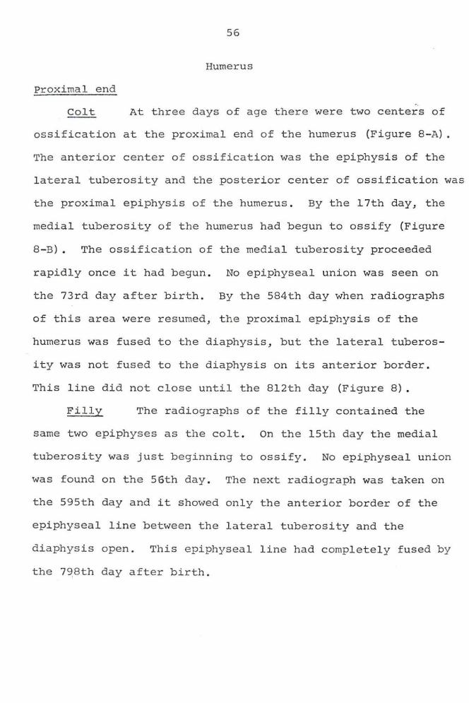
56
Humerus
Proximal end
Colt At three days of age there were two centers of
ossification at the proximal end of the humerus (Figure 8-A) .
The anterior center of ossification was the epiphysis of the
lateral tuberosity and the posterior center of ossification was
the proximal epiphysis of the humerus. By the 17th day, the
medial tuberosity of the humerus had begun to ossify (Figure
8 - B) . The ossification of the medial tuberosity proceeded
rapidly once it had begun . No epiphyseal union was seen on
the 73rd day after birth. By the 584th day when radiographs
of this area were resumed, the proximal epiphysis of the
humerus was fused to the diaphysis, b ut the latera l tuber os -
ity was not fused to the diaphysis on its anterior border.
This line did not close until the 812th day (Figure 8) .
Filly The radiographs of the filly contained the
same two epiphyses as the colt. On the 15th day the medial
tuberosity was just beginning to ossify . No epiphyseal union
was found on the 56th day . The next radiograph was taken on
the 595th day and it showed only the anterior border of the
epiphyseal line between the lateral tuberosity and the
diaphysis open. This epiphyseal line had completely fused by
the 798th day after birth.
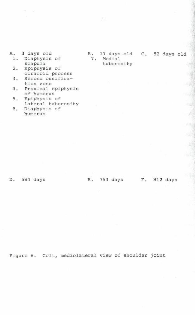
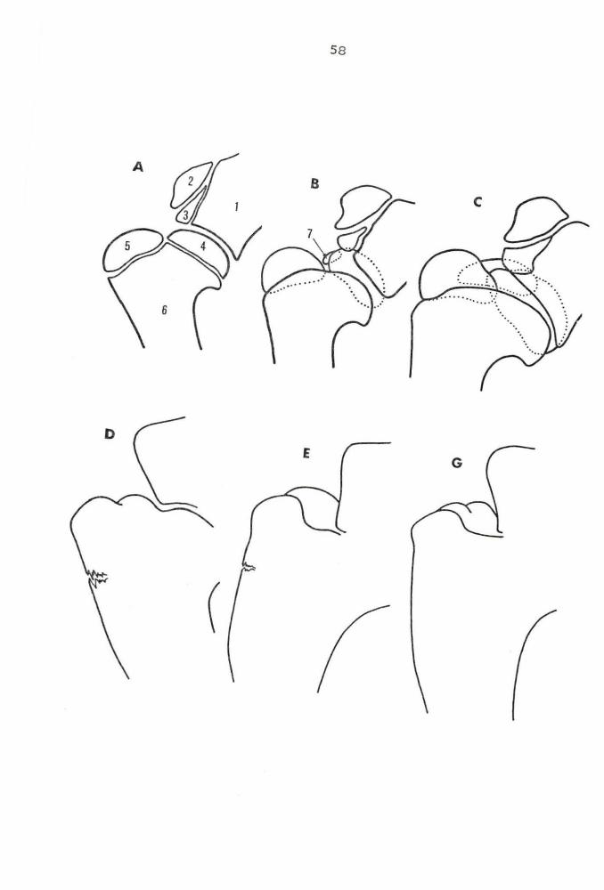
A. 3 days old 1 . Diaphysis of
scapula 2 . Epiphysis of
coracoid process 3 . Second ossifica-
tion zone 4 . Proximal epiphysis
of humerus 5 . Epiphysis of
lateral tuberosity 6 . Di aphysis of
humerus
D. 584 days
B. 17 days old 7 . Medial
tuberosity
E . 753 days
c . 52 days old
F . 812 days
Figure 8 . Colt, mediolateral view of shoulder joint
58
D
(
59
Scapula
Distal end
Colt On the 3rd day there were two centers of ossi-
fication on the distal end of the scapulae (Figure 8 - A) • The
anterior center was thought to be the center 6f ossification
of the coracoid proc~ss and the second center, the center of
ossification of par t o f the tuber scapula. These centers were
ununited after 73 days . These centers fused to the diaphysis
sometime between the 73rd day and 584th day, when this radio-
graphic view was taken next (Figure 8) •
Filly The two centers of ossification seen in the
colt were also seen in the filly until the 15th day. After
the 15th day no clear radiograph was found until the 595th
day when no signs of the open epiphyseal line were present .
60
DISCUSSION
Two tables were prepared to help with the discussion of
epiphyseal closure times. Table 3 was compiled to show the
anatomical closure times given by Sisson and Grossman (52),
Lesbre (32), Zietzschmann and Krolling (66), and Rooney (42).
Table 4 was made to compare the radiographic closure times of
Tohara (61) and Schmidt (48) with those found in this study.
The colt and the filly were kept separate in this table to
allow better comparison.
The radiographic closure times of the proximal end of
the second phalanx were found to be similar to those of
Tohara, but were about one month later than those found by
Schmidt. The radiographic closure times were two to three
months earlier than the anatomical closure ages. The closure
times of the colt a nd the filly were almost identical for
this epiphyseal line, the former being by the 24lst day and
the l atter by the 229th day.
The distal epiphysis of the first phalanx was united at
birth in the filly, but did not unite until sometime between
the 17th and 31st day in the colt. This epiphyseal line was
not seen by Schmidt or Tohara after birth. Anatomically
Sisson and Grossman stated it was closed before birth, while
the other anatomical works stated it closed after birth.
The proximal epiphysis of the first phalanx was united
to the diaphysis earlier in the filly (229 days) than in the
61
colt (269 days) . Again these unions agreed closely with those
of Tohara, but were later than those of Schmidt. The radio-
graphic closure times again were in advance of the anatomical
closure times.
There was considerable difference between the age Tohara
gave for closure of the distal epiphyseal line of the third
metacarpal bone and the ages determined in this study.
Tohara stated that closure took place in the 17th to 18th
months. This was more than twice as long as this study showed
and almost three times longer than Schmidt had reported .
This epiphysis was fused by the 213th day in the colt and by
the 229th day i n the filly . Tohara's fusion time agreed with
the anatomical time given by Sisson and Grossman, but was
longer than the times given by the other anatomical references .
The distal epiphysis of the u lna was not completely fused
to the distal radial epiphysis in these foals until the 196th
and 269th days . This was 2~ and 5 months longer than Schmidt
had found and it was more than twice as long as Lesbre ' s
anatomical study had shown . In the colt this union took
place 2~ months earlier than in the filly.
The proximal epiphysis of the ulna was ossified in both
these foals at birth . This agreed with Tohara, but not with
Kupfer. This epiphysis was f used on the 812th day in the
colt and on the 907th day in the filly, whic h was earlier
than the "about 3~ years" reported in all the anatomical
62
studies. The closure was completed more than three months
later in the filly than in the colt in this study. Schmidt
did not determine the fusion time of this epiphysis .
The distal epiphysis of the radius was fused on the
726th day in the colt and on the 708th day in the filly.
This is just a few days less than two years of age in both
horses. All of the anatomical studies stated this fusion
occurred at "about 3~ years", while Schmidt said it was
closed by the age of 3 years. The "epiphyseal scar" reported
by Smith (55) was present in the area of this epiphyseal line
in both horses on their third birthdays. The width of this
scar continually decreased after union, but it was still
present at age three.
The proximal epiphysis of the radius was said to be
fused before one year of age by Schmidt. It was fused in the
colt by the 416th day and in the filly by the 427th day.
These ages were just slightly less than the 15 to 18 months
reported in the anatomical studies.
Epiphyseal closure times for both the medial epicondyle
and the distal epiphysis of the humerus were given in this
study. In all the other works cited, only a closure time for
the distal epiphysis of the humerus was given. Whether these
workers actually referred to the later closing epiphyseal
line of the medial epicondyle was not stated.
The distal epiphysis of the humerus was united to the
63
diaphysis by the 332nd day in the colt and by the 37lst day
in the filly, but the medial epicondyle did not fuse until
the 416th day in the colt and the 455th day in the filly .
The latter agreed with the 15 to 18 months given in the
anatomical reports, but were later than the "before one year"
given by Schmidt.
At the proximal end of the humerus two epiphyses were
described and again the anatomical reports did not say if
they were referring to one or both of these epiphyses. It
was assumed that their "about 3~ years" referred to both
epiphyseal lines, since the proximal epiphysis of the humerus
was fused sometime before the 21st month in both of these
horses. The lateral tuberosity of the humerus was complete-
ly fused in the colt by the 812th day and in the fill y by
the 798th day. Schmidt did not establish closure times for
this area. The medial tuberosity of the humerus was found
to ossify separately from the lateral tuberosity and it was
still separate at the end of the second month.
The coracoid process of the scapula was fused sometime
before the 21st month in both these foals, but no definite
fusion time was determined. Schmidt said this fusion took
place before the 18th month and the anatomical studies gave
a fusion time between 10 and 12 months.
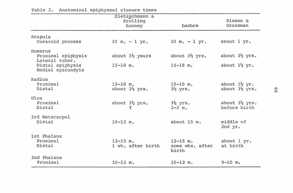
Table 3. Anatomi cal epiphyseal closur e times
Scapula Coracoid process
Humerus Proximal epiphysis Lateral tuber. Distal epiphysis Medial epicondyle
Radius Proximal Distal
Ulna Proximal Distal
3rd Metacarpal Distal
1st Phalanx Proximal Distal
2nd Phalanx Proxima l
Zietzschmann & Krol ling Rooney
10 m. - 1 yr .
about 3~ years
15-18 m.
15-18 m. about 3~ yrs .
about 3~ yrs. ?
10-12 m.
12 - 15 m. 1 wk. after birth
10-12 m.
Lesbre
10 m. - 1 yr.
about 3~ yrs.
15-18 m.
15- 18 m. 3~ yrs.
3~ yrs. 2-3 m.
about 15 m.
12-15 m. some wks . after birth
10 - 12 m.
Sisson & Grossman
about 1 yr.
about 3~ yrs.
about l~ yr .
about l~ yr. about 3~ yrs.
about 3~ yrs. before birth
middle of 2nd yr.
about 1 yr . at birth
9-10 m.
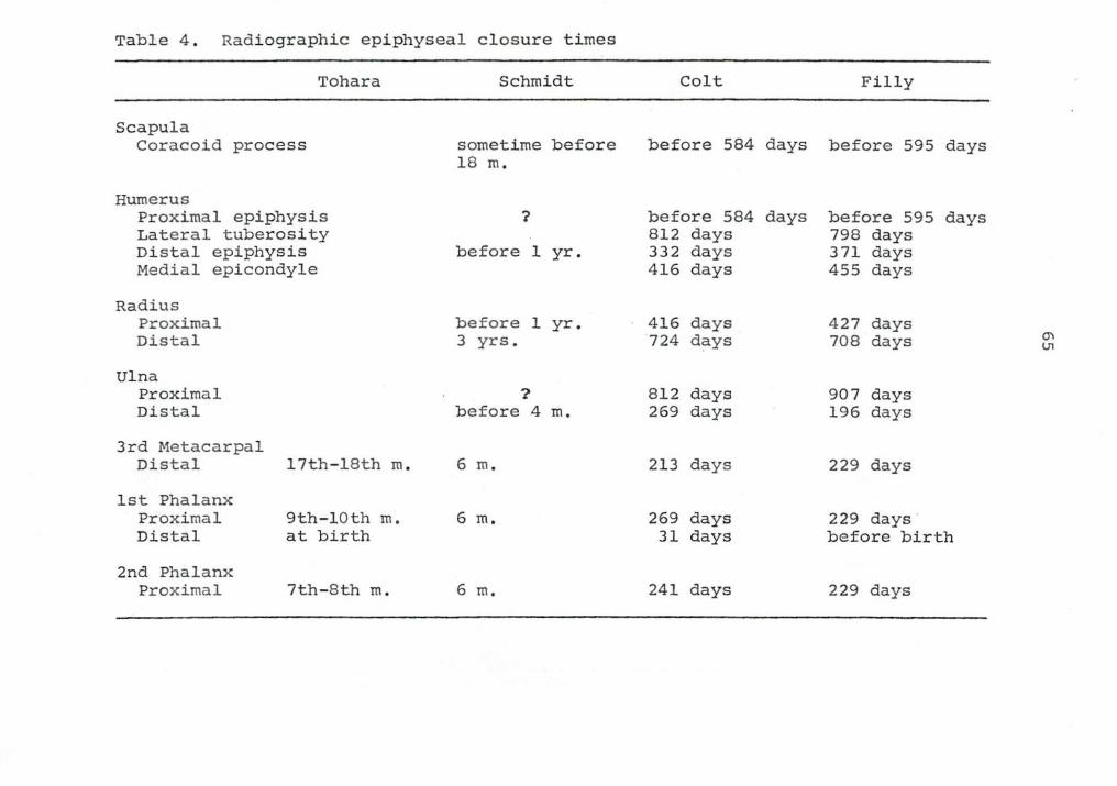
Table 4 . Radiographic epiphyseal closure times
Tohara
Scapula Coracoid process
Humerus Proximal epiphysis Lateral tuberosity Distal epiphysis Medial epicondyle
Radius Proximal Distal
Ulna Proximal Distal
3rd Metacarpal
Schmidt
sometime before 18 m.
?
before 1 yr.
before 1 yr. 3 yrs.
? before 4 m.
Distal 17th-18th m. 6 m.
1st Phalanx Proximal Distal
2nd Phalanx Proxima l
9th-10th m. at birth
7th-8th m.
6 m.
6 m.
Colt Filly
before 584 days before 595 days
before 584 days 812 days 332 days 416 days
416 days 724 days
812 days 269 days
213 days
269 days 31 days
241 days
before 595 days 798 days 371 days 455 days
427 days 708 days
907 days 196 days
229 days
229 days before birth
229 days
CTI Ul
66
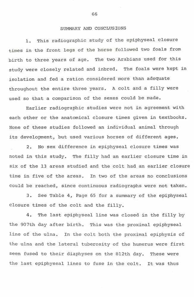
SUMMARY AND CONCLUSIONS
1. This radiographic study of the epiphyseal closure
times in the front legs of the horse followed two foals from
birth to three years of age. The two Arabians used for this
study were closely related and inbred. The foals were kept in
isolation and fed a ration considered more than adequate
throughout the entire three years. A colt and a filly were
used so that a comparison of the sexes could be made.
Earlier radiographic studies were not in agreement with
each other or the anatomical closure times given in textbooks .
None of these studies followed an individual animal through
its development, but used various horses of different ages.
2. No sex difference in epiphyseal closure times was
noted in this study. The filly had an earlier closure time in
six of the 13 areas studied and the colt had an earlier closure
time in five of the areas. In two of the areas no conclusions
could be reached, since continuous radiographs were not taken.
3 . See Table 4, Page 65 for a summary of the epiphyseal
closure times of the colt and the filly.
4 . The last epiphyseal line was closed in the filly by
the 907th day after birth. This was the proximal epiphyseal
line of the ulna. In the colt both the proximal epiphysis of
the ulna and the lateral tuberosity of the humerus were first
seen fused to their diaphyses on the 812th day. These were
the last epiphyseal lines to fuse in the colt. It was thus
67
concluded that all growth in length of the fore limbs of these
two horses was c ompleted before they were two and one half
years old.
5. It was found that the distal epiphysis of the first
phalanx can be fused or partially open at birth . This epi-
physeal line was closed at birth in the filly, but was open in
the cente r in the colt at three days of age . Complete closure
was observed by the 31st day in the colt.
6 . The epiphyseal closure times of the ulna varied be-
tween the colt and the filly more than any of the other closure
times. Fusion of the proximal end of the ulna occurred first
in the c olt, while the distal end of the ulna was fused first
in the filly .
7 . The radiographic closure times of this study were
earlier than the anatomical times in Table 1, with t wo ex-
ceptions . The radiographic fusion of the distal epiphyses of
the radius and the ulna in both the colt and the filly occur-
red a t a later date than reported in the anatomical studies.
The fusion of the distal epiphysis of the f irst phalanx f used
in the colt after birth, while Sisson and Grossman claimed
fusion t ook place before birth .
8 . The chronological order of epiphyseal fusion of the
front legs in both the colt and the fi lly was very s imilar .
The fusion of the distal ends of the radius a nd the ulna were
not in the same order in these foals . Fusion of the open
68
epiphyseal lines below the carpus occurred by 229 days of age
in the filly, but these closure times varied in the colt.
9. All of the epiphyseal lines below the carpus were
fused by the end of the ninth month.
69
ACKNOWLEDGEMENTS
The author wishes to express his gratitude to a ll the
members of the veterinary staff who helped in many ways with
this project.
The author is particularly indebted to Dr . M. A. Emmer-
son for his radiographic advice and assistance, and for his
help in preparing this paper throughout and to Dr . B . w. Kingrey, who gave encouragement and assistance in numerous
ways. Dr. Emmerson is also thanked for organi zing this
project as we ll as for his early technical work .
Dr . L. R . Hu tchinson and Dr. H. Nelson are thanked for
their early technical work on this project .
The assistance given by many students with this project
was greatly appreciated.
Lou Facto and Dan Hillmann are thanked for their help
in preparing the figures used in this paper.
The author is indebted to his wife, Helena, not only
for her assistance in the early preparation of this draft,
but for her patience and understanding throughout the course
of this endeavor .
70
LITERATURE CITED
1 . Adams, o. R. Lameness in horses. Philadelphia, Pa., Lea and Febiger. 1962.
2 . Andersen, A. c. and Floyd, M. Growth and development of the femur in the beagle . American Journal of Veterinary Research 24: 348-351. 1963.
3. Bassett, c . A. L. Current concepts of bone formation. The Journal of Bone and Joint Surgery 44-A: 1217- 1244 . 1962 .
4 . Bloom, w. and Bloom, M. A. Calcification and ossifica-tion calcification of developing bones in embryonic and n ewborn rats. Anatomical Record 78: 497- 524. 1940 .
5. and Fawcett, D. w. A textbook of histology.
6.
8th ed. Philadelphia, Pa., W. B. Saunders Company. 1962.
Bressou, c., Pomriaskinsky-Kobozieff, N. A., and Kobozieff, N. Etude radiologique de l'aspect du squelette normal de la main du chien. Recueil de cine vJterinaire 133: 449-464. 195 7 .
,
~
Mede-
7. , and Etude radiologique de l'ossification de squelette du pied du chat aux divers stades de son evolution, de la na i ssance a l'age adulte. Recueil de M~decine Vet~rinaire 135: 611 - 626 . 1959 .
8. and Etude radiologique de l ' ossification du squellette de la main du chat aux divers stades de son evolution, de la naissance ~ l'age adulte . Recueil de Medecine Vet4rinaire 135: 547-564 . 1959 .
9 . Carlson, w. D. Veterinary Radiology. Philadelphia, Pa . , Lea and Febiger. 1961.
10 . Dawson, A. B. Further studies on epiphyseal union in the skeleton of the rat. Anatomical Record 60: 83 - 86 . 1934 .
11. The age order of epiphyseal union in the long
12.
bones of the albino rat. Anatomical Record 31: 1018 . 1925 .
Ellenberger, W. and Baum, H. Anatomie der Haustiere. 18 . Springer-Verlag. 1943 .
Handbuch der Vergleichenden Auflage . Berlin, Germany,
71
13 . Emara, M. Some observations on epiphyseal union of long bones in young Egyptian cattle and its importance as an aid in the estimation of age . Veterinary Record 49: 1534-1537. 1937.
14. Enlow, D. H. A study of the post-nata l growth and re -modeling of bone . American Journal of Anatomy 110: 79-102 . 1962 .
15. Ewart, J. c. The development of the skeleton of the limbs of the horse, with observations on polydactyly . Journal of Anatomy and Physiology 28, New Series 8 : 236- 256 . 1894 .
16. The development of the skeleton of the limbs of the horse, with observations on polydactyly . Journal of Anatomy and Physiology 28, New Series 8, part 2: 342 -369 . 1894 .
17. Flecker, H. Roentgenographic observations of the times of appearance of epiphyses and their fusion with the diaphyses . Journal of Anatomy 67: 118- 164 . 1932 .
18 . Time of appearance and fusion of ossification center s as observed by roentgenographic methods . Ameri-can Journal of Roentgenology and Radium Therapy 47: 97-159 . 1942 .
19. Francis, c. c . , Werle, P. P. and Behm, A. Appearance of centers of ossification from birth to five years . Ameri -can Journal of Physical Anthropology 24: 273-299 . 1939 .
20 . Greulich, W. W. and Pyle, s. I . Radiographic atlas of Skeletal development of the hand and wrist . 2nd ed . Stanford, Calif . , Stanford University Press . 1959.
21. Habel, R . E., Barrett, R. B . , Diesem, c. D. and Roenigk, w. J . Nomenclature for radiologic anatomy . Journal of the American Veterinary Medical Association 142: 38-41. 1963.
22 . Ham, A. w. and Leeson, T . s . Histology . delphia, Pa., J. B. Lippincott Company .
4th ed . 1961 .
Phi la-
23. Hanlon, G. F. Skeletal maturation and abnormal bone growth in the dog . The Minnesota Veterinarian 3: 6- 9 . 1963 .
24 . Hare, W. c . D. Radiographic anatomy of the canine pec-toral limb Part 2 . Developing limb. Journal of the Amer-ican Veterinary Medical Assoc iation 135 : 305-310 . 1959.
72
25. • The age at which epiphyseal union takes place in the limb bones of the dog. Wiener Tierarztliche Monatsschrift 9/72: 224-245. 1960.
26. Hill, A. H. Fetal a ge asses sment by centers of ossifica-tion. American Journal of Physical Anthropology 24: 251-272. 1939.
27. Hoerr, N. L., Pyle, s. I. and Francis, c. c. Radio-graphic atlas of skeletal development of the foot and ankle. Springfield, Ill., Charles c. Thomas. 1962.
28. Ingalls, T. H. Epiphyseal growth: normal sequence of events at the epiphyseal plate. Endocrinology 29: 710-719. 1941.
29. Kanan, c. v. Some observations on ossification of the bones of the appendicular skeleton of Carnelus dromedarius. Acta morphologica neerlando-scandinavica 4: 254-260. 1961.
30. Koch, w. The age order of epiphyseal union in the skele-ton of the European bison (Bo s bonasus L.). Anatomical Record 61: 371-376. 1935. ~-
31. K~pfer, M. Beitrage zum Modus der ossifikationsvorgange in der Anlage des Extremitatenskelettes bei den Equiden der Verknocherungsprozess in der Pf erde- und Eselglied-masse auf Grund Rontgenologischer Untersuchungen. Denkschriften der Schweizerischen Naturforschenden Gesellschaft Memoires de la societe Helvetique des Sciences Naturelles 67, No. 67: 1-352. 1931.
32. Lesbre, M. F.-x. Contribution a l'etude de !'ossifica-tion du squellette d e s mammiferes dome stiques .principale-ment aux points de vue de sa marche et de sa chronologie. Annales de la Soci~t~ d'Agriculture Sciences et Industrie de Lyon 5, 7 serie. 1897.
33. Menees, T. o. and Holly, L. E. The ossification in the extremities of the new-born. American Journal of Roentgenology and Radium The rapy 28: 389-390. 1932.
34. Nissen, H. w. and Riese n, A. H. onse t of ossification in the epiphyses and short bones of the extremities in chimpanzees. Growth 13: 45-70. 1949.
35. Patte n, B. M. Foundations of embryology. New York, N. Y., McGraw-Hill Book Co., Inc. 1958 .
73
~
36. Pomriaskinsky-Kobezieff, N. and Kobezieff, N. Etude radiologique de l'aspect du squelette, normal de l a main du chien aux divers stades de son ~volution de la nais-sance a l'age adulte (1) . Recueil de M~decine Veterinaire 130: 617-646. 1954 .
37. Pryor, J . W. Differences in the time of development of c enters of ossification in the male and female skeleton. Anatomical Record 25 : 252-273 . 1923.
38 . Time of ossification of the bones of the hand of the male and female, and union of t he epiphyses with the diaphyses . American Journal of Physical Anthro-pology 8: 401-410. 1925.
39. Pyle, s. I. and Hoerr, N. L . Radiographic atlas of skeletal development of the knee. Springfield, Ill., Charles c. Thomas . 1955.
40 . , Stuart, H. c . , Cornoni, J., and Reed, R. B. Onsets, completions, and spans of the osseous stage of development in representative bone growth c enters of the extremities. Monographs of the Society for Research in Child Development 26, No . 1: 1-126. 1961.
41 . Retterer , E . Contribution au d~veloppement du squelette des extremit~s chez l es mammif~res . Journal de l'ana-t omie et de Physiologie Norrnales et Pathologiques de l'Homrnes et des Animaux 20: 467-614 . 1884 .
42 . Rooney, J . R. Normal bone structure . Gabel , Johnson and Riley, eds . Equine Surgery. pp . 407-409. Wheaton, Ill., nary Publications, Inc. 1963 .
In Bone, Catcott, Medicine and American Veteri-
43. Rosenberg, A. Ueber die Entwicklung des Extremitaten-skeletes bei einigen durch Reductionen ibrer Gliedmassen characterisirten Wirbelthieren . Zeitschrift fur Wissen-schaftliche Zoologie 23: 116-170. 1873.
44. Roshdesstwenskaja, G. Wachstum und Entwicklung der Extremitaten bei Fohlen i m ersten Lebensjahr. Pfer-dezucht u . Pferdesport 30 (105): 30-32. 1960 . Original not available for examination ; abstracted in Landwirt-schaftliches zentralblatt der Deutschen Akademie der Landwirtschaftswissenschaften zu Berlin Abteilung 4 Veterinarmedizin 6 : 1311 . 1961.
74
45. Rozhdestvenskaya, G. A. Bone formation in limb bones of foals during the first year of life. Trudy Vsesoynznyi nauchno-issledovatel' skii Institut Konevodstva 23: 321-330. 1960. Original not available for examination; cited in Index Veterinarius 29 No. 4: 21. 1961.
46 . Saarni, I. Die Intrauterine Entwicklung der Extremi-tatenknochen des Pferdes. Inaugural-Dissertation. Giessen, Germany. 1921.
47 . Schlotthauer, c. F. and Janes, J. M. The time of closure of the lower femoral epiphyses and upper tibial epiphyses in the dog as determined by roentgenogram . American Journal of Veterinary Research 13: 90. 1952.
48. Schmidt, G. Epiphysen und Apophysen in der Rontgeno-logischen Darstellung in den Vorder-und Hinterextremitaten der Fohlen. Inaugural-Dissertation . Hannover, Germany. 1960.
49 . Seoudi, R. X-Ray examination of epiphyseal union as an aid to the estimation of the age in dogs. British Veterinary Journal 104: 150-155. 1948 .
50. Siegling, J. A. Growth of the epiphyses. Journal of Bone and Joint Surgery 23: 23-36. 1941 .
51 . Sisson, s . A text-book of veterinary anatomy. 1st ed.
52 .
53 .
Philadelphia, Pa., W. B. Saunders Company. 1910 .
animal s . Company.
and Grossman, J. D. The anatomy of the domestic 4th ed. Phila delphia, Pa ., W. B. Saunders 1956.
Smith, R . N. of the sheep.
Fusion of the epiphyses of the limb bones The Veterinary Record 68 : 257-259. 1956.
54. Radiological observations on the limbs of young greyhounds. The Journal of Small Animal Practice . 1, No. 2: 84-90 . 1960 .
55 . and Allcock, J. Epiphysial fusion in the grey-hound. The Veterinary Record 72, No. 5: 75-79 . 1960 .
56 . Spark, C. and Dawson, A. B. The order and time of ap-pearance of centers of ossification in the fore and hind limbs of the albino rat, with special reference to the possible influence of the sex factor. American Journal of Anatomy 41: 411-445. 1928.
75
57 . Stevenson, P . H. Age order of epiphyseal union in man. American Journal of Physical Anthropology 7: 53 - 93 . 1924 .
58. Stoss, A. o. Tierarztliche Geburtskunde und Gynakologie einschliesslich der Krankleiten der Neugeborenen. Stuttgart, Germany, Ferdinand Enke. 1928 .
59 . Struthers, J. On the development of the bones of the foot of the horse, and of digital bones generally; and on a case of polydactyly in the horse. Journal of Anatomy and Physiology 28, New Series 8: 51- 62 . 1893 -1894 .
60. Todd, T . w. and Todd, A. W. The epiphyseal union pattern of the ungulates with a note on sirenia. American Journal of Anatomy 63: 1-36. 1938.
61 . Tohara, s. Radiographical studies on the ossification of leg-bones of horses. Japanese Journal of Veterinary Science 12, No . 1: 1-12. 1950. Original available but not translated: English summary used.
62. Trau tmann, A. and Fiebiger, J . histology of domestic animals. Publishing Associates. 1957.
Fundamentals of the Ithaca, N. Y., Comstock
63 . Van Wagnener, G. and Asling, c. E. Roentgenographic estimation of bone age in the rhesus monkey (Macaca mul atta). American Journal of Anatomy 103: 163-185. 1958 .
64.
65.
Walker, D. G. and Wirtschafter, z. T. the rat skeleton a laboratory atlas. Charles c . Thomas. 1957.
The genesis of Springfield, Ill . ,
Washburn, s. L . world monkeys. 1943 .
The sequence of epiphyseal union in old American Journal of Anatomy 72: 339-360 .
66. Zietzschmann, o. and Krolling, o. Lehrbuch der Entwick-lungschichte der Haustiere . Berlin, Germany, Paul Parey . 1955.
67 . Zuck, T . T. guinea pig.
The a g e orde r of epiphyseal union in the Anatomical Record 70: 389-399 . 1938.


![Accuracy of scoring of the epiphyses at the knee joint ... · Accuracy of scoring of the epiphyses at the knee joint (SKJ) ... Cameriere et al. [51] in 2012 studied the frontal ra-diographs](https://img.pdfslide.us/doc/110x75/5e330b20da1b036ec55f05c2/accuracy-of-scoring-of-the-epiphyses-at-the-knee-joint-accuracy-of-scoring-of.jpg)