Embed Size (px)
Citation preview

1
SKELETAL TISSUESCHAPTER 7
By John McGillSupplement Outlines: Beth WyattOriginal PowerPoint: Jack Bagwell
INTRODUCTION TO THE SKELETAL SYSTEM
• STRUCTURE– Organs: Bones– Related Tissues: Cartilage and Ligaments
• PRIMARY FUNCTION– Support
• PRIMARY TISSUES OF THE SKELETAL SYSTEM– BONE TISSUE– CARTILAGE
• Connective Tissues
TYPES OF BONES
• LONG: Long and Narrow• Humerus and femur• SHORT: Cube/Box-
Shaped– carpus and tarsus– found in parts of skeleton
that require strength and limited movement
• FLAT: Flat and Thin• IRREGULAR: Complex
Shapes

2
MACROSCOPIC STRUCTURE:
Long Bones• DIAPHYSIS
– Shaft– Composed of Compact
Bone• EPIPHYSES
– Both Ends Composed of Cancellous Bone
• ARTICULAR CARTILAGE– “Joining Cartilage”– Covers Epiphyses (Thin
Layer)– Provides Cushioning at
Joints
MACROSCOPIC STRUCTURE:
Long Bones• PERIOSTEUM
– Bone’s Covering– White– Thin but Tough– “Welded” to Underlying Bone– Contains Blood Vessels
• MEDULLARY (MARROW) CAVITY– Space Within the Diaphysis– Contains Bone Marrow
• ENDOSTEUM– Lines the Medullary Cavity– Thin
MACROSCOPIC STRUCTURE:SHORT, FLAT, IRREGULAR BONES
• Inner Portion: Cancellous Bone– “spongy bone”
• Surfaces: Compact Bone– dense and solid
• Periosteum Present

3
MICROSCOPIC STRUCTURE OF BONE: COMPACT BONE:
• HAVERSIAN SYSTEMS (OSTEONS)– Microscopically,
Compact Bone is Composed of Haversian Systems
– Haversian Systems: Microscopic Structural Units of Compact Bone
Microscopic structure - Haversian System
• Haversian system (osteon)-consists of the canal and surrounding structures
• Lamellae – concentric layers of calcified matrix
• Lacunae – “little lakes”; where the bone cells live
• Canaliculi – very small canals that radiate from the lacunae; carry nutrients
• Haversian canal – central canal which carries blood vessels
• FUNCTION OF HAVERSIAN SYSTEMS– Blood Supply to Compact Bone– Periosteum Haversian Canals
Canalculi Lacunae
BONE (MICROSCOPIC VIEW)
canaliculi osteocyte in lacunae
Haversian canal
ossified matrix (lamellae)

4
CANCELLOUS BONE: TRABECULAE
• Trabeculae: Needlelike Pieces of Bone (Surround Spaces)
• Contains Osteocytes• How Cancellous Bone Gets Its Blood Supply: • From Bone Marrow by Diffusion (Periosteum
Bone Marrow Openings in Trabeculae)
BONE TISSUE (OSSEOUS TISSUE)• COMPONENTS: MATRIX, PROTEIN FIBERS, CELLS
– Typical Connective Tissue• COMPOSITION OF BONE MATRIX
– INORGANIC COMPONENTS• Minerals (Esp. Ca and Phosphate)• Forms hydroxyapatite• Constitute Approx. 65% of Bone Matrix• Gives Matrix Hardness and Strength
– ORGANIC COMPONENTS• Complex Mixture of Carbohydrates and Proteins• Gives Matrix Strength
• PROTEIN FIBERS: COLLAGENOUS– Also Gives Matrix Strength
• *NOTE: Matrix with Protein Fibers Means Hardness and Strength
BONE CELLS• OSTEOBLASTS
– Bone-Forming Cells– Location: Periosteum (Primarily)
• OSTEOCLASTS– Bone-Destroying Cells– Location: Endosteum (Primarily)
• OSTEOCYTES– Bone Cells (Mature Osteoblasts)– Locations:
• 1) Compact Bone: Lacunae• 2) Cancellous Bone: Trabeculae

5
BONE MARROW (MYELOID TISSUE)• Tissue Type:
Connective Tissue (Reticular)
• LOCATIONS– Long Bones:
• Medullary Cavity• Epiphyses:
– Spaces in Cancellous Bone
– Short, Flat, Irregular Bones:
• Spaces in CancellousBone
BONE MARROW TYPES: RED MARROW
• DESCRIPTION/FUNCTIONS– Red in Color Because
Functions in Hematopoiesis• LOCATIONS
– Children: All Bones Contain Red Marrow
– Adults: Certain Bones Contain Red Marrow
• Flat Bones of the Skull• Sternum, Ribs, Vertebrae• Pelvic Bones• Epiphyses of Humerus and Femur
BONE MARROW TYPES: YELLOW MARROW
• DESCRIPTION/FUNCTIONS– Yellow in Color Because
Contains Largely Adipose Tissue
– Yellow Marrow Was Once Red Marrow, Now Yellow B/C
– It No Longer Functions in Hematopoiesis
• LOCATIONS– Most Bones in Adults Contain
Yellow Marrow

6
Functions of Bones• Support – support the weight of the rest of
the body• Protection – protect the delicate body parts• Movement – muscles attach to bone and
allow movement• Mineral storage – calcium, phosphorous,
and other minerals are stored in the bone• Hematopoiesis – red marrow plays an
important role in the formation of red blood cells, some flat bones also play a role here
DEVELOPMENT OF BONE
(OSTEOGENESIS)• How Bones Form in the Fetus• INTRAMEMBRANOUS OSSIFICATION
– DEFINITION• “Within Membrane Bone
Formation”• Method by Which Flat Bones
Form• MECHANISM
– Connective Tissue Membrane – Cells Develop Into Osteoblasts– Secrete Organic Matrix and
Collagenous Fibers– Calcification Occurs
•Intramembranous bone formation in a fetal pig skull. •Flat bones of the skull develop by IO. •Embryonic mesenchyme cells form a membrane (Mes) & •differentiate into osteoblasts that•form bony spicules or cancellousbone (CsB). •Eventually osteonsform.
DEVELOPMENT OF BONE (OSTEOGENESIS)• ENDOCHONDRAL OSSIFICATION
– DEFINITION• “Within Cartilage Bone
Formation”• Method by Which Most Bones
Form– MECHANISM
• Cartilage Model • Periosteum Forms • Cells Develop Into
Osteoblasts• Secrete Organic Matrix and
Collagenous Fibers• Calcification Occurs
– *Note: In Both Types of Ossification:
• Osteoclasts Resorb Bone • Forms Medullary Cavity,
Spaces in Cancellous Bone
ENDOCHONDRAL OSSIFICATION
• Embryonal hyaline cartilage precedes bone formation.
• Inner cells change into osteoblasts cells in the perichondrium.
• Osteoblasts form the periosteum.

7
Summary: Endochondral Ossification
• Bone forms from a cartilage model• Osteoblasts begin to calcify the cartilage• Osteoblasts and osteoclasts are constantly
reshaping the bone• Centers of ossification appear in the
epiphyses• Epiphyseal plate is site of continued bone
growth; indicates the bone is not yet mature.
Osteogenesis (Bone formation)• The cartilaginous skeleton is changed to bone in one
of two ways:• Intramembranous ossification – happens in some flat
bones of body– 1st step – cells differentiate into osteoblasts (centers of
ossification)– 2nd – cells secrete ground substance– 3rd – ground substance is calcified– 4th – trabelculae appear and join to form spongy bone– 5th - layer of spongy bone is covered on both sides by
compact bone– 6th – growth occurs by appositional growth – the addition of
osseous tissue to its outer surface
Bone Growth - Animation
•http://www.anatomy.gla.ac.uk/fab/tutorial/generic/bonet.html

8
FETAL SKELETON
BONE GROWTH AND RESORPTION• How Bones Increase in Size after Birth• Involves Bone Resorption : Destruction• BONE GROWTH
– FLAT BONES (Also Short, Irregular Bones)• APPOSITIONAL GROWTH
– Growth By Adding to the Surfaces
– LONG BONES• GROWTH IN LENGTH – EPIPHYSEAL PLATE• Epiphyseal Plate: Layer of Hyaline Cartilage That Lies B/T Epiphyses
and Diaphysis• Didn’t Ossify During the Fetal Period (Purpose: To Allow Bone
Growth in Length)• Epiphyseal Plate 1) Thickens and 2) Ossifies Repeatedly• When Growth in Length is Complete, Cells in EP Stop Mitosis and
the Entire Plate Ossifies, What Remains is Epiphyseal Line
EPIPHYSEAL PLATE

9
Epiphyseal Plate• The epiphyseal plate
allows for growth in bones.
Zones of the Epiphyseal Plate
GROWTH IN DIAMETER – COMBINED ACTION OF OSTEOBLASTS AND OSTEOCLASTS
• Osteoblasts(Periosteum) Build New Bone on the Outer Surface
• Osteoclasts(Endosteum) Destroy Bone from the Inner Surface of the Medullary Cavity (Enlarges Med. Cavity)

10
BONE GROWTH AND RESORPTION
• BONE RESORPTION– Osteoclasts (Endosteum) Destroy Bone
from the Inner Surface of the MedullaryCavity
BONE GROWTH AND RESORPTION• BONE GROWTH AND RESORPTION
THROUGHOUT LIFE– Both Growth and Resorption Go On
Throughout Life, But at Different Rates• From Infancy Young Adulthood: Growth
EXCEEDS Resorption (Bones Grow and are Thick)
• During Late 20’s/Early 30’s: Growth EQUALS Resorption (Bones Remain Relatively Constant)
• From Mid 30’s/Early 40’s Old Age: Resorption EXCEEDS Growth (Bones Become Thinner, More Susceptible to Fracture and Disease)
BONE GROWTH AND RESORPTION
• BONES RESPONSE TO STRESS– Bone Stress = Weight Bearing Applied
to Bones– Bone Stress Increases the Activity of the
Osteoblasts (Helps Offset the Effects of Aging on Bones)
11
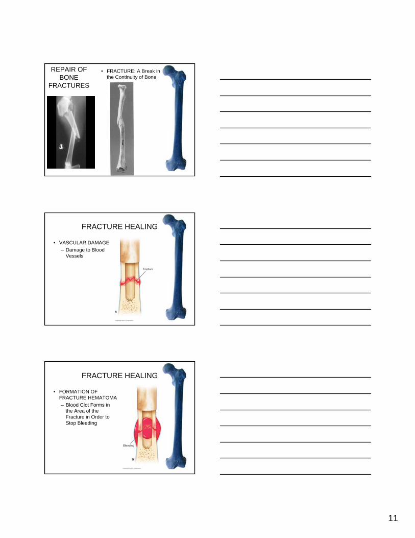
REPAIR OF BONE
FRACTURES
• FRACTURE: A Break in the Continuity of Bone
FRACTURE HEALING
• VASCULAR DAMAGE– Damage to Blood
Vessels
FRACTURE HEALING
• FORMATION OF FRACTURE HEMATOMA– Blood Clot Forms in
the Area of the Fracture in Order to Stop Bleeding

12
FRACTURE HEALING
• FORMATION OF CALLUS TISSUE– Thickened Repair
Tissue That Binds the Ends of the Bones Together (Reason That the Fracture is Aligned and Immobilized)
FRACTURE HEALING
• REPLACEMENT BY BONE– Callus Tissue
Becomes Bone (Action of Osteoblasts), Remodeled by Osteoclasts
Epiphyseal Plate Fracture

13
CARTILAGE• CHARACTERISTICS
– MATRIX• FIRM/FLEXIBLE GEL
– PROTEIN FIBERS• COLLAGENOUS
– CELLS• CHONDROCYTES• Chondrocytes Lie in
Lacunae– AVASCULAR: Oxygen
and Nutrients by Diffusion
CARTILAGE: Types
• Hyaline• Elastic• Fibrocartilage
HYALINE CARTILAGE• Most Abundant and
Common• Shiny• Semitransparent• Locations:
– Articular Cartilage– Costal Cartilages– Cartilage Rings in
Trachea and Bronchi– Tip of Nose

14
ELASTIC CARTILAGE• Has Fewer Collagenous
Fibers Compared to Hyaline
• In Addition, Contains Elastic Fibers
• Locations: – External Ear– Epiglottis– Eustachian Tube
FIBROCARTILAGE
• Cartilage With the Most CollagenousFibers
• Locations: – Symphysis Pubis– Intervertebral Disks– Menisci in Knee
GROWTH OF CARTILAGE• INTERSTITIAL (ENDOGENOUS)
GROWTH– DEFINITION: “Growth From Within”– OCCURS WHEN: During Childhood and
Adolescence• APPOSITIONAL (EXOGENOUS)
GROWTH– DEFINITION: “Growth by Adding to the
Surfaces”– OCCURS WHEN: During Adulthood


![Accuracy of scoring of the epiphyses at the knee joint ... · Accuracy of scoring of the epiphyses at the knee joint (SKJ) ... Cameriere et al. [51] in 2012 studied the frontal ra-diographs](https://img.pdfslide.us/doc/110x75/5e330b20da1b036ec55f05c2/accuracy-of-scoring-of-the-epiphyses-at-the-knee-joint-accuracy-of-scoring-of.jpg)