Embed Size (px)
Citation preview
1
THE ACTIVE SITE OF OGL PROVIDES UNIQUE INSIGHTS INTO CYTOPLASMIC
OLIGOGALACTURONATE -ELIMINATION†
D. Wade Abbott1,*
, Harry J. Gilbert1, and Alisdair B. Boraston
2,*
1 Complex Carbohydrate Research Center
University of Georgia, 315 Riverbend Road, Athens, GA 30602, USA
2 Department of Biochemistry and Microbiology, University of Victoria, P.O. Box 3055 STN CSC,
Victoria, British Columbia V8W 3P6, Canada *Correspondence should be addressed to: [email protected] or [email protected]
†This research was supported by The Georgia Research Alliance (HJG) and the Natural Sciences and
Engineering Research Council of Canada (ABB). Running Title: Structure of an intracellular pectate lyase
Keywords: Pectin, Pectate Lyase, Carbohydrate Active Enzyme, Beta-elimination, Manganese
Oligogalacturonate lyases (OGLs, now
also classified as pectate lyase family 22) are
cytoplasmic enzymes found in pectinolytic
members of Enterobacteriaceae, such as the
enteropathogen Yersinia enterocolitica. OGLs
utilize a -elimination mechanism to
preferentially catalyze the conversion of
saturated and unsaturated digalacturonate
(GalA2) into monogalaturonate (GalA) and the
4,5-unsaturated GalA-like molecule, DKI. To
provide mechanistic insights into the specificity
of this enzyme activity we have characterized
the OGL from Y. enterocolitica, YeOGL, on
oligogalacturonides and determined its three-
dimensional X-ray structure to 1.65 Å. The
model contains a Mn2+
atom in the active site,
which is coordinated by three histidines, one
glutamine, and an acetate ion. The acetate
mimics the binding of the uronate group of
galactourono-configured substrates. These
findings, in combination with enzyme kinetics
and metal supplementation assays, provide a
framework for modeling the active site
architecture of OGL. This enzyme appears to
contain a histidine for the abstraction of the -
proton in the -1 subsite, a residue that is highly
conserved throughout the OGL family and
represents a unique catalytic base among pectic
active lyases. In addition we present a
hypothesis for an emerging relationship
observed between the cellular distribution of
pectate lyase folding and their distinct metal
coordination chemistries.
The distal gut microbiota of monogastric
animals is a bioreactor consisting of 10-100
trillion bacteria from up to a thousand different
species [1-2]. This diverse community, referred to
as the “forgotten organ” [3], unlocks the rich
source of chemical energy present within the
glycosidic bonds of plant cell wall polysaccharides
ingested as part of the host diet. These potential
substrates are indigestible by the full repertoire of
glycoside hydrolases and lyases encoded within
the human genome. Consistent with this biological
role, analysis of the gut metagenome has revealed
an enormous arsenal of enzymes active in the
modification and utilization of plant cell wall
polysaccharides [2, 4]. Recently, using
transcriptomic technology some of the catabolic
strategies employed by the model gut microbe
Bacteroides thetaiotaomicron have illuminated
the dynamic relationship that exists between
dietary regime, host glycan assemblies, and the
composition of the microbiota [2, 5-6]. However,
how these induced enzymes operate at the
biochemical level and deconstruct complex target
substrates still remains poorly understood.
By comparison to the natural flora, the
processes by which enteropathogenic bacterial
populations interact within this dynamic
microecosystem has received significantly less
attention in the literature. In particular, the
mechanisms by which pathogens compete for
nutrients during infection, colonization and disease
progression remains to be determined. At the turn
of this century, the presence of hexuronide
transport and utilization systems responsive to
KdgR (2-keto-3-deoxygluconate repressor protein)
were identified in the genomes of several gut
pathogens [7-8]. These observations have provided
http://www.jbc.org/cgi/doi/10.1074/jbc.M110.153981The latest version is at JBC Papers in Press. Published on September 17, 2010 as Manuscript M110.153981
Copyright 2010 by The American Society for Biochemistry and Molecular Biology, Inc.
by guest on February 3, 2018http://w
ww
.jbc.org/D
ownloaded from
2
a platform for exploring the capability of
infectious agents, such as Yersinia enterocolitica,
to metabolize pectic plant cell wall
polysaccharides.
Pectin is chemically the most complex
carbohydrate found in nature. It is located within
the primary cell walls and middle lamella of most
terrestrial plants, indicating that it is involved in
intercellular interactions [9]. Pectin is a negatively
charged and heavily hydrated polysaccharide that
crosslinks the load bearing cellulosic components
of the cell wall. This class of structural
carbohydrate is defined by the presence of a
galacturonic acid (GalA) component within its
backbone, interconnected through [-1,4]
linkages. The abundance and composition of its
decorations, however, varies significantly, which
forms the basis for its classification into three
major subtypes: homogalacturonan (HG),
rhamnogalacturonan I (RG-I) and
rhamnogalacturonan II (RG-II) [9]. The identity of
these side chains and the inherent charge status of
uronate groups in HG are critical factors for
regulating the function of distinct pectic
polysaccharides during various phytophysiological
events. For example, methylesterification of HG,
which neutralizes the GalA subunits, regulates
dynamic and reversible packing of fibers in a
process called “gellation.” [10]
The pectic network is an energy-rich
substrate targeted for catabolism by pectinolytic
microorganisms. This complex carbohydrate is an
amenable substrate, when compared to crystalline
structural polysaccharides, due to its polarity,
hydration level, and flexibility. Extracellular
depolymerization of pectin is associated with the
activity of secreted pectinases from dedicated
phytopathogens and saprophytes. Conversely, the
intracellular stages of pectic utilization, involving
reactions that convert pectic fragments released by
upstream extracellular processes into smaller
metabolites, are common to a wider diversity of
pectinolytic bacteria. Recently we reported several
advances towards understanding the molecular
basis for polygalacturonate depolymerization and
transport using the enteropathogen Y.
enterocolitica as a model organism (see [11] for a
recent review, Figure 1). The mechanisms
governing the intracellular conversion of
oligogalacturonides into downstream metabolites
remains less understood.
The highly-conserved cytosolic pathway
was first documented in Erwina chrysanthemi
(now renamed: Dickeya dadantii Ech586) using
biochemical and genetic approaches in the 1980-
1990s (see [12] for an extensive review) (Figure
1). In the initial stages, intracellular di- and
trigalacturonides are processed into saturated
monogalacturonate (GalA) and a 4,5-unsaturated
GalA-like monosaccharide (DKI, 5-keto-4-
deoxyuronate) by the combined activities of an
exo-acting family 2 pectate lyase (referred to as
YePL2B in Y. enterocolitica, [13-14]), and
oligogalacturonate lyase (YeOGL/YePL22 in Y.
enterocolitica), a novel lyase family preferentially
active on digalacturonides [14] (see Figure 2 for a
generalized reaction mechanism). GalA and DKI
then undergo a short parallel series of chemical
modifications culminating in the production of 2-
keto-3-deoxygluconate (KDG), the central
metabolite in the regulation of pectin inducible
genes. Despite this understanding of the pathway,
the molecular processes that define substrate
recognition and catalytic mechanism within each
stage of the intracellular catabolism of pectic
fragments still remain to be defined.
In this study we have explored in further
detail the mechanism of OGL activity by using the
oligogalacturonate lyase from Y. enterocolitica,
YeOGL. These findings illuminate several features
of oligogalacturonide -elimination, which is
distinct from polysaccharide lyases, define a new
family of cytoplasmic pectate lyase within the
carbohydrate active enzyme database (CAZy,
www.cazy.org, [15], and present a hypothesis to
explain the relationship between cellular
localization of pectate lyases and the coordination
of distinct metal cofactors.
Experimental Procedures
Molecular Cloning, Gene Expression, Protein
Production and Purification – Recombinant
YeOGL was prepared as according to techniques
previously described [16]. Briefly, the full length
gene locus YE1876 (ogl), which encodes a 388
amino acid 44.2 kDa protein, was amplified by
polymerase chain reaction from Y. enterocolitica
subsp. enterocolitica 8081 genomic DNA (ATCC
by guest on February 3, 2018http://w
ww
.jbc.org/D
ownloaded from

3
number 23715TM
) using forward
(ATATGCTAGCATGGCTAAAGGTAAACAA
ATCCCCCTGA) and reverse
(TATACTCGAGCTATTTCCAGACTGATTCA
GGCAATGTGGCT) oligonucleotide primers. The
gene fragment was restricted using engineered
NheI-5’and XhoI-3’sites, and ligated into pET28a
digested with complementary restriction enzymes
to create the expression vector pET28-YeOGL.
YeOGL encoded by pET28-YeOGL contains a
thrombin cleavable N-terminal 6X Histag. The
construct was transformed into chemically
competent BL21-pLysS (DE3) cells and 1.0 L
cultures of LB were grown supplemented with 50
g ml-1
kanamycin. When cultures reached an
optical density of ~0.6 (at 600 nm) the temperature
was decreased to 16°C and recombinant gene
expression was induced by the addition of
isopropylthiogalactopyranoside to a final
concentration of 0.2 mM. Induced cultures were
incubated overnight with shaking. Cells were then
pelleted, resuspended in binding buffer (500 mM
NaCl, 20 mM Tris-HCl pH 8.0), and lysed by
French Press. The lysate was clarified by
centrifugation at 17,000 g for 45 min and the
supernatant loaded directly onto a nickel-charged
immobilized metal affinity chromatography
column. After extensive washing with binding
buffer, bound polypeptides were eluted by a step-
wise 5 mM to 500 mM imidazole gradient in
binding buffer. Collected protein eluate was
analyzed by SDS-PAGE for purity, pooled, and
used for biochemical assays following buffer
dialysis to remove imidazole. Protein dedicated for
crystallization trials was further processed by
buffer exchanging into 20 mM Tris-HCl, pH 8.0, 2
mM CaCl2, and treating with 1.5 U of thrombin at
room temperature overnight. The extent of protein
cleavage was evaluated by SDS-PAGE, and then
YeOGL was concentrated in a stirred ultrafiltration
device at 4°C under nitrogen pressure. Protein was
loaded onto a S-200 gel filtration column in 20
mM Tris-HCl to further purify the protein. The
major protein peak was detected by UV
absorbance, pooled, and concentrated to 12.5 mg
ml-1
for crystallization trials.
Crystallization and Structure Solution – YeOGL
was screened for crystallization conditions using
the hanging-drop vapor diffusion method by
mixing with an equal volume of mother liquor and
incubating at 18°C. Crystals formed in 0.2 M
CaCl2, 0.1 M NaOAc, pH 4.8, 20 % PEG 3350
and were cryoprotected with 15% ethylene glycol.
Diffraction data were collected at the Canadian
Light Source on beamline CMCF1 and processed
using iMOSFLM and SCALA [17] (see Table I for
a list of data collection and refinement statistics).
The structure was solved with PHASER [18] using
the V. parahaemolyticus homolog of YeOGL
(VpOGL, unpublished, PDB ID: 3C5M, 68%
identity) as the search model. The YeOGL model
was corrected and completed by successive rounds
of manual building in COOT [19] and refinement
with REFMAC [20], all within the CCP4 suite of
programs [21]. Waters were added using the
ARP/WARP option in REFMAC and then visually
inspected prior to structure deposition. The final
coordinates were validated with SFCHECK [22]
and PROCHECK [23]. The modeled GalA
complex was constructed by exporting a
monogalacturonate in an energy minimized state
from the Glycam Biomolecule Builder [24] and
superimposing the uronate group with the ligated
acetate ion. The monosaccharide was modeled
with the axial O4 projecting towards solvent and
H5 towards H242, the closest basic amino acid in
the active site with appropriate geometry for
proton abstraction.
Kinetic Assays – Activity of YeOGL on
digalacturonate was first demonstrated using thin-
layer chromatography. Enzyme digests were set up
with 0.1 M enzyme and 1.0 mM substrate in
Tris-HCl, pH 7.0 (a previously reported condition
for EcOGL [14]), and incubated for 10 min at
37°C. At 0, 1 and 5 min aliquots were removed
and assayed for the formation of monosaccharide
products by loading TLC silica gel plates with
aluminum backing (EMD: 55553-7) and run in 1-
butanol/acetic acid/water (2:1:1, v/v) solvent for 4
hours. Plates were dried and developed using
orcinol sulphuric acid reagent (sulphuric
acid/ethanol 3:70 v/v, orcinol 1%). Sugars were
visualized by heating. Activity of YeOGL was
quantified using wild-type enzyme at 0.5 M and
substrate concentrations ranging from 0.025 mM
to 6.4 mM. The formation of product was
monitored by absorbance at 230nm. The highest
concentration points had an inhibitory effect on
activity, an observation commonly observed for
pectate lyases [25], and were excluded from the
by guest on February 3, 2018http://w
ww
.jbc.org/D
ownloaded from

4
Michaelis-Menton calculations. All assays for
wild-type and catalytic mutants were performed in
0.1 mM Tris-HCl, pH 7.0, supplemented with 1.0
mM MnCl2 at 25°C. The molar extinction
coefficient used to quantify DKI production (the
unsaturated reaction product) was 4,600 M-1
[26].
Reactions were performed in triplicate for
statistical significance and the data processed
using GraphPad Prisim5® software.
Metal supplementation assays were
performed as described for YePL2A [13]. Each
reaction series was run in triplicate for statistical
accuracy. Control reactions were setup using 0.5
M YeOGL and 0.2 mM GalA2 in 0.1 mM Tris-
HCl, pH 7.0. Activities were ablated with 2 mM
EDTA, and recovered by the addition of a 10 mM
divalent cation-chloride salt, which saturates the
chelating agent on a molar basis (EDTA:Metal,
5:2). In the presence of MnCl2, the reaction rate
was increased above that of the wild-type enzyme
activity.
Results and Discussion
Structure of YeOGL – Although the existence of
oligogalaturonate lyases (OGLs) has been known
for some time no representative structure has been
reported in the literature and thus the structural
features that contribute to the function of these
enzymes remained unknown. To explore the
structural basis of OGL activity, we determined
the high-resolution X-ray crystal structure of
YeOGL. The entire 388 amino acids of OGL plus
572 water molecules, one manganese ion, two
calcium ions, and one acetate ion (a component of
the crystallization buffer) in bidentate coordination
to the manganese ion are visible within the
electron density (to be deposited in proof). The
metal was determined to be Mn2+
based upon high
resolution structural data supporting appropriate
covalent coordination bond lengths, ligated
nitrogen chemistry, coordination sphere symmetry,
and anomalous signal (see below). YeOGL adopts
a seven-bladed -propeller fold (Figure 3) that has
structural similarity to the WD40 family of
proteins [27-28]. A DALI-Lite v3 search [29]
revealed the highest similarity to OGL from V.
parahaemolyticus (VpOGL), solved by the
Northeast Structure Genomics initiative
(unpublished, PDB ID: 3C5M, Z value = 59.1,
RMSD = 1.1 Å, with 68% identity over 373
aligned amino acids), which was used as the
search model to solve the structure of YeOGL.
Other than these two bacterial lyases, however, the
closest structural homologs are proteins that
display very little sequence and functional
similarity. The next two closest hits belong to
components of eukaryotic enzymes, including the
B subunit of the Protein Phosphatase 2A
holoenzyme ([28], PDB ID: 3DW8, Z value =
22.1, RMSD = 3.2 Å, with 10 % identity over 286
aligned amino acids) and the histone-binding
protein RBBP7 ([27], PDB ID: 3CFS, Z value =
22.1, RMSD = 3.1 Å, with 10 % identity over 290
aligned amino acids). These observations suggest
that the seven-bladed -propeller is a plastic fold
capable of accommodating diverse functionalities.
Each propeller in YeOGL consists of a
repeating antiparallel 4 stranded -substructure
(labeled A-D using nomenclature described for
other structural homologs) that flows from the
core of the protein towards the outer surface of the
protein (Figure 3A and 3B). The innermost strands
(strand A) are connected to the outermost strand
(strand D) of the preceding propeller by loops that
vary greatly in length and structure and congregate
on the upper surface of the protein, consistent with
other seven-bladed propeller proteins [27-28]. One
of these loop segments, a stretch of 11 amino acids
connecting strands B and C of propeller 6 (P305-
E316), could not be modeled as the density in this
region was too poor. This is also observed in the
structure of VpOGL, suggesting that the absence
of density is due to flexibility in this loop region
rather than crystallographic packing effects. The
“pinwheel-like” fold is capped at the C-terminus
by two N-terminal antiparallel strands (7C and
7D) that deviate from the contiguous repeating -
structures, to form the two outer strands of the
seventh propeller. The C-terminal sequence exits
through the bottom of the protein and projects into
solvent. The only noticeable -helical structure
(1) is a twelve amino acid sequence (Asp154-
Lys165) within the third propeller that lies on the
protein surface.
The solvent accessible electrostatic
surface model of YeOGL reveals that there is a
prominent tunnel lined with basic amino acids
burrowing from the surface towards a core metal
by guest on February 3, 2018http://w
ww
.jbc.org/D
ownloaded from
5
binding site (Figure 3C). Metal coordination
pockets are a signature substructure within pectate
lyases and a key feature of the -elimination
chemistry, highlighting that this is the location of
the active site in YeOGL. The walls of the tunnel
are formed by the several loop regions, an
observation consistent with the active sites of
other WD40 enzymes [27-28], and capped by the
-helix (1). Surprisingly, the tunnel appears to
exit into solvent on both sides of the protein
reminiscent of the possessive cellobiohydrolase
[30], as opposed to displaying a “pocket-like”
structure more common within exo-acting
enzymes [31]. In the full length YeOGL, however,
it is likely that the rear of the active site is
occluded by the missing loop segment as the loop
truncation occurs near this region. One entrance to
the active site would require the substrate and
product to access and depart the active site through
the same portal.
The Unique Metal Binding Pocket of a
Cytoplasmic Pectate Lyase – All described pectate
lyase families utilize a catalytic metal during -
elimination of GalA. Mechanistically, uronate
coordination contributes to electron delocalization
and acidification of the -proton at C5 of the
aglycone sugar (Figure 2). Calcium is the only
metal that has been found in the active sites of
extracellular lyases; interestingly however, this
metal also plays several other significant roles in
the modification of pectic fragments and
regulation of pectin ultrastructure. These include
(1) the “egg-box” model [32], a dynamic and
physiologic process by which divalent cations
crosslink the uronate groups, enabling the
reversible gellation and solubilization of
homogalacturonate chains; and (2) bridging
substrate uronate groups and protein contact points
at distal subsites within pectate lyases, interactions
contribute to the binding energy and substrate
specificity. These reactions tend to be transient,
however, and do not require an intact metal
binding pocket in the absence of enzyme-substrate
complexes. The prominent and diverse roles of
calcium in pectinase activity and pectin structure
may be linked to the prevalence of this metal
cofactor within the plant cell wall (~1 mM).
By comparison to extracellular lyases, the
more specialized role of catalytic metals in pectate
lyases that operate within the confines of bacterial
membranes has only just begun to be defined. The
initial two cases revealed that two E. chrysanthemi
(Dickeya dadantii Ech586) enzymes, PelW (a
family 2 PL) and EcOGL (a family 22 PL), could
harness novel metal cofactors including
manganese, nickel and cobalt [14]. In contrast to
calcium, coordination of these transition metals
requires signature nitrogen ligands presented by
histidine residues [33]. Recently, we provided a
structure-function explanation for manganese
coordination by two homologs of PelW from Y.
enterocolitica, YePL2A and YePL2B [13]. Within
the coordination pocket of these enzymes, two
histidine residues were identified that contribute to
the octahedral geometry at distances consistent
with the binding of manganese.
To further explore the emerging
relationship between pectate lyase location and
differential metal coordination chemistry, we have
dissected the manganese binding pocket within
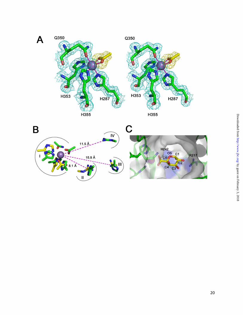
YeOGL. The coordination sphere is composed of
three histidines (H287, H353, and H355) and a
glutamine (Q350) residue that display coordinate
bond distances ranging from 2.1-2.2 Å for all six
ligands, consistent with compositions and
distances described for other manganese
complexes [33]. Q350 was modeled with O as the
donor atom and N hydrogen-bond contact with an
ordered water. This is the most stable tautomeric
form as N lacks a free electron pair for covalent
coordinate bond formation. The remaining two
atoms that complete the octahedral geometry was
determined to be the oxygen atoms of an acetate
carboxylate group, which is supported by B-factor
and difference map analysis (see Table II and
Figure 4A). Acetate is a component of the
crystallization buffer and mimics the chemistry of
the GalA uronate group.
This analysis defines the third pectate
lyase that coordinates Mn2+
, two of which are
cytoplasmic (PL2B/OGL) and one of which is
periplasmic (PL2A) ([13-14] Figure 1). These
select enzymes contrast the more abundant
examples of extracellular secreted pectate lyases
that exclusively utilize calcium. The molecular
basis for the relationship between cellular
distribution and metal selectivity had yet to be
elucidated. Although the possibility of metals
by guest on February 3, 2018http://w
ww
.jbc.org/D
ownloaded from

6
contributing different mechanistic effects to the
reaction must be considered, as calcium and
transition metals display distinct electron orbital
structures; it appears likely that this relationship
may reflect the distribution of trace elements
within prokaryotes. Although the roles of
intracellular calcium in bacteria are still poorly
understood, emerging evidence has revealed that
similar to eukaryotes, calcium is a signaling
molecule in many bacterial processes such as
transport, cell differentiation and the maintenance
of cell structure [34-35]. As a result, calcium
levels within the cell are limiting and tightly
regulated (100-300 nM) [36-37]. Therefore,
intracellular pectate lyases may have evolved
unique chemistries tailored for the coordination of
metal cofactors (Mn2+
as opposed to Ca2+
) that are
available in sufficient concentrations within the
microenvironment that they function in. The
observation that PL2A utilizes manganese and is
operational within the periplasm, however,
provides an interesting twist on this possibility as
the calcium utilizing pectate lyase, PelX (PL9) has
also been determined to be functional within the
periplasm [7, 38]. Analysis of the primary
structure of the intact YePL2A polypeptide reveals
the presence of a signature Lys-Arg di-peptide in
its secretion signal. This sequence marker
indicates that YePL2A is folded within the
cytoplasm and subsequently transported intact
through a TAT-pathway across the inner
membrane [39-40]. Y. enterocolitica lacks a PelX
homolog, and therefore may have diversified its
collection of pectinases by evolving to secrete a
PL2 homolog that would be active in the upstream
stages of the pathway. These enzymes are
observed in only a few pectinolytic microbes;
however, in almost every case they are found in
tandem PL2A and PL2B pairs (www.cazy.org,
[7]). Typically secreted pectinases transit through
a SEC-mediated pathway in an unfolded state, and
therefore, would incorporate a catalytic metal
within a folding microenvironment where calcium
is prevalent. This observation is reminiscent of the
targeted folding and selective metal acquisition of
the Synechocystis metalloproteins MncA and
CucA [41].
Structural Analysis of the YeOGL -elimination
Machinery - evolutionary convergence of the
catalytic machinery in pectate lyases has been
observed in families 1, 2, and 10 [13, 42].
Although these enzymes have no sequence
relatedness and adopt unique folds, they display a
constellation of functional residues within their
active site that is structurally conserved. These
include the components of the metal binding
pocket involved in -proton acidification; a basic
residue, typically an arginine, which operates as
the Brønstead base during H5 abstraction; and a
secondary arginine that flanks the substrate by
binding to its C2 and C3 hydroxyl groups.
Therefore, based upon this premise and in order to
dissect the molecular basis of -elimination within
the active site of YeOGL, we have explored the
evidence for structural convergence between
YeOGL and YePL2A. These two enzymes,
unrelated at the sequence level, represent the only
two characterized pectate lyases available in the
database in complex with a Mn2+
ion. Comparing
the active sites of these two lyases is complicated
by the presence of the two alternate conformations
of YePL2A that have been described, which
represent distinct structural snapshots across the
catalytic landscape of the enzyme. The Mn2+
complex, or “open” form is poised for substrate
binding; whereas the GalA3-Michaelis complex, or
“closed” form has locked its two catalytic arms
around the substrate, resulting in a rearrangement
of the core -elimination machinery [13]. The
lack of a metal in the YePL2A-GalA3 complex
results in a shift of the substrate orientation within
the active site as the uronate group is not anchored
by coordination. Preliminary rounds of analysis
determined that the metal bound complex of
YeOGL and YePL2A (with both enzymes lacking
sugar substrate) display the greatest level of
structural similarity in the alignment of their
catalytic residues. Therefore, in the following
discussion we will describe the superimposition of
the two active sites using the metal as a central
reference point.
Four main substructures were indentified
(I-IV) (Figure 4B). (I) The metal binding site
shares several structurally conserved donor atoms
within both enzymes, including two histidines.
Interestingly, the Q350 of YeOGL did not align
with any candidate residues from YePL2A. This
glutamine, which also binds an ordered water
molecule, may be functionally similar to the
catalytic amides from family PL9 and PL10
by guest on February 3, 2018http://w
ww
.jbc.org/D
ownloaded from

7
lyases. It has been proposed that amide groups
proximal to the uronate may stabilize the enolate-
enolate intermediate through hydrogen bonding
[42-43] (Figure 2). (II) The Brønstead base from
YePL2A (R171) structurally aligns with H242 in
YeOGL, the only basic residue with suitable
proximity and geometry for abstracting the H5 of a
bound GalA substrate. Histidine catalyzed -
elimination has been proposed for the family PL8
Streptcoccus pneumoniae hyaluronate lyase
(SpPL8) [44]; however, to the best of our
knowledge this is the first description of a similar
role for this residue in pectate lyases. The
remaining two substructures, (III) and (IV),
represent two potential substrate stabilizing
residues in YeOGL. (III) The H211 in YeOGL
superimposes well with the R272 of YePL2A in its
open conformation; however, this position is distal
to the metal binding site (~15.9 Å) and in an
unfavorable position to interact directly with the
substrate. In YePL2A this is explained through a
change in enzyme conformation; following GalA3
binding R272 undergoes a positional shift of 4.4 Å
towards the aglycone sugar. Further analysis of the
YeOGL structure reveals that the arginine
displayed in (IV) (R217) most likely interacts with
GalA in subsite +1 (bond cleavage occurs between
the sugars at subsites -1 and +1) based upon its
configuration, as both Nof the guanidine-group
are pointing towards the metal binding pocket
consistent with other pectate lyase structures,
functional group chemistry, and proximity. In
YeOGL, the two R217 nitrogen atoms are 11.2 Å
and 11.5 Å, respectively from the coordinated
metal, which is very similar to the distances
between the guanidine group of R272 (10. 9 Å and
11.0 Å) to a bound water molecule in the GalA3-
Michaelis complex of YePL2A (not shown). This
observation suggests that there is more
conformational flexibility within the active site of
YePL2A than YeOGL, and that the active site of
YeOGL presents another strong example of the
structural convergence of the -elimination
machinery in pectate lyases. The potential role of
this conserved highly conserved arginine in
catalytic proton transfer to the glycosidic oxygen
of the scissile bond, in YeOGL and other pectate
lyases, remains to be established [42, 45].
Crystallographic investigations into the
molecular basis of YeOGL recognition of
oligogalacturonides have proven unsuccessful.
Fortuitously, however, the coordination of an
acetate molecule to the Mn2+
ion provides a
framework for predicting the position of a GalA
molecule within the active site (Figure 4C). A
YeOGL-GalA model was constructed by
superposing the uronate group of GalA with the
bound acetate group. To select between the two
possible orientations, the monosaccharide was
positioned such that the axial-hydrogen is pointing
towards the candidate Brønstead base, H242; as
opposed to towards the top of the active site, a
region lacking any definable basic residue suitable
for potential proton abstraction. In this
configuration, the O2 hydroxyl of GalA is directed
towards the guanidine group of R217. Although
the model does not account for subtle changes in
conformation and orientation that may be induced
by binding energy, the two molecules are already
approaching hydrogen bond distance. O1 is
pointing towards the core of the enzyme and the
rear exit of the solvent tunnel, and the axial O4 is
positioned towards the tunnel entrance to
accommodate polymerization of another GalA.
Based upon these observations, it is likely that in
the native complex GalA undergoes a small
rotation around its planar axis so that O2 and O3
both interact with R217, and O4 extends closer
towards the center of the solvent tunnel to alleviate
any steric clash with the wall of the enzyme.
Transition Metal Utilization by YeOGL – to test
the viability of our model and interrogate the role
of Mn2+
in the elimination of oligogalacturonate
we have characterized the active site by enzyme
kinetics and metal supplementation assays.
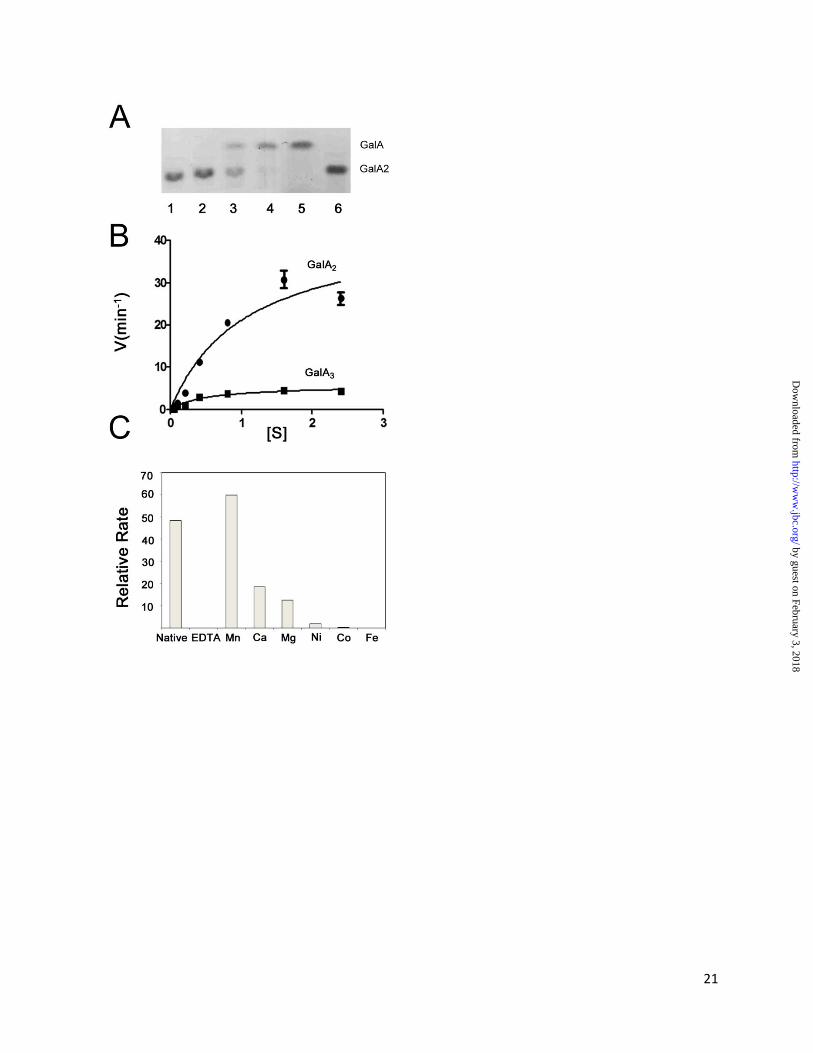
Preliminary assays revealed that YeOGL is active
on GalA2 (Figure 5A). Calculated kinetic values
(see Figure 5B and Table III) suggest that YeOGL
has an identical activity to its EcOGL homolog
[14], a result that is not surprising when
considering the high level of amino acid
conservation between the two proteins. The ~8-
fold higher maximal velocity of YeOGL on GalA2
(43.67 ± 5.16 min-1
) compared with GalA3 (5.70 ±
0.49 min-1
), underpins the complementary role of
YePL2B/PelW. YePL2B/PelW is dedicated to the
conversion of cytoplasmic trisaccharides into
substrates for OGL, a process which will
ultimately increase the efficiency of the upstream
stages of cytoplasmic oligogalacturonide
by guest on February 3, 2018http://w
ww
.jbc.org/D
ownloaded from
8
catabolism (see Figure 1). Previously, a cluster of
transition metals, ranging from the 7-10th row of
the fourth period, were previously shown to be
capable of assisting in the catalytic activity of
EcOGL to varying degrees
(Mn2+
>Co2+
>Fe2+
>Ni2+
) [14]. This finding
indicated that the metal coordinating machinery of
this lysase family is tailored to utilizing transition
metals. Similar recovery assays performed for
YeOGL also revealed a preferential relationship
with Mn2+
, as other cofactors failed to restore
similar levels of enzyme activity (Figure 5C).
These assays support the structural observations
for tailored metal binding within the OGL family
discussed above.
Evolution of the OGL Family – EcOGL was first
discovered in 1968 (originally referred to as
oligogalacturonide transeliminase, [46]) and
known to be found in diverse species of
Enterobacteriaceae, including Y. enterocolitica for
over a decade [8]; however, a detailed analysis of
the relatedness of orthologs and its classification
as a novel polysaccharide lyase family in the
CAZy database has been lacking. Therefore, to
provide insight into the functional relatedness of
the family we did a BLAST search using the
YeOGL as a query sequence. Initial searches
identified 46 family members (with a low-end
cutoff value of ≥10 for several outlying sequences
as determined by BH and PMC at CAZy.org,
personal communication) that partition into two
clusters, one of which contains ~85% of the entries
(relatedness value of 53-97%), including the
characterized enzymes from E. chrysanthemi
(CAA43990) and Y. enterocolitica (CAL11955)
and the uncharacterized structural homolog from
V. parahaemolyticus (BAC61431). The smaller
seven member out-group (relatedness threshold
value of 10-52%) consists of uncharacterized
putative enzymes from Candidatus Solibacter
usitatus (ABJ83750, ABJ85020, and ABJ83823),
Roseiflexus castenholizii (ABU58452 and
ABU58528), and single entries from Geobacillus
sp. and Haloterrigena turkmenica. The presence of
OGL in each of these species indicates that these
microorganisms have a propensity to utilize pectin
as an energy source.
To compare the functional residues across
the OGL the family, a ClustalW alignment [47]
was performed on sequences from each
representative species; redundant entries from
individual strains were omitted for clarity (Figure
6). There is high level sequence conservation
across the entire family. Inspection of the
putative catalytic histidine (H242) and stabilizing
arginine (R217) reveals that they are virtually
invariant. Only one sequence (ABJ83823) from S.
usitatus displays unique residues at these
positions, which in addition contains a completely
remodeled metal binding site and raises the
question of whether it is even a functional lyase.
Closer analysis of the other two entries from S.
usitatus (ABJ83750 and ABJ85020) may provide
insight into the potential relationship between
metal selectivity and cellular localization we have
described above. Both these putative enzymes
contain bona fide signal peptides. The lack of a
consensus TAT sequence (not shown) suggests
that they are targeted for a general secretion
pathway and therefore are folded outside the
cytoplasm where Ca2+
is abundant. Closer
investigation of catalytic machinery in these
homologs reveals that they contain the putative
catalytic histidine; however, noticeable
substitutions are present within the metal binding
site. The H353 and H355 from YeOGL are
replaced with a glutamate and asparagine residue,
respectively. These functional groups contribute
oxygen donor atoms during coordinate bond
formation, indicating a potential loss of Mn2+
and
gain of Ca2+
binding specificity. In addition,
although H287 is conserved in the primary
structure, it is located in a stretch of amino acids
displaying very little similarity in the S. usitatus
proteins, and therefore, it is difficult to justify a
spatial and functional conservation of the histidine
at this site. These two predicted enzymes from the
OGL family, therefore, provide further support for
the hypothesis that pectate lyase catalytic metal
selectivity is based upon the location of enzyme
folding rather than mechanistic effects.
The observation that Yersinia pestis, the
causative agent of the bubonic plague, possesses
an OGL homolog is intriguing. In addition to this
cytoplasmic lyase, the genome of every known Y.
pestis strain was shown to contain genetic
evidence of a well-developed pectin utilization
pathway ([7], www.cazy.org). These observations
appear inconsistent with traditional life-cycle
by guest on February 3, 2018http://w
ww
.jbc.org/D
ownloaded from

9
models of the pathogen, as Y. pestis is widely
considered an enzootic disease and is not known to
colonize the digestive tract of its mammalian host.
It is difficult to rationalize therefore where the
microbe would have access to dietary pectin and
concomitantly why it would contain a pathway
tailored to its catabolism. Recently, however, it
has been reported that Y. pestis can persist within
soil [48-50], a process that may be facilitated by
scavenging dietary carbohydrates such as pectin.
Utilizing plant cell wall polysaccharides as a
transient energy source may explain the
mechanistic basis of how Y. pestis lies dormant
within and passages through the environment as it
waits for an opportunity to colonize new host
reservoirs. This possibility awaits further
investigation, however, as the presence of pectin
utilization genes within Y. pestis may more simply
reflect the evolutionary history of this genus, and
this pathway may in fact be a decommissioned
remnant containing non-functional enzymes.
Conclusion
Early structural analysis suggested that
PLs harness a structurally conserved -helix fold
to modify and dismantle pectin [51-52]. Structural
biology within the past decade, however, has
illuminated the great structural diversity in
proteins folds that actually exists within nature for
this class of carbohydrate-active enzyme (this
work, [13, 42]). Analysis of the catalytic
substructures within diverse PL families has
established a new paradigm for our understanding
of PL structure-function relationships: the
remarkable structural convergence of active site
chemistry. Tailoring distinct protein scaffolds with
highly similar catalytic apparatus strongly
suggests that the -elimination machinery and the
stereochemical identity of their target substrate
place heavy selective pressure on the evolutionary
processes driving the structural convergence of
these enzyme families. Beyond the core features of
the PL active site, however, several variations on
this theme have begun to surface with the increase
of available structural information. First, it appears
that the OGL family utilizes a histidine as a
Brønstead base during H5-abstraction. Outside of
family 9 pectate lyases, which utilize a lysine, all
other described examples exclusively display
catalytic arginines. The significance of this
chemical substitution likely reflects the localized
OGL environment. Indeed, the optimal pH of
YeOGL is between 7.3-7.7 (EcOGL [14], YeOGL
not shown) consistent with the buffered solution of
the bacterial cell. Additionally, this pH optimum is
more alkaline than the pKa of histidine (pKa =
6.1), an observation that contrasts that of
extracellular lyases. Secreted enzymes display a
pH optimum around 9.0 [25], which requires
localized pKa effects perturbing the catalytic
arginine (pKa = 12.5) to display a more acidic
character. Second, the metal binding site of
YeOGL possesses three histidines and a glutamine,
tailored for the coordination of Mn2+
. This is the
second lyase, representing two different PL
families with unique protein folds, that harnesses a
metal cofactor other than calcium. What
distinguishes these two enzyme families from
previous complexes is that YeOGL and YePL2B
are cytoplasmic lyases, and therefore fold and
operate in an environment that limits the
availability of calcium ions. The periplasmic
homolog of YePL2B, YePL2A, contains a TAT-
secretion signal indicating in its signal peptide,
indicating that it is folded within the cell and
secreted into the periplasm intact. This observation
suggests that it is the environment in which the
enzyme is folded that dictates the metal cofactor
selection within the active site of pectate lyases
and not the mechanistic influences.
As the field continues to advance it will be
exciting to discover whether the vast arsenal of
uncharacterized pectate lyases encoded within the
distal gut microbiome also display structural
convergence in the architecture of their catalytic
site and a metal selectivity dependent on cellular
localization, or if this dynamic and densely
populated community has evolved novel platforms
for the -elimination of pectate.
Acknowledgments: We wish to thank B.
Henrissat and P.M. Coutinho for their assistance in
defining the cutoff parameters for the OGL family.
A.B.B. is a Canada Research Chair in Molecular
Interactions and a MSFHR Career Scholar. H.J.G.
is a Georgia Research Alliance Eminent Scholar in
Bioenergy.
by guest on February 3, 2018http://w
ww
.jbc.org/D
ownloaded from

10
Table I: Crystallographic statistics for the structure of YeOGL.
Data Collection Refinement
Space group P212121 Resolution (Å) 35.42-1.65 (1.69-1.65)
Cell dimensions No. reflections 44824
a (Å) 66.65 Rwork / Rfree 0.18/0.20
b (Å) 75.51 No. atoms 3527
c ( Å) 78.51 Protein 3019
() 90.00 Acetate/ion 4/3
() 90.00 Water 497
() 90.00 B-factors (Å2)
Resolution (Å) 30.00-1.65 Protein 15.4
Rmerge 0.085 (0.35) Acetate/ion 19.0 /12.0
I / I 14.9 (4.0) Water 30.8
Completeness (%) 97.7 (85.5) R.m.s deviations
Multiplicity 6.8 (4.4) Bond lengths (Å) 0.012
Average Mosaicity 0.44 Bond angles () 1.554
Ramachandran Statistics
Favored 98.9%
Allowed 0.5%
Outlier 0.5% (Gly373)
by guest on February 3, 2018http://w
ww
.jbc.org/D
ownloaded from

11
Table II: Atomic distances and temperature factors for the manganese coordination pocket of
YeOGL.
Ligand Distance B-factor Occupancy
Mn - 7.5 Å2 1.0
NH287 2.1 Å 9.5 Å2
1.0
OQ350 2.2 Å 10.2 Å2 1.0
NH353 2.2 Å 8.7 Å2 1.0
NH355 2.1 Å 8.3 Å2 1.0
OAACT 2.1 Å 17.0 Å2 1.0
OBACT 2.3 Å 16.2 Å2 1.0
Table III: Kinetic values of YeOGL
Substrate KM (mM) Kcat (min-1
) Kcat/KM (min-1
mM-1
)
GalA2 1.07 ± 0.28* 43.67 ± 5.16 40.81
GalA3 0.52 ± 1.2 5.70 ± 0.49 10.96
*Standard deviations are based upon the rate calculations for three independently performed experiments.
by guest on February 3, 2018http://w
ww
.jbc.org/D
ownloaded from

12
REFERENCES
1. Backhed, F., et al., Host-bacterial mutualism in the human intestine. Science, 2005. 307(5717): p.
1915-20.
2. Martens, E.C., et al., Complex glycan catabolism by the human gut microbiota: the Bacteroidetes
Sus-like paradigm. J Biol Chem, 2009. 284(37): p. 24673-7.
3. O'Hara, A.M. and F. Shanahan, The gut flora as a forgotten organ. EMBO Rep, 2006. 7(7): p.
688-93.
4. Sonnenburg, J.L., et al., Glycan foraging in vivo by an intestine-adapted bacterial symbiont.
Science, 2005. 307(5717): p. 1955-9.
5. Martens, E.C., et al., Coordinate regulation of glycan degradation and polysaccharide capsule
biosynthesis by a prominent human gut symbiont. J Biol Chem, 2009. 284(27): p. 18445-57.
6. Martens, E.C., H.C. Chiang, and J.I. Gordon, Mucosal glycan foraging enhances fitness and
transmission of a saccharolytic human gut bacterial symbiont. Cell Host Microbe, 2008. 4(5): p.
447-57.
7. Rodionov, D.A., M.S. Gelfand, and N. Hugouvieux-Cotte-Pattat, Comparative genomics of the
KdgR regulon in Erwinia chrysanthemi 3937 and other gamma-proteobacteria. Microbiology,
2004. 150(Pt 11): p. 3571-90.
8. Rodionov, D.A., et al., Transcriptional regulation of transport and utilization systems for
hexuronides, hexuronates and hexonates in gamma purple bacteria. Mol Microbiol, 2000. 38(4):
p. 673-83.
9. Caffall, K.H. and D. Mohnen, The structure, function, and biosynthesis of plant cell wall pectic
polysaccharides. Carbohydr Res, 2009. 344(14): p. 1879-900.
10. Gustafsson, M., et al., Novel Polynuclear Nickel(II) Complex: Hydrazine, Sulfato, and Hydroxo
Bridging in an Unusual Metal Hexamer. Crystal Structure and Magnetic Properties of
[Ni(6)(N(2)H(4))(6)(SO(4))(4)(OH)(2)(H(2)O)(8)](SO(4))(H(2)O)(10). Inorg Chem, 2010.
11. Abbott, D.W. and A.B. Boraston, Structural biology of pectin degradation by
Enterobacteriaceae. Microbiol Mol Biol Rev, 2008. 72(2): p. 301-16, table of contents.
12. Hugouvieux-Cotte-Pattat, N., et al., Regulation of pectinolysis in Erwinia chrysanthemi. Annu
Rev Microbiol, 1996. 50: p. 213-57.
13. Abbott, D.W. and A.B. Boraston, A family 2 pectate lyase displays a rare fold and transition
metal-assisted beta-elimination. J Biol Chem, 2007. 282(48): p. 35328-36.
14. Shevchik, V.E., et al., The exopolygalacturonate lyase PelW and the oligogalacturonate lyase
Ogl, two cytoplasmic enzymes of pectin catabolism in Erwinia chrysanthemi 3937. J Bacteriol,
1999. 181(13): p. 3912-9.
15. Cantarel, B.L., et al., The Carbohydrate-Active EnZymes database (CAZy): an expert resource for
Glycogenomics. Nucleic Acids Res, 2009. 37(Database issue): p. D233-8.
16. Boraston, A.B., et al., Binding specificity and thermodynamics of a family 9 carbohydrate-
binding module from Thermotoga maritima xylanase 10A. Biochemistry, 2001. 40(21): p. 6240-
6247.
17. Evans, P., Scaling and assessment of data quality. Acta Crystallogr D Biol Crystallogr, 2006.
62(Pt 1): p. 72-82.
18. McCoy, A.J., et al., Phaser crystallographic software. J Appl Crystallogr, 2007. 40(Pt 4): p. 658-
674.
19. Emsley, P. and K. Cowtan, Coot: model-building tools for molecular graphics. Acta Crystallogr
D Biol Crystallogr, 2004. 60(Pt 12 Pt 1): p. 2126-32.
20. Murshudov, G.N., A.A. Vagin, and E.J. Dodson, Refinement of macromolecular structures by the
maximum-likelihood method. Acta Crystallogr D Biol Crystallogr, 1997. 53(Pt 3): p. 240-55.
21. Potterton, L., et al., Developments in the CCP4 molecular-graphics project. Acta Crystallogr D
Biol Crystallogr, 2004. 60(Pt 12 Pt 1): p. 2288-94.
by guest on February 3, 2018http://w
ww
.jbc.org/D
ownloaded from

13
22. Vaguine, A.A., J. Richelle, and S.J. Wodak, SFCHECK: a unified set of procedures for
evaluating the quality of macromolecular structure-factor data and their agreement with the
atomic model. Acta Crystallogr D Biol Crystallogr, 1999. 55(Pt 1): p. 191-205.
23. Roman A Laskowski, M.W.M., David S Moss and Janet M Thornton, PROCHECK: a program
to check the stereochemical quality of protein structures Journal of Applied Crystallography.,
1993. 26(283): p. 283-291.
24. Kirschner, K.N., et al., GLYCAM06: a generalizable biomolecular force field. Carbohydrates. J
Comput Chem, 2008. 29(4): p. 622-55.
25. Tardy, F., et al., Comparative analysis of the five major Erwinia chrysanthemi pectate lyases:
enzyme characteristics and potential inhibitors. J Bacteriol, 1997. 179(8): p. 2503-11.
26. Scavetta, R.D., et al., Structure of a plant cell wall fragment complexed to pectate lyase C. Plant
Cell, 1999. 11(6): p. 1081-92.
27. Murzina, N.V., et al., Structural basis for the recognition of histone H4 by the histone-chaperone
RbAp46. Structure, 2008. 16(7): p. 1077-85.
28. Xu, Y., et al., Structure of a protein phosphatase 2A holoenzyme: insights into B55-mediated Tau
dephosphorylation. Mol Cell, 2008. 31(6): p. 873-85.
29. Holm, L., et al., Searching protein structure databases with DaliLite v.3. Bioinformatics, 2008.
24(23): p. 2780-1.
30. Rouvinen, J., et al., Three-dimensional structure of cellobiohydrolase II from Trichoderma reesei.
Science, 1990. 249(4967): p. 380-6.
31. Davies, G. and B. Henrissat, Structures and mechanisms of glycosyl hydrolases. Structure, 1995.
3(9): p. 853-9.
32. Liners, F., et al., Monoclonal Antibodies against Pectin: Recognition of a Conformation Induced
by Calcium. Plant Physiol, 1989. 91(4): p. 1419-1424.
33. Harding, M.M., Geometry of metal-ligand interactions in proteins. Acta Crystallogr D Biol
Crystallogr, 2001. 57(Pt 3): p. 401-11.
34. Dominguez, D.C., Calcium signalling in bacteria. Mol Microbiol, 2004. 54(2): p. 291-7.
35. Michiels, J., et al., The functions of Ca(2+) in bacteria: a role for EF-hand proteins? Trends
Microbiol, 2002. 10(2): p. 87-93.
36. Gangola, P. and B.P. Rosen, Maintenance of intracellular calcium in Escherichia coli. J Biol
Chem, 1987. 262(26): p. 12570-4.
37. Gilroy, S., et al., Role of Calcium in Signal Transduction of Commelina Guard Cells. Plant Cell,
1991. 3(4): p. 333-344.
38. Shevchik, V.E., et al., Characterization of the exopolygalacturonate lyase PelX of Erwinia
chrysanthemi 3937. J Bacteriol, 1999. 181(5): p. 1652-63.
39. DeLisa, M.P., et al., Genetic analysis of the twin arginine translocator secretion pathway in
bacteria. J Biol Chem, 2002. 277(33): p. 29825-31.
40. De Buck, E., E. Lammertyn, and J. Anne, The importance of the twin-arginine translocation
pathway for bacterial virulence. Trends Microbiol, 2008. 16(9): p. 442-53.
41. Tottey, S., et al., Protein-folding location can regulate manganese-binding versus copper- or
zinc-binding. Nature, 2008. 455(7216): p. 1138-42.
42. Charnock, S.J., et al., Convergent evolution sheds light on the anti-beta -elimination mechanism
common to family 1 and 10 polysaccharide lyases. Proc Natl Acad Sci U S A, 2002. 99(19): p.
12067-72.
43. Jenkins, J., et al., The crystal structure of pectate lyase Pel9A from Erwinia chrysanthemi. J Biol
Chem, 2004. 279(10): p. 9139-45.
44. Li, S., et al., Structural basis of hyaluronan degradation by Streptococcus pneumoniae
hyaluronate lyase. EMBO J, 2000. 19(6): p. 1228-40.
45. Seyedarabi, A., et al., Structural insights into substrate specificity and the anti beta-elimination
mechanism of pectate lyase. Biochemistry, 2010. 49(3): p. 539-46.
by guest on February 3, 2018http://w
ww
.jbc.org/D
ownloaded from

14
46. Moran, F., S. Nasuno, and M.P. Starr, Oligogalacturonide trans-eliminase of Erwinia carotovora.
Arch Biochem Biophys, 1968. 125(3): p. 734-41.
47. Thompson, J.D., D.G. Higgins, and T.J. Gibson, CLUSTAL W: improving the sensitivity of
progressive multiple sequence alignment through sequence weighting, position-specific gap
penalties and weight matrix choice. Nucleic Acids Res, 1994. 22(22): p. 4673-80.
48. Ayyadurai, S., et al., Long-term persistence of virulent Yersinia pestis in soil. Microbiology,
2008. 154(Pt 9): p. 2865-71.
49. Drancourt, M., L. Houhamdi, and D. Raoult, Yersinia pestis as a telluric, human ectoparasite-
borne organism. Lancet Infect Dis, 2006. 6(4): p. 234-41.
50. Eisen, R.J., et al., Persistence of Yersinia pestis in soil under natural conditions. Emerg Infect
Dis, 2008. 14(6): p. 941-3.
51. Jenkins, J. and R. Pickersgill, The architecture of parallel beta-helices and related folds. Prog
Biophys Mol Biol, 2001. 77(2): p. 111-75.
52. Jenkins, J., O. Mayans, and R. Pickersgill, Structure and evolution of parallel beta-helix proteins.
J Struct Biol, 1998. 122(1-2): p. 236-46.
53. Read, R.J., Improved Fourier coefficients for maps using phases from partial structures with
errors. Acta Cryst.A, 1986. 42: p. 140-149.
by guest on February 3, 2018http://w
ww
.jbc.org/D
ownloaded from

15
FIGURE LEGENDS
Figure 1: General pectin degradation pathway found in pectinolytic bacteria from
Enterobacteraciae [11]. Extracellular pectin depolymerization is primarily restricted to phytopathogens
and involves a consortium of enzymes that liberate fragments from the highly polymerized pectic network
within the plant cell wall. Pectinases involved in these processes include pectate lyases (PLs),
polygalacturonases (GH28s) and carbohydrate esterases (CE8). Products diffuse into the periplasmic
space through anionic porins belonging to the KdgM family where a specialized collection of proteins
operate to retain and concentrate (CBM32), and process pectic substrates into mono-, di-, and
trisaccharides (PLs and exoGH28). Intracellular transport of oligogalacturonides is facilitated by integral
membrane systems, including the ATP-dependent transporter TogMNAB. Within the cell, pectin
catabolism remains poorly understood at the structural level; however, a pathway has been established
based upon genetics, biochemistry, and comparative genomics [7, 12]. There are two branches of
metabolism dedicated to saturated monogalacturonate (GalA: UxaA, UxaB, UxaC), and unsaturated
monogalacturonate (DKI: KduI and KduD), the product of pectate lyases. These pathways converge to
produce 2-keto-3-deoxygluconate (KDG) which is the inducer of KdgR, a repressor protein that regulates
the expression of virtually every pectin utilization gene known. KDG is converted into the metabolites 3-
phosphoglyceraldehyde (3-PGA) and pyruvate by a two-step phosphorylation and hydrolysis reaction
path catalyzed by KdgK and KdgA, respectively.
Figure 2: General -elimination reaction mechanism for catalysis by pectate lyases. 1,4-linked
galacturonate substrates adopting the relaxed 4C1 chair conformation present the ideal geometry for an
anti periplanar transelimination with the C5 hydrogen and C4 hydroxyl group both orientated in axial
configurations. The rate limiting step is the acidification and deprotonation of C5, a process that is
facilitated by the effects of the electrophilic uronate group, catalytic divalent metals, and localized basic
residues within the active site, which draw charge from the C5 carbon and increase the reactivity of its
lone hydrogen atom. Abstraction occurs via general base attack by an arginine (Family 1, 2, 3, 10) or
lysine (Family 9) [43], the high pKa of these residues accounting for the basic pH optima observed across
the PL landscape. Recently, the contributions of an ancillary lysine in family 1 PLs and asparagines in
family 9, have been proposed to influence the catalytic rate by stabilizing the enolate-enolate intermediate
[45]. These residues have been implicated in either hydrogen donation and or bonding to the nascent
oxyanion of the uronate group following electron transfer to the transient double bond between C5 and
C6. Decomposition of the intermediate occurs by protonation of the scissile glycosidic oxygen and
elimination of the axial O4 by electron shuttling from the C5-C6 bond to the C4-C5 bond of the sugar
ring. This unsaturation distorts the relaxed chair structure of the pyranose as the trigonal planar geometry
of C4 and C5 results in migration of C4 into the plane of C2, C3 and C5. The substrate is shown is a
disaccharide, with the R-group depicting a second GalA or DKI molecule (note the Asn residue shown
may be equivalent to Q350).
Figure 3: -propeller fold of YeOGL. (A) Structural alignment of OGLs from Y. enterocolitica
(YeOGL), E. chrysanthemi (EcOGL), and V. parahaemolyticus (VpOGL). The secondary structure
elements of YeOGL are displayed above the aligned primary structure. Amino acids targeted for
mutagenesis are indicated with black triangles below the sequences. (B) Cartoon representation of YeOGL
structure colored from the N-terminus (blue) to C-terminus (red). The seven -propellers are labeled (1-7)
with each strand (A-D) packing from the core towards the surface. The three bound metal ions are
displayed as spheres with the catalytic manganese colored in purple and the surface bound calciums in
light blue. (C) Electrostatic surface potential of YeOGL. The protein is centered to display the entrance to
the active site that is lined with several basic patches.
Figure 4: The active site of YeOGL. (A) The octahedral metal binding site is displayed in wall-eyed
stereo format with the weighted maximum-likelihood [20]/σA [53] 2Fobs - Fcalc (cyan) map (cyan)
by guest on February 3, 2018http://w
ww
.jbc.org/D
ownloaded from

16
contoured to 1.0 σ (0.43 e-/Å
3). The coordinated acetate ion (yellow sticks) is shown with a Fobs-Fcalc omit
map, produced from phases generated during refinements with the acetate molecule excluded from the
model, contoured at 2.5 σ (0.18 e-/A
3). This map demonstrates the high quality of unbiased electron
density for this molecule. The three histidines (H287, H353, and H355) and glutamine (Q350) are shown
in stick representation, and the Mn2+
ion is modeled as a sphere. (B) Superimposition of catalytic
substructures within the active site of YePL2A (yellow, PDB ID: 2V8J) and YeOGL (green). Four aligned
regions are shown: (I) metal coordination pocket, (II) catalytic base, and (III-IV) potential residues
involved in substrate stabilization. Distances of the residues from the metal are labeled. (C) Cut-away
surface representation of the active site with a GalA modeled in the +1 subsite. The GalA uronate group
of the aglycone is superimposed with the coordinated acetate ion with the axial H5 pointing towards the
candidate Brønstead base H242, O2 the stabilizing R217, and O4 is presented for polymerization.
Figure 5: Activity of YeOGL. (A) TLC gel of GalA22 digestion. Lanes are loaded as follows: (1) No
enzyme control, (2-4) increasing reaction time points (0-5 minutes), (5) GalA standard, and (6) GalA2
standard. (B) Reaction rates plotted velocity (min-1
) against substrate concentration (mM). (C) Metal
supplementation assays. Rates shown represent quantified changes in UV absorbance.
Figure 6: Pylogenetic tree of the OGL family. Related sequence targets are shown as a phylogram. The
out-group is indicated with an asterix. The inset panel displays the level of conservation of functional
residues for the seven out-group sequences and the putative secreted pectate lyases are indicated with
black triangles.
by guest on February 3, 2018http://w
ww
.jbc.org/D
ownloaded from

D. Wade Abbott, Harry J. Gilbert and Alisdair B. BorastonCYTOPLASMIC OLIGOGALACTURONATE BETA-ELIMINATIONTHE ACTIVE SITE OF OGL PROVIDES UNIQUE INSIGHTS INTO
published online September 17, 2010J. Biol. Chem.
10.1074/jbc.M110.153981Access the most updated version of this article at doi:
Alerts:
When a correction for this article is posted•
When this article is cited•
to choose from all of JBC's e-mail alertsClick here
by guest on February 3, 2018http://w
ww
.jbc.org/D
ownloaded from


![Weg51005 OGL D6 System Book[1]](https://img.pdfslide.us/doc/110x75/5475be40b4af9fba5b8b4569/weg51005-ogl-d6-system-book1.jpg)