Embed Size (px)
Citation preview

211
The ABA-1 allergen of Ascaris lumbricoides : sequence
polymorphism, stage and tissue-specific expression, lipid
binding function, and protein biophysical properties
Y. XIA "†, H. J. SPENCE"†, J. MOORE", N. HEANEY", L. MDERMOTT",#, A. COOPER#,
D. G. WATSON$, B. MEI%†, R. KOMUNIECKI% and M. W. KENNEDY"*
" Division of Infection and Immunity, Institute of Biomedical and Life Sciences, Joseph Black Building, University ofGlasgow, Glasgow G12 8QQ, Scotland, UK# Department of Chemistry, University of Glasgow, Glasgow G12 8QQ, Scotland, UK$ Department of Pharmaceutical Sciences, University of Strathclyde, Glasgow G1 1XW% Department of Biology, University of Toledo, Toledo, OH 43606, USA
(Received 18 June 1999; revised 20 August 1999; accepted 20 August 1999)
The ABA-1 protein of Ascaris lumbricoides (of humans) and Ascaris suum (of pigs) is abundant in the pseudocoelomic fluid
of the parasites and also appears to be released by the tissue-parasitic larvae and the adult stages. The genes encoding the
polyprotein precursor of ABA-1 (aba-1) were found to be arranged similarly in the two taxa, comprising tandemly
repeating units encoding a large polyprotein which is cleaved to yield polypeptides of approximately 15 kDa which fall
into 2 distinct classes, types A and B. The polyprotein possibly comprises only 10 units. The aba-1 gene of A. lumbricoides
is polymorphic, and the majority of substitutions observed occur in or near predicted loop regions in the encoded proteins.
mRNA for ABA-1 is present in infective larvae within the egg, and in all parasitic stages, but was not detectable in
unembryonated eggs. ABA-1 mRNA was confined to the gut of adult parasites, and not in body wall or reproductive
tissues. Recombinant protein representing a single A-type unit for the A. lumbricoides aba-1 gene was produced and found
to bind retinol (Vitamin A) and a range of fatty acids, including the pharmacologically active lipids lysophosphatidic acid,
lysoplatelet activating factor, and there was also evidence of binding to leukotrienes. It failed to bind to any of the
anthelmintics screened. Differential Scanning Calorimetry showed that the recombinant protein was highly stable, and
unfolded in a single transition at 90±4 °C. Analysis of the transition indicated that the protein occurs as a dimer and that
the dimer dissociates simultaneously with the unfolding of the monomer units.
Key words: Ascaris lumbricoides, ABA-1 allergen, polymorphism, nematode polyprotein allergens}antigens, fatty acid
binding protein.
The ABA-1 protein of Ascaris lumbricoides (of
humans) and Ascaris suum (of pigs) is abundant in
the pseudocoelomic fluid of the parasites and also
appears to be released by the tissue-parasitic larvae
and the adult stages (Kennedy & Qureshi, 1986;
Kennedy et al. 1987a, 1989). ABA-1 is probably
Allergen A of A. suum (Ambler et al. 1973; Christie
et al. 1990). It is the target of IgE antibody responses
in both infected humans and rodents (Tomlinson et
* Corresponding author: Division of Infection and
Immunity, Joseph Black Building, University of Glasgow,
Glasgow G12 8QQ, Scotland, UK. Tel: 44 (0) 141-330-
5819. Fax: 44 (0) 141-330-4600. E-mail : malcolm.
kennedy!bio.gla.ac.uk
† Present addresses: Dr Yu Xia, Department of
Pathology, School of Medicine, University of Louisville,
Louisville, KY40292; Dr Heather J. Spence, Beatson
Institute of Cancer Research, University of Glasgow,
Glasgow G61 1QH; Dr Baisong Mei, Department of
Nutritional Sciences, University of California, Berkeley,
Berkeley, CA 94720, USA.
al. 1989; Christie et al. 1990; Kennedy, Fraser &
Christie, 1991; Fraser, Christie & Kennedy, 1993;
McSharry et al. 1999), but it remains to be
established whether its allergenic activity is an
intrinsic property of the protein or merely due to
generalized IgE potentiation by the infection
(Christie, Fraser & Kennedy, 1992; Jarrett & Miller,
1982). There is evidence, however, that a homologue
from filarial nematodes is also associated with
Th2}IgE responses (Paxton et al. 1993; Yazdan-
bakhsh et al. 1995; Allen, Lawrence & Maizels,
1995). Only a subset of Ascaris-infected people
produce antibody to ABA-1 (Kennedy et al. 1990;
McSharry et al. 1999), which is probably due to
genetic restriction of the immune repertoire, as has
been formally demonstrated in rodents (Kennedy et
al. 1987b ; Tomlinson et al. 1989; Christie et al.
1990; Kennedy et al. 1991) and also applies to
ABA-1 homologues in other nematode infections
(Kwan-Lim & Maizels, 1990; Allen et al. 1995). In
those humans who respond to ABA-1 there is an
association between IgE antibody to ABA-1 and
Parasitology (2000), 120, 211–224. Printed in the United Kingdom " 2000 Cambridge University Press

Y. Xia and others 212
relative resistance to infection, although, as with
many such studies, this remains an associative rather
than a causal link (McSharry et al. 1999).
Homologues of ABA-1 have been described in a
wide range of nematode species, and DNA sequences
encoding them arise frequently from the screening of
cDNA expression libraries with antibody from
infected hosts (e.g. Culpepper et al. 1992; Poole et al.
1992; Tweedie et al. 1993; Ajuh et al. 1995; de Graaf
et al. 1995; Yahiro, Cain & Butler, 1998). These
proteins are unusual in being produced as large
polyproteins which are post-translationally cleaved
to yield multiple copies of approximately 15 kDa
polypeptides (Tweedie et al. 1993; Selkirk et al.
1993; Spence et al. 1993; Poole et al. 1996). This
unusual means of synthesis has led to the term
Nematode Polyprotein Allergens}antigens (NPAs).
The individual units withing the polyprotein pre-
cursors can be highly diverse in amino acid sequence
(Britton et al. 1995), although some species have
several units with identical or very similar sequences
(Culpepper et al. 1992; Poole et al. 1992; Tweedie et
al. 1993; Ajuh et al. 1995; Yahiro et al. 1998). The
NPA gene (aba-1) of A. suum is known to have two
distinct classes of repeat unit ; type A (9 very similar
units) and type B (1 unit), whose sequences show
only 50% identity (Moore et al. 1999).
Parasite-derived ABA-1 or recombinant forms of
single NPA units from several species have been
found to bind a variety of small lipids including fatty
acids and retinol (Vitamin A) (Kennedy et al.
1995a–c ; Moore et al. 1999). In the case of ABA-1,
fluorescence methods have revealed subtle differ-
ences in the ligand-binding sites of type A and type
B proteins, although with no fundamental disparity
in their binding activities (Moore et al. 1999).
A. lumbricoides and A. suum are able to infect
either humans or pigs, but cross-infection with
adult worms is rare in endemic areas in which
genetic studies have shown that the Ascaris of
humans and pigs exist essentially as reproductively
isolated populations and could be considered as
distinct taxa (Anderson, 1995; Anderson, Romero-
Abel & Jaenike, 1993, 1995; Anderson & Jaenike,
1997; Peng et al. 1998; Zhu et al. 1999). It is
conceivable, however, that the tissue-invasive larval
stages of A. suum infect humans and cause pul-
monary hypersensitivity reactions. Such cross-infec-
tion may also influence the development of
immunity to A. lumbricoides. It is therefore of
interest to know whether the ABA-1 allergen
differs between the two species, and whether poly-
morphisms exist.
Here we report on the aba-1 gene of A.
lumbricoides, using parasites recovered from humans
at 2 different geographical locations, and show that
A. lumbricoides ABA-1 protein is similar to that of A.
suum, and that the gene is polymorphic. We also
provide new information on the organization of the
aba-1 gene, and on the biochemical and structural
properties of the protein.†
Parasite and host materials
A. lumbricoides worms from Guatamala were a gift
from Dr T. J. C. Anderson (Oxford University), and
A. lumbricoides worms from China were kindly
provided by Professor Weidong Peng (Nanchang,
China); the latter were genetically typed as carrying
alleles unique to human-derived Ascaris. A. suum
worms were obtained from the intestines of infected
pigs at local abattoirs. Eggs, 1st-stage larvae (L1),
infective larvae (here designated 2nd-stage; L2) and
lung-stage larvae (taken to be a mixture of 3rd- and
4th-stage larvae, L3}4) of A. suum were obtained by
standard methods (Kennedy & Qureshi, 1986). Gut
and body wall tissues were dissected from adult
parasites and stored in liquid nitrogen prior to
extraction of nucleic acids. Ascaris pseudocoelomic
fluid (¯Ascaris body fluid, ABF) and excretory–
secretory (ES) materials were prepared as described
previously (Kennedy & Qureshi, 1986). Antisera to
infection with A. lumbricoides or A. suum, or to
specific antigens were raised in mice or rabbits as
described (Kennedy et al. 1987a). The sera from
humans naturally infected with A. lumbricoides were
collected from Nigeria (McSharry et al. 1999) or
The Gambia (a kind gift of Dr Adrian Hill, Oxford
University).
Plasmids
Plasmid vectors pT7 Blue and the expression vector
pET-15b were purchased from Novagen. pHS10,
containing a 1±2 kb of A. suum aba-1 cDNA inserted
in pBluescript vector, was constructed as described
previously (Spence et al. 1993).
Preparation of DNA and RNA
Ascaris genomic DNA was isolated from eggs and
adult worms using standard methods (Sambrook,
Fritsch & Maniatis, 1989), with minor modifications.
Plasmid DNA was extracted using a Magic
Minipreps DNA Purification Kit (Promega). A.
suum total RNA from various developmental stages
(unembryonated eggs, L1, L2 or L3}4 stage larvae)
and from different adult tissues (body wall}muscle,
ovary, testis or intestine) was isolated using either
the method described by Chomczynski & Sacchi
(1987) or the TRIzolTM reagent (Gibco–BRL). The
integrity of both DNA and RNA preparations was
evaluated by agarose gel electrophoresis.
† DNA sequences from this study (but not shown) have
been submitted to the EMBL, GenBank and DDBJ
Nucleotide Sequence Databases under the accession codes
U86091 to U86102.

The ABA-1 allergen protein of Ascaris lumbricoides 213
Table 1. Nucleotide sequences of the oligonucleotide primers and their
code names
Primer Sequence*
HS10N 5{-ggaattcCATCATTTCACCCTTG-3{HS10C 5{-ggaattcCCTCCTTCGTCGCGAAG-3{RepN 5{-gggaattcCATACAATGGAACACTATC-3{RepC 5{-gggaattcCCTCCTTCGTCGATGATG-3{Lum1 5{-GAAGAAGCAATATATCGCCG-3{Lum2 5{-CTTGACTGAGCCATTTCAG-3{Lum3 5{-cgcggatccCCATCATTTCACCCTTGAAAG-3{Lum4 5{-gcgggatccTCACGAAGTATGTGCTGCAGC-3{
* Lower cases represent restriction enzyme (EcoRI or BamHI) site and clamp
sequence.
Southern and Northern blotting
Genomic DNA (1 µg) was digested with different
restriction endonucleases (Promega or Appligene)
using standard methods (Sambrook et al. 1989),
fractionated in a 0±8% (w}v) of agarose gel and
transferred to a Hybond-N nylon membrane
(Amersham) according to the conditions recom-
mended by the manufacturer. Southern hybridi-
zation with the [α-$#P]dCTP-labelled probes was
carried out using standard procedures. Northern
dot–blot analysis was performed with RNA (20 µg)
dot-blotted on to the Hybond-N nylon. Each volume
of RNA sample was mixed with 2 volumes of
formamide, 0±7 volume of 37% formaldehyde and
0±2 volume of 20¬SSC, and incubated at 68 °C for
15 min. Samples were then loaded on to the nylon
membrane pre-wetted with DEPC-treated water
and 6¬SSC. The RNA was fixed to the membrane,
hybridized with $#P-labelled probe and washed as
described for Southern blot analysis.
PCR amplification and sequencing of A.
lumbricoides aba-1 repeat units
PCR was performed on A. lumbricoides genomic
DNA to amplify repeat units of the aba-1 gene. The
amplification was carried out for 30 cycles at
94 °C}45 s, 55 °C}45 s and 72 °C}2±5 min using a
DNA Thermal Cycler (Perkin–Elmer Cetus). Six
oligonucleotide primers (Table 1) were used for
PCR. Primers HS10N and HS10C were designed
complementary to the extreme 5{ and 3{ regions of a
399 bp unit (type A) of A. suum cDNA clone pHS10
(Spence et al. 1993). Primers RepN and RepC were
designed complementary to the extreme 5{ and 3{regions of the 402 bp divergent type B unit of A.
suum cDNA clone pJM33 (Moore et al. 1999). The
remaining two primers (Lum1 and Lum2) were
designed complementary to the internal sequences of
the pHS10 repeat unit ; Lum1 was designed as a
forward primer hybridizing at the 3{ region (positions
324–343) of pHS10, and Lum2 designed as a reverse
primer hybridizing to the 5{ region (positions 37–55)
of pHS10. The amplified DNA fragmentss were
purified from low melting temperature agarose gels
(1% [w}v], SeaPlaque GTG agarose, FMC) using
WizardTM PCR Preps DNA Purification System
(Promega) and cloned into the plasmid vector
pT7Blue (Novagen). The constructs were sub-
sequently used to transform E. coli DH5α strain cells
(Gibco–BRL). Clones containing the insert were
identified by direct colony PCR and restriction
endonuclease digestion. Both the coding and the
non-coding strands of the DNA inserts were se-
quenced by the Sanger dideoxy method either using
the Sequenase 2.0 Kit (US Biochemical) or the ABI
Prism Dye Terminator Cycle Sequencing Ready
Reaction Kit on an ABI 373 DNA Sequencing
System (Perkin–Elmer Corporation). Nucleotide
and deduced amino acid sequences were compared
using Wisconsin GCG (Genetic Computer Group)
package, version 8.0 for Unix. Similarity and identity
between each sequence were determined by the
GCG GAP program.
Recombinant A. lumbricoides ABA-1 type A
protein
DNA encoding 1 repeat unit of A. lumbricoides aba-
1 (aba-1r1), originally amplified from DNA of a
single specimen of human-derived A. lumbricoides
from Guatemala, using primers HS10N and HS10C,
was subcloned into the pET-15b expression vector
(Novagen) using a PCR amplification procedure.
Briefly, 2 oligonucleotide primers with BamH1
restriction sites (Lum3 and Lum4, Table 1) were
designed complementary to the 5{ and 3{ ends of this
aba-1r1 sequence but omitting sequence encoding
the terminal 4 arginines. The reverse primer Lum4
incorporated a stop codon. Amplification was carried
out for 30 cycles at 94 °C}45 s, 55 °C}45 s and
72 °C}1±5 min. The amplified DNA was purified
from a 1% (w}v) SeaPlaque GTG agarose gel.
Using WizardTM PCR Preps DNA Purification
System (Promega), digested with BamHI and ligated
into the BamHI digested and dephosphorylated
pET-15b. The pET-15b construct, containing A.

Y. Xia and others 214
lumbricoides aba-1r1 unit (designated as pAL2), was
transformed into two E. coli strains. The first,
NovaBlue, was used for initial cloning of target
DNA into pET vectors and for maintaining
plasmids; the second, BL21 (DE3), was used for
target gene expression. Colonies were screened by
direct colony PCR and the DNA insert was se-
quenced to ensure the correct sequence and orien-
tation. Transformants containing the pAL2 clone
were grown to an absorbance of 0±6 units at 600 nm,
then induced with 1 m isopropyl β--thiogalacto-
pyranoside at 20 °C for 3–4 h. Cells were harvested
by centrifugation at 7000 g, resuspended in
binding buffer (5 m imidazole, 0±5 NaCl, 20 m
Tris, pH 7±9) and sonicated on ice at 10 µm amplitude
for 30 bursts of 30 s. Following centrifugation at
15000 g, affinity purification was carried out by
applying the supernatant to His-Bind metal chelation
resin and elution carried out according to manu-
facturer’s instructions (Novagen). The N-terminal
histidine tag was removed by overnight incubation
of the fusion protein with thrombin (Sigma) at a
concentration of 0±5 units per mg of protein at room
temperature, the cleaved tag removed by dialysis,
and the purified recombinant 15±5 kDa protein
checked for purity by SDS–PAGE. The recom-
binant protein from the pAL2 clone was designated
rAlABA-1A. Recombinant ABA-1A1 from A. suum
was produced in an identical manner (rAsABA-1A;
Moore et al. 1999).
Immunological assays
Detection of antibody in infected or immunized
mice or rabbits, or naturally infected humans was
carried out using radioimmunoprecipitation com-
bined with SDS–PAGE and autoradiography, or
immunoblotting of SDS–PAGE separated antigen
preparations and visualization with an alkaline
phosphatase conjugate in an ECL system
(Amersham). Immunoblotting of A. suum pseudo-
coelomic fluid was carried out using rabbit antisera
and peroxidase-conjugated secondary antibody for
the visualization steps.
Spectrofluorimetry and fluorescence-based ligand
binding
Residual detergent was removed from solutions of
rAlABA-1 by passage through an Extracti-Gel D
column (Pierce). Fluorescence emission spectra were
recorded at 20 °C with a SPEX FluoroMax spectro-
fluorimeter (Spex Industries, Edison, NJ), using
2 ml samples in a silica cuvette. Raman scattering by
solvent water was corrected for where necessary
using appropriate blank solutions. The fluorescent
fatty acid analogues 11-((5-dimethylamino-
naphthalene-1-sulphonyl)amino)undecanoic acid
(DAUDA), and dansyl--α-aminocaprylic acid
(DACA) were obtained from Molecular Probes
and Sigma, respectively. All-trans-retinol, oleic acid
and5-[dimethylamino]naphthalene-1-sulphonamide
(dansylamide) were also obtained from Sigma, and
cis-parinaric acid obtained from Molecular Probes.
The excitation wavelengths used for DAUDA,
DACA, retinol and parinaric acid were 345, 345, 350
and 319 nm, respectively. The dansylated fatty acids
were stored as stock solutions of approximately
1 mg}ml in ethanol, in the dark at ®20 °C, and
freshly diluted in phosphate-buffered saline (PBS;
171 m NaCl, 3±35 m KCl, 10 m Na#HPO
%,
1±8 m KH#PO
%; pH 7±2) to 1 µ before use in the
fluorescence experiments. Competitors of fluore-
scent fatty acid binding were prepared as stock
solutions in ethanol at approximately 10 m and
diluted in PBS or ethanol for use. Free retinol is
poorly soluble and unstable in aqueous solution, so it
was dissolved and diluted in ethanol immediately
before use and binding to proteins was tested by
addition of typically 5 µl of this directly to a cuvette
containing protein in PBS.
For estimation of dissociation constant of pro-
tein:fatty acid binding, 5 µl or 10 µl samples of
protein were added successively to 2 ml of DAUDA
at approximately 1 µ, and the fluorescence
measured at 483 nm, with λexcitation
¯345 nm. The
concentration of the ethanol stock solution of
DAUDA was checked by absorbance of a 1:10
dilution in methanol at 335 nm, using an extinction
coefficient e$$&
of 4400 w" cmw" (Haugland, 1992).
The concentration of retinol was estimated by
absorbance of a solution of retinol in ethanol at
325 nm, with an e$#&
of 52480 w" cmw". For the
titration experiments, retinol in ethanol was added in
5 µl aliquots to 2 ml of a solution of test protein and
mixed immediately. The Kd
was estimated with
correction for the fluorescence of free retinol added
to a cuvette containing only PBS, as previously
described (Cogan et al. 1976). The concentration of
protein was estimated by absorbance at 280 nm,
using an extinction coefficient of e#)!
¯10810
cmw" w", based on the amino acid composition of
the recombinant protein (Gill & von Hippel, 1989).
Fluorescence data were corrected for dilution where
necessary, and fitted by standard non-linear re-
gression techniques (using Microcal ORIGIN soft-
ware) to a single non-competitive binding model to
give estimates of the dissociation constant (Kd) and
maximal fluorescence intensity (Fmax
). Similar non-
linear regression methods were used to analyse
results of competition experiments in which test
ligands were progressively added to DAUDA}protein mixtures.
Gas chromatography}mass spectrometry (GC–MS)
Purified rAlABA-1A was produced in E. coli as
described above, but, in order to avoid loss of ligand,

The ABA-1 allergen protein of Ascaris lumbricoides 215
Fig. 1. Schematic representation of the known region of the Ascaris aba-1 mRNA (derived from Moore et al. 1999).
Also shown are the positions at which the various oligonucleotides should anneal to initiate amplification in PCR and
the direction of their extension.
was not passed down the detergent removal column
after elution from the affinity chromatography
column. Protein in PBS (1 mg}ml) was acidified
with 200 µl of 1 HCl and was extracted with 2 ml
ethyl acetate. The extract was evaporated to dryness
under a stream of nitrogen and 100 µl of N,O-
(bis)trimethylsilyacetamide was added, and 2 µl of
the resulting solution were injected into the GC–MS
system. GC–MS was carried out using a Hewlett–
Packard 5988 A machine. The instrument was fitted
with an HP1 column (12 m¬0±2 mm i.d.¬0±33 µm
film) and the GC was programmed from 60 °C at
10 °C per minute to 320 °C. The mass spectrometer
was operated in the electron impact mode and the
scan range was 50–800 atomic mass units.
Differential Scanning Calorimetry (DSC)
Experiments were performed with the rAlABA-1A
(1–2 mg}ml) by standard procedures (Cooper &
Johnson, 1994) using a Microcal MC2-D instrument
at a scan rate of 60 °C}h over a 20–110 °C range.
Normalized excess heat capacity data were analysed
by standard procedures, using Microcal ORIGIN
software.
Semantics
The gene encoding the ABA-1 polyprotein of A.
lumbricoides and A. suum is termed aba-1, and the
general term used for the genes encoding nematode
polyprotein allergens}antigens, of which aba-1 is a
member, is npa. Protein or gene sequences from A.
lumbricoides are prefixed by Al, and those from A.
suum by As. The 4 different type A unit sequences
(A1, A2, A3 and A4) refer to the protein sequences
found in A. suum, there being several synonymous}silent differences in the DNA sequence which were
useful in assembling the known contiguous cDNA
sequence from A. suum (Fig. 1; Moore et al. 1999).
Fig. 1 illustrates the structure of the mRNA
encoding the ABA-1 polyprotein of A. suum
(AsABA-1). Because of the large size of NPA
mRNAs, complete cDNA sequences have been
difficult to obtain and are only available from one
nematode species (Dictyocaulus viviparus ; Britton et
al. 1995). The genomic sequence is available for
Caenorhabditis elegans (from cosmids VC5 and
F27B10). For the ABA-1 of A. suum, the available
cDNAs together represent 10 tandemly repeated
units, each having a consensus processing proteinase
cleavage site at the junctions (ArgArgArgArg). The
units fall into 2 distinct classes, types A and B
(Moore et al. 1999). The 6 units at the 3{ end are
identical in encoded amino acid sequence (AsABA-
1A1), and the first 41 amino acids of each are
identical to that provided by N-terminal amino acid
sequencing of parasite-derived ABA-1 (Christie et
al. 1990). The 3 units upstream are slight variants
(AsABA-1A2, AsABA-1A3 and AsABA-1A4), but
the next is substantially different (AsABA-1B),
although all possess the conserved amino acid
positions common to all NPAs (Trp15, Cys64 and
Cys120 in the numbering for the type A unit in Fig.
1; Kennedy et al. 1995b).
Gene organization
Southern blotting and hybridization with DNA
encoding a type A unit was performed to examine
the organization of the aba-1 genes of both A.
lumbricoides and A. suum. Genomic DNA was
digested with EcoR1 (which has no site within the

Y. Xia and others 216
*
Fig. 2. Organization of the aba-1 gene in Ascaris lumbricoides and A. suum. Southern blot analysis of A. lumbricoides
and A. suum genomic DNA probed with the insert encoding ABA-1A1 of A. suum (clone HS10). (A) A. lumbricoides
genomic DNA was digested with EcoRI (track A), XbaI (B), Sau3A (C) or Nru1 (D) and electrophoresed on a 0±8%
agarose gel. The gel was capillary blotted onto Hybond N and probed with the insert encoding AsABA-1A1. (B)
Similar analysis of A. suum genomic DNA digested with EcoRI (track A), Nru1 (B), Sau3A (C), or XbaI (D). (C)
Progressive digestion of A. lumbricoides genomic DNA with the following concentrations of XbaI for 1 h:5 units of
enzyme (A), 1 unit (B), and 0±5 units (C). The gel was capillary blotted on to Hybond N and probed with the insert
encoding AsABA-1A1. For each blot, λHindIII}EcoRI markers were run on the same gel and their positions are
indicated in kbp. (D) Western blot of the adult pseudocoelomic fluid of A. lumbricoides probed with polyclonal rabbit
antiserum raised to the recombinant ABA-1A1 protein of A. suum. The protein sample was electrophoresed under
reducing conditions on a 5–25% gradient SDS–PAGE gel. The positions of standard proteins are as indicated
in kDa, and the band corresponding to relative mobility 150 kDa is indicated (*).
known repeat type A units of A. suum), and a
Southern blot probed with DNA encoding a type A
unit of A. suum (derived from clone pHS10, sequence
AsABA-1A1; Spence et al. 1993) (Fig. 2A and B). In
both cases, this revealed a single DNA species of
approximately 9 kbp, probably representing the full
or near full length aba-1 gene in single copy.
Digestion with Sau3A (which also has no site
within the known A. suumaba-1 sequences) produced
a single 5 kbp band with A. lumbricoides DNA
(Fig. 2A). In A. suum DNA, however, digestion
with Sau3A produced a more complex set of
fragments, indicative of restriction sites in unknown
5{ repeat units, and}or polymorphism within the
population of A. suum (Fig. 2B). Consistent with
the latter possibility was the finding that a similar
Southern blot carried out with DNA isolated from
bulk cultures of infective larvae produced an even
more complex pattern of bands (not shown). New
Sau3A sites are more likely to occur through
mutation than are EcoR1 sites because they comprise
4 bp rather than 6 bp.
Digestion of A. lumbricoides or A. suum DNA with
enzymes having sites within each unit of the known
A. suum sequences (XbaI and NruI) produced
dominant bands at approximately 400 bp, commen-
surate with DNA encoding a single unit protein (Fig.
2A and B). The closeness in size of the units in both
cDNA and genomic DNA indicates that the region
of aba-1 encoding the A type units is either devoid of
introns, or has extremely small introns. NruI also
produced a fragment of 800 bp in A. suum, indicating
the existence of at least one repeat unit in which the
restriction site is absent. In addition to the dominant
400 bp fragment, XbaI also produced a faint band
of approximately 200 bp in A. lumbricoides DNA,
possibly indicating that a second site for this enzyme
exists in a minority of the units. Both enzymes also
produced fragments of considerably larger sizes
which are probably due to restriction sites at the
extreme ends of the aba-1 gene or in flanking
genomic sequence.
Progressive digestion of A. lumbricoides genomic
DNA with XbaI (Fig. 2C) produced, first, a single

The ABA-1 allergen protein of Ascaris lumbricoides 217
Fig. 3. Northern dot blot to show stage- and tissue-
specificity of ABA-1 expression. mRNA from the
different life-cycle stages and tissues of adult Ascaris
suum worms was spotted on to a nylon membrane and
probed with $#P-labelled insert cDNA from clone pAL-
2 and autoradiographed. mRNA was obtained from
unembryonated eggs (D0), eggs containing L1 larvae
(L1), eggs containing fully-developed infective larvae
(L2), lung-stage larvae (L3), or the following tissues
dissected from adult worms, muscle}body wall (M),
ovary (O), testis (T) and intestine (I).
band of poorly digested DNA, then approximately
10 bands spaced 400 bp apart, and culminated in a
dominant band at 400 bp, presumably representing a
single unit length. A very similar result was obtained
with A. suum DNA (not shown). This effect has
been observed for other NPA-encoding genes, and is
taken to reflect the repetitive nature of the genes
(Selkirk et al. 1993; Tweedie et al. 1993).
More direct evidence that A. lumbricoides ABA-1
is produced as a polyprotein was gained from an
immunoblot in which pseudocoelomic fluid from A.
lumbricoides was probed with rabbit antibody raised
against recombinant type A repeat protein (rAsABA-
1A1) from A. suum and showed a ladder of bands
interpretable as polyproteins at progressive stages of
processing (Fig. 2D). A strong band at approxi-
mately 150 kDa was also evident, which is com-
mensurate with the size of putative unprocessed
polyprotein comprising 10 units.
The analysis therefore showed that the aba-1 gene
is similarly organized in A. lumbricoides and A. suum,
and that polymorphisms exist in the A. lumbricoides
aba-1 gene.
Stage- and tissue-specific expression
Total RNA was extracted from different tissues of
adult A. suum and Northern dot blots were probed
with DNA encoding a type A unit (in this case A.
lumbricoides Alaba-1r1). This demonstrated that the
gene was transcribed at a high level in the gut, at a
substantially lower level in the muscle}body wall
tissue, and not at all in reproductive tissue (Fig. 3).
Moreover, mRNA was not detected in un-
embryonated eggs of A. suum, but was present in
developing larvae within the egg, and in the later
tissue-invasive stages.
Sequence of A. lumbricoides ABA-1 and
polymorphisms
Fig. 1 illustrates the positions at which the different
oligonucleotide primers (Table 1) used for the PCR-
based isolation of NPA genomic sequences from A.
lubricoides will hybridize, and the direction of their
extension (using the known structure of the A. suum
mRNA for illustration). Convergent PCR primers
(HS10N and HS10C) were used to amplify aba-1
units from genomic DNA of a single human-derived
specimen from Guatemala or a mixture of genomic
DNA from 10 specimens from China. This provided
6 unique sequences (Alaba-1r1 to Alaba-1r6) that
were slightly different from A. suum ABA-1A1, and
are aligned in Fig. 4B. This PCR procedure will
sample at different positions within this part of the
array because the primers derived from type A units
will hybridize and initiate amplification of DNA
encoding any one of the type A units. Also because
of the repeated nature of the coding sequence,
amplicons of 400, 800 and 1200 bp were produced,
although only those of 400 bp were sequenced.
A similar procedure was used to examine the type
B unit in A. lumbricoides using primers designed
either to amplify DNA encoding a type B repeat
alone or contiguously with the type A repeat
immediately downstream (Table 1). PCR using the
primersRepN}RepCproducedampliconsofapproxi-
mately 400 bp in length, indicating the existence of a
single type B unit in the A. lumbricoides aba-1 gene
(not shown). Amplicons of approximately 800 and
1200 bp were then generated using the primers
RepN}HS10C, and of approximately 455, 855 and
1255 bp with primers RepN}Lum2 indicative of
type A units immediately 3{ of a type B unit.
However, when a forward primer (HS10N or
LUM1) from a type A repeat was used in conjunction
with a reverse primer (RepC) from type B, no
amplified product was observed, indicating that
there are no A-type units upstream of the B-type
(not shown). DNA fragments (455 and 855 bp from
RepN}Lum2 and 800 bp from RepN}HS10C) were
cloned into pT7Blue, and the inserts from 6 clones
(including 3 with 455 bp insert, 2 with 855 bp insert
and 1 with 800 bp insert) were sequenced on both
strands. This provided six type B unit (aligned in
Fig. 4A) sequences (Alaba-1d1 to Alaba-1d3 and
Alaba1dr1 to Alaba1dr3) and 3 type A unit sequences
(Alaba-1dr1 to Alaba-1dr3) (aligned in Fig. 4B)
immediately downstream of the type B unit.
The alignment reveals the existence of 23 di-
morphic sites, and one tetramorphic site in the type
A and B units. The substitutions are a mixture of
amino acids with similar or dissimilar properties (as
indicated in Fig. 4). One region in which sub-
stitutions are particularly prevalent was evident
(positions 33–38; Type A).
Recombinant A. lumbricoides ABA-1 allergen
In order to examine the immunological and bio-
chemical properties of ABA-1 from A. lumbricoides,
the Alaba-1r1 cDNA was expressed in E. coli.

Y. Xia and others 218
Fig. 4. Alignment of sequences of type A and B units of ABA-1 from Ascaris lumbricoides and A. suum. The
alignment was produced using the MultAlin program (Corpet, 1988) set for the Dayhoff comparison matrix (Dayhoff
et al. 1978), and the single amino acid code is used. Residues identical to the sequence in the first line are indicated
by dots (.). Positions in which a gap has been introduced to optimize the alignment are indicated by dashes (–).
Positions where differences occur are as indicated in the relevant sequence line. The consensus line shows whether or
not a substitution is with amino acids with similar () or different (®) properties in terms of their substitution in
proteins using the groupings GASTP, DEQN, C, VIML, KRH and FYW (Dayhoff et al. 1978; Bordo & Argos,
1991). The new DNA and translated protein sequences have been entered in GenBank under the accession codes
U86091 to U86102, and the other sequences used are entered under L03211 and AF051702. Sequences derived from
A. lumbricoides are prefixed ‘Al’, and those from A. suum are prefixed ‘As’. The Type A and Type B units are
grouped separately, and those in which the PCR amplicon encodes a contiguous Type A and Type B sequence are

The ABA-1 allergen protein of Ascaris lumbricoides 219
A
B
Fig. 5. Ligand binding by recombinant Ascaris
lumbricoides ABA-1 protein. (A) Recombinant AlABA-
1A protein was added to an approximately 1 µ solution
of DAUDA and produced an enhancement of
fluorescence and a blue-shift in emission from 543 nm to
477 nm, which is similar to that with parasite-derived
ABA-1 (Kennedy et al. 1995b). Also illustrated is a
typical result of the addition of a natural, non-
fluorescent ligand (here lysophosphatidic acid) to a
DAUDA:rABA-1 mixture in which the DAUDA probe
is competitively displaced from the binding site into
solvent, with a resultant decrease in fluorescence
emission. Progressive addition of this ligand resulted
in complete reversal of the change in the emission
of a DAUDA:rAlABA-1 mixture and provided an
estimate of the dissociation constant for rABA-
1: lysophosphatidylethanolamine (not shown). Values
for this and other ligands are given in Table 2.
λexcitation
¯345 nm. (B) Retinol binding to recombinant
AlABA-1A. A solution of retinol (Vitamin A) was added
to 2 ml of buffer in the fluorescence cuvette containing
rAlABA-1. The effect of addition of oleic is also shown.
λexcitation
¯350 nm.
Alaba-1dr1, Alaba-1dr2, and Alaba-1dr3. Although the entire cDNA from mRNA is not yet available for the ABA-1
polyprotein, it is likely from our previous DNA and protein sequencing work that Type A units are the most
abundant form. The recombinant protein used in subsequent experiments was derived from DNA encoding the
Alaba-1r1 sequence, but without the COOH-terminal four arginines. The parts of the protein which are predicted to
form helical secondary structure (Kennedy et al. 1995b) are underlined.
Table 2. Ligand binding by recombinant AlABA-
1A protein from Ascaris lumbricoides
(The binding studies were carried out either by com-
petitive displacement of a bound fluorescent ligand
(DAUDA), or be detecting changes in the fluorescence of
natural ligands (e.g. retinol, parinaric acid). The value for
the dissociation constant (Kd) for rAlABA-1A:DAUDA
interaction was calculated as previously described, and this
value then used to estimate the apparent dissociation
constant (Kapp
) for non-fluorescent compounds in com-
petitive titration experiments (Kennedy et al. 1995b).)
Binding No binding
Oleic acid (Kapp
¯0±72 µ) Tocopherol
Retinol Tocopherol acetate
cis Parinaric acid β-carotene
trans Parinaric acid Mebendazole
Arachidonic acid Albendazole
Lysophosphatidic acid
(Kapp
¯3±3 µ)
Thiabendazole
Oxibendazole
Lysophosphatidyl ethanolamine Piperazine
Lysophosphatidyl choline Tetramisole
Platelet activating factor Pyrantel
Lysoplatelet activating factor DEC
Leukotrienes B4, C4, D4, E4* Levamisole
Bilirubin (Kapp
¯5±3 µ)
DAUDA (Kd¯1±37 µ)
DACA
* Binding with these compounds was weak but detectable.
Antibodies against the protein were detected in the
serum of mice and rabbits infected with A. suum or
A. lumbricoides using both immunoprecipitation and
immunoblotting assays (not shown) and in humans
naturally infected with A. lumbricoides (McSharry et
al. 1999). In the latter case, it was found that only a
subset of infected people produced detectable anti-
bodies to rAlABA-1A, but that these were the same
as those found to respond to parasite-derived ABA-
1. In mice infected with A. suum, only those of the
H-2s haplotype are known to respond to AsABA-1,
and serum antibody from such mice also bound
rAlABA-1A (not shown).
Ligand binding
rAlABA-1A was found to bind the fluorescently
tagged fatty acid DAUDA, and retinol (Vitamin A)
(Fig. 5). The fluorescence emission of DAUDA was
strongly enhanced and blue-shifted (543 to 475 nm)
upon binding, which is similar in degree to that with
parasite-derived ABA-1 (Kennedy et al. 1995b), and

Y. Xia and others 220
Fig. 6. Differential Scanning Calorimetry analysis of
recombinant AlABA-1A. Protein solution (0±9 mg}ml,
2 ml) was scanned from 20 °C to 110 °C yielding a single
cooperative unfolding transition at 90 °C. Reasonable
numerical agreement between calorimetric
(∆Hcal
¯58±0 kcal}mol) and van’t Hoff enthalpies
(∆HvH
¯59±5 kcal}mol) analysed assuming dimer
concentration supports the idea that the protein unfolds
cooperatively as the dimer.
is indicative of entry of the fluorophore into a highly
apolar environment (Macgregor & Weber, 1986).
DAUDA was found to be competitively displaced
from the protein’s binding site by natural fatty acids
and related lipids, examples of which are also shown
in Fig. 5. This provided an assay for screening
anthelmintics and biologically important lipids for
binding (Table 2). None of the anthelmintics
examined could displace the fluorescent probe in this
assay system. Lipids associated with inflammatory
processes were found to bind, including lyso-
phosphatidic acid, lyso-platelet activating factor, and
certain eicosanoids (arachidonic acid, leukotrienes).
Dissociation constants were estimated in fluo-
rescence titration experiments, or indirectly in
competitive titration experiments, revealing that the
binding affinities were of a similar order of mag-
nitude to that for other lipid transport proteins
(Wilkinson & Winton, 1986; Thumser et al. 1994).
To examine the hydrophobic ligands which bind
to rAlABA-1A in a biological context, a sample of
the affinity-purified protein, but with no further
treatment, was subjected to extraction against ethyl
acetate and the extracted material subjected to GC-
MS. The mass spectra revealed single ions consistent
with the presence of palmitoleic, palmitic, oleic and
stearic acids. No evidence for significant amounts of
retinol was found, which would be consistent with
the fact that E. coli does not synthesize retinoids.
Calorimetry
DSC showed that rAlABA-1A requires very high
temperatures before unfolding (Tm¯90±4 °C; Fig. 6)
and only a single transition was observed. Reasonable
numerical agreement between calorimetric and
van’t Hoff enthalpies (analysed assuming dimer
concentration) supports the idea that the protein
unfolds cooperatively as the dimer. That is, the
dimer dissociates and the monomer units unfold
simultaneously, indicating that the structural in-
tegrity of the monomer units depends on inter-
molecular interactions in the dimer and}or that the
dimer interaction depends on the structural integrity
of the monomer partners.
This analysis of the aba-1 genes of A. lumbricoides
and A. suum reveals them to be similarly organized,
and each encodes a polyprotein of at least 10 repeat
units. This is the same number as that already
known from contiguous sequence from A. suum
(Moore et al. 1999). D. viviparus and C. elegans have
NPA genes which contain 11 and 10 units, re-
spectively (Britton et al. 1995; C. elegans genomic
cosmids VC5 and F27B10), so it is conceivable that
there are no more units to be found in aba-1. A
notable feature of aba-1 which Ascaris and Toxocara
spp. appear to have in common with filarial nema-
todes, but not with D. viviparus and C. elegans, is
close similarities in the sequences of several con-
tiguous repeat units (Culpeper et al. 1992; Poole et
al. 1992; Tweedie et al. 1993; Paxton et al. 1993;
Yahiro et al. 1998). It might be relevant that the
ascaridids and the filarial nematodes are in the same
clade within the nematoda, and that D. viviparus and
C. elegans belong to another, distantly related, clade
(Blaxter, 1998; Blaxter et al. 1998).
The current analysis indicated that a large part or
all of the coding region of the A. lumbricoides aba-1
gene is without introns, as is thought to be the case
for the homologues in other species (Tweedie et al.
1993; Paxton et al. 1993). A more direct analysis of
part of the genomic sequence of aba-1 indeed found
no introns within the repeating units, except for a
very large intron (4 kb) in the region encoding the
short COOH-terminal extension peptide of the
polyprotein (Spence, 1994). The npa gene of C.
elegans has, however, very small introns predicted to
interrupt several, but not all, of the units of the
polyprotein (I. L. Johnstone, personal communi-
cation). Such introns are sufficiently small that they
could be missed in experiments involving partial
digestion of genomic DNA carried out in this and
other studies (Culpepper et al. 1992; Poole et al.
1992; Tweedie et al. 1993). It is therefore possible
that npa genomic DNA of all nematodes, including
ascaridids and filariae, have stretches devoid of
introns, but the existence of small introns cannot be
excluded at this stage.
The only other known cases of polyproteins
encoded in intronless genes are certain structural
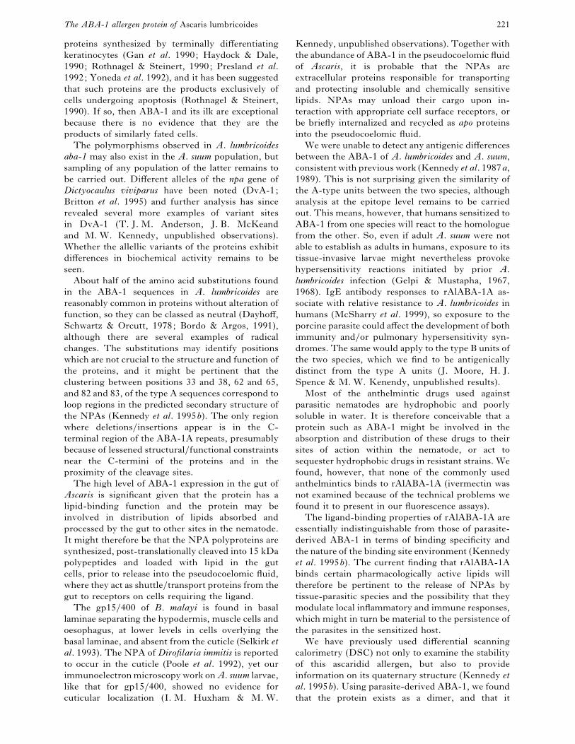
The ABA-1 allergen protein of Ascaris lumbricoides 221
proteins synthesized by terminally differentiating
keratinocytes (Gan et al. 1990; Haydock & Dale,
1990; Rothnagel & Steinert, 1990; Presland et al.
1992; Yoneda et al. 1992), and it has been suggested
that such proteins are the products exclusively of
cells undergoing apoptosis (Rothnagel & Steinert,
1990). If so, then ABA-1 and its ilk are exceptional
because there is no evidence that they are the
products of similarly fated cells.
The polymorphisms observed in A. lumbricoides
aba-1 may also exist in the A. suum population, but
sampling of any population of the latter remains to
be carried out. Different alleles of the npa gene of
Dictyocaulus viviparus have been noted (DvA-1;
Britton et al. 1995) and further analysis has since
revealed several more examples of variant sites
in DvA-1 (T. J. M. Anderson, J. B. McKeand
and M. W. Kennedy, unpublished observations).
Whether the allellic variants of the proteins exhibit
differences in biochemical activity remains to be
seen.
About half of the amino acid substitutions found
in the ABA-1 sequences in A. lumbricoides are
reasonably common in proteins without alteration of
function, so they can be classed as neutral (Dayhoff,
Schwartz & Orcutt, 1978; Bordo & Argos, 1991),
although there are several examples of radical
changes. The substitutions may identify positions
which are not crucial to the structure and function of
the proteins, and it might be pertinent that the
clustering between positions 33 and 38, 62 and 65,
and 82 and 83, of the type A sequences correspond to
loop regions in the predicted secondary structure of
the NPAs (Kennedy et al. 1995b). The only region
where deletions}insertions appear is in the C-
terminal region of the ABA-1A repeats, presumably
because of lessened structural}functional constraints
near the C-termini of the proteins and in the
proximity of the cleavage sites.
The high level of ABA-1 expression in the gut of
Ascaris is significant given that the protein has a
lipid-binding function and the protein may be
involved in distribution of lipids absorbed and
processed by the gut to other sites in the nematode.
It might therefore be that the NPA polyproteins are
synthesized, post-translationally cleaved into 15 kDa
polypeptides and loaded with lipid in the gut
cells, prior to release into the pseudocoelomic fluid,
where they act as shuttle}transport proteins from the
gut to receptors on cells requiring the ligand.
The gp15}400 of B. malayi is found in basal
laminae separating the hypodermis, muscle cells and
oesophagus, at lower levels in cells overlying the
basal laminae, and absent from the cuticle (Selkirk et
al. 1993). The NPA of Dirofilaria immitis is reported
to occur in the cuticle (Poole et al. 1992), yet our
immunoelectron microscopy work on A. suum larvae,
like that for gp15}400, showed no evidence for
cuticular localization (I. M. Huxham & M. W.
Kennedy, unpublished observations). Together with
the abundance of ABA-1 in the pseudocoelomic fluid
of Ascaris, it is probable that the NPAs are
extracellular proteins responsible for transporting
and protecting insoluble and chemically sensitive
lipids. NPAs may unload their cargo upon in-
teraction with appropriate cell surface receptors, or
be briefly internalized and recycled as apo proteins
into the pseudocoelomic fluid.
We were unable to detect any antigenic differences
between the ABA-1 of A. lumbricoides and A. suum,
consistent with previous work (Kennedy et al. 1987a,
1989). This is not surprising given the similarity of
the A-type units between the two species, although
analysis at the epitope level remains to be carried
out. This means, however, that humans sensitized to
ABA-1 from one species will react to the homologue
from the other. So, even if adult A. suum were not
able to establish as adults in humans, exposure to its
tissue-invasive larvae might nevertheless provoke
hypersensitivity reactions initiated by prior A.
lumbricoides infection (Gelpi & Mustapha, 1967,
1968). IgE antibody responses to rAlABA-1A as-
sociate with relative resistance to A. lumbricoides in
humans (McSharry et al. 1999), so exposure to the
porcine parasite could affect the development of both
immunity and}or pulmonary hypersensitivity syn-
dromes. The same would apply to the type B units of
the two species, which we find to be antigenically
distinct from the type A units (J. Moore, H. J.
Spence & M. W. Kenendy, unpublished results).
Most of the anthelmintic drugs used against
parasitic nematodes are hydrophobic and poorly
soluble in water. It is therefore conceivable that a
protein such as ABA-1 might be involved in the
absorption and distribution of these drugs to their
sites of action within the nematode, or act to
sequester hydrophobic drugs in resistant strains. We
found, however, that none of the commonly used
anthelmintics binds to rAlABA-1A (ivermectin was
not examined because of the technical problems we
found it to present in our fluorescence assays).
The ligand-binding properties of rAlABA-1A are
essentially indistinguishable from those of parasite-
derived ABA-1 in terms of binding specificity and
the nature of the binding site environment (Kennedy
et al. 1995b). The current finding that rAlABA-1A
binds certain pharmacologically active lipids will
therefore be pertinent to the release of NPAs by
tissue-parasitic species and the possibility that they
modulate local inflammatory and immune responses,
which might in turn be material to the persistence of
the parasites in the sensitized host.
We have previously used differential scanning
calorimetry (DSC) not only to examine the stability
of this ascaridid allergen, but also to provide
information on its quaternary structure (Kennedy et
al. 1995b). Using parasite-derived ABA-1, we found
that the protein exists as a dimer, and that it

Y. Xia and others 222
appeared to undergo a 2-phase unfolding process
which is not the same as observed here for the
recombinant AlABA-1A, or the NPA from a
different species (Kennedy et al. 1995c). The 2-
phase transition seen with parasite-derived ABA-1
might therefore have been an artifact of molecular
heterogeneity in the parasite-derived preparation
(which is most likely given current knowledge of the
aba-1 gene), or contamination with other types of
protein.
It has previously been noted that the allergens of
ascaridids are heat stable, retaining their allergenicity
following cooking (Audicana et al. 1997) or even
autoclaving (Christie, Dunbar & Kennedy, 1993).
This could be due to the IgE epitopes being resistant
to heat denaturation (as, for example, would be short
linear peptide epitopes), and}or that the allergens are
structurally stable at high temperatures or capable of
refolding efficiently upon cooling. The calorimetry
experiments reported here showed that rAlABA-1A
is highly heat stable, and that unfolding requires
temperatures in excess of 90 °C. We have also found
that the NPA proteins refold efficiently, although
their binding function is compromised (L.
McDermott, A. Cooper & M. W. Kennedy, un-
published observations). This high degree of stab-
ility is a characteristic of 4 bundle alpha helix
proteins, a class of protein to which ABA-1 possibly
belongs (Kennedy et al. 1995a), and is a type of
structure known for certain carrier proteins of
invertebrates (Branden & Tooze, 1999). Of signi-
ficance, calorimetry showed that the recombinant
AlABA-1A exists as a dimer in solution at the
concentrations used. This increase in effective size
will presumably limit the protein’s diffusability
within, and loss from, the nematode. Moreover,
existence as a homodimer means that cross-linking
of IgE receptors on mast cells will be facilitated, even
where the number of epitopes on a monomer unit is
limited. We have yet to establish, however, whether
NPA proteins naturally exist as homodimers or as
heterodimers with other members of the array from
which they are derived, and whether dimerisation is
necessary for their ligand binding activities.
The organization of the gene encoding the ABA-1
polyprotein of A. lumbricoides appears to be similarly
organized to that for A. suum, and the encoded
proteins have virtually indistinguishable biochemical
activities. The polymorphisms of the gene found
here in the former may also occur in A. suum, but it
is too early yet to say whether any divergence
between the 2 taxa is reflected in differences in the
ABA-1 proteins which are unique to either. It is
likely, however, that the ABA-1 allergens of A.
lumbricoides and A. suum are for all intents and
purposes immunologically indistinguishable. In
terms of the biology of the parasite, the new ligand-
binding data contribute to the idea that NPAs
secreted by nematode parasites could modulate their
tissue and immunological environments through
their binding of pharmacologically active lipids.
Note added to proof
A recent paper by P.L. Geenen et al. (Journal of
Parasitology (1999) 85, 616–622) has convincingly
settled the debate as to the developmental stage of
the infective stage larva in the egg of Ascaris – it is
the third stage (L3), and not the L2 stage as used in
our paper. The convention used in our paper follows
the previously standard nomenclature, and is thus
incorrect, but the naming and source of the various
larval stages used is detailed in Materials and
Methods and in the legend to Fig. 3.
This work was supported by the Wellcome Trust and the
Medical Research Council (UK). Nick Heaney was
supported by a Wellcome Trust Vacation Scholarship. We
are also indebted to Dr Tim Anderson for providing us
with A. lumbricoides from Guatemala and Professor
Weidong Peng for providing us with parasites from China.
We also wish to acknowledge that Dr Barry Kingston had
unpublished evidence for the large size of the aba-1mRNA several years before it was appreciated that it
encoded a large polyprotein.
, . ., , . ., , ., , .
, . . (1995). Loa loa – structural diversity of
a 15-kDa repetitive antigen. Experimental Parasitology
81, 145–153.
, . ., , . . , . . (1995).
Fine specificity of the genetically controlled immune-
response to native and recombinant gp15}400
(polyprotein allergen) of Brugia malayi. Infection and
Immunity 63, 2892–2898.
, ., , . ., , . , . . .
(1973). Characterisation of an allergen extracted from
Ascaris suum. Determination of the molecular weight,
isoelectric point, amino acid and carbohydrate content
of the native allergen. Immunochemistry 10, 815–820.
, . . . (1995). Ascaris infections in humans
from North America: molecular evidence for cross-
infection. Parasitology 110, 215–219.
, . . . , . (1997). Host specificity,
evolutionary relationships and macrogeographic
differentiation among Ascaris populations from
humans and pigs. Parasitology 115, 325–342.
, . . ., -, . . , .
(1993). Genetic structure and epidemiology of Ascaris
populations: patterns of host affiliation in Guatemala.
Parasitology 107, 319–334.
, . . ., -, . . , .
(1995). Mitochondrial DNA and Ascaris
microepidemiology: the composition of parasite
populations from individual hosts, families and
villages. Parasitology 110, 221–229.
, ., , . ., , .
, . . (1997). Cooking and freezing may
not protect against allergenic reactions to ingested
Anisakis simplex antigens in humans. Veterinary
Record 140, 235.

The ABA-1 allergen protein of Ascaris lumbricoides 223
, . (1998). Caenorhabditis elegans is a nematode.
Science 282, 2041–2046.
, . ., , ., , . ., , . .,
, ., , ., , . .,
, . ., , ., , . ., , . .
, . . (1998). A molecular evolutionary
framework for the phylum Nematoda. Nature, London
392, 71–75.
, . , . (1991). Suggestions for ‘safe’
residue substitutions in site-directed mutagenesis.
Journal of Molecular Biology 217, 721–729.
, . , . (1999). Introduction to Protein
Structure. 2nd Edn. Garland Publishing, New York.
, ., , ., , . . , . .
(1995). Extensive diversity in repeat unit sequences of
the cDNA encoding the polyprotein antigen}allergen
from the bovine lungworm Dictyocaulus viviparus.
Molecular and Biochemical Parasitology 72, 77–88.
, . , . (1987). Single-step
method of RNA isolation by acid guanidinium
thiocyanate-phenol-chloroform extraction. Analytical
Biochemistry 162, 156–159.
, . ., , . . , . . (1992).
Comparison between the MHC-restricted antibody
repertoires to Ascaris antigens elicited by adjuvant-
assisted immunisation or infection. Parasite
Immunology 14, 59–73.
, . ., , . , . . (1993). The
ABA-1 allergen of the nematode Ascaris suum : epitope
stability, mass spectrometry, and N-terminal sequence
comparison with its homologue in Toxocara canis.
Clinical and Experimental Immunology 92, 125–132.
, . ., , ., , . ,
. . (1990). N-terminal amino acid sequence identity
between a major allergen of Ascaris lumbricoides and
Ascaris suum, and MHC-restricted IgE responses to
it. Immunology 69, 596–602.
, ., , ., , . , .
(1976). Binding affinities of retinol and related
compounds to retinol binding proteins. European
Journal of Biochemistry 65, 71–78.
, . , . . (1994). Differential
Scanning Calorimetry. In Methods in Molecular
Biology, Vol. 22: Microscopy, Optical Spectroscopy,
and Macroscopic Techniques (ed. Jones, C., Mulloy, B.
& Thomas, A. H.), pp. 125–136. Humana Press,
Totowa, N. J.
, . (1988). Multiple sequence alignment with
hierarchical clustering. Nucleic Acids Research 16,
10881–10890.
, ., , . ., , ., -,
., , . . , . (1992). Molecular
characterization of a Dirofilaria immitis cDNA
encoding a highly immunoreactive antigen. Molecular
and Biochemical Parasitology 54, 51–62.
, . ., , . . , . . (1978).
A model of evolutionary changes in proteins. In Atlas
of Protein and Sequence Structure (ed. Dayhoff, M. O.,
vol. 5, Suppl. 3, pp. 345–362. National Biomedical
Research Foundation, Washington D.C.
, . ., , . ., , .,
, ., , . . , .
(1995). Cloning and sequencing of an
excretory}secretory antigen from Ostertagia ostertagi
fourth-stage larvae containing multiple tandem
repeats. Molecular and Biochemical Parasitology 72,
239–241.
, . ., , . . , . . (1993).
Heterogeneity amongst infected children in IgE
antibody repertoire to the antigens of the parasitic
nematode Ascaris. International Archives of Allergy
and Immunology 100, 283–286.
, .-., , . ., , . ., , .
, . . (1990). Organization, structure, and
polymorphisms of the human preprofilaggrin gene.
Biochemistry 29, 9432–9440.
, . . , . (1967). Seasonal
pneumonitis with eosinophilia : a study of larval
ascariasis in Saudi Arabia. American Journal of
Tropical Medicine and Hygiene 16, 646–657.
, . . , . (1968). Ascaris pneumonia.
American Journal of Medicine 44, 377–389.
, . . , . . (1989). Calculation of
protein extinction coefficients from amino acid
sequence data. Analytical Biochemistry 182, 319–326.
, . . (1992). Handbook of Fluorescent Probes
and Research Chemicals, 5th Edn, Molecular Probes
Inc., Oregon, USA.
, . . , . . (1990). Filaggrin, an
intermediate filament-associated protein: structural
and functional implications of a cDNA from rat.
DNA and Cell Biology 9, 251–261.
, . . . , . . . (1982). Production
and activities of IgE in helminth infection. Progress in
Allergy 31, 178–233.
, . . , . (1986). Stage-specific
secreted antigens of the parasitic larval stages of the
nematode Ascaris. Immunology 58, 515–522.
, . ., , . . , . . (1991).
MHC class II (I-A) region control of the IgE
antibody repertoire to the ABA-1 allergen of the
nematode Ascaris. Immunology 72, 577–579.
, . ., , . . ., , . .
, . (1987b). Genetic (major histocompatibility
complex?) control of the antibody repertoire to the
secreted antigens of Ascaris. Parasite Immunology 9,
269–273.
, . ., , ., , . ., -
, ., , . . , . . (1989).
Antigenic relationships between the surface-exposed,
secreted, and somatic materials of the nematode
parasites Ascaris lumbricoides, Ascaris suum, and
Toxocara canis. Clinical and Experimental Immunology
75, 493–500.
, . ., , ., -, .
, . . (1987a). Homology and heterology
between the secreted antigens of the parasitic larval
stages of Ascaris lumbricoides and Ascaris suum.
Clinical and Experimental Immunology 67, 20–30.
, . ., , . ., , . .
, . . (1990). The specificity of the antibody
response to internal antigens of Ascaris : heterogeneity
in infected humans, and MHC (H-2) control of the
repertoire in mice. Clinical and Experimental
Immunology 80, 219–224.

Y. Xia and others 224
, . ., , . ., , . ., ,
. . , . (1995a). The gp15}400 polyprotein
antigen of Brugia malayi binds fatty acid and
retinoids. Molecular and Biochemical Parasitology 71,
41–50.
, . ., , ., , . ., , . .,
, . . , . (1995b). The ABA-1
allergen of the parasitic nematode Ascaris suum : fatty
acid and retinoid binding function and structural
characterization. Biochemistry 34, 6700–6710.
, . ., , ., , . ., , . .
, . (1995c). The DvA-1 polyprotein of the
parasitic nematode Dictyocaulus viviparus : a small
helix-rich lipid-binding protein. Journal of Biological
Chemistry 270, 19277–19281.
-, . . , . . (1990). MHC and non-
MHC-restricted recognition of filarial surface-antigens
in mice transplanted with adult Brugia malayi
parasites. Journal of Immunology 145, 1912–1920.
, . . , . (1986). Estimation of the
polarity of the protein interior by optical
spectroscopy. Nature, London 319, 70–73.
, ., , ., , . . , . .
(1999). Natural immunity to Ascaris lumbricoides
associated with immunoglobulin E antibody to ABA-1
allergen and inflammation indicators in children.
Infection and Immunity 67, 484–489.
, ., , ., , . ., , . .,
, . , . . (1999). Sequence-
divergent units of the ABA-1 polyprotein array of the
nematode Ascaris have similar fatty acid and retinol
binding properties but different binding site
environments. The Biochemical Journal 340, 337–343.
, . ., , ., , .,
, ., , . . , . . (1993).
Primary structure and IgE response to the repeat
subunit of gp15}400 from human lymphatic filarial
parasites. Infection and Immunity 61, 2827–2833.
, ., , . . ., , . , . .
(1998). Genetic variation in sympatric Ascaris
populations from humans and pigs in China.
Parasitology 117, 355–361.
, . ., , . ., , . ., , . .,
, . . , . . (1992). Cloning of
a cuticular antigen that contains multiple tandem
repeats from the filarial parasite Dirofilaria immitis.
Proceedings of the National Academy of Sciences, USA
89, 5986–5990.
, . ., , . ., , . ., , . .
, . . (1996). Carboxy-terminal sequence
divergence and processing of the polyprotein antigen
from Dirofilaria immitis. Molecular and Biochemical
Parasitology 82, 51–65.
, . ., , . ., , .,
, . , . . (1992).
Characterization of the human epidermal profilaggrin
gene. Genomic organization and identification of a S-
100-like calcium binding domain at the amino
terminus. Journal of Biological Chemistry 267,
23772–23781.
, . . , . . (1990). The repeating
structure of the units for mouse filaggrin and a
comparison of the repeating units. Journal of
Biological Chemistry 265, 1862–1865.
, ., , . . , . (1989).
Molecular Cloning: A Laboratory Manual, 2nd Edn.
Cold Spring Harbour Laboratory Press, Cold Spring
Harbour, NY.
, . ., , . ., , . . ,
. . (1993). Localization, turnover and conservation
of gp15}400 in different stages of Brugia malayi.
Parasitology 107, 449–457.
, . . (1994). Molecular cloning and
characterisation of the gene encoding the Ascaris
allergen ABA-1. Ph.D. thesis, University of Glasgow.
, . ., , ., , . , . .
(1993). A cDNA encoding repeating units of the
ABA-1 allergen of Ascaris. Molecular and Biochemical
Parasitology 57, 339–344.
, . . ., , ., , . . ,
. . (1994). Effect on ligand binding of arginine
mutations in recombinant rat liver fatty acid-binding
protein. The Biochemical Journal 297, 103–107.
, . ., , . ., , . .,
, ., , . . , . .
(1989). MHC restriction of the antibody repertoire to
secretory antigens, and a major allergen, of the
nematode parasite Ascaris. Journal of Immunology 143,
2349–2356.
, ., , . ., , ., , . .,
, . . , . . (1993). Brugia
pahangi : a surface-associated glycoprotein (gp15}400)
is composed of multiple tandemly repeated units and
processed from a 400-kDa precursor. Experimental
Parasitology 76, 156–164.
, . . . , . . (1986). Studies on
fatty acid-binding proteins. The detection and
quantification of the protein from rat liver using a
fluorescent fatty acid analogue. The Biochemical
Journal 238, 419–424.
, ., , . , . . (1998). Identification,
characterization and expression of Toxocara canis
nematode polyprotein allergen TBA-1. Parasite
Immunology 20, 351–357.
, ., , . ., , .,
, ., , ., , ., , . .
, . . (1995). Differential antibody isotype
reactivity to specific antigens in human lymphatic
filariasis – gp15}400 preferentially induces
immunoglobulin-E (IgE), IgG4, and IgG2. Infection
and Immunity 63, 3772–3779.
, ., , ., , . ., , ., ,
. ., , . . , . . (1992). The
human loricin gene. Journal of Biological Chemistry
267, 18060–18066.
, . ., , . ., , . ., , .
, . . (1999). Characterisation of Ascaris from
human and pig hosts by a nuclear ribosomal DNA
sequences. International Journal for Parasitology 29,
469–478.