Embed Size (px)
Citation preview

Forum For osteopathic thoughtThe AAO
Official Publication of the American Academy of Osteopathy®
tradition shapes the Future Volume 21 number 1 march 2011
JOURNAL
OMT of Isolated Chronic Sphenoidal Sinusitis in a Post-Sinus Surgery Patient: A Case Report...pg. 24

The AAO Journal Volume 21, Issue 1, March 2011Page 2
The American Academy of Osteopathy® is your voice . . .... in teaching, advocating, and researching the science, art and philosophy of osteopathic medicine, emphasizing the integration of osteopathic principles, practices and manipulative treatment in patient care.
• Access to the active members section of the AAO website which will be enhanced in the coming months to include many new features including resource links, job bank, and much more.
• Discounts in advertising in AAO publications, on the website, and at the AAO’s Convocation.
• Access to the American Osteopathic Board of Neuromusculoskeletal Medicine—the only existing certifying board in manual medicine in the medical world today.
• Maintenance of an earned Fellowship program to recognize excellence in the practice of osteopathic manipulative medicine.
• Promotion of research on the efficacy of osteopathic medicine.
• Supporting the future of the profession through the Undergraduate American Academy of Osteopathy on college campuses.
• Your professional dues are deductible as a business expense.
If you have any questions regarding membership or renewal of membership, please contact Susan Lightle, Director of Member Relations, at (317) 879-1881 or [email protected] you for supporting the American Academy of Osteopathy®.
The AAO Membership Committee invites you to join the American Academy of Osteopathy® as a 2010-2011 member. The AAO is your professional organization. It fosters the core principles that led you to choose to become a Doctor of Osteopathy.
For just $5.01 a week (less than a large specialty coffee at your favorite coffee shop) or just 71 cents a day (less than a bottle of water), you can become a member of the specialty professional organization dedicated to the core principles of your profession!
Your membership dues provide you with:• A national advocate for osteopathic manipulative medicine
(including appropriate reimbursement for OMM services) with osteopathic and allopathic professionals, public policy makers, the media and the public.
• Referrals of patients through the Find a Physician tool on the AAO website, as well as calls to the AAO office.
• Discounts on quality educational programs provided by AAO at its Annual Convocation and workshops.
• New online courses (coming soon).• Networking opportunities with your peers.• Discounts on publications in the AAO Bookstore. • Free subscription to the AAO Journal published
electronically four times annually.• Free subscription to the new AAO online member newsletter.
CLASSIFIED ADSSEATTLE OMT PRACTICE OPPORTunITy
Contact Stephen Cavanaugh, DO at [email protected] or (206) 834-5438. Website for the practice is SeattleDO.com.
PRACTICE AVAILABLEOMM practice in Arizona available at no cost. Contact Craig Hoffbauer, DO, at (928) 634-0091.
PHySICIAn OPEnInG In DEnVERLocated in the heart of Denver, CO, Osteopathic Integrative Medicine is a single-physician practice seeking a compassionate and experienced Board Certified/Eligible NMM/OMM physician for part-time or full-time employment. Responsibilities include patient evaluation, treatment and management. If you are interested in joining a rapidly growing, patient focused practice, please contact Dave Zarou at [email protected], or provide your CV to Lori Gulmantovicz at [email protected] or fax: (303) 217-5708.
PLACE yOuR AD HERELooking to sell office equipment, hire a board certified DO or present a similar inquiry to the osteopathiccommunity? Place a classified ad in the AAO Journal or AAO Member Newletter! Contact Tessa at the AAO office at (317) 879-1881 for details.

Volume 21, Issue 1 March 2011 The AAO Journal Page 3
3500 DePauw BoulevardSuite 1080
Indianapolis, IN 46268P: (317) 879-1881F: (317) 879-0563
www.academyofosteopathy.org
American Academy of Osteopathy®
Richard A Feely, DO, FAAO ......................................... PresidentMichael A. Seffinger, DO .....................................President-ElectDiana L. Finley, CMP .................................... Executive Director
Editorial Advisory BoardRaymond J. Hruby, DO, FAAOMurray R. Berkowitz, DO, MA, MS, MPHDenise K. Burns, DO, FAAOEileen L. DiGiovanna, DO, FAAOEric J. Dolgin, DOClaire M. Galin, DOWilliam J. Garrity, DOStephen I. Goldman, DO, FAAOStefan L. J. Hagopian, DO, FAAOHollis H. King, DO, PhD, FAAOJohn McPartland, DOStephen F. Paulus, DO, MSPaul R. Rennie, DO, FAAOMark E. Rosen, DOWalter Witryol, MD
The AAO JournalRaymond J. Hruby, DO, FAAO ........................... Scientific EditorMurray R. Berkowitz, DO, MA, MS, MPH.........Associate EditorDiana Finley, CMP ........................................... Supervising EditorTessa Boeing ....................................................... Managing Editor
The AAO Journal is the official publication of the American Academy of Osteopathy®. Issues are published in March, June, September, and December each year.
Send address/e-mail address changes to: [email protected]
The AAO Journal is not responsible for statements made by any contributor. Although all advertising is expected to conform to ethical medical standards, acceptance does not imply endorsement by this journal.Opinions expressed in The AAO Journal are those of authors or speakers and do not necessarily reflect viewpoints of the editors or official policy of the American Academy of Osteopathy® or the institutions with which the authors are affiliated, unless specified.
Forum for Osteopathic Thought
JOURNALTHE AAO
Official Publication of the American Academy of Osteopathy®
TradiTion ShapeS The FuTure • Volume 201number 1 • marCh 2011
The mission of the American Academy of Osteopathy® is to teach, advocate and research the science, art and philosophy of osteopathic medicine, emphasizing the integration of osteopathic principles, practices and manipulative treatment in patient care.
Advertising Rates for The AAO Journal; official publication of the American Academy of Osteopathy®. The AAO and AOA affiliate organizations and members of the Academy are entitled to a 20 percent discount on advertising in this journal. Call the AAO at (317) 879-1881 for more information.
Advertising Rates
Placed 1 time Placed 2 times Placed 4 times
Full Page 7.5” x 10” $600 $575 $550
Half Page 7.5” x 5” $400 $375 $350
Third Page 7.5” x 3.3” $300 $275 $200
Fourth Page 3.75” x 5” $200 $175 $150
Professional Card 3.5” x 2” $60
Classified $1.00 per word
in this issue:AAO Calendar of Events ...................................................................... 27CME Certification of Home Study Forms ............................................ 33
editorial:Letters to the Editor ................................................................................ 4Osteopathic Scholarship, Research and Publication ............................... 8 Murray R. Berkowitz, DO, MS, MPHA Clinician’s Advice to Students............................................................10
Robert C. Clark, DO, MS
regular Features:View From the Pyramids ........................................................................ 6 Raymond J. Hruby, DO, FAAOFrom the Archives ................................................................................. 36
Component Society Calendar of Events ............................................... 44
original contributions:The 2010 Northup Memorial Lecture: Low Freqeuncy Oscillations in Human Physiology and Cranial Osteopathy ..................................... 12 Kenneth Nelson, DO, FAAO, FACOFPOsteopathic Manipulative Treatment of Isolated Chronic Sphenoidal Sinusitis in a Post-Sinus Surgery Patient:A Case Report Murray R. Berkowitz, DO, MS, MPH; Rani Shah, OMS IIIA “System’”of Cranial Manipulattion....................................................28 David S. Miller, DOMaintaining McManis Treatment Tables .............................................. 34 Robert C. Clark, DO, MSEvaluating the Perception of Motion in Osteopathic Medical Students, Residents and Physicians ...................................................................... 38
Craig Chappell, DO; Godwin Dogbey, PhD;Kevin Dankart, OMS IV

The AAO Journal Volume 21, Issue 1, March 2011Page 4
Letters to the EditorDear Editor,
I feel that an error in terminology occurred in the September 2010 issue of The AAO Journal. “CRI” is used repeatedly in the article, “Various methods of CRI palpation of body parts, their diagnostic values and interpretation of findings” by Krishnahari Pribadi, MD, appearing in Volume 20, Issue 3, pp. 9-20.
I believe the term “CRI” should be restricted to the situation where one is measuring the rate or amplitude of the primary respiratory mechanism in the head for research purposes, as John and Rachel Woods used it when they coined the term doing their research. In my interpretation, the CRI has no real clinical application. I feel this use of the term is commonplace to the point that most people do not recognize its proper definition, and I feel this is a misuse of the term. This article further authenticates what I feel is its misuse.
I believe the applicable term is “primary respiratory mechanism” or “primary respiration.” Other terms, such
as “cranial rhythm,” are less desirable because they are less generalizable. Primary respiration and primary respiratory mechanism are fully generalizable to the whole body. These terms describe the physiology that leads to metabolism in the whole organism. They also solve the issue Dr. Pribadi brings up of the cosmos affecting the CRI. Primary respiration is affected directly by the cosmos and all other outside influences. One does not feel the CRI at acupuncture points on the rest of the body. One feels the effect of the breath of life, that is, primary respiration. Primary respiration is consistent with the term Qi, which the Chinese describe as a similar phenomenon.
I use the PRM to test my patients in the manner Dr. Pribadi describes in this article, and it is valuable to have a reference article for this type of diagnosis.
R. Paul Lee, DO, FAAO
Dear Editor,
I am submitting a table (see next page) listing various factors capable of influencing the primary respiratory mechanism as reflected by changes in the CRI frequency and amplitude as detected by the cranial palpation method during specifically designed experiments. The experiments were done by:
1. Exposing the subject to various materials by having the subject touch the materials or containers while the CRI was being monitored and CRI changes were recorded;
2. For distant objects such as heavenly bodies, having the subject look at various distant objects while the CRI was monitored and the CRI changes were recorded;
3. Detecting the CRI while the subject experienced different treatment modalities, exercises, activities, mental and spiritual states, etc.
These experiments demonstrated that the PRM is indeed very sensitive to many factors at the physical, mental, energetic as well as spiritual levels. One should be aware of this phenomenon when one is conducting medical diagnosis and treatment using the cranial concept. I am also suggesting we use different materials, substances and experiences to influence the PRM for treatment purposes,
and combine the various healing methods with cranial manipulation synergistically to achieve optimal results.
This fact may explain the therapeutic results of many healing devices and methods, such as gemstones, crystals, magnetic devices, acupuncture, aromatherapy, health accessories from woods, ceramics containing magnetic energy, energized healing water, Lourdes water, zam-zam water, meditation, yoga, relaxation methods, spiritual therapies and prayers, etc. On the other hand, many man-made materials, substances and chemicals are deleterious to the PRM, and therefore may cause health problems. We should be aware of the influence of spiritual and emotional negative forces and negative intention in the environment. The readers are welcome to repeat the experiments. I suggest the detection of the CRI changes be performed by using objective measuring devices to obtain objective results.
Respectfully yours,
Krishnahari S. Pribadi, MDJakarta, Indonesia, February 15, 2011

Volume 21, Issue 1 March 2011 The AAO Journal Page 5
negative craniotropic(Reducing CRI frequency and amplitude):ColorBlack, red, blue, purple, yellow, orange, pink
Gemstones Lapis lazuli, topaz, opal, quartz, synthetic gemstones
MetalsAlloyed metals, heavy metals, steel, nickel, copper
natural substancesSome plants, herbs (echinacea), fruits, vegetables, lactose
Fabrics/materialsSynthetic materials such as polyester, polyvinyl, nylon, rayon, plastics, formica, man-made leather, melamine, teflon, etc., most synthetic drugs, artificial colors, artificial sweeteners such as saccharine, cyclamate, aspartame, erythritol, sucralose, chemical preservatives
Food and drinks containing man-made chemicals and additives, chemical residues, heavy metals
Most synthetic chemicals Food additives, oils, solvents, pesticides, perfumes, cleaning agents, detergents, latex, artificial rubber, dehydrogenated cooking oils, MSG, taste enhancers, etc.
Natural gas, compression engine exhaust fumes, methane, liquid gas, fumes, polluted air, dyes, etc.
Man-made electromagnetic wavesHigh tension wires, TV, computer monitors, CPU, electronic keyboards, cellular phones, antenna towers, electromagnetic motors, fans, air conditioning, etc.
Electric bulbs, neon lights
Airplanes, cars, trains,planets, rontgen apparatus
Nuclear energy and radioisotopes, radiation
Heavenly bodiesCores of comets
Sky, clouds
Cosmic space, high altitude, space travel, low gravity
Certain illnessesSleep state, distress, depression, severe pain, head trauma, mental trauma, coma, Alzheimer’s, brain damage, migraine headache, cancer, schizophrenia, autism, ADHD, ADD, serious illnesses, dying, poor immunity state, most viral infections esp. EBV, hepatitis, dengue, ebola, hepatitis, flu, poliomyelitis
Negative emotional states, lying
Negative spiritual forces
Iso-craniotropic(Not altering CRI)
Color Green
GemstonesDiamond, peridot, pearls, emerald,gold, silver, platinum, titanium
natural materialsCotton, wool, leather, silk, woods, animal fibers, etc.
natural substancesSome herbal remedies, some homeopathic remedies, some essential oils
Natural sugar,sorbitol
Spring water, healing water, unpolluted water, grain alcohol, most juices, wines, most fruits and vegetables, unspoiled meat
Fresh, unpolluted fish, expressed oils (olive, safflower oils, etc)
Love, happiness, peacefulness, tranquility, classical music, poem, meditation, relaxation (although may induce still-point)
Positive spiritual forcesState of health
Positive craniotropic(Increasing frequency and amplitude)
ColorViolet
GemstonesAmatis
Some plants, herbal remedies, especially aphrodisiac, tonic, stimulating essential oils
Excitement, high energy, sexual tension, anger, agitation (orgasm induces prolonged still point)
Stress, fever, acute pain, mania, agitation, certain cancers, HIV infection
Zero-craniotropic(Inducing still point)
Acupuncture, altered state of consciousness, bio-energy therapy,aromatherapy, contemplation,cranial manipulation, craniosacral therapy, intensive psychotherapy, relaxation methods, meditation, orgasm, praying, somato-emotional release, spiritual forces, Tai Chi,therapeutic massage, transcendental experience, yoga
Various factors capable of influencing the primary respiratory mechanism causing CRI changes

The AAO Journal Volume 21, Issue 1, March 2011Page 6
View From the Pyramids
Integrating Osteopathic Principle into Daily Practice Raymond J. Hruby, DO, FAAO
O steopathic medicine has always been a profession which deals with treating the whole patient. Therefore its philosophy has always been a holistic
one, and, with the concept of holism, is the implied concept of preventive medicine. Osteopathic medicine is a truly prevention-oriented approach to health care. In addition to its role in present-day care, one can also look at some of the reasons why osteopathic medicine will be the ideal system of health care for the future.
Osteopathy’s roots in holism and prevention go back thousands of years, to the early Chinese and Greek periods. Chinese medicine has always considered health to be a positive state and not just the absence of disease. Thus, when the body is healthy, it is healthy all over. Likewise, when the body is sick, it is sick all over. Treatment of illness must therefore include consideration of the whole person in order to be fully effective.
The ancient Greeks had a similar approach to health and disease. Medieval historian Henry Sigerist1 points out that “the (ancient Greek) physicians had an explanation for health. Health, they believed, was a condition of perfect equilibrium. When the forces or humors or whatever constituted the human body were perfectly balanced, man (or woman)2 was healthy. Disturbed balance resulted in disease. This is still the best general explanation we have.” This was also the view of Hippocratic medicine, and it is from these early Greek and Chinese ideas that osteopathic medicine draws it major principles.
The other major, modern-day school of medicine, known as allopathic medicine, has its roots in the ancient Greek school of Aesculapius,3 which concentrates on disease and miracle cures. If the cause of each and every disease can be determined, so this manner of thinking goes, so also can the requisite cure. This is the single-cause, single-cure of disease and health. Modern allopathic medicine has a strong and vested interest and pride in this disease-oriented method. Dr. Franz Ingelfinger, the late Editor Emeritus of the New England Journal of Medicine and a noted spokesman for allopathic medicine, gives this description of the role of the physician: “The physician, of
course, would be aware of the multiple influences that bear on health--the economic, marital, political, environmental and all other factors that determine how a given individual feels... but the doctor’s basic responsibility is cure. Yes, cure... his primary concern, inspite of all the utopian claims to the contrary, is sickness, not overall health.”4
Contrast this with the words of Andrew Taylor Still, founder of osteopathic medicine, who said simply, “To find health should be the object of the doctor. Anyone can find disease.”5
Let us revisit the well-known basic principles of osteopathic medicine as given to us by Dr. Still:
1) Body unity. Dr. Still felt the body was an integrated set of systems that functioned as a unit. Each system depends on all the other systems in order to function properly. Thus, as mentioned earlier, when the body is healthy, it is healthy all over; and when the body is sick, it is sick all over.
2) The body has an inherent tendency toward health. This idea is summed up very nicely by Northup,6 who states: “Basically, all treatment should be designed to support, stimulate, and in some instances initiate, the body’s trend toward health. The three Rs of medicine--repair, remove and relieve--are not sufficient unto themselves. Relief, removal or repair is necessary and helpful but is primarily designed to cope with the byproducts of disease rather than with the disease itself. It is in the field of prevention and the support of health that osteopathic medicine maintains an emphasis.”
3) The interrelationship between structure and function. This principle states that abnormalities in the structure of the body can produce abnormalities in bodily functions. Dr. Still felt that if all the parts of the body were in good mechanical order, then there would be proper nutrition, oxygen and nerve supply to all tissues of the body. Any abnormality here could result in a deviance in the body’s ability to resist disease. Thus, the structural system of the body had a direct influence on the function of the internal organ system.

Volume 21, Issue 1 March 2011 The AAO Journal Page 7
Dr. Still believed that rational treatment was based on the application of these principles. He founded osteopathic medicine, and the first school of osteopathy (the American School of Osteopathy) to “... improve our present system of surgery, obstetrics and treatments of diseases generally, and place the same on a more rational and scientific basis.”7
We should be clear about this last statement. Dr. Still’s purpose was to improve upon a system of medicine, not replace it. As Siehl points out,8 “He did not say ‘no drugs.’ He merely stated that the drugs then in vogue were harmful and largely useless. He did not say ‘no surgery’... Dr. Still did not say to avoid doing procedures other than palpatory diagnosis. He emphasizes that other methods were necessary.”
This philosophy has been the foundation of osteopathic medicine since its inception. However, in general, the American health care system has been disease-oriented. And although this has been the case, the American public has developed an awareness of, and a desire for, prevention and wellness rather than crisis-oriented care. This is not a mere trend, but a style of thinking that is here to stay. The health care system of the future will be the one that meets these needs. Osteopathic medicine, with its firm
foundation in prevention and wellness, is in the most ideal position to fulfill this need for all of us.
References
1. Sigerist HE. Medicine and Human Welfare. New Haven, CT: Yale University Press, 1941.57.
2. Words in parentheses added by author.3. Jonas S. Health-Oriented Physician Education and Osteopathic
Medicine. The D.O. April 1982. 110.4. Ingelfinger FJ. The Physicians Contribution To The Health System.
New England Journal of Medicine. 2 September 1976. 295:565-66.
5. Northup GW. Osteopathic Medicine: An American Reformation, 2nd Edition. Chicago: American Osteopathic Association, 1979. 31.
6. Ibid, 2.7. Booth ER. History of Osteopathy and Twentieth Century
Medicinal Practice. Cincinnati: The Press of Jennings and Graham, 1905. 80.
8. Siehl D. Andrew Taylor Still Memorial Lecture: The Osteopathic Difference--Is It Only Manipulation? JAOA. 1984. 83 (5): 349.

The AAO Journal Volume 21, Issue 1, March 2011Page 8
Osteopathic Scholarship, Research and PublicationMurray R. Berkowitz, DO, MA, MS, MPH
“The mission of the American Academy of Osteopathy is to teach, advocate, and research the science, art and philosophy of osteopathic medicine, emphasizing the integration of osteopathic principles, practices and manipulative treatment in patient care (emphasis added).”
All aspects of the human experience are enhanced by way of research and scholarship. This is especially true of the areas of science, technology, and health care. Clearly, humanity is profoundly affected–for both good and bad–by the application of the results of research and scholarship in these fields. Health care crosses disciplinary lines and ranges from the “bench” to the bedside. It is not until we have thoroughly researched a problem and the proposed solution, and tested them on real patients with real health problems, that we then choose to apply them in the clinical settings of our practices on our own patients. This is the essential underpinning of Evidence-Based Medicine. In order for this evidence to be available to physicians and other health providers, it must first be published. Therein lies the problem.
The osteopathic profession has increased its infrastructure to perform osteopathically-oriented research. Notable in this regard is the Osteopathic Research Center (ORC) based at the University of North Texas Health Sciences Center (UNTHSC) in Fort Worth, TX. Our founder (Dr. Andrew Taylor Still) and our various pioneers (i.e., Drs. William Garner Sutherland, Fred Mitchell, Sr., Frank Chapman, Larry Jones, etc.) reported and taught what they observed. What most people–both inside and outside of the osteopathic profession–have long felt is lacking is large-scale research that supports what we have observed and been teaching. As we all know, this requires funding. While the “gold standard” regarding medical evidence is the randomized controlled/clinical trial (i.e., experimental studies), observational studies (i.e., cohort, case-control, case report/series) are also not to be dismissed. We are all familiar with the observational Framingham Study (now in its third generation), and no one argues with the validity or significance of its results.
Until there is enough funding and enough experimental research—that is, large-scale, multi-center, randomized, clinical—we need to rely on observational studies. We need to publish these observational studies. Case reports and case series can form the underpinnings for meta-analyses, which require that the studies forming the
basis for the meta-analysis have been previously published in the medical literature. Furthermore, as anyone who has studied Evidence-Based Medicine knows, meta-analyses are considered even more significant and valuable than randomized studies. The key here is that the studies must have been published.
Again, the results of these studies are to be found in publications. It matters not whether the publication medium is print or online. The problem here is the decline of osteopathic literature and journals. January 1, 2011, saw the loss of one of the few osteopathic medicine journals–Osteopathic Medicine and Primary Care. On January 12, Tyler Cymet, DO, reported that the journal Chiropractic & Osteopathy changed its name to Chiropractic & Manual Therapies. The January 18 AOA Daily Report reported that, in response to concerns about The Journal of the American Osteopathic Association (JAOA), the AOA formed a task force (the JAOA Realignment Task Force) to study ways to improve the JAOA. In a telephone conversation with Michael Fitzgerald (the AOA Director of Publications), this task force is studying ways to realign the JAOA along the lines of the tenets of osteopathic medicine and to make it more relevant to the increasing number of specialists and subspecialists in the profession. For example, former JAOA Associate Editor Felix J. Rogers, DO, pointed out to the AOA Board of Trustees in October 2010 that OMT is used in every appropriate patient in his cardiology practice. I remain concerned by the recent removal of at least two peer-reviewed medical journals bearing the words “Osteopathy” or “Osteopathic Medicine” in their title.
On the other hand, also in January of this year, our esteemed colleague, Michael A. Seffinger, DO, MS, FAAFP, put forth the idea of creating “a new online journal called Neuromusculoskeletal Medicine/Osteopathic Manipulative Medicine–a specialty journal with articles by and for NMM/OMM specialists and other physician manual medicine specialists.” I replied, “I thought that was the role of the AAOJ. Perhaps we should change the name of the AAOJ, then we might get an increase in readership and ads to help support its publication. Thoughts and/or comments?” At first, I thought, “We already have a journal,” but upon further reflection, I now feel we do indeed need even more venues for publishing osteopathic medical literature.
continued on pg. 11

Volume 21, Issue 1 March 2011 The AAO Journal Page 9
Seeking a full-time Osteopathic Physician specializing in Osteopathic Manipulative
Medicine
Mount Clemens Regional Medical Center (MCRMC) is a 288 bed acute care teaching hospital located in southeastern Michigan. MCRMC Center for Osteopathic Medicine is an osteopathic manipulative medicine clinic located in the hospital’s medical office building. The Center is open five days a week receiving referrals from physicians in Family Medicine, Internal Medicine, Neurology, Orthopedics, Neurosurgery, Pain
management, Cardiology, ENT, Pediatrics and Obstetrics. The Center is also involved in training students and residents.
Candidates require residency training in Neuromusculoskeletal Medicine/ Osteopathic Manipulative Medicine or equivalent certification or board eligibility (C-NMM/OMM or C-SPOMM).
Duties include patient evaluation, care and treatment, educational programs for house staff, and assisting in the review and implementation of hospital policy with regard to conforming to accrediting osteopathic standards.
If you’re interested in joining our two-physician practice, please provide your CV to:Kathryn Martin RN, Physician Business Development e-mail :[email protected]:586.741.4127
Michigan, the Great Lakes State, offers pristine lakeshores of the great lakes and many inland lakes, championship golf courses, camping, fishing, hunting, skiing, canoeing, and exhilarating sailing.Professional sports teams and numerous cultural events are located in our metropolitan area.

The AAO Journal Volume 21, Issue 1, March 2011Page 10
A Clinician’s Advice to StudentsRobert C. Clark, DO, MS
“patients have confidence in a doctor who displays a quiet confidence in himself.” G.M. McCole (An Analysis of the Osteopathic Lesion, 1925)
I have been in practice for a few years—32 to be exact. I converse with many of my patients as I perform Osteopathic Manipulative Treatment (OMT). When I was a student and OMT Fellow at Kirksville College of Osteopathic Medicine, Paul Kimberly, DO, FAAO, always suggested I talk less and work more. He was very quiet when he treated. To this day, I still converse with many patients. But some want quiet and they get it. One student commented that OMT is very right-brain oriented, and I am fairly left-brain oriented. He thought I talked to distract my left-brain and free the right brain to work!
At the 2009 AAO Convocation, my son went to “Evening with the Stars” with me to get a treatment. I was coaching some students and assigned a couple of them to treat my son. He got annoyed with the students. He told them rather pointedly that he came seeking treatment and that I had assigned them to treat him. He was offended when they asked him for permission for everything they wanted to do. In his opinion, any reasonable and intelligent person by his request and presence had given permission for the students to do what was reasonable and appropriate for his complaint and condition. And any reasonable and intelligent doctor or student would know that!
Before I forget, he is 26 years old, stands six feet tall, weighs 290 pounds and has super fast reaction time and astounding strength. Anyone who meets him will never consider doing anything inappropriate! Also, he has been treated by me for 26 years and has been to Convocation many times. He has been treated by a number of my colleagues and other students.
Students should stop and think about who comes to AAO Convocation because, other than newborn babies, people who ask for treatment at AAO Convocation are knowledgeable patients. To constantly ask for permission from knowledgeable patients is demeaning.
Communication with patients is important, but if everything is a request for permission, the impression being given is that the person making the request is clearly lacking confidence and knowledge of his or her professional skills. Suffice it to say in a moderate voice, “I need for you to do X.” Stating, “Please lie on the table on
your back face up,” is far better than asking, “Is it ok if you lie down on the table, etc.?”
Another piece of advice from my son and me is to let the patient steer the conversation. In other words, if the patient wants to talk, the patient will talk. If not, be quiet. Sutherland is attributed with saying, “be still and know.” It is good advice, and when I am not getting the results I want, I become still and the answers come to me.
I have heard a lot of students prattle incessantly to patients about the theoretical mechanism of action of the chosen treatment technique. Stop that! Most patients do not want to know. My son stopped during his treatment to remind one student that I had taught the subject and was at one time an OMM department chair. He had heard it all before and, as a computer technician, had helped me develop presentations when Power Point© was new. He then asked the student if, when his computer broke down, he wanted the technician to explain to the student how the operating system and the hardware worked or just get the thing fixed? It is not hard to guess the answer to that question.
Believe it or not, I occasionally explain mechanisms of action to patients. But I do so on a limited basis. When? 1.) When the patient asks. 2.) When I do a linea alba release because the procedure is unpleasant, takes some time to do properly and most patients ask why I am doing it.
The point of all this commentary is that repeatedly asking for permission to do every aspect of examination and treatment and long-winded explanations of mechanisms of action are screaming demonstrations of a lack of confidence. It is also a tremendous waste of time!
In physical diagnosis classes, we were taught to ask questions. We were told the best questions were open-ended, and that leading questions were biased questions. So why does seemingly every student ask the patient, “Am I hurting you?” or, “Tell me if the treatment hurts” while doing OMT? Do you make similar comments just before you start an IV or blood draw or give an injection?
Remember, patients are people and most people want to please and affirm others. So leading questions or instructions are not productive. They often give false positive answers. Remember people are smart and

Volume 21, Issue 1 March 2011 The AAO Journal Page 11
will instinctively protect themselves. They rarely need reminders to do what is a natural reaction.
It is necessary to communicate with our patients. Informed consent is a requirement today. But we can be brief and to the point. Our conversations with patients can be cordial and pleasant. But they must be professional! We must be confident in ourselves and, by being confident in ourselves, our patients will be confident in us.
Osteopathic Scholarship...continued from page 8
We need more osteopathic medicine research. We need more data on which to base our conclusions. The osteopathic profession is actively taking crucial steps toward developing a research cadre and performing larger-scale, multi-center research. Meanwhile, we need to have more data come from even observational studies. From these published observations, we can develop meta-analyses. From these meta-analyses will come further evidence-based osteopathic medicine. Everyone has the potential to contribute to this effort. We all have experienced an “interesting case”—the rare finding or an unexpected outcome—in our practices. We can learn from you, but only if you let us know what you observed. Increase the osteopathic data. Write and submit your cases.
NEW at the AAO Bookstore!
Outline of Osteopathic Maniputive Procedures: Memorial Edition
by Paul E. Kimberly, DO, FAAOPurchase your copy at
www.academyofosteopathy.org(select “AAO Bookstore” from the left-hand menu)
AAO Member Price: $90.00List Price: $100.00
(plus shipping and handling)

The AAO Journal Volume 21, Issue 1, March 2011Page 12
“Vibration is an accepted fact in science. Solid bodies are composed of atoms which are vibrating at almost infinite velocities. One substance differs from another mainly in the modulus of vibritility, the different planes of substance representing the planes of gradually increasing vibratility. The higher vibratility governs and molds the lower, just as the sun centralizes the solar system. The most refined vibrations mean life and light, with all their accompaniments, to the planets in the solar system. In man, this vibratile characteristic also predominates, for within his organism he combines the higher and lower grades of vibratility in connection with mind, brain, bone, muscle, blood. So long as these combined vibratilities are in harmony, the organism enjoys life and health.”1
As J. Martin Littlejohn observed more than one hundred years ago, human physiology is dynamic.
AbstractThe use of laser-Doppler flowmetry provides the
opportunity to study Cranial Osteopathy in the context of quantifiable aspects of human physiology. Six studies, spanning a period from 1998 to the present, are reviewed that support the following conclusions:
1. Palpation of the CRI tracks identifiable frequencies in bloodflow velocity.
2. Cranial palpation alone may be employed as sham treatment in future research into the clinical impact of cranial manipulation.
3. Cranial manipulation appears to exert effects upon baroreflex physiology.
4. Cranial manipulation affects the low-frequency (0.10-0.20 Hz) signal, and to a lesser extent the very low-frequency (0.003-0.05 Hz) signal in bloodflow velocity, and does so in a manner consistent with the type of manipulative procedure being employed.
5. A frequency signal of 0.08 Hz (0.04-0.11 Hz) has been identified in the flowmetry record that is closely related to the 0.10-0.20 Hz signal. Both are demonstrated to be affected by cranial manipulation, in this case CV-4.
6. Although not everyone appears to be palpating at the same frequency, everyone tracks the 0.10-0.20 Hz signal, with the majority tracking at 0.04-0.11 Hz or 1 CRI cycle to 2 low-frequency bloodflow velocity waves.
7. A new normative range for the CRI of 2-7 cpm, as palpated by experienced examiners, has been identified.
Figure 1. Above, (a) waves upon waves upon waves; the time-domain record of a complete bloodflow velocity record demonstrating the heart rate upon the low frequency baroreflex wave upon the very low frequency wave, and (b) filtered record showing respiratory, low and very low frequency components only. Below, Fourier transform, frequency-domain spectrum demonstrating the 1) very low frequency, 2) low frequency, 3) high frequency respiration, and 4) heart rate spectral peaks.
The 2010 northup memorial lecture: low Frequency Oscillations in Human Physiology and Cranial OsteopathyKenneth nelson, DO, FAAO, FACOFP

Volume 21, Issue 1 March 2011 The AAO Journal Page 13
Everything in life is changing with time, but not necessarily at the same rate. Holistically, human physiology may be considered in the context of waves upon waves upon waves (Figure 1, above, a), wherein each independent vibrational frequency influences, and is influenced by, those frequencies above and below it. Within the broad spectrum of physiologic rhythms, one area is of particular interest to practitioners of osteopathic manipulative medicine—the frequency range from 0.003 to 0.50 Hz (0.18 to 30 cpm). In cardiovascular physiology, this range is subdivided, by spectral peaks, into very low-frequency (0.003 to 0.05 Hz, 0.18 to 3.0 cpm), low frequency (0.10 to 0.20 Hz, 6.0 to 12 cpm) and high frequency (0.25 to 0.50 Hz, 15 to 30 cpm) components.2 The very low frequency peak reflects autonomic (parasympathetic) and renin-angiotensin interaction. The low frequency spectral peak is predominantly the result of sympathetic, baroreflex activity. The activity in the high-frequency area, pulmonary respiration, impacts the cardiovascular system through the interaction of the autonomic (parasympathetic and sympathetic) nervous system.3
In osteopathic manipulative medicine, there is the cranial rhythmic impulse (CRI) described as a palpable manifestation of a primary (cellular) respiratory mechanism (PRM).4 The rate of the CRI, first measured by Woods and Woods in 1961,5 has since been measured repeatedly, with a reported range of 2 to 14 cpm (0.03 to 0.23 Hz).6-16 This frequency range encompasses the low-frequency peak between 0.10 and 0.20 Hz in cardiovascular physiology.
In the mid-nineteenth century, activity in the 0.10 to 0.20 Hz frequency range was observed in blood pressure, independent of pulmonary respiration.17,18 This low frequency rhythm has since been identified as Traube-Hering waves,19-21 Mayer waves22 or Traube-Hering-Mayer waves.23 To avoid confusion, rather than using eponyms in the discussion that follows, the oscillations will be identified by their frequencies.
Oscillations in the low frequency range of 0.10-0.20 Hz have been identified throughout human physiology—in blood pressure,17,18,22,24 heart-rate variability,22,25-27 pulmonary blood flow,20 peripheral blood flow20,22,25,26,29 muscle sympathetic tone,27 cerebral blood flow movement of the cerebrospinal fluid,21,28,30-32 and cerebral cortical cellular activity.33-36 Because these phenomena occupy the same frequency range as the CRI, it was decided to monitor that particular frequency in a known physiologic phenomenon to provide insight into Cranial Osteopathy.
Peripheral vascular manifestations of the low frequency (0.10-0.20 Hz) rhythm are readily measured by laser-Doppler flowmetry and may be recorded
simultaneously with cranial osteopathic procedures. In the basic science protocols described below, where the low frequency (0.10-0.20 Hz) rhythm was monitored, a laser-Doppler perfusion monitor (Transonic Systems Inc.) was employed to determine Doppler velocity of circulating blood that was then digitized for subsequent data reduction (WinDaq Data Acquisition & Playback Software, Transonic Systems).
This method provides time domain records that may be obtained simultaneously with cranial diagnostic and therapeutic procedures. These records provide striking illustrations of what cranial practitioners have been describing for years. They lend themselves to the identification of the interaction between the practitioner and subject, and for determination of the rate of the CRI. The recorded bloodflow velocity record is the result of a very complex group of physiological processes, with multiple contributing frequencies resulting in waves upon waves upon waves (Figure 1, above, a). Because of this complexity, visually identifying where any given intervention actually has an effect is extremely difficult, if not impossible.
However, because these complex visual records are digital, the data may be converted mathematically through a Fourier transformation (FT) (Figure 1, below). This provides frequency-domain spectra that clearly identify the frequencies of individual spectral peaks (location on the x axis), their power (height of any given spectral peak, y axis) and dispersion or irregularity (width of a spectral peak measured at half height) that result in the complex waves upon waves upon waves of the visual time-domain
Figure 2: Protocol 1, Compressed laser-Doppler flowmeter bloodflow velocity (waveform) and flexion-extension records (vertical event marks) from two subjects.

The AAO Journal Volume 21, Issue 1, March 2011Page 14
records. FT spectra may be filtered and inverse Fourier transformations performed to create time-domain records that focus upon the contribution of any spectral area to the observed time domain record (Figure 1, above, b). Frequency-domain records also may be comparatively analyzed to determine where in the complex waveform an intervention has had effect. This may be done by comparing the relative height of consecutive measurements of the same spectral peak—or by subtracting one FT spectrum from another, and thereby calculating the changes that have occurred in frequency, power and dispersion throughout the entire spectrum as a magnitude difference spectrum (Figures 9, bottom and 12).
These methods provide opportunities to study Cranial Osteopathy in the context of quantifiable aspects of human physiology through cutaneous bloodflow velocity. The following protocols were implemented by our group—Thomas Glonek, PhD, Nicette Sergueef, DO (Fr), and me—with able assistance in the first protocol from Celia M. Lipinski, DO, and Arina R. Chapman, DO. These studies, spanning a period from 1998 to the present, represent our attempt to quantify the CRI, and demonstrate the effect of cranial manipulation upon the vibrations manifest in human bloodflow velocity.
protocol 1: Comparing low-frequency bloodflow velocity waves with cranial palpation.23
First, it was appropriate to establish a correlation between the palpated CRI and the 0.10-0.20 Hz oscillation.
Twelve subjects participated in this study. With the laser-Doppler probe affixed to the subject’s earlobe, they rested quietly on an osteopathic manipulative treatment (OMT) table. A baseline flowmetry record was then obtained. Next, an experienced examiner blinded to the laser-Doppler record, monitored the CRI. As they palpated, they identified the CRI, saying “f” for flexion/external rotation and “e” for extension/internal rotation. At each verbal indication, an event mark was entered into the computer by the recording technician.
Figure 2 is the compressed laser-Doppler flowmetry time-domain records of two subjects. The palpation of the CRI is indicated by the vertical event marks on the right side of each record.
The flowmetry records for each subject were Fourier transformed and dissected, removing frequencies above 0.50 Hz. Inverse Fourier transformation was performed on the remaining data, resulting in a time-domain record of frequencies below 0.50 Hz. This demonstrated that the dominant, low frequency wave phenomena, apparent in the original flowmetry records, represented the low frequency
Figure 3: Protocol 1, Expanded laser-Doppler flowmeter, relative blood velocity record of Subject 2. (Top) Flowmeter trace revealing cardiac cycle fine structure. The double-headed arrow indicates the wavelength of one low frequency cycle. (Bottom) Low frequency waveform component only of the top trace. The bottom waveform was created from the top waveform by filtering (removing) the high frequency cardiac component, leaving only the very low frequency, low frequency and respiratory components. Inverse Fourier transformation of this filtered data generates the bottom trace. Both traces are in register with respect to time, and the event markers indicate the positions of the palpatory findings.
Figure 4: Protocol 1, Inverse Fourier transform, time-domain spectra from Subject 2. (Top) All frequency-domain data used in the inverse computation. (Bottom) Only the frequency component lying below 0.5 cycles/sec. (30 cycles/min.) used. The bottom spectrum is the oscillation resulting from very low, low and respiratory frequency contributions. The insert box shows the portion of the Fourier transform spectrum used to compute the inverse very low, low and respiratory frequency spectrum.

Volume 21, Issue 1 March 2011 The AAO Journal Page 15
(0.10-0.20 Hz) wave and not harmonic aberrations from some other frequency (Figures 3 and 4).
Of the twelve subjects, eleven provided data suitable for analysis. Six hundred thirteen low frequency wave peaks (maxima) and troughs (minima) were visually identified. One hundred sixty-six flexion/external rotation events and 162 extension/internal rotation events (n = 328) were identified. These were associated equally between low frequency maxima (n = 164) and minima (n = 164).
There was no correlation between palpation (flexion/external rotation, extension/internal rotation) and the low frequency wave maximum or minimum in the flowmetry record (Pearson’s R value, -0.085; approximate significance, 0.123). So, the time of each palpation event was compared with the time recorded for the nearest maximum or minimum in the flowmetry record. The paired t-test, in this case, showed no statistical difference between the flowmetry low frequency (0.10-0.20 Hz) wave record and the palpated CRI. With 328 data pairs, both groups of time values were highly correlated (correlation = 1.000; significance = 0.000). Even though, during the length of the recording, the low frequency (0.10-0.20 Hz) waves demonstrated a frequency modulation of up to twenty percent, the palpation events precisely mirrored the oscillating flowmetry wave.
Protocol 2: Effecting low-frequency bloodflow velocity waves by cranial manipulation.37
If the palpable CRI and low frequency (0.10-0.20 Hz) bloodflow velocity oscillations are temporally concomitant, the question arises—does cranial manipulation exert an effect upon the low frequency oscillations?
Twenty-three subjects were randomly divided into control (n=13) and experimental (n=10) groups. The laser-Doppler probe was affixed to the subject’s earlobe. Subjects rested quietly on an OMT table. A baseline flowmetry record was obtained, followed by cranial manipulation (experimental group) or sham intervention (control). The sham intervention consisted of five minutes of cranial palpation using a biparietal modification vault-hold. Subjects in the experimental group received an individually determined cranial treatment, applied until a therapeutic endpoint was achieved (five to ten miuntes). Immediately following the sham or manipulative intervention, a five-minute post-intervention laser-Doppler recording was acquired. During the entire process the subjects in both groups remained on the treatment table; the laser-Doppler recording was continuous, and the probe was undisturbed.
The effects of the cranial treatment seen in Figure 5, although visually exceptional, are consistent with changes induced in all the subjects. Figure 6, a compressed
continuous flowmetry record (approximately 30 minute duration), demonstrates the progressive organization resulting from the increased low-frequency wave activity readily seen from the end of the treatment period through the post-treatment period.
Protocol 3: Effecting low-frequency bloodflow velocity waves on demand.38
Since individually determined cranial manipulation changed bloodflow velocity, it was decided to see if an effect could be obtained on demand, using palpation
Figure 5: Protocol 2, Laser Doppler blood flow recording of two individuals before and after cranial manipulation.
Figure 6: Protocol 2, Continuous flowmetry record of approximately 30 minute duration. Although the record is greatly compressed to afford visualization in its entirety, the progressive organization resulting from the increased low frequency wave activity can be readily seen from the end of the treatment period through the post-treatment period. The bottom tracings are the contributions to the flowmetry record from very low, low and respiratory frequencies before and after manipulation.

The AAO Journal Volume 21, Issue 1, March 2011Page 16
only, alternating with incitant bitemporal rocking. This alternating palpation and manipulation sequence was continued for a total of 35 minutes. (maximum recording time for an uninterrupted laser-Doppler record). To eliminate the possibility that there might be an independent oscillation in bloodflow physiology, two different time sequences were decided on for the protocol. Five minute and seven minute intervals, both divisible into 35, were chosen. The timing of the treatment/non-treatment sequence was established for each subject before the initiation of the protocol.
Fifteen subjects participated. The laser-Doppler probe was placed in the midline on the subject’s forehead. It was felt that the previously used ear site was too close to the temporomastoid region (area being manipulated), and could therefore be directly affected by the intervention.
The subjects rested on the OMT table with their heads on the practitioner’s hands, in position for the manipulative procedure. A baseline bloodflow velocity record was obtained. Following this, incitant bitemporal rocking was performed synchronous with the subject’s CRI. The manipulation was stopped, and, without changing hand placement, a period of cranial palpation only followed. This alternating sequence continued uninterrupted for the maximum laser-Doppler recording time.
Figure 7 shows the compressed, 35 minute long flowmetry records for two subjects treated with cranial manipulation at five (Subject 1) and seven minute (Subject
2) intervals. Event marks (EM) identify where cranial manipulation was started and stopped. Expansion of the first and third non-treatment/treatment pairs of the flowmetry record for Subject 1 (Figure 8) clearly shows the low frequency (0.10-0.20 Hz) wave, and the amplifying effect upon it resulting from incitant manipulation.
Using FT, the very low frequency, low frequency, high frequency and cardiac rate signals were identified to determine which changed. Signal intensities as a function of the respective component’s frequency for Subject 1, third non-treatment segment (Figure 9, top) and the third treatment segment (Figure 9, center), are plotted in Figure 9 bottom. It demonstrates that the incitant cranial manipulation increased the very low frequency signal, and greatly increased the low frequency signal. Additionally, the heart rate can be seen, from the resultant sinusoidal shape for the cardiac signal, to have increased from approximately 70 to 82 beats per minute.
Protocol 4: Effecting low-frequency bloodflow velocity waves by Compression of the Fourth Ventricle (CV-4).39
Because incitant cranial manipulation affected the amplitude of the low frequency oscillations, it was decided to study the response to compression of the fourth ventricle (CV-4), a manipulative procedure that, during its application, is intended to dampen the CRI. CV-4 offers the advantage of having a specific starting point, and a generally agreed upon physiologic end point—the still point. This endpoint is then reportedly followed by
Figure 7: Protocol 3, Compressed laser-Doppler-fiowmetry, relative blood velocity waveforms, of two subjects treated by cranial manipulation at designated five minute (Subject 1) and seven minute (Subject 2) intervals. Event marks (EM) indicate points in time when cranial manipulation started and stopped.
Figure 8: Protocol 3, Expansion of the laser-Doppler flowmetry record of Subject 1–the top record showing the initial resting segment followed by the first treatment segment, and the bottom record showing the analogous segment pair beginning at 18 minutes, both demonstrating that incitant cranial treatment amplifies the power of the low frequency oscillation.

Volume 21, Issue 1 March 2011 The AAO Journal Page 17
amplification of the CRI. This allowed us to measure the duration of time the CV-4 procedure was applied and any impact it had on bloodflow velocity.
Twenty-eight experienced cranial practitioners performed the CV-4, each with a different subject (n=26; two subjects participated twice). One physician plus one subject at one treatment constituted one statistical case.
The physician sat at the head of an OMT table. The subject, lying supine with the laser-Doppler probe attached to the midline of their forehead, rested quietly for an equilibration period. A baseline record of five to seven minutes, the Control (C) segment (Figure 10, Control), was then obtained. During the Control segment period, no treatment was administered, but the subject’s head rested upon the physician’s hands in the appropriate position for palpatory diagnosis and treatment using CV-4. At the end of
the Control segment, the physician was instructed to begin implementation of CV-4, and upon the treating physicians’ indication that they had started, an event mark was entered into the record by the technician (Figure. 10).
The Treatment (T) phase lasted until the physician indicated that they had obtained their therapeutic goal. At this point, a second event mark was entered into the flowmetry record, indicating the end of the Treatment segment (Figure 10, Treatment). The physicians removed their hands from contact with the subject’s head, and the Response (R) to treatment was followed for an additional five to seven minutes (Figure 10, Response). Both treating physicians and subjects were blinded to operations at the computer console.
The duration of Treatment for the CV-4 procedure from the 28 individual records (Table 1) was computed
Figure 9: Protocol 3, Fourier-transformation magnitude spectra–component intensity is plotted as a function of component frequency. Subject 1: third non-treatment segment (top) and third treatment segment (center), with the (1) very low, (2) low, (3) respiratory, and (4) cardiac frequencies identified. Magnitude difference spectrum–Subject 1: third non-treatment/treatment segment (bottom), with the low frequency oscillation increased (2) and the heart rate increased (4) from approximately 70 to 82 during cranial manipulation.
Figure 10: Protocol 4, Real time demonstration of the flowmetry record of a CV-4, consisting of a baseline bloodflow velocity record, the Control segment, the Treatment segment, and the post-treatment, Response segment. The event marks were entered into the record by the research technician upon verbal indication by the treating physician at the onset and culmination of treatment. The inserts (1, 2 and 3) are representative segments (approx. two minutes each) of the flowmetry record, the amplitude of the low velocity oscillation, and (4) the portion of the record immediately before and following the still point.

The AAO Journal Volume 21, Issue 1, March 2011Page 18
(Control), the CV-4 treatment period (Treatment), and the immediate response period (Response), were identified for intergroup comparisons. Within each segment, a four to six minute portion of the record was selected. The shortest of these segments, for each subject, was identified, and its duration, to the nearest 0.01 second noted. Portions of the remaining two segments from that record, each of identical duration as the shortest segment, were extracted for FT.
FT spectra, for each of the segments, were then computed to generate 60 frequency-domain spectra (Figure 11). Point-by-point subtraction, generating Control minus Treatment (C-T), Treatment minus Response (T-R), and Control minus Response (C-R) difference spectra, was then carried out (Figure 12). The resulting difference spectra were plotted and then integrated to obtain spectral signal areas.
by measuring the time elapsed on the flowmetry record between the first event mark, when the physician started the procedure, and the event mark indicating they had attained their therapeutic goal. The mean duration of Treatment was 4.43 minutes, range 8.65 minutes. (minimum 1.42, maximum 10.07), a standard deviation ± 2.22 minutes, and a variance of 4.94. This duration is consistent with a published report of three to seven minutes for CV-4 application.40
The impact of the CV-4 procedure was then determined. Among the 28 CV-4 records obtained, high-frequency noise in eight records (29 percent) made them unsatisfactory for data reductions and statistical analyses. The remaining 20 records, ranging from 15-24 minute duration, were usable. Each of these records contained the three continuously linked segments (total waveform segments = 60) separated by the event marks. These segments (Figure 10), the pre-treatment resting period,
Figure 11: Protocol 4, Fourier transformation of each segment, (1) Control, (2) Treatment, and (3) Response of the CV-4 procedure, with the low frequency (A) and cardiac (C) components indicated.
Figure 12: Protocol 4, Difference spectra comparing the component parts (Control, Treatment, and Response) of the CV-4 procedure, with the low frequency (A) and cardiac components (C) indicated. (1) Control minus Treatment, (2) Treatment minus Response, and (3) Control minus Response

Volume 21, Issue 1 March 2011 The AAO Journal Page 19
From these difference spectra, signal areas were computed from three signals in the low frequency region. The 0.02 Hz signal, represents physiological activity in the range of the very low frequency wave; the 0.10 Hz signal represents activity consistent with the low frequency wave. A new minor signal at 0.08 Hz was resolved in flowmetry data but not reported in earlier work. Sufficient data at this point were accumulated verifying the existence of this minor resonance. Additionally, areas were computed from both the low and high frequency halves of the cardiac signal (centered at approximately 1.10 Hz) and minimum and maximum frequency components of the cardiac signal were recorded.
To determine significance among the three groups (C-T, T-R, C-R) for each selected signal area and frequency value, Analysis of Variance (ANOVA) was used. Seven scalar variables were considered, consisting of the areas of the signals centered at 0.02, 0.08, and 0.10 Hz; areas of the lower frequency and higher frequency cardiac bands; and the frequencies at the maximum amplitude (either positive or negative) of both cardiac bands. Also evaluated were pair-wise comparisons between group pairs, using Scheffé, Bonferroni, Tukey, and Least-Significant Difference (LSD) range tests (respectively, from most conservative).
Significant differences were identified for the minor signal at 0.08 Hz (significance = .041) and the low frequency signal at 0.10 Hz (significance = .000). There was no significant difference for the very low frequency signal at 0.02 Hz, or for any of the four cardiac signal variables. Using the Scheffé range test, significant differences were found only for the 0.10 Hz area variable at the alpha .05 level; however, the 0.08 Hz signal did exhibit parallel differences at the .072 level. Therefore, it is believed that both signals are affected together, and in the same sense, by the CV-4. The differences in significance between the two variables most likely reflect the much lower signal-to-noise ratio of the minor 0.08 Hz signal than fundamental differences in the behavior of each signal band with manipulation.
The variable that demonstrates the largest mean difference in response to CV-4 is the low-frequency area of the 0.10 Hz signal, where all three combinations, C-R, C-T, and T-R, are significantly different from each other.
Protocol 5: The Rate of the Cranial Rhythmic Impulse.15
It is important to establish normative values when studying physiologic phenomena. We therefore measured the rate of the CRI per minute and determined how clinicians palpate the CRI in comparison to the laser-Doppler flowmetry record.
The CRI rate was determined from the records of 44 different examiners, each palpating a different subject. The examiners were experienced osteopathic physicians attending various professional meetings. Each palpated a different subject, who was recruited randomly from attendees at the same meetings.
The laser-Doppler probe was placed onto one earlobe, and the subject rested quietly on an OMT table. Examiners were seated at the head of the table. With a contact position of their preference, the examiners palpated their subject’s CRI. As they palpated, they said “f,” indicating the perception of the flexion/external rotation, and “e” indicating extension/internal rotation. At each verbal indication, an event mark was entered into the computer by the recording technician. Continuous, unbroken records
Figure 13: Protocol 5, Palpation of the CRI as compared to the laser-Doppler blood flow velocity record. (Above) This figure shows the low frequency oscillation (oscillating trace) and CRI (palpation of “flexion/extension” vertical event marks) in a 2:1 ratio. (Below) Compressed flowmetry record demonstrating the 2:1 ratio. This is the most frequently encountered low frequency oscillation (LF) to CRI ratio demonstrated by skilled examiners.
Figure 14: Protocol 5, Bloodflow velocity record and CRI (palpation of flexion/extension”) in a 1:1 ratio.

The AAO Journal Volume 21, Issue 1, March 2011Page 20
were recorded for each subject. The recording length—five to fifteen minute duration—was determined by the examiner.
A portion of each record was selected for computation where the CRI was palpated consistently, without large “palpatory gaps.” Calculating from 44 records acquired, the mean rate (cpm) for the palpated CRI was 4.54, with a range of 7.26 (minimum 1.25, maximum 8.51). The standard deviation was 2.08, the standard error 0.313, and the variance 4.32.
The vast majority of examiners in this study palpated the CRI such that a flexion event was perceived coincident with one low frequency oscillation and an extension event perceived coincident with the next low frequency oscillation. This resulted in a ratio of palpated CRI to recorded low frequency (0.10-0.20 Hz) oscillations of 1:2 (Figure 13). It is worthwhile to note that infrequently an examiner palpated the CRI at a 1:1 ratio to the low-frequency oscillation (Figure 14).
During flowmetry recording, irregularities were observed, resulting in gaps in both the palpatory and flowmetry records. In some instances, these gaps were recognized and reported by the examiners as “still points”41 (Figure 15).
Protocol 6: The Rate of the Cranial Rhythmic Impulse; A Second Method of Assessment.16
A second protocol, currently under review for publication, measuring the rate of the CRI is reported here. This study provides a statistical N of 727 subjects, consisting of several smaller groups from 16 to 86 individuals. Participants palpated CRI rates on each other. Half of each group acted as examiners while the other half were subjects. The examiners palpated the CRI using the classically described vault hold.42,43 They were not told how long they would be palpating, only to count the number of complete biphasic CRI cycles that they palpated during the acquisition period. The number of cycles each examiner reported was kept private so no one was aware of the rates other participants reported. Following this, the pairs exchanged positions and the protocol was repeated. The statistician then computed the CRI rate in cycles/minute for each recorded value by dividing the total number of CRI cycles counted per subject by the time allowed at each measurement session.
The mean reported CRI rate (N = 727) was 6.88 ± 4.45 cycles per minute. This group was subdivided by experience level and it is of interest to note that examiners with the greatest experience level palpated at a rate of 4.78 ± 2.57.
Figure 15: Protocol 5, Pause in the palpation record coincidental with a decrease in low frequency oscillation amplitude. Several examiners have commented on the perception of a “still point” at such times, although they were blinded to the flowmerty record.
Figure 16: A graphic representation of quantified rates for the cranial rhythmic impulse reported in the literature over the past 45 years. It is of interest to note that (with the exception of Woods, 10-14 cpm) when palpation is employed to obtain data, the reported rate tends to be lower (3-9 cpm), while, if data is obtained by instrumentation of any type, the reported rate tends to be faster (7-14 cpm).
Discussion
From the above studies the following observations may be made:
Protocol 1 demonstrated that the CRI and the low frequency (0.10-0.20 Hz) bloodflow velocity waves are concomitant phenomena. The bloodflow velocity waves demonstrated a frequency modulation of up to 20 percent
Volume 21, Issue 1 March 2011 The AAO Journal Page 21
that was precisely mirrored by the palpation record. This frequency modulation was also reported by Lockwood and Degenhardt in their analysis of Frymann’s 1971 data from instrumental measurement of the CRI.12,44
Additionally, it is of interest to note that the palpated CRI in this study was consistently palpated such that the ratio of the CRI to the low frequency (0.10-0.20 Hz) oscillations was 1:2 (Figure 2). This relationship was recognized retrospectively when additional flowmetry records were analyzed to measure the rate of the CRI (see Protocol 5) (Figure 13).
Protocol 2 demonstrated that cranial manipulation, specifically directed at cranial patterns of individual subjects, affected bloodflow velocity oscillations. The amplitude of the very low frequency (0.003-0.05 Hz) wave decreased, and that of the low frequency (0.10-0.20 Hz) wave increased. It is of interest to note here that cranial manipulation has been demonstrated to exert a comparable effect on similar frequency oscillations (0.08-0.20 Hz) in intracranial fluid content as measured by transcranial bioimpedence.32
Because the low frequency wave in bloodflow velocity is mediated by sympathetic, baroreflex activity,3 cranial manipulation can be inferred to affect the autonomic nervous system. Additionally, since the control palpation did not greatly affect bloodflow velocity oscillations, control palpation may be used as a sham treatment in future research.
Protocol 3 demonstrated that incitant cranial manipulation can, on demand, alter the physiologic parameters of bloodflow velocity. The low frequency (0.10-0.20 Hz) component increased most markedly and the very low frequency component (0.003-0.05 Hz) increased to some degree. These effects occurred within a few seconds and, in this instance, the flowmetry record returned to near baseline levels within fractions of a minute after the intervention was stopped. Fourier transform analysis, however, revealed that the flowmetry record does not return precisely to baseline following intervention, rather it exhibits a small residual effect with a considerably longer half life. This persistent residual amplification may, in part, account for the therapeutic effect of some forms of cranial manipulation.
Protocol 4 demonstrated that the duration of the CV-4 was 4.43 ± 2.22 minutes, consistent with the previously published report of three to seven minutes.40 During its application, bloodflow velocity was affected in a manner consistent with what would be expected from descriptions of the impact of CV-4 upon the CRI.43 As the occiput was held in extension to decrease the amplitude of the CRI, the low-frequency oscillation was damped and essentially
eliminated when a still point was obtained (Figures 10 and 11). The therapeutic impact of CV-4 is said to be increased amplitude of the CRI that enhances the fluid motion of the PRM.43 Following CV-4, the amplitude of the low-frequency wave in bloodflow velocity increases (Figure 10).
Protocol 5 provides a normative rate for the CRI, and insight into previously unexplained discrepancies in its reported rate. Also, by observing the relationship between the palpated CRI and bloodflow velocity, an explanation may be advanced for the difficulties encountered when sequentially comparing palpated rates for the CRI for the purpose of establishing inter-rater reliability.
The rate of the CRI first reported as 10-14 cpm5 has remained the accepted rate in the majority of osteopathic textbooks.42,43,45-47 Review of the literature, however, reveals an interesting paradox. Studies using palpation tend to report lower rates for the CRI7-9,11,13 than those obtained by instrumentation.6,10,12,14,32 This occurs independently of the type of instrumentation, such as plethysmography applied to the upper extremity,6 infrared light reflected from acupuncture needles implanted into the cranial bones of human subjects,10 retrospective analysis of data obtained by Frymann using a pressure transducer placed upon the head,12 and fluctuation of intracranial fluid content using transcranial electrical bioimpedance.14,32 (Figure and Table 16)
The palpated CRI rate in this study (4.54±2.08 cpm, 0.04-0.11 Hz) is consistent with the lower rates obtained by palpation and reported by the majority of previous investigators7-9,11,13 (Figure 16). The inconsistency between palpation and instrumentation may be explained by the observation that the majority of examiners in the current study palpated such that a flexion event was perceived coincident with one low frequency oscillation and an extension event perceived coincident with the next low frequency oscillation. (Figure 13) This resulted in a ratio of palpated CRI to recorded low frequency (0.10-0.20 Hz) oscillations of 1:2. If instrumental measurement of the CRI tracks the dominant low-frequency oscillation, then the discrepancy between the palpated and instrumental measurements is explained.
There is, however, the issue of the higher palpated rate (10-14 cpm) consistent with the rates obtained by instrumentation, reported by Woods and Woods,5 and identified in osteopathic textbooks.42,43,45-47 Infrequently, an examiner will palpate the CRI at a 1:1 ratio to the low frequency oscillation (Figure 14).
The difference between these palpation to flowmetry ratios may be explained by the observation from Protocol 4 of the previously unreported 0.08 Hz (4.5 cpm) frequency

The AAO Journal Volume 21, Issue 1, March 2011Page 22
wave in bloodflow velocity. The reported rate for the CRI in this study is 2.46 to 6.62 (4.54±2.08) cpm, or 0.04-0.11 Hz. The low frequency wave between 6 and 12 cpm (0.10-0.20 Hz) is twice as fast. So, it may be concluded that the majority of individuals track the 0.04-0.11 Hz frequency, while some individuals track the faster 0.10-0.20 Hz frequency.
Protocol 6, with a reported rate for the CRI of 4.78 ± 2.57, underscores the Protocol 5 rate of 4.54±2.08. This rate was identified in both protocols using skilled osteopathic examiners. The methods by which this result was obtained, however, were entirely different.
ConclusionFrom the above, the following conclusions can be drawn: 1. Palpation of the CRI tracks identifiable frequencies in bloodflow velocity (Protocol 1).2. Cranial palpation alone may be employed as sham treat-ment in future research into the clinical impact of cranial manipulation (Protocol 2).3. Cranial manipulation appears to exert effects upon baro-reflex physiology (Protocols 2, 3 and 4).4. Cranial manipulation affects the low frequency (0.10-0.20 Hz) signal, and to a lesser extent the very low frequen-cy (0.003-0.05 Hz) signal in bloodflow velocity, and does so in a manner consistent with the type of manipulative procedure being employed (Protocols 2, 3 and 4).5. A frequency signal, 0.08 Hz (0.04-0.11 Hz), has been identified in the flowmetry record that is closely related to the 0.10-0.20 Hz signal. Both are demonstrated to be affect-ed by cranial manipulation, in this case CV-4 (Protocol 4). 6. Although not everyone appears to be palpating the CRI at the same frequency, everyone tracks the 0.10-0.20 Hz signal, with the majority tracking at 0.04-0.11 Hz or 1 CRI cycle to 2 low frequency bloodflow velocity waves (Protocol 5).7. A new normative range for the CRI of 2-7 cpm, as pal-pated by experienced examiners, has been identified (Proto-cols 5 and 6).
Human physiology abounds with oscillating phenomena in the low frequency (0.10-0.20 Hz) range. Many of these phenomena can be directly or indirectly linked to oscillations in the autonomic nervous system, particularly, but not limited to, the sympathetic nervous system. The CRI, with reported rates ranging from 2 to 14 cpm (0.04-0.23 Hz), shares the spectral frequency band with these physiologic phenomena. It is naïve, however, to therefore draw the conclusion that these measurable phenomena are the PRM, or even the CRI. They are not. But they are certainly linked to one another, and offer points of access through which the elusive aspects of cranial osteopathy may be studied.
The above protocols represent only the beginning of the work that needs to be done. They provide potential explanation for the physiology underlying the PRM. The conclusions offered, although controversial to some, cannot be denied. Although the oldest protocol involving flowmetry was published a decade ago, these studies have not, as yet, been replicated. It also must be acknowledged that these studies provide no clinical validation of Cranial Osteopathy. They address only basic scientific issues and offer no understanding as to how modulation of low frequency physiological oscillations provides any therapeutic benefit. The door has been opened for further study. The need is urgent.
As Dr. Littlejohn indicated, the body will respond optimally when therapeutic procedures are applied rhythmically and at the proper frequency. He proposed that the therapeutic effect of osteopathic treatment is through the use of the physiological frequencies to affect the oscillations that share those frequencies.1 Thus, it is suggested that appropriately applied OMT entrains physiological phenomena, replacing dissonance with enhanced power and harmonic resonance.48,49
References1. Littlejohn JM. The physiological basis of the therapeutic law. J
Am Osteopath Assoc. 1902. 2: 42-60.2. Eckberg DL. Physiological basis for human autonomic rhythms.
Ann Med 2000. 32(5): 341-349.3. Akselrod S, Gordon D, Ubel FA, Shannon DC, Barger AC, Cohen
RJ. Power spectral analysis of heart rate fluctuation: A quantitative probe of beat-to-beat cardiovascular control. Science 1981. 213: 220-221.
4. Sutherland WG. The Cranial Bowl. Mankato, MN: Free Press Company. 1939. Reprinted 1986.
5. Woods JM, Woods RH. A physical finding relating to psychiatric disorders. J Amer Osteopath Assoc. 1961. 60: 988-993.
6. Upledger JE, Karni Z. Strain plethysmography and the cranial rhythm. Proc XII Internat Conf Med Biol Eng. Jerusalem, Israel. Aug 19-24. 1979. Part IV. 69.5.
7. Fernandez D, Lecine A. L’enregistrement de l’onde de Traube-Herring et de la palpation cranienne simultanee. Kinesitherapie Scientifique. 1990. 292: 33-40.
8. Norton JM, Sibley G, Broder-Oldach R. Characterization of the cranial rhythmic impulse in healthy human adults. AAO Journal. 1992. 2(3): 9-12, 26.
9. Wirth-Pattullo V, Hayes KW. Interrater reliability of craniosacral rate measurements and their relationship with subjects’ and examiners’ heart and respiratory rate measurements. Physical Therapy. 1994. 74: 908-916.
10. Zanakis MF, Cebelenski RM, Dowling D, Lewandoski MA, Lauder CT, Kirchner KT, Hallas BH. The cranial kinetogram: Objective quantification of cranial mobility in man. J Am Osteopath Assoc. 1994; 94(9): 761-EOA.
11. McAdoo J, Kuchera ML. Reliability of cranial rhythmic impulse palpation. J Am Osteopath Assoc. 1995. 95(8): 491-EOA.
12. Lockwood MD, Degenhardt BF. Cycle-to-cycle variability attributed to the primary respiratory mechanism. J Am Osteopath Assoc. 1998. 98(1): 35-43.

Volume 21, Issue 1 March 2011 The AAO Journal Page 23
13. Hanten WP, Dawson DD, Iwata M, Seiden M, Whitten FG, Zink T. Craniosacral rhythm: Reliability and relationships with cardiac and respiratory rates. J Orthop Sports Phys Ther. 1998. 27(3): 213-218.
14. Moskalenko YE, Kravchenko TI, Gaidar BV, Vainshtein GB, Semernya VN, Maiorova NF, Mitrofanov VF. Periodic mobility of cranial bones in humans. Human Physiol. 1999. 25(1): 62-70.
15. Nelson KE, Sergueef N, Glonek T. Recording the Rate of the Cranial Rhythmic Impulse. J Am Osteopath Assoc. 2006. 106(6): 337-41.
16. Sergueef N, Greer MA, Nelson KE, Glonek T. The palpated cranial rhythmic impulse (CRI): Its normative rate and examiner experience. International Journal of Osteopathic Medicine. 2010. Under review.
17. Traube L. Über periodische Tätigkeitsänderungen des Vasomotorischen und Hemmungs-Nervenzentrums. Cbl Med Wiss. 1865. 56: 881-885.
18. Hering E. Über Athembewegungen des Gefässsystems. Sitzungb d k Akad d W math naturw. 1869. 60: 829-856-Table III.
19. Hulett GD. Chapter II. Some Fundamental Considerations, Subsection, The Cell Doctrine Insufficient. Principles of Osteopathy. Kirksville, MO: Journal Printing Company. 1903. 27-32.
20. Szidon JP, Cherniack NS, Fishman AP. Traube-Hering waves in the pulmonary circulation of the dog. Science. 1969;164:75-76.
21. Jenkins CO, Campbell JK, White DN. Modulation resembling Traube-Hering waves recorded in the human brain. Europ Neurol. 1971. 5: 1-6.
22. Peñáz J. Mayer Waves: History and methodology. Automedica. 1978. 2: 135-141.
23. Nelson KE, Sergueef N, Lipinski CM, Chapman AR, Glonek T. The cranial rhythmic impulse related to the Traube-Hering-Mayer oscillation: Comparing laser-Doppler flowmetry and palpation. J Am Osteopath Assoc. 2001. 101: 163-173.
24. Hyndman BW. The role of rhythms in homeostasis. Kybernetik. 1974. 15: 227-236.
25. Akselrod S, Gordon D, Madwed JB, Snidman NC, Shannon DC, Cohen RJ. Hemodynamic regulation: Investigation by spectral analysis. Amer J Physiol. 1985. 49: H867-H875.
26. Fuller BF. The effects of stress-anxiety and coping styles on heart rate variability. Internat J Psychophysiol. 1992. 12: 81-86.
27. Saul PJ, Rea RF, Eckberg DL, Berger RD, Cohen RJ. Heart rate and muscle sympathetic nerve variability during reflex changes of autonomic activity. Am J Physiol. 1990. 258: H713-721.
28. White DN. The early development of neurosonology: III. Pulsatile echoencephalography and Doppler techniques. Ultrasound Med Biol. 1992. 18: 323-376.
29. Mevio E, Bernardi L. Phasic changes in human nasal and skin blood flow: relationship with autonomic tone. Ann Otol Rhinol Laryngol. 1994. 103(10): 789-795.
30. Clarke MJ, Lin JC. Microwave sensing of increased intracranial water content. Invest Radiol. 1983. 18: 245-248.
31. Hara K, Nakatani S, Ozaki K, Ikeda T, Mogami H. Detection of B waves in the oscillation of intracranial pressure by fast Fourier transform. Med Inform. 1990. 15(2): 125-131.
32. Moskalenko YE, Frymann V, Weinstein GB, Semernya VN, Kravchenko TI Markovets SP, Panov AA, Maiorova NF. Slow Rhythmic Oscillations within the Human Cranium: Phenomenology, Origin and Informational Significance. Human Physiol. 2001. 27(2): 171-178.
33. Vern BA, Schuette WH, Leheta B, Juel VC, Radulovacki M. Low-frequency oscillations of cortical oxidative metabolism in waking and sleep. J Cerebral Blood Flow. Metab. 1988. 8:215-226.
34. Vern BA, Leheta BJ, Juel VC, LaGuardia J, Graupe P, Schuette WH. Interhemispheric synchrony of slow oscillations of cortical blood volume and cytochrome aa3 redox state in unanesthetized rabbits. Brain Res. 1997. 775: 233-239.
35. Vern BA, Leheta BJ, Juel VC, LaGuardia J, Graupe P, Schuette WH. Slow oscillations of cytochrome oxidaseredox state and blood volume in unanesthetized cat and rabbit cortex: Intethemispheric synchrony. Advances in Experimental Medicine. 1998. 454: 561-570.
36. Kiviniemi V. Spontaneous blood oxygen fluctuation in awake and sedated brain cortex – A BOLD fMRI study. Oulu, Finland: Oulu University Press. 2004.
37. Sergueef N, Nelson KE, Glonek T. The effect of cranial manipulation upon the Traube Hering Meyer oscillation. Alternative Therapies in Health and Medicine. Nov/Dec 2002. 8(6): 74-76.
38. Nelson KE, Sergueef N, Glonek T. Cranial Manipulation Induces Sequential Changes in Blood Flow Velocity on Demand. AAO Journal. Sep. 2004. 14(3): 15-17.
39. Nelson KE, Sergueef N, Glonek T. The Effect of an Alternative Medical Procedure (CV-4) upon Low Frequency Oscillations in Cutaneous Blood flow Velocity. J Manip Phys Ther. 2006. 29(8): 626-36.
40. Becker RE. Brooks RE, editor. Life in Motion. Portland, OR: Rudra Press. 1997. 107.
41. Ward RC, editor. Glossary of Osteopathic Terminology. Foundations for Osteopathic Medicine. 2nd edition. Baltimore, MD: Williams and Wilkins, 2003. 1251.
42. King HH, Lay E. Osteopathy in the cranial field. Ward RC, editor. Foundations for Osteopathic Medicine. 2nd edition. Baltimore, MD: Lippincott Williams & Wilkins. 2003. 985-1001.
43. Magoun HI. Osteopathy in the Cranial Field. 2nd edition. Kirksville, MO: The Journal Printing Company. 1966.
44. Frymann VM. A study of the rhythmic motions of the living cranium. J Am Osteopath Assoc. 1971. 70:928-945.
45. Upledger JE, Vredevoogd JD. Craniosacral Therapy. Chicago: Eastland Press. 1983.
46. Sergueef N. Le B.A.BA du crânien. Paris: SPEK. 1986.47. DiGiovanna E, Schiowitz S, editors. An Osteopathic Approach to
Diagnosis and Treatment. 2nd edition. Philadelphia: J.B. Lippincott Raven Co. 1997.
48. MacPartland J, Mein EA. Entrainment of the cranial rhythmic impulse. Altern Ther Health Med. 1997. 3:40-45.
49. Nelson KE. The primary respiratory mechanism. AAO Journal. Winter 2002. 12(4): 25-34.
Accepted for Publication: November 2010
Address correspondence to:Kenneth E. Nelson, DO, FAAOCCOM/MWU555 31st St.Downers Grove, IL 60515E-mail: [email protected]
Editor’s Note: During the AOA OMED in San Francisco in October 2010, Richard A. Feely, DO, FAAO, presented the 2010 Thomas L. Northup, DO, Lecture Award to Dr. Nelson. This article reflects Dr. Nelson’ s speech given at that time.

The AAO Journal Volume 21, Issue 1, March 2011Page 24
Osteopathic Manipulative Treatment of Isolated Chronic Sphenoidal Sinusitis in a Post-Sinus Surgery Patient:A Case ReportRani Shah, OMS III and Murray R. Berkowitz, DO, MA, MS, MPH
AbstractIsolated chronic sphenoidal sinusitis is extremely
rare. One major medical center in New York City reported only 132 total cases seen in over two decades. Following sinus surgery, a 24-year-old female presented with isolated chronic sphenoid sinus disease. Somato-visceral reflexes, cervical and thoracic hypertonicity, blocked lymphatic drainage and restricted cranial movement can exacerbate any chronic sinusitis. After removing these somatic dysfunctions, her classic symptoms of chronic sinusitis (postnasal drip and inability to equalize pressures in both ears) resolved, and her pain, tenderness, and asymmetry either decreased or completely resolved. Use of these techniques in an individual with chronic sinusitis proved to be beneficial. Applied to any patient presenting in the primary care setting with symptoms of acute sinusitis, allergy, or asthma, this may reduce the chance for development of chronic sinusitis and the attendant risks associated with isolated chronic sphenoidal sinusitis.
Introduction and EpidemiologySinusitis, inflammation of the sinuses, is a common
chief complaint in many of today’s primary care offices.1 When patients complain of symptoms lasting longer than 12 weeks, they are labeled with chronic sinusitis, an illness that is often much harder to treat than its acute counterpart.2 While antibiotics are considered an important, integral part of therapy,3 osteopathic manipulative treatment (OMT) can be adjunctively used to help minimize symptoms. Treatment aimed at the cervical and thoracic spine, lymphatic drainage and cranial manipulation have been proven to help resolve symptomatology in patients with sinusitis,4 along with consideration for somato-visceral reflexes.1
The etiology of chronic sinusitis is often secondary to allergy, anatomic abnormalities or untreated/undertreated acute sinusitis. The precise epidemiology of chronic sinusitis is unclear, largely due to imprecise or incorrect use of terminology and difficulties in defining cases.5 Gwaltney reported sinusitis to be part of the “common cold syndrome” and co-morbid in 87 percent of cases of patients with colds.6 Van Cauwenberge and Watelet reported the
prevalence of chronic sinusitis to be 15 percent of the general population.7 They also reported chronic sinusitis to be sequalae in 25-30 percent of patients with allergies and 43 percent of patients with asthma. Gordts, Clement, and associates reported the overall prevalence of chronic sinusitis to be 40 percent in the general population.8,9
Sphenoidal sinusitis is reported in 2.5 percent of cases of chronic sinusitis involving multiple sinuses,8 but isolated sphenoidal sinusitis is extremely rare. Lawson and Reino found only 132 cases of isolated sphenoidal sinusitis at Mount Sinai Medical Center in New York in the 22-year period from 1974 to 1996.10
HistoryThe patient is a 24-year-old white female who
presented to the OMM Clinic at the Philadelphia College of Osteopathic Medicine–Georgia campus with chronic sinusitis and left neck pain. Her sinusitis, which has been ongoing for the past three years, has become a problem. She complained of postnasal drip and decreased motion in her tympanic membrane, causing an inability of her “ears to pop.” The left neck pain started one year ago, and has become worse since school began. The pain is achy and constant which becomes better when lying down and worse while sitting. Other than her chronic sinusitis, her medical problem list only includes ADHD. She is currently taking Yasmin, Adderall XR 30 mg BID, Patanase prn, and has been using the Neti pot at least once a day. Her surgical history and list of outpatient procedures includes removal of hemangioma from forehead in 1994, wisdom teeth removed in 2004, a tonsillectomy in 2005, reset broken nose in 2005, and septoplasty and sinus surgery in 2009. There was no related family history, and she denied alcohol, tobacco, and recreational drug use.
Physical ExamOn physical exam, her pulse was 70 beats per minute,
blood pressure was 110/75 and respiratory rate was 14. The patient was alert, awake, and oriented to time, person, and place, well-groomed, and in no acute distress. Her head was normocephalic and atraumatic. Extraocular muscles were intact and pupils were equally round and reactive to

Volume 21, Issue 1 March 2011 The AAO Journal Page 25
light. Her nasal cavities demonstrated clear congestion and a blue-gray appearance, but no erythema or recent epistaxis was appreciated. Her tongue was midline and her throat did not show erythema or exudate. Her neck was supple and no masses or thyromegaly were appreciated. Her cardiac exam revealed regular rate and rhythm with no murmurs, gallops or rubs. Her lungs were clear to auscultation bilaterally, and her abdomen was soft and non-tender with no hepatosplenomegaly and positive bowel sounds. Her neurologic examination showed +2 deep tendon reflexes, 5/5 muscle strength all around, normal sensation, normal gait, negative Romberg, and cranial nerves II-XII were grossly intact.
Her osteopathic structural exam revealed somatic dysfunction at numerous locations. She had restricted left cervical range of motion. She also demonstrated tenderness at occipito-atlantal joint on the right at the insertion of splenius capitis muscle, right C4 and C7 bilaterally. Her segmental dysfunctions included C3 ERLSL C4 FRRSR, C7 NRLSR, T2 NRLSR, T4-6 ERRSR and an elevated first rib on the right. Her standing flexion, seated flexion, spring and sacral rock test were negative.
TreatmentSince the patient presented with somatic dysfunctions
in the cervical and thoracic spine, osteopathic manipulative treatment to remove the somatic dysfunctions was performed. Still technique, soft tissue and muscle energy were used in the cervical region, and HVLA directed towards the thoracic region helped normalize the spine, increase range of motion and decrease pain after treatment. Her first rib that was held in inhalation was treated with Still technique, which normalized the rib and decreased asymmetry post-treatment. Her right shoulder range of motion also increased after treatment. She was told to use ice prn for inflammation in her cervical spine and counseled to drink more water. She was also prescribed Ibuprofen 800 mg with food TID x 4-5 days for inflammation or pain secondary to any treatment reaction beginning within the next 24-48 hours and Tylenol 1000 mg po every 4-6 hours prn for breakthrough pain. Although we did not treat her sinusitis or prescribe antibiotics for her chronic sinusitis, we did consider that plan of action if her problems continued the following Monday, four days post-treatment.
The next day, the patient returned to the office complaining of continued postnasal drip. Her physical exam revealed purulent postnasal drip and discharge in her oropharynx, along with purulent congestion in the right nare and erythema and inflammation in both nares. Her right maxillary sinus was also tender to palpation. She was prescribed Amoxicillin 875 mg BID x 14 days.
Twenty-five days later, after her trial of Amoxicillin failed, she came back to the office presenting with the same left cervical pain as on the initial visit and decreased motion in her tympanic membrane. She reported her postnasal drip was still present and the color changed from green to a more clear color. On physical exam, her initial frontal and maxillary sinus pain disappeared, but her right and left nares were still erythematous and without epistaxis and congestion. Her oropharynx was also erythematous but no exudate was appreciated. Her neck was supple with paravertebral muscle tenderness on the left side. Her osteopathic exam revealed the following somatic dysfunctions: her right occipitomastoid suture was compressed on the right side, left temporal bone internally rotated, CRI was 6 with amplitude of 2/5, right torsion of sphenobasilar synchondrosis, left splenius capitis muscle tenderness and right splenius cervicis muscle tenderness. She also had the following segmental somatic dysfunctions: C2 NRRSR, C3 NRLSL, T1 NRRSL and T2 NRRSL.
She was given osteopathic manipulative treatment to normalize her somatic dysfunctions. Her thoracic spine was treated with HVLA and cervical spine with HVLA, ME, soft tissue and Still techniques. Both areas were normalized with decreased tenderness and increased range of motion after treatment. Osteopathic cranial manipulation was added to the treatment as a possible means to help decrease her symptoms caused by chronic sinusitis. We performed CV-4, normalized her externally rotated left temporal bone, decompressed her right occipitomastoid suture, oscillated her vomer and stimulated her trigeminal nerve. Afterwards, her cranial rhythmic impulse increased to eight with increased amplitude of 3-4/5.
She returned the following day complaining of the same “clogged ears” bilaterally and continued postnasal drip with a slight green tint. On physical exam, her CRI was unchanged from the previous visit, her left temporal bone was internally rotated and C1 FRLSR.Treatment included articulatory techniques to her cervical somatic dysfunction, auricular drainage technique bilaterally, Galbreath technique bilaterally and mandibular drainage technique bilaterally. C1 was normalized after treatment and CRI was increased to 9 with amplitude of 4/5. Tympanic membrane mobility also increased bilaterally after treatment. She was prescribed Ciprofloxacin 500 mg BID x 14 days and Guaifenesin with Codeine one to two teaspoons every eight hours as needed for cough.
The patient returned to office the next morning. She had started her Ciprofloxacin the previous day and had only slight mobility of the left tympanic membrane. Physical examination revealed intact tympanic membranes with no cerumen bilaterally and no mobility of her left tympanic

The AAO Journal Volume 21, Issue 1, March 2011Page 26
membrane. We decided to continue the Ciprofloxacin and performed auricular drainage, Galbreath technique and mandibular drainage on the left ear. These cranial techniques were successful and resulted in increased left tympanic membrane mobility after treatment. She came back the following day with the same dysfunctions, which were treated with the same lymphatic drainage techniques used the day before, and there was once again, improved tympanic mobility. She was told to return to the office if there was any worsening of her condition.
Although her problems could not be completely treated in one visit, increased numbers of visits should be expected with any chronic sinusitis. Eventually, her cervical and thoracic spine had decreased pain and tenderness, her tympanic membrane regained mobility and her cranial dysfunctions fixed.
DiscussionThe patient’s recurrent cervical pain, internally rotated
temporal bone and continued feeling of unequal pressures in her ears were most likely a secondary problem from her chronic sinusitis. Therefore, all osteopathic treatments were directed toward removing those secondary conditions. In addition to osteopathic treatment, it was necessary to consider the causes of her chronic sinusitis and as reported by Shah, since she does not present with nasal polyps or an asthma allergy, bacteria such as S. aureus, anaerobes and Enterics are the most likely offending agents.3 Therefore, Ciprofloxacin was chosen, and, in the span of one week, her symptoms drastically improved.
According to Shaw and Shaw,1 somato-viscero reflexes are implicated in the causes of cervical and thoracic hypertonicity. The paranasal sinuses are supplied by preganglionic sympathetic fibers that originate from the upper thoracic region, synapse in the superior cervical ganglion in the upper cervical region, join with the carotid plexus fibers and continue to ascend as postganglionic fibers that ultimately pass through the sphenopalatine ganglion. Since sympathetic nerves cause vasoconstriction in the nasal passageways, through somato-visceral reflexes, pain would most likely be expected to localize to the upper cervical and thoracic areas.1
The patient returned to the office frequently for left cervical pain. Most likely, her recurrent cervical pain was partially due to her constant post-graduate studying, but it has been shown that cervical somatic dysfunction is often a common complaint in patients with chronic sinusitis, with occipito-atlantal dysfunction being the most common.4,11 Removing the somatic dysfunctions can decrease recovery time, which is why we decided to include these cervical techniques. HVLA, Still, articulatory, muscle energy and
soft tissue techniques were used to normalize the cervical somatic dysfunctions. This also helps improve lymphatic flow that can be blocked by the hypertonic cervical musculature in splenius cervicis and capitis muscles.1
Since lymphatic techniques have proven to play a key role, her lymphatic blockages were addressed next. Continuous auricular drainage, mandibular drainage and Galbreath techniques enhanced drainage towards the retropharyngeal nodes and deep cervical nodes. Performing these techniques one to two times per day, also promoting nerve tension relaxation, seems to show the most improvement as previously reported by Deason12 and seen in our clinic. Because the mucociliary elevator can be damaged in chronic sinusitis, promoting drainage via manipulation helps move the bacteria, toxins, and other substances towards the lymph nodes to remove the debris.4 Enhancing lymphatic drainage will result in stimulation of the immune system and decrease swelling and inflammation1.
Finally, as mentioned by Dr. Sutherland, stimulation of the sphenopalatine ganglion through the vomer bone and cranial treatment will help control the patient’s symptoms.13 This will result in stimulation of the parasympathetic nerves, causing the secretions to become thin and profuse. According to research performed in the past, the amount of work done by the nose decreases post treatment. Similarly, sensory fibers from the trigeminal nerve need to be stimulated also. This has shown to decrease reflex symptoms which results in a reduction in frequency of sneezing and headaches. CV-4 was also performed on this patient, which has been shown to increase lymphatic pump potency13.
Our patient presented with isolated chronic sphenoidal sinusitis. Although our patient did have sinus surgery, it is possible for a patient to still have symptoms. Causes of continued sinusitis, as well as presentation of isolated sphenoidal sinusitis, include immunodeficiency, autoimmune or granulomatous diseases, allergic rhinitis, fungal infection and unresolved anatomic irregularities or scarring.10 These will need to be evaluated if the patient’s symptoms remain refractory to osteopathic manipulative treatment. Although isolated chronic sphenoidal sinusitis is extremely rare, osteopathic manipulative treatments directed toward relieving hypertonic muscles, cervical and thoracic somatic dysfunctions, promoting lymphatic drainage and treating cranial dysfunctions, as well as the use of Ciprofloxacin to empirically treat possible bacterial etiology, resulted in a reduction of symptoms and significant improvement of tympanic membrane mobility bilaterally. Research is needed to demonstrate the effectiveness of the application of osteopathic manipulation

Volume 21, Issue 1 March 2011 The AAO Journal Page 27
in the presentation of acute sinusitis, allergy or asthma to preventing or decreasing the development of chronic sinusitis and the attendant risks associated with isolated chronic sphenoidal sinusitis.
References
1. Shaw HH and Shaw MA. Osteopathic Management of Ear, Nose, and Throat Disease. Foundations for Osteopathic Medicine, 2nd Edition. Ward RC. Philadelphia: Lippincott, Williams & Wilkins, 2003. 370–381.
2. Rubin MA, Gonzales R, Sande MA. Chapter 31: Pharyngitis, Sinusitis, Otitis, and Other Upper Respiratory Tract Infections. Fauci AS, Braunwald E, Kasper DL, Hauser SL, Longo DL, Jameson JL, Loscalzo J. Harrison’s Principles of Internal Medicine, 17th Edition. http://www.accessmedicine.com/content.aspx?aID=2883486.
3. Shah AR, Tami TA, Chapter 14: Acute & Chronic Sinusitis. Lalwani AK. Current Diagnosis & Treatment in Otolaryngology—Head & Neck Surgery, 2nd Edition. http://www.accessmedicine.com/content.aspx?aID=2824938.
4. DiGiovanna E, Schiowitz S, Dowling D. An Osteopathic Approach to Diagnosis and Treatment, 3rd Edition. Philadelphia: Lippincott, Williams & Wilkins, 2005 170.
5. Clement PA, Gordts F. Epidemiology and Prevalence of Aspecific Chronic Sinusitis. International Journal of Pediatric Otorhinolarygology. 49 Suppl 1(1999): S101-S103.
6. Gwaltney, Jr. JM. Acute Community-Acquied Sinusitis. Clinical Infectious Diseases. 1996 (23):1209-1225.
7. Van Cauwenberge P and Watelet JB. Epidemiology of Chronic Rhinosiusitis. Thorax. 55 Suppl 2(2000): S20-S21.
8. Gordts F, Clement PA, Buisseret T. Prevalence of Sinusitis Signs in a Non-ENT Population. Journal of Oto-Rhino-Laryngology, Head and Neck Surgery.1996 (58): 315-319.
9. Gordts F, Clement PA, Destrycker A, Desprechins B, Kaufman L. Prevalence of Sinusitis Signs on MRI in a Non-ENT Pediatric Population. Rhinology. December 1997. 35(4): 154-157.
10. Lawson W and Reino AJ. Isolated Sphenoid Sinus Disease: An Analysis of 132 Cases. The Laryngoscope. December 1997.107.1590-1595.
11. Kuchera M and Kuchera W. Osteopathic Considerations in Systemic Dysfunction, Revised 2nd Edition. Columbus, OH: Greyden Press, 1994. 16-17.
12. Deason J. Diseases of the Head and Neck. Kirksville, MO: Journal Printing Company, 1921. 180.
13. Magoun H, Sutherland W. Osteopathy in the Cranial Field. Denver: 1951. 174.
Accepted for Publication: December 2010Address correspondence to:Rani Shah, OMS III Murray R. Berkowitz, DO, MA, MS, MPHDepartment of Osteopathic Manipulative MedicinePhiladelphia College of Osteopathic Medicine–Georgia 625 Old Peachtree Road NWSuwanee, Georgia 30024
AAO Calendar of EventsMark your calendar for these upcoming Acad-
emy meetings and educational courses.
march 16-20 AAO Convocation Broadmoor Hotel, Colorado Springs, COJune 10-12 Osteopathic Considerations in Sports Medicine Combined with a little GOLF! Kurt Heinking, DO, FAAO Mark McKeigue, DO CCOM, Downers Grove, IL July 9-10 AAO Board of Trustees Meeting Indianapolis, INJuly 12-14 AOA Board of Trustees Meeting Chicago, ILJuly 14-17 AOA House of Delegates Meeting Chicago, ILaugust 25-28 Rapid OMT for the Busy Practi- tioner (Co-Sponsored by Florida East Orlando Hospital) Ann Habenicht, DO, FAAO William T. Crow, DO, FAAO Contemporary Resort at Walt Disney World, Orlando, FLSeptember 23-25 Seated Facet Release: Techniques of the Still Family Karen Steele, DO, FAAO Virginia Beach, VA october 7-9 Prolotherapy Weekend Mark Cantieri, DO, FAAO George Pasquarello, DO, FAAO UNECOM, Biddeford, MEoctober 30 AAO Board of Trustees Meeting Orlando, FLoctober 31 Progressive Inhibitions of Neuromusculoskeletal Strucutres (Pre-OMED) Dennis Dowling, DO, FAAO Orlando, FLoct. 31-nov. 3 AAO Program at OMED Kendi Hensel, DO, PhD Orlando, FLdecember 9-11 Visceral Approach to the Sacrum and Pelvis Kenneth Lossing, DO (location TBA)

The AAO Journal Volume 21, Issue 1, March 2011Page 28
a “System” of Cranial manipulation: an original Contribution on Osteopathic TechniqueDavid S. Miller, DO
AbstractA comprehensive system of Cranial Manipulation
is presented. Indirect Cranial Techniques followed by Direct Cranial Techniques for the treatment of Cranial Strain Patterns are described. This is analogous to “Still” Techniques1 utilized elsewhere in the body. Direct and Indirect Sutural Decompression is revisited. Indirect Sphenobasilar Symphysis (SBS) Decompression is discussed. Lift Techniques of the frontal and parietals are employed for their effects on the falx cerebri and tentorium cerebelli. Venous Sinus Techniques are employed for their vascular decongestive effects and their effects on the meninges. After rechecking for balance and symmetry, Ventricular Techniques are employed for complete system balance.
IntroductionI would begin by saying there is no valid “system”
for manipulation anywhere in the body; however, as with many practitioners, over the years I’ve found there is a sequence of treating specific findings in the cranium in a specific manner, which consistently yield substantial changes rapidly. These changes tend not to re-occur without recurrent trauma.
I would emphasize these are advanced cranial techniques for practitioners with years of experience. These techniques are for those who have developed excellent palpatory skills, have understanding of sutural anatomy, know directions for decompression of sutures and know the locations of bevel changes, as a minimum requirement.
Before I begin, I would like to discuss why I am writing this. Over the years I have had the opportunity to witness and receive treatment from a number of practitioners, with a wide disparity of treatment styles. Some have a feather touch, others are more mechanistic, still others deal with the fluidic nature of the cranium and some give even more ethereal treatments focusing on the energetics. In my opinion, treatments should address all of these aspects sequentially. For example, in a patient that was struck in the face, structurally removing the strain from a severely internally rotated zygoma and decompressing the involved sutures and sphenobasilar synchondrosis (SBS), prior to performing the more ethereal treatments that deal
with the fluidic and energetic nature of the cerebrum would be appropriate.
I would comment that I don’t necessarily do all these techniques to everyone. This is not a cookbook. I do not necessarily treat the sutures or strain patterns first. If the history prompts me, if there is a really compressed Coronal or Occipitomastoid Suture, I might release the involved sutures first.2 If they come in with a severe headache, I might start with a Suboccipital Tension Release3 or Venous Sinus Technique.4
It is important to emphasize that prior to treating the cranium with the described techniques, treatment of the cervical spine must be completed. This is especially true with older individuals with cervical degenerative disc disease. I always treat the patient from toe to head first, but regardless of age, the pelvis and sacrum must be treated to free the sacrum between the ilia. This allows for a greater degree of freedom of the dura via the core link5 when treating the cranium.
Treatment Sequence Begin with palpation for inherent motion within the
cranium with a standard vault hold. Identify the primary strain pattern present, and the quality and amplitude of the cranial rhythmic impulse. If there is no cranial rhythmic impulse, or if there is poor amplitude of the cranial rhythmic impulse, I begin by performing peripheral sutural decompression. Discussion with Viola Frymann, DO, FAAO, regarding this, revealed that it is recommended to begin treatment with sutural decompressions first, prior to SBS Decompression. I then perform Indirect SBS Decompression.
Sutural Decompression Treatments Assess the cranium for tenderness or palpable sutural
tissue texture changes. During all of the treatments for sutural dysfunction, the understanding of sutural anatomy is of paramount importance.6 Decompression must occur transversely across the suture, with attention to where the bevel changes to avoid jamming the sutures.
Begin assessing the sutures systematically. I start with the Petrosquamosal Suture by palpating along the nuchal line, with transverse index or middle fingertips meeting at

Volume 21, Issue 1 March 2011 The AAO Journal Page 29
CME QuIZ
The purpose of the quiz found on page 33 is to provide a convenient means of self-assessment for your reading of the scientific content in “A ‘System’ of Cranial Manipulation” by David S. Miller, DO.
Answer each question listed. The correct answers will be published in the June 2011 issue of the The AAO Journal.
To apply for Category 2-B CME credit, transfer your answers to the AAOJ CME quiz application form answer sheet on page 33. The AAO will record the fact that you submitted the form for Category 2-B CME credit and will forward your test results to the AOA Division of CME for documentation. You must have a 70 percent accuracy in order to receive CME credits.
the inion. Does the nuchal line feel linear across its distance or does it feel like one side is more cephalad, and the other side more caudad? The presence of inequality of these landmarks is due to compression of the Petrosquamosal Suture on the cephalad side.
Direct decompression of this suture may be accomplished by the Mason Jar Technique.2 Begin with a five-finger Unilateral Temporal Hold on the side of the compression. With the fingers of the opposite hand aligned vertically, posteriorly on the occiput on the same side of the compression, follow the occiput anteriorly as it moves into flexion. At the apex of this motion, externally rotate the temporal bone on an oblique axis on the side of the compression. This motion decompresses the Petrosquamosal Suture. Recheck after decompression should show the fingers are now aligned transversely along the nuchal line without cephalad/caudad disparity.
Next, assess the Occipitomastoid Suture. This large suture is usually painful on the side of dysfunction. There is usually a palpable prominence where the sutural dysfunction is. The decompressive vector essentially follows along the jaw line. Direct decompression occurs transversely across the suture with the Interphalangeal joint of the thumb inferior to the suture, and the other thumb superior to the suture. Directly engage the sutural barrier, wait approximately 30 seconds to allow for plastic elongation and deformation of the soft tissues that compose the suture. In time, there should be a palpable softening and release of the suture. Maintain stabilization of the mastoid portion of the suture while following the separation of the occipital portion cephalad toward the vertex. Re-check the suture. After decompression, the tissue texture changes, and tenderness associated with the compression should be markedly improved or resolved. It is important to know that, with Occipitomastoid dysfunction, it is important to treat bilaterally, as the function of these sutures are reciprocally interrelated.
The Parietosquamous Suture2 is directly decompressed as follows. With the patient’s head turned away from the side of the dysfunction, and one thumb inferior to the suture and the other superior to the suture, directly engage the suture in a superior/inferior direction. In my experience, decompression is best accomplished by anchoring the squamous portion in the direction of the mastoid, waiting for plastic elongation and deformation of the soft tissue component, and following the parietal portion superiorly. Re-check after decompression should reveal no tissue texture changes or tenderness of the suture.
Coronal Sutural anatomy is extremely important to consider when performing direct sutural decompression.
Direct disengagement of this suture has been previously described by Barral.2 The head is turned away from the side of the dysfunction. Usually there is a depression on the side of the sutural compression where the bevel changes. It is more caudad, inferior when compared to the unaffected side. When viewing the suture from above, it appears as a “T” where the coronal suture meets the sagittal suture. Hand placement is with both thenar eminences on either side of one arm of the top portion of the “T”. The frontal hand should be lateral to the bevel change point, and the parietal hand should be medial to the bevel change but lateral to the sagittal suture. Directly decompress the suture in a posteroanterior (PA) direction, waiting for plastic elongation and deformation of the sutural tissues. When tissue relaxation begins, follow the motion until the barrier is engaged. Upon engagement of the new barrier anteriorly, change the focus of the distraction from anterior transversely across the coronal suture to along the coronal suture. After feeling some give in this direction, finish with a new level of direct decompression transverse across the coronal suture. After disengagement, re-check to ensure that the depression, where the bevel change of the coronal suture was, is no longer present.
The next suture to evaluate is the Sphenosquamosal Suture. Palpation at the pterion should reveal a tender side for treatment. If the patient presents with exquisite tenderness and/or a true migraine, I perform an intraoral SS pivot technique, which has been previously described.7 If not a major finding, but tenderness is present, an external SS pivot may alternatively be performed. This can readily be accomplished by combining a Can’t Hook Technique, with a Unilateral Temporal Hold.7

The AAO Journal Volume 21, Issue 1, March 2011Page 30
Treatment begins with the hand on the side of the sutural compression in a five-finger unilateral temporal hold position. The thumb of the other hand holds the ipsilateral great wing of the sphenoid, while the other fingers hold the contralateral frontal eminence. In flexion, the sphenoid is tipped forward off the temporal, whiIe the temporal is externally rotated on an oblique axis.
Although there are many techniques for SBS decompression,7 I employ an indirect technique and compress the head further (fronto-occipitally). With these techniques, it is important to understand that they are not “feather touch” techniques, and that many of the techniques presented here are advanced techniques that require meeting, or even slightly exceeding, the amount of force that was put into the cranium to cause the Cranial Strain Pattern, compression of the SBS or the involved suture. A very good basic cranial technique knowledge is crucial to ensure the proper axes are engaged, or the proper directions are employed (for sutural decompression for instance) to ensure the sutures are not jammed or compressed further.
When compressing on an anteroposterior axis, you engage the barrier, compressing the SBS further and “listen to the tissues” for the slight creaking of the mechanism toward your hands and away. When it moves away from your hands, you take up the slack a series of times until it gets to a Still point. As Rachel Brooks says, “like a tiny boat on quiet water”. Here, you must wait at the barrier, sometimes from 30 seconds to possibly many minutes—as long as it takes until the Still point has passed and you feel the mechanism begin to try to move again. Tune in to this movement. After a while, it should begin to wax and wane and try to overcome inertia. When the inertia is about to be overcome, release rapidly and allow the full expansion of the cranium to occur. Re-check with a Vault Hold to insure that quality of amplitude has been restored.
If there was SBS Compression, you might find upon re-evaluation there is now better amplitude; however a pathologic, Lateral Strain Pattern is present. Alternately, the cranium may now have a physiologic strain present. I make a distinction between the two presently. Although, one could argue that any strain of the cranium is pathologic, Lateral, Superior and Inferior Vertical Strains are always pathologic, whereas Torsions and Side-bending Rotation Strain Patterns can be “normal” physiologic compensatory adaptations. Torsions and Side-bending Rotation Strain Patterns can be pathophysiologic when they are traumatically induced, symptomatic or severe. Now that the compressive elements have been removed, these strain patterns can be readily treated.
For the treatment of Cranial Strain Patterns, I employ
an Indirect, followed by a Direct technique. This is similar to the Still Techniques revisited by Van Buskirk utilized elsewhere in the body.1 It is performed with the same speed and specificity as seen in the only video footage of the Great Lightning Bonesetter, A.T. Still himself. I might only spend two to three minutes treating three major cranial strain patterns superimposed upon one another. I do this to rapidly “get the strains out of the way,” so I may spend the majority of the treatment time tuning into the inherent potency of the cerebrospinal fluid (utilizing the ventricular techniques described below) once everything is balanced.
For example, in the treatment of a Right Lateral Strain with the hands in a Vault Hold, both index fingers are moving toward the left while the fifth fingers are moving towards the right. This movement is followed (in time) with the cranial rhythm impulse indirectly to its end range of motion. Just at its zenith, before the motion starts to return to the opposite direction, follow the motion to the barrier and firmly follow through the barrier, with the right hand moving posteriorly and the left hand moving anteriorly. This is best accomplished with concominant cervical rotation toward the right.
The palpatory experience here is as if the cranium is a sphere between your hand, and you are rotating it in the opposite direction of the Lateral Strain, in this case toward the right.
With Torsions, the Vault Hold is again utilized. For example, in a Right Torsion, recall the right great wing of the sphenoid rises in flexion, while the left great wing descends. The palpatory experience is as if both hands are on two contiguous spheres rotating in opposite directions. The right sphere rotates toward you, while the left sphere rotates away from you.
Treatment involves the same Indirect followed by Direct Technique previously described. However, when following the right great wing of the sphenoid upward in flexion, allow the head to rotate slightly toward the left. This allows further tissue drag of the torsion superiorly. When you feel the phase changing toward extension, tell the patient to lift their chin toward the ceiling while you rotate their head slightly toward the right. Follow the right great wing inferiorly and lift the left great wing superiorly, effectively directly twisting the cranium in a left torsion pattern, while asking the patient to drop their chin.
For Side-bending Rotation (SBR) Strains, a Vault Hold is utilized. For example, with a Right Side-bending Rotation Strain, in flexion there is an expansile motion palpable on the right, and when indirectly following this motion, it moves inferiorly towards the patient’s feet.

Volume 21, Issue 1 March 2011 The AAO Journal Page 31
TOURO UNIVERSITY CALIFORNIAAssistant Professor
Osteopathic Manipulative Medicine
Touro University California, a rapidly growing university offering graduate educational programs in health sciences and education, currently has an excellent opportunity on our Mare Island campus for an Assistant Professor. Reports to the Chair of Osteopathic Manipulative Medicine.
The Department of Osteopathic Manipulative Medicine (OMM) has a full time position available. The applicant should have interest and experience in clinical practice and teaching osteopathic manipulative medicine in a variety of settings. The applicant should have interest and experience in clinical practice and teaching osteopathic manipulative medicine in a variety of settings. Higher levels of experience encouraged to apply as well.
Qualifications:Board certified in OMM/NMM or eligible to sit for certification•Board certification in other specialties with OMM skills also desirable•Clinical practice experience•Licensed or ability to be licensed in the State of California•Unrestricted DEA licensure•Graduate of an AOA‐approved osteopathic college•Residency training and teaching experience desirable•Research experience or interest desirable•
Letters of interest and current curriculum vitae are being accepted at this time and will continue until a suitable candidate is hired. The position is available beginning immediately. Information and inquiries should be sent to:
John C. Glover, DO, FAAOChairman, Department of Osteopathic Manipulative MedicineTouro University‐California1310 Johnson LaneVallejo, CA 94592(707) 638‐5219, Fax (707) 638‐5255, e‐mail: [email protected]
Touro University is an Equal Opportunity/Affirmative Action Employer
TOURO UNIVERSITYC A L I F O R N I A
Treatment involves following this motion to its end range. At the zenith, ask the patient to lift their chin toward the ceiling while encouraging them to relax their neck. Upon relaxation, directly engage the opposite motion, carrying their head into a left side-bending position while asking them to drop their chin. This is repeated one or two times, usually with a greater degree of relaxation from the patient, as they are now aware of what they will experience.
This “around the world” motion of the cranium, from Right Side-bending to chin up into Left Side-bending with chin down, effectively stretches the tentorium maximally toward the right, then maximally toward the left. This motion causes external rotation of the right temporal and then the left temporal by proxy. I have noticed in people who have had SBR strains for years (no prior cranial treatment history), there is a persistence of the strain in their falx, albeit more subtle, which can be maintained even after indirect treatment of the SBR Strain (a palpable hint of a continued Side-bending Rotation strain) after indirect treatment. After successful direct treatment utilizing this technique, this palpable hint of persistent strain disappears.
After performing this type of treatment, always reassess the cranium as you did at the beginning of the treatment, for amplitude, rate and the presence or absence of strain patterns. It is not uncommon for you to now feel a different strain pattern than the initial strain pattern. This is NOT because you incorrectly identified the strain pattern previously, but because of the presence of Superimposed Somatic Dysfunction. For Inferior Vertical Strains, a Fronto-occipital. Hold is utilized. For example, with an Inferior Vertical Strain, you are seated to the right of the patient, the left hand cradles the occiput and the right hand spans the frontal region with a thumb-index or thumb-middle fingertip contact on the great wings of the sphenoid. Recall with an Inferior Vertical Strain, when the Basiocciput rises in flexion, the Basiphenoid paradoxically descends.
The palpatory experience parallels this motion. It is as if both hands are following counterclockwise rotating contiguous spheres. The left hand rises on the bottom-right quadrant of the left sphere, while the right hand descends on the top-left quadrant of the right sphere.
For Superior Vertical Strains, a Fronto-occipital Hold is utilized. For example, with a Superior Vertical Strain, you are seated to the right of the patient. The left hand cradles the occiput and the right hand spans the frontal region with a thumb-index or thumb-middle fingertip contact on the great wings of the sphenoid. Recall with a Superior Vertical Strain, when the Basisphenoid rises in
flexion, the Basiocciput paradoxically descends.
The palpatory experience parallels this motion. It is as if both hands are following clockwise, rotating contiguous spheres. The left hand rises on the bottom-left quadrant of the left sphere, while the right hand ascends on the top-left quadrant of the right sphere.
Treatment involves the same Indirect followed by Direct Technique previously described, with obligatory reassessment afterward.
It is at this point in the system that I would address any facial involvement, as the face effectively hangs from the Cranial Vault, which has been effectively treated at this point. I provide standard treatments to the face as previously described by others7 based on my clinical findings. The specifics of which are beyond the scope of this treatise. The Temporomandibular Joint and its dysfunction will be addressed in a future article.
Dural Techniques I consider Venous Sinus techniques and Lift
Techniques as dural techniques for the purpose of this system of manipulation because, although these techniques do so much more, they either directly or indirectly treat the

The AAO Journal Volume 21, Issue 1, March 2011Page 32
dura, creating softening and increasing the flexibility of the dura.
I perform the Frontal Lift Technique7 different than I was taught. It made sense to me to add a second degree of separation once I had lifted the frontal bone anteriorly to then lift it superiorly, disengaging it from the ethmoid complex and face. At this time, I add a Metopic Suture Spread7 unless I have performed the Metopic Sutural Spread as the final stage of Venous Sinus Technique. The Parietal Lift is performed as previously described.7
Ventricular Techniques By now the cranium should be a balanced mechanism
with “good amplitude and rhythm.” These techniques focus on the fluidic nature of the cranium, as if the cerebrum acts as a pulsating orb with the coiling and uncoiling of the Ram’s Horn, with the flexion and extension phases of the Cranial Ryhthmic Impulse.
I will give pause at this time to give credit for where I learned the following two techniques to Phillipe Druelle from the College de Etudes de Osteopathique in Montreal, Quebec, Canada.
You begin the Lateral Ventricle Technique by utilizing a Vault Hold. The expansion and contraction of the flexion and extension phases are followed like a standard CV4.7 The flexion phase is discouraged and extension is encouraged. The focus of the light contact on the cranium is applied across the palmar area near the metacarpalphalangeal joints of the second through fifth digits bilaterally. With continued discouragement of flexion and continued encouragement of extension, you eventually effectively induce a Still point.
Here, you wait for possibly up to many minutes—as long as it takes until the Still point has passed and you feel the mechanism begin to overcome inertia again. Tune in to this movement, and, perhaps on the second or third cycle of attempting to move, allow it until you feel the full amplitude of the cranial mechanism return.
The Third Ventricle Technique continues as an extension of the Lateral Ventricle Technique. Jealous teaches a Compression of the Third Ventricle;7 however as described, it is qualitatively different.
Perhaps this would be more appropriately entitled Expansion of the Third Ventricle (EV3), since you are not compressing the Third Ventricle, but the end result is expansion. During the flexion phase, follow the coiling forward of the cerebrum. Change the focus of the light palmar contact on the cranium from the previous focus of all metacarpalphalangeal joints in the Lateral Ventricle Technique to only the second metacarpalphalangeal joint.
Discourage the flexion phase and encourage the extension phase until you reach a new Still point. Wait here for possibly two or even three minutes—as long as it takes until the Still point has passed and you feel the mechanism begin to try to move again. Tune in to this movement, and it should begin to try to overcome its inertia. On its second or third attempt, allow this movement until you feel full amplitude of normal flexion and extension of the cranial mechanism return.
I will mention now that sometimes when I focus on the third ventricle between my MCPs, it feels as if the ventricle is “off center.” At this time, I sometimes give a very gentle Low Amplitude/Low Velocity “nudge” toward the midline. Afterwards, I will re-check, and the ventricle will feel balanced in the midline. After doing this technique, I always repeat the Third VentricleTechnique, sometimes multiple times, with each successive repition I achieve a new deeper Still point, increasing CSF outflow from the cranium to the withering fields.
“There is nothing more difficult to plan, more doubtful of success, nor more dangerous to manage than the creation of a new system. For the initiator has the enmity of all who would profit by the preservation of the old system, and merely lukewarm defenders in those who would gain by the new one.” -Machiavelli, 1513
References1. Van Buskirk R. The Still Technique Manual, 2nd Edition.
Indianapolis: American Academy of Osteopathy, 2006. 2. Barral JP. Trauma, 1st Edition. Seattle: Eastland Press, 1999. 261-
264.3. Digiovanna E, Schiowitz S. An Osteopathic Approach to
Diagnosis and Treatment, 3rd Edition. Philadelphia: Lippincott, Williams &Wilkins, 2005.137-8.
4. Digiovanna E, Schiowitz S. An Osteopathic Approach to Diagnosis and Treatment, 2nd Edition. Philadelphia: Lippincott-Raven, 1997. 409.
.5 Chaitow L. Cranial Manipulation, Theory and Practice, 2nd Edition. Edinburgh: Elsevier, 2005.
6. Pick M. Cranial Sutures, 1st Edition. Seattle: Eastland Press,1999.7. Liem T. Cranial Osteopathy, 2nd Edition. Seattle: Eastland Press,
2009.Accepted for Publication: February 2011
Address correspondence to:
David S. Miller, [email protected]
Click on the link below to read Dr. Miller’s article AtypicalPathologic Somatic Dysfunctions: Techniques Revisitedfrom the December 2010 AAO Journal:http://www.academyofosteopathy.org/AAOJ/10DecAAOJpdf

Volume 21, Issue 1 March 2011 The AAO Journal Page 33
CME CErtifiCation of HoME Study forM
This is to certify that I, ____________________________ Please print name
READ the following articles for AOA CME credits.
name of Article: A “System” of Cranial Manipulation
Author(s): David S. Miller, DO
Publication: The AAO Journal, Volume 21, No. 1, March 2011, pp. 28-32.
Complete the quiz below by circling the correct answer. Mail your completed answer sheet to the AAO. The AAO will forward your completed test results to the AOA. You must have a 70 percent accuracy in order to receive CME credits.
1. Prior to performing SBS Decompression, what technique(s) should be performed first?
A. Treatment of SBS Strain PatternsB. Frontal and Parietal Lift TechniquesC. Peripheral Suture DecompressionsD. Venous Sinus Drainage Techniques
2. Cranial sutural decompression should occur transversely across the sutural lines.
A. TrueB. False
3. The treatment of spehenobasilar strain patterns proposed by Miller uses which approach?
A. Direct onlyB. Indirect onlyC. Indirect followed by directD. Direct followed by indirect
4. Miller’s approach to cranial treatment considers the venous sinus technique to be what type of technique?
A. SuturalB. EnergeticC. FluidD. Dural
Mail this page with your quiz answers to:American Academy of Osteopathy®
3500 DePauw Blvd, Suite 1080Indianapolis, IN 46268
Category 2-B credit may be granted for these articles.00____________ AOA No. Full Name ______________________________________ (Please print)
Street Address ___________________________________
City, State, Zip___________________________________
Signature _______________________________________
FOR OFFICE USE ONLYCategory: 2-B Credits _________ Date: ____________
December 2010 AAO Journal CME quiz answers:
1. D2. C3. A
Answer sheet to March 2011 AAOJ CME quiz will ap-pear in the June 2011 issue.
american osteopathic association continuing medical education
This CME Certification of Home Study Form is intended to document individual review of articles in the American Academy of Osteopathy Journal under the criteria described for Category 2-B CME credit.

The AAO Journal Volume 21, Issue 1, March 2011Page 34
Maintaining McManis Treatment TablesRobert C. Clark, DO, MS
Maintaining McManis TreatmentTablesMcManis tables
are ruggedly built and designed to provide years of service. However, any machine, no matter how well designed and built, requires periodic service to keep function at peak performance levels. I am very impressed with the reliability of the overall
design and substantial construction of McManis tables.
I have seen only a few tables with broken parts. The types of breaks suggested the cause of the failure was physical abuse. In one example, there was an inherent design weakness of a square tang (peg) in a round hole, but still, how does that explain the failure of a piece of ductile iron that is 1/8 inch thick? This material is designed to take a lot of load!
The owner’s manual, if one can even find one, presents maintenance guidelines for the McManis table. “To keep the table working easily and smoothly, clean and oil frequently. Where friction exists, and no oil holes have been provided, oil
with a small brush or oily rag.” Maintenance is mostly cleaning and lubrication. The following is a distillation of the necessary maintenance for keeping a McManis table in good working condition.
Ideally, the table should be lubricated monthly, but a lightly used table should easily go six months before requiring lubrication. Concurrent with lubrication, use a cloth to remove all dust and grime from the threads and bearing surfaces.
Dust on moving parts is very hard on the mechanism. Inspect and remove dust from metal surfaces at least weekly!
LubricationAll exposed
unfinished metal parts need to be coated periodically with a light oil such as 3-In-One® oil, WD-40® or Boeshield® T-9. 3-In-One® oil is a surface lubricant while WD-40® will penetrate into the pores of the metal and Boeshield® T-9 coats the metal with an oil-impregnated wax.
Spray lubricants are slightly easier to use than liquid lubricants. A cloth is needed to spread the lubricant and wipe up drips and overspray.
The following instructions apply to all models of McManis Tables:
Start with the split leaf at the head of the table. Raise the head of the table. Coat the rods with a thin coat of oil. Open and close the split leaves several times to work the oil into the frame and guide rods. Lightly lubricate the hinge on the split leaf that holds the table in the up position. Lubricate the crank shaft that opens and closes the split leaf (Figure 1).
Working from underneath and the side of the table, lubricate the three metal rods that support the drop leaf section (foot end) of the table (Figure 2).
Figure 1
Figure 2
Figure 3
Figure4
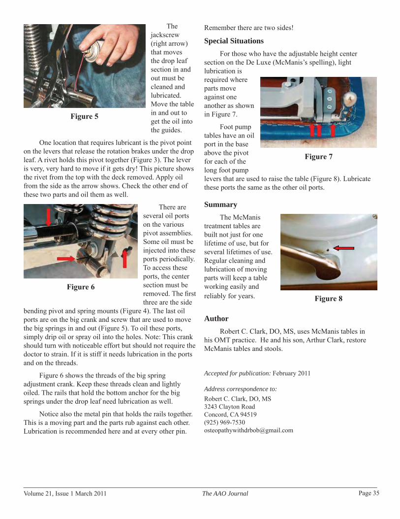
Volume 21, Issue 1 March 2011 The AAO Journal Page 35
The jackscrew (right arrow) that moves the drop leaf section in and out must be cleaned and lubricated. Move the table in and out to get the oil into the guides.
One location that requires lubricant is the pivot point on the levers that release the rotation brakes under the drop leaf. A rivet holds this pivot together (Figure 3). The lever is very, very hard to move if it gets dry! This picture shows the rivet from the top with the deck removed. Apply oil from the side as the arrow shows. Check the other end of these two parts and oil them as well.
There are several oil ports on the various pivot assemblies. Some oil must be injected into these ports periodically. To access these ports, the center section must be removed. The first three are the side
bending pivot and spring mounts (Figure 4). The last oil ports are on the big crank and screw that are used to move the big springs in and out (Figure 5). To oil these ports, simply drip oil or spray oil into the holes. Note: This crank should turn with noticeable effort but should not require the doctor to strain. If it is stiff it needs lubrication in the ports and on the threads.
Figure 6 shows the threads of the big spring adjustment crank. Keep these threads clean and lightly oiled. The rails that hold the bottom anchor for the big springs under the drop leaf need lubrication as well.
Notice also the metal pin that holds the rails together. This is a moving part and the parts rub against each other. Lubrication is recommended here and at every other pin.
Figure 5
Remember there are two sides!
Special SituationsFor those who have the adjustable height center
section on the De Luxe (McManis’s spelling), light lubrication is required where parts move against one another as shown in Figure 7.
Foot pump tables have an oil port in the base above the pivot for each of the long foot pump levers that are used to raise the table (Figure 8). Lubricate these ports the same as the other oil ports.
SummaryThe McManis
treatment tables are built not just for one lifetime of use, but for several lifetimes of use. Regular cleaning and lubrication of moving parts will keep a table working easily and reliably for years.
AuthorRobert C. Clark, DO, MS, uses McManis tables in
his OMT practice. He and his son, Arthur Clark, restore McManis tables and stools.
Accepted for publication: February 2011
Address correspondence to:Robert C. Clark, DO, MS 3243 Clayton Road Concord, CA 94519(925) 969-7530 [email protected]
Figure 6
Figure 7
Figure 8

The AAO Journal Volume 21, Issue 1, March 2011Page 36
From The Archives From: Fryette, HH. Principles of Osteopathic Technique. Carmel, CA: American Academy of osteopathy, 1954. 61-64.
Articular AdjustmentIn the beginning, osteopathic “adjustment” meant
articular adjustment, but since then we have broadened the meaning of adjustment almost as much as we have the meaning of the word “lesion.” We now speak of dietetic adjustment, occupational adjustment and climatic adjustment, all kinds of physical adjustments, as well as a multitude of psychological adjustments. “Adjustment” is a very good word. It conveys a sense of harmony, peace and contentment, all of which are concomitant to health. To the degree that we are in harmonious adjustment within ourselves and with our environment, we are apt to be well.
The principles of anatomical adjustment may be split up ad infinitum, but for practical purposes they are simple and generally applicable.
Before we begin a mechanical adjustment, we assume that the operator has made a correct diagnosis mechanically and pathologically. If one cannot definitely and clearly visualize every mechanical angle and pathological factor involved, one should not proceed with adjustment until he can.
Check again the angles, planes and degrees of limited motion, and estimate the reverse angles and planes and the estimated degree of force required to overcome that degree of resistance. There is no way for one to tell another how to acquire this art, which in time becomes subconscious.
Many say that when we have positioned a lesion for correction that we should take all the slack out of the ligaments and then thrust it a bit farther. I am very definitely opposed to this method.
My father once asked Dr. Still about this point. Dr. Still said, “If you had a horse tied to a post (they still drove horses in those days) and you wanted to untie him, you wouldn’t first frighten him so that he would pull back on the rope to hold it tight during the untying operation, would you?”
I do not like the word “thrust” because, as it has been used, it implies too much force, and force often implies discomfort. A thrust is said to be a violent or sudden push. It is at the moment of the thrust that the sequence of fear, tension and pain sometimes occurs. As long as I do not know a better word, however, I shall use “thrust.”
If the angles and planes are properly visualized and the patient is thoroughly relaxed, no great amount of force is required to move a bone in line of correction.
The character of the thrust should be more in the nature of a tap, not a push with a follow through. It is sudden. The velocity should be high, the amplitude low. It should start from near neutral, not from extreme flexion or extreme extension. If these principles are properly applied, the inertia of the body mass is sufficient to hold it in position for the operation. No pain or soreness ensues as when less brains and more muscle are used. As an example of what I mean, I used to glue a block of wood to the top of a common laboratory table, and then move the table about the room by pushing on the block, but by tapping the block lightly with a tack hammer at a high velocity and low amplitude, if the force were applied exactly parallel to the articulation, the block would easily be knocked off without moving the table. It must be observed that in this process there is no “follow through,” which would result in a push.
In the application of our thrust or stroke, we have borrowed an expression from golf, which is called “timing.”
In golf, good timing or bad timing is all within one’s self. It is really coordination. It is the problem of controlling the club head in such a manner that it will be traveling at its greatest velocity at the instant it meets the ball.
Our timing in making mechanical adjustments is very much the same—except that the golf ball sits still until it is hit, while the patient may not. The golf ball does not know that it is going to be hit, but the patient does. The patient, realizing this, has a subconscious tendency to flinch. If he flinches at the instant of impact, or a little before, the operation is a failure. The tendency then is for the operator to try it again using more force. By this time the patient has been hurt and is really tightened up. Instead of this procedure, the operator should sense the condition of his patient and so time his adjustment that it is made while the patient is relaxed. Never fight tense or sore muscles.
There are cases, I am one of them, who are extremely sensitive, who have abnormally quick reflexes which are often beyond their control. If, under these conditions, it is still thought to be desirable to adjust them, they should be given a sedative, not too much, but just enough to slow down their reflexes.
The practice of medicine might be reduced to the practice of nutrition, for perfect nutrition means perfect health. One of the main factors which controls nutrition is circulation. Dr. Still said, “The rule of the artery is supreme.”

Volume 21, Issue 1 March 2011 The AAO Journal Page 37
Gravity TreatmentOne of the important factors which impedes circulation is
gravity. Dr. C. R. Nelson has said that “Gravity kills your patient.” Gravity, as we have remarked, is an inexorable factor. It is the factor that places a constant load upon the supporting structures.
First, visualize our circulatory system in the horizontal position. Under these conditions, the heart has no great load to lift, and during physical exercise, the action of the skeletal muscles is nearly sufficient to keep the blood circulating. The heart acts as a regulator as well as a pump. Under these conditions, there is little cause for heart failure.
Now, visualize our circulatory system in the perpendicular position. Everything above the heart is relatively anemic, which includes our brains—a thought worth considering. As a result of this local anemia, our brains function poorly, some of us have lost our hair, our eyes and ears fail, our teeth decay and our sinuses become infected. Even the apices of our lungs often show signs of anemia.
On the other hand, everything below the heart prolapses and becomes congested, and in many cases the heart breaks down from overload.
As we remarked, when we have been in the upright posture all day, everything above the heart is anemic, everything below it congested. When we lie down at night, it requires a great deal of work on the part of the heart, and considerable time, to equalize the blood—in other words, to get the body into condition to begin to rest.
We have said that gravity is inexorable—we cannot get rid of it unless we should walk on all fours and that would be inconvenient. Under these circumstances I have found it a very good idea to stand on our heads, not literally, as the Yogi do, but to hang over a table or a high bed for a minute or two and allow gravity to reverse its processes.
The results of this simple gravity treatment are amazing. It not only equalizes the circulation quickly, but tends to replace the abdominal viscera, which have been sagging all day.
This treatment is an absolute must with all of my sinus cases.
I am often asked if this treatment is dangerous. There are contra indications for it, of course. The doctor must be the judge of that, but I have never had any bad results from it.
Touro University Nevada College of Osteopathic Medicine (TUNCOM) is seeking an osteopathic physician (D.O.) for a full-time faculty position in the Department of Osteopathic Manipulative Medicine. Responsibilities include teaching, patient care and program development. In addition, there are opportunities for conducting research.
Qualifications: Graduate of an AOA-approved osteopathic medical collegeSatisfactory completion of an AOA-approved internship Certified by the American Osteopathic Board of Special Proficiency in Osteopathic Manipulative Medicine or American Osteopathic Board of Neuromusculoskeletal Medicine. The applicant must also be licensable in the State of Nevada.
TUNCOM offers a competitive salary and benefits package. Touro University Nevada is an EEO.
In addition, Southern Nevada has year-round sunshine with excellent weather, close proximity to Zion, Bryce and Grand Canyon National Parks, world-class food and entertainment in Henderson and neighboring Las Vegas, and convenient access to an international airport.
Information regarding this position may be obtained by contacting: Paul R. Rennie, DO, FAAO Touro University Nevada College of Osteopathic MedicineDepartment of Osteopathic Manipulative Medicine874 American Pacific Drive, Henderson, NV 89014(702) [email protected]
Application Closing Date: Open until filled.
C
M
Y
CM
MY
CY
CMY
K
OMM position advertising.pdf 1 10/27/2009 3:49:54 PM
PAIn ALTERnATIVES, InCSeeking a physician to join dynamic expanding multi-practitioner Osteopathic practice. Osteopathic manipula-tive office-based care and inpatient consultative services are the cornerstones of this practice which treats new-borns through geriatrics.
This practice participates in Osteopathic medical educa-tion of students, interns, residents, faculty and the medi-cal community. We are affiliated with a large award-winning Osteopathic training hospital system recognized nationally for clinical excellent.
Come practice at the cutting edge of Osteopathic Medi-cine!
Physicians need to have completed or be board certified in Neuromusculoskeletal Medicine.
For further information or to send a CV please contact:
Kelley Beloff, MSW, CMM2510 Commons Blvd., Suite 240
Beavercreek, OH 45431Phone: 937/429.8620 • Fax: 937/429-8629

The AAO Journal Volume 21, Issue 1, March 2011Page 38
Evaluating the Perception of Motion in Osteopathic Medical Students, Residents and PhysiciansCraig Chappell, DO; Godwin Dogbey, PhD; Kevin Dankart, OMS IV
AbstractHypothesis
Perception of motion can be objectified. The ability to palpate motion improves with level of training.
Methods
A six-inch, fluid-filled sphere was attached to a syringe via a rigid conduit. The syringe was mounted to a pump that was programmed to deliver and withdraw fluid over predetermined intervals. With a closed system, fluid was pumped into and out of the sphere inducing various amounts of expansion and contraction. Participants placed their hands on the ball and verbalized what they felt as fluid was introduced or withdrawn. Responses were recorded and correlated with the participants’ level of osteopathic training.
Results
Palpation of motion was objectified. First-year medical students and residents are able to correctly identify motion better than physicians.
Conclusion
The ability to correctly perceive motion appears to be independent of training. This device proved beneficial in objectifying perceived motion and may assist future students in training.
IntroductionPalpation plays a key diagnostic role in osteopathic
medicine. “Palpation involves the use of the hands and fingers to gather information through the sense of touch. The palmar surface of the fingers and finger pads is more sensitive than the fingertips and is used whenever discriminatory touch is needed for determining position, texture, size, consistency, masses, fluid, and crepitus.”1 Although palpation may be a subjective experience, it can often be used in detecting states such as health versus disease and normal versus non-normal states. Palpation is used to detect lymphadenopathy, masses, chest-wall compliance, tactile fremitus of the lungs, pulsation of vessels and the point of maximum impulse of the heart.
In osteopathic medical training, instructors guide students in laboratory environments where they spend extensive time working with one another to learn how to distinguish normal versus non-normal palpatory findings, as well as to improve their palpatory and diagnostic skills.
Palpation becomes even more complex when students are introduced to osteopathy in the cranial field, where the perception of cranial motion is attempted. There has been a growing body of literature that discusses different aspects of palpation.2,3,4 These aspects include two-point discrimination, nerve receptor involvement, improving palpatory skills utilizing static or mechanical devices, and pressures used by practitioners when palpating. However, to the authors’ best knowledge, there have been no studies that objectify palpation by quantitatively assessing the palpation of motion within the context of when motion can be detected and suggests a device that could be used in medical training to develop the palpation skills of trainees, particularly in osteopathic medical education. This study was designed to evaluate and quantify the perception of motion and determine whether the ability to perceive such motion improves with time and/or training among osteopathic medical students, residents and physicians.
Materials and MethodsThis study uses a mechanical device as a means to
gauge and quantify the palpatory skills of medical students, residents and physicians.
Figure 1: ne-1050-X2 pump with pressure gauge and syringe attached

Volume 21, Issue 1 March 2011 The AAO Journal Page 39
Assembly of Device
A six-inch spherical ball was attached to a large-bore, semi-rigid conduit (3/8” braided PVC) via a ¼” fully threaded nipple, which was inserted into a ball. The nipple was then secured to the ball. To provide a leak-proof system, the opposite end of the conduit was attached to a pressure gauge and then to a permanently modified 160 cc syringe via another ¼” nipple. The system (ball, conduit, pressure gauge and syringe) was then filled with fluid, and care was taken to ensure that no air remained in the system. The syringe was mounted to a programmable syringe pump (NE-1050-X2 by New Era Pump Systems, Inc.) capable of precisely (+/- one percent of injected/withdrawn amount) injecting and withdrawing relatively small and large volumes of fluid over programmed intervals (Figure 1). The ball and conduit were then secured to a modified Osteopathic Manipulative Medicine (OMM) table to ensure a fixed position. The conduit was invested tightly into a soft foam material and then covered in black vinyl similar to the OMM table top (Figure 2). The foam and vinyl served two functions, one of which was aesthetic and the other to dampen any vibration emitted by the syringe pump. The pump and pressure gauge were placed on a separate table, and care was taken to ensure the OMM table and pump table were not in contact to avoid any transmission of vibration from the syringe pump to the OMM table. The table containing the pump and gauge was then concealed with a blind so the pump was not visible to the study participants.
Ball diameter at base line was 155.55 mm and was measured using electronic digital calipers (+/- 0.02 mm accuracy). It was presumed that fluid is not compressible and that all fluid injected from the syringe would end up in the ball, given the low working pressures and the rigidity of the conduit. Working under this presumption, the starting volume of the sphere was calculated using the intial ball
diameter of 155.5 5mm (volume = 4/3 π r3). With each change in volume from injecting or withdrawing fluid, the theoretical diameter changes were calculated from the relationship diameter = 2r, where r = radius of the sphere (Table 1).
The pump was programmed to inject a specified amount of fluid over three seconds, followed by a randomly assigned pause, after which the same amount of fluid was withdrawn over three seconds, returning the ball to a baseline state. Pauses were randomized from three to six seconds to prevent any rhythmic pattern. Example: four- second pause, inject 12 cc over three seconds; six-second pause, withdraw 12 cc over threeseconds; five-second pause, inject 21 cc over three seconds, etc. The amount of fluid introduced was randomized and ranged from zero cc to 42 cc of fluid in 3-cc increments. This means that zero cc to 42 cc in intervals 3 cc of fluid were all injected or withdrawn over three seconds during the course of the test. This predetermined sequence was repeated twice and could be accomplished in eight minutes.
SubjectsParticipants in the study consisted of osteopathic
medical students, residents and physicians. Specifically, the subject composition was first-year medical students (OMS I), second-year medical students (OMS II), third- and fourth-year medical students (OMS III and IV), residents and practicing osteopathic physicians. Participants were recruited from Ohio University College of Osteopathic Medicine and the 2010 annual American Academy of Osteopathy Convocation in Colorado Springs, CO. After reading an instructional poster, which served as informed consent, subjects were able to participate in this study, which was approved by the institutional review board at Ohio University (approval number 09X125). Participants were asked to place both hands on the ball utilizing a hold similar to that of the “vault hold” as described in
Figure 2: The complete set up Table 1: relationship of changing volume and diameter

The AAO Journal Volume 21, Issue 1, March 2011Page 40
osteopathy in the cranial field (See Figure 2 and Figure 3). Participants also had to wear noise-cancelling headphones that played white noise in order to cancel any sound from the pump. Participants were instructed to verbalize what they felt using the words “bigger” or “smaller” as they felt it. It was presumed that when no response was given, then no motion was felt. Participants were also informed that the ball would not be moving the majority of the time secondary to the assigned pauses. No instructions were given concerning hand contact with the ball or contact pressure.
Participants were then asked to put the headphones on and place their hands comfortably on the ball in a vault-hold type position. When the participant was ready, he or she would tell the tester to start. Three seconds after pressing the start button, 45 cc of fluid was injected over three seconds, followed by a three-second pause, then 45 cc of fluid was withdrawn from the ball over three seconds. This initial introduction of motion served as a primer to allow the participant to experience the motion being tested. Following the initial practice motion, the test began after a randomly assigned pause. The remainder of the test consisted of recording the participants’ responses, or lack of responses, to the perceived ball motion.
Data Collection and AnalysesAll responses were recorded in the time period in
which they were spoken, and all testing was performed by the primary author. Correct responses were given in the time period at which the motion occurred, and the perception of motion was consistent with what happened mechanically. Incorrect responses were those that described anything other than that which happened mechanically. For
example, if the pump was injecting fluid and the participant said “smaller,” this would be an incorrect response because the ball was actually getting larger, as would any response that detected change in size during a randomly assigned pause. Other data recorded included level of training, specialty and number of years in practice (Table 2).
All testing was performed from March 2010 to June 2010. Criteria for participants included all osteopathic medical students and physicians. Two foreign-trained osteopaths participated as well. Groups were compared based on level of training and were broken down intoOMS I, OMS II, OMS III and IV, osteopathic residents, and osteopathic physicians. Repeated measures multivariate analysis of variance was used to determine whether significant differences existed over time and/or among the years of training with respect to correctly feeling the changes in ball size during injection and withdrawal phases. The outcome variables were the total number of correct detections of motion when volume changes with the two injections and the two withdrawals of liquid phases of the ball. Statistical significance was set at p ≤ .05.
ResultsResults from the repeated measures MANOVA
indicated a statistically significant difference in total correct responses (correct detections of motion) within the four successive rounds of injections and withdrawals of liquid into and out of the ball (Wilks’ Λ = 0.356, F (3,111) = 66.92, p < .001). In particular, more correct responses were registered on average on the injection phase than the withdrawal phase. There were also statistically significant
Table 2: Physician and resident specialty data and total participants in each group
Figure 3: Vault Hold

Volume 21, Issue 1 March 2011 The AAO Journal Page 41
differences between the levels of training, namely OMS I, OMS II, OMS III & IV, residents and physicians (F (4, 113) = 3.187, p = .016). Post-hoc analyses indicated the statistically significant differences emanated from the difference in the total correct responses between the OMS I and physicians (p = .035) (Figure 4). On the whole, while residents outperformed all other groups in terms of the average total number of correct responses and the least number of incorrect pauses detected (Figure 5), the differences were not statistically significant.
DiscussionPalpation is an important tool used every day by
osteopathic physicians of many specialties. It can be utilized to objectively aid a physician in the diagnosis of infectious processes through the palpation of tactile fremitus in the lungs or an enlarged lymph node; to detect the heaves or lifts of a diseased heart; to uncover an underlying malignancy of a breast mass or enlarged prostate; or to detect the subtle musculoskeletal and cranial changes associated with somatic dysfunction.¹ According to Foundations of Osteopathic Medicine, trained individuals can perceive the motion of tissue as diminutive as one micrometer; however, the source of this information could not be confirmed.5
Prior research has shown that two-point discrimination is greater in upper-class chiropractic students than in first-year students; however, these skills were not maintained into practice. Conversely, it was shown that palpatory abilities were not only maintained, but also continued to improve in professional practice, leading the authors to propose that two-point discrimination may not be a dependable marker for measuring palpatory ability.6,7 Thus, it was hypothesized in our study that the objectified perception of minute motion could be applied as a reliable indicator of palpatory abilities. In accordance with this previous research, it was also hypothesized that the ability to palpate motion would improve with level of training.
The results of this study demonstrate that when all levels of training (i.e., OMS I, OMS II, OMS III and IV, residents and physicians) were compared, first-year osteopathic medical students were able to correctly detect motion significantly more often than physicians. This does not correspond with the hypothesis that palpatory skills should improve with the level of osteopathic training.6,7 However, these results could be due to several reasons, including lack of confidence in OMM abilities secondary to lack of exposure and practice of OMM during the clinical years of osteopathic training (OMS III & OMS IV).8 Furthermore, some participants admitted to being able to perceive fluid motion or turbulence during the injection and
withdrawal of fluid. Thus, more highly trained individuals, especially practicing physicians, may be able to detect this subtle fluid motion consequently, leading to confusion between diameter change and internal fluid motion and hence more incorrect answers during pauses (Figure 5).
Another limitation of this study was the effect of gravity on the fluid in the ball. Gravity may have produced a larger change in diameter in the transverse plane compared to the vertical plane. However, if participants were properly employing the vault hold technique, their fingertips should primarily have been overlying the transverse plane, allowing better perception of motion.
The strengths of this study include the number of participants (N = 118). Moreover, all testing was done by the primary author, ensuring each trial was administered the
Figure 5: number of incorrect pauses out of 60 trials on pauses by level of training
Figure 4: number of Average Total Correct Responses by Level of Training for the Injection Rounds, Withdrawal Rounds and Overall

The AAO Journal Volume 21, Issue 1, March 2011Page 42
same to every participant and the same instructions were given. Finally, all movement of the ball was objectively measured.
If this study were to be replicated, there are some improvements that the authors think may increase the validity of the results. First, the viscosity of the injected and withdrawn fluid from the ball could be increased to reduce fluid turbulence, and consequently, the amount of fluid motion perceived. This could eliminate the possibility of participants mistaking fluid motion for change in diameter.
Another resolution to this same variable would be to inject the fluid into an inner bladder that would act as a baffle placed inside the ball. This would remove the sensation of fluid motion by preventing direct contact between the fluid and inner surface of the ball. Conclusion
The results of this study suggest that first-year osteopathic medical students are able to correctly palpate motion significantly more often than practicing osteopathic physicians. It seems the ability to palpate motion does not improve with training. However, additional studies are needed to address the issue of internal fluid motion discussed previously. Finally, the authors suggest that the development and objective assessment of palpation skills of trainees in osteopathic medical education can be further enhanced by the device developed for this study.
AcknowledgementsWe would like to thank the Centers for Osteopathic
Research and Education (CORE) for funding and support. Thanks to Ken Willeard and Robert Baker for participation in photos. And thanks to Robert Evans, DO, and Corie McCament for editing.
References1. Seidel HM, Ball JW, Dains JE, and Benedict GW. Mosby’s Guide
to Physical Examination, 5th Edition. Henderson L, Ed. St. Louis: Mosby, 2003.
2 Zegarra-Parodi R, de Chauvigny de Blot P, Rickards LD, Renard EO. Cranial palpation pressures used by osteopathy students: effects of standardized protocol training. The Journal of the American Osteopathic Association. February 2009. 109 (2): 79-85.
3. Howell JN, Conatser RR, Williams RL, Burns JM, Eland DC. Palpatory diagnosis training on the virtual haptic back: performance improvement and user evaluations. Journal of the American Osteopathic Association. January 2008. 108 (1): 29-36.
4. Howell JN, Conatser RR, Williams RL, Burns JM, Eland DC. The virtual haptic back: A simulation for training in palpatory diagnosis. BMC Medical Education. April 2008. 8 (14): 10.1186/1472-6920-8-14.
5. Ward RC. Palpatory Skills: An Introduction. Kappler RE, Ed. Foundations for Osteopathic Medicine. Baltimore: Williams &
Wilkins, 1997. 475.6. Chandhok PS, Bagust J. Differences between the cutaneous
two-point discrimination thresholds of chiropractic students at different stages in a 5-year course. Journal of Manipulative and Physiological Therapeutics. October 2002. 25 (8): 521-525.
7. Foster IE, Bagust J. Cutaneous two-point discrimination thresholds and palpatory sensibility in chiropractic students and field chiropractors. Journal of Manipulative and Physiological Therapeutics. September 2004. 27 (7): 466-471.
8. Quinn T, Best M, Fotopoulos TJ, Ball LD, Ruston VJ. Attitudes and confidence levels of fourth-year osteopathic medical students towards osteopathic manipulative medicine. The AAO Journal. September 2010. 20 (3): 23-28.
Accepted for publication: February 2011
Address correspondence to:Craig Chappell, DOOhio University College of Osteopathic MedicinePlus-One Resident4329 Harvard Road Erie, PA 16509Phone: (801) 898-6114
Godwin Dogbey PhDOhio University College of Osteopathic Medicine CORE Research Biostatistician030D Grosvenor HallAthens, Ohio 45701Phone: (740) 593-2370
Kevin Dankart, OMS IV Lake Erie College of Osteopathic Medicine3742 Oak Forest Toledo OH, 43614Phone: (419) 351-3328

Volume 21, Issue 1 March 2011 The AAO Journal Page 43
5. Manuscripts must be published with the correct name(s) of the author(s). No manuscripts will be published anonymously, or under pseudonyms or pen names.
6. For human or animal experimental investigations, include proof that the project was approved by an appropriate institutional review board, or when no such board is in place, that the manner in which informed consent was obtained from human subjects.
7. Describe the basic study design; define all statistical methods used; list measurement instruments, methods, and tools used for independent and dependent variables.
8. In the “Materials and Methods” section, identify all interventions that are used which do not comply with approved or standard usage.
FLOPPy, CD-ROM or DVDWe encourage and welcome a CDROM or DVD containing the material submitted in hard copy form. Though we prefer receiving materials saved in rich text format on a CD-ROM or via Email, materials submitted in paper format are acceptable.AbstractProvide a 150-word abstract that summarizes the main points of the paper and its conclusions.Illustrations1. Be sure that illustrations submitted are clearly labeled.2. Photos and illustrations should be submitted as a 5” x 7”
glossy black and white print with high contrast. On the back of each photo, clearly indicate the top of the photo. If photos or illustrations are electronically scanned, they must be scanned in 300 or higher dpi and saved in .jpg format.
3. Include a caption for each figure.PermissionsObtain written permission from the publisher and author to use previously published illustrations and submit these letters with the manuscript. You also must obtain written permission from patients to use their photos if there is a possibility that they might be identified. In the case of children, permission must be obtained from a parent or guardian.ReferencesReferences are required for all material derived from the work of others. Cite all references in numerical order in the text. If there are references used as general source material, but from which no specific information was taken, list them in alphabetical order following the numbered journals.For journals, include the names of all authors, complete title of the article, name of the journal, volume number, date and inclusive page numbers. For books, include the name(s) of the editor(s), name and location of publisher and year of publication. Give page numbers for exact quotations.Editorial ProcessingAll accepted articles are subject to copy editing. Authors are responsible for all statements, including changes made by the manuscript editor. No material may be reprinted from The AAO Journal without the written permission of the editor and the author(s).
The American Academy of Osteopathy® (AAO) Journal is a peer-reviewed publication for disseminating information on the science and art of osteopathic manipulative medicine. It is directed toward osteopathic physicians, students, interns and residents, and particularly toward those physicians with a special interest in osteopathic manipulative treatment.The AAO Journal welcomes contributions in the following categories:Original Contributions: Clinical or applied research, or basic science research related to clinical practice.Case Reports: Unusual clinical presentations, newly recognized situations or rarely reported features.Clinical Practice: Articles about practical applications for general practitioners or specialists.Special Communications: Items related to the art of practice, such as poems, essays and stories.Letters to the EditorComments on articles published in The AAO Journal or new information on clinical topics. Letters must be signed by the author(s). No letters will be published anonymously, or under pseudonyms or pen names.Book ReviewsReviews of publications related to osteopathic manipulative medicine and to manipulative medicine in general.noteContributions are accepted from members of the AOA, faculty members in osteopathic medical colleges, osteopathic residents and interns and students of osteopathic colleges. Contributions by others are accepted on an individual basis.
SubmissionSubmit all papers (in word format) to:
American Academy of Osteopathy3500 DePauw Blvd, Suite 1080Indianapolis, IN 46268Email: [email protected]
Editorial ReviewPapers submitted to The AAO Journal may be submitted for review by the Editorial Board. Notification of acceptance or rejection usually is given within three months after receipt of the paper; publication follows as soon as possible thereafter, depending upon the backlog of papers. Some papers may be rejected because of duplication of subject matter or the need to establish priorities on the use of limited space.Requirements for manuscript submissionManuscript1. Type all text, references and tabular material using upper and
lower case, double-spaced with one-inch margins. Number all pages consecutively.
2. Submit original plus two copies. Retain one copy for your files.
3. Check that all references, tables and figures are cited in the text and in numerical order.
4. Include a cover letter that gives the author’s full name and address, telephone number, institution from which work initiated and academic title or position.
Instructions to Authors

The AAO Journal Volume 21, Issue 1, March 2011Page 44
Component Societies and affiliated organizations upcoming Calendar of Events
april 6 - 9Towards an Integrated Medicine
The Register of Osteopaths of Italy and The European Institute for Evidence-Based Osteopathic Medicine’s
International Congress of Osteopathic MedicineFlorence, Italy
Web site: www.osteopatia2011.it
april 13 - 16Meeting Future Health Care Needs:
The Role of Interprofessional EducationThe American Association of Colleges of Osteopathic Medicine
and American Osteopathic Directors and Medical Educators Joint Annual Meeting
Marriott Baltimore Waterfront, Baltimore, MDContact: Beth Martino (AAACOM) or Penny Friske (AODME)
Phone: (301) 968-4189 or (312) 202-8211Email: [email protected] or [email protected]
Web site: www.aacom.org/events/annualmtg/
april 28 - may 1Indiana Osteopathic Association 114th Annual Convention
Sheraton - Keystone at the Crossing, Indianapolis, INPhone:(800) 942-0501
E-mail: [email protected] Web site: http://www.inosteo.org/
may 4-7Pennsylvania Osteopathic Medical Association
103rd Annual Clinical Assembly and Scientific SeminarValley Forge Convention Center, King of Prussia, PA
CME: 41 AOA credits (35 Category 1A and 6 Category 1B)Phone: (717) 939-9318 Fax: (717) 939-7255
E-mail: [email protected] Website: www.poma.org
may 13 - 15Intro to Osteopathic Medicine and Evaluation & Treatment: Pelvis
OMM at UNECOM, Biddeford, ME CME: 20 Category 1A AOA credits
Phone: (207) 602-2589 Fax: (207) 602-5957E-mail: [email protected]
Web site: www.une.edu/com/cme
June 2 - 6Basic Course:Osteopathy in the Cranial Field
PNWUCOM, Yakima, WACourse Director: Melicien Tettambel, DO, FAAOCME: 40 Category 1A AOA credits anticipated
Contact: Joy CunninghamPhone: (509) 469-1520 Fax: (509) 453-1808
Email: [email protected] site: www.sctf.com
June 3 - 6Biodynamics of Osteopathy: Phase II
UNECOM, Biddeford, ME CME: Up to 22 Category 1A AOA credits
Contact: Joan Hankinson Phone: (207) 781-7900
E-mail: [email protected] Web site: osteopathichealthcareofmaine.com
June 11 - 15Introductory Course:
Osteopathy in the Cranial FieldCME: 40 Category 1A AOA credits
Hyatt Regency, Indianapiolis, INCourse Director: Michael J. Porvaznik, DOPhone: (317) 594-0411 Fax: (317) 594-9299
E-mail: [email protected] site:www.cranialacademy.org
June 15 - 19Texas Osteopathic Medical Association and Texas American College of Osteopathic Family Physicisians Fourth Annual
Joint ConventionThe Fairmont Hotel, Dallas, TX
Phone: (512) 708-8662 Fax: (512) 708- 1415E-mail: [email protected]
Web site: http://www.txosteo.org
June 16 - 19Annual Conference—Clinical Pearls: A Tribute to the
Journal of the Osteopathic Cranial Association 1948-1958Hyatt Regency, Indianapiolis, IN
Course Director: Daniel J. Kary, DO, FAAOCME: 22 Category 1A AOA credits
Phone: (317) 594-0411 Fax: (317) 594-9299E-mail: [email protected] site:www.cranialacademy.org
July 22 - 24Intro to Osteopathic Medicine and
Evaluation & Treatment: Lumber SpineOMM at UNECOM, Biddeford, ME CME: 20 Category 1A AOA credits
Phone: (207) 602-2589 Fax: (207) 602-5957E-mail: [email protected]
Web site: www.une.edu/com/cme