Embed Size (px)
Citation preview
Crystal structure of BlaB
1
The 1.5 Å structure of Chryseobacterium meningosepticum
Zn- -lactamase in complex with the inhibitor, D-captopril
Isabel García-Sáez§, Julie Hopkins§, Cyril Papamicael◊, Nicola Franceschini¶,
Gianfranco Amicosante¶, Gian Maria Rossolini¥, Moreno Galleni‡, Jean-Marie Frère‡,
and Otto Dideberg§*
§ Laboratoire de Cristallographie Macromoléculaire, Institut de Biologie Structurale Jean-
Pierre Ebel (CNRS-CEA), 41, rue Jules Horowitz, F-38027 Grenoble Cedex 1, France.
◊ Oxford Center for Molecular Sciences, England
South Parks Road, GB-OXFORD, OX1 3QY- United Kingdom
¶ Dipartimento di Scienze e Tecnologie Biomediche, Università di L'Aquila, I-67100
L'Aquila, Italy.
¥ Dipartimento di Biologia Moleculare, Sezione di Microbiologia, Università di Siena, I-
53100 Siena, Italy.
‡ Laboratoire d'Enzymologie, Centre d'Ingénierie des Protéines, Institut de Chimie, Université
de Liège, Sart Tilman, B-4000 Liège, Belgium.
Running title: Crystal structure of BlaB
*Corresponding author: Phone: +(33) 4 38 78 56 09
Fax: +(33) 4 38 78 54 94
E-mail: [email protected]
Copyright 2003 by The American Society for Biochemistry and Molecular Biology, Inc.
JBC Papers in Press. Published on April 8, 2003 as Manuscript M301062200 by guest on February 24, 2020
http://ww
w.jbc.org/
Dow
nloaded from
Crystal structure of BlaB
2
Summary
The crystal structure of the class-B β-lactamase, BlaB, from the pathogenic bacterium,
Chryseobacterium meningosepticum, in complex with the inhibitor, D-captopril, has been
solved at 1.5 Å resolution. The enzyme has the typical αβ/βα metallo-β-lactamase fold and
the characteristic two metal binding sites of members of the subclass B1, in which two Zn
ions were identified. D-captopril, a diastereoisomer of the commercial drug, Captopril, acts as
an inhibitor by displacing the catalytic hydroxyl ion required for antibiotic hydrolysis and
intercalating its sulfhydryl group between the two Zn ions.
Interestingly, D-captopril is located on one side of the active-site cleft. The X-ray
structure of the complex of the closely related enzyme, IMP-1, with a mercaptocarboxylate
inhibitor, which also contains a sulfhydryl group bound to the two Zn ions, shows the ligand
to be located on the opposite side of the active-site cleft. A molecule generated by fusion of
these two inhibitors would cover the entire cleft, suggesting an interesting approach to the
design of highly specific inhibitors.
by guest on February 24, 2020http://w
ww
.jbc.org/D
ownloaded from
Crystal structure of BlaB
3
Introduction
Antibiotic resistance of pathogenic bacteria is a major clinical concern, since it has
increased significantly during recent years, rendering antimicrobial therapy progressively less
effective. One mechanism by which bacteria can escape the action of antibiotics is the
production of metallo-β-lactamases (MBLs) or class B β-lactamases (1). These enzymes were
initially described as a curiosity in innocuous strains of Bacillus cereus (2), but were
subsequently identified in several pathogenic species responsible for human infections in
which the enzymes are either encoded by resident chromosomal genes or by horizontally
acquired genes carried on mobile genetic elements (3-7). Class B β-lactamases have a broad
spectrum of substrates, including penicillins, cephalosporins, and carbapenems, some of
which are not substrates for active-site serine β-lactamases (3, 4). At present, no inhibitors of
these enzymes are used clinically, making them a potential source of antibiotic resistance.
The MBL family has been divided into three-different subclasses, B1, B2, and B3, on
the basis of sequence similarities (8). MBLs have two metal-binding sites (the His-site, or first
site, and the Cys-site, or second site), which bind Zn ions required for enzyme activity. In
general, both the mono-Zn (Zn in the His-site) and di-Zn forms are active, but have different
kinetic properties (9); (10) the exception is CphA from Aeromonas hydrophyla, which is
inhibited by the presence of the second zinc ion (11). In the mono-Zn enzyme, a water-
mediated mechanism for the hydrolysis of the antibiotic β-lactam ring has been suggested, in
which a water molecule is activated by the presence of the Zn ion in the His-site, then carries
by guest on February 24, 2020http://w
ww
.jbc.org/D
ownloaded from
Crystal structure of BlaB
4
out nucleophilic attack against the carbonyl carbon of the β-lactam. Residues Asp120 and
Cys221 from the second site would help keep the active water molecule in position. Asp120
would also act as a general base transferring the proton from the water molecule to the
nitrogen atom of the β-lactam ring, causing bond cleavage and opening of the ring, leading to
inactivation of the antibiotic (12, 13). In the bi-Zn enzyme, a hydroxide ion is positioned close
to the two Zn ions. The second Zn would also help to anchor Asp120 (14) and would orient
the β-lactam ring by binding to the β-lactam nitrogen of the antibiotic (15). Although both the
mono- and di-Zn forms exist, it has been hypothesized that only the mono-Zn form is present
under normal physiological conditions (16).
Due to the great interest in MBL inhibition, several classes of inhibitors have been
described (17); these include (18) phenazines (19), ketone derivatives of L- and D-alanine and
trifluoromethyl alcohol (20), thioesters (21, 22), biphenyl tetrazoles (23, 24), amino acid-
derived hydroxamates (25) thiols (26), (27-30) and tricyclic natural products (31). However,
despite many of these having good inhibitory properties, only a few thiols have a broad
spectrum of inhibition for MBLs. In this context, the analysis of the three-dimensional
structures of MBLs in complex with potential inhibitors becomes a powerful tool for rational
drug design. At present, the structures of Bacteroides fragilis CcrA complexed with 4-
morpholineethanesulfonic acid, biphenyl tetrazole, or a tricyclic natural product (32) and of
(23, 31) Pseudomonas aeruginosa IMP-1 complexed with mercaptocarboxylate (30) or with
succinic acid derivatives (33) have been reported.
by guest on February 24, 2020http://w
ww
.jbc.org/D
ownloaded from
Crystal structure of BlaB
5
BlaB is a chromosome-encoded MBL produced by Chryseobacterium
meningosepticum. It is a member of subclass B1 (34), (35, 36) has a broad substrate profile,
and is produced constitutively (37). Chryseobacterium meningosepticum is an ubiquitous
Gram-negative rod bacterium of clinical relevance, since it can cause neonatal meningitis,
adult septicemia, and nosocomial infections, and is resistant to most β-lactams, including
carbapenems (38, 39).
D-captopril (1-(D-3-mercapto-2-methylpropionyl)-D-proline) is a diastereoisomer of
the commercial drug Captopril (1-(D-3-mercapto-2-methylpropionyl)-L-proline), used
clinically to control high blood pressure. It acts as an angiotensin converting enzyme inhibitor
and increases plasma level of the vasodilator, bradykinin (40). D-captopril, which was
synthesized to test its inhibitory effect on MBLs, is a good competitive inhibitor of BlaB (Ki
= 70-100 µM) at neutral pH (Dr. H. W. Adolph, personal communication).
In this work, we describe the first high-resolution (1.5 Å) three-dimensional structure
of BlaB complexed with D-captopril.
by guest on February 24, 2020http://w
ww
.jbc.org/D
ownloaded from
Crystal structure of BlaB
6
Experimental Procedures
Synthesis of D-captopril.
D-captopril was synthesized from commercially available compounds, starting with D-
proline (41, 42). The final hydrolysis reaction was carried out using 1 N NaOH under an argon
atmosphere.
Expression, purification, and crystallization of the D-captopril complex
The overexpression, purification, and characterization of the protein have been
described (34). For crystallization, the hanging drop method was used. Crystals were obtained
by mixing 2 µl of protein (5 mg/ml in 10 mM Na cacodylate, 100 µM Zn acetate, 100 µM
DTT, pH 6.5) and 1 µl of the reservoir solution (28% PEG 4000, 0.2 M Na acetate, and 0.1 M
Tris-HCl, pH 8.4) at 8°C; crystals appeared after approximately three weeks. A crystal was
soaked for approximately 5 weeks in previously equilibrated drops of the reservoir solution
containing 2 mM D-captopril, then the crystal was flash cooled in liquid nitrogen using
precipitant containing 15% glycerol as cryoprotectant, and stored in liquid nitrogen for
subsequent data collection at a synchrotron source.
Data collection and processing
Diffraction data sets were collected at the ID14-4 beamline of the European
Synchrotron Radiation Facility (ESRF) in Grenoble. A data set of 360 frames at λ= 0.9792 Å
was collected at 1.5 Å resolution with 0.5° oscillation steps. Data integration performed using
by guest on February 24, 2020http://w
ww
.jbc.org/D
ownloaded from
Crystal structure of BlaB
7
DENZO/SCALEPACK (43) gave an initial ambiguity in the space group determination, since,
according to the automated indexing procedure, the crystal could be cubic or trigonal, with
similar distortion indexes. The data were re-indexed using DENZO/SCALEPACK and scaled
using SCALA (44), trying different space groups to solve this ambiguity. After this stage, the
crystal was considered to be cubic I23 or I213 with a=b=c=112.85 Å, α=β=γ=90° Rsym =
0.117 (1 molecule/asymmetric unit) or trigonal R3, with a=b=159.69 Å c=97.82 Å, and
α=β=90° γ= 120° Rsym = 0.119 (3 or 4 molecules/asymmetric unit) (Table 1).
TABLE 1
Structural determination and refinement.
The structure of the MBL, BcII, from Bacillus cereus at 1.8 Å resolution (45) with no ions or
water molecules was used as a model for molecular replacement using the program AMoRe
(46). Since the X-ray data could be indexed in three space groups (I213, I23, and R3), various
tests were performed. In brief, data indexed in R3 were used successfully in AMoRe with a
BcII model performing a four molecules/asymmetric unit search (c-factor= 36.3%, R-factor=
48.0%). With these solutions and using a ncs-averaging map calculated using the CNS
program (47), a partial model of BlaB was built using TURBO (48). A full model of BlaB at
1.5 Å was made using ARP/WARP (49). From the initial (2Fo-Fc) and (Fo-Fc) maps, the
inhibitor, D-captopril, was localized close to the Zn ions in the active site. Further cycles of
energy minimization, B-factor refinement, and water picking were performed using CNS
programs (47). Models for D-captopril were not included until their conformations were well
defined by the unbiased Fo-Fc, φcalc electron density maps. D-captopril molecules in each
by guest on February 24, 2020http://w
ww
.jbc.org/D
ownloaded from
Crystal structure of BlaB
8
of the four monomers were modeled. The model was refined without any non-
crystallographic-symmetry restrains, yielding Rworking= 18.95% and Rfree= 20.76%.
by guest on February 24, 2020http://w
ww
.jbc.org/D
ownloaded from
Crystal structure of BlaB
9
Results
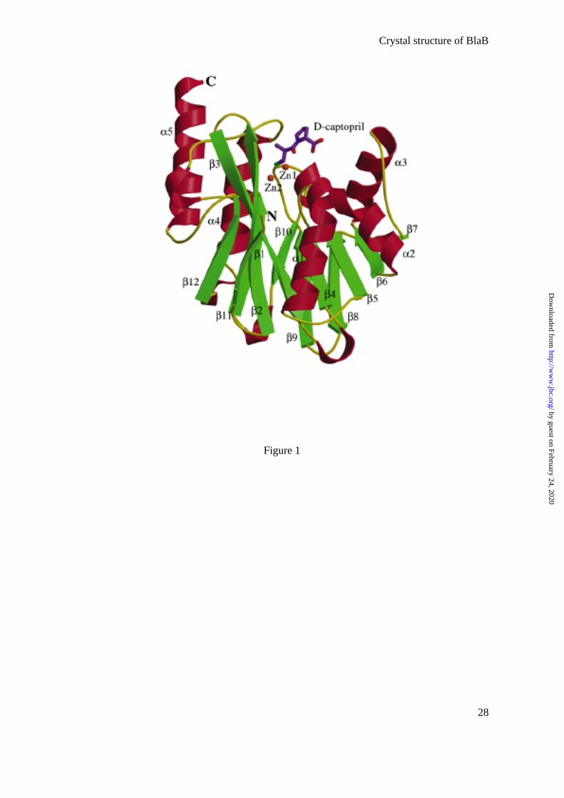
The BlaB monomer shares a structural feature with other members of the MBL family,
this being an αβ/βα fold, in which a compact core of two β-sheets is surrounded by α-helices
(Fig. 1). The crystal, which shows a cubic pseudo-symmetry, belongs to the space group R3
and has four monomers per asymmetric unit (A, B, C, and D).
FIGURE 1
The four monomers are practically identical, with a few local differences due to
slightly different environments or to effects of crystal packing (e.g. monomers A, B, and D
are missing the last four C-terminal residues, while monomer C is missing the last six). The
root mean square deviation between the conserved Cα atoms of the four molecules is about
0.1 Å. The active site of BlaB shares a feature with other subclass B1 structures, namely that
two Zn ions (Zn1 and Zn2) occupy the two metal-binding sites, the His-site (His116, His118,
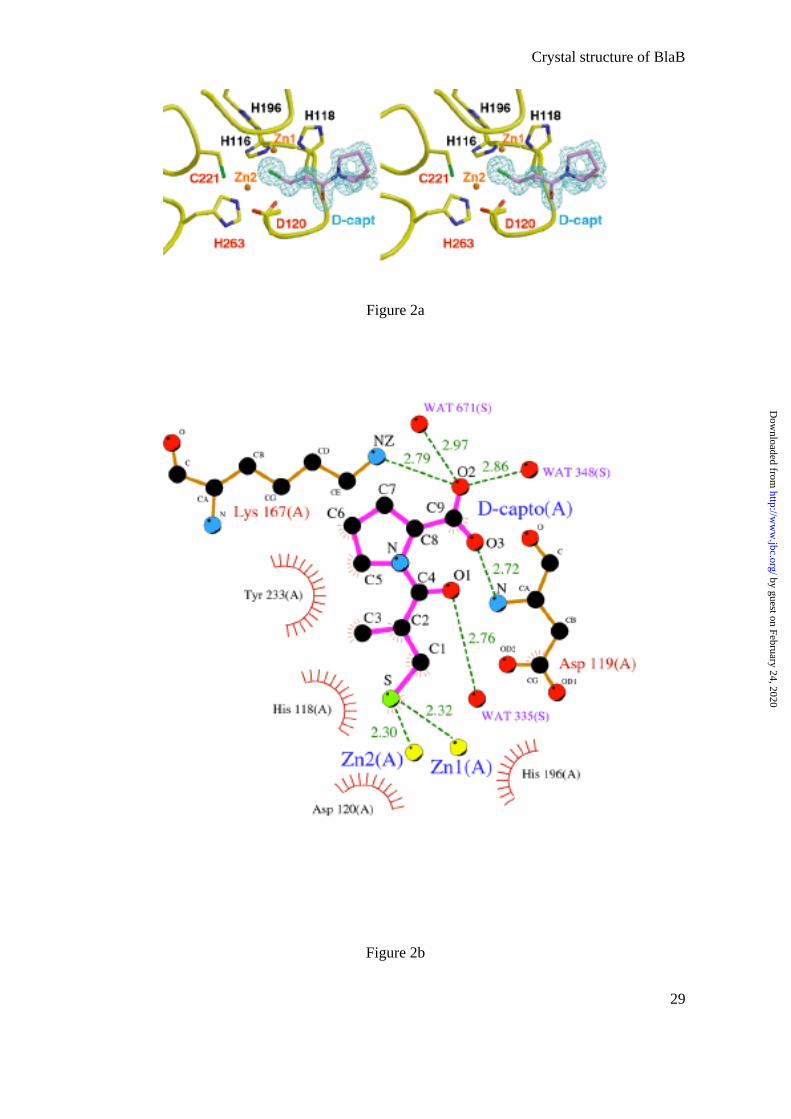
His196) and the Cys-site (Asp120, Cys221, His263) (Fig. 2a,c). A clear electron density
corresponding to a molecule of D-captopril was found close to the active site. The inhibitor
intercalates its sulfhydryl group between the two Zn ions (distance S-Zn1, 2.32 Å; S-Zn2,
2.30 Å in monomer A) (Fig.2). The sulfur atom is also close to the NE2 of His196 (3.34 Å)
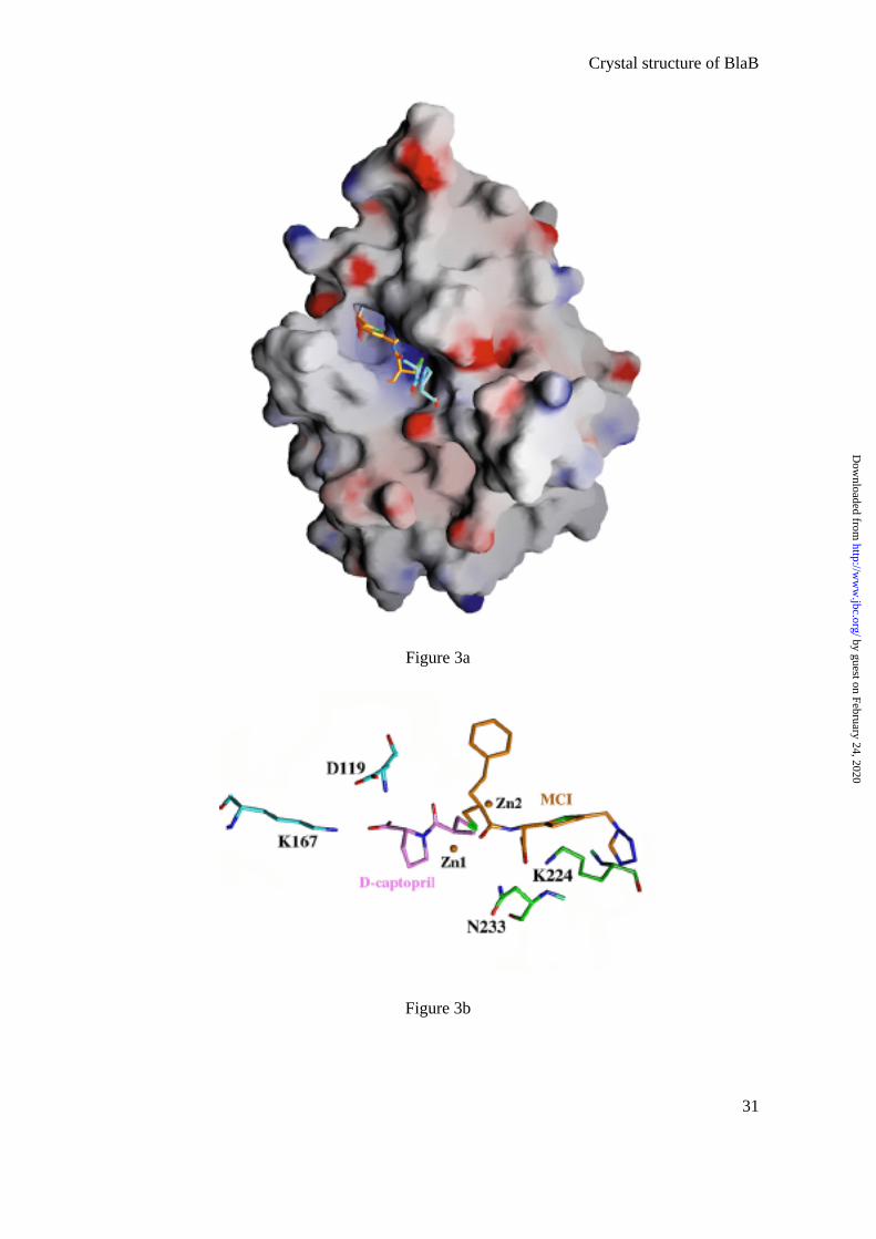
(Table 2). The inhibitor is located in the positively charged groove leading to the active site
(Fig. 3a) and is present in the four molecules/asymmetric unit with full occupancy.
TABLE 2
FIGURE 2a,2b,2c
by guest on February 24, 2020http://w
ww
.jbc.org/D
ownloaded from
Crystal structure of BlaB
10
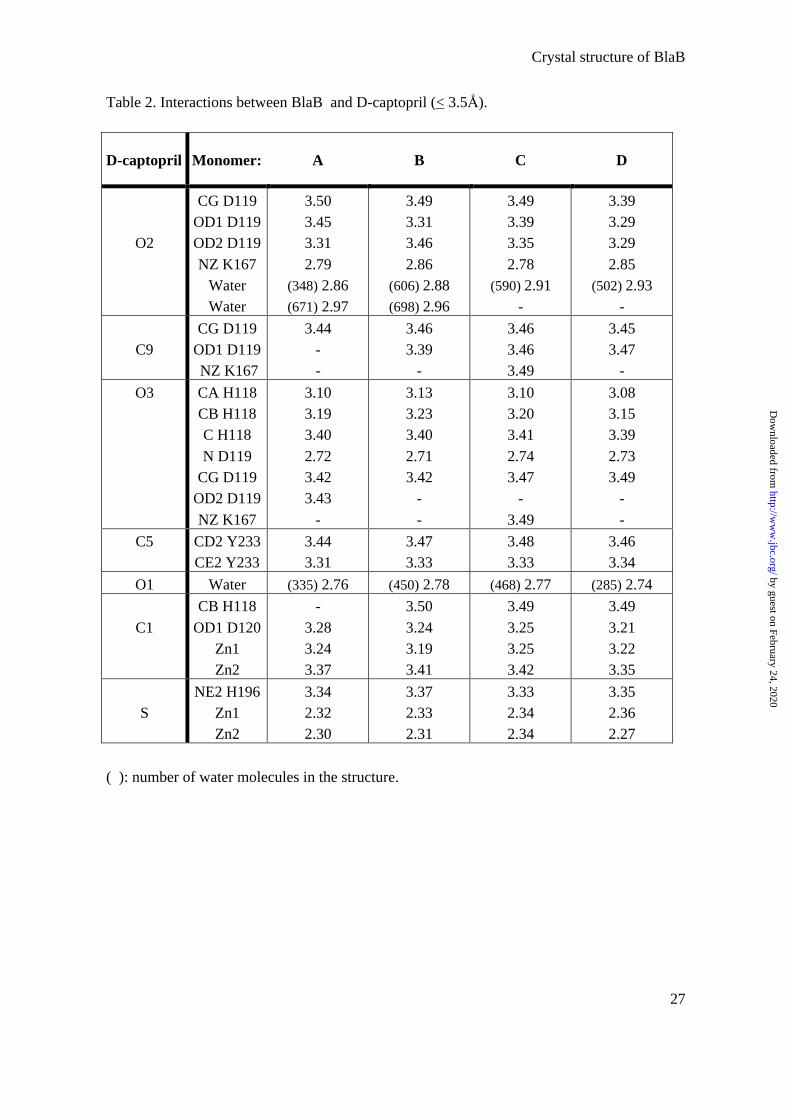
Other interactions enhance the binding of the inhibitor molecule. The carboxylic group
of D-captopril is stabilized by hydrogen bonds between its O2 and the NZ of Lys167 and two
water molecules; the other carboxylic oxygen, O3, is bound to the amide main-chain of
Asp119. The O1 carbonyl oxygen of the inhibitor also makes a hydrogen bond with another
water molecule (Fig. 2b). In addition, the inhibitor is stabilized by several hydrophobic
contacts between its carbons and His118, Asp120, and Tyr233 (Fig. 2b and Table 2).
FIGURE 3a, 3b
by guest on February 24, 2020http://w
ww
.jbc.org/D
ownloaded from
Crystal structure of BlaB
11
Discussion
Structural comparisons
The sequence of BlaB shows 25 - 35 % identity with those of other subclass B1 β-
lactamases. The secondary structure topology of the four available X-ray structures (BcII,
CcrA, BlaB, and IMP-1) is very similar, the only difference being that the first short β-strand
seen in BcII and CcrA is absent in BlaB and IMP-1. Pairwise superposition of the Cα atoms of
BlaB and BcII (1BMC), Ccra (1ZNB), or IMP-1 (1JJE) gives rmsd of 1.5, 1.4 and 1.9 Å,
respectively.
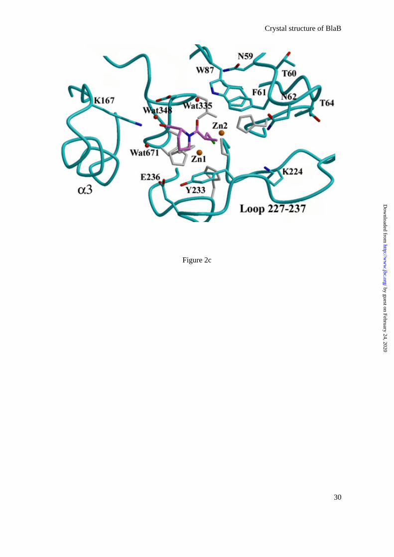
In subclass B1, there is a loop between β2 and β3 (residues 61-65), which is
disordered in the structure of native BcII (12). However, in the presence of an inhibitor, the
loop is stabilized (30), transforming the active site groove into a tunnel-shaped cavity. In the
BlaB structure, the loop is also well defined, probably due to the presence of D-captopril in
the active site (Fig. 2c). Interestingly, D-captopril has no direct interaction with any residues
in the loop (Fig. 2b,c; Table 2). The flexibility in the loop may help to accommodate the
inhibitor, which itself may help to stabilize the loop. This stabilization takes place indirectly
by Trp87, which makes a double hydrogen bond between its NE1 and the OD1 of Asp119 and
the water molecule that hydrogen bonds to the O1 carbonyl oxygen of D-captopril (Table 2).
Trp87 also makes hydrophobic contacts with the active site residue, His263. All these
interactions determine the position of Trp87, which establishes hydrophobic interactions
between its ring and two residues, Phe61 and Asn59. The loop is strongly stabilized by
by guest on February 24, 2020http://w
ww
.jbc.org/D
ownloaded from
Crystal structure of BlaB
12
hydrogen bonds between the carbonyl oxygens and the amide nitrogens of the backbone of
this region (57-69). In the apical part of the loop, facing the solvent, a water molecule forms a
bridge between the ND2 of Asn62 and the carbonyl oxygen of Phe61, which also makes a
hydrogen bond with the amide backbone of Thr64. In addition, the amide backbone of Phe61
makes hydrogen bonds with the carbonyl oxygen of the main-chain of Thr64. A second water
molecule makes hydrogen bonds with this bridging water molecule and the hydroxyl group of
Tyr67. In BlaB, the loop appears to be closer to the rest of the protein than in the other three
structures, with the hydrophobic residues, Phe61, Tyr67, and Trp87, pointing towards the
inside of the protein and creating a hydrophobic environment, even though the loop does not
interact directly with the inhibitor. In addition, the loops in BlaB and BcII are in different
positions e.g. the distance between equivalent residues in BlaB and BcII, is 5.44Å for Phe61,
and 5.77Å for Asn62. Interestingly, in subclass B1 MBLs, bulky hydrophobic residues are
often found at positions 61, 67, and 87.
In all MBL structures, the Asp or Asn residue at residue 84 has an ϕ angle outside the
allowed range (ϕ ∼ 60 ± 1°) and plays an important structural role, contributing to the
architecture of the active site.
Of the residues which are highly conserved and / or potentially involved in substrate
binding in subclass B1 β-lactamases, three positions are particularly interesting in the BlaB
structure, these being 224, 233, and 236 (Fig. 2c).
Lys224 is very well defined in the electron density map (NZ, B factor of 13.5 Å2). The
NZ makes three hydrogen bonds with the carbonyl oxygen of the preceding amino acid
by guest on February 24, 2020http://w
ww
.jbc.org/D
ownloaded from
Crystal structure of BlaB
13
(Ile223) and two water molecules. Lys224 is assumed to interact with the carboxyl group of
the β-lactam antibiotic (30). However, Lys224 is not found in subclass B3 MBLs and no
equivalent residue is observed in a similar spatial position in the L1 (15) or FEZ-1 structures
(50).
Based on X-ray crystallographic structures, Asn233 and Zn1 were thought to form an
oxyanion hole to polarize the carbonyl group of the β-lactam antibiotic (50, 51). In BlaB,
Asn233 is replaced by Tyr and the hydroxyl of the tyrosine is 7.88 Å away from Zn1 (Fig.
2c). Interestingly, Glu236, which is an aspartic acid residue in the BcII, CcrA, and IMP-1
enzymes, makes three hydrogen bonds with OH Tyr233, NEZ His118 (a zinc ligand), and a
water molecule, whereas Asp236 in the other three X-ray structures only hydrogen bonds to
NEZ His118 and a water molecule. Consequently, no oxyanion hole is observed in the present
structure. However, the observed position of Tyr233 could be the result of complex
formation, since C3, C5, and C6 of D-captopril make hydrophobic contacts with the tyrosine
side chain. Nevertheless, rotation around χ1 is sterically forbidden and only a large
conformational change of the 227-237 loop (Fig. 2c) upon substrate binding could bring OH
Tyr233 or other side chain residues into positions where they could form part of the oxyanion
hole.
Binding of the inhibitor
The main interaction between BlaB and D-captopril occurs through intercalation of the
sulfur atom of the inhibitor between the two Zn ions, leading to the removal of the activated
water (OH- group) (Fig. 2a,b; Table 2). A similar situation has been seen in the three-
by guest on February 24, 2020http://w
ww
.jbc.org/D
ownloaded from
Crystal structure of BlaB
14
dimensional structure of IMP-1 in complex with a mercaptocarboxylate inhibitor (MCI: (2-
mercaptomethyl-4-phenyl-butyrylimino)-(5-tetrazol-1-ylmethyl-thiophen-2-yl)-acetic acid)
(30).
Structural superposition of the structures of the two protein-inhibitor complexes shows
that the inhibitors intercalate their sulfur groups almost identically between the two Zn ions of
the active site. A striking observation is that, although they are located in different parts of the
active site, together, the two inhibitors cover the entire active site cavity (Fig. 3a). The fusion
of these two inhibitor molecules could be a good starting point for generating a new family of
more effective inhibitors (Fig. 3b). Based on the structural superposition, using this new
molecule, many interactions would be retained, including those between the S atom and the
two Zn ions, the carboxylate groups and the main-chain amide nitrogens (N119 and N233),
the carboxylate groups and the NZ of two lysines (Lys167 and Lys224), and many
hydrophobic contacts.
In the crystal structure of IMP-1 complexed with a succinic acid derivative (33), one
oxygen of the carboxylic group of the inhibitor is located between the two Zn ions, displacing
the hydroxyl. Curiously, in the three-dimensional structure of the subclass B3 MBL FEZ-1
complexed with D-captopril (50), the inhibitor is not located in the active site, because a
sulfate ion from the crystallization solution intercalates one of its oxygen atoms between the
two Zn ions. In the complex of Bacteroides fragilis CcrA with a biphenyl tetrazole (23), the
tetrazole ring of the inhibitor acts as an extra ligand for the second Zn, changing its
by guest on February 24, 2020http://w
ww
.jbc.org/D
ownloaded from
Crystal structure of BlaB
15
coordination and provoking a distortion of the active site and the removal of the water
molecules from the active site.
Taking together our results and all currently known three-dimensional structures of
MBLs complexed with inhibitors (23, 30, 32, 33), we can conclude that there is a high
variability in the modes of binding of the inhibitors, this depending on the β-lactamase
sequence and the size and chemical properties of the inhibitor. However, one feature is
constant, this being the intercalation of a charged group between the Zn ions and the removal
of the bridging hydroxide directly involved in water-mediated hydrolysis of the substrate.
Moreover, the new BlaB structure presented here highlights the structural diversity in the
MBL family. The evolution of broad substrate profile enzymes, such as BlaB, poses a serious
threat to antibacterial therapy and a detailed analysis of inhibitor-enzyme complexes is vital
for the design of new inhibitors. by guest on February 24, 2020http://w
ww
.jbc.org/D
ownloaded from
Crystal structure of BlaB
16
References
1. Ambler, R. P., Daniel, M., Fleming, J., Hermoso, J. M., Pang, C., and Waley, S. G.
(1985) FEBS Lett. 189, 207-211
2. Kubawara, S. and Abraham, E. P. (1967) Biochem. J. 103, 27C-30C
3. Rasmussen, B. A. and Bush, K. (1997) Antimicrob. Agents Chemother. 41, 223-232
4. Bush, K. (1998) Clin. Infect. Dis. 27 Suppl. 1, S48-53
5. Senda, K., Arakawa, Y., Ichiyama, S., Nakashima, K., Ito, H., Ohsuka, S., Shimokata,
K., Kato, N., and Ohta, M. (1996) J. Clin. Microbiol. 34, 2909-2913
6. Laraki, N., Galleni, M., Thamm, I., Riccio, M. L., Amicosante, G., Frère, J.-M., and
Rossolini, G. M. (1999) Antimicrob. Agents Chemother. 43, 890-901
7. Lauretti, L., Riccio, M. L., Mazzariol, A., Cornaglia, G., Amicosante, G., Fontana, R.,
and Rossolini, G. M. (1999) Antimicrob. Agents Chemother. 43, 1584-1590
8. Galleni, M., Lamotte-Brasseur, J., Rossolini, G. M., Spencer, J., Dideberg, O., and
Frère, J.-M. (2001) Antimicrob. Agents Chemother. 45, 660-663
9. Paul-Soto, R., Hernandez-Valladares, M., Galleni, M., Bauer, R., Zeppezauer, M.,
Frère, J.-M., and Adolph, H. W. (1998) FEBS Lett. 438, 137-140
10. Paul-Soto, R., Bauer, R., Frère, J.-M., Galleni, M., Meyer-Klaucke, W., Nolting, H.,
Rossolini, G. M., de Seny, D., Hernandez-Valladares, M., Zeppezauer, M., and
Adolph, H. W. (1999) J. Biol. Chem. 274, 13242-13249
11. Hernandez-Valladares, M., Felici, A., Weber, G., Adolph, H. W., Zeppezauer, M.,
Rossolini, G. M., Amicosante, G., Frère, J.-M., and Galleni, M. (1997) Biochemistry
by guest on February 24, 2020http://w
ww
.jbc.org/D
ownloaded from
Crystal structure of BlaB
17
36, 11534-11541
12. Carfi, A., Parès, S., Duée, E., Galleni, M., Duez, C., Frère, J.-M., and Dideberg, O.
(1995) EMBO J. 14, 4914-4921
13. Diaz, N., Suarez, D., and Merz, K. M. J. (2001) J. Am. Chem. Soc. 123, 9867-9879
14. Rasia, R. M. and Vila, A. J. (2002) Biochemistry 41, 1853-1860
15. Ullah, J. H., Walsh, T. R., Taylor, I. A., Emery, D. C., Verma, C. S., Gamblin, S. J.,
and Spencer, J. (1998) J.Mol.Biol. 284, 125-136
16. Wommer, S., Rival, S., Heinz, U., Galleni, M., Frère, J.-M., Franceschini, N.,
Amicosante, G., Rasmussen, B., Bauer, R., and Adolph, H. W. (2002) J. Biol. Chem.
277, 24142-24147
17. Di Modugno, E. and Felici, A. (1999) Curr. Opin. Anti-Infective investig. Drugs 1, 26-
39
18. Payne, D. J., Du, W., and Bateson, J. H. (2000) Expert Opin. Investig. Drugs 9, 247-
261
19. Gilpin, M. L., Fulston, M., Payne, D., Cramp, R., and Hood, I. (1995)
J.Antibiot.Tokyo. 48, 1081-1085
20. Walter, M. W., Felici, A., Galleni, M., Soto, R. P., Adlington, R. M., Baldwin, J. E.,
Frère, J.-M., Gololobov, M., and Schofield, C. J. (1996) Bioorg. Med. Chem. Lett. 6,
2455-2458
21. Payne, D. J., Bateson, J. H., Gasson, B. C., Khushi, T., Proctor, D., Pearson, S. C., and
Reid, R. (1997) FEMS Microbiol. Lett. 157, 171-175
by guest on February 24, 2020http://w
ww
.jbc.org/D
ownloaded from
Crystal structure of BlaB
18
22. Greenlee, M. L., Laub, J. B., Balkovec, J. M., Hammond, M. L., Hammond, G. G.,
Pompliano, D. L., and Epstein-Toney, J. H. (1999) Bioorg. Med. Chem. Lett. 9, 2549-
2554
23. Toney, J. H., Fitzgerald, P. M., Grover-Sharma, N., Olson, S. H., May, W. J.,
Sundelof, J. G., Vanderwall, D. E., Cleary, K. A., Grant, S. K., Wu, J. K., Kozarich, J.
W., Pompliano, D. L., and Hammond, G. G. (1998) Chem.Biol. 5, 185-196
24. Toney, J. H., Cleary, K. A., Hammond, G. G., Yuan, X., May, W. J., Hutchins, S. M.,
Ashton, W. T., and Vanderwall, D. E. (1999) Bioorg. Med. Chem. Lett. 9, 2741-2746
25. Walter, M. W., Valladares, M. H., Adlington, R. M., Amicosante, G. M., Baldwin, J.
E., Frère, J.-M., Galleni, M., and Rossolini, G. M. S. C. J. (1999) Bioorg. Chem. 27,
35-40
26. Goto, M., Takahashi, T., Yamashita, F., Koreeda, A., Mori, H., Ohta, M., and
Arakawa, Y. (1997) Biol. Pharm. Bull 20, 1136-1140
27. Page, M. I. and Laws, A. P. (1998) J. Chem. Soc. Chem. Commun. 1609-1618
28. Scrofani, S. D., Chung, J., Huntley, J. J., Benkovic, S. J., Wright, P. E., and Dyson, H.
J. (1999) Biochemistry 38, 14507-14514
29. Yang, K. W. and Crowder, M. W. (1999) Arch. Biochem. Biophys. 368, 1-6
30. Concha, N. O., Janson, C. A., Rowling, P., Pearson, S., Cheever, C. A., Clarke, B. P.,
Lewis, C., Galleni, M., Frère, J.-M., Payne, D. J., Bateson, J. H., and Abdel-Meguid,
S. S. (2000) Biochemistry 39, 4288-4298
31. Payne, D. J., Hueso-Rodriguez, J. A., Boyd, H., Concha, N. O., Janson, C. A., Gilpin,
by guest on February 24, 2020http://w
ww
.jbc.org/D
ownloaded from
Crystal structure of BlaB
19
M., Bateson, J. H., Cheever, C., Niconovich, N. L., Pearson, S., Rittenhouse, S., Tew,
D., Diez, E., Perez, P., De La Fuente, J., Rees, M., and Rivera-Sagredo, A. (2002)
Antimicrob. Agents Chemother. 46, 1880-1886
32. Fitzgerald, P. M. D., Wu, J. K., and Toney, J. H. (1998) Biochemistry 37, 6791-6800
33. Toney, J. H., Hammond, G. G., Fitzgerald, P. M., Sharma, N., Balkovec, J. M., Rouen,
G. P., Olson, S. H., Hammond, M. L., Greenlee, M. L., and Gao, Y. D. (2001) J. Biol.
Chem. 276, 31913-31918
34. Rossolini, G. M., Franceschini, N., Riccio, M. L., Mercuri, P. S., Perilli, M., Galleni,
M., Frère, J.-M., and Amicosante, G. (1998) Biochem. J. 332 ( Pt 1), 145-152
35. Bellais, S., Poirel, L., Leotard, S., Naas, T., and Nordmann, P. (2000) Antimicrob.
Agents Chemother. 44, 3028-3034
36. Woodford, N., Palepou, M. F., Babini, G. S., Holmes, B., and Livermore, D. M.
(2000) Antimicrob. Agents Chemother. 44, 1448-1452
37. Bloch, K. C., Nadarajah, R., and Jacobs, R. (1997) Medicine (Baltimore) 76, 30-41
38. von Graevenitz (1995) Manuel of Clinical Microbiology, 6th edn. (Murray, Baron,
Pfaller, Tenover, Yolken, eds) American Society for Microbiology, Washington D.C.
520-532
39. Fraser, S. L. and Jorgensen, J. H. (1997) Antimicrob. Agents Chemother. 41, 2738-
2741
40. Smith, W. H. and Ball, S. G. (2000) Basic Res. Cardiol. 95 Suppl 1, I8-14
41. Suh, J. T., Skiles, J. W., Williams, B. E., Youssefyeh, R. D., Jones, H., Loev, B.,
by guest on February 24, 2020http://w
ww
.jbc.org/D
ownloaded from
Crystal structure of BlaB
20
Neiss, E. S., Scwab, A., Mann, W. S., Khandwala, A., Wolf, P. S., and Weinryb, I.
(1985) J. Med. Chem. 28, 57-66
42. Skiles, J. W., Suh, J. T., Williams, B. E., Menard, P. R., Barton, J. N., Loev, B., Jones,
H., Neiss, E. S., Schwab, A., Mann, W. S., Khandwala, A., Wolf, P. S., and Weinryb,
I. (1986) J. Med. Chem. 29, 784-796
43. Otwinowski, Z. and Minor, W. (1997) Methods Enzymol. 276, 307-326
44. CCP4 (1994) Acta Crystallogr. D50, 760-763
45. Carfi, A., Duée, E., Galleni, M., Frère, J.-M., and Dideberg, O. (1998) Acta
Crystallogr. D54, 313-323
46. Navaza, J. (1994) Acta Crystallogr. A50, 157-163
47. Brünger, A. T., Adams, P. D., Clore, G. M., DeLano, W. L., Gros, P., Grosse-
Kunstleve, R. W., Jiang, J. S., Kuszewski, J., Nilges, M., Pannu, N. S., Read, R. J.,
Rice, L. M., Simonson, T., and Warren, G. L. (1998) Acta Crystallogr. D54, 905-921
48. Roussel, A. and Cambillau, C. (1991) Mountain View, CA, USA, Silicon Graphics
Geometry Partners Directory 88
49. Perrakis, A., Sixma, T. K., Wilson, K. S., and Lamzin, V. S. (1997) Acta Crystallogr.
D53, 448-455
50. Garcia-Sáez, I., Mercuri, P. S., Papamicael, C., Kahn, R., Frère, J.-M., Galleni, M.,
Rossolini, G. M., and Dideberg, O. (2003) J. Mol. Biol. 325, 651-660
51. Concha, N. O., Rasmussen, B. A., Bush, K., and Herzberg, O. (1996) Structure 4, 823-
836
by guest on February 24, 2020http://w
ww
.jbc.org/D
ownloaded from
Crystal structure of BlaB
21
52. Berman, H. M., Westbrook, J., Feng, Z., Gilliland, G., Bhat, T. N., Weissig, H.,
Shindyalov, I. N., and Bourne, P. E. (2000) Nucleic Acids Res. 28, 235-242
53. Kraulis, P. J. (1991) J. Appl. Crystallogr. 24, 946-950
54. Esnouf, R. M. (1999) Acta Crystallogr. D 55, 938-940
55. Wallace, A. C., Laskowski, R. A., and Thornton, J. M. (1995) Protein Eng. 8, 127-134
56. Nicholls A. (1992) GRASP: graphical representation and analysis of surface
properties. New York: Columbia University
by guest on February 24, 2020http://w
ww
.jbc.org/D
ownloaded from
Crystal structure of BlaB
22
Keywords: crystal structure; antibiotic resistance; metallo-β-lactamase complex; D-captopril;
metallo-β-lactamase inhibition.
Acknowledgements
We thank Dr E. Gordon for help with the beamline ID-14-4 at the European
Synchrotron Research Facility, Grenoble, France. An EU grant Human and Mobility N° ERB-
FMRX-CT98-0232 and a grant from the Belgian Government (PAI P5/33) supported this
work.
Footnote: Research collaboratory for Structural Bioinformatics, (52) Protein Data Bank =
PDB # 1m2x.
by guest on February 24, 2020http://w
ww
.jbc.org/D
ownloaded from
Crystal structure of BlaB
23
Figure legends
Fig.1: Structure of BlaB in complex with D-captopril
Three-dimensional structure of BlaB from C. meningosepticum in complex with D-captopril.
α-helices, β-strands, and loops are shown in red, green, and yellow respectively. Zn ions are
represented as dark orange spheres. The N- and C-termini are labeled. The figure was
produced using MOLSCRIPT (53)
Fig.2: Coordination of D-captopril by BlaB
a) Stereoview of the active site of BlaB in complex with D-captopril. The calculated
Fo-Fc map at 1.5 σ shows the presence of the inhibitor. The inhibitor atoms were not included
in the phase calculation. The residues labeled in black belong to the His-site and those in red
to the Cys-site. Zinc ions are represented as brown spheres. The figure was produced using
BOBSCRIPT (54).
b) Protein-ligand interactions between BlaB and D-captopril depicted in monomer A
using LIGPLOT (55). In the schematic drawing, strong interactions are shown as dashed
green lines. Ligand and protein hydrophobic contacts are represented as curved red combs.
c) Active-site cleft of BlaB. Zinc ions and water molecules are represented in brown
and red spheres, respectively. D-captopril is displayed in sticks (carbon, nitrogen and sulfur
atoms colored in violet, blue and green, respectively). Amino acid residues cited in the text
are labeled and Zn ligands are colored in gray.
by guest on February 24, 2020http://w
ww
.jbc.org/D
ownloaded from
Crystal structure of BlaB
24
Fig. 3: Two inhibitors in the active site cleft
a) GRASP representation (56) of BlaB complexed with both D-captopril (carbons
colored in sky-blue) and the mercaptocarboxylate inhibitor (carbons colored in orange) from
the IMP-1 complex (30) after structural superposition of the BlaB and IMP-1 complexes.
Both molecules intercalate their sulfur atoms between the two Zinc atoms of the active site,
but are localized in different areas of the active-site cavity.
b) Close-up view of the two inhibitors showing strong interactions between the two
inhibitors and BlaB.
by guest on February 24, 2020http://w
ww
.jbc.org/D
ownloaded from
Crystal structure of BlaB
25
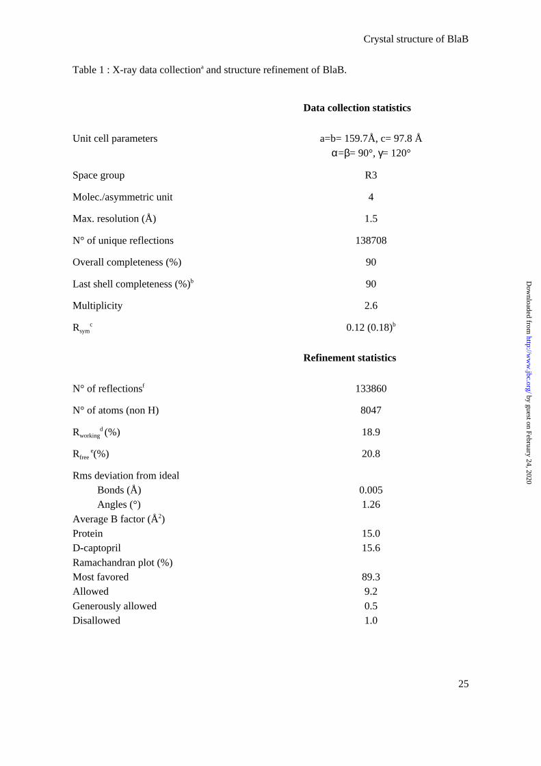
Table 1 : X-ray data collectiona and structure refinement of BlaB.
Data collection statistics
Unit cell parameters a=b= 159.7Å, c= 97.8 Å
α=β= 90°, γ= 120°
Space group R3
Molec./asymmetric unit 4
Max. resolution (Å) 1.5
N° of unique reflections 138708
Overall completeness (%) 90
Last shell completeness (%)b 90
Multiplicity 2.6
Rsymc 0.12 (0.18)b
Refinement statistics
N° of reflectionsf 133860
N° of atoms (non H) 8047
Rworkingd (%) 18.9
Rfree e(%) 20.8
Rms deviation from idealBonds (Å)Angles (°)
0.0051.26
Average B factor (Å2)ProteinD-captopril
15.015.6
Ramachandran plot (%)Most favoredAllowed
Generously allowedDisallowed
89.39.2
0.51.0
by guest on February 24, 2020http://w
ww
.jbc.org/D
ownloaded from
Crystal structure of BlaB
26
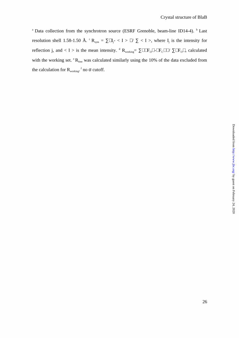
a Data collection from the synchrotron source (ESRF Grenoble, beam-line ID14-4). b Last
resolution shell 1.58-1.50 Å. c Rsym = ∑Ij- < I > / ∑ < I >, where Ij is the intensity for
reflection j, and < I > is the mean intensity. d Rworking= ∑FO-FC/ ∑FO, calculated
with the working set. e Rfree was calculated similarly using the 10% of the data excluded from
the calculation for Rworking. f no σ cutoff.
by guest on February 24, 2020http://w
ww
.jbc.org/D
ownloaded from
Crystal structure of BlaB
27
Table 2. Interactions between BlaB and D-captopril (< 3.5Å).
D-captopril Monomer: A B C D
O2
CG D119OD1 D119OD2 D119
NZ K167WaterWater
3.503.453.31
2.79(348) 2.86(671) 2.97
3.493.313.46
2.86(606) 2.88(698) 2.96
3.493.393.35
2.78(590) 2.91
-
3.393.293.29
2.85(502) 2.93
-
C9CG D119
OD1 D119 NZ K167
3.44--
3.463.39
-
3.463.463.49
3.453.47
-
O3 CA H118CB H118C H118
N D119CG D119
OD2 D119
NZ K167
3.103.193.40
2.723.423.43
-
3.133.233.40
2.713.42
-
-
3.103.203.41
2.743.47
-
3.49
3.083.153.39
2.733.49
-
-
C5 CD2 Y233CE2 Y233
3.443.31
3.473.33
3.483.33
3.463.34
O1 Water (335) 2.76 (450) 2.78 (468) 2.77 (285) 2.74
C1
CB H118
OD1 D120Zn1Zn2
-
3.283.243.37
3.50
3.243.193.41
3.49
3.253.253.42
3.49
3.213.223.35
SNE2 H196
Zn1Zn2
3.342.322.30
3.372.332.31
3.332.342.34
3.352.362.27
( ): number of water molecules in the structure.
by guest on February 24, 2020http://w
ww
.jbc.org/D
ownloaded from
Crystal structure of BlaB
28
Figure 1
by guest on February 24, 2020http://w
ww
.jbc.org/D
ownloaded from
Crystal structure of BlaB
29
Figure 2a
Figure 2b
by guest on February 24, 2020http://w
ww
.jbc.org/D
ownloaded from
Crystal structure of BlaB
30
Figure 2c
by guest on February 24, 2020http://w
ww
.jbc.org/D
ownloaded from
Crystal structure of BlaB
31
Figure 3a
Figure 3b
by guest on February 24, 2020http://w
ww
.jbc.org/D
ownloaded from
DidebergAmicosante, Gian Maria Rossolini, Moreno Galleni, Jean-Marie Frère and Otto
Isabel Garcia-Saez, Julie Hopkins, Cyril Papamicael, Nicola Franceschini, Gianfrancocomplex with the inhibitor, D-captopril
-lactamase inβThe 1.5 A structure of chryseobacterium meningosepticum Zn-
published online April 8, 2003J. Biol. Chem.
10.1074/jbc.M301062200Access the most updated version of this article at doi:
Alerts:
When a correction for this article is posted•
When this article is cited•
to choose from all of JBC's e-mail alertsClick here
by guest on February 24, 2020http://w
ww
.jbc.org/D
ownloaded from


![Captopril - media.dav-medien.demedia.dav-medien.de/sample/9783804737426_p.pdf · Pharmakokinetik: Captopril PB [%] 25–30 BV [%] 70–75 HWZ [h] 2 tmax [h] 1–1,5 WE [min] 15–30](https://img.pdfslide.us/doc/110x75/5d502b5788c993f62d8b4eff/captopril-mediadav-pharmakokinetik-captopril-pb-2530-bv-7075.jpg)