Embed Size (px)
Citation preview
The Patient witThe Patient witLocalization of NDisease and Sel
Neuropatholo
St A KSteven A. KanThe Edward S. Har
th Visual Loss:th Visual Loss: Neuropathologic p gect Diseases of ogic Interest
M D Ph De, M.D., Ph.D.rkness Eye Institute
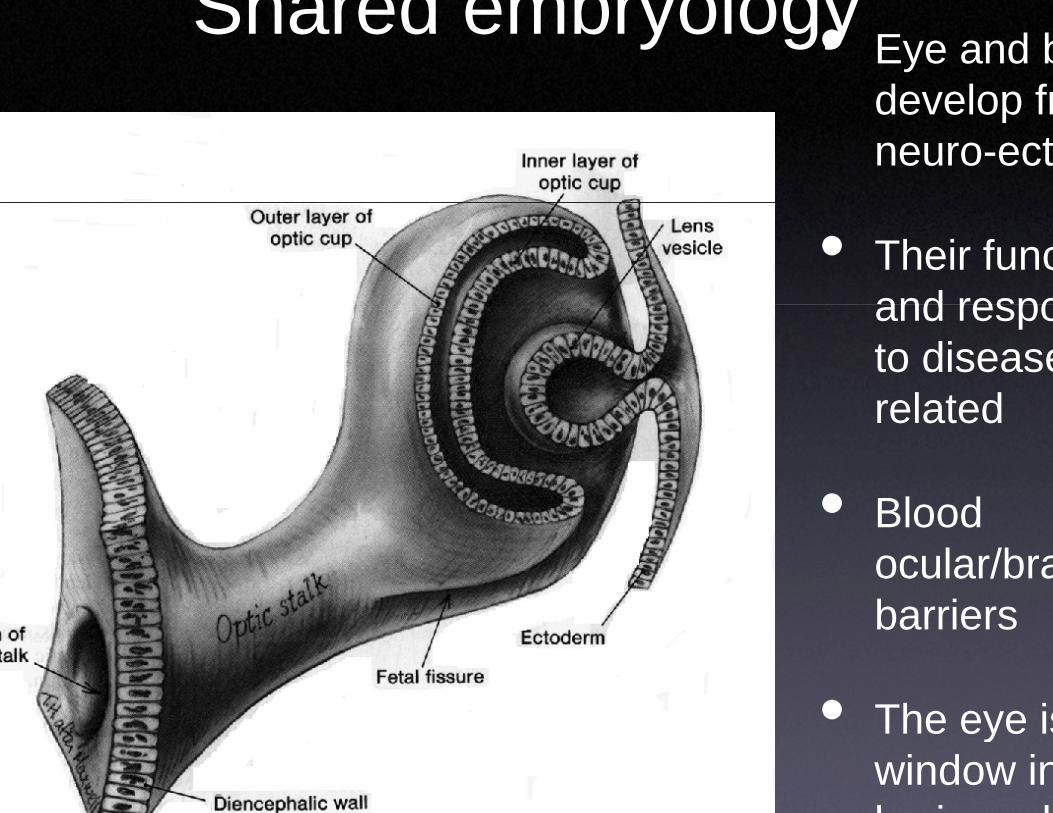
Shared em • Eye and bdevelop fr
mbryologydevelop frneuro-ect
• Their funcand respoand respoto diseaserelatedrelated
• Blood• Blood ocular/brabarriersbarriers
• The eye is• The eye iswindow inbrain and
Localizacharacterizatiocharacterizatio
visvis
• Pattern of visual loPattern of visual lolesion site
• Disease course ant lsymptoms may cla
ation and on of impairedon of impaired ionion
oss may identify theoss may identify the
nd accompanying if it tarify its nature
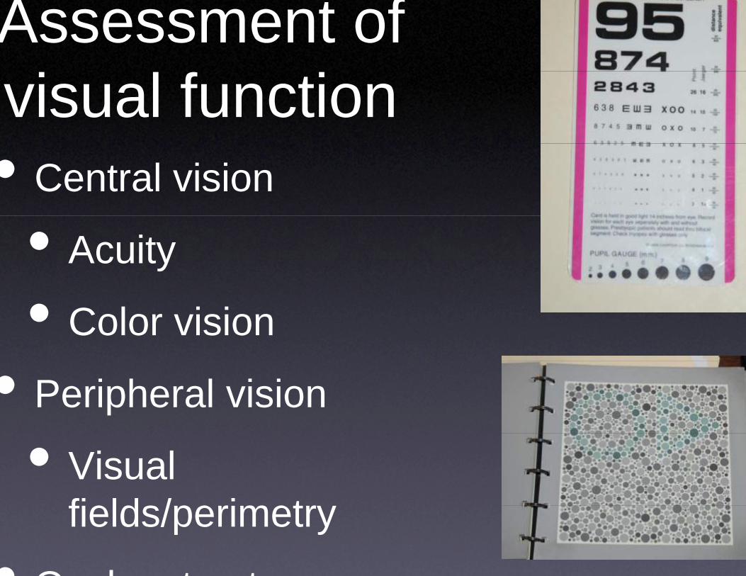
Assessment ovisual function• Central vision
• Acuity
• Color vision
• Peripheral vision
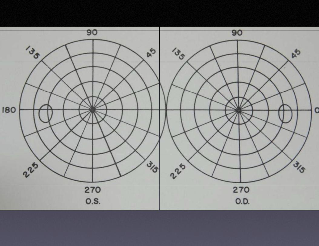
• Visual fi ld / i tfields/perimetry
• O l t t
of n
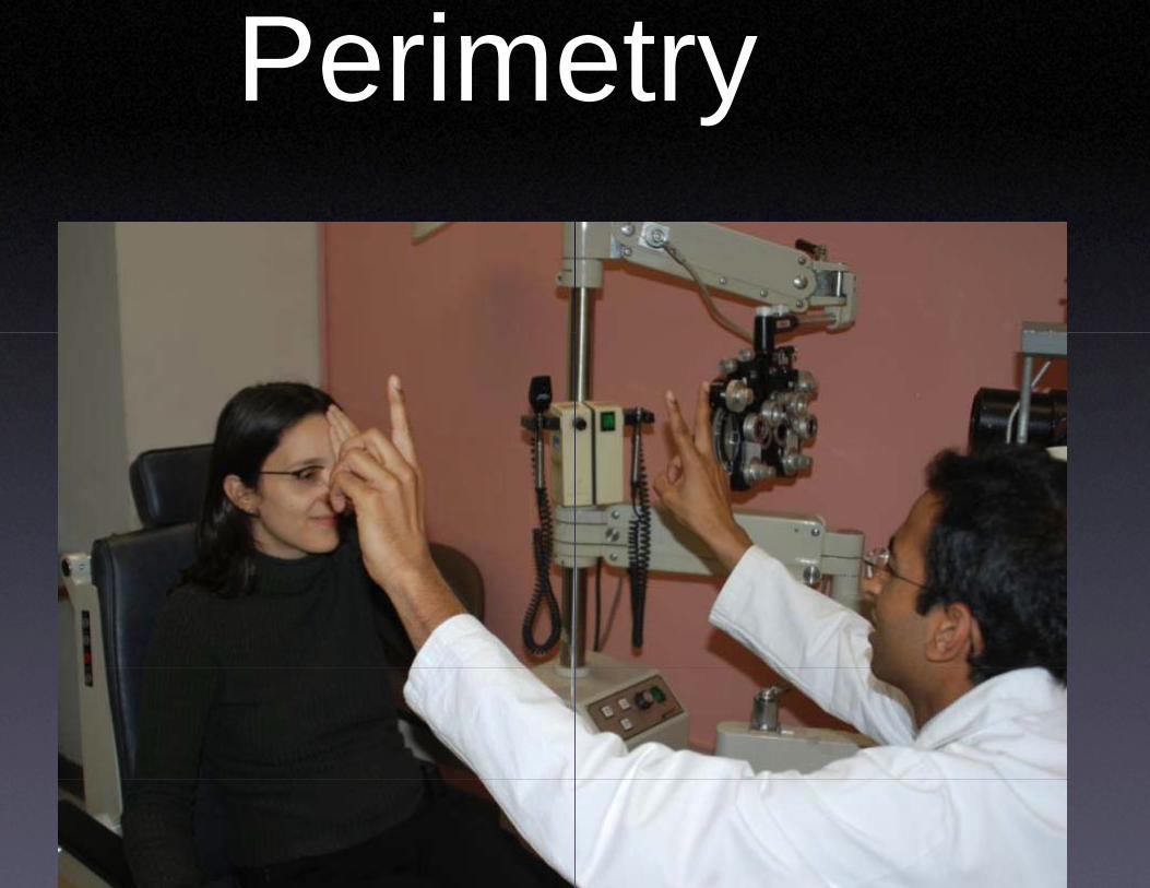
Perimeetryy
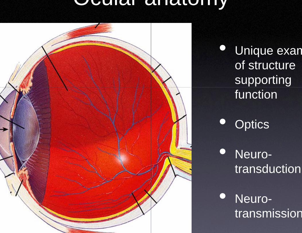
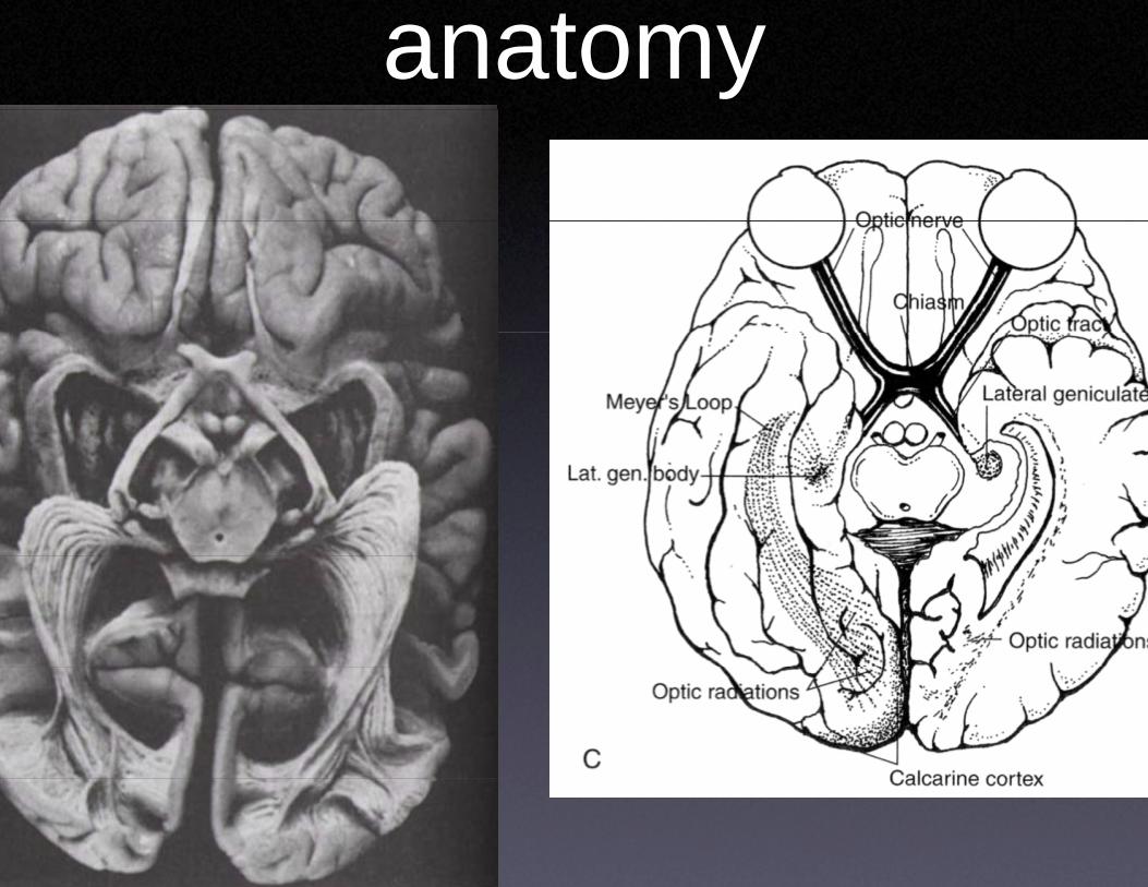
Ocular aanatomy
• Unique examof structure supporting function
• Optics
• Neuro-transduction
• Neuro-transmission
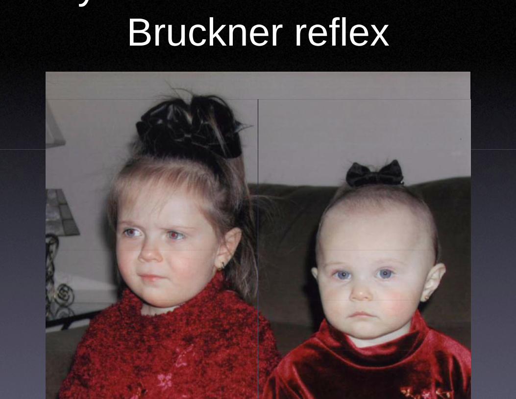
yBruckneer reflex
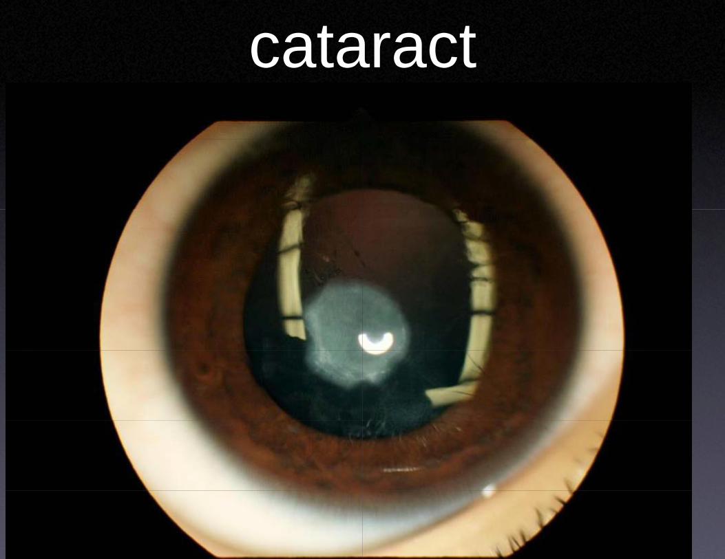
cataaract
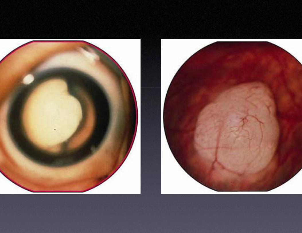
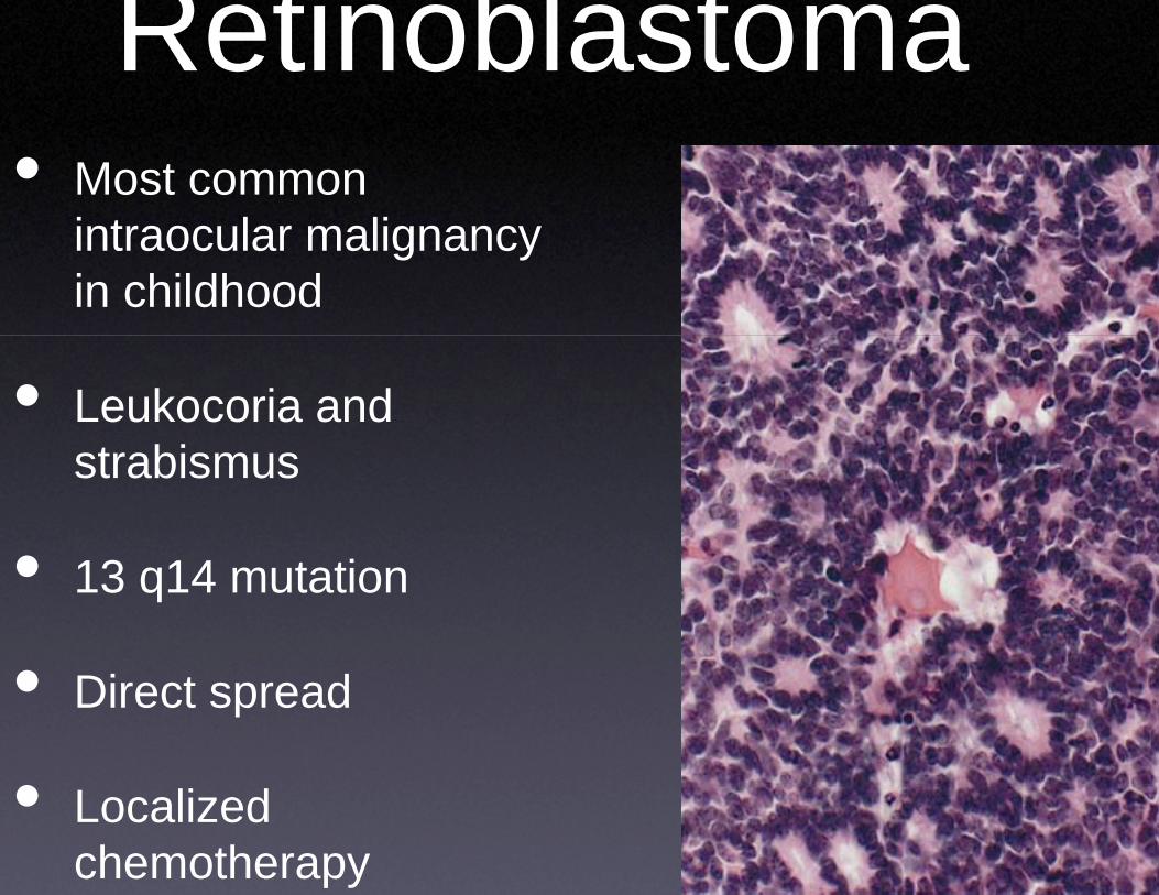
Retinobla• Most common
i t l liintraocular malignancy in childhood
• Leukocoria and t bistrabismus
• 13 14 t ti• 13 q14 mutation
•• Direct spread
•• Localized chemotherapy
astoma
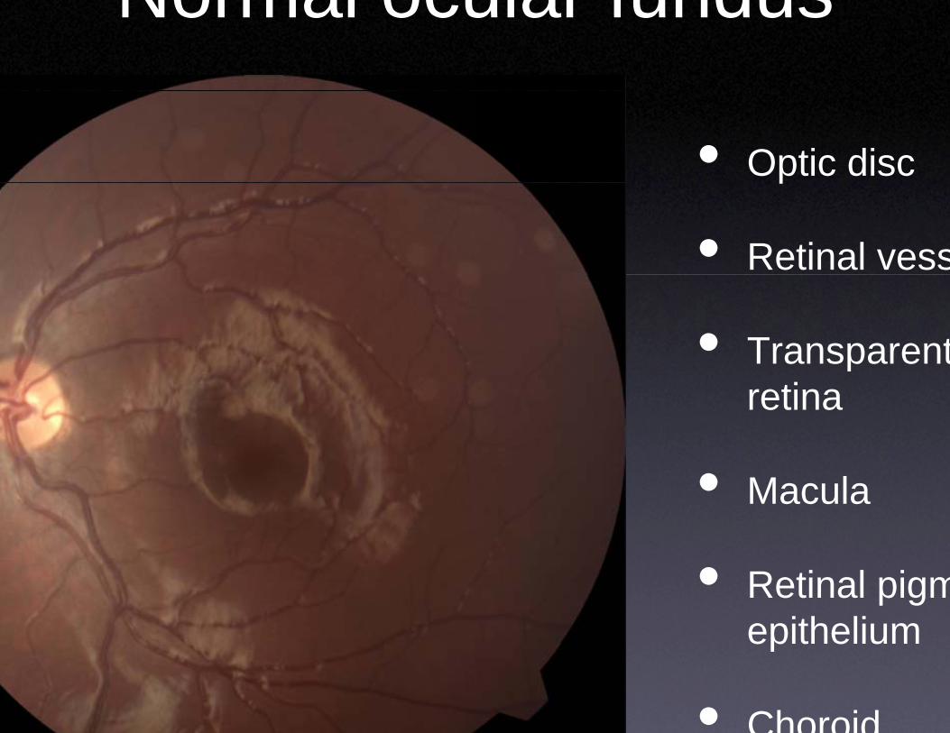
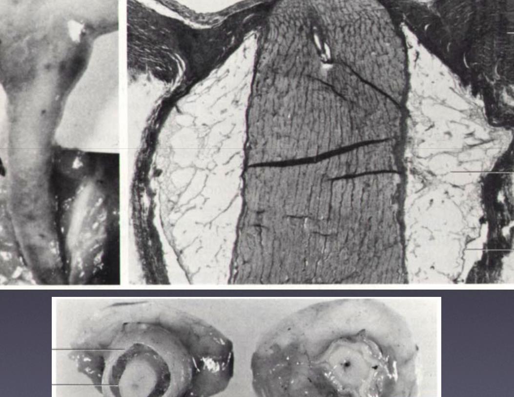
Normal ocuular fundus
• Optic discp
• Retinal vess
• Transparentpretina
• Macula
• Retinal pigmepitheliumepithelium
• Choroid
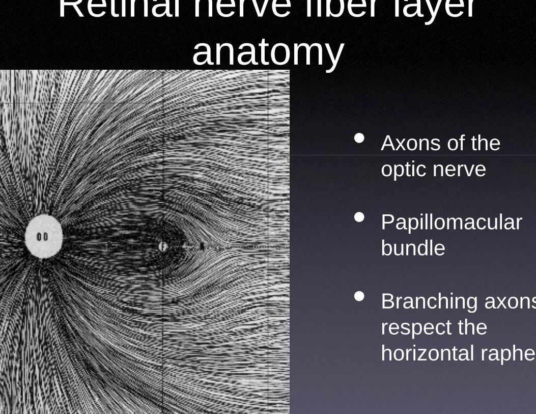
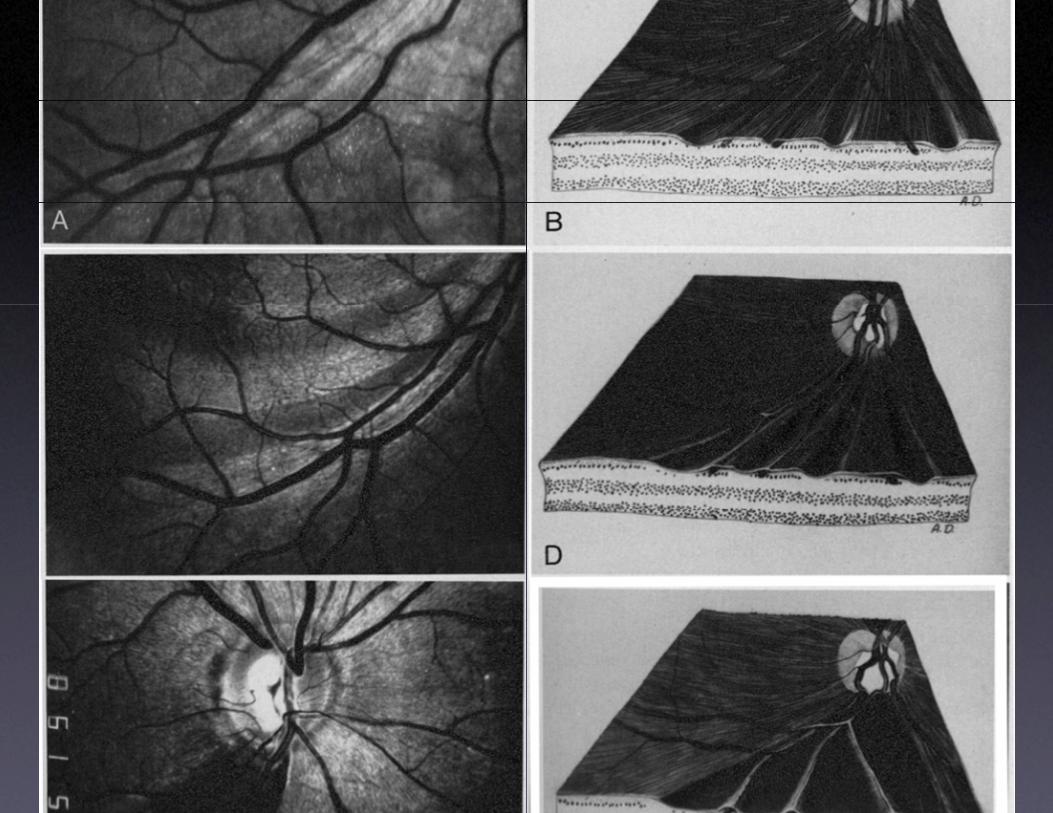
Retinal nervanatanat
ve fiber layer tomytomy
• Axons of the optic nerve
• Papillomacular bundle
• Branching axonsgrespect the horizontal raphep
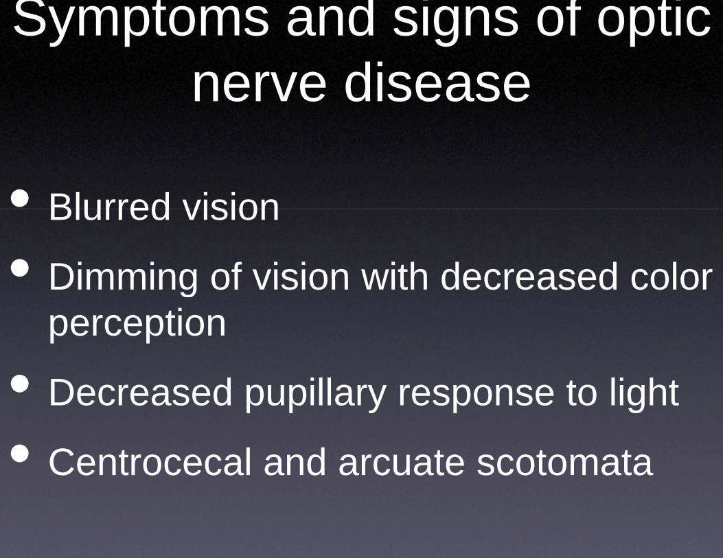
Symptoms anddnerve d
• Blurred visionBlurred vision
• Dimming of vision wDimming of vision wperception
• Decreased pupillar
• Centrocecal and a
d signs of optic didisease
with decreased colorwith decreased color
ry response to light
rcuate scotomata
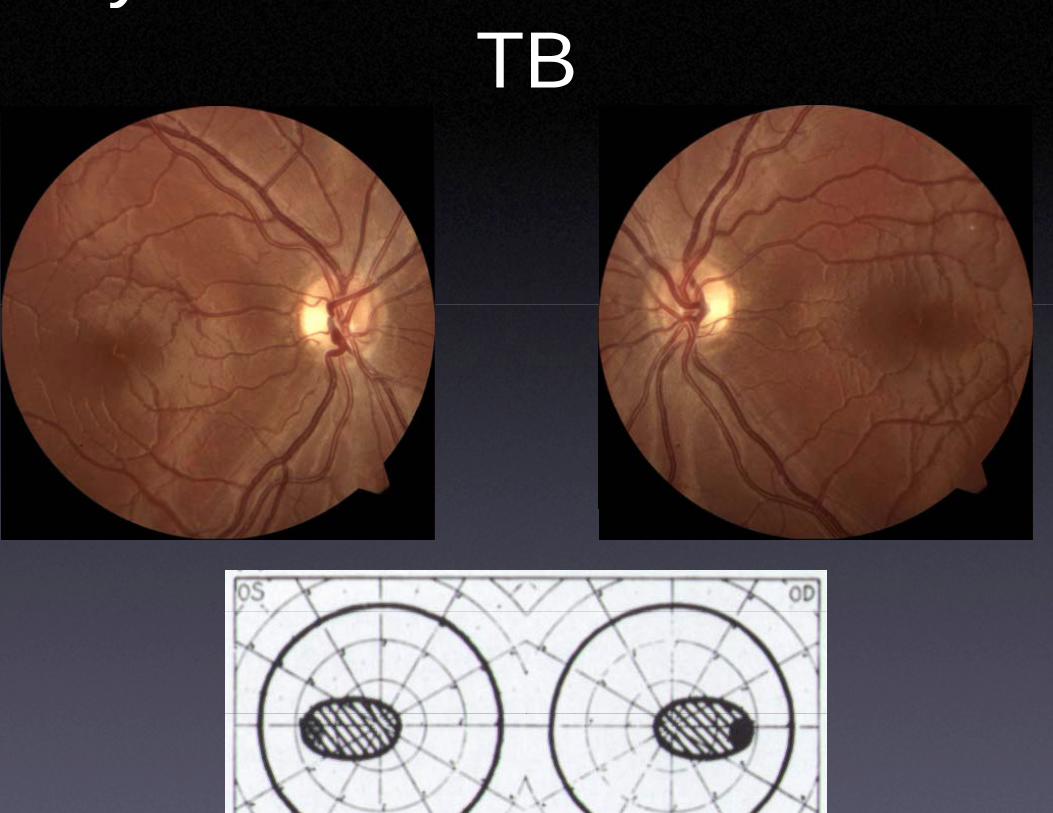
yTB
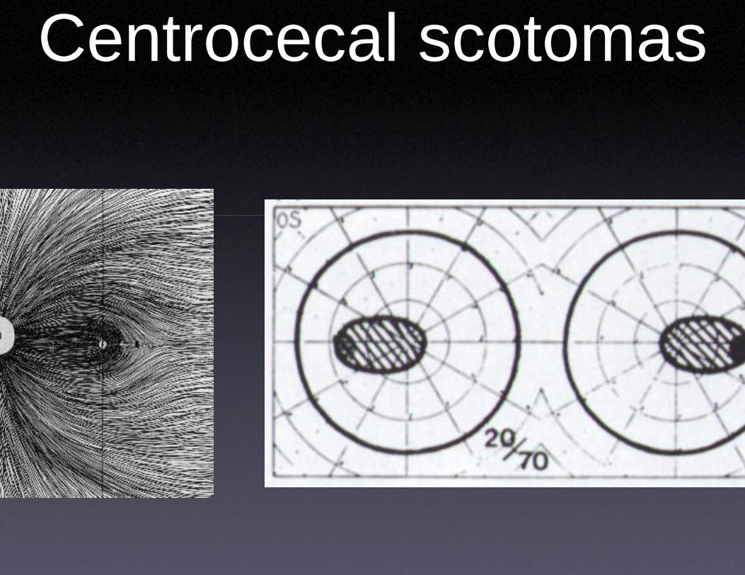
Centrocecaal scotomas
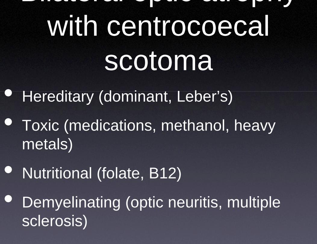
Bilateral opwith centwith cent
scotoscoto• H dit (d i• Hereditary (dominan
• T i ( di ti• Toxic (medications, metals)
• Nutritional (folate, B
• Demyelinating (opticsclerosis)
ptic atrophy trocoecaltrocoecal omaomat L b ’ )nt, Leber’s)
th l hmethanol, heavy
12)
c neuritis, multiple
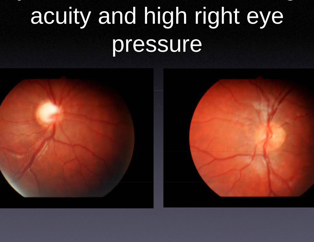
yacuity and h
pres
gigh right eye sure
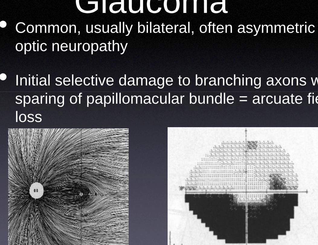
Glauc• Common usually bilaCommon, usually bila
optic neuropathy
• Initial selective damagsparing of papillomacloss
comaateral often asymmetricateral, often asymmetric
ge to branching axons wular bundle = arcuate fie
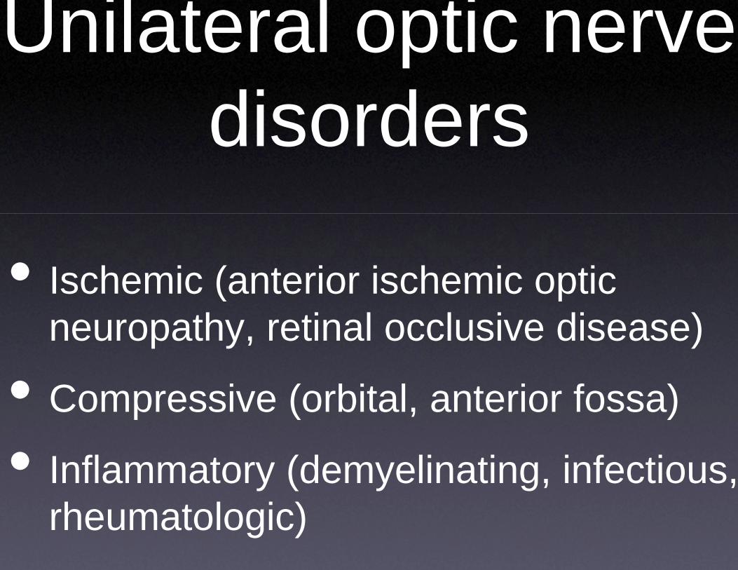
Unilateral odisordisor
• Ischemic (anteriorIschemic (anterior neuropathy, retinal
• Compressive (orbit
• Inflammatory (demrheumatologic)
optic nerve rdersrders
ischemic opticischemic optic l occlusive disease)
tal, anterior fossa)
myelinating, infectious,
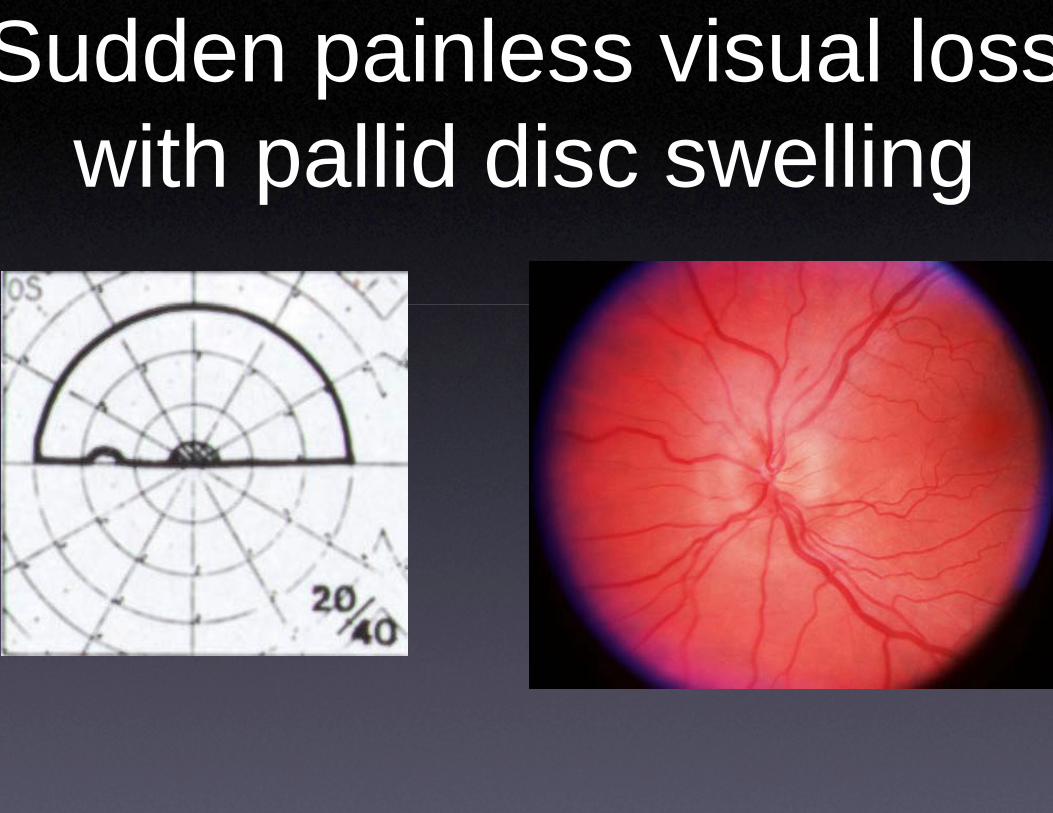
Sudden painlewith pallid dp
ess visual lossisc swellingg
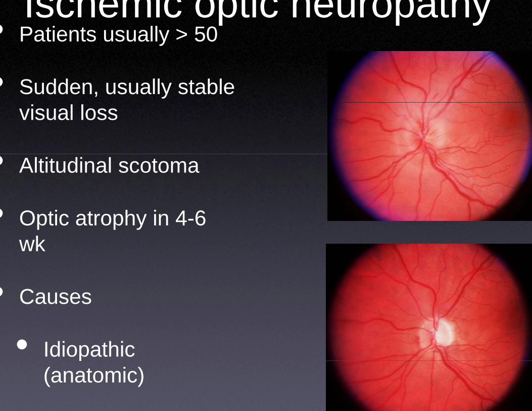
• Patients usually > 50Ischemic optic
• Sudden, usually stable visual loss
• Altitudinal scotoma
• Optic atrophy in 4-6 wk
• Causes
• Idiopathic p(anatomic)
c neuropathy
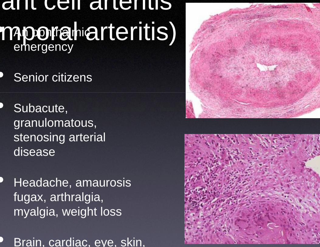
ant cell arteritimporal arteritis• An ophthalmicmporal arteritis• An ophthalmic
emergency
• Senior citizens
• Subacute, granulomatousgranulomatous, stenosing arterial diseasedisease
• Headache, amaurosisHeadache, amaurosis fugax, arthralgia, myalgia, weight lossy g , g
• Brain, cardiac, eye, skin,
ss)s)
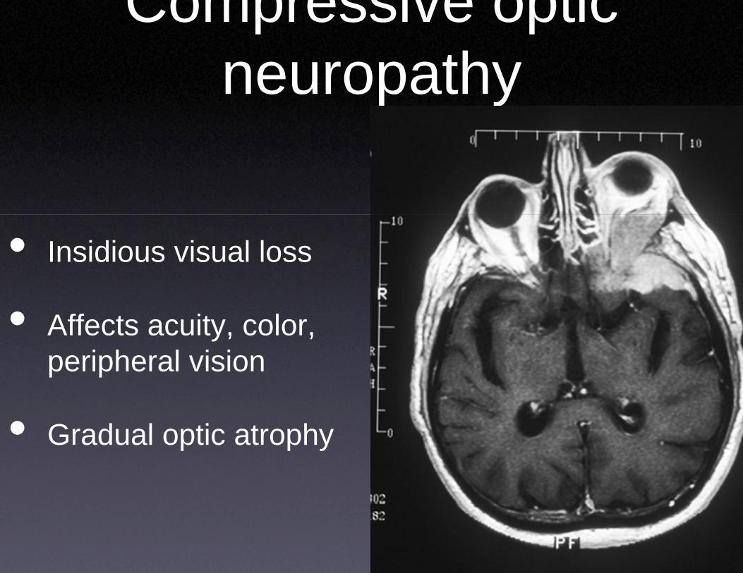
Compressneuroneuro
• Insidious visual loss
• Affects acuity, color, peripheral visionperipheral vision
• Gradual optic atrophy• Gradual optic atrophy
sive optic opathyopathy
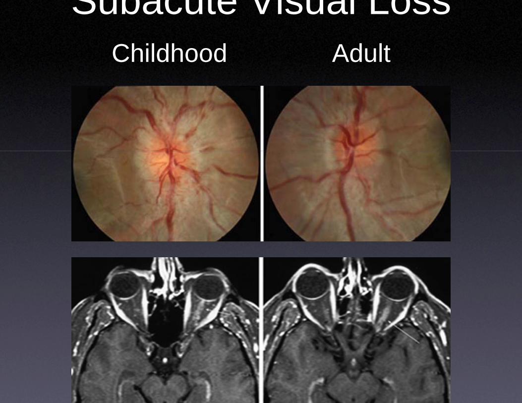
Subacute VChildhoodChildhood
Visual LossAdultAdult
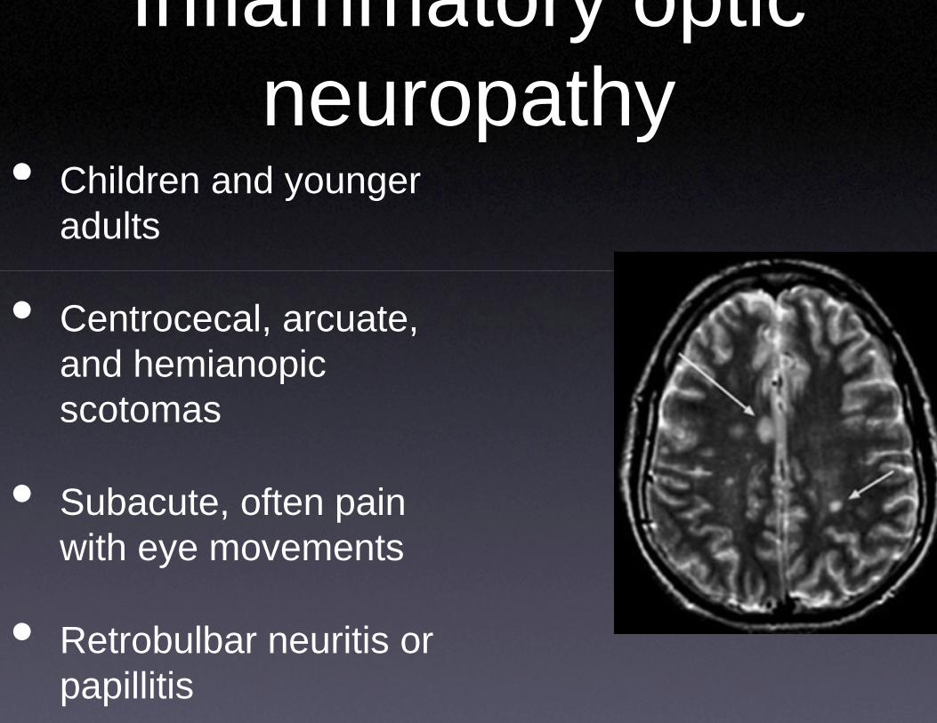
Inflammaneuro
• Children and younger
neuroChildren and younger adults
• Centrocecal, arcuate, and hemianopicand hemianopic scotomas
• Subacute, often pain with eye movementswith eye movements
• Retrobulbar neuritis or• Retrobulbar neuritis or papillitis
tory optic opathyopathy
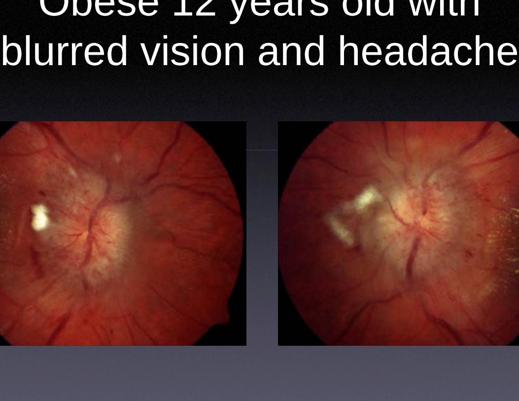
Obese 12 yeblurred vision ablurred vision a
ears old with and headacheand headache
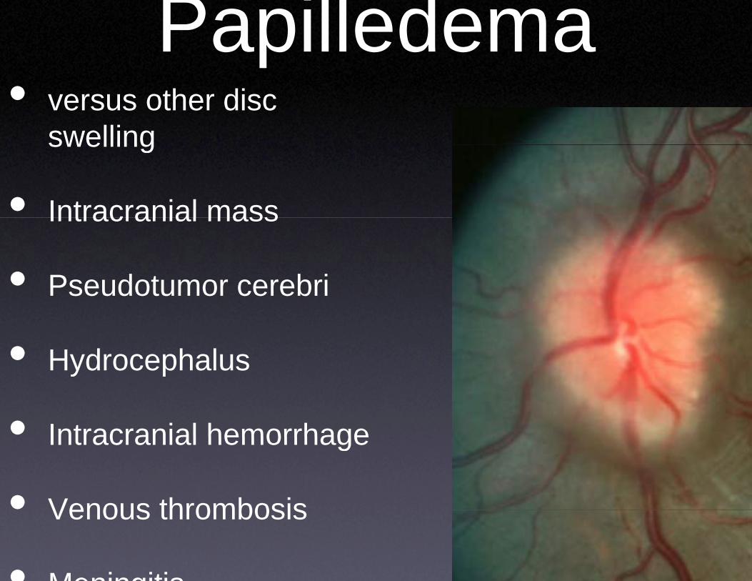
Papille• versus other disc
swellingswelling
• Intracranial massIntracranial mass
• Pseudotumor cerebriPseudotumor cerebri
• HydrocephalusHydrocephalus
• Intracranial hemorrhageIntracranial hemorrhage
• Venous thrombosisVenous thrombosis
• Meningitis
edema
anattomy
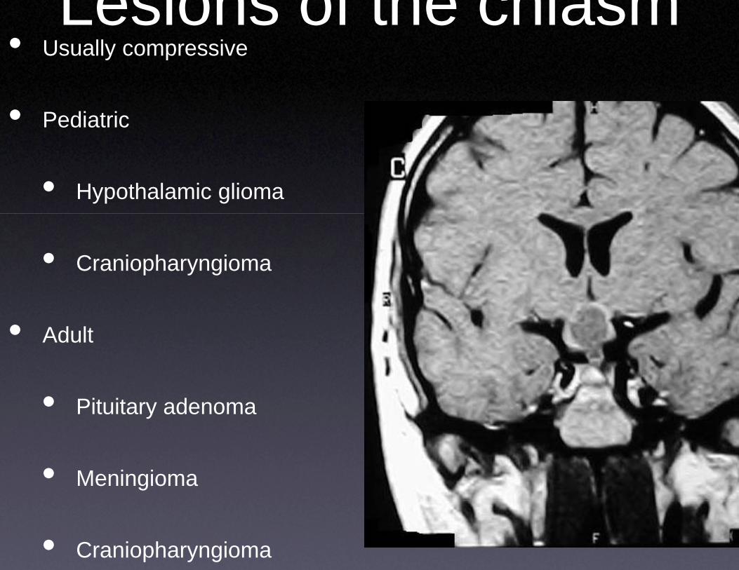
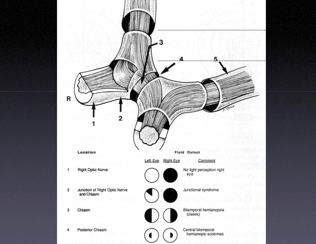
• Usually compressiveLesions of
• Pediatric
• Hypothalamic glioma
• Craniopharyngioma
• Adult
• Pituitary adenoma
• Meningioma
• Craniopharyngioma
the chiasm
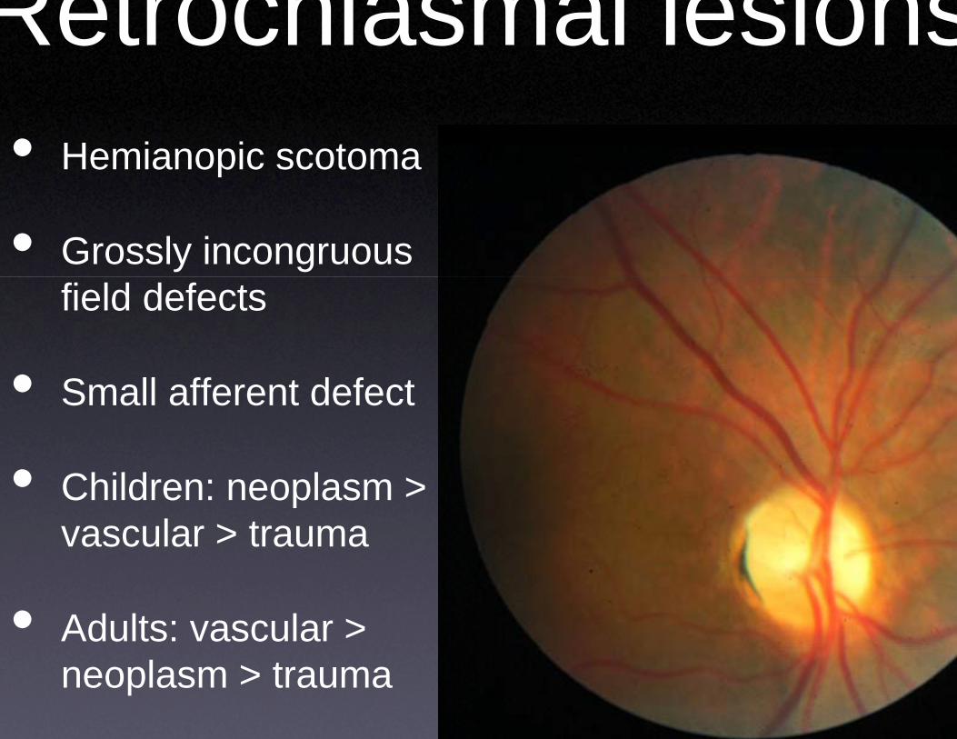
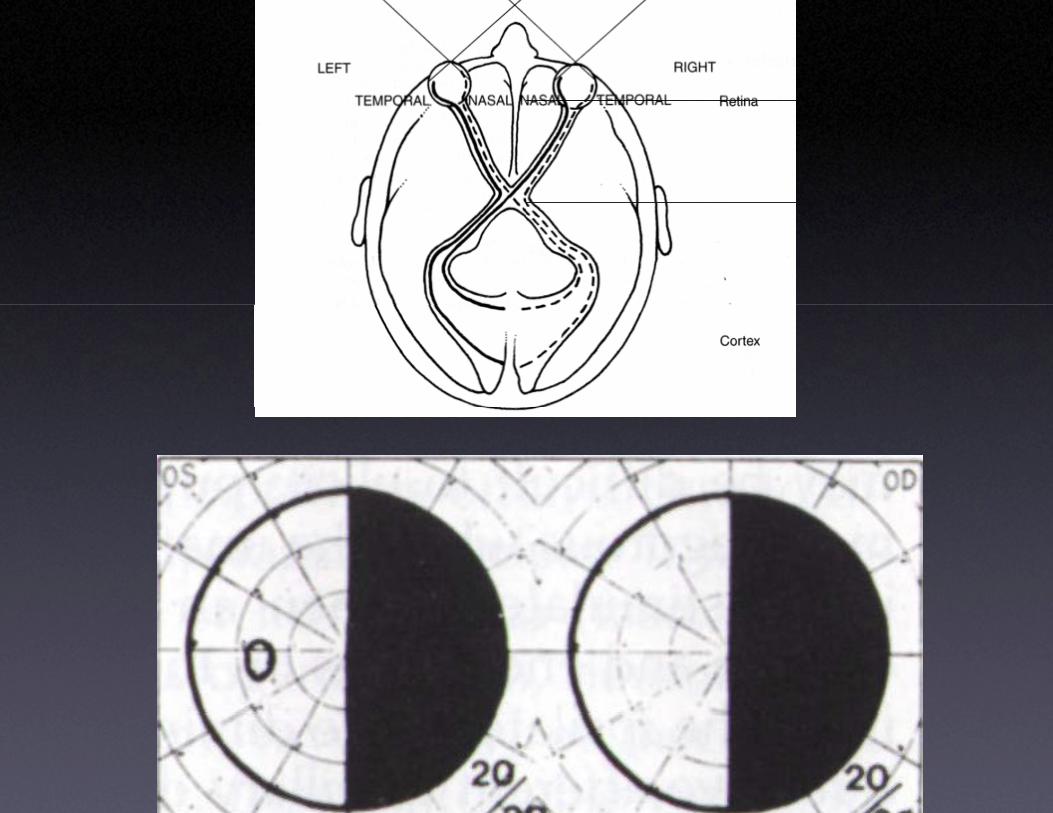
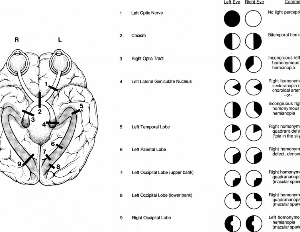
Retrochiasm• Hemianopic scotoma
• Grossly incongruous field defects
• Small afferent defect
• Children: neoplasm > vascular > trauma
• Adults: vascular > neoplasm > trauma
mal lesions
manifestationdisea• 10 year old unable10 year old unable
year of upper and lkweakness
• Slurred speech andE ti ll l bilEmotionally labile,
• Ataxia, Babinski repalpable liver slowpalpable liver, slowblepharospasm
pns of systemic asesto walk because of 1to walk because of 1
lower extremity
d drooling for 3 years, h t ihypertonic
flex, intention tremor, wed saccadeswed saccades,
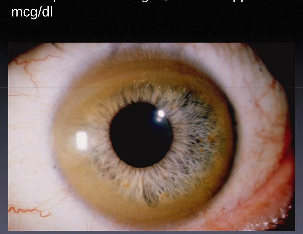
p gmcg/dl
, pp
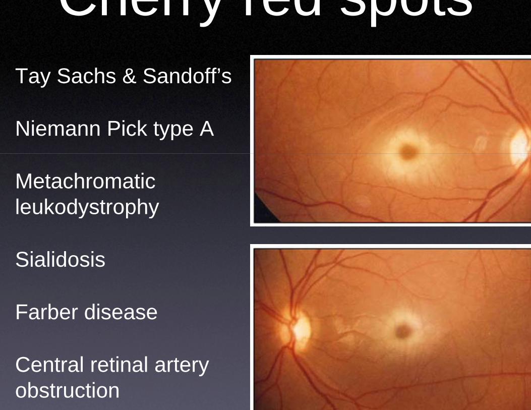
Cherry reTay Sachs & Sandoff’s
Niemann Pick type A
Metachromatic leukodystrophyleukodystrophy
SialidosisSialidosis
Farber diseaseFarber disease
C t l ti l tCentral retinal artery obstruction
ed spots
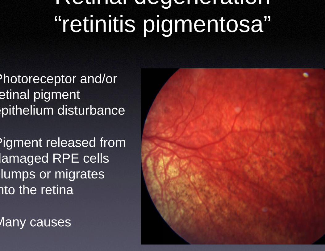
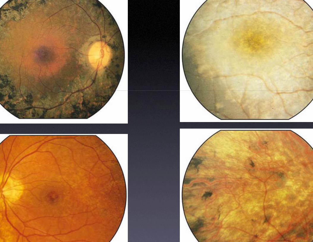
Retinal deg“retinitis pigretinitis pig
Photoreceptor and/or etinal pigmentetinal pigment
epithelium disturbance
Pigment released from damaged RPE cellsdamaged RPE cells clumps or migrates nto the retinanto the retina
Many causesMany causes
generationgmentosa”gmentosa
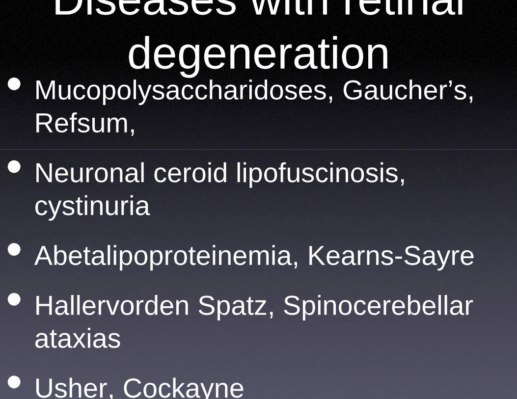
Diseases wdegene• Mucopolysacchariddegene
ucopo ysacc a dRefsum,
• Neuronal ceroid lipti icystinuria
• Ab t li t i• Abetalipoproteinem
• H ll d S t• Hallervorden Spatzataxias
• Usher, Cockayne
with retinal erationdoses, Gaucher’s, erationdoses, Gauc e s,
pofuscinosis,
i K Smia, Kearns-Sayre
S i b llz, Spinocerebellar
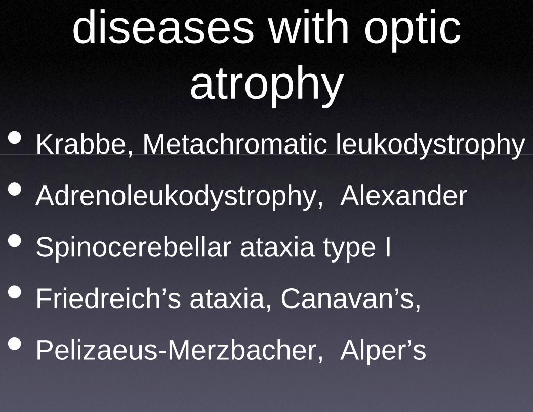
diseases atro
• Krabbe, Metachrom,
• Adrenoleukodystroy
• Spinocerebellar atap
• Friedreich’s ataxia
• Pelizaeus-Merzbac
with optic pophy
matic leukodystrophy
p yy p y
ophy, Alexanderp y,
axia type Iyp
, Canavan’s, , ,
cher, Alper’s, p


![Robust Visual Localization with Dynamic Uncertainty ...oa.upm.es/52666/1/INVE_MEM_2017_283898.pdf · Robust Visual Localization with Dynamic Uncertainty ... [1,32]. The core of the](https://img.pdfslide.us/doc/110x75/5f3ae7a8ab42636b3535ec4e/robust-visual-localization-with-dynamic-uncertainty-oaupmes526661invemem2017.jpg)