Embed Size (px)
Citation preview

Textbook of EndodontologySecond Edition
Edited by
Gunnar BergenholtzPreben Hørsted-BindslevClaes Reit


Textbook of Endodontology


Textbook of EndodontologySecond Edition
Edited by
Gunnar BergenholtzPreben Hørsted-BindslevClaes Reit
This edition first published 2010© 2003 Blackwell Munksgaard© 2010 Blackwell Publishing Ltd
Blackwell Publishing was acquired by John Wiley & Sons in February 2007. Blackwell’s publishing programme has been merged with Wiley’s global Scientific, Technical, and Medical business to form Wiley-Blackwell.
First edition published 2003Second edition 2010
Registered officeJohn Wiley & Sons Ltd, The Atrium, Southern Gate, Chichester, West Sussex, PO19 8SQ, United Kingdom
Editorial offices9600 Garsington Road, Oxford, OX4 2DQ, United Kingdom2121 State Avenue, Ames, Iowa 50014-8300, USA
For details of our global editorial offices, for customer services and for information about how to apply for permission to reuse the copyright material in this book please see our website at www.wiley.com/wiley-blackwell.
The right of the author to be identified as the author of this work has been asserted in accordance with the Copyright, Designs and Patents Act 1988.
All rights reserved. No part of this publication may be reproduced, stored in a retrieval system, or transmitted, in any form or by any means, electronic, mechanical, photocopying, recording or otherwise, except as permitted by the UK Copyright, Designs and Patents Act 1988, without the prior permission of the publisher.
Wiley also publishes its books in a variety of electronic formats. Some content that appears in print may not be available in electronic books.
Designations used by companies to distinguish their products are often claimed as trademarks. All brand names and product names used in this book are trade names, service marks, trademarks or registered trademarks of their respective owners. The publisher is not associated with any product or vendor mentioned in this book. This publication is designed to provide accurate and authoritative information in regard to the subject matter covered. It is sold on the understanding that the publisher is not engaged in rendering professional services. If professional advice or other expert assistance is required, the services of a competent professional should be sought.
Library of Congress Cataloging-in-Publication DataTextbook of endodontology/edited by Gunnar Bergenholtz, Preben Hørsted-Bindslev, Claes Reit. — 2nd ed. p. ; cm. Includes bibliographical references and index. ISBN 978-1-4051-7095-6 (hardback: alk. paper) 1. Endodontics. I. Bergenholtz, Gunnar.
II. Hørsted-Bindslev, Preben. III. Reit, Claes. [DNLM: 1. Dental Pulp Diseases—therapy. 2. Periapical Diseases—therapy. WU 230 T355 2010] RK351.T49 2003 617.6�342—dc22
2009024733
A catalogue record for this book is available from the British Library.
Set in 9.5/12.5pt Palatino by Gray Publishing, Tunbridge Wells, KentIllustrations by Jens Lund KirkegaardPrinted in Singapore
1—2010
v
Contents
List of Contributors xi Preface xiii
1 Introduction to endodontology 1 Claes Reit, Gunnar Bergenholtz and Preben Hørsted-Bindslev
Endodontology 1 The dawn of modern endodontology 2 The objective of endodontic treatment 3 Clinical problems and solutions 3 The diagnostic dilemma 5 The tools of treatment 6 Extraction and dental implant? 6 References 6
Part 1 The Vital Pulp
2 The dentin–pulp complex: structures, functions and responses to adverse influences 11 Leif Olgart and Gunnar Bergenholtz
Introduction 11 Constituents and normal functions of the dentin–pulp complex 11 Basal maintenance 18 Appropriate responses of the healthy pulp to non-destructive stimuli 19 Responses to external threats 19 Effects of potentially destructive stimuli 23 References 30
3 Dentinal and pulpal pain 33 Matti Närhi
Introduction 33 Classification of nerve fibers 33 Morphology of intradental sensory innervation 33 Function of intradental sensory nerves under normal conditions 36 Sensitivity of dentin: hydrodynamic mechanism in pulpal A-fiber activation 37 Responses of intradental nerves to tissue injury and inflammation 39 Local control of pulpal nociceptor activation 42 Dentin hypersensitivity 42 Pain symptoms and pulpal diagnosis 43 References 44
4 Treatment of vital pulp conditions 47 Preben Hørsted-Bindslev and Gunnar Bergenholtz
Introduction 47 Clinical scenarios 47 Treatment options 48 Factors influencing choice of treatment 50 Management of exposed pulps by direct pulp capping/partial pulpotomy 52 Pulpectomy 59
vi Contents
Emergency treatment 65 References 69
5 Endodontics in primary teeth 73 Ingegerd Mejàre
Introduction 73 The normal pulp 73 Pulpal inflammation in the primary tooth 73 Wound dressings – characteristics, modes of action and reported clinical success rates 75 Objectives of pulp treatment 79 Operative treatment procedures 79 Indications and contraindications for pulp treatment in primary teeth 85 Future directions 85 References 88
Part 2 The Necrotic Pulp
6 The microbiology of the necrotic pulp 95 Gunnel Svensäter, Luis Chávez de Paz and Else Theilade
Introduction 95 Evidence for the essential role of microorganisms in apical periodontitis 95 Routes of microbial entry to the pulpal space 96 Modes of colonization 97 Ecological determinants for microbial growth in root canals 98 Methods for studying the root canal microflora 103 Composition of the endodontic microflora 106 Association of signs and symptoms with specific bacteria 109 Concluding remarks 110 References 110
7 Apical periodontitis 113 Zvi Metzger, Itzhak Abramovitz and Gunnar Bergenholtz
Introduction 113 The nature of apical periodontitis 113 Interactions with the infecting microbiota 118 Clinical manifestations and diagnostic terminology 123 References 126
8 Systemic complications of endodontic infections 128 Nils Skaug and Vidar Bakken
Introduction 128 Acute periapical infections as the origin of metastatic infections 128 Chronic periapical infections as the origin of metastatic infections 135 References 138
9 Treatment of the necrotic pulp 140 Paul Wesselink and Gunnar Bergenholtz
Introduction 140 Objectives and general treatment strategies 140 Scheme for a routine procedure in root canal therapy 143 Considerations in complex cases 152
Effects of root canal therapy on the intracanal microbiota 153 Management of symptomatic lesions 153 References 156
Part 3 Endodontic Treatment Procedures
10 The surgical microcope 163 Pierre Machtou
Introduction 163 Components 163 Ergonomics and working techniques 164 Microinstrumentation 167 Critical steps 167 Concluding remarks 168 References 168
11 Root canal instrumentation 169 Lars Bergmans and Paul Lambrechts
Introduction 169 Principles of root canal instrumentation 169 Root canal system anatomy 170 Procedural steps 174 Endodontic instruments 180 Instrumentation techniques 183 Limitations of root canal instrumentation 186 Preventing procedural mishaps 188 References 190
12 Root canal filling materials 193 Gottfried Schmalz and Preben Hørsted-Bindslev
Introduction 193 Requirements 194 Gutta-percha cones 198 Sealers 202 Materials for retrograde fillings (root-end fillings) and replantation 214 Mandibular nerve injuries 215 References 216
13 Root filling techniques 219 Paul Wesselink
Introduction 219 Specific objectives 219 Selecting a root canal filling material 219 Root filling techniques for gutta-percha 221 Root filling techniques employing gutta-percha and sealer 224 Procedures prior to root canal filling 229 Assessing root filling quality 229 Filling of the pulp chamber and coronal restoration 230 Conclusions and recommendations 231 References 231
Contents vii
Part 4 Diagnostic Considerations and Clinical Decision Making
14 Diagnosis of pulpal and periapical disease 235 Claes Reit and Kerstin Petersson
Introduction 235 Evaluation of diagnostic information 235 Diagnostic strategy 237 Clinical manifestations of pulpal and periapical inflammation 238 Collecting diagnostic information 238 Diagnostic classification 247 References 253
15 Diagnosis and management of endodontic complications after trauma 255 John Whitworth
Introduction 255 Common dental injuries 255 Dental trauma and its consequences 258 General considerations in the management of dental trauma 267 Diagnostic quandaries – to remove or review the pulp after trauma? 273 Pulp regeneration – the dawn of a new era? 274 References 274
16 The multidimensional nature of pain 277 Ilana Eli and Peter Svensson
Introduction 277 Neurobiological factors affecting the pain experience 278 Psychological factors affecting the pain experience 280 Gender and pain 282 Special populations 284 Management and treatment of pain 285 Concluding remarks 287 References 287
17 Clinical epidemiology 290 Claes Reit and Lise-Lotte Kirkevang
Introduction 290 Clinical epidemiology 290 Diagnosis 292 Cause 292 Prevalence, frequency and incidence 293 Risk for apical periodontitis 295 Treatment 296 Prognosis 296 Longevity of root filled teeth 297 Back to the case 298 References 298
18 Endodontic decision making 301 Claes Reit
The outcome of endodontic treatment 301 Factors influencing treatment outcome 302 Prevalence of endodontic “failures” 304
viii Contents
Variation in the management of periapical lesions in endodontically treated teeth 304 Clinical decision making: descriptive projects 305 Endodontic retreatment decision making: a normative approach 306 Concluding remarks 311 References 311
Part 5 The Root Filled Tooth
19 The root filled tooth in prosthodontic reconstruction 317 Eckehard Kostka
Introduction 317 Problems associated with root filled teeth as abutments 317 Core build-ups 322 Clinical techniques 325 Prosthodontic reconstruction 327 References 332
20 Non-surgical retreatment 335 Pierre Machtou and Claes Reit
Introduction 335 Indications 335 Access to the root canal 335 Access to the apical area 339 Instrumentation of the root canal 342 Antimicrobial treatment 344 Preventive retreatment 346 Prognosis 346 References 346
21 Surgical endodontics 348 Peter Velvart
Introduction 348 General outline of the procedure 349 Pain control after surgery 361 Bone healing 362 Prognosis 362 References 364
Failures after surgical endodontics 366 Thomas von Arx
Index 371
Contents ix

xi
List of Contributors
Editors
Gunnar Bergenholtz Institute of Odontology, The Sahlgrenska Academy at University of Gothenburg, Sweden
Preben Hørsted-Bindslev School of Dentistry, Faculty of Health Sciences, Aarhus University, Denmark
Claes Reit Institute of Odontology, The Sahlgrenska Academy at University of Gothenburg, Sweden
Contributors
Itzhak Abramovitz Hebrew University and Hadassa Faculty of Dental Medicine, Hebrew University, Jerusalem, Israel
Thomas von Arx School of Dental Medicine, University of Berne, Switzerland
Vidar Bakken Faculty of Medicine and Dentistry, University of Bergen, Norway
Lars Bergmans School of Dentistry, University of Leuven, Belgium
Luis Chávez de Paz Faculty of Odontology, Malmö University, Sweden
Ilana Eli The Maurice and Gabriela Goldschleger School of Dental Medicine, Tel Aviv University, Israel
Lise-Lotte Kirkevang School of Dentistry, Faculty of Health Sciences, Aarhus University, Denmark
Eckehard Kostka School of Dental Medicine, Charité, Medical Faculty of the Berlin Humboldt University, Germany
Paul Lambrechts School of Dentistry, University of Leuven, Belgium
Pierre Machtou Denis Diderot School of Dentistry, Paris 7 University, France
Ingegerd Mejàre Faculty of Odontology, Malmö University, Sweden
Zvi Metzger The Maurice and Gabriela Goldschleger School of Dental Medicine, Tel Aviv University, Israel
Matti Närhi Faculty of Medicine, University of Kuopio, Finland
Leif Olgart Karolinska Institute, Stockholm, Sweden
Kerstin Petersson Faculty of Odontology, Malmö University, Sweden
Gottfried Schmalz School of Dentistry, University of Regensburg, Germany
Nils Skaug deceased
Gunnel Svensäter Faculty of Odontology, Malmö University, Sweden
Peter Svensson School of Dentistry, Faculty of Health Sciences, Aarhus University, Denmark
Else Theilade School of Dentistry, Faculty of Health Sciences, Aarhus University, Denmark
Peter Velvart Private practice, Zürich, Switzerland
Paul Wesselink Academic Center for Dentistry Amsterdam (ACTA), The Netherlands
John Whitworth School of Dental Science, Newcastle University, UK
xii List of Contributors
xiii
Preface
The Textbook of Endodontology is intended to serve the educational needs of dental students, as well as of dental practitioners seeking updates on endodontic theories and techniques. The primary aim has been to provide an understanding of the biological processes involved in pulpal and periapical pathologies and how that knowl-edge impinges on clinical management, and to present that information in an easily accessible form. Therefore, we have supplemented the core text with numerous fig-ures and photographs, as well as with boxes highlighting key facts, important clinical procedures and key research. Case studies are given at the end of some chapters in order to further illustrate topics described in the text. In these various ways, the book provides information both at a foundation level, and at a more detailed level for the graduating student and practitioner. The key information boxes are color coded as an easy-to-use navigational aid for readers. Core concepts are colored pink, while advanced concepts are purple. Clinical procedures are coded green and key literature boxes are blue. Although not designed to provide a comprehensive review of the literature, this book is also intended to
stimulate the reader to delve into the research that forms our current knowledge base in endo dontology. To aid the reader, a selective reference list is provided and comments have been added to especially weighty or use-ful references. Important and interesting investigations are presented in the core and advanced concept boxes, and we hope that these features will encourage the student to carry on with his or her own exploration of the subject area. This is the second edition of the book, which features three new chapters reflecting the use of the surgical microscope, diagnosis and management of endodontic complications subsequent to dental trauma, and endo-dontic epidemiology. The dedicated support of our co-authors – 23 highly respected clinicians and scientists – who, in addition to the editors, have contributed to this book, is greatly appreciated. We thank them all sincerely for their time, effort and endurance during the editing process.
Gunnar BergenholtzPreben Hørsted-Bindslev
Claes Reit

1
Chapter 1
Introduction to endodontologyClaes Reit, Gunnar Bergenholtz and Preben Hørsted-Bindslev
Endodontology
The word ”endodontology” is derived from the Greek language and can be translated as ”the knowledge of what is inside the tooth”. Thus, endodontology concerns structures and processes within the pulp chamber. But what about ”knowledge”? What does it actually mean to ”know” things? Most people would probably say that knowledge has something to do with truth and provid-ing reasons for things. It is often believed that dental and medical knowledge is simply scientific knowledge – science is based on research and deals with how things are constructed and work. But as practicing dentists we also need other types of knowledge. Although it is important to know about tooth anatomy and how to produce good root canal preparations for example, we must also develop good judgment and ability to make the ”right” clinical decisions. There are at least three dif-ferent forms of knowledge that the dental practitioner requires and, in a tradition that goes all the way back to Aristotle, we will refer to the Greek terms for these forms: episteme, techne and phronesis (1).
Episteme
Episteme is the word for theoretical–scientific knowledge. The opposite is doxa, which refers to “belief” or “opin-ion”. There is a massive body of epistemic knowledge within endodontology, for example on the biology of the pulp, the microorganisms that inhabit root canals, the procedures and materials used in the clinical practice of endodontology (endodontics) and the outcome of endodontic therapies. Science produces “facts”. It must be understood that modern science is an industry and is affected by many factors, both internal and external. Although this is not the place to discuss the philosophy of science, the concept of “truth” and the growth of sci-entific knowledge is not unproblematic. There has been substantial contemporary philosophical discussion reflecting on epistemic knowledge, and the interested reader is referred to one of the many good introductory texts that are available (3).
The results of science are presented in lectures, articles and textbooks. So from a student’s point of view the learning situation is rather straightforward, provided that the subject is structured well and ample time given for reading and reflection. This book, in large part, is composed of epistemic knowledge.
Techne
The first person to challenge the deeply intrenched theo-retical concept of knowledge was the British philosopher Gilbert Ryle. In his book The Concept of Mind (10) he introduces “knowing-how” and distinguishes it from “knowing-that”. “Knowing-how” is practical in nature and concerns skills and the performance of certain actions. This concept of knowledge implies the ability not only to do things, but also to understand what you are doing. To say that you have practical knowledge, it is not enough to produce things out of mere routine or habit. You have to “know” what you are doing and be able to argue about it. Practice must be combined with reflection. The idea that there is a tacit or silent dimen-sion of knowledge has had a great impact on the contem-porary discussion. Michael Polanyi, for example, said that “We know more than we can tell” (9). When trying to explain how we master practical things such as riding a bicycle or recognizing a face, it is not possible to articu-late verbally all the knowledge that we have. Certain important aspects are “tacit”. Likewise, it is not sufficient to teach students about root canal preparation simply by asking them to read a book or presenting the subject matter in a lecture. It has to be demonstrated. Knowledge is very often transmitted by the act of doing. A substantial body of endodontic knowledge must be characterized as techne. It is not possible to learn all about the procedures in endodontology by studying a text-book. Observing a good clinical instructor, watching other dentists at work, performing the procedures one-self and reflecting on what has been learned are all important.
2 Introduction to endodontology
Phronesis
According to Aristotle, phronesis is the ability to think about practical matters. This can be translated as “practi-cal wisdom” (5) and is concerned with why we might decide to act in one way rather than in another. When thinking about the “right” action or making the “right” decision we enter the territory of moral philosophy. The person who has practical wisdom has good moral judg-ment. Modern ethical thinking has been influenced significantly by ideas that originated during the enlight-enment. Morality is concerned with human actions and there are certain principles that can separate “right” from “wrong” decisions. Jeremy Bentham (2) and the utilitari-ans launched the utility principle and Immanuel Kant (6) invented the categorical imperative, each creating a tra-dition with great impact on today’s medical ethics and decision making. Aristotle, on the other hand, believed that there are no explicit principles to guide us. He understood practical wisdom as a combination of understanding and experi-ence and the ability to read individual situations correct-ly. He thought that phronesis could be learnt from one’s own experience and by imitating others who had already mastered the task. He stressed the cultivation of certain character traits and the habit of acting wisely. The clinical situation demands that the dentist exercises practical wisdom, “to do the right thing at the right moment”. In order to develop phronesis, theoretical studies of moral theory and decision-making principles might be helpful. Neoaristotelians such as Martha Nussbaum (8) have sug-gested that reading literature should be part of any aca-demic curriculum, the idea being that it increases our knowledge and understanding of other people. However, the essence of phronesis has to be learnt from practice.
Concepts of endodontology
From the above it can be concluded that endodontology encompasses not only theoretical thinking but also the practical skills of a craftsperson and the practical thinking needed for clinical and moral judgment. Unfortunately, through the years, undue prestige has been given to theoretical–scientific thinking and this has hindered the development of a rational discussion of the other types of knowledge. The serious student of endodontology has to investigate all three aspects, but, as argued above, there are limits to what can be communicated within the covers of a textbook.
The dawn of modern endodontology
It all started with a speech at the McGill University in Montreal. In the morning of October 3, 1910, Dr William
Hunter gave a talk entitled “The role of sepsis and anti-sepsis in medicine”. Hunter said that:
“In my clinical experience septic infection is without exception the most prevalent infection operating in medicine, and a most important and prevalent cause and complication of many medical diseases. Its ill-effects are widespread and extend to all systems of the body. The relation between these effects and the sepsis that causes them is constantly overlooked, because the existence of the sepsis is itself overlooked. For the chief seat of that sepsis is the mouth; and the sepsis itself, when noted, is erroneously regarded as the result of various conditions of ill-health with which it is associated – not, as it really is, an important cause or complication. “Gold fillings, gold caps, gold bridges, gold crowns, fixed dentures, built in, on, and around diseased teeth, form a veritable mausoleum of gold over a mass of sepsis to which there is no parallel in the whole realm of medicine or surgery. The whole con-stitutes a perfect gold trap of sepsis.”
The cited text was published in the Lancet in 1911. But Hunter’s words rapidly spread and were intensively dis-cussed among laymen and given banner headlines in the newspapers. Essentially, Hunter proposed that micro-organisms from a dental focus of infection can spread to other body compartments and cause serious systemic disease. The fear that illnesses and even those of chronic or of unknown origin were caused by oral infections, brought thousands of people to the waiting rooms of dentists with demands to have their teeth removed. As a result of the focal infection theory teeth were extracted in enormous numbers. Although not directly stated by Hunter, teeth with necrotic pulps were seen as one of the main causes of “focal infection”. Laboratory studies had disclosed the presence of bacteria in the dead pulp tissue. In the 1920s, dental radiography came into general use and radio-lucent patches around the apices of teeth with necrotic pulps indicating an inflammatory bone lesion were pos-sible to detect. If such teeth were extracted and cultured, microorganisms were often recovered from the attached soft tissue. It became virtually incontestable that pulpally diseased teeth should be removed. Reflecting on this period in the history of dentistry, Grossman (4) wrote: “The focal infection theory promul-gated by William Hunter in 1910 gave dentistry in gener-al, and root canal treatment in particular, a black eye from which it didn’t recover for about 30 years.” However, in hindsight, this period can also be regarded as the dawn of modern endodontology. Researchers started to question and oppose the clinical consequences of the focal infection theory. Microbiologists began mapping out the microflora of infected root canals. Pathologists
Introduction to endodontology 3
investigated the reaction patterns of the pulp and peri-apical tissues and came to understand the protective power of the host defense mechanisms. Clinicians invented aseptic methods to treat the root canal, and radiography made it possible to confine the procedures to within the root canal space. It was further demon-strated that root canal infections could be combated suc-cessfully and it became obvious that root canal infections were not such a serious threat to the human organism as once believed. Pulpally compromised teeth could there-fore be spared and endodontic treatment became a necessary skill of the modern dentist.
The objective of endodontic treatment
The consequences of inflammatory lesions in the pulp and periapical tissue (Fig. 1.1) have tormented human-kind for thousands of years. Historically, therefore, the main task of endodontic treatment has been to cure toothache due to inflammatory lesions in the pulp (pulpitis) and the periapical tissue (apical periodontitis). For a long period of time a commonly used method to remedy painful pulps was to cauterize the tissue with a red-hot wire or with chemicals such as acid. In 1836, arsenic was introduced to devitalize the pulp, a method that would be used for well over 100 years. Procedures to remove the pulp without toxic chemicals were intro-duced in the early part of the 19th century and small, hooked instruments were used. The advent of local anes-thesia at the beginning of the 20th century made pulpec-tomy a painless procedure. Signs of root canal infection, such as abscesses with fistulae, were also dealt with historically using highly toxic chemicals. These substances were introduced to the root canal, and forced through the foramen into the fis-tula. Often the treatment was more damaging than the
disease condition itself, and the tooth and parts of the surrounding bone were often lost in the process. While relief of pain is still a primary goal of endodontic treatment, patients also may want to exclude the com-promised tooth, as both a general and local health hazard. This means that intra- as well as extraradicular infections should be eradicated and that materials implanted in the root canal should be innocuous and not cause adverse tissue reactions or systemic complications. Using modern endodontic treatment procedures, these treatment objec-tives can be attained in the large majority of cases.
Clinical problems and solutions
The vital pulp
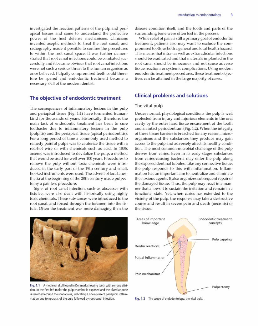
Under normal, physiological conditions the pulp is well protected from injury and injurious elements in the oral cavity by the outer hard tissue encasement of the tooth and an intact periodontium (Fig. 1.2). When the integrity of these tissue barriers is breached for any reason, micro-organisms and the substances they produce may gain access to the pulp and adversely affect its healthy condi-tion. The most common microbial challenge of the pulp derives from caries. Even in its early stages substances from caries-causing bacteria may enter the pulp along the exposed dentinal tubules. Like any connective tissue, the pulp responds to this with inflammation. Inflam-mation has an important aim to neutralize and eliminate the noxious agents. It also organizes subsequent repair of the damaged tissue. Thus, the pulp may react in a man-ner that allows it to sustain the irritation and remain in a functional state. Yet, when caries has extended to the vicinity of the pulp, the response may take a destructive course and result in severe pain and death (necrosis) of the tissue.
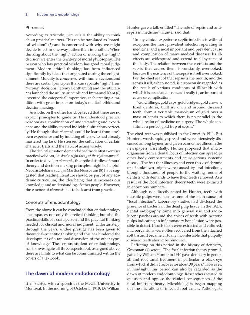
Fig. 1.1 A medieval skull found in Denmark showing teeth with serious attri-tion. In the first left molar the pulp chamber is exposed and the alveolar bone is resorbed around the root apices, indicating a once-present periapical inflam-mation due to necrosis of the pulp followed by root canal infection.
Dentin reactions
Areas of importantknowledge
Pulpal inflammation
Pain mechanisms
Pulp capping
Pulpectomy
Endodontic treatmentconcepts
Fig. 1.2 The scope of endodontology: the vital pulp.
4 Introduction to endodontology
An inflamed or injured pulp may have to be removed and replaced with a root filling – a procedure termed pulpectomy. This measure is undertaken especially in cases when the condition of the pulp is such that an inflammatory breakdown is deemed imminent. A mani-fest infection may otherwise develop in the root canal system. A pulpectomy procedure is carried out under local anesthesia and with the use of specially designed root canal instruments. These instruments remove the dis-eased pulp and prepare the canal system so that it can be filled properly. The purpose of the filling is to prevent microbial growth and multiplication in the pulpal cham-ber. Thus, pulpectomy is a measure primarily aimed at preventing the development of a manifest root canal infection and painful sequelae. Pulpectomy may also be carried out any time a pulp is directly exposed to the oral environment. This may occur after clinical excavation of caries or after a traumatic insult or iatrogenic injury. If the exposure is fresh and the pulp judged not to be seriously inflamed it may not have to be removed. If the open wound is treated with a proper dressing and protected from the oral environ-ment by pulp capping, healing and repair of the wound
are possible. For common terminologies used to specify the endodontic disease conditions and their treatments, see Core concept 1.1.
The necrotic pulp
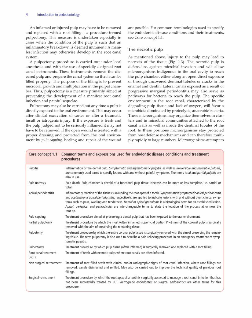
As mentioned above, injury to the pulp may lead to necrosis of the tissue (Fig. 1.3). The necrotic pulp is defenseless against microbial invasion and will allow microorganisms indigenous to the oral cavity to reach the pulp chamber, either along an open direct exposure or through uncovered dentinal tubules or cracks in the enamel and dentin. Lateral canals exposed as a result of progressive marginal periodontitis may also serve as pathways for bacteria to reach the pulp. The specific environment in the root canal, characterized by the degrading pulp tissue and lack of oxygen, will favor a microbiota dominated by proteolytic, anaerobic bacteria. These microorganisms may organize themselves in clus-ters and in microbial communities attached to the root canal walls as well as inside the dentinal tubules of the root. In these positions microorganisms stay protected from host defense mechanisms and can therefore multi-ply rapidly to large numbers. Microorganisms attempt to
Core concept 1.1 Common terms and expressions used for endodontic disease conditions and treatment procedures
Pulpitis Inflammation of the dental pulp. Symptomatic and asymptomatic pulpitis, as well as irreversible and reversible pulpitis, are commonly used terms to specify lesions with and without painful symptoms. The terms total and partial pulpitis are also in use.
Pulp necrosis Pulp death. Pulp chamber is devoid of a functional pulp tissue. Necrosis can be more or less complete, i.e. partial or total.
Apical periodontitis Inflammatory reaction of the tissues surrounding the root apex of a tooth. Symptomatic/asymptomatic apical periodontitis and acute/chronic apical periodontitis, respectively, are applied to indicate lesions with and without overt clinical symp-toms such as pain, swelling and tenderness. Dental or apical granuloma is a histological term for an established lesion. Apical, periapical and periradicular are interchangeable terms to state the location of the process at or near the root tip.
Pulp capping Treatment procedure aimed at preserving a dental pulp that has been exposed to the oral environment.
Partial pulpotomy Treatment procedure by which the most (often inflamed) superficial portion (1–2 mm) of the coronal pulp is surgically removed with the aim of preserving the remaining tissue.
Pulpotomy Treatment procedure by which the entire coronal pulp tissue is surgically removed with the aim of preserving the remain-ing tissue. The term pulpotomy is also used to describe a pain-relieving procedure in an emergency treatment of symp-tomatic pulpitis.
Pulpectomy Treatment procedure by which pulp tissue (often inflamed) is surgically removed and replaced with a root filling.
Root canal treatment Treatment of teeth with necrotic pulps where root canals are often infected. (RCT)
Non-surgical retreatment Treatment of root filled teeth with clinical and/or radiographic signs of root canal infection, where root fillings are removed, canals disinfected and refilled. May also be carried out to improve the technical quality of previous root fillings.
Surgical retreatment Treatment procedure by which the root apex of a tooth is surgically accessed to manage a root canal infection that has not been successfully treated by RCT. Retrograde endodontics or surgical endodontics are other terms for this procedure.
Introduction to endodontology 5
invade the periodontal tissues via the apical foramen or any other portal of exit from the root canal, and may do so before the host defense has been effectively organized. Once established, however, organisms will normally be held back but not eliminated from the root canal space. A chronic inflammatory lesion will ensue, normally around the root tip, and remain for as long as no treatment is initiated. The periapical tissue reaction is often visible in a radio-graph as a localized radiolucency because the adjacent bone has been resorbed in the course of the inflammatory process. The condition may or may not be associated with pain, tooth tenderness and various degrees of swelling. Treatment of the necrotic pulp is by root canal treatment (RCT) and is aimed to combat the intracanal infection. The canal is cleaned with files in order to remove microbes as well as their growth substrate. Owing to the complex anatomy of the root, instruments cannot reach all parts of the canal system and therefore antimicrobial substances are added to disinfect the canal. In order to avoid reinfection and to prevent surviving microbes from growing, the canal is then sealed with a root filling.
The root filled tooth
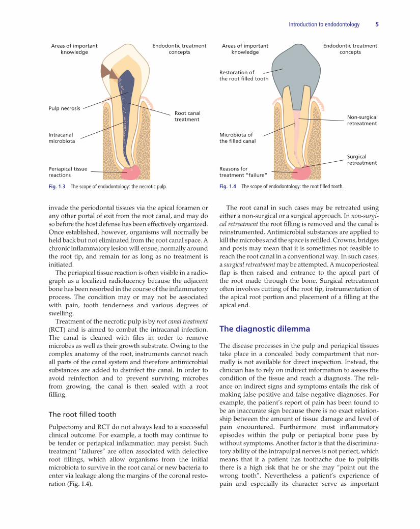
Pulpectomy and RCT do not always lead to a successful clinical outcome. For example, a tooth may continue to be tender or periapical inflammation may persist. Such treatment “failures” are often associated with defective root fillings, which allow organisms from the initial microbiota to survive in the root canal or new bacteria to enter via leakage along the margins of the coronal resto-ration (Fig. 1.4).
The root canal in such cases may be retreated using either a non-surgical or a surgical approach. In non-surgi-cal retreatment the root filling is removed and the canal is reinstrumented. Antimicrobial substances are applied to kill the microbes and the space is refilled. Crowns, bridges and posts may mean that it is sometimes not feasible to reach the root canal in a conventional way. In such cases, a surgical retreatment may be attempted. A mucoperiosteal flap is then raised and entrance to the apical part of the root made through the bone. Surgical retreatment often involves cutting of the root tip, instrumentation of the apical root portion and placement of a filling at the apical end.
The diagnostic dilemma
The disease processes in the pulp and periapical tissues take place in a concealed body compartment that nor-mally is not available for direct inspection. Instead, the clinician has to rely on indirect information to assess the condition of the tissue and reach a diagnosis. The reli-ance on indirect signs and symptoms entails the risk of making false-positive and false-negative diagnoses. For example, the patient’s report of pain has been found to be an inaccurate sign because there is no exact relation-ship between the amount of tissue damage and level of pain encountered. Furthermore most inflammatory episodes within the pulp or periapical bone pass by without symptoms. Another factor is that the discrimina-tory ability of the intrapulpal nerves is not perfect, which means that if a patient has toothache due to pulpitis there is a high risk that he or she may “point out the wrong tooth”. Nevertheless a patient’s experience of pain and especially its character serve as important
Pulp necrosis
Intracanalmicrobiota
Periapical tissuereactions
Root canaltreatment
Areas of importantknowledge
Endodontic treatmentconcepts
Fig. 1.3 The scope of endodontology: the necrotic pulp.
Restoration ofthe root filled tooth
Microbiota ofthe filled canal
Reasons fortreatment “failure”
Non-surgicalretreatment
Surgicalretreatment
Areas of importantknowledge
Endodontic treatmentconcepts
Fig. 1.4 The scope of endodontology: the root filled tooth.
6 Introduction to endodontology
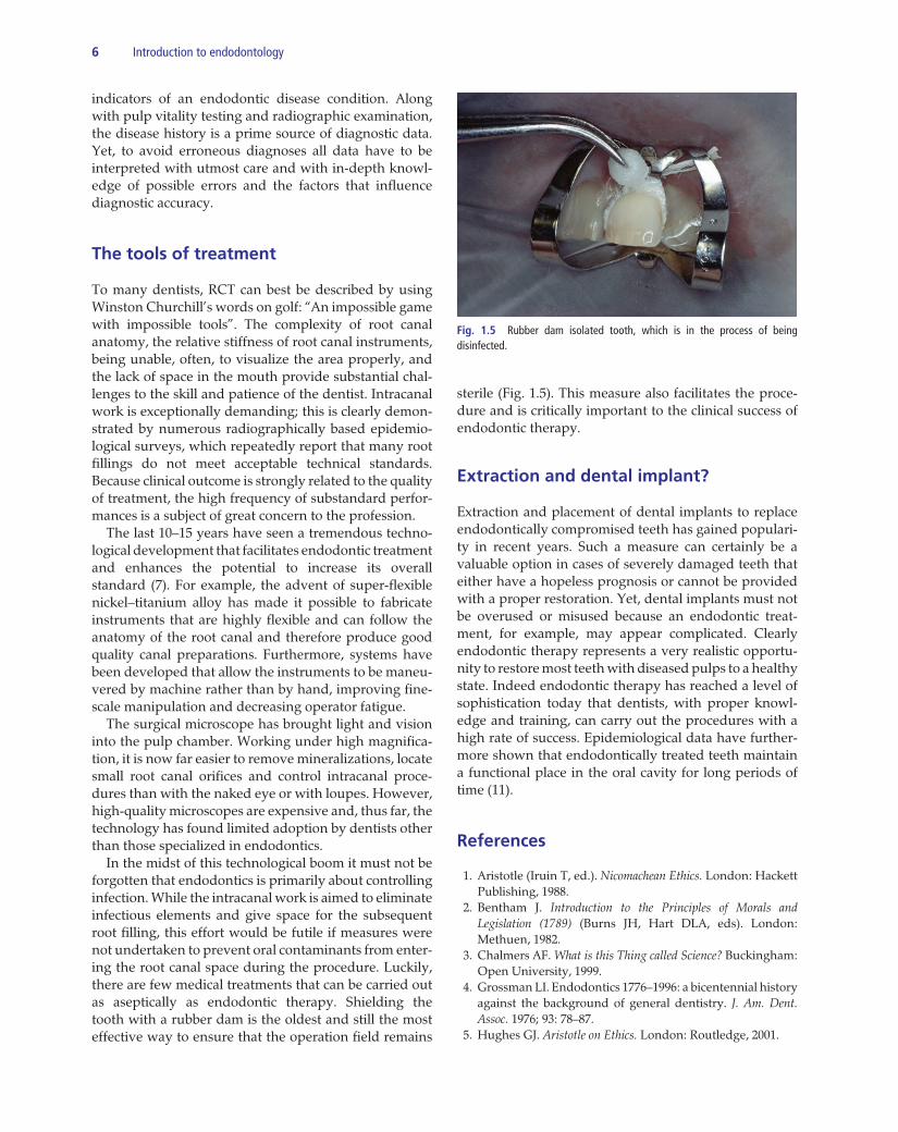
sterile (Fig. 1.5). This measure also facilitates the proce-dure and is critically important to the clinical success of endodontic therapy.
Extraction and dental implant?
Extraction and placement of dental implants to replace endodontically compromised teeth has gained populari-ty in recent years. Such a measure can certainly be a valuable option in cases of severely damaged teeth that either have a hopeless prognosis or cannot be provided with a proper restoration. Yet, dental implants must not be overused or misused because an endodontic treat-ment, for example, may appear complicated. Clearly endodontic therapy represents a very realistic opportu-nity to restore most teeth with diseased pulps to a healthy state. Indeed endodontic therapy has reached a level of sophistication today that dentists, with proper knowl-edge and training, can carry out the procedures with a high rate of success. Epidemiological data have further-more shown that endodontically treated teeth maintain a functional place in the oral cavity for long periods of time (11).
References
1. Aristotle (Iruin T, ed.). Nicomachean Ethics. London: Hackett Publishing, 1988.
2. Bentham J. Introduction to the Principles of Morals and Legislation (1789) (Burns JH, Hart DLA, eds). London: Methuen, 1982.
3. Chalmers AF. What is this Thing called Science? Buckingham: Open University, 1999.
4. Grossman LI. Endodontics 1776–1996: a bicentennial history against the background of general dentistry. J. Am. Dent. Assoc. 1976; 93: 78–87.
5. Hughes GJ. Aristotle on Ethics. London: Routledge, 2001.
indicators of an endodontic disease condition. Along with pulp vitality testing and radiographic examination, the disease history is a prime source of diagnostic data. Yet, to avoid erroneous diagnoses all data have to be interpreted with utmost care and with in-depth knowl-edge of possible errors and the factors that influence diagnostic accuracy.
The tools of treatment
To many dentists, RCT can best be described by using Winston Churchill’s words on golf: “An impossible game with impossible tools”. The complexity of root canal anatomy, the relative stiffness of root canal instruments, being unable, often, to visualize the area properly, and the lack of space in the mouth provide substantial chal-lenges to the skill and patience of the dentist. Intracanal work is exceptionally demanding; this is clearly demon-strated by numerous radiographically based epidemio-logical surveys, which repeatedly report that many root fillings do not meet acceptable technical standards. Because clinical outcome is strongly related to the quality of treatment, the high frequency of substandard perfor-mances is a subject of great concern to the profession. The last 10–15 years have seen a tremendous techno-logical development that facilitates endodontic treatment and enhances the potential to increase its overall standard (7). For example, the advent of super-flexible nickel–titanium alloy has made it possible to fabricate instruments that are highly flexible and can follow the anatomy of the root canal and therefore produce good quality canal preparations. Furthermore, systems have been developed that allow the instruments to be maneu-vered by machine rather than by hand, improving fine-scale manipulation and decreasing operator fatigue. The surgical microscope has brought light and vision into the pulp chamber. Working under high magnifica-tion, it is now far easier to remove mineralizations, locate small root canal orifices and control intracanal proce-dures than with the naked eye or with loupes. However, high-quality microscopes are expensive and, thus far, the technology has found limited adoption by dentists other than those specialized in endodontics. In the midst of this technological boom it must not be forgotten that endodontics is primarily about controlling infection. While the intracanal work is aimed to eliminate infectious elements and give space for the subsequent root filling, this effort would be futile if measures were not undertaken to prevent oral contaminants from enter-ing the root canal space during the procedure. Luckily, there are few medical treatments that can be carried out as aseptically as endodontic therapy. Shielding the tooth with a rubber dam is the oldest and still the most effective way to ensure that the operation field remains
Fig. 1.5 Rubber dam isolated tooth, which is in the process of being disinfected.

Introduction to endodontology 7
6. Kant I. Foundations of the Metaphysics of Morals (1785). Indianapolis: Bobbs–Merrill, 1959.
7. Molander A, Caplan D, Bergenholtz G, Reit C. Improved root-filling quality among general dental practitioners edu-cated in nickel titanium rotary instrumentation. Int. Endod. J. 2007; 40: 254–60.
8. Nussbaum M. Poetic Justice. The Literary Imagination and Public Life. Boston: Beacon Press, 1995.
9. Polanyi M. Personal Knowledge: Towards a Postcritical Philosophy. London: Routledge, 1958.
10. Ryle G. The Concept of Mind. London: Penguin, 1949.11. Salehrabi R, Rotstein I. Endodontic treatment outcomes in a
large patient population in the USA: an epidemiological study. J. Endod. 2004; 30: 846–50.


9
Part 1The Vital Pulp

11
Chapter 2
The dentin–pulp complex: structures, functions and responses to adverse influencesLeif Olgart and Gunnar Bergenholtz
Introduction
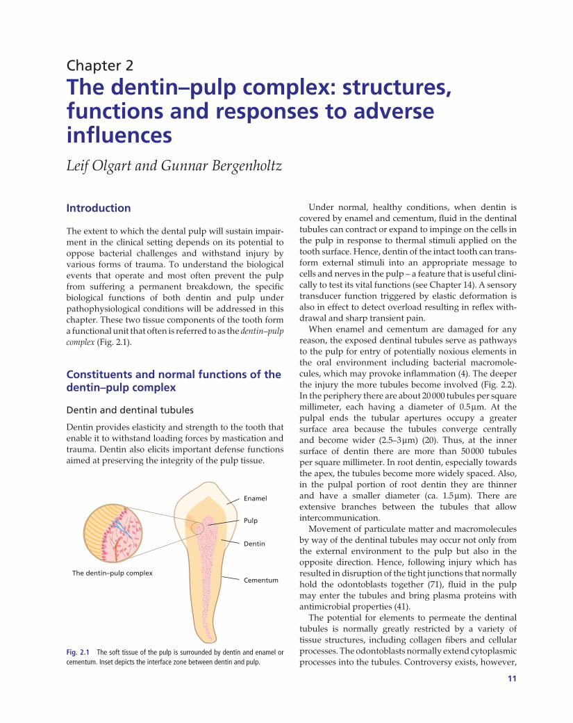
The extent to which the dental pulp will sustain impair-ment in the clinical setting depends on its potential to oppose bacterial challenges and withstand injury by various forms of trauma. To understand the biological events that operate and most often prevent the pulp from suffering a permanent breakdown, the specific biological functions of both dentin and pulp under pathophysiological conditions will be addressed in this chapter. These two tissue components of the tooth form a functional unit that often is referred to as the dentin–pulp complex (Fig. 2.1).
Constituents and normal functions of the dentin–pulp complex
Dentin and dentinal tubules
Dentin provides elasticity and strength to the tooth that enable it to withstand loading forces by mastication and trauma. Dentin also elicits important defense functions aimed at preserving the integrity of the pulp tissue.
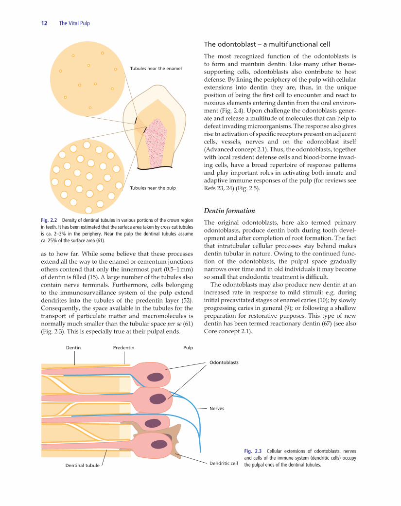
Under normal, healthy conditions, when dentin is covered by enamel and cementum, fluid in the dentinal tubules can contract or expand to impinge on the cells in the pulp in response to thermal stimuli applied on the tooth surface. Hence, dentin of the intact tooth can trans-form external stimuli into an appropriate message to cells and nerves in the pulp – a feature that is useful clini-cally to test its vital functions (see Chapter 14). A sensory transducer function triggered by elastic deformation is also in effect to detect overload resulting in reflex with-drawal and sharp transient pain. When enamel and cementum are damaged for any reason, the exposed dentinal tubules serve as pathways to the pulp for entry of potentially noxious elements in the oral environment including bacterial macromole-cules, which may provoke inflammation (4). The deeper the injury the more tubules become involved (Fig. 2.2). In the periphery there are about 20 000 tubules per square millimeter, each having a diameter of 0.5 μm. At the pulpal ends the tubular apertures occupy a greater surface area because the tubules converge centrally and become wider (2.5–3 μm) (20). Thus, at the inner surface of dentin there are more than 50 000 tubules per square millimeter. In root dentin, especially towards the apex, the tubules become more widely spaced. Also, in the pulpal portion of root dentin they are thinner and have a smaller diameter (ca. 1.5 μm). There are extensive branches between the tubules that allow intercommunication. Movement of particulate matter and macromolecules by way of the dentinal tubules may occur not only from the external environment to the pulp but also in the opposite direction. Hence, following injury which has resulted in disruption of the tight junctions that normally hold the odontoblasts together (71), fluid in the pulp may enter the tubules and bring plasma proteins with antimicrobial properties (41). The potential for elements to permeate the dentinal tubules is normally greatly restricted by a variety of tissue structures, including collagen fibers and cellular processes. The odontoblasts normally extend cytoplasmic processes into the tubules. Controversy exists, however,
The dentin–pulp complex
Dentin
Pulp
Cementum
Enamel
Fig. 2.1 The soft tissue of the pulp is surrounded by dentin and enamel or cementum. Inset depicts the interface zone between dentin and pulp.
12 The Vital Pulp
as to how far. While some believe that these processes extend all the way to the enamel or cementum junctions others contend that only the innermost part (0.5–1 mm) of dentin is filled (15). A large number of the tubules also contain nerve terminals. Furthermore, cells belonging to the immunosurveillance system of the pulp extend dendrites into the tubules of the predentin layer (52). Consequently, the space available in the tubules for the transport of particulate matter and macromolecules is normally much smaller than the tubular space per se (61) (Fig. 2.3). This is especially true at their pulpal ends.
The odontoblast – a multifunctional cell
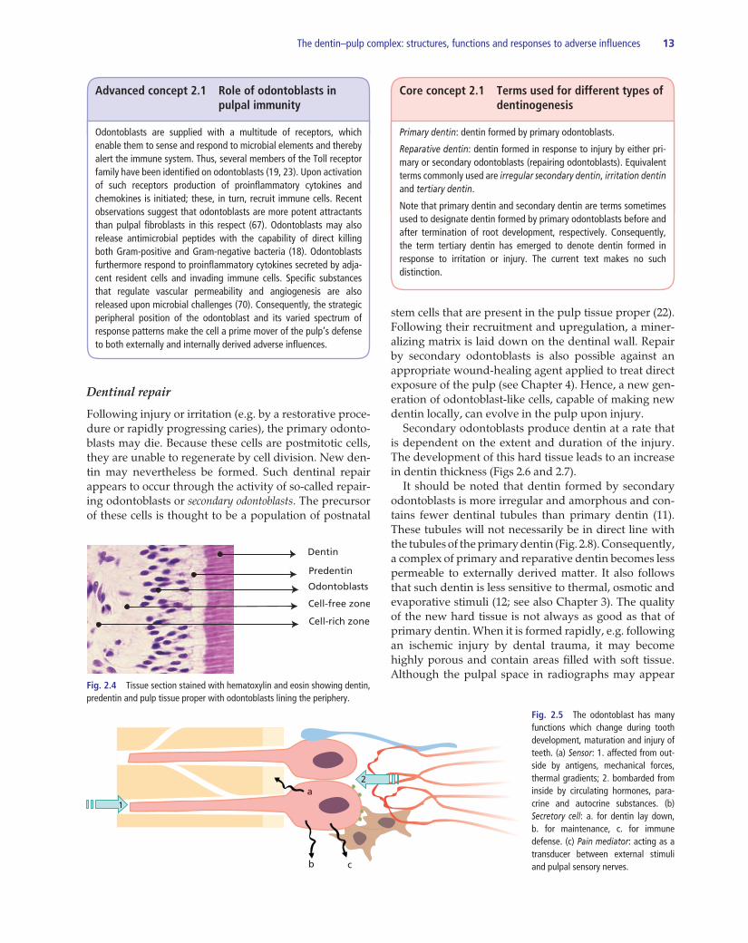
The most recognized function of the odontoblasts is to form and maintain dentin. Like many other tissue- supporting cells, odontoblasts also contribute to host defense. By lining the periphery of the pulp with cellular extensions into dentin they are, thus, in the unique position of being the first cell to encounter and react to noxious elements entering dentin from the oral environ-ment (Fig. 2.4). Upon challenge the odontoblasts gener-ate and release a multitude of molecules that can help to defeat invading microorganisms. The response also gives rise to activation of specific receptors present on adjacent cells, vessels, nerves and on the odontoblast itself (Advanced concept 2.1). Thus, the odontoblasts, together with local resident defense cells and blood-borne invad-ing cells, have a broad repertoire of response patterns and play important roles in activating both innate and adaptive immune responses of the pulp (for reviews see Refs 23, 24) (Fig. 2.5).
Dentin formation
The original odontoblasts, here also termed primary odontoblasts, produce dentin both during tooth devel-opment and after completion of root formation. The fact that intratubular cellular processes stay behind makes dentin tubular in nature. Owing to the continued func-tion of the odontoblasts, the pulpal space gradually narrows over time and in old individuals it may become so small that endodontic treatment is difficult. The odontoblasts may also produce new dentin at an increased rate in response to mild stimuli: e.g. during initial precavitated stages of enamel caries (10); by slowly progressing caries in general (9); or following a shallow preparation for restorative purposes. This type of new dentin has been termed reactionary dentin (67) (see also Core concept 2.1).
Tubules near the enamel
Tubules near the pulp
Fig. 2.2 Density of dentinal tubules in various portions of the crown region in teeth. It has been estimated that the surface area taken by cross cut tubules is ca. 2–3% in the periphery. Near the pulp the dentinal tubules assume ca. 25% of the surface area (61).
Dendritic cell
Dentin
Dentinal tubule
Predentin Pulp
Odontoblasts
Nerves
Fig. 2.3 Cellular extensions of odontoblasts, nerves and cells of the immune system (dendritic cells) occupy the pulpal ends of the dentinal tubules.
The dentin–pulp complex: structures, functions and responses to adverse influences 13
b c
2a
1
Fig. 2.5 The odontoblast has many functions which change during tooth development, maturation and injury of teeth. (a) Sensor: 1. affected from out-side by antigens, mechanical forces, thermal gradients; 2. bombarded from inside by circulating hormones, para-crine and autocrine substances. (b) Secretory cell: a. for dentin lay down, b. for maintenance, c. for immune defense. (c) Pain mediator: acting as a transducer between external stimuli and pulpal sensory nerves.
Dentinal repair
Following injury or irritation (e.g. by a restorative proce-dure or rapidly progressing caries), the primary odonto-blasts may die. Because these cells are postmitotic cells, they are unable to regenerate by cell division. New den-tin may nevertheless be formed. Such dentinal repair appears to occur through the activity of so-called repair-ing odontoblasts or secondary odontoblasts. The precursor of these cells is thought to be a population of postnatal
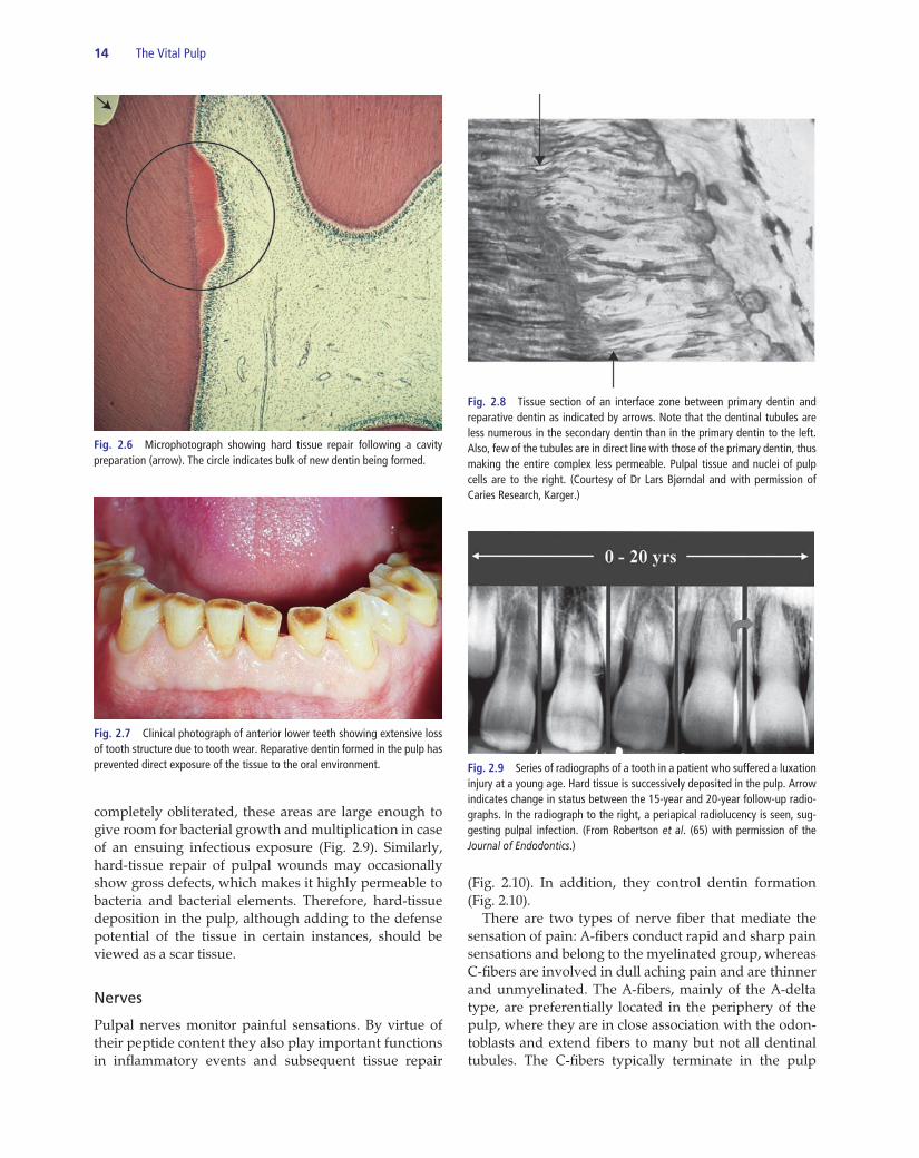
stem cells that are present in the pulp tissue proper (22). Following their recruitment and upregulation, a miner-alizing matrix is laid down on the dentinal wall. Repair by secondary odontoblasts is also possible against an appropriate wound-healing agent applied to treat direct exposure of the pulp (see Chapter 4). Hence, a new gen-eration of odontoblast-like cells, capable of making new dentin locally, can evolve in the pulp upon injury. Secondary odontoblasts produce dentin at a rate that is dependent on the extent and duration of the injury. The development of this hard tissue leads to an increase in dentin thickness (Figs 2.6 and 2.7). It should be noted that dentin formed by secondary odontoblasts is more irregular and amorphous and con-tains fewer dentinal tubules than primary dentin (11). These tubules will not necessarily be in direct line with the tubules of the primary dentin (Fig. 2.8). Consequently, a complex of primary and reparative dentin becomes less permeable to externally derived matter. It also follows that such dentin is less sensitive to thermal, osmotic and evaporative stimuli (12; see also Chapter 3). The quality of the new hard tissue is not always as good as that of primary dentin. When it is formed rapidly, e.g. following an ischemic injury by dental trauma, it may become highly porous and contain areas filled with soft tissue. Although the pulpal space in radiographs may appear
Advanced concept 2.1 Role of odontoblasts in pulpal immunity
Odontoblasts are supplied with a multitude of receptors, which enable them to sense and respond to microbial elements and thereby alert the immune system. Thus, several members of the Toll receptor family have been identified on odontoblasts (19, 23). Upon activation of such receptors production of proinflammatory cytokines and chemokines is initiated; these, in turn, recruit immune cells. Recent observations suggest that odontoblasts are more potent attractants than pulpal fibroblasts in this respect (67). Odontoblasts may also release antimicrobial peptides with the capability of direct killing both Gram-positive and Gram-negative bacteria (18). Odontoblasts furthermore respond to proinflammatory cytokines secreted by adja-cent resident cells and invading immune cells. Specific substances that regulate vascular permeability and angiogenesis are also released upon microbial challenges (70). Consequently, the strategic peripheral position of the odontoblast and its varied spectrum of response patterns make the cell a prime mover of the pulp’s defense to both externally and internally derived adverse influences.
Core concept 2.1 Terms used for different types of dentinogenesis
Primary dentin: dentin formed by primary odontoblasts.
Reparative dentin: dentin formed in response to injury by either pri-mary or secondary odontoblasts (repairing odontoblasts). Equivalent terms commonly used are irregular secondary dentin, irritation dentin and tertiary dentin.
Note that primary dentin and secondary dentin are terms sometimes used to designate dentin formed by primary odontoblasts before and after termination of root development, respectively. Consequently, the term tertiary dentin has emerged to denote dentin formed in response to irritation or injury. The current text makes no such distinction.
Dentin
Predentin
Odontoblasts
Cell-free zone
Cell-rich zone
Fig. 2.4 Tissue section stained with hematoxylin and eosin showing dentin, predentin and pulp tissue proper with odontoblasts lining the periphery.
14 The Vital Pulp
completely obliterated, these areas are large enough to give room for bacterial growth and multiplication in case of an ensuing infectious exposure (Fig. 2.9). Similarly, hard-tissue repair of pulpal wounds may occasionally show gross defects, which makes it highly permeable to bacteria and bacterial elements. Therefore, hard-tissue deposition in the pulp, although adding to the defense potential of the tissue in certain instances, should be viewed as a scar tissue.
Nerves
Pulpal nerves monitor painful sensations. By virtue of their peptide content they also play important functions in inflammatory events and subsequent tissue repair
(Fig. 2.10). In addition, they control dentin formation (Fig. 2.10). There are two types of nerve fiber that mediate the sensation of pain: A-fibers conduct rapid and sharp pain sensations and belong to the myelinated group, whereas C-fibers are involved in dull aching pain and are thinner and unmyelinated. The A-fibers, mainly of the A-delta type, are preferentially located in the periphery of the pulp, where they are in close association with the odon-toblasts and extend fibers to many but not all dentinal tubules. The C-fibers typically terminate in the pulp
Fig. 2.6 Microphotograph showing hard tissue repair following a cavity preparation (arrow). The circle indicates bulk of new dentin being formed.
Fig. 2.7 Clinical photograph of anterior lower teeth showing extensive loss of tooth structure due to tooth wear. Reparative dentin formed in the pulp has prevented direct exposure of the tissue to the oral environment.
Fig. 2.8 Tissue section of an interface zone between primary dentin and reparative dentin as indicated by arrows. Note that the dentinal tubules are less numerous in the secondary dentin than in the primary dentin to the left. Also, few of the tubules are in direct line with those of the primary dentin, thus making the entire complex less permeable. Pulpal tissue and nuclei of pulp cells are to the right. (Courtesy of Dr Lars Bjørndal and with permission of Caries Research, Karger.)
Fig. 2.9 Series of radiographs of a tooth in a patient who suffered a luxation injury at a young age. Hard tissue is successively deposited in the pulp. Arrow indicates change in status between the 15-year and 20-year follow-up radio-graphs. In the radiograph to the right, a periapical radiolucency is seen, sug-gesting pulpal infection. (From Robertson et al. (65) with permission of the Journal of Endodontics.)