Embed Size (px)
Citation preview
Dr. Reem Al-Dhalaan
1
Multiple Factors Which Influence Post/Dowel Selection:
Amount of coronal tooth structure
Tooth anatomy Position of the tooth in the arch
Root length Root width
Canal configuration Functional requirements of the tooth
Torquing force Stresses
Development of hydrostatic pressure Post design
Post material Material compatibility
Bonding capability Core retention Retrievability
Esthetics Crown material
Fernandes AS et al; Int J Prosthodont. 2001
PROSTHODONTIC MANAGEMENT OF ENDODONTICALLY TREATED TEETH;
Factors Determining Post Selection, Foundation Restorations and Review of Success & Failure Data
The longevity of endodontically involved teeth has been greatly enhanced by continuing developments made in endodontic therapy and restorative procedures. It has been reported that a large number of endodontically treated teeth are restored to their original function with the use of intraradicular devices. These devices vary from a conventional custom cast post and core to one visit techniques, using commercially available prefabricated post systems. In the last few decades, various prefabricated posts systems have been developed. The selection of post design is important, because it may have an influence on the longevity of the tooth (Sorensen JA et al 1990). HISTORICAL PERSPECTIVE The concept of using the root of a tooth for retention of a crown is not new (Shillingburg HT et al; 1982). In the 1700s Fauchard inserted wooden dowels in canals of teeth to aid in crown retention. Over time the wood would expand in the moist environment to enhance retention of the dowel until, unfortunately, the root would often fracture vertically. Additional efforts to develop crowns retained with posts or dowels in the 1800s were limited by the failure of the “endodontic” therapy of the era. Several of the 19th century versions of dowels also used wooden “pivots” but some dentists reported the use of metal posts favored by Black (1869) in which a porcelain-faced crown was secured by a screw passing into a gold-lined root canal. A device developed by Clark in the mid-1800s was extremely practical for its time because it included a tube that allowed drainage from the apical area or the canal (Prothero JH; 1921). The Richmond crown was introduced in 1878 and incorporated a threaded tube in the canal with a screw-retained crown. It was later modified to eliminate the threaded tube and was redesigned as a 1-piece dowel and crown (Hampson EL et al; 1958, and Demas NC et al; 1957), which lost its popularity quickly because they were not practical. This was obviously evident when divergent paths of insertion of the post space and remaining tooth structure existed, especially for abutments of fixed partial dentures. One piece dowel-crown restorations also presented problems when the crown or FPD required removal and replacement. These difficulties led to development of a post and core restoration as a separate entity with an artificial crown cemented over a core and remaining tooth structure. With the advent of scientific endodontic therapy in the 1950s, the challenges increased for restorative dentistry. Teeth that were extracted without hesitations were now successfully treated with predictable endodontic therapy; and a satisfactory restorative solution was necessary.
Dr. Reem Al-Dhalaan
2
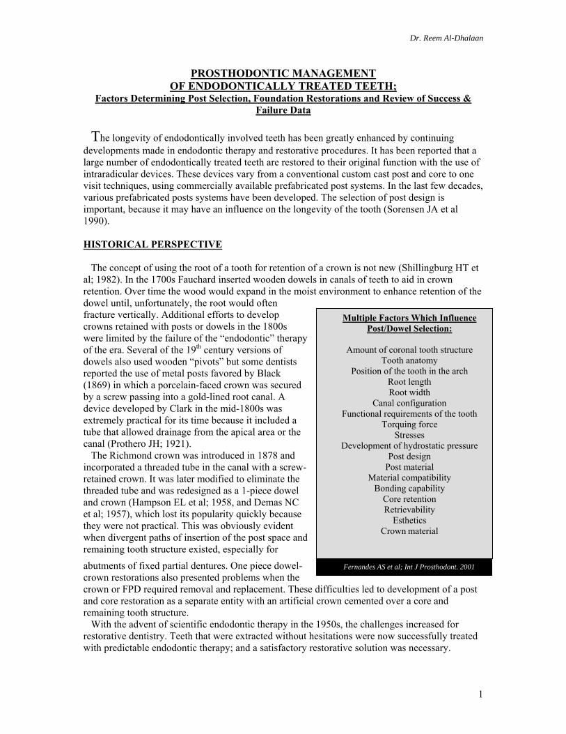
Figure 1: Excessive preparation of canal may cause perforation of proximal depression in root
surfaces and encourages root fracture
FACTORS INFLUENCING POST SELECTION
I. Root Length The length and shape of the remaining root determines the length of the post (Holmes DC et al, 1996). It has been demonstrated that the greater the post length, the better the retention and stress distribution (Standlee JP et al; 1978 & 1972). It may not always be possible to use a long post, especially when the remaining root is short or curved. Mattison CD et al (1984) and Kvist T et al (1989) suggest that is important to preserve 3 to 5 mm of apical gutta percha to maintain the apical seal. When the root length is short, the clinician must decide whether to use a longer post or maintain the recommended apical seal and use a parallel-sided threaded post. The use of reinforced composite luting agents may compensate for the reduced post length (Nissan J et al; 2001). For molars with short roots the placement of more than one post will provide additional retention for the core foundation restoration (Hirshfeld Z et al; 1972).
II. Tooth Anatomy Root anatomy such as root curvature, mesio-distal width, and labio-lingual dimension dictates post selection (Nissan J et al; 2001). A consideration of the root size and length is important, because improper post space preparation and use of large diameter posts present the risk of apical or lateral perforation. Moreover, an active post can initiate cracks in the thin dentinal wall. Radiographs help evaluate the root length, width, anatomic variations, canal structure, and the surrounding hard tissue structures. Occasionally they can be misleading because of proximal root concavities and magnification factor, thus a use of a grid is recommended so that the length, diameter, and design of the post can be correctly determined (Frommer HH; 1996). Gutmann (1992) reviewed the anatomic considerations and stated that roots of maxillary centrals and laterals, and also mandibular premolars have sufficient bulk to accommodate most post systems.
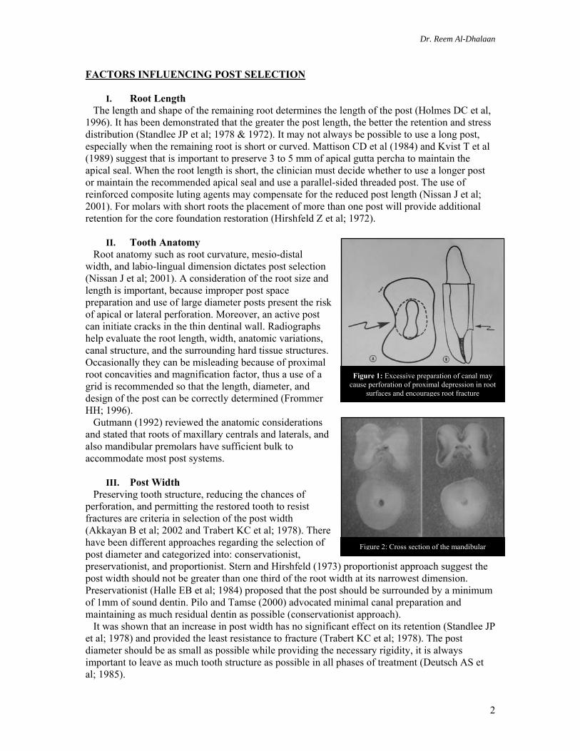
III. Post Width Preserving tooth structure, reducing the chances of perforation, and permitting the restored tooth to resist fractures are criteria in selection of the post width (Akkayan B et al; 2002 and Trabert KC et al; 1978). There have been different approaches regarding the selection of post diameter and categorized into: conservationist, preservationist, and proportionist. Stern and Hirshfeld (1973) proportionist approach suggest the post width should not be greater than one third of the root width at its narrowest dimension. Preservationist (Halle EB et al; 1984) proposed that the post should be surrounded by a minimum of 1mm of sound dentin. Pilo and Tamse (2000) advocated minimal canal preparation and maintaining as much residual dentin as possible (conservationist approach). It was shown that an increase in post width has no significant effect on its retention (Standlee JP et al; 1978) and provided the least resistance to fracture (Trabert KC et al; 1978). The post diameter should be as small as possible while providing the necessary rigidity, it is always important to leave as much tooth structure as possible in all phases of treatment (Deutsch AS et al; 1985).
Figure 2: Cross section of the mandibular
Dr. Reem Al-Dhalaan
3
IV. Canal Configuration and Post Adaptability Canal configuration aids in making a choice between a custom designed post and a prefabricated post (Ash M jr. et al; 1993, and Smith TC et al; 1997). Often a dilemma arises in funnel-shaped canals, whether to use a parallel sided post and fill the remaining post spaced with cement or to use a tapered post that closely adapt to the canal wall. A third option is to use large prefabricated parallel-sided posts. It has been suggested that if a canal requires extensive preparation a well adapted cast post and core restoration will be more retentive that a prefabricated post that does not match the canal shape (Cohen BI et al; 1996). In addition, root reinforcement with composite is suggested for wide canals (Saupe WA et al; 1996). A well adapted tapered post provided increased resistance to fracture, however on fracture it resulted in an extensive loss of tooth structure (Sorenson JA et al; 1990 and Tjan AH et al; 1985). Morgano and Milot (1993) stated that 44% of the cast posts were less than one half to one quarter the length of the clinical crown and that the failure rate reported was the result of compromised length rather than post type. In another investigation they reported custom cast posts success rate to be more than 90% after 5 years in function.
Root Canal Configuration Elliptical
Circular Buccolingual Mesiodistal
Maxillary central incisor Maxillary first premolar (two roots)
Mandibular second premolar Maxillary molars (DB roots)
Maxillary lateral incisor Maxillary canine
Mandibular incisors Mandibular canine
Maxillary first premolar (single root)
Mandibular first premolar Maxillary second premolar
Maxillary molars (MB roots) Mandibular molars (M & D roots)
Maxillary molars (palatal roots)
Weine FS:Endodontic Therapy; 1989
V. Coronal Structure The amount of remaining coronal tooth structure is also a critical factor in determining the post selection. Hoag and Dwyer (1982) determined that the amount of tooth structure present is more important than the material from which the post and core is fabricated (amalgam, resin, or cast gold).The bulk of the tooth above the restorative margin should be at least 1.5mm to 2mm to achieve resistance form (Barkhordar RA et al; 1989). The use of cast post and cores in restoring endodontically treated teeth with moderate to severe coronal tooth loss has demonstrated a success rate of 90.6% after 5 years of service (Bergman B et al; 1989). When ample coronal dentin remains non-metal posts such as a carbon fiber posts were deemed successful (Sidoli GE et al; 1997, Stockton LW et al; 1999, Barkhordar RA et al; 1989)
VI. Position of the tooth in the arch It is considered one of the important criterions in the restoration of endodontically treated teeth.
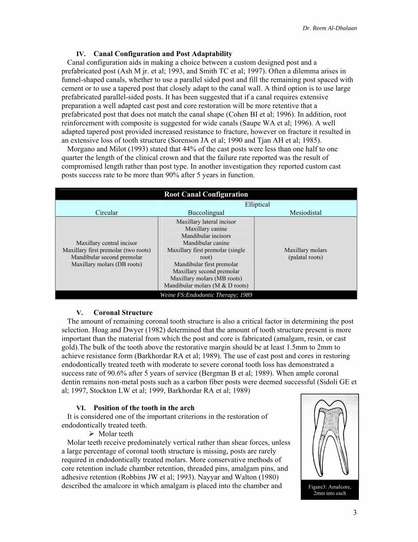
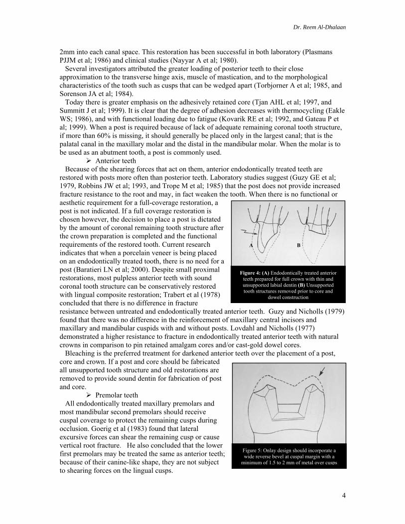
Molar teeth Molar teeth receive predominately vertical rather than shear forces, unless a large percentage of coronal tooth structure is missing, posts are rarely required in endodontically treated molars. More conservative methods of core retention include chamber retention, threaded pins, amalgam pins, and adhesive retention (Robbins JW et al; 1993). Nayyar and Walton (1980) described the amalcore in which amalgam is placed into the chamber and Figure3: Amalcore,
2mm into each
Dr. Reem Al-Dhalaan
4
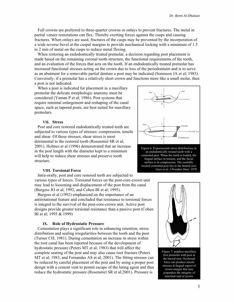
Figure 4: (A) Endodontically treated anterior teeth prepared for full crown with thin and unsupported labial dentin (B) Unsupported tooth structures removed prior to core and
dowel construction
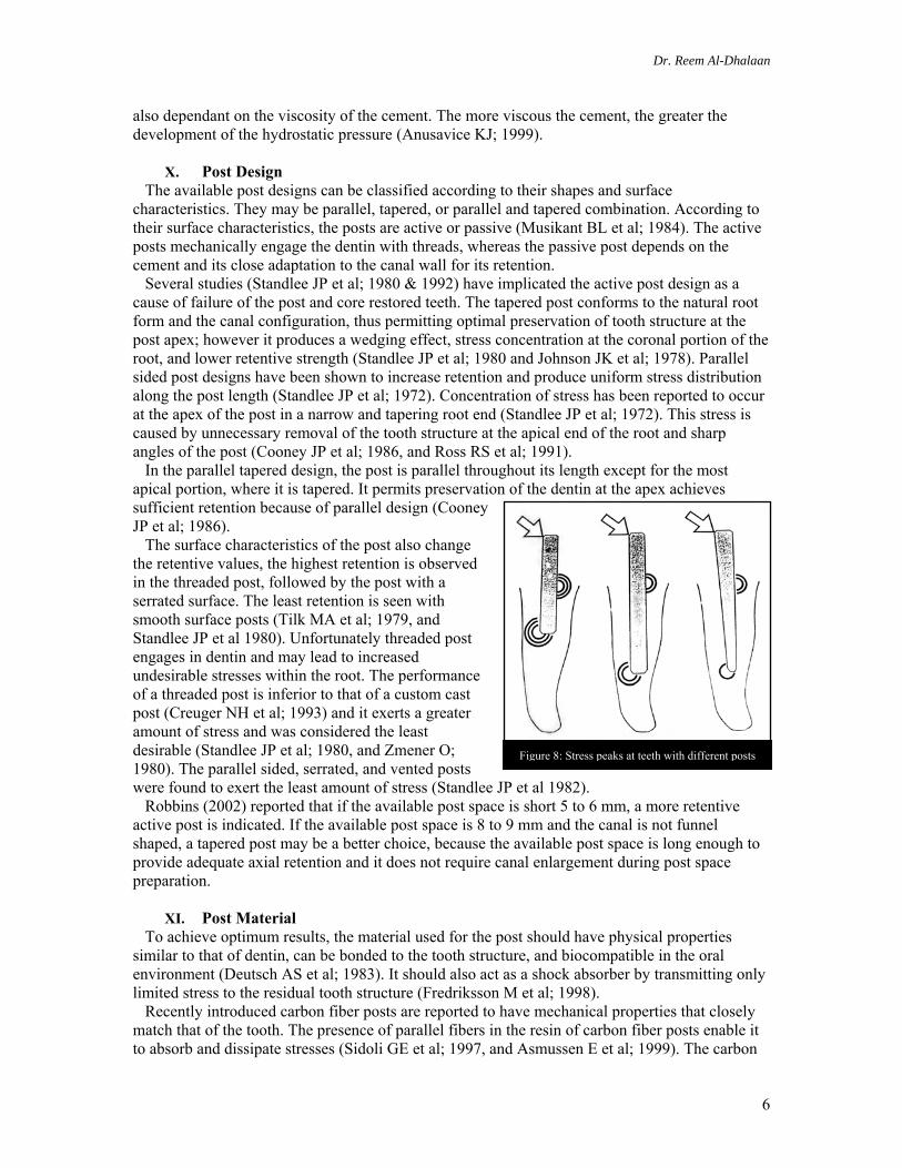
Figure 5: Onlay design should incorporate a wide reverse bevel at cuspal margin with a
minimum of 1.5 to 2 mm of metal over cusps
2mm into each canal space. This restoration has been successful in both laboratory (Plasmans PJJM et al; 1986) and clinical studies (Nayyar A et al; 1980). Several investigators attributed the greater loading of posterior teeth to their close approximation to the transverse hinge axis, muscle of mastication, and to the morphological characteristics of the tooth such as cusps that can be wedged apart (Torbjorner A et al; 1985, and Sorenson JA et al; 1984). Today there is greater emphasis on the adhesively retained core (Tjan AHL et al; 1997, and Summitt J et al; 1999). It is clear that the degree of adhesion decreases with thermocycling (Eakle WS; 1986), and with functional loading due to fatigue (Kovarik RE et al; 1992, and Gateau P et al; 1999). When a post is required because of lack of adequate remaining coronal tooth structure, if more than 60% is missing, it should generally be placed only in the largest canal; that is the palatal canal in the maxillary molar and the distal in the mandibular molar. When the molar is to be used as an abutment tooth, a post is commonly used.
Anterior teeth Because of the shearing forces that act on them, anterior endodontically treated teeth are restored with posts more often than posterior teeth. Laboratory studies suggest (Guzy GE et al; 1979, Robbins JW et al; 1993, and Trope M et al; 1985) that the post does not provide increased fracture resistance to the root and may, in fact weaken the tooth. When there is no functional or aesthetic requirement for a full-coverage restoration, a post is not indicated. If a full coverage restoration is chosen however, the decision to place a post is dictated by the amount of coronal remaining tooth structure after the crown preparation is completed and the functional requirements of the restored tooth. Current research indicates that when a porcelain veneer is being placed on an endodontically treated tooth, there is no need for a post (Baratieri LN et al; 2000). Despite small proximal restorations, most pulpless anterior teeth with sound coronal tooth structure can be conservatively restored with lingual composite restoration; Trabert et al (1978) concluded that there is no difference in fracture resistance between untreated and endodontically treated anterior teeth. Guzy and Nicholls (1979) found that there was no difference in the reinforcement of maxillary central incisors and maxillary and mandibular cuspids with and without posts. Lovdahl and Nicholls (1977) demonstrated a higher resistance to fracture in endodontically treated anterior teeth with natural crowns in comparison to pin retained amalgam cores and/or cast-gold dowel cores. Bleaching is the preferred treatment for darkened anterior teeth over the placement of a post, core and crown. If a post and core should be fabricated all unsupported tooth structure and old restorations are removed to provide sound dentin for fabrication of post and core.
Premolar teeth All endodontically treated maxillary premolars and most mandibular second premolars should receive cuspal coverage to protect the remaining cusps during occlusion. Goerig et al (1983) found that lateral excursive forces can shear the remaining cusp or cause vertical root fracture. He also concluded that the lower first premolars may be treated the same as anterior teeth; because of their canine-like shape, they are not subject to shearing forces on the lingual cusps.
A B
Dr. Reem Al-Dhalaan
5
Full crowns are preferred to three-quarter crowns or onlays to prevent fractures. The metal in partial veneer restorations can flex. Thereby exerting forces against the cusps and causing fractures. When onlays are used, fractures of the cusps may be prevented by the incorporation of a wide reverse bevel at the cuspal margins to provide mechanical locking with a minimum of 1.5 to 2 mm of metal on the cusps to reduce metal flexing. When restoring an endodontically treated premolar, a decision regarding post placement is made based on the remaining coronal tooth structure, the functional requirements of the tooth, and an evaluation of the forces that acts on the tooth. If an endodontically treated premolar has increased functional stresses acting on the crown due to loss of the periodontuim and is to serve as an abutment for a removable partial denture a post may be indicated (Sorenson JA et al; 1985). Conversely, if a premolar has a relatively short crown and functions more like a small molar, then a post is not indicated. When a post is indicated for placement in a maxillary premolar the delicate morphologic anatomy must be considered (Yaman P et al; 1986). Post systems that require minimal enlargement and reshaping of the canal space, such as tapered posts, are best suited for maxillary premolars.
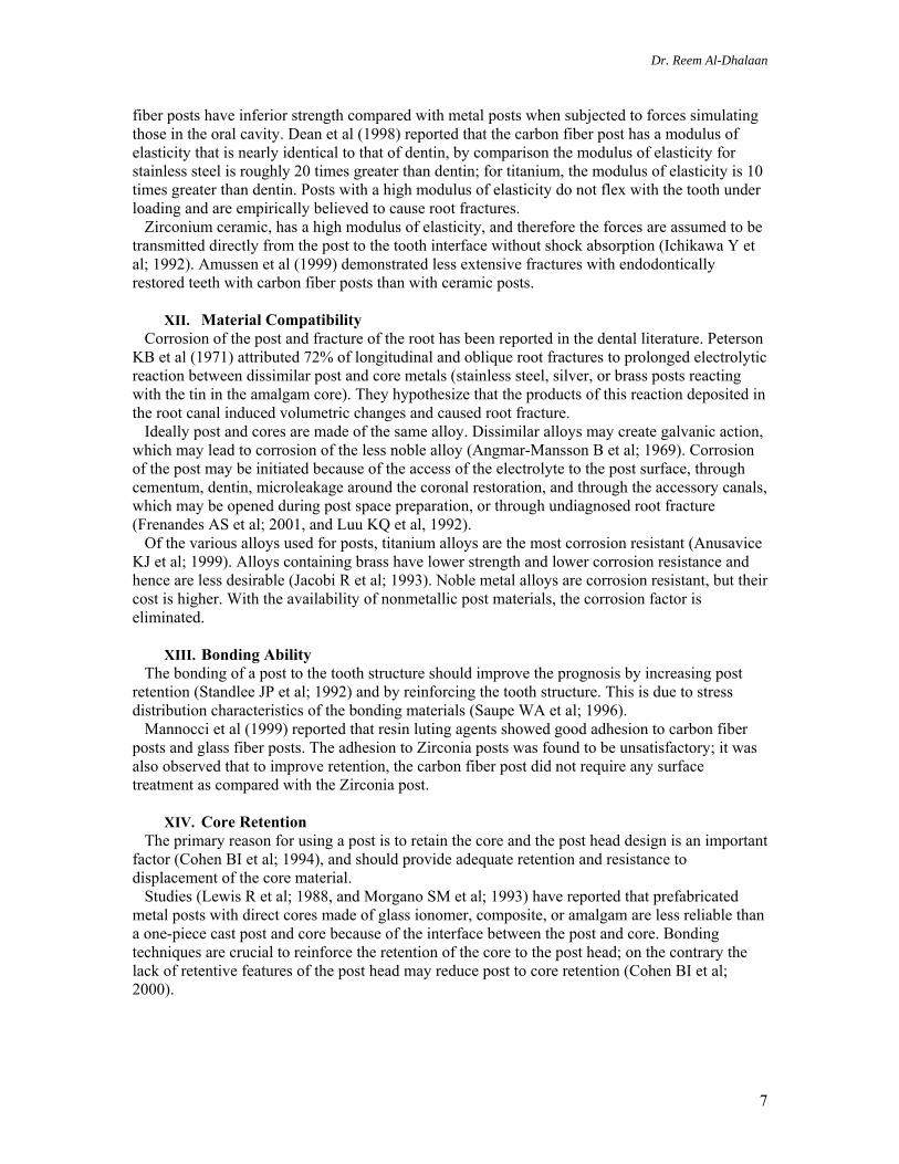
VII. Stress Post and core restored endodontically treated teeth are subjected to various types of stresses: compression, tensile and shear. Of these stresses, shear stress is most detrimental to the restored tooth (Rosenstiel SR et al; 2001). Holmes et al (1996) demonstrated that an increase in the post length with the diameter kept to a minimum will help to reduce shear stresses and preserve tooth structure.
VIII. Torsional Force Intra-orally, post and core restored teeth are subjected to various types of forces. Torsional forces on the post-core-crown unit may lead to loosening and displacement of the post from the canal (Burgess JO et al; 1992, and Cohen BI et al; 1995). Burgess et al (1992) emphasized on the importance of an antirotational feature and concluded that resistance to torsional forces is integral to the survival of the post-core-crown unit. Active post designs provide greater torsional resistance than a passive post (Cohen BI et al; 1995 & 1999)
IX. Role of Hydrostatic Pressure Cementation plays a significant role in enhancing retention, stress distribution and sealing irregularities between the tooth and the post (Turner CH; 1981). During cementation an increase in stress within the root canal has been reported because of the development of hydrostatic pressure (Peters MT et al; 1983) that will affect the complete seating of the post and may also cause root fracture (Peters MT et al; 1983, and Fernandes AS et al; 2001). The fitting stresses can be reduced by careful placement of the post and by using a proper post design with a cement vent to permit escape of the luting agent and thus reduce the hydrostatic pressure (Rosenstiel SR et al;2001). Pressure is
Figure 6: Experimental stress distributions in an endodontically treated tooth with a
cemented post. When the tooth is loaded, the lingual surface in tension, and the facial surface is in compression. The centrally
located cemented post lies in the neutral axis Guzy et al, J Prosthet Dent; 1979
Figure 7: pulpless maxillary first premolar with post in the buccal root. Occlusual force can produce tensile
stresses at lingual aspect of crown margin that may
jeopardize the integrity of marginal seal of crown.
Dr. Reem Al-Dhalaan
6
Figure 8: Stress peaks at teeth with different posts
also dependant on the viscosity of the cement. The more viscous the cement, the greater the development of the hydrostatic pressure (Anusavice KJ; 1999).
X. Post Design The available post designs can be classified according to their shapes and surface characteristics. They may be parallel, tapered, or parallel and tapered combination. According to their surface characteristics, the posts are active or passive (Musikant BL et al; 1984). The active posts mechanically engage the dentin with threads, whereas the passive post depends on the cement and its close adaptation to the canal wall for its retention. Several studies (Standlee JP et al; 1980 & 1992) have implicated the active post design as a cause of failure of the post and core restored teeth. The tapered post conforms to the natural root form and the canal configuration, thus permitting optimal preservation of tooth structure at the post apex; however it produces a wedging effect, stress concentration at the coronal portion of the root, and lower retentive strength (Standlee JP et al; 1980 and Johnson JK et al; 1978). Parallel sided post designs have been shown to increase retention and produce uniform stress distribution along the post length (Standlee JP et al; 1972). Concentration of stress has been reported to occur at the apex of the post in a narrow and tapering root end (Standlee JP et al; 1972). This stress is caused by unnecessary removal of the tooth structure at the apical end of the root and sharp angles of the post (Cooney JP et al; 1986, and Ross RS et al; 1991). In the parallel tapered design, the post is parallel throughout its length except for the most apical portion, where it is tapered. It permits preservation of the dentin at the apex achieves sufficient retention because of parallel design (Cooney JP et al; 1986). The surface characteristics of the post also change the retentive values, the highest retention is observed in the threaded post, followed by the post with a serrated surface. The least retention is seen with smooth surface posts (Tilk MA et al; 1979, and Standlee JP et al 1980). Unfortunately threaded post engages in dentin and may lead to increased undesirable stresses within the root. The performance of a threaded post is inferior to that of a custom cast post (Creuger NH et al; 1993) and it exerts a greater amount of stress and was considered the least desirable (Standlee JP et al; 1980, and Zmener O; 1980). The parallel sided, serrated, and vented posts were found to exert the least amount of stress (Standlee JP et al 1982). Robbins (2002) reported that if the available post space is short 5 to 6 mm, a more retentive active post is indicated. If the available post space is 8 to 9 mm and the canal is not funnel shaped, a tapered post may be a better choice, because the available post space is long enough to provide adequate axial retention and it does not require canal enlargement during post space preparation.
XI. Post Material To achieve optimum results, the material used for the post should have physical properties similar to that of dentin, can be bonded to the tooth structure, and biocompatible in the oral environment (Deutsch AS et al; 1983). It should also act as a shock absorber by transmitting only limited stress to the residual tooth structure (Fredriksson M et al; 1998). Recently introduced carbon fiber posts are reported to have mechanical properties that closely match that of the tooth. The presence of parallel fibers in the resin of carbon fiber posts enable it to absorb and dissipate stresses (Sidoli GE et al; 1997, and Asmussen E et al; 1999). The carbon
Dr. Reem Al-Dhalaan
7
fiber posts have inferior strength compared with metal posts when subjected to forces simulating those in the oral cavity. Dean et al (1998) reported that the carbon fiber post has a modulus of elasticity that is nearly identical to that of dentin, by comparison the modulus of elasticity for stainless steel is roughly 20 times greater than dentin; for titanium, the modulus of elasticity is 10 times greater than dentin. Posts with a high modulus of elasticity do not flex with the tooth under loading and are empirically believed to cause root fractures. Zirconium ceramic, has a high modulus of elasticity, and therefore the forces are assumed to be transmitted directly from the post to the tooth interface without shock absorption (Ichikawa Y et al; 1992). Amussen et al (1999) demonstrated less extensive fractures with endodontically restored teeth with carbon fiber posts than with ceramic posts.
XII. Material Compatibility Corrosion of the post and fracture of the root has been reported in the dental literature. Peterson KB et al (1971) attributed 72% of longitudinal and oblique root fractures to prolonged electrolytic reaction between dissimilar post and core metals (stainless steel, silver, or brass posts reacting with the tin in the amalgam core). They hypothesize that the products of this reaction deposited in the root canal induced volumetric changes and caused root fracture. Ideally post and cores are made of the same alloy. Dissimilar alloys may create galvanic action, which may lead to corrosion of the less noble alloy (Angmar-Mansson B et al; 1969). Corrosion of the post may be initiated because of the access of the electrolyte to the post surface, through cementum, dentin, microleakage around the coronal restoration, and through the accessory canals, which may be opened during post space preparation, or through undiagnosed root fracture (Frenandes AS et al; 2001, and Luu KQ et al, 1992). Of the various alloys used for posts, titanium alloys are the most corrosion resistant (Anusavice KJ et al; 1999). Alloys containing brass have lower strength and lower corrosion resistance and hence are less desirable (Jacobi R et al; 1993). Noble metal alloys are corrosion resistant, but their cost is higher. With the availability of nonmetallic post materials, the corrosion factor is eliminated.
XIII. Bonding Ability The bonding of a post to the tooth structure should improve the prognosis by increasing post retention (Standlee JP et al; 1992) and by reinforcing the tooth structure. This is due to stress distribution characteristics of the bonding materials (Saupe WA et al; 1996). Mannocci et al (1999) reported that resin luting agents showed good adhesion to carbon fiber posts and glass fiber posts. The adhesion to Zirconia posts was found to be unsatisfactory; it was also observed that to improve retention, the carbon fiber post did not require any surface treatment as compared with the Zirconia post.
XIV. Core Retention The primary reason for using a post is to retain the core and the post head design is an important factor (Cohen BI et al; 1994), and should provide adequate retention and resistance to displacement of the core material. Studies (Lewis R et al; 1988, and Morgano SM et al; 1993) have reported that prefabricated metal posts with direct cores made of glass ionomer, composite, or amalgam are less reliable than a one-piece cast post and core because of the interface between the post and core. Bonding techniques are crucial to reinforce the retention of the core to the post head; on the contrary the lack of retentive features of the post head may reduce post to core retention (Cohen BI et al; 2000).
Dr. Reem Al-Dhalaan
8
XV. Retrievability Ideally the post system selected should be such that if the endodontic treatment fails or the post fractures, it is easy for the clinician to retrieve the post without substantial loss of tooth structure (Lewis R et al; 1988). Unfortunately, the retrievability of a metal post, especially the cast post and core system is difficult and involves removal of tooth structure around the post, which could further weaken the tooth. Carbon fiber posts have an advantage over metallic and ceramic posts in that the removal is relatively easy, rapid, and predictable (Freedman GA et al; 2001). Post removal can be preformed by means of conventional rotary instruments and solvents. Other commercially available systems to remove posts include the Masseran Kit, Post Removal System, Endodontic extractors, and ultrasonic unit Roto-Pro bur and a combination of tube extractors with cyanoacrylate will aid in post removal by breaking up the cement (Cohen S et al; 2002). Abbott (2002) reported that post removal is a predictable procedure, if appropriate techniques and devices are used, and root fracture is a rare occurrence. He also concluded the following:
(1) Masseran kit and ultrasonics are effective in removing fractured cast posts and parallel-sided posts.
(2) Unscrewing of the threaded screw posts. (3) Eggler post removal (Ruddle) in retrieving cast posts. (4) Ultrasonics in retrieving parallel-sided posts. XVI. Esthetics
The post and core material should be esthetically compatible with the crown and the surrounding tissues (Tamse A; 1988). Freedman (2001) and Vichi (2000) have emphasized the need to have the color of the foundation restoration as close to that of natural dentin. The use of a custom cast post would compromise esthetics as the gray tint of the metal may show through the thin root wall. The overlying gingival tissue would also appear darker or grayish (Saupe WA et al; 1996). This esthetic concern has led to the development of esthetic posts made from reinforced resins or ceramics in an effort to eliminate the color deficiency. Another alternative to an esthetic post and core system is the use of a 1.6 mm or more thick opaque porcelain fused to the core portion of cast post and core in order to eliminate the grayish effect of cast metal (Hochstedler J et al; 1996, and Nakamura T et al; 2002). Also the use of ceramic core material such as IPS Empress Cosmo core is advocated. The availability of different cement shades permits minor esthetic corrections under all ceramic crowns (Vichi A et al; 2000).
DENTAL POSTS Wagnild et al (2002) summarized the ideal physical properties of a post that include:
(1) Maximum protection of the root. (2) Adequate retention within the root. (3) Biocompatible / noncorrosive (4) Maximum retention of the core and crown. (5) Maximum protection of the crown margin cement seal. (6) Pleasing esthetics (7) Radiopaque
CAST POSTS AND CORES The custom–cast post has a long history of clinical success. They provide excellent service for endodontically treated teeth with moderate to severe damage. A 6-year retrospective study of 96 endodontically treated teeth with extensive loss of tooth structure and restored with cast dowel cores indicated a 90.6% success rate (Bergman B et al; 1989), and are best applied to single rooted teeth, especially incisors and canines. However when it is compared to parallel
Dr. Reem Al-Dhalaan
9
Figure 9: Custom cast post and core
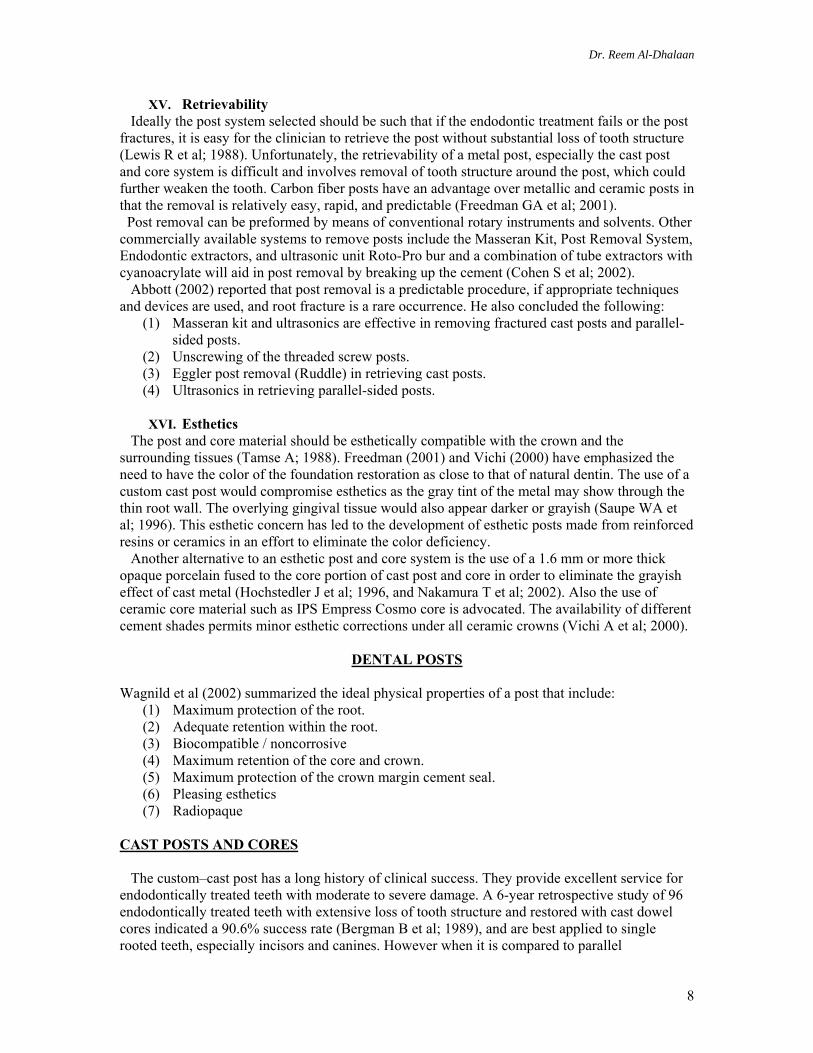
prefabricated posts, both in vitro (Chan RW et al; 1982, and Lovdahl PE et al; 1977) and in vivo (Sorenson JA et al; 1984, and Torbjorner A et al; 1995) its superiority is questionable; although this can be attributed to the severe damage of teeth restored with the cast post. There are circumstances in which the custom-cast post is the restoration of choice (Robbins JW et al; 1990) including the following:
(1) When multiple cores are being placed in the same arch. It is most cost effective to prepare multiple post spaces, make an impression and fabricate the posts in the laboratory.
(2) When post and cores are being placed in small teeth such as mandibular incisors. In these circumstances it is often difficult to retain the core material on the head of the post.
(3) When the angle of the core must be changed in relation to the post. Prefabricated posts should not be bent; therefore the custom cast post best fulfills this requirement.
(4) When an all ceramic crown restoration is placed it is necessary to have a core that approximates the color of the natural tooth structure. If a large core is being placed in a high-stress situation, resin composite may not be the material of choice due to the fact that it tends to deform under a load (Kovarik RE et al; 1992, and Gateau P et al; 1999). In this circumstance the post and core can be cast in metal, and porcelain can be fired to the core to stimulate the color of natural tooth structure (Hochstedler J et al; 1996, and Nakamura T et al; 2002). The core can then be etched with hydrofluoric acid and the all-ceramic crown can be bonded to the core.
(5) Cast posts and cores are the restorative method of choice for endodontically treated anterior teeth with moderate to severe destruction (Morgano M et al; 1993).
The customized cast post and core posses’ superior adaptation to the root canal, associated with little or no stress with installation, and high strength in comparison to the prefabricated post. On the other hand custom cast posts is considered time consuming complex procedure, less retentive than parallel-sided posts, and acts as a wedge during occlusual load transfer. Its recommended use is with elliptical or flared canals. Molars often perform satisfactory with direct cores retained by engaging the pulpal chamber and a portion of the root canals (Nayyar A et al; 1980 & 1988), and retention of the core can be augmented by placement of one or more prefabricated intraradicular posts. Premolars may be restored with both custom cast posts and cores or prefabricated post(s) with direct cores.
Criteria for Cast Post and Core Design
(1) Adequate post length This will assist retention, and distribution of coronal forces through the roots. Shorter posts may increase the possibility of root fractures due to stress concentration on the gingival margin (Weine FS; 1982). Inadequate post length is considered a common cause of post failure. The ideal post is approximately two thirds the length of the root, leaving 4 to 5 mm of root cal filling within the canal. Perel and Muroff (1972) recommended that the post be at least half the length of root in bone. When there is bone loss from periodontal disease, the post length would be longer.
(2) Minimal alteration of the internal root canal anatomy It is essential to leave adequate dentin for support and distribution of post stresses. Excessive preparation may cause perforation of the proximal depressions in the root surface, limiting function and increasing root fracture.
Dr. Reem Al-Dhalaan
10
(3) Protection of the root against vertical root fracture This is accomplished by:
1. The post and core should have a positive occlusual seat to avoid the wedge like action of the post (Schnell F; 1978).
2. Cohen et al (1976) concluded that a metal margin should surround and protect the root from vertical fracture (the ferrule effect)
3. The post is vented by flattening a small portion of the buccal or lingual post along the length to allow escape of the cement and reduce the hydraulic pressure.
4. The post should have a passive fit without a wedging effect. (4) Antirotational features
Many cast posts resist rotational forces because they are oblong in cross section. However, the cast post for round canals, such as the maxillary incisor requires locking notches or keyways incorporated into the canal to resist rotational movement (Gutmann JL et al; 1977, and Dewhirst RB et al; 1969). Methods of fabricating cast posts and cores
A. Direct Technique A reliable method is direct fabrication of the pattern first described by Barker (1963), using numerous materials: wax with a plastic rod as a carrier (Barker BC; 1963, Dewhirst RB et al; 1969, and Gentile D; 1965), wax with a dental bur, acrylic resin with a solid plastic sprue (DeDomenico RJ; 1977, and Stern N; 1972) , and a core of acrylic resin with an endodontic file coated with wax that adapted to the prepared canal (Miller AW; 1978). Custom cast dowel cores require 2 visits which is a primary disadvantage of the direct method.
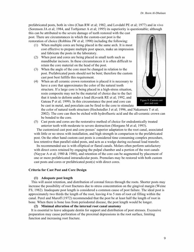
B. Indirect Technique Success of the indirect method depends on the accuracy of the impression replicating the internal surface of the prepared root canal. Impression material is injected into the post space (Sall HD; 1977), and a rigid object is inserted in the canal before the initial set of impression material to
A
B
Figure 10: Indirect Custom Cast Post Technique. (A) Wire reinforcement,
(B) Impression material
Figure 11: Sectional custom cast post / Indirect technique
Dr. Reem Al-Dhalaan
11
A B
strengthen this impression and minimize potential for distortion which includes toothpicks (Michnick BT et al; 1978), wire (McLean JW; 1967), paper clips (Baraban DJ; 1967), and plastic sprues (Mazzuchelli L; 1972). The indirect method conserves chair time by delegating the pattern for the post and core to a dental laboratory.
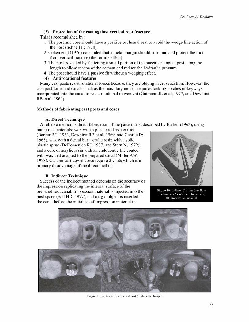
PREFABRICATED POSTS A recent nationwide survey of dentists in the U.S.A indicated that 40% of general dentists used prefabricated posts most of the time, and the most popular prefabricated
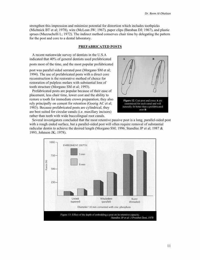
post was parallel sided serrated post (Morgano SM et al; 1994). The use of prefabricated posts with a direct core reconstruction is the restorative method of choice for restoration of pulpless molars with substantial loss of tooth structure (Morgano SM et al; 1993). Prefabricated posts are popular because of their ease of placement, less chair time, lower cost and the ability to restore a tooth for immediate crown preparation; they also rely principally on cement for retention (Goerig AC et al; 1983). Because prefabricated posts are cylindrical, they are best suited for circular canals (i.e. maxillary incisors) rather than teeth with wide buccolingual root canals. Several investigators concluded that the most retentive passive post is a long, parallel-sided post with a rough ended surface, but a parallel-sided post will often require removal of substantial radicular dentin to achieve the desired length (Morgano SM; 1996, Standlee JP et al; 1987 & 1993, Johnson JK; 1978).
Figure 12: Cast post and cores A are customized for each canal and will
naturally fit better than a prefabricated post B
Figure 13: Effect of the depth of embedding a post on its retentive capacity. Standlee JP et al: J Prosthet Dent, 1978
Dr. Reem Al-Dhalaan
12
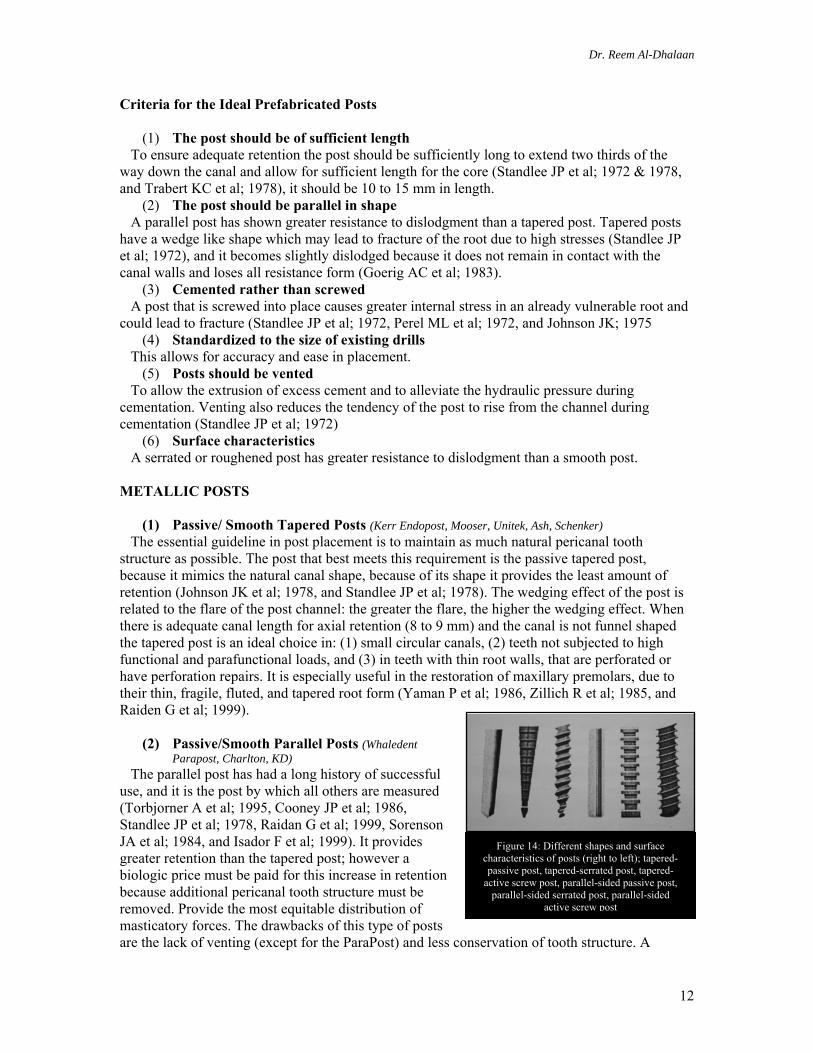
Figure 14: Different shapes and surface characteristics of posts (right to left); tapered-passive post, tapered-serrated post, tapered-
active screw post, parallel-sided passive post, parallel-sided serrated post, parallel-sided
active screw post
Criteria for the Ideal Prefabricated Posts
(1) The post should be of sufficient length To ensure adequate retention the post should be sufficiently long to extend two thirds of the way down the canal and allow for sufficient length for the core (Standlee JP et al; 1972 & 1978, and Trabert KC et al; 1978), it should be 10 to 15 mm in length.
(2) The post should be parallel in shape A parallel post has shown greater resistance to dislodgment than a tapered post. Tapered posts have a wedge like shape which may lead to fracture of the root due to high stresses (Standlee JP et al; 1972), and it becomes slightly dislodged because it does not remain in contact with the canal walls and loses all resistance form (Goerig AC et al; 1983).
(3) Cemented rather than screwed A post that is screwed into place causes greater internal stress in an already vulnerable root and could lead to fracture (Standlee JP et al; 1972, Perel ML et al; 1972, and Johnson JK; 1975
(4) Standardized to the size of existing drills This allows for accuracy and ease in placement.
(5) Posts should be vented To allow the extrusion of excess cement and to alleviate the hydraulic pressure during cementation. Venting also reduces the tendency of the post to rise from the channel during cementation (Standlee JP et al; 1972)
(6) Surface characteristics A serrated or roughened post has greater resistance to dislodgment than a smooth post. METALLIC POSTS
(1) Passive/ Smooth Tapered Posts (Kerr Endopost, Mooser, Unitek, Ash, Schenker) The essential guideline in post placement is to maintain as much natural pericanal tooth structure as possible. The post that best meets this requirement is the passive tapered post, because it mimics the natural canal shape, because of its shape it provides the least amount of retention (Johnson JK et al; 1978, and Standlee JP et al; 1978). The wedging effect of the post is related to the flare of the post channel: the greater the flare, the higher the wedging effect. When there is adequate canal length for axial retention (8 to 9 mm) and the canal is not funnel shaped the tapered post is an ideal choice in: (1) small circular canals, (2) teeth not subjected to high functional and parafunctional loads, and (3) in teeth with thin root walls, that are perforated or have perforation repairs. It is especially useful in the restoration of maxillary premolars, due to their thin, fragile, fluted, and tapered root form (Yaman P et al; 1986, Zillich R et al; 1985, and Raiden G et al; 1999).
(2) Passive/Smooth Parallel Posts (Whaledent Parapost, Charlton, KD)
The parallel post has had a long history of successful use, and it is the post by which all others are measured (Torbjorner A et al; 1995, Cooney JP et al; 1986, Standlee JP et al; 1978, Raidan G et al; 1999, Sorenson JA et al; 1984, and Isador F et al; 1999). It provides greater retention than the tapered post; however a biologic price must be paid for this increase in retention because additional pericanal tooth structure must be removed. Provide the most equitable distribution of masticatory forces. The drawbacks of this type of posts are the lack of venting (except for the ParaPost) and less conservation of tooth structure. A
Dr. Reem Al-Dhalaan
13
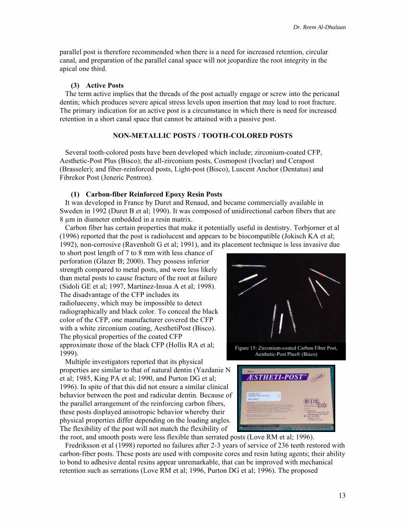
Figure 15: Zirconium-coated Carbon Fiber Post, Aesthetic-Post Plus® (Bisco)
parallel post is therefore recommended when there is a need for increased retention, circular canal, and preparation of the parallel canal space will not jeopardize the root integrity in the apical one third.
(3) Active Posts The term active implies that the threads of the post actually engage or screw into the pericanal dentin; which produces severe apical stress levels upon insertion that may lead to root fracture. The primary indication for an active post is a circumstance in which there is need for increased retention in a short canal space that cannot be attained with a passive post.
NON-METALLIC POSTS / TOOTH-COLORED POSTS Several tooth-colored posts have been developed which include; zirconium-coated CFP, Aesthetic-Post Plus (Bisco); the all-zirconium posts, Cosmopost (Ivoclar) and Cerapost (Brasseler); and fiber-reinforced posts, Light-post (Bisco), Luscent Anchor (Dentatus) and Fibrekor Post (Jeneric Pentron).
(1) Carbon-fiber Reinforced Epoxy Resin Posts It was developed in France by Duret and Renaud, and became commercially available in Sweden in 1992 (Duret B et al; 1990). It was composed of unidirectional carbon fibers that are 8 µm in diameter embedded in a resin matrix. Carbon fiber has certain properties that make it potentially useful in dentistry. Torbjorner et al (1996) reported that the post is radiolucent and appears to be biocompatible (Jokisch KA et al; 1992), non-corrosive (Ravenholt G et al; 1991), and its placement technique is less invasive due to short post length of 7 to 8 mm with less chance of perforation (Glazer B; 2000). They possess inferior strength compared to metal posts, and were less likely than metal posts to cause fracture of the root at failure (Sidoli GE et al; 1997, Martinez-Insua A et al; 1998). The disadvantage of the CFP includes its radiolueceny, which may be impossible to detect radiographically and black color. To conceal the black color of the CFP, one manufacturer covered the CFP with a white zirconium coating, AesthetiPost (Bisco). The physical properties of the coated CFP approximate those of the black CFP (Hollis RA et al; 1999). Multiple investigators reported that its physical properties are similar to that of natural dentin (Yazdanie N et al; 1985, King PA et al; 1990, and Purton DG et al; 1996). In spite of that this did not ensure a similar clinical behavior between the post and radicular dentin. Because of the parallel arrangement of the reinforcing carbon fibers, these posts displayed anisotropic behavior whereby their physical properties differ depending on the loading angles. The flexibility of the post will not match the flexibility of the root, and smooth posts were less flexible than serrated posts (Love RM et al; 1996). Fredriksson et al (1998) reported no failures after 2-3 years of service of 236 teeth restored with carbon-fiber posts. These posts are used with composite cores and resin luting agents; their ability to bond to adhesive dental resins appear unremarkable, that can be improved with mechanical retention such as serrations (Love RM et al; 1996, Purton DG et al; 1996). The proposed
Dr. Reem Al-Dhalaan
14
advantages of CFP are that it can be bonded to dentin (Purton DG et al; 1996), making it significantly more flexible than metal posts (Assmussen E et al; 1999). The laboratory data indicate that the bond strength of a composite core material to a CFP is less than the mechanical retention of composite core to a metal post (Millstein P et al; 1999, and Purton DG et al; 1996). It has been reported Triolo et al (1999) that the bond strength to CFP can be increased with air abrasion, whereas Drummond et al (1999) reported decreased bond strength after air abrasion. The literature does support the notion that the nature of the fractures is more favorable with the CFP than with the metal post (Dean JP et al; 1998, Isador F et al; 1996, King PA; 1990, Hollis RA et al; 1999, Martinez-Insua A et al; 1998, and Sidoli GE et al; 1997) in all but one study that reported opposite results (Stockton L et al; 1999). A laboratory study by Drummond et al (1999)
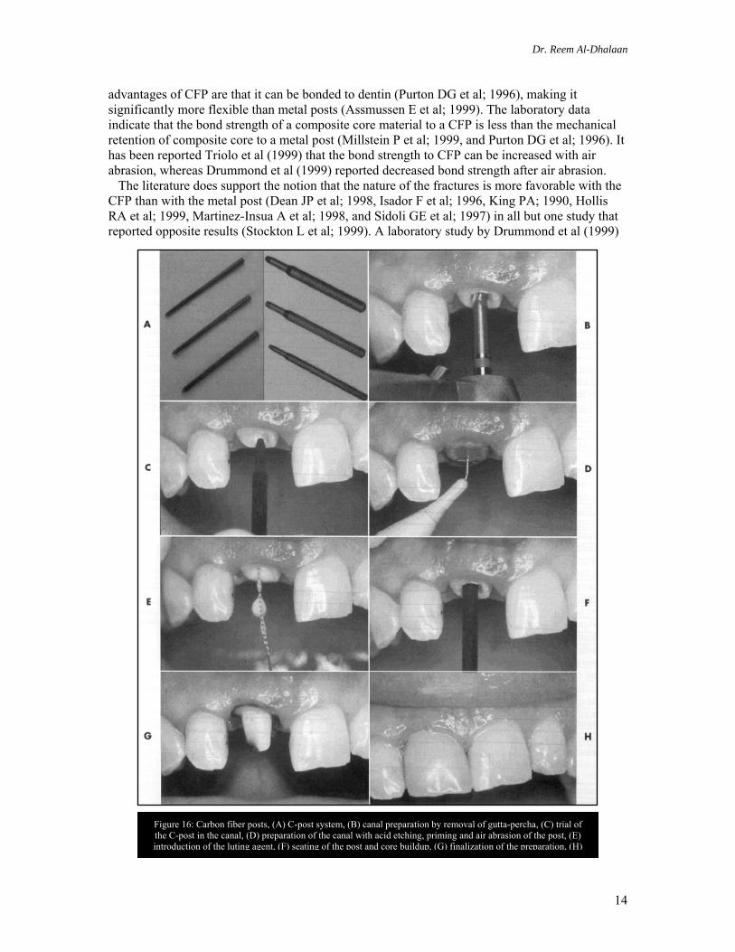
Figure 16: Carbon fiber posts, (A) C-post system, (B) canal preparation by removal of gutta-percha, (C) trial of the C-post in the canal, (D) preparation of the canal with acid etching, priming and air abrasion of the post, (E) introduction of the luting agent, (F) seating of the post and core buildup, (G) finalization of the preparation, (H)
Dr. Reem Al-Dhalaan
15
reported a significant decrease in flexural strength after cyclic and thermal loading. The clinical data regarding the success of the CFP are favorable. Glazer (2000) concluded that the use of carbon reinforced resin posts in premolars, especially mandibular premolars, may be associated with a higher failure rate and shorter longevity than in anterior teeth. Fredriksson et al (1998) reported no failures in a mean duration of 32 months. Ferrari et al (2000) compared the CFP to custom-cast post over 4 years. They reported an 11% failure of the custom cast post, whereas there were no failures of the CFP. In another study (2000) he reported a failure rate of 3.2% of CFPs after 1 to 6 years. Manocci et al (1998) reported in a 3-yearclinical study comparing CFPs and custom cast post. Only one CFP failed because of post dislodgment, whereas 10 of the custom cast posts failed due to root fractures.
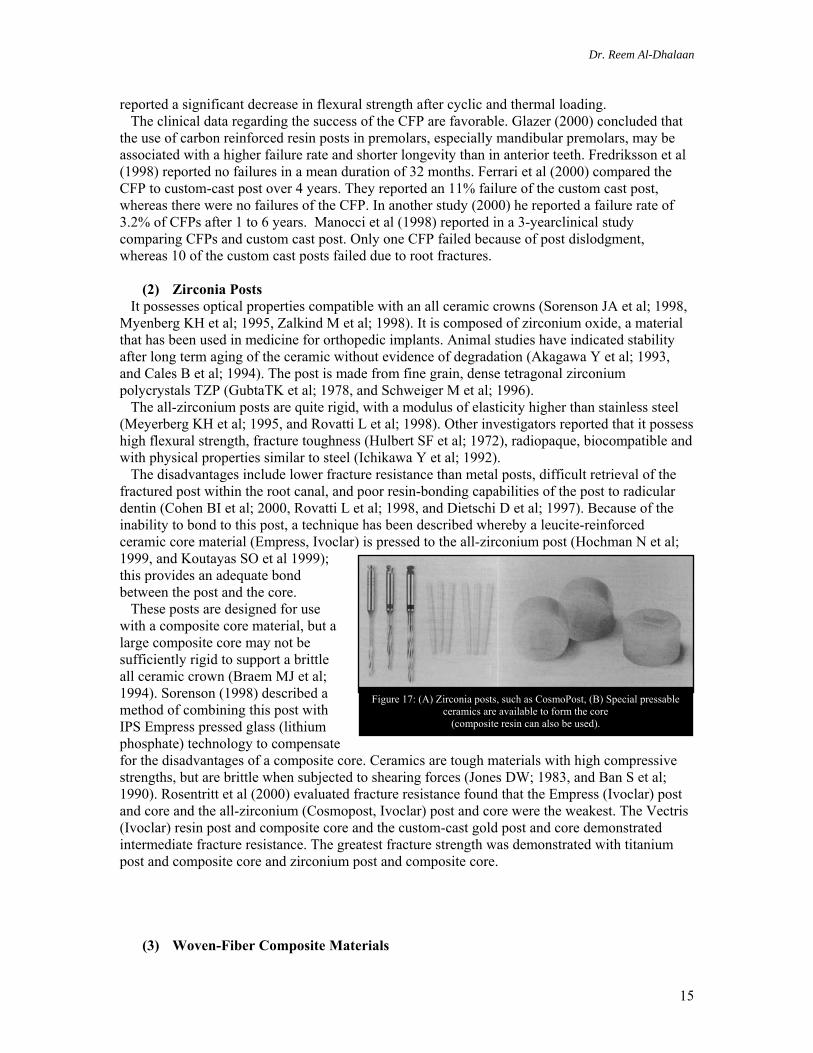
(2) Zirconia Posts It possesses optical properties compatible with an all ceramic crowns (Sorenson JA et al; 1998, Myenberg KH et al; 1995, Zalkind M et al; 1998). It is composed of zirconium oxide, a material that has been used in medicine for orthopedic implants. Animal studies have indicated stability after long term aging of the ceramic without evidence of degradation (Akagawa Y et al; 1993, and Cales B et al; 1994). The post is made from fine grain, dense tetragonal zirconium polycrystals TZP (GubtaTK et al; 1978, and Schweiger M et al; 1996). The all-zirconium posts are quite rigid, with a modulus of elasticity higher than stainless steel (Meyerberg KH et al; 1995, and Rovatti L et al; 1998). Other investigators reported that it possess high flexural strength, fracture toughness (Hulbert SF et al; 1972), radiopaque, biocompatible and with physical properties similar to steel (Ichikawa Y et al; 1992). The disadvantages include lower fracture resistance than metal posts, difficult retrieval of the fractured post within the root canal, and poor resin-bonding capabilities of the post to radicular dentin (Cohen BI et al; 2000, Rovatti L et al; 1998, and Dietschi D et al; 1997). Because of the inability to bond to this post, a technique has been described whereby a leucite-reinforced ceramic core material (Empress, Ivoclar) is pressed to the all-zirconium post (Hochman N et al; 1999, and Koutayas SO et al 1999); this provides an adequate bond between the post and the core. These posts are designed for use with a composite core material, but a large composite core may not be sufficiently rigid to support a brittle all ceramic crown (Braem MJ et al; 1994). Sorenson (1998) described a method of combining this post with IPS Empress pressed glass (lithium phosphate) technology to compensate for the disadvantages of a composite core. Ceramics are tough materials with high compressive strengths, but are brittle when subjected to shearing forces (Jones DW; 1983, and Ban S et al; 1990). Rosentritt et al (2000) evaluated fracture resistance found that the Empress (Ivoclar) post and core and the all-zirconium (Cosmopost, Ivoclar) post and core were the weakest. The Vectris (Ivoclar) resin post and composite core and the custom-cast gold post and core demonstrated intermediate fracture resistance. The greatest fracture strength was demonstrated with titanium post and composite core and zirconium post and composite core.
(3) Woven-Fiber Composite Materials
Figure 17: (A) Zirconia posts, such as CosmoPost, (B) Special pressable ceramics are available to form the core
(composite resin can also be used).
Dr. Reem Al-Dhalaan
16
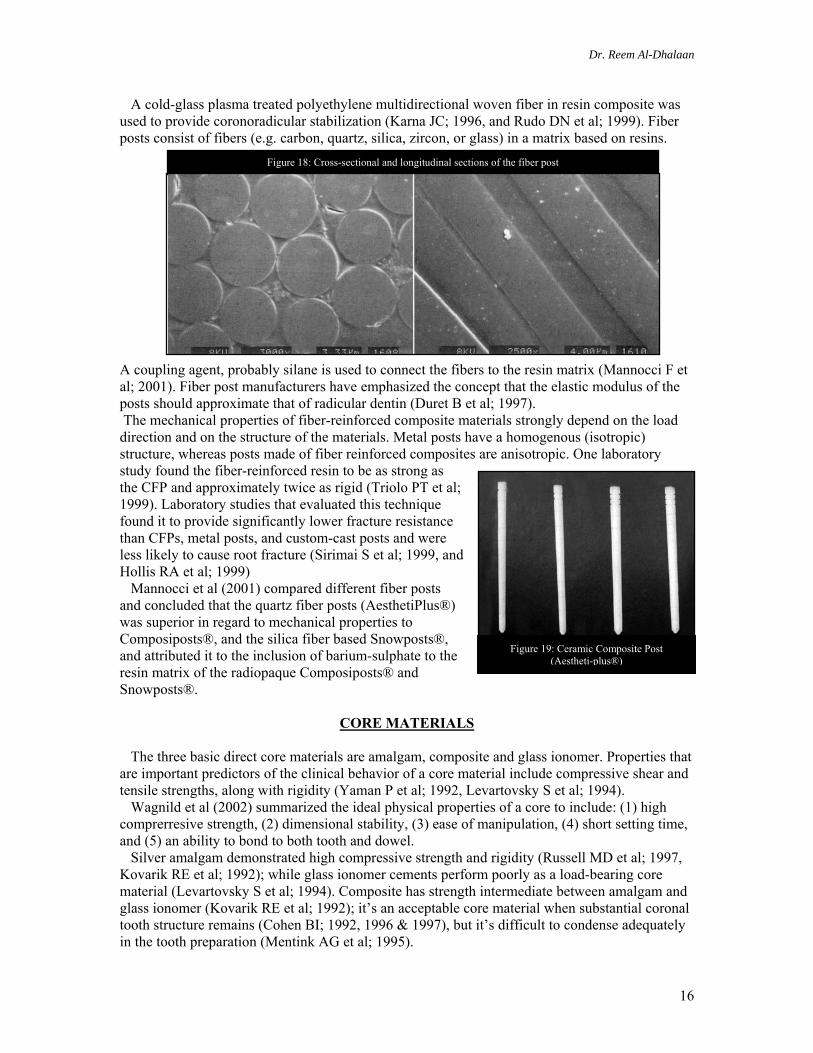
Figure 18: Cross-sectional and longitudinal sections of the fiber post
A cold-glass plasma treated polyethylene multidirectional woven fiber in resin composite was used to provide coronoradicular stabilization (Karna JC; 1996, and Rudo DN et al; 1999). Fiber posts consist of fibers (e.g. carbon, quartz, silica, zircon, or glass) in a matrix based on resins.
A coupling agent, probably silane is used to connect the fibers to the resin matrix (Mannocci F et al; 2001). Fiber post manufacturers have emphasized the concept that the elastic modulus of the posts should approximate that of radicular dentin (Duret B et al; 1997). The mechanical properties of fiber-reinforced composite materials strongly depend on the load direction and on the structure of the materials. Metal posts have a homogenous (isotropic) structure, whereas posts made of fiber reinforced composites are anisotropic. One laboratory study found the fiber-reinforced resin to be as strong as the CFP and approximately twice as rigid (Triolo PT et al; 1999). Laboratory studies that evaluated this technique found it to provide significantly lower fracture resistance than CFPs, metal posts, and custom-cast posts and were less likely to cause root fracture (Sirimai S et al; 1999, and Hollis RA et al; 1999) Mannocci et al (2001) compared different fiber posts and concluded that the quartz fiber posts (AesthetiPlus®) was superior in regard to mechanical properties to Composiposts®, and the silica fiber based Snowposts®, and attributed it to the inclusion of barium-sulphate to the resin matrix of the radiopaque Composiposts® and Snowposts®.
CORE MATERIALS The three basic direct core materials are amalgam, composite and glass ionomer. Properties that are important predictors of the clinical behavior of a core material include compressive shear and tensile strengths, along with rigidity (Yaman P et al; 1992, Levartovsky S et al; 1994). Wagnild et al (2002) summarized the ideal physical properties of a core to include: (1) high comprerresive strength, (2) dimensional stability, (3) ease of manipulation, (4) short setting time, and (5) an ability to bond to both tooth and dowel. Silver amalgam demonstrated high compressive strength and rigidity (Russell MD et al; 1997, Kovarik RE et al; 1992); while glass ionomer cements perform poorly as a load-bearing core material (Levartovsky S et al; 1994). Composite has strength intermediate between amalgam and glass ionomer (Kovarik RE et al; 1992); it’s an acceptable core material when substantial coronal tooth structure remains (Cohen BI; 1992, 1996 & 1997), but it’s difficult to condense adequately in the tooth preparation (Mentink AG et al; 1995).
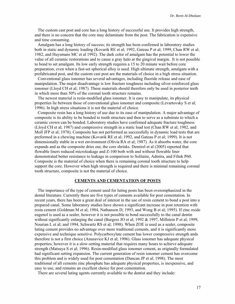
Figure 19: Ceramic Composite Post (Aestheti-plus®)
Dr. Reem Al-Dhalaan
17
The custom cast post and core has a long history of successful use. It provides high strength, and there is no concern that the core may delaminate from the post. The fabrication is expensive and time consuming. Amalgam has a long history of success; its strength has been confirmed in laboratory studies both in static and dynamic loading (Kovarik RE et al; 1992, Gateau P et al; 1999, Chan RW et al; 1982, and Huysmans MC et al 1992). The dark color of amalgam has the potential to lower the value of all ceramic restorations and to cause a gray halo at the gingival margin. It is not possible to bond to set amalgam. Its low early strength requires a 15 to 20 minute wait before core preparation, even when a fast-set spherical alloy is used. High ultimate strength, amalgam with a prefabricated post, and the custom cast post are the materials of choice in a high stress situation. Conventional glass ionomer has several advantages, including fluoride release and ease of manipulation. The major disadvantage is low fracture toughness including silver-reinforced glass ionomer (Lloyd CH et al; 1987). These materials should therefore only be used in posterior teeth in which more than 50% of the coronal tooth structure remains. The newest material is resin-modified glass ionomer. It is easy to manipulate, its physical properties lie between those of conventional glass ionomer and composite (Levartovsky S et al; 1996). In high stress situations it is not the material of choice. Composite resin has a long history of use due to its ease of manipulation. A major advantage of composite is its ability to be bonded to tooth structure and then to serve as a substrate to which a ceramic crown can be bonded. Laboratory studies have confirmed adequate fracture toughness (Lloyd CH et al; 1987) and compressive strength in a static load test (Chan RW et al; 1982, and Moll JFP et al; 1978). Composite has not performed as successfully in dynamic load tests that are preformed in a chewing machine (Kovarik RE et al; 1992, and Gateau P et al; 1999). It is not dimensionally stable in a wet environment (Olivia RA et al; 1987). As it absorbs water, the core expands and as the composite dries out, the core shrinks. Demirel et al (2005) reported that flowable liners reduced microleakage and Z-100 both with and without flowable liner demonstrated better resistance to leakage in comparison to Solitaire, Admira, and Filtek P60. Composite is the material of choice when there is remaining coronal tooth structure to help support the core. However when high strength is required and there is minimal remaining coronal tooth structure, composite is not the material of choice.
CEMENTS AND CEMENTATION OF POSTS The importance of the type of cement used for luting posts has been overemphasized in the dental literature. Currently there are five types of cements available for post cementation. In recent years, there has been a great deal of interest in the use of resin cement to bond a post into a prepared canal. Some laboratory studies have shown a significant increase in post retention with resin cement (Goldman M et al; 1984, Nathanson D; 1993, and Wong B et al; 1995). If zinc oxide eugenol is used as a sealer, however it is not possible to bond successfully to the canal dentin without significantly enlarging the canal (Burgess JO et al; 1992 & 1997, Millstein P et al; 1999, Nourian L et al; and 1994, Schwartz RS et al; 1998). When ZOE is used as a sealer, composite luting cement provides no advantage over more traditional cements, and it is significantly more expensive and technique sensitive. Polycarboxylate cement has lower compressive strength ands therefore is not a first choice (Anusavice KJ et al; 1996). Glass ionomer has adequate physical properties; however it is a slow-setting material that requires many hours to achieve adequate strength (Matsuya S et al; 1996). Resin-modified glass ionomer cement, as originally formulated had significant setting expansion. The current generation of resin ionomer cement has overcome this problem and is widely used for post cementation (Duncan JP et al; 1998). The most traditional of all cements zinc phosphate has adequate physical properties, is inexpensive, and easy to use, and remains an excellent choice for post cementation. There are several luting agents currently available to the dentist and they include:
Dr. Reem Al-Dhalaan
18
• Zinc phosphate cement 1. Extremely successful standard cement. 2. High solubility in the oral cavity. 3. Lack of true adhesion. 4. Adequate physical properties. 5. Ease of application. 6. Inexpensive.
• Polycarboxylate 1. Provides a weak chemical bond to dentin. 2. Undergoes plastic deformation after cyclic loading. 3. Less retentive in comparison to zinc phosphate; (low compressive strength).
• Glass ionomer cement 1. Provides a weak chemical bond to dentin. 2. Fluoride release and anticariogenic effect. 3. Requires several days or even several weeks to reach it maximum strength so it’s
unsuitable as a luting agent for posts (Matsuya S et al; 1996). • Resin-modified glass ionomer cement
1. Fluoride release and anticariogenic effect. 2. Insoluble. 3. Provide good retention of prosthesis. 4. Imbibes water and expands with time and there is anecdotal evidence that volumetric
expansion of the cement will fracture all ceramic crowns and should be avoided for cementation of posts because it will likely cause vertical root fracture (Miller MB; 1996).
• Adhesive resin cement 1. There is greater retention for posts cemented with adhesive resins (Duncan JP et al;
1998). 2. Mendosa and Eakle (1994) reported that some posts did not seat completely in post
channels because of premature setting of the resin. 3. Resin cements have also been suggested as a method to reinforce pulpless teeth. 4. Lowest solubility among all
cements. 5. Highest compressive strength.
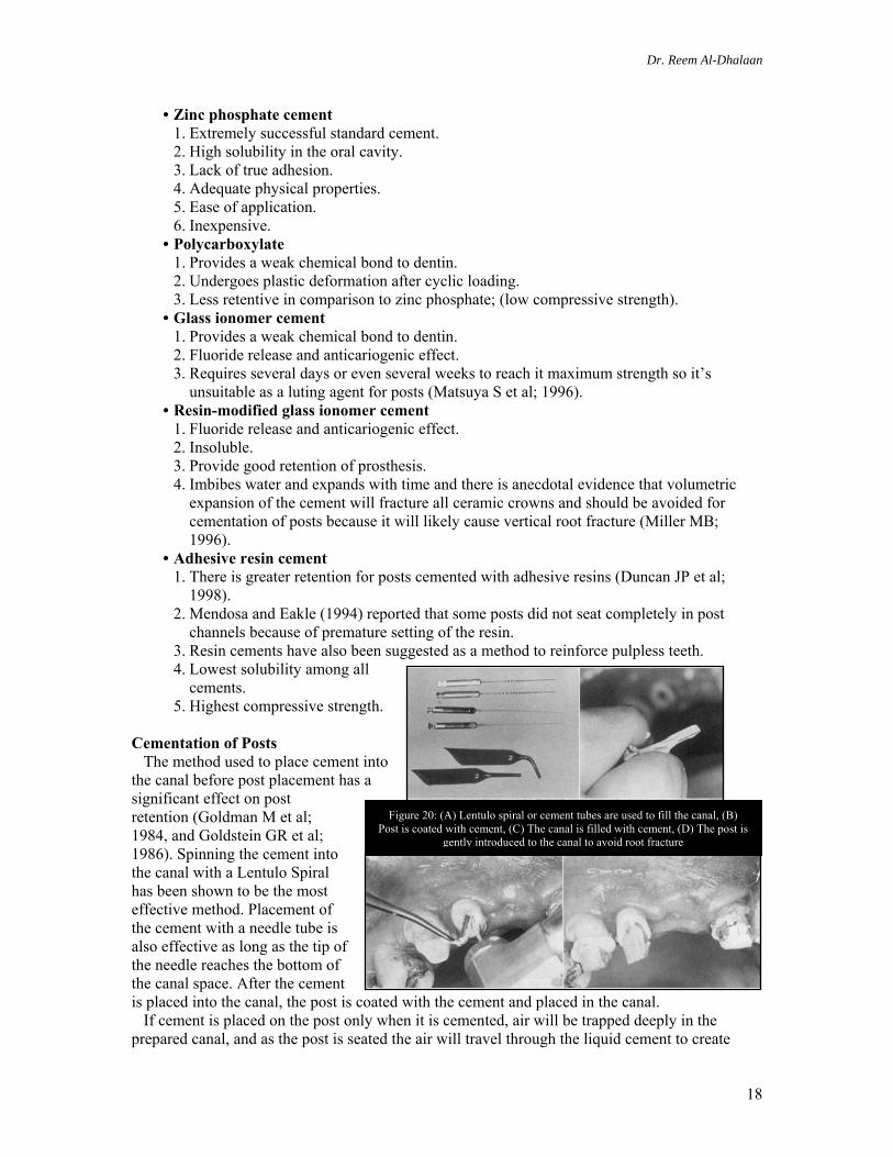
Cementation of Posts The method used to place cement into the canal before post placement has a significant effect on post retention (Goldman M et al; 1984, and Goldstein GR et al; 1986). Spinning the cement into the canal with a Lentulo Spiral has been shown to be the most effective method. Placement of the cement with a needle tube is also effective as long as the tip of the needle reaches the bottom of the canal space. After the cement is placed into the canal, the post is coated with the cement and placed in the canal. If cement is placed on the post only when it is cemented, air will be trapped deeply in the prepared canal, and as the post is seated the air will travel through the liquid cement to create
Figure 20: (A) Lentulo spiral or cement tubes are used to fill the canal, (B) Post is coated with cement, (C) The canal is filled with cement, (D) The post is
gently introduced to the canal to avoid root fracture
Dr. Reem Al-Dhalaan
19
voids that will compromise the physical properties of the cement film. Filling the canal with cement before seating the post will avoid air entrapment and ensure a dense uniform cement lute (Jacobi R et al; 1993). Tjan et al (1992) demonstrated that voids within adhesive resin cement were responsible for the expected low retentive values for post retention, due to oxygen inhibition of resin polymerization.
DEFINITIVE RESTORATIONS
Kanca et al (1988) reported a high success rate when restoring endodontically treated posterior teeth with intracoronal direct placement composite restorations; however the long term strengthening effect of the composite is questionable; since it is known that the strength of dentin bonding decreases over time (Hashimoto M et al; 2000) with load fatigue (Fissore B et al; 1991) and thermocycling (Eakle WS et al; 1986). In addition, it is postulated that a portion of sensory feedback mechanism is lost when the neurovascular pulpal tissue is removed during root canal therapy (Randow K et al; 1986). Clinically a person can inadvertently bite with significantly more force on an endodontically treated tooth than on a vital tooth. Sorenson (1984) and Hoag (1982) have both demonstrated that the essential element in the long term success of a posterior endodontically treated tooth is the placement of cuspal coverage restoration. Laboratory studies (Guzy GE et al; 1979, Trope M et al; 1985, and Lovdahl PE et al; 1977) indicate the fracture resistance of an endodontically treated anterior tooth with conservative access is approximately equal to that of a vital tooth. A simple bonded composite is the restoration of choice. When at least 50% of the coronal tooth structure, including enamel remains intact, enamel bonded porcelain veneer may be the restoration of choice. A laboratory study confirms the efficacy of the porcelain veneer in this circumstances (Magne P et al; 2000) and a post is not indicated (Baratieri LN et al; 2000). When the decision is made to place a crown for esthetic or functional reasons a post may be indicated. The decision to place a post in an anterior tooth is made based on the amount of remaining coronal tooth structure after the crown preparation and the functional requirements of the tooth. The maxillary lateral incisors and the mandibular incisors are smaller teeth; a post is commonly indicated before crown placement. In maxillary central incisors and canine teeth, however the decision should be made after crown preparation. Prior to any restoration, existing endodontically treated teeth should be assessed for the following: (1) good apical seal, (2) no sensitivity to percussion, and (3) no active inflammation.
RETENTION AND RESISTANCE
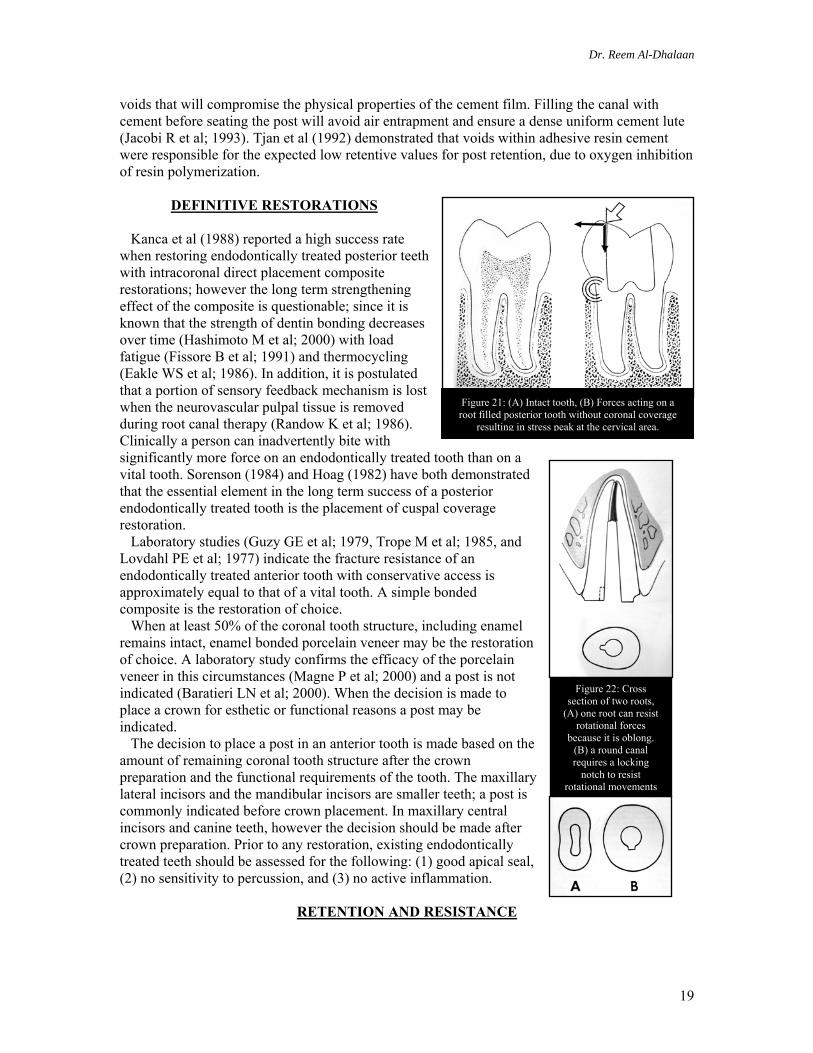
Figure 21: (A) Intact tooth, (B) Forces acting on a root filled posterior tooth without coronal coverage
resulting in stress peak at the cervical area.
Figure 22: Cross section of two roots,
(A) one root can resist rotational forces
because it is oblong. (B) a round canal requires a locking
notch to resist rotational movements
Dr. Reem Al-Dhalaan
20
The terms retention and resistance are commonly used interchangeably and incorrectly. Retention is defined as that which resists a tensile or pulling force; resistance is that which opposes any force other than a tensile force. There are three factors that provide retention for a post: (1) post configuration, (2) post length, and the (3) cement. The first factor is post configuration which can be active or passive, and tapered or parallel. A tapered passive post is ideal when the canal has not been overelarged and is of adequate length (Johnson JK et al; 1978, and Standlee JP et al; 1978). Adequate length in an anterior tooth is considered to be 8 mm of post space plus 4 to 5 mm of remaining gutta percha at the apex (Neagley RL et al; 1969). If more retention is required because of a decreased canal length or increased functional requirements (i.e., the tooth is a FPD/RPD abutment), a less tooth-conserving passive parallel post may be indicated. As available canal length for post placement decreases an active post is required. The third retention feature is the cement it provides important retention to the post and core; however no cement can compensate for a poorly designed post. The most important consideration in the long term success of post-retained restorations is the resistance form (Isador F et al; 1999, and Lambjerg-Hansen H et al; 1997). Resistance form is provided by three factors: (1) antirotation, (2) crown bevel, and (3) vertical remaining tooth structure. These three factors work together to provide resistance form so if one of the features is decreased long term success would require that one or both of the remaining two be increased. The first feature is antirotation, in molars it’s commonly achieved by the square shape of the tooth; however premolars and anterior teeth are commonly more round. When a round post is placed antirotation is essential to prevent shear forces from breaking the cement seal. Antirotation can be provided by vertical remaining tooth structure below the margin of the core. In the absence of significant vertical tooth structure, antirotation must be incorporated in the post and core with slots or pins. The second resistance feature is the crown bevel. With the advent of all-ceramic crowns and ceramometal crowns with porcelain labial margins, a crown bevel is seldom placed on an anterior tooth. For a bevel to provide significant resistance, it must be at least 1.5mm long (Libman W et al; 1995). Biological width requirements generally prevent the placement of this 1.5mm bevel especially in anterior teeth. The third and most important resistance feature is vertical remaining tooth structure above crown margin. Sorenson et al (1988) shown that only 2 mm of vertical remaining tooth structure doubles the resistance form. Regarding anterior teeth it is most important that this vertical remaining tooth structure be on the facial and lingual surfaces. Starr (1992) indicated that increased vertical tooth height can be gained by crown-lengthening; however in anterior teeth, single-tooth crown lengthening often results in unacceptable aesthetics. The treatment of choice, before placement of the restoration, is orthodontic extrusion. Several investigators (Mentick AG et al; 1998, Standlee JP et al; 1980, Thorsteinsson TS et al; 1992, Derand T; 1977, Leary JM et al; 1989, Peters MCRB et al; 1983, and Yaman SD et al; 1998) analyzed the influence of post design on stress distribution; the following conclusions have been drawn:
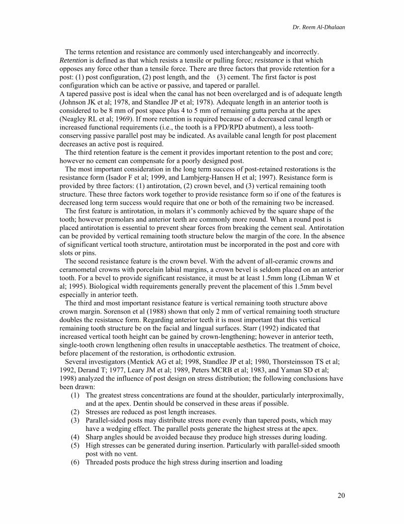
(1) The greatest stress concentrations are found at the shoulder, particularly interproximally, and at the apex. Dentin should be conserved in these areas if possible.
(2) Stresses are reduced as post length increases. (3) Parallel-sided posts may distribute stress more evenly than tapered posts, which may
have a wedging effect. The parallel posts generate the highest stress at the apex. (4) Sharp angles should be avoided because they produce high stresses during loading. (5) High stresses can be generated during insertion. Particularly with parallel-sided smooth
post with no vent. (6) Threaded posts produce the high stress during insertion and loading
Dr. Reem Al-Dhalaan
21
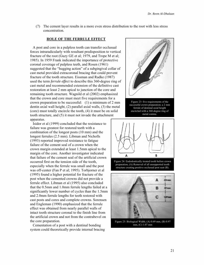
Figure 24: Endodontically treated tooth before crown preparation, (A) Removal of all unsupported tooth structure creating positive occlusual post seat (B).
Figure 23: five requirements of the successful crown preparation; a 2 mm
ferrule of dentinal axial height encircled with a 360-degree ring of
metal coping.
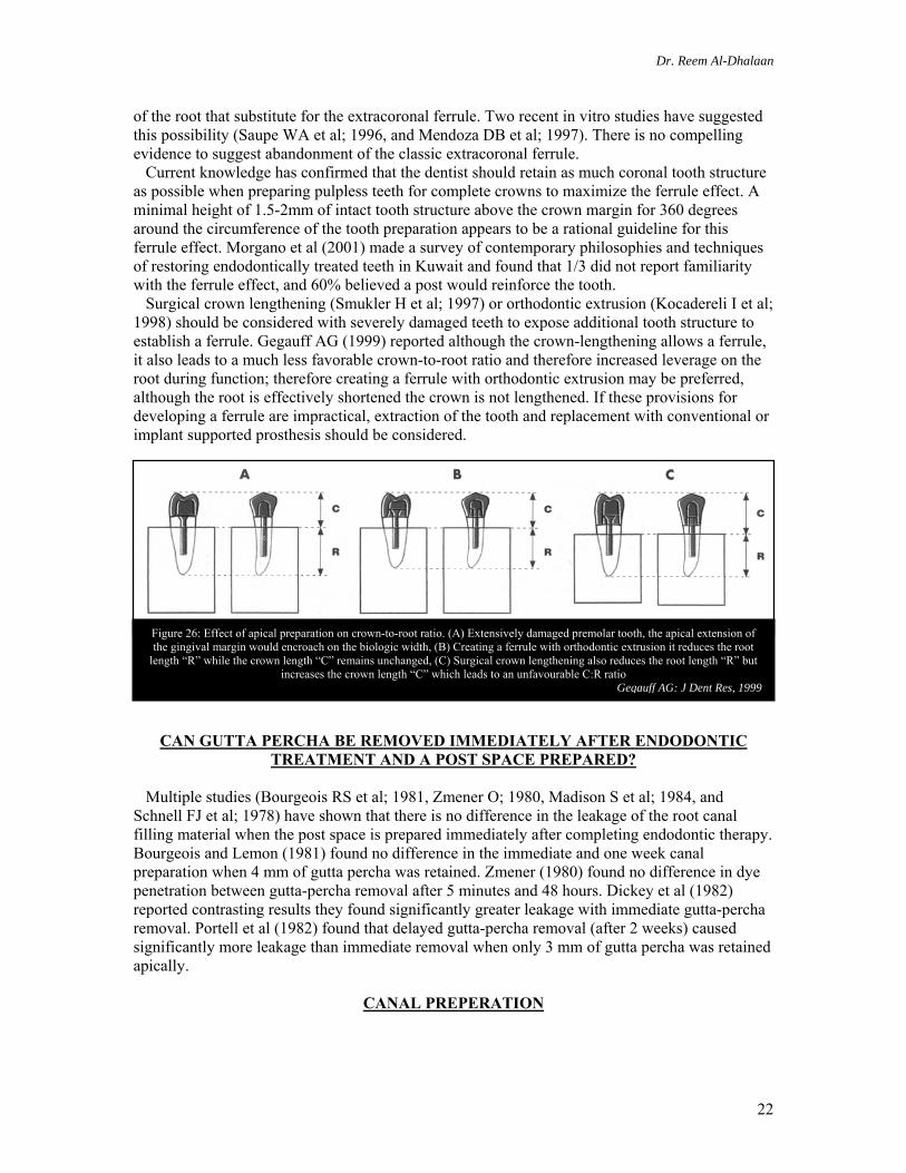
Figure 25: Biological Width; (A) 0.69 mm, (B) 0.97 mm, (C) 1.07 mm
(7) The cement layer results in a more even stress distribution to the root with less stress concentration.
ROLE OF THE FERRULE EFFECT
A post and core in a pulpless tooth can transfer occlusual forces intraradicularly with resultant predisposition to vertical fracture of the root (Guzy GE et al; 1979, and Trope M et al; 1985). In 1959 Frank indicated the importance of protective coronal coverage of pulpless teeth, and Rosen (1961) suggested that the “hugging action” of a subgingival collar of cast metal provided extracoronal bracing that could prevent fracture of the tooth structure. Eissman and Radke (1987) used the term ferrule effect to describe this 360-degree ring of cast metal and recommended extension of the definitive cast restoration at least 2 mm apical to junction of the core and remaining tooth structure. Wagnild et al (2002) emphasized that the crown and core must meet five requirements for a crown preparation to be successful: (1) a minimum of 2 mm dentin axial wall height, (2) parallel axial walls, (3) the metal (core) must totally encircle the tooth, (4) it must be on solid tooth structure, and (5) it must not invade the attachment apparatus. Isidor et al (1999) concluded that the resistance to failure was greatest for restored teeth with a combination of the longest posts (10 mm) and the longest ferrules (2.5 mm). Libman and Nicholls (1993) reported improved resistance to fatigue failure of the cement seal of a crown when the crown margin extended at least 1.5mm apical to the margin of the core. Another investigator indicated that failure of the cement seal of the artificial crown occurred first on the tension side of the tooth, especially when the ferrule was small and the post was off-center (Fan P et al; 1995). Torbjorner et al (1995) found a higher potential for fracture of the post when the cemented crowns did not provide a ferrule effect. Libman et al (1995) also concluded that the 0.5mm and 1.0mm ferrule lengths failed at a significantly lower number of cycles than the 1.5mm and 2.0mm ferrule lengths for teeth restored with cast posts and cores and complete crowns. Sorensen and Engleman (1990) emphasized that the ferrule effect was obtained from nearly parallel walls of intact tooth structure coronal to the finish line from the artificial crown and not from the contrabevel on the core preparation. Cementation of a post with a dentinal bonding system could theoretically provide internal bracing
Dr. Reem Al-Dhalaan
22
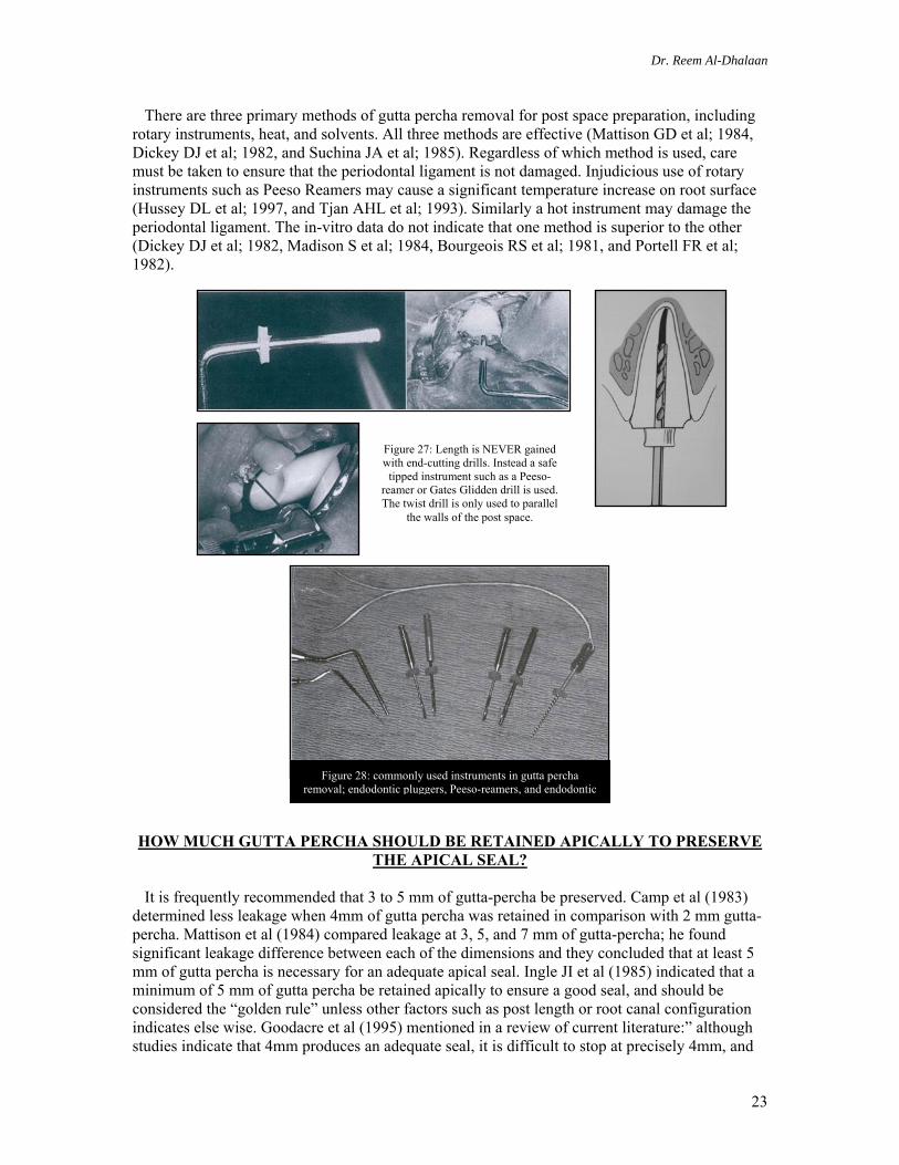
of the root that substitute for the extracoronal ferrule. Two recent in vitro studies have suggested this possibility (Saupe WA et al; 1996, and Mendoza DB et al; 1997). There is no compelling evidence to suggest abandonment of the classic extracoronal ferrule. Current knowledge has confirmed that the dentist should retain as much coronal tooth structure as possible when preparing pulpless teeth for complete crowns to maximize the ferrule effect. A minimal height of 1.5-2mm of intact tooth structure above the crown margin for 360 degrees around the circumference of the tooth preparation appears to be a rational guideline for this ferrule effect. Morgano et al (2001) made a survey of contemporary philosophies and techniques of restoring endodontically treated teeth in Kuwait and found that 1/3 did not report familiarity with the ferrule effect, and 60% believed a post would reinforce the tooth. Surgical crown lengthening (Smukler H et al; 1997) or orthodontic extrusion (Kocadereli I et al; 1998) should be considered with severely damaged teeth to expose additional tooth structure to establish a ferrule. Gegauff AG (1999) reported although the crown-lengthening allows a ferrule, it also leads to a much less favorable crown-to-root ratio and therefore increased leverage on the root during function; therefore creating a ferrule with orthodontic extrusion may be preferred, although the root is effectively shortened the crown is not lengthened. If these provisions for developing a ferrule are impractical, extraction of the tooth and replacement with conventional or implant supported prosthesis should be considered.
CAN GUTTA PERCHA BE REMOVED IMMEDIATELY AFTER ENDODONTIC
TREATMENT AND A POST SPACE PREPARED?
Multiple studies (Bourgeois RS et al; 1981, Zmener O; 1980, Madison S et al; 1984, and Schnell FJ et al; 1978) have shown that there is no difference in the leakage of the root canal filling material when the post space is prepared immediately after completing endodontic therapy. Bourgeois and Lemon (1981) found no difference in the immediate and one week canal preparation when 4 mm of gutta percha was retained. Zmener (1980) found no difference in dye penetration between gutta-percha removal after 5 minutes and 48 hours. Dickey et al (1982) reported contrasting results they found significantly greater leakage with immediate gutta-percha removal. Portell et al (1982) found that delayed gutta-percha removal (after 2 weeks) caused significantly more leakage than immediate removal when only 3 mm of gutta percha was retained apically.
CANAL PREPERATION
Figure 26: Effect of apical preparation on crown-to-root ratio. (A) Extensively damaged premolar tooth, the apical extension of the gingival margin would encroach on the biologic width, (B) Creating a ferrule with orthodontic extrusion it reduces the root
length “R” while the crown length “C” remains unchanged, (C) Surgical crown lengthening also reduces the root length “R” but increases the crown length “C” which leads to an unfavourable C:R ratio
Gegauff AG: J Dent Res, 1999
Dr. Reem Al-Dhalaan
23
There are three primary methods of gutta percha removal for post space preparation, including rotary instruments, heat, and solvents. All three methods are effective (Mattison GD et al; 1984, Dickey DJ et al; 1982, and Suchina JA et al; 1985). Regardless of which method is used, care must be taken to ensure that the periodontal ligament is not damaged. Injudicious use of rotary instruments such as Peeso Reamers may cause a significant temperature increase on root surface (Hussey DL et al; 1997, and Tjan AHL et al; 1993). Similarly a hot instrument may damage the periodontal ligament. The in-vitro data do not indicate that one method is superior to the other (Dickey DJ et al; 1982, Madison S et al; 1984, Bourgeois RS et al; 1981, and Portell FR et al; 1982). HOW MUCH GUTTA PERCHA SHOULD BE RETAINED APICALLY TO PRESERVE
THE APICAL SEAL?
It is frequently recommended that 3 to 5 mm of gutta-percha be preserved. Camp et al (1983) determined less leakage when 4mm of gutta percha was retained in comparison with 2 mm gutta-percha. Mattison et al (1984) compared leakage at 3, 5, and 7 mm of gutta-percha; he found significant leakage difference between each of the dimensions and they concluded that at least 5 mm of gutta percha is necessary for an adequate apical seal. Ingle JI et al (1985) indicated that a minimum of 5 mm of gutta percha be retained apically to ensure a good seal, and should be considered the “golden rule” unless other factors such as post length or root canal configuration indicates else wise. Goodacre et al (1995) mentioned in a review of current literature:” although studies indicate that 4mm produces an adequate seal, it is difficult to stop at precisely 4mm, and
Figure 27: Length is NEVER gained with end-cutting drills. Instead a safe
tipped instrument such as a Peeso-reamer or Gates Glidden drill is used. The twist drill is only used to parallel
the walls of the post space.
Figure 28: commonly used instruments in gutta percha removal; endodontic pluggers, Peeso-reamers, and endodontic
Dr. Reem Al-Dhalaan
24
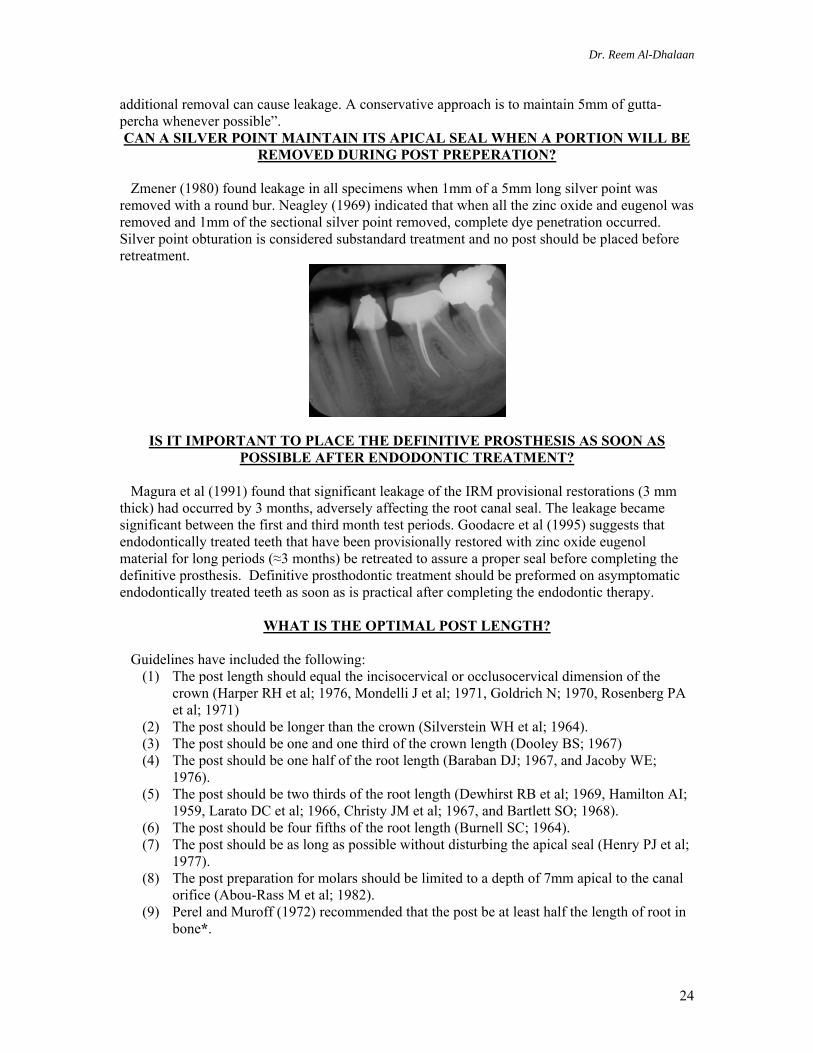
additional removal can cause leakage. A conservative approach is to maintain 5mm of gutta-percha whenever possible”. CAN A SILVER POINT MAINTAIN ITS APICAL SEAL WHEN A PORTION WILL BE
REMOVED DURING POST PREPERATION?
Zmener (1980) found leakage in all specimens when 1mm of a 5mm long silver point was removed with a round bur. Neagley (1969) indicated that when all the zinc oxide and eugenol was removed and 1mm of the sectional silver point removed, complete dye penetration occurred. Silver point obturation is considered substandard treatment and no post should be placed before retreatment.
IS IT IMPORTANT TO PLACE THE DEFINITIVE PROSTHESIS AS SOON AS POSSIBLE AFTER ENDODONTIC TREATMENT?
Magura et al (1991) found that significant leakage of the IRM provisional restorations (3 mm thick) had occurred by 3 months, adversely affecting the root canal seal. The leakage became significant between the first and third month test periods. Goodacre et al (1995) suggests that endodontically treated teeth that have been provisionally restored with zinc oxide eugenol material for long periods (≈3 months) be retreated to assure a proper seal before completing the definitive prosthesis. Definitive prosthodontic treatment should be preformed on asymptomatic endodontically treated teeth as soon as is practical after completing the endodontic therapy.
WHAT IS THE OPTIMAL POST LENGTH?
Guidelines have included the following: (1) The post length should equal the incisocervical or occlusocervical dimension of the
crown (Harper RH et al; 1976, Mondelli J et al; 1971, Goldrich N; 1970, Rosenberg PA et al; 1971)
(2) The post should be longer than the crown (Silverstein WH et al; 1964). (3) The post should be one and one third of the crown length (Dooley BS; 1967) (4) The post should be one half of the root length (Baraban DJ; 1967, and Jacoby WE;
1976). (5) The post should be two thirds of the root length (Dewhirst RB et al; 1969, Hamilton AI;
1959, Larato DC et al; 1966, Christy JM et al; 1967, and Bartlett SO; 1968). (6) The post should be four fifths of the root length (Burnell SC; 1964). (7) The post should be as long as possible without disturbing the apical seal (Henry PJ et al;
1977). (8) The post preparation for molars should be limited to a depth of 7mm apical to the canal
orifice (Abou-Rass M et al; 1982). (9) Perel and Muroff (1972) recommended that the post be at least half the length of root in
bone*.
Dr. Reem Al-Dhalaan
25
(10) To minimize stress in the dentin and in the post, the post should extend more than 4mm apical to the bone crest to decrease dentin stress*.
* Usually are used in periodontally involved teeth. From these varied suggestions it becomes apparent that length is an important aspect of clinical success. Sorenson and Mortinoff (1984) found in a retrospective evaluation of 1,273 endodontically treated teeth a 98.5% clinical success rate when the post was equal to or greater than the crown length. Johnson and Sakumura (1978) found that posts that were three fourths or more of the root length were 20% to 30% more retentive than posts that were one half of the root length or equal in length to the crown. Leary, Aquilino and Svare (1987) found that posts with a length at least three fourths of the length of the root offered the greatest rigidity and least root deflection. Zillich and Corcoran (1984) concluded when posts were two thirds of the root length, many of the average and short root length teeth had compromised apical seals. When the post was equal to the crown length an adequate seal was only possible on teeth with average or long root lengths. With short rooted teeth, even the shorter post guideline of being equal to the crown length produced a compromised apical seal. Shillingburg et al (1982) noted that making the post length equal the clinical crown would cause post to encroach on the 4 mm “safety zone” of gutta percha on some teeth. Wagnild et al (2002) indicated that alveolar bone height also influences dowel length. Occlusual forces generate the least risk to the remaining tooth structure and surrounding bone when a dowel extends apical to the alveolar crest. It was found that short stiff dowels transfer forces to the unsupported root extending above the alveolus and can cause root fracture. Assif et al (1993) concluded that when pre-restorative root canal therapy is indicated for elongated, periodontally involved teeth, it is extremely important to retain as much radicular dentin as possible. These teeth are subject to fracture because of increased leverage caused by greater crown length and the smaller diameter of root structure at the alveolar crest. Conventional post guidelines do not apply on the severely periodontally compromised teeth. The dowel is rarely as long as the clinical crown and often will not reach to the alveolar crest. Thus the apical end of the dowel should not be at the level of the alveolar crest; it should terminate above or below the alveolus by at least 4mm. The bony crest and dowel terminus are both stress concentrators and coincident placement increases fracture potential.
HOW IMPORTANT IS POST DIAMETER?
It has been recommended (Stern N et al; 1973 and Johnson JK et al; 1976) that post diameter be one third of the root diameter. Hunter et al (1989) and Krupp et al (1979) determined that post length is more important than the diameter in determining cervical stresses. Mattison (1982) found that the stress in the tooth generally increases as the post diameter increases. Trabert et al (1978) found that increasing post diameter decreases the tooth’s resistance to fracture. Deutsch et al (1985) found a six-fold increase in root fracture potential of threaded posts for each millimeter that the tooth diameter is decreased, and post diameter increased. Using finite element analysis, Peters et al (1983) found higher tooth stresses for the small diameter design (1 mm) compared with the larger diameter (1.5 – 2mm). Abou-Rass et al (1982) prepared post spaces using a variety of instrument diameters and recorded the incidence of perforations. He suggested safe instrument sizes and indicate that maximal post tip diameters
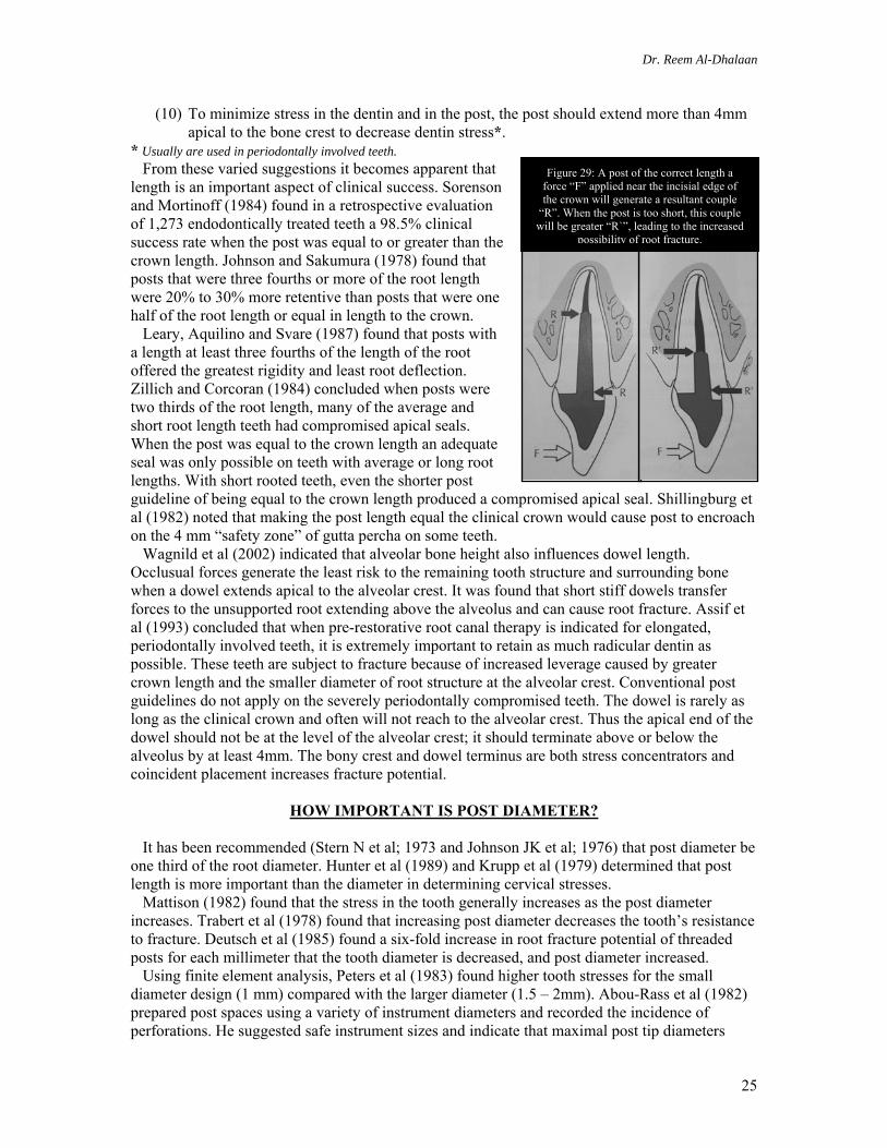
Figure 29: A post of the correct length a force “F” applied near the incisial edge of the crown will generate a resultant couple
“R”. When the post is too short, this couple will be greater “R`”, leading to the increased
possibility of root fracture.
Dr. Reem Al-Dhalaan
26
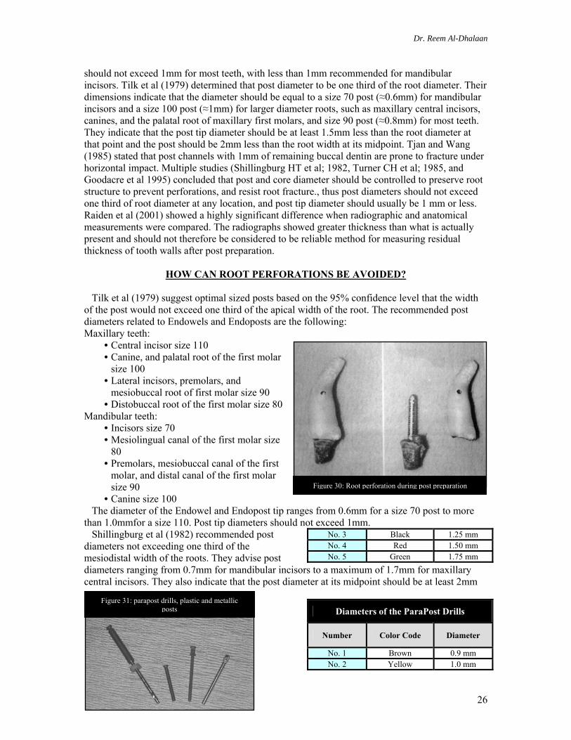
Figure 31: parapost drills, plastic and metallic posts
should not exceed 1mm for most teeth, with less than 1mm recommended for mandibular incisors. Tilk et al (1979) determined that post diameter to be one third of the root diameter. Their dimensions indicate that the diameter should be equal to a size 70 post (≈0.6mm) for mandibular incisors and a size 100 post (≈1mm) for larger diameter roots, such as maxillary central incisors, canines, and the palatal root of maxillary first molars, and size 90 post (≈0.8mm) for most teeth. They indicate that the post tip diameter should be at least 1.5mm less than the root diameter at that point and the post should be 2mm less than the root width at its midpoint. Tjan and Wang (1985) stated that post channels with 1mm of remaining buccal dentin are prone to fracture under horizontal impact. Multiple studies (Shillingburg HT et al; 1982, Turner CH et al; 1985, and Goodacre et al 1995) concluded that post and core diameter should be controlled to preserve root structure to prevent perforations, and resist root fracture., thus post diameters should not exceed one third of root diameter at any location, and post tip diameter should usually be 1 mm or less. Raiden et al (2001) showed a highly significant difference when radiographic and anatomical measurements were compared. The radiographs showed greater thickness than what is actually present and should not therefore be considered to be reliable method for measuring residual thickness of tooth walls after post preparation.
HOW CAN ROOT PERFORATIONS BE AVOIDED?
Tilk et al (1979) suggest optimal sized posts based on the 95% confidence level that the width of the post would not exceed one third of the apical width of the root. The recommended post diameters related to Endowels and Endoposts are the following: Maxillary teeth:
• Central incisor size 110 • Canine, and palatal root of the first molar
size 100 • Lateral incisors, premolars, and
mesiobuccal root of first molar size 90 • Distobuccal root of the first molar size 80
Mandibular teeth: • Incisors size 70 • Mesiolingual canal of the first molar size
80 • Premolars, mesiobuccal canal of the first
molar, and distal canal of the first molar size 90
• Canine size 100 The diameter of the Endowel and Endopost tip ranges from 0.6mm for a size 70 post to more than 1.0mmfor a size 110. Post tip diameters should not exceed 1mm. Shillingburg et al (1982) recommended post diameters not exceeding one third of the mesiodistal width of the roots. They advise post diameters ranging from 0.7mm for mandibular incisors to a maximum of 1.7mm for maxillary central incisors. They also indicate that the post diameter at its midpoint should be at least 2mm
Diameters of the ParaPost Drills
Number Color Code Diameter
No. 1 Brown 0.9 mm No. 2 Yellow 1.0 mm
No. 3 Black 1.25 mm No. 4 Red 1.50 mm No. 5 Green 1.75 mm
Figure 30: Root perforation during post preparation
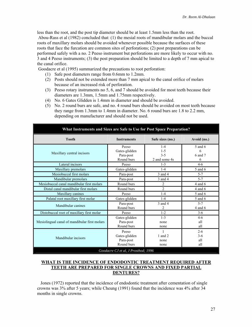
Dr. Reem Al-Dhalaan
27
less than the root, and the post tip diameter should be at least 1.5mm less than the root. Abou-Rass et al (1982) concluded that: (1) the mesial roots of mandibular molars and the buccal roots of maxillary molars should be avoided whenever possible because the surfaces of these roots that face the furcation are common sites of perforations; (2) post preparations can be performed safely with a no. 2 Peeso instrument but perforations are more likely to occur with no. 3 and 4 Peeso instruments; (3) the post preparation should be limited to a depth of 7 mm apical to the canal orifice. Goodacre et al (1995) summarized the precautions to root perforation:
(1) Safe post diameters range from 0.6mm to 1.2mm. (2) Posts should not be extended more than 7 mm apical to the canal orifice of molars
because of an increased risk of perforation. (3) Peeso rotary instruments no 5, 6, and 7 should be avoided for most teeth because their
diameters are 1.3mm, 1.5mm and 1.75mm respectively. (4) No. 6 Gates Glidden is 1.4mm in diameter and should be avoided. (5) No. 2 round burs are safe, and no. 4 round burs should be avoided on most teeth because
they range from 1.3mm to 1.4mm in diameter. No. 6 round burs are 1.8 to 2.2 mm, depending on manufacturer and should not be used.
What Instruments and Sizes are Safe to Use for Post Space Preparation?
Tooth Instruments Safe sizes (no.) Avoid (no.)
Maxillary central incisors
Peeso Gates-glidden
Para-post Round burs
1-4 1-5 3-5
2 and some 4s
5 and 6 6
6 and 7 6
Lateral incisors Peeso 1-3 4-6 Maxillary premolars Gates-glidden 1-4 5 and 6
Mesiobuccal first molars Para-post 3 and 4 5-7 Mandibular premolars Para-post 3 and 4 5-7
Mesiobuccal canal mandibular first molars Round burs 2 4 and 6 Distal canal mandibular first molars Round burs 2 4 and 6
Maxillary canines Peeso 1-4 5 and 6 Palatal root maxillary first molar Gates-glidden 1-4 5 and 6
Mandibular canines Para-post Round burs
3 and 4 2
5-7 4 and 6
Distobuccal root of maxillary first molar Peeso 1-2 3-6
Mesiolingual canal of mandibular first molars Gates-glidden
Para-post Round burs
1-3 none none
4-6 all all
Mandibular incisors
Peeso Gates-glidden
Para-post Round burs
1 1 and 2
none none
2-6 3-6 all all
Goodacre CJ et al, J Prosthod; 1996
WHAT IS THE INCIDENCE OF ENDODONTIC TREATMENT REQUIRED AFTER
TEETH ARE PREPARED FOR SINGLE CROWNS AND FIXED PARTIAL DENTURES?
Jones (1972) reported that the incidence of endodontic treatment after cementation of single crowns was 3% after 5 years; while Cheung (1991) found that the incidence was 4% after 34 months in single crowns.
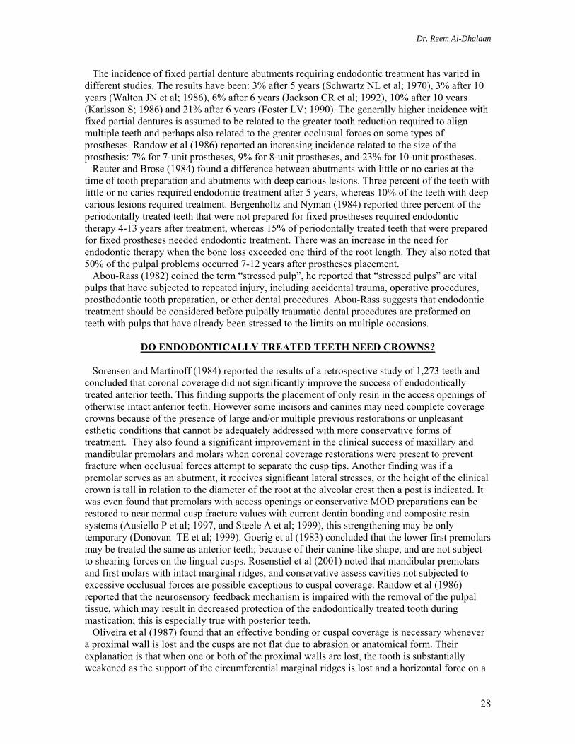
Dr. Reem Al-Dhalaan
28
The incidence of fixed partial denture abutments requiring endodontic treatment has varied in different studies. The results have been: 3% after 5 years (Schwartz NL et al; 1970), 3% after 10 years (Walton JN et al; 1986), 6% after 6 years (Jackson CR et al; 1992), 10% after 10 years (Karlsson S; 1986) and 21% after 6 years (Foster LV; 1990). The generally higher incidence with fixed partial dentures is assumed to be related to the greater tooth reduction required to align multiple teeth and perhaps also related to the greater occlusual forces on some types of prostheses. Randow et al (1986) reported an increasing incidence related to the size of the prosthesis: 7% for 7-unit prostheses, 9% for 8-unit prostheses, and 23% for 10-unit prostheses. Reuter and Brose (1984) found a difference between abutments with little or no caries at the time of tooth preparation and abutments with deep carious lesions. Three percent of the teeth with little or no caries required endodontic treatment after 5 years, whereas 10% of the teeth with deep carious lesions required treatment. Bergenholtz and Nyman (1984) reported three percent of the periodontally treated teeth that were not prepared for fixed prostheses required endodontic therapy 4-13 years after treatment, whereas 15% of periodontally treated teeth that were prepared for fixed prostheses needed endodontic treatment. There was an increase in the need for endodontic therapy when the bone loss exceeded one third of the root length. They also noted that 50% of the pulpal problems occurred 7-12 years after prostheses placement. Abou-Rass (1982) coined the term “stressed pulp”, he reported that “stressed pulps” are vital pulps that have subjected to repeated injury, including accidental trauma, operative procedures, prosthodontic tooth preparation, or other dental procedures. Abou-Rass suggests that endodontic treatment should be considered before pulpally traumatic dental procedures are preformed on teeth with pulps that have already been stressed to the limits on multiple occasions.
DO ENDODONTICALLY TREATED TEETH NEED CROWNS?
Sorensen and Martinoff (1984) reported the results of a retrospective study of 1,273 teeth and concluded that coronal coverage did not significantly improve the success of endodontically treated anterior teeth. This finding supports the placement of only resin in the access openings of otherwise intact anterior teeth. However some incisors and canines may need complete coverage crowns because of the presence of large and/or multiple previous restorations or unpleasant esthetic conditions that cannot be adequately addressed with more conservative forms of treatment. They also found a significant improvement in the clinical success of maxillary and mandibular premolars and molars when coronal coverage restorations were present to prevent fracture when occlusual forces attempt to separate the cusp tips. Another finding was if a premolar serves as an abutment, it receives significant lateral stresses, or the height of the clinical crown is tall in relation to the diameter of the root at the alveolar crest then a post is indicated. It was even found that premolars with access openings or conservative MOD preparations can be restored to near normal cusp fracture values with current dentin bonding and composite resin systems (Ausiello P et al; 1997, and Steele A et al; 1999), this strengthening may be only temporary (Donovan TE et al; 1999). Goerig et al (1983) concluded that the lower first premolars may be treated the same as anterior teeth; because of their canine-like shape, and are not subject to shearing forces on the lingual cusps. Rosenstiel et al (2001) noted that mandibular premolars and first molars with intact marginal ridges, and conservative assess cavities not subjected to excessive occlusual forces are possible exceptions to cuspal coverage. Randow et al (1986) reported that the neurosensory feedback mechanism is impaired with the removal of the pulpal tissue, which may result in decreased protection of the endodontically treated tooth during mastication; this is especially true with posterior teeth. Oliveira et al (1987) found that an effective bonding or cuspal coverage is necessary whenever a proximal wall is lost and the cusps are not flat due to abrasion or anatomical form. Their explanation is that when one or both of the proximal walls are lost, the tooth is substantially weakened as the support of the circumferential marginal ridges is lost and a horizontal force on a
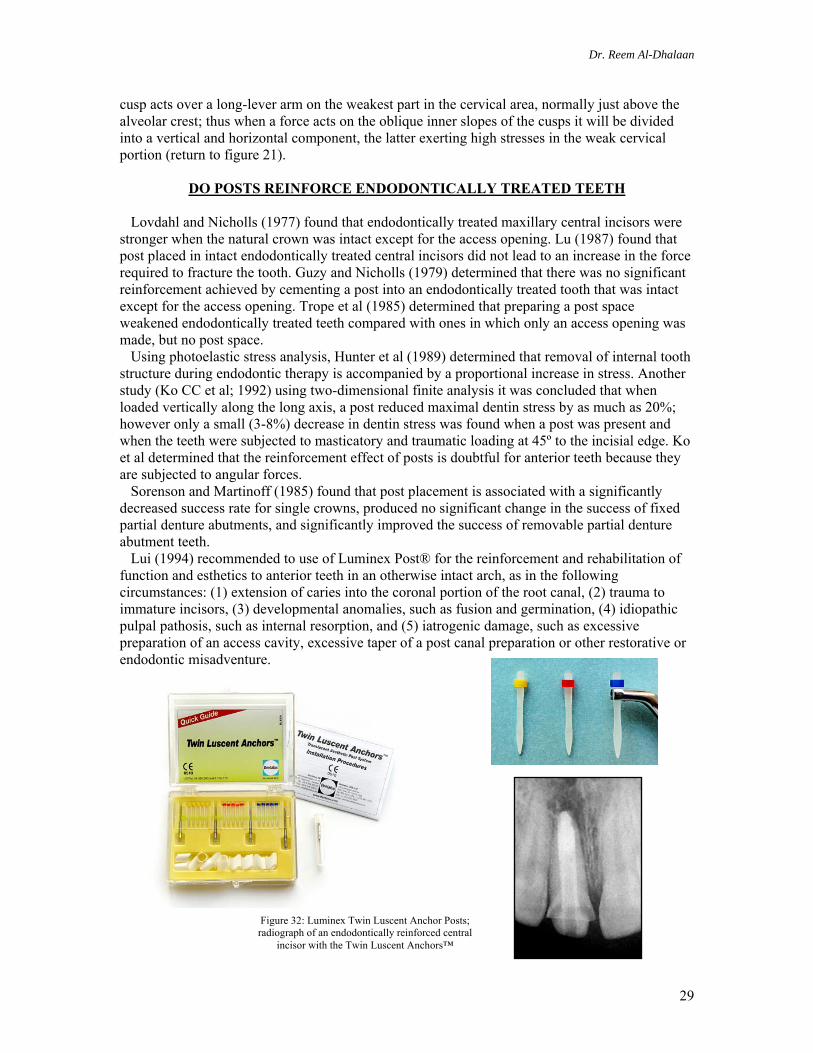
Dr. Reem Al-Dhalaan
29
cusp acts over a long-lever arm on the weakest part in the cervical area, normally just above the alveolar crest; thus when a force acts on the oblique inner slopes of the cusps it will be divided into a vertical and horizontal component, the latter exerting high stresses in the weak cervical portion (return to figure 21).
DO POSTS REINFORCE ENDODONTICALLY TREATED TEETH
Lovdahl and Nicholls (1977) found that endodontically treated maxillary central incisors were stronger when the natural crown was intact except for the access opening. Lu (1987) found that post placed in intact endodontically treated central incisors did not lead to an increase in the force required to fracture the tooth. Guzy and Nicholls (1979) determined that there was no significant reinforcement achieved by cementing a post into an endodontically treated tooth that was intact except for the access opening. Trope et al (1985) determined that preparing a post space weakened endodontically treated teeth compared with ones in which only an access opening was made, but no post space. Using photoelastic stress analysis, Hunter et al (1989) determined that removal of internal tooth structure during endodontic therapy is accompanied by a proportional increase in stress. Another study (Ko CC et al; 1992) using two-dimensional finite analysis it was concluded that when loaded vertically along the long axis, a post reduced maximal dentin stress by as much as 20%; however only a small (3-8%) decrease in dentin stress was found when a post was present and when the teeth were subjected to masticatory and traumatic loading at 45º to the incisial edge. Ko et al determined that the reinforcement effect of posts is doubtful for anterior teeth because they are subjected to angular forces. Sorenson and Martinoff (1985) found that post placement is associated with a significantly decreased success rate for single crowns, produced no significant change in the success of fixed partial denture abutments, and significantly improved the success of removable partial denture abutment teeth. Lui (1994) recommended to use of Luminex Post® for the reinforcement and rehabilitation of function and esthetics to anterior teeth in an otherwise intact arch, as in the following circumstances: (1) extension of caries into the coronal portion of the root canal, (2) trauma to immature incisors, (3) developmental anomalies, such as fusion and germination, (4) idiopathic pulpal pathosis, such as internal resorption, and (5) iatrogenic damage, such as excessive preparation of an access cavity, excessive taper of a post canal preparation or other restorative or endodontic misadventure.
Figure 32: Luminex Twin Luscent Anchor Posts; radiograph of an endodontically reinforced central
incisor with the Twin Luscent Anchors™
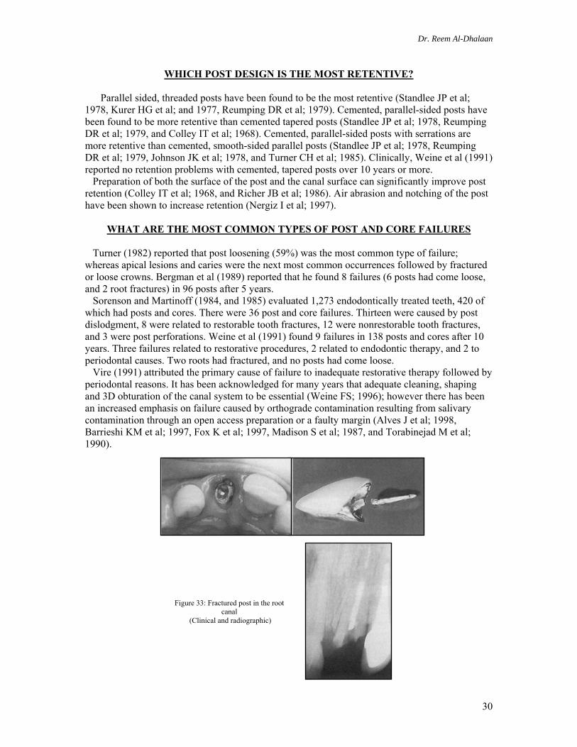
Dr. Reem Al-Dhalaan
30
WHICH POST DESIGN IS THE MOST RETENTIVE? Parallel sided, threaded posts have been found to be the most retentive (Standlee JP et al; 1978, Kurer HG et al; and 1977, Reumping DR et al; 1979). Cemented, parallel-sided posts have been found to be more retentive than cemented tapered posts (Standlee JP et al; 1978, Reumping DR et al; 1979, and Colley IT et al; 1968). Cemented, parallel-sided posts with serrations are more retentive than cemented, smooth-sided parallel posts (Standlee JP et al; 1978, Reumping DR et al; 1979, Johnson JK et al; 1978, and Turner CH et al; 1985). Clinically, Weine et al (1991) reported no retention problems with cemented, tapered posts over 10 years or more. Preparation of both the surface of the post and the canal surface can significantly improve post retention (Colley IT et al; 1968, and Richer JB et al; 1986). Air abrasion and notching of the post have been shown to increase retention (Nergiz I et al; 1997).
WHAT ARE THE MOST COMMON TYPES OF POST AND CORE FAILURES
Turner (1982) reported that post loosening (59%) was the most common type of failure; whereas apical lesions and caries were the next most common occurrences followed by fractured or loose crowns. Bergman et al (1989) reported that he found 8 failures (6 posts had come loose, and 2 root fractures) in 96 posts after 5 years. Sorenson and Martinoff (1984, and 1985) evaluated 1,273 endodontically treated teeth, 420 of which had posts and cores. There were 36 post and core failures. Thirteen were caused by post dislodgment, 8 were related to restorable tooth fractures, 12 were nonrestorable tooth fractures, and 3 were post perforations. Weine et al (1991) found 9 failures in 138 posts and cores after 10 years. Three failures related to restorative procedures, 2 related to endodontic therapy, and 2 to periodontal causes. Two roots had fractured, and no posts had come loose. Vire (1991) attributed the primary cause of failure to inadequate restorative therapy followed by periodontal reasons. It has been acknowledged for many years that adequate cleaning, shaping and 3D obturation of the canal system to be essential (Weine FS; 1996); however there has been an increased emphasis on failure caused by orthograde contamination resulting from salivary contamination through an open access preparation or a faulty margin (Alves J et al; 1998, Barrieshi KM et al; 1997, Fox K et al; 1997, Madison S et al; 1987, and Torabinejad M et al; 1990).
Figure 33: Fractured post in the root canal
(Clinical and radiographic)
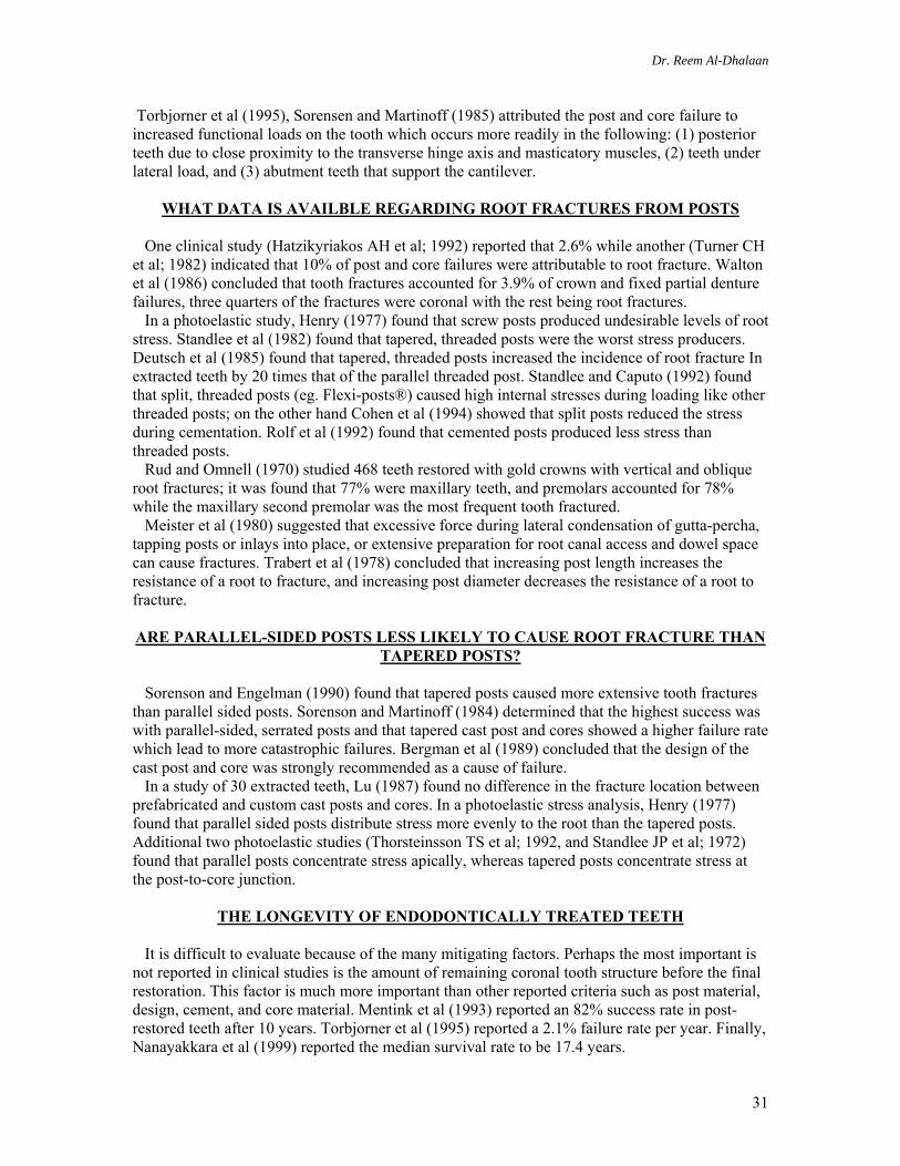
Dr. Reem Al-Dhalaan
31
Torbjorner et al (1995), Sorensen and Martinoff (1985) attributed the post and core failure to increased functional loads on the tooth which occurs more readily in the following: (1) posterior teeth due to close proximity to the transverse hinge axis and masticatory muscles, (2) teeth under lateral load, and (3) abutment teeth that support the cantilever.
WHAT DATA IS AVAILBLE REGARDING ROOT FRACTURES FROM POSTS
One clinical study (Hatzikyriakos AH et al; 1992) reported that 2.6% while another (Turner CH et al; 1982) indicated that 10% of post and core failures were attributable to root fracture. Walton et al (1986) concluded that tooth fractures accounted for 3.9% of crown and fixed partial denture failures, three quarters of the fractures were coronal with the rest being root fractures. In a photoelastic study, Henry (1977) found that screw posts produced undesirable levels of root stress. Standlee et al (1982) found that tapered, threaded posts were the worst stress producers. Deutsch et al (1985) found that tapered, threaded posts increased the incidence of root fracture In extracted teeth by 20 times that of the parallel threaded post. Standlee and Caputo (1992) found that split, threaded posts (eg. Flexi-posts®) caused high internal stresses during loading like other threaded posts; on the other hand Cohen et al (1994) showed that split posts reduced the stress during cementation. Rolf et al (1992) found that cemented posts produced less stress than threaded posts. Rud and Omnell (1970) studied 468 teeth restored with gold crowns with vertical and oblique root fractures; it was found that 77% were maxillary teeth, and premolars accounted for 78% while the maxillary second premolar was the most frequent tooth fractured. Meister et al (1980) suggested that excessive force during lateral condensation of gutta-percha, tapping posts or inlays into place, or extensive preparation for root canal access and dowel space can cause fractures. Trabert et al (1978) concluded that increasing post length increases the resistance of a root to fracture, and increasing post diameter decreases the resistance of a root to fracture. ARE PARALLEL-SIDED POSTS LESS LIKELY TO CAUSE ROOT FRACTURE THAN
TAPERED POSTS?
Sorenson and Engelman (1990) found that tapered posts caused more extensive tooth fractures than parallel sided posts. Sorenson and Martinoff (1984) determined that the highest success was with parallel-sided, serrated posts and that tapered cast post and cores showed a higher failure rate which lead to more catastrophic failures. Bergman et al (1989) concluded that the design of the cast post and core was strongly recommended as a cause of failure. In a study of 30 extracted teeth, Lu (1987) found no difference in the fracture location between prefabricated and custom cast posts and cores. In a photoelastic stress analysis, Henry (1977) found that parallel sided posts distribute stress more evenly to the root than the tapered posts. Additional two photoelastic studies (Thorsteinsson TS et al; 1992, and Standlee JP et al; 1972) found that parallel posts concentrate stress apically, whereas tapered posts concentrate stress at the post-to-core junction.
THE LONGEVITY OF ENDODONTICALLY TREATED TEETH
It is difficult to evaluate because of the many mitigating factors. Perhaps the most important is not reported in clinical studies is the amount of remaining coronal tooth structure before the final restoration. This factor is much more important than other reported criteria such as post material, design, cement, and core material. Mentink et al (1993) reported an 82% success rate in post-restored teeth after 10 years. Torbjorner et al (1995) reported a 2.1% failure rate per year. Finally, Nanayakkara et al (1999) reported the median survival rate to be 17.4 years.
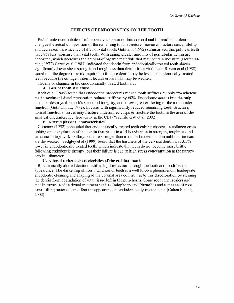
Dr. Reem Al-Dhalaan
32
EFFECTS OF ENDODONTICS ON THE TOOTH Endodontic manipulation further removes important intracoronal and intraradicular dentin, changes the actual composition of the remaining tooth structure, increases fracture susceptibility and decreased translucency of the nonvital tooth. Gutmann (1992) summarized that pulpless teeth have 9% less moisture than vital teeth. With aging, greater amounts of peritubular dentin are deposited, which decreases the amount of organic materials that may contain moisture (Helfer AR et al; 1972).Carter et al (1983) indicated that dentin from endodontically treated teeth shows significantly lower shear strength and toughness than dentin from vital teeth. Rivera et al (1988) stated that the degree of work required to fracture dentin may be less in endodontically treated teeth because the collagen intermolecular cross-links may be weaker. The major changes in the endodontically treated tooth are:
A. Loss of tooth structure Reeh et al (1989) found that endodontic procedures reduce tooth stiffness by only 5% whereas mesio-occlusual-distal preparation reduces stiffness by 60%. Endodontic access into the pulp chamber destroys the tooth’s structural integrity, and allows greater flexing of the tooth under function (Gutmann JL; 1992). In cases with significantly reduced remaining tooth structure, normal functional forces may fracture undermined cusps or fracture the tooth in the area of the smallest circumference, frequently at the CEJ (Wagnild GW et al; 2002).
B. Altered physical characteristics Gutmann (1992) concluded that endodontically treated teeth exhibit changes in collagen cross-linking and dehydration of the dentin that result in a 14% reduction in strength, toughness and structural integrity. Maxillary teeth are stronger than mandibular teeth, and mandibular incisors are the weakest. Sedgley et al (1999) found that the hardness of the cervical dentin was 3.5% lower in endodontically treated teeth; which indicate that teeth do not become more brittle following endodontic therapy; but their failure is due to high stress concentration at the narrow cervical diameter.
C. Altered esthetic characteristics of the residual tooth Biochemically altered dentin modifies light refraction through the tooth and modifies its appearance. The darkening of non-vital anterior teeth is a well known phenomenon. Inadequate endodontic cleaning and shaping of the coronal area contributes to this discoloration by staining the dentin from degradation of vital tissue left in the pulp horns. Some root canal sealers and medicaments used in dental treatment such as Iodophores and Phenolics and remnants of root canal filling material can affect the appearance of endodontically treated teeth (Cohen S et al; 2002).
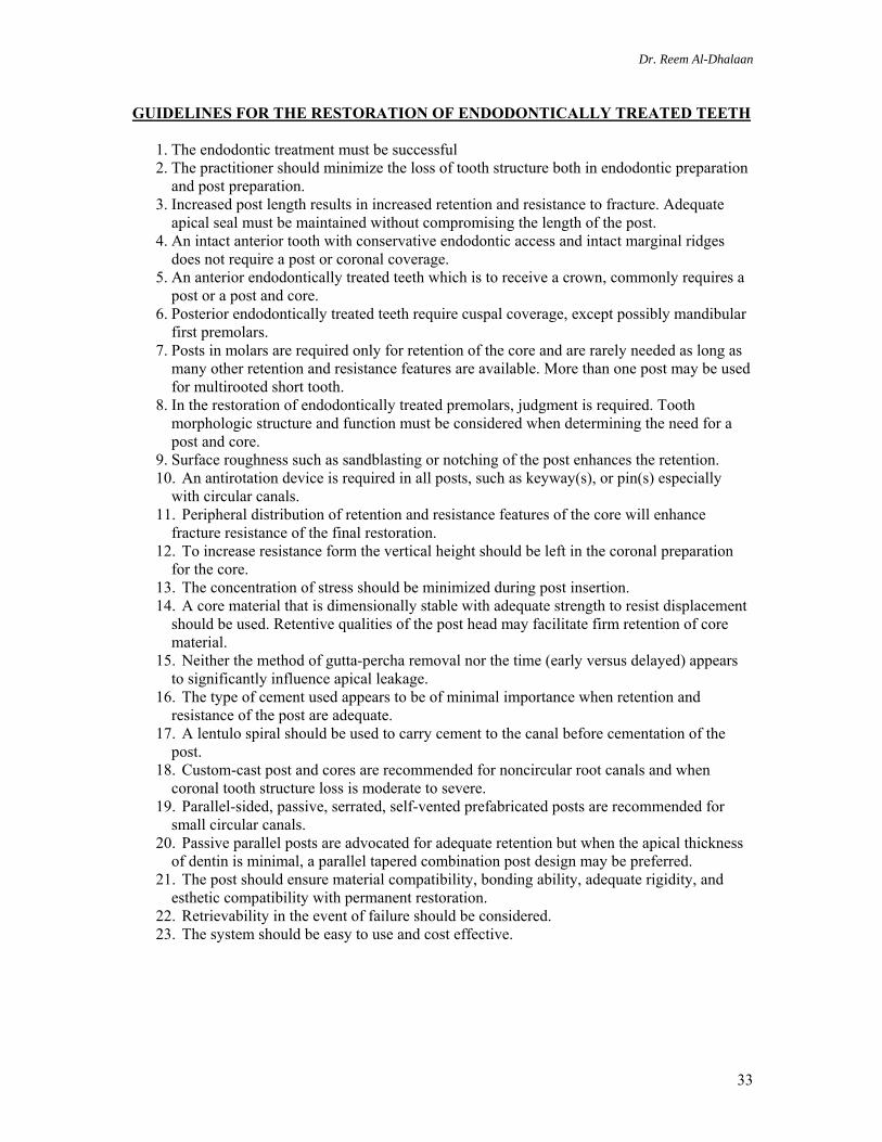
Dr. Reem Al-Dhalaan
33
GUIDELINES FOR THE RESTORATION OF ENDODONTICALLY TREATED TEETH
1. The endodontic treatment must be successful 2. The practitioner should minimize the loss of tooth structure both in endodontic preparation
and post preparation. 3. Increased post length results in increased retention and resistance to fracture. Adequate
apical seal must be maintained without compromising the length of the post. 4. An intact anterior tooth with conservative endodontic access and intact marginal ridges
does not require a post or coronal coverage. 5. An anterior endodontically treated teeth which is to receive a crown, commonly requires a
post or a post and core. 6. Posterior endodontically treated teeth require cuspal coverage, except possibly mandibular
first premolars. 7. Posts in molars are required only for retention of the core and are rarely needed as long as
many other retention and resistance features are available. More than one post may be used for multirooted short tooth.
8. In the restoration of endodontically treated premolars, judgment is required. Tooth morphologic structure and function must be considered when determining the need for a post and core.
9. Surface roughness such as sandblasting or notching of the post enhances the retention. 10. An antirotation device is required in all posts, such as keyway(s), or pin(s) especially
with circular canals. 11. Peripheral distribution of retention and resistance features of the core will enhance
fracture resistance of the final restoration. 12. To increase resistance form the vertical height should be left in the coronal preparation
for the core. 13. The concentration of stress should be minimized during post insertion. 14. A core material that is dimensionally stable with adequate strength to resist displacement
should be used. Retentive qualities of the post head may facilitate firm retention of core material.
15. Neither the method of gutta-percha removal nor the time (early versus delayed) appears to significantly influence apical leakage.
16. The type of cement used appears to be of minimal importance when retention and resistance of the post are adequate.
17. A lentulo spiral should be used to carry cement to the canal before cementation of the post.
18. Custom-cast post and cores are recommended for noncircular root canals and when coronal tooth structure loss is moderate to severe.
19. Parallel-sided, passive, serrated, self-vented prefabricated posts are recommended for small circular canals.
20. Passive parallel posts are advocated for adequate retention but when the apical thickness of dentin is minimal, a parallel tapered combination post design may be preferred.
21. The post should ensure material compatibility, bonding ability, adequate rigidity, and esthetic compatibility with permanent restoration.
22. Retrievability in the event of failure should be considered. 23. The system should be easy to use and cost effective.
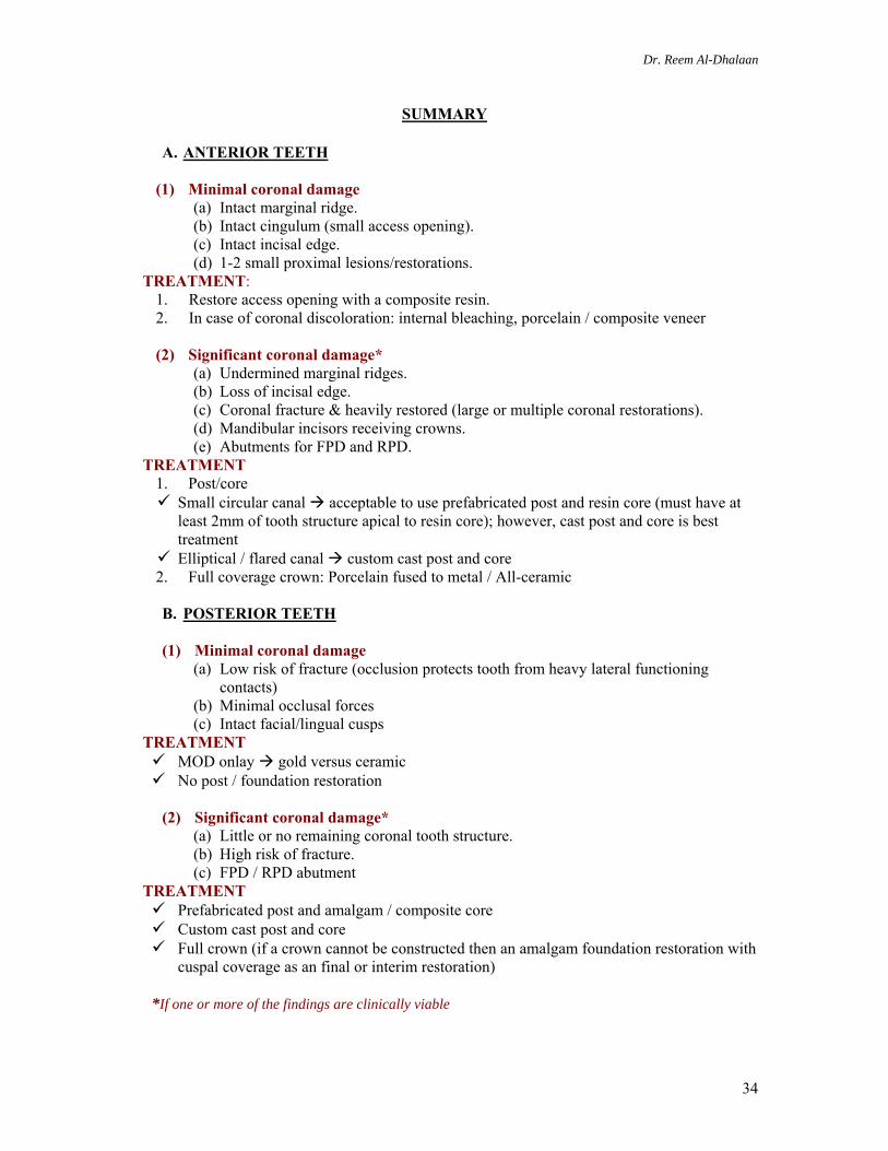
Dr. Reem Al-Dhalaan
34
SUMMARY
A. ANTERIOR TEETH
(1) Minimal coronal damage (a) Intact marginal ridge. (b) Intact cingulum (small access opening). (c) Intact incisal edge. (d) 1-2 small proximal lesions/restorations.
TREATMENT: 1. Restore access opening with a composite resin. 2. In case of coronal discoloration: internal bleaching, porcelain / composite veneer
(2) Significant coronal damage*
(a) Undermined marginal ridges. (b) Loss of incisal edge. (c) Coronal fracture & heavily restored (large or multiple coronal restorations). (d) Mandibular incisors receiving crowns. (e) Abutments for FPD and RPD.
TREATMENT 1. Post/core
Small circular canal acceptable to use prefabricated post and resin core (must have at least 2mm of tooth structure apical to resin core); however, cast post and core is best treatment
Elliptical / flared canal custom cast post and core 2. Full coverage crown: Porcelain fused to metal / All-ceramic
B. POSTERIOR TEETH (1) Minimal coronal damage
(a) Low risk of fracture (occlusion protects tooth from heavy lateral functioning contacts)
(b) Minimal occlusal forces (c) Intact facial/lingual cusps
TREATMENT MOD onlay gold versus ceramic No post / foundation restoration
(2) Significant coronal damage*
(a) Little or no remaining coronal tooth structure. (b) High risk of fracture. (c) FPD / RPD abutment
TREATMENT Prefabricated post and amalgam / composite core Custom cast post and core Full crown (if a crown cannot be constructed then an amalgam foundation restoration with
cuspal coverage as an final or interim restoration) *If one or more of the findings are clinically viable
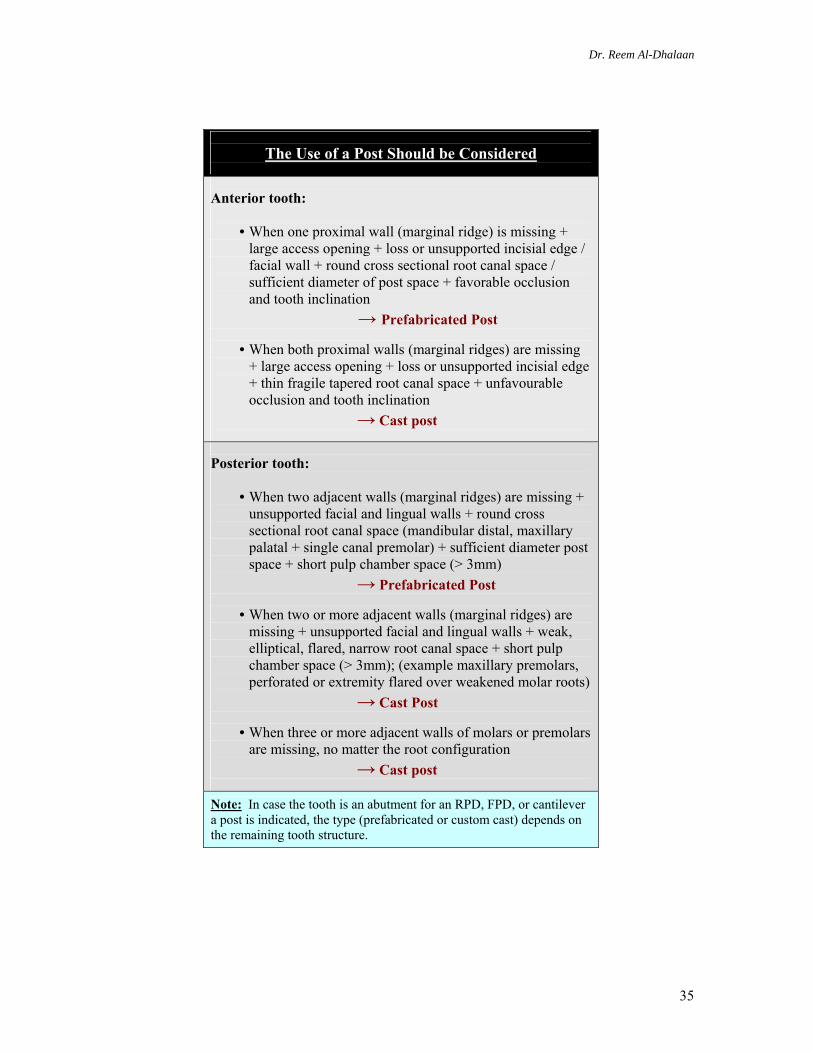
Dr. Reem Al-Dhalaan
35
The Use of a Post Should be Considered
Anterior tooth:
• When one proximal wall (marginal ridge) is missing + large access opening + loss or unsupported incisial edge / facial wall + round cross sectional root canal space / sufficient diameter of post space + favorable occlusion and tooth inclination
→ Prefabricated Post
• When both proximal walls (marginal ridges) are missing + large access opening + loss or unsupported incisial edge + thin fragile tapered root canal space + unfavourable occlusion and tooth inclination
→ Cast post
Posterior tooth:
• When two adjacent walls (marginal ridges) are missing + unsupported facial and lingual walls + round cross sectional root canal space (mandibular distal, maxillary palatal + single canal premolar) + sufficient diameter post space + short pulp chamber space (> 3mm)
→ Prefabricated Post • When two or more adjacent walls (marginal ridges) are
missing + unsupported facial and lingual walls + weak, elliptical, flared, narrow root canal space + short pulp chamber space (> 3mm); (example maxillary premolars, perforated or extremity flared over weakened molar roots)
→ Cast Post • When three or more adjacent walls of molars or premolars
are missing, no matter the root configuration → Cast post
Note: In case the tooth is an abutment for an RPD, FPD, or cantilever a post is indicated, the type (prefabricated or custom cast) depends on the remaining tooth structure.