Embed Size (px)
Citation preview

9/29/2020
1
Edward B. Singleton, MD
Thierry A.G.M. Huisman, MD, PD, FICIS, EDiNR, EDiPNR
Radiologist-in-Chief and
Edward B. Singleton Chair of Pediatric Radiology
Professor of Radiology, Pediatrics, Neurosurgery, Obstetrics and Gynecology
Imaging in cerebellar ataxia
Page 1
I have nothing to disclose
No relevant financial relations interfering with my presentation
No reference of any unlabeled or unapproved use of drugs
Disclosure
Page 2
For all I know about the cerebellum, I am very grateful to
Prof. Eugen Boltshauser and Prof. Andrea Poretti
Disclosure
Page 3
Ataxia: all cerebellar?
Ataxia: static, progressive, acute, intermittent, episodic?
Ataxia: Gait/truncal, limb, speech, eye movements?
Ataxia: Cognition?
Inherited/developmental versus acquired/disruptive?
Atrophy versus hypoplasia?
Introduction
Page 4
The word “ataxia,” comes from the Greek word “α-τάξις “ (a
taxis) meaning “lack of order”
www.ataxia.org:
• Ataxia is a rare neurological disease. It is progressive (?) –
affecting a person’s ability to walk, talk, and use fine motor skills
• These symptoms are caused by damage to the cerebellum (?)
Ataxia
www.ataxia.orgPage 5
Lack of coordination
Slurred speech
Trouble eating and swallowing
Impaired fine motor skills
Difficulty walking, gait abnormalities
Eye movement abnormalities
Tremors
Heart problems
Ataxia
www.ataxia.org
9/29/2020
2
Page 6
If you can pronounce, write and recognize
Rhombencephalosynapsis
you most likely do not have cerebellar ataxia!!!
Ataxia, fun fact
Page 7
Cerebellar
Sensory
Vestibular
Optic
Epileptic pseudo-ataxia
Functional/psychogenic
Ataxia ~ affected systems
Page 8
Comprehensive history of patient and family
Complete interview
• Acute
• Non-progressive
• Progressive
• Intermittent
• Episodic
Ataxia ~ How to diagnose?
• Past medical history
• Family history
• Social history
• Toxin exposure
• Medication
Page 9
Comprehensive history of patient and family
Complete interview
• Acute
• Subacute
• Non-progressive
• Chronic or progressive
• Intermittent
• Episodic
• Difficult to differentiate between static and slowly progressive
Ataxia ~ How to diagnose?
Page 10
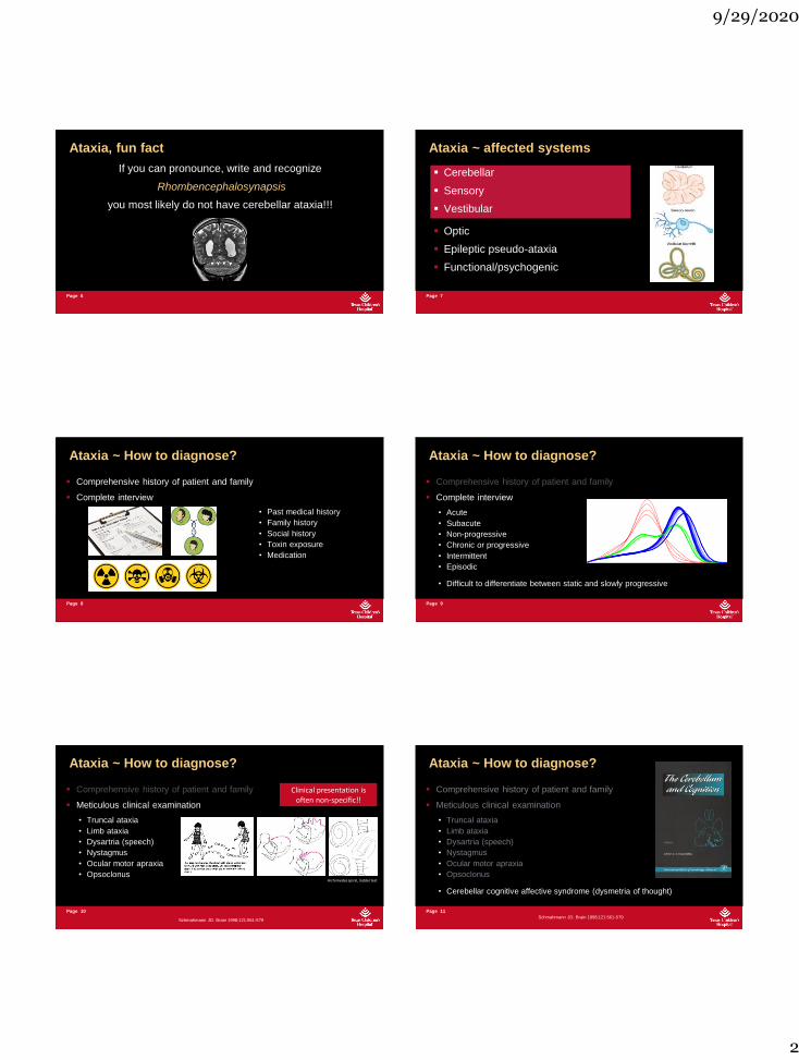
Comprehensive history of patient and family
Meticulous clinical examination
• Truncal ataxia
• Limb ataxia
• Dysartria (speech)
• Nystagmus
• Ocular motor apraxia
• Opsoclonus
Ataxia ~ How to diagnose?
Clinical presentation is often non-specific!!
Archimedes spiral, ladder test
Schmahmann JD. Brain 1998;121:561-579
Page 11
Comprehensive history of patient and family
Meticulous clinical examination
• Truncal ataxia
• Limb ataxia
• Dysartria (speech)
• Nystagmus
• Ocular motor apraxia
• Opsoclonus• c
• Cerebellar cognitive affective syndrome (dysmetria of thought)
Ataxia ~ How to diagnose?
Schmahmann JD. Brain 1998;121:561-579
9/29/2020
3
Page 12
Comprehensive history of patient and family
Meticulous clinical examination
Targeted additional investigations
• Laboratory (blood and urine)
• Molecular genetics
• Anatomical and functional imaging (MRI)
of the CNS (not limited to the cerebellum)
Ataxia ~ How to diagnose?
Clinical presentation is often non-specific!!
Page 13
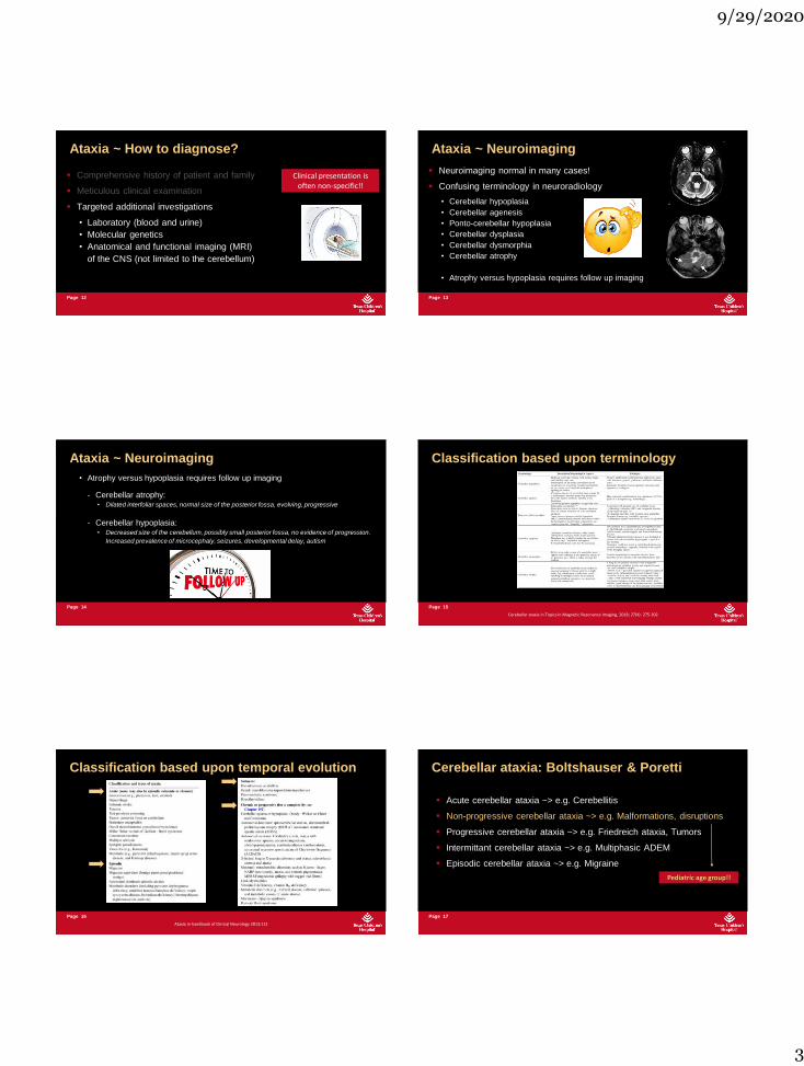
Neuroimaging normal in many cases!
Confusing terminology in neuroradiology
• Cerebellar hypoplasia
• Cerebellar agenesis
• Ponto-cerebellar hypoplasia
• Cerebellar dysplasia
• Cerebellar dysmorphia
• Cerebellar atrophy
• Atrophy versus hypoplasia requires follow up imaging
Ataxia ~ Neuroimaging
Page 14
• Atrophy versus hypoplasia requires follow up imaging
- Cerebellar atrophy: • Dilated interfoliar spaces, normal size of the posterior fossa, evolving, progressive
- Cerebellar hypoplasia: • Decreased size of the cerebellum, possibly small posterior fossa, no evidence of progression.
Increased prevalence of microcephaly, seizures, developmental delay, autism
Ataxia ~ Neuroimaging
Page 15
Classification based upon terminology
Cerebellar ataxia in Topics in Magnetic Resonance Imaging, 2018; 27(4): 275-302
Page 16
Classification based upon temporal evolution
Ataxia in handbook of Clinical Neurology 2013;112
Page 17
Acute cerebellar ataxia ~> e.g. Cerebellitis
Non-progressive cerebellar ataxia ~> e.g. Malformations, disruptions
Progressive cerebellar ataxia ~> e.g. Friedreich ataxia, Tumors
Intermittant cerebellar ataxia ~> e.g. Multiphasic ADEM
Episodic cerebellar ataxia ~> e.g. Migraine
Cerebellar ataxia: Boltshauser & Poretti
Pediatric age group!!
9/29/2020
4
Page 18
For the purpose of the image based classification/presentation
1. Acquired/disruptive cerebellar ataxia
2. Inherited (genetic) and developmental cerebellar ataxia
Cerebellar ataxia
Page 19
Congenital/perinatal onset
- Global hypoxic ischemic injury to the supratentorial brain~> Abnormal cerebellar development
- Intra-uterine cerebellar hemorrhage~> Prenatal cerebellar disruption
~> Unilateral cerebellar hypo-/dysplasia ~ complete aplasia
~> Not genetic in origin, low recurrence risk
1. Acquired cerebellar ataxia
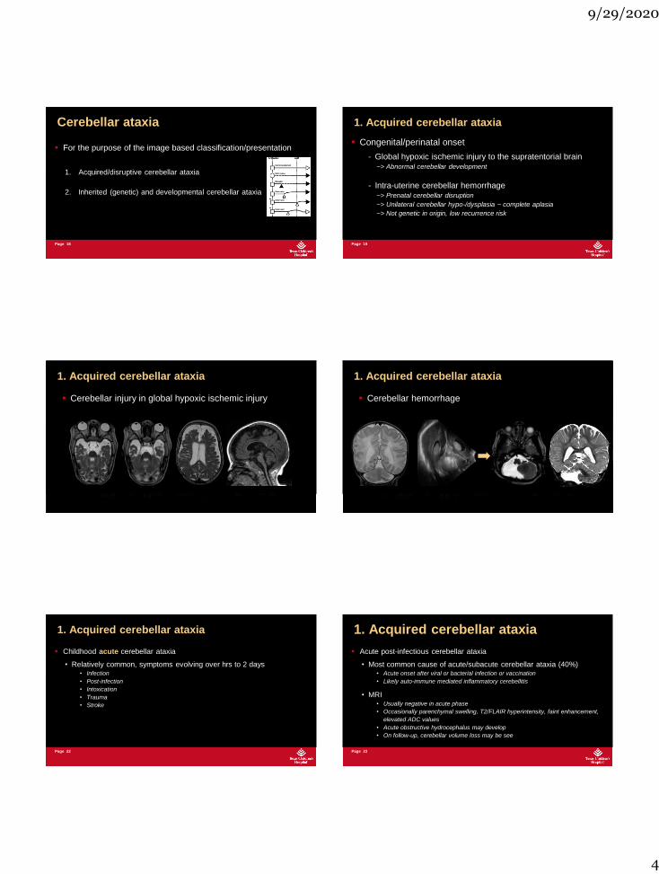
Cerebellar injury in global hypoxic ischemic injury
1. Acquired cerebellar ataxia
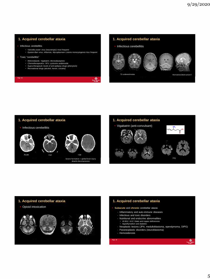
Cerebellar hemorrhage
1. Acquired cerebellar ataxia
Page 22
Childhood acute cerebellar ataxia
• Relatively common, symptoms evolving over hrs to 2 days• Infection
• Post-infection
• Intoxication
• Trauma
• Stroke
1. Acquired cerebellar ataxia
Page 23
Acute post-infectious cerebellar ataxia
• Most common cause of acute/subacute cerebellar ataxia (40%)• Acute onset after viral or bacterial infection or vaccination
• Likely auto-immune mediated inflammatory cerebellitis
• MRI • Usually negative in acute phase
• Occasionally parenchymal swelling, T2/FLAIR hyperintensity, faint enhancement,
elevated ADC values
• Acute obstructive hydrocephalus may develop
• On follow-up, cerebellar volume loss may be see
1. Acquired cerebellar ataxia
9/29/2020
5
Page 24
Infectious cerebellitis
• Varicella-zoster virus (neurotropic) most frequent
• Epstein-Barr virus, influenza, Mycoplasmam Listeria monocytogenes less frequent• d
Toxic “cerebellitis”
• Metronidazole, Vigabatrin, Benzodiazepines
• Chemotherapeutics: 5FU, cystosine arabinoside
• Supra-therapeutic levels of anti-epilepsy drugs (phenytoin)
• Recreational drugs (alcohol, heroin, cocaine)
1. Acquired cerebellar ataxia
Infectious cerebellitis
Herniation/obstruction!!T2 underestimates
1. Acquired cerebellar ataxia
Infectious cerebellitis
+1d +2dAcute
Severe herniation -> global brain injury despite decompression
1. Acquired cerebellar ataxia
Vigabatrin (anti-convulsant)
F/U
1. Acquired cerebellar ataxia
Opioid intoxication
1. Acquired cerebellar ataxia
Page 29
Subacute and chronic cerebellar ataxia
- Inflammatory and auto-immune diseases
- Infectious and toxic disorders
- Nutritional and endocrine abnormalities• Vit B12, Vit E, folate and copper deficiencies
• Hypothyroidism and diabetes
- Neoplastic lesions (JPA, medulloblastoma, ependymoma, DIPG)
- Paraneoplastic disorders (neuroblastoma)
- Hemosiderosis
1. Acquired cerebellar ataxia
9/29/2020
6
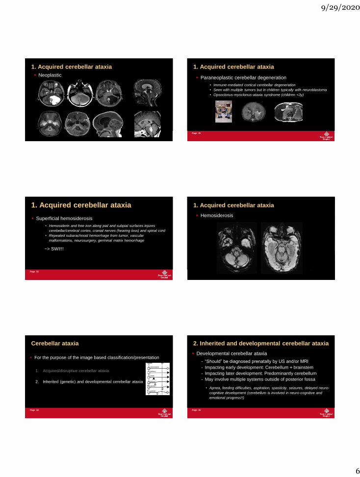
Neoplastic
1. Acquired cerebellar ataxia
Page 31
Paraneoplastic cerebellar degeneration
• Immune-mediated cortical cerebellar degeneration
• Seen with multiple tumors but in children typically with neuroblastoma
• Opsoclonus-myoclonus-ataxia syndrome (children <3y)
1. Acquired cerebellar ataxia
Page 32
Superficial hemosiderosis
• Hemosiderin and free iron along pial and subpial surfaces injures
cerebellar/cerebral cortex, cranial nerves (hearing loss) and spinal cord
• Repeated subarachnoid hemorrhage from tumor, vascular
malformations, neurosurgery, germinal matrix hemorrhage
~> SWI!!!
1. Acquired cerebellar ataxia
Hemosiderosis
1. Acquired cerebellar ataxia
Page 34
For the purpose of the image based classification/presentation
1. Acquired/disruptive cerebellar ataxia
2. Inherited (genetic) and developmental cerebellar ataxia
Cerebellar ataxia
Page 35
Developmental cerebellar ataxia
- “Should” be diagnosed prenatally by US and/or MRI
- Impacting early development: Cerebellum + brainstem
- Impacting later development: Predominantly cerebellum
- May involve multiple systems outside of posterior fossa
• Apnea, feeding difficulties, aspiration, spasticity, seizures, delayed neuro-
cognitive development (cerebellum is involved in neuro-cognitive and
emotional progress!!)
2. Inherited and developmental cerebellar ataxia

9/29/2020
7
Page 36
Genetic malformations
• Predominantly cerebellar• Dandy-Walker Malformation, Rhombencephalosynapsis
• Cerebellar dysplasia + cysts: Poretti-Boltshauser syndrome (LAMA1), Alpha-
dystroglycanopathies
• Cerebellum and Brainstem• Ponto-cerebellar hypoplasia (PCH)
• Dystroglycanopathies (Walker Warburg, Muscle-Eye Brain, Fukuyama muscular
dystrophy)
• Ciliopathies (Joubert Syndrome) and Tubulinopathies
2. Inherited and developmental cerebellar ataxia
Predominant cerebellar
• Cerebellar ataxia is NOT a feature of Chiari 1 or
posterior fossa arachnoid cysts!!!!
2. Inherited and developmental cerebellar ataxia
Predominant cerebellar
Dandy Walker Malformation (+)
2. Inherited and developmental cerebellar ataxia
Predominant cerebellar
Rhombencephalosynapsis
2. Inherited and developmental cerebellar ataxia
Ciliopathy: Joubert Syndrome
Cerebellar and brainstem
2. Inherited and developmental cerebellar ataxia
Tubulinopathy
Cerebellar and brainstem
2. Inherited and developmental cerebellar ataxia
9/29/2020
8
Page 42
Autosomal metabolic and recessive inherited ataxias
• Most common causes of genetic ataxias in children
• Typically slowly progressive with gait and limb ataxia + sensori-motor
polyneuropathy
• Additional organs may be affected (myocardium, pancreas)
• Friedreich ataxia
• Louis-Bar syndromeMost frequent etiologies
2. Inherited and developmental cerebellar ataxia
Page 43
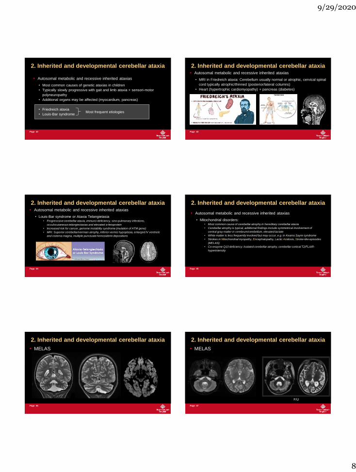
Autosomal metabolic and recessive inherited ataxias
• MRI in Friedreich ataxia: Cerebellum usually normal or atrophic, cervical spinal
cord typically atrophic/thinned (posterior/lateral columns)
• Heart (hypertrophic cardiomyopathy) + pancreas (diabetes)
2. Inherited and developmental cerebellar ataxia
Page 44
Autosomal metabolic and recessive inherited ataxias
• Louis-Bar syndrome or Ataxia Telangietasia• Progresssive cerebellar ataxia, immuno-deficiency, sino-pulmonary infections,
occulocutaneous telangiectasias and elevated α-fetoprotein
• Increased risk for cancer, genome instability syndrome (mutation of ATM gene)
• MRI: Superior cerebellar/vermian atrophy, inferior vermis hypoplasia, enlarged IV ventricle
and cisterna magna, multiple punctuate hemosiderin depositions
2. Inherited and developmental cerebellar ataxia
Page 45
Autosomal metabolic and recessive inherited ataxias
• Mitochondrial disorders: • Most common cause of cerebellar atrophy in hereditary cerebellar ataxia
• Cerebellar atrophy is typical, additional findings include symmetrical involvement of
central gray matter in cerebrum/cerebellum, elevated lactate
• White matter is less frequently involved but may occur, e.g. in Kearns Sayre syndrome
• Strokes in Mitochondrial myopathy, Encephalopathy, Lactic Acidosis, Stroke-like episodes
(MELAS)
• Co-enzyme Q10 deficiency: Isolated cerebellar atrophy, cerebellar cortical T2/FLAIR-
hyperintensity
2. Inherited and developmental cerebellar ataxia
Page 46
MELAS
2. Inherited and developmental cerebellar ataxia
Page 47
MELAS
F/U
2. Inherited and developmental cerebellar ataxia
9/29/2020
9
Page 48
Autosomal metabolic and recessive inherited ataxias
• Lysosomal storage disorders• GM2-gangliosidosis ~> severe, isolated cerebello-vermian atrophy
• Congenital disorders of glycosylation type 1a (CDG Type 1a)• Isolated cerebellar atrophy
• Marinesco Sjogren syndrome• Cerebellar ataxia with bilateral cataracts, developmental delay
• Autosomal recessive spastic ataxia of Charlevoix-Saguenay• Atrophy of anterior vermis and superior cerebellar peduncles and tigroid pons
• Sjogren-Larsson syndrome• Neurocutaneous syndrome with congenital ichthyosis, spastic di/tetraplegia
and mental retardation
2. Inherited and developmental cerebellar ataxia
Prodi E, et al. Eur J Radiol 2013;20:138-146
Page 49
T2 T1
Autosomal metabolic and recessive inherited ataxias
• Hypomyelinating leukodystrophies
- Genetic white matter disorder with permanent deficit in amount of myelin
- Neurological (cerebellar ataxia), developmental and systemic findings
- MRI: high T2-sigal intensity, mild T1-hypointensity• Vanishing white matter
• H-ABC
• PMD
• 4H leukodystrophy
• Salla disease
• Cockayne syndrome
2. Inherited and developmental cerebellar ataxia
2y 8mo
8y 2mo
T2wT1w
T1w T2w
Hypomyelinating leukodystrophy with atrophy of Cerebellum and Basal Ganglia : H-ABC
2. Inherited and developmental cerebellar ataxia
Page 51
Hypomyelination with Hypodontia and Hypogonadotropic Hypogonadism (4H)
MRI: Hypomyelination of WM, progressive cerebellar atrophy and T2-
hypointensity of thalami and/or pallidum
T2w T1w
2. Inherited and developmental cerebellar ataxia
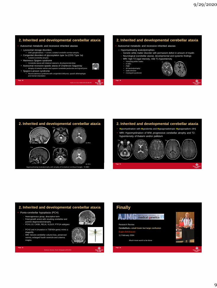
Page 52
Ponto-cerebellar hypoplasia (PCH)
- Heterogeneous group, descriptive term
- Fetal growth arrest with resulting cerebellar and
pontine degeneration/atrophy
- PCH1-10, CASK, RELN, VLDLR, PTF1A subtypes
- PCH2 and 4 (mutation in TSEN54 gene) mimic a
dragonfly
- MRI: Severe cerebellar volume loss, preserved
vermis, enlarged fourth ventricle and cisterna
magna
Bosemani, Huisman, Poretti. Radiographics2015;35(1)
2. Inherited and developmental cerebellar ataxia
Page 53
Finally
Research Review
Cerebellum—small brain but large confusion
Eugen Boltshauser
11 February 2004
Much more work to be done

9/29/2020
10