Embed Size (px)
Citation preview
Teratogens: a public health issue – a Brazilian overview
Thiago Mazzu-Nascimento1,2, Débora Gusmão Melo3, Giorgio Gianini Morbioli1,2,4, Emanuel Carrilho1,2,
Fernanda Sales Luiz Vianna 5,6, André Anjos da Silva5,7 and Lavinia Schuler-Faccini5,6
1Instituto de Química de São Carlos, Universidade de São Paulo, São Carlos, SP, Brazil.2 Instituto Nacional de Ciência e Tecnologia de Bioanalítica, Campinas, SP, Brazil.3Departamento de Medicina, Centro de Ciências Biológicas e da Saúde, Universidade Federal de São
Carlos, São Carlos, SP, Brazil.4School of Chemistry and Biochemistry, Georgia Institute of Technology, Atlanta, GA, USA.5Sistema Nacional de Informação sobre Agentes Teratogênicos (SIAT), Hospital de Clínicas de Porto
Alegre, Porto Alegre, RS, Brazil.6Programa de Pós-Graduação em Genética e Biologia Molecular, Departamento de Genética, Instituto de
Biociências, Universidade Federal do Rio Grande do Sul, Porto Alegre, RS, Brazil.7UNIVATES University, Lajeado, RS, Brazil.
Abstract
Congenital anomalies are already the second cause of infant mortality in Brazil, as in many other middle-incomecountries in Latin America. Birth defects are a result of both genetic and environmental factors, but a multifactorial eti-ology has been more frequently observed. Here, we address the environmental causes of birth defects – orteratogens – as a public health issue and present their mechanisms of action, categories and their respective mater-nal-fetal deleterious effects. We also present a survey from 2008 to 2013 of Brazilian cases involving congenitalanomalies (annual average of 20,205), fetal deaths (annual average of 1,530), infant hospitalizations (annual aver-age of 82,452), number of deaths of hospitalized infants (annual average of 2,175), and the average cost of hospital-izations (annual cost of $7,758). Moreover, we report on Brazilian cases of teratogenesis due to the recent Zika virusinfection, and to the use of misoprostol, thalidomide, alcohol and illicit drugs. Special attention has been given to theZika virus infection, now proven to be responsible for the microcephaly outbreak in Brazil, with 8,039 cases under in-vestigation (from October 2015 to June 2016). From those cases, 1,616 were confirmed and 324 deaths occurreddue to microcephaly complications or alterations on the central nervous system. Congenital anomalies impact lifequality and raise costs in specialized care, justifying the classification of teratogens as a public health issue.
Keywords: Birth defects, teratogens, Zika virus, pregnancy, public health.
Received: July 01, 2016; Accepted: October 07, 2016.
Background
Congenital anomalies are among the major causes of
infant death worldwide (Brazilian Ministry of Health,
MoH) (Egbe et al., 2015). In the United States, as in other
high-income countries, birth defects are the main cause of
infant mortality, being responsible for 1 out of 5 infant
deaths (Petrini et al., 2002). Within the main causes of in-
fant deaths in the United States, congenital malformations,
deformations and chromosomal abnormalities appear in
first place, representing 20.3% of total infant deaths in 2013
(Xu et al., 2016). These are also the reasons for 12% of all
pediatric hospitalizations (Egbe et al., 2015). In Latin Ame-
rica, congenital anomalies are between the second and fifth
cause of death in children under 1 year of age (Bronberg et
al., 2014). In Brazil, since 2000, they are the second main
cause of infant death (Passos-Bueno et al., 2014), and
among the three leading causes of infant hospitalizations,
responsible for 37% of pediatric hospital admissions. In ad-
dition, the hospital mortality rate in children with malfor-
mations accounted for 9.8% of total deaths, almost twice as
in children without malformations (Horovitz et al., 2005).
Congenital anomalies affect about 1 in 33 liveborns,
with an estimated 3.2 million newborns with birth defects
per year. Moreover, in 2013 a worldwide estimate showed
that nearly 276,000 newborns die before one month of life
every year, as a result of these congenital anomalies (World
Health Organization, 2015a). From those, 10 to 25% pres-
Send correspondence to Lavinia Schuler-Faccini. Departamentode Genética, Instituto de Biociências, Universidade Federal do RioGrande do Sul, Caixa postal 15053, Campus Agronomia,91501-970 Porto Alegre, RS, Brazil. E-mail:[email protected]
Review Article
Genetics and Molecular Biology, 40, 2, 387-397 (2017)
Copyright © 2017, Sociedade Brasileira de Genética. Printed in Brazil
DOI: http://dx.doi.org/10.1590/1678-4685-GMB-2016-0179
ent specific genetic inheritance, 10% are caused by envi-
ronmental factors, such as teratogens exposure, and 65 to
75% remain with unknown causes, what may include poly-
genic diseases, multifactorial factors (gene-environment
interactions), spontaneous development abnormalities and
synergistic interactions with teratogens (Brent, 2001).
Teratogens are environmental agents such as drugs,
viruses, lack of nutrients, and physical or chemical ele-
ments that upon contact with embryo/fetus can cause
congenital anomalies, generating permanent functional or
morphological changes in the newborn (Shepard, 1982).
Among the main reasons for pregnant women having
contact with teratogenic substances is the association of
preexisting public health problems (such as lack of medical
care, drug and alcohol consumption, lack of basic sanita-
tion) to other social issues like poverty and illiteracy (Reid-
path and Allotey, 2003; World Health Organization,
2015a). Therefore, the offspring of socially disadvantaged
women are more vulnerable to birth defects, causing an im-
pact on infant mortality and health expenses with special-
ized care, qualifying teratogens as a public health problem.
Teratogenic agents
Ancient Egypt wall paintings suggest the long time
existence of congenital abnormalities, such as clubfoot and
achondroplasia (Barrow, 1971). Children with congenital
malformations were labeled as “monsters” by the ancients
(Garcias and Schüler-Faccini, 2004). Other regions, such as
the Americas, Australia and Pacific Islands also have early
records of primitive sculptures revealing concerns with
congenital anomalies, which have even inspired some
mythological figures (Barrow, 1971). Only in the 1930s
malformations started to be scientifically studied in animal
models. Pioneer studies were performed in pig offspring,
addressing dietary vitamin A deficiency in pregnant sows,
which caused a complete absence of eye globe and ocular
tissue in the newborns (Hale, 1933).
Between the 1950s and 1960s thalidomide was exten-
sively used as sedative and to treat morning nausea during
pregnancy in Europe, Australia, Canada, Japan and Brazil.
Thalidomide use during pregnancy caused limb reduction
defects in thousands of newborns, leading to its ban in most
countries since 1961 (Kim and Scialli, 2011). The thalido-
mide tragedy caused an increasing interest about drug ex-
posure during pregnancy and the mechanism of action of
teratogenic agents on embryo-fetal abnormality develop-
ment.
Humans are exposed to millions of potential deleteri-
ous substances and hazardous conditions daily. However,
only a small part of these substances have been tested in an-
imals and even fewer were confirmed as a teratogenic for
humans, as teratogenicity studies cannot be conducted in
humans due to ethical reasons (Kalter, 2003).
Principles of Teratology
Teratology studies establish relationships between
environmental agents and anatomical and physiological
changes in the fetus (Finnell, 1999; Kalter, 2003). We pres-
ent below the six basic principles that determine terato-
genic effects (Wilson, 1977; Finnell, 1999).
Maternal-fetal genotype
A teratogenic effect depends on maternal-fetal geno-
type and on how embryos interact with their surroundings.
Due to these conditions, newborns exposed to a specific
dose of the same teratogenic can show different pheno-
types. Teratogens are capable of interacting with some
genes, modifying morpho-functional patterns and resulting
in either a major susceptibility or resistance to harmful sub-
stances. Some biochemical pathways can also respond in
distinct ways to different agents, affecting even more mal-
formation patterns (Cassina et al., 2012).
Mechanisms of action
The most common mechanisms of action of terato-
gens are hyperacetylation, cholesterol imbalance, alteration
of folate metabolism and folate antagonism, retinoic acid
imbalance, endocrine disruption, vascular disruption and
oxidative stress (Giavini and Menegola, 2012).
Hyperacetylation
Hyperacetylation may occur due to the inhibition of
histone deacetylase enzyme (HDAC), and the acetylation
status of the histone affects the modulation of chromatin
structure and gene expression, interfering with the embry-
onic development. HDAC inhibitors have been used as
anticonvulsant drugs (valproic acid) and for cancer treat-
ment, once they prevent tumorigenesis. Among antican-
cerdrugs are trichostatin A (TSA), apicidin and sodium
butyrate. These drugs are able to induce hyperacetylation in
animal embryos, leading to congenital malformations, such
as neural tube and axial skeletal defects (Menegola et al.,
2005).
Cholesterol imbalance
A high amount of cholesterol is required for fetal de-
velopment. This biomolecule is supplied by the mother dur-
ing early pregnancy and transported to the fetus across the
placenta, while during late pregnancy, cholesterol biosyn-
thesis will depend on the fetus’ own production
(Waterham, 2006). Drugs used for the treatment of hyper-
cholesterolemia, such as statins, act by blocking HMG-
CoA reductase, the enzyme involved with cholesterol
biosynthesis. HMG-CoA reductase is converted to meva-
lonate (Charlton-Menys and Durrington, 2008), interrupt-
ing the synthesis of cholesterol, and thus can cause adverse
effects on the developing fetus (Edison and Muenke 2004).
388 Mazzu-Nascimento et al.
Alteration in folate metabolism and folate antagonism
Folate or water-soluble vitamin B acts as a co-enzyme
in biochemical reactions, as a receiver or donor of one-
carbon units, and it is involved in purine and pyrimidine
synthesis and in DNA methylation. Increased cell growth
and tissue proliferation during embryogenesis demands an
increase in DNA synthesis, for which the presence of folate
is essential. Some drugs can compete with dihydrofolate
reductase and block the conversion of folate to tetrahydro-
folate. Among these drugs are methotrexate, sulfasalazine,
triamterene and trimethoprim. Other drugs, such as
anti-epileptic drugs, can interfere in folate absorption or in-
fluence folate degradation (including valproic acid and
carbamazepine phenytoin). The most common birth defects
involving these drugs are neural tube defects, orofacial
clefts and limb defects (van Gelder et al., 2010).
Retinoic acid imbalance
An imbalance between synthesis and degradation of
retinoic acid can lead to an excess or deficiency of this acid,
resulting in deleterious effects on cells and embryos, once
this Vitamin A precursor is closely related to vertebrate
morphogenesis. However, retinoic acid is also a signaling
molecule in neural crest cells, which will originate various
cell types and structures, such as intramembranous bone,
cartilage, peripheral nerves and Schwann cells, and mus-
cles, amongst others. Drugs such as isotretinoin can con-
tribute to retinoic acid imbalance and lead to craniofacial
and axial skeleton malformations (Giavini and Menegola,
2012).
Endocrine disruptors
Endocrine disruptors may interfere with the release of
hormones and in reactions mediated by hormone receptors
(van Gelder et al., 2010). Diethylstilbestrol, oral contracep-
tives, fertility treatment drugs and other endocrine disrup-
tive chemicals, which can include bisphenol A and
phthalates, act as endocrine disruptors (van Gelder et al.,
2010). These teratogenic substances can cross the placenta
and lead to fetal genital malformations (Raman-Wilms et
al., 1995)
Vascular disruption
Changes in the development of veins, arteries and
capillaries will disturb blood perfusion in fetal tissues.
These maternal-fetal blood disturbances can include hyper-
perfusion, hypoperfusion, hypoxia and obstruction, and are
caused by anatomical problems, maternal chronic diseases
or exposure to teratogenic agents during pregnancy, such as
misoprostol, phenytoin, cocaine, ergotamine, and some
vasodilator and vasoconstrictor drugs. Structural birth de-
fects are the most commonly reported, particularly limb de-
fects (Holmes, 2002).
Oxidative stress
Oxidative damage to cellular macromolecules such as
lipids, proteins, DNA and RNA is caused by reactive oxy-
gen species (ROS), which provide oxidation-reduction re-
actions (Wells et al., 1997). Exogenous ROS sources
include ultraviolet light (UV), UVA and UVB radiation,
ionizing radiation, and chemical agents, while endogenous
sources are related to cellular metabolism or oxidase en-
zymes (Hansen, 2006; van Gelder et al., 2010; Giavini and
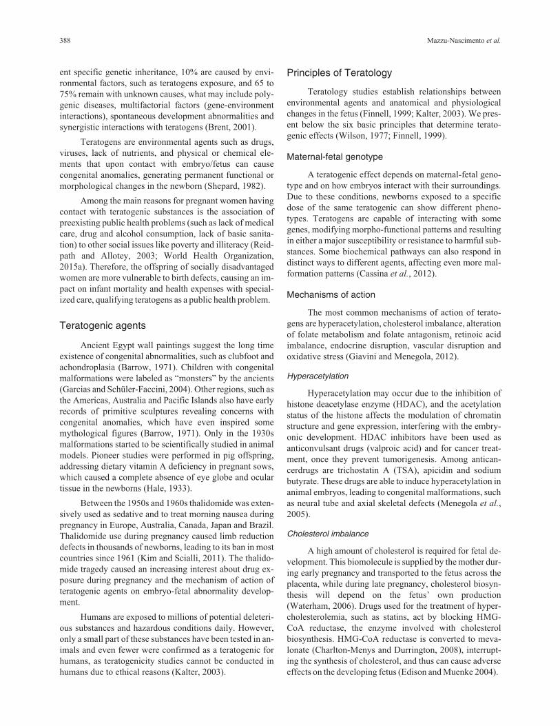
Menegola 2012). Some of these agents (also called prote-
ratogens) can be bioactivated by embryonic cytochrome
P450 enzymes. Their teratogenic effect will depend on the
intracellular balance between proteratogen bioactivation,
molecular target damage, maternal proteratogen elimina-
tion, and repair of damaged cells (Winn and Wells, 1995;
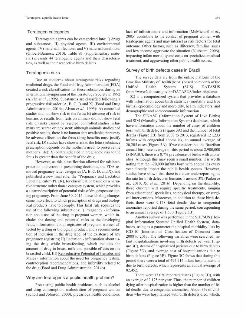
Wells et al., 1997), as illustrated in Figure 1. Among drugs
that induce oxidative stress are thalidomide, valproic acid,
phenytoin, alcohol, (van Gelder et al., 2010), and anti-
cancer drugs (Conklin 2004).
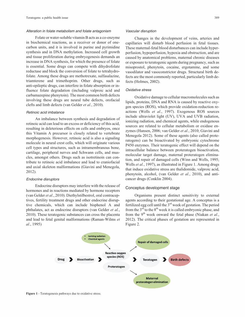
Conceptus development stage
Organisms present distinct sensitivity to external
agents according to their gestational age. A conceptus is a
fertilized egg cell until the 3rd week of gestation. The period
from the 3rd to the 8th week it is called embryonic phase, and
from the 9th week onward the fetal phase (Niakan et al.,
2012). The critical phases of gestation are represented in
Figure 2.
Teratogens: a public health issue 389
Figure 1 - Teratogenesis pathways due to oxidative stress.
The two initial weeks after fertilization, in which the
zygote is undergoing mitotic cell division is called the
‘all-or-nothing’ phase; in case a contact with a teratogenic
agent occurs, it can result either in spontaneous abortion or
in a normal embryo-fetal development. If teratogenic expo-
sure occurs between the 3rd and 8th week of gestation, a pe-
riod in which most of the morphological structures develop,
it can lead to considerable phenotypical changes in the em-
bryo, such as alterations in the central nervous system,
limbs and face. From the 9th week of gestation some organs
are still developing, like external genitalia and brain, and
exposure to teratogens can culminate in functional abnor-
malities. However most morphological characteristics are
preserved from this phase onward (Shepard ,1979; Niakan
et al., 2012).
Nature of the agent
Teratogenic agents can affect the embryo in different
ways, according to the teratogen’s nature. Physical terato-
genic agents, such as ionizing radiation, affect the embryo
directly. Drugs and other chemical substances on the other
hand are previously metabolized by the mother’s organism
before reaching the fetus. This metabolism can either acti-
vate or inactivate relevant metabolites, resulting in differ-
ent teratogenic susceptibilities (Finnell, 1999; Gilbert-
Barness, 2010; Niakan et al., 2012).
Dose-response relationship
Considering basic principles of dose-response, pro-
longed teratogen exposure leads to worsened embryo-fetal
sequelae. Dose is also a major factor, with evidence of short
exposures to high teratogen doses leading to more deleteri-
ous effects (Cohlan, 1963; van Gelder et al., 2010).
Final manifestation
The resulting teratogenic effects are spontaneous
abortion, fetal loss, embryo-fetal morphological abnormal-
ities, intrauterine growth restriction, and functional disabil-
ities, such as intellectual disability (Finnell, 1999; Gilbert-
Barness, 2010).
Teratologists use Shepard’s seven criteria to establish
human teratogenicity (Shepard, 1994): 1) proven exposure
to the agent during the critical stages of prenatal develop-
ment; 2) consistent findings on two or more high-quality
epidemiological studies (control of confounding factors,
sufficient number of cases, exclusion of positive and nega-
tive bias, prospective studies, if possible); 3) careful delin-
eation of clinical cases; 4) rare environmental exposure that
is associated with the rare defect (three or more cases, at
least); 5) teratogenicity in experimental animals; 6) the as-
sociation should make biological sense; and 7) experimen-
tal proof that the agent acts in an unaltered state. Criteria
1-4 are considered essential. criteria 5, 6, and 7 are helpful
but not essential.
390 Mazzu-Nascimento et al.
Figure 2 - Critical stages of human embryological development.
Teratogen categories
Teratogenic agents can be categorized into: I) drugs
and substances, II) physical agents, III) environmental
agents, IV) maternal infections, and V) maternal conditions
(Gilbert-Barness, 2010). Table S1 (supplementary mate-
rial) presents 44 teratogenic agents and their characteris-
tics, as well as their respective birth defects.
Teratogenic risks
Due to concerns about teratogenic risks regarding
medicinal drugs, the Food and Drug Administration (FDA)
created a risk classification for these substances during an
international symposium of the Teratology Society in 1992
(Alván et al., 1995). Substances are classified following a
progressive risk order (A, B, C, D and X) (Food and Drug
Administration, 2014a; Alván et al., 1995): A) controlled
studies did not show risk to the fetus; B) absence of risk to
humans or results from tests on animals did not show fetal
risk; C) risks cannot be rejected; controlled studies in hu-
mans are scarce or inexistent; although animals studies had
positive results, there is no human data available; there may
be adverse effects on the fetus (drug benefits must justify
fetal risk; D) studies have shown risk to the fetus (substance
prescription depends on the mother’s need, to preserve the
mother’s life); X) contraindicated in pregnancy; risk to the
fetus is greater than the benefit of the drug.
However, as this classification allowed for misinter-
pretation and errors in prescribing decisions, the FDA re-
moved pregnancy letter categories (A, B, C, D, and X), and
published a new final rule, the “Pregnancy and Lactation
Labeling Rule” (PLLR), for classification based on a narra-
tive structure rather than a category system, which provides
a clearer description of potential risks of drug exposure dur-
ing pregnancy. From June 30, 2015, these labeling changes
came into effect, to which prescription of drugs and biolog-
ical products have to comply. This final rule requires the
use of the following subsections: I) Pregnancy - informa-
tion about use of the drug in pregnant women, which in-
cludes the dosing and potential risks to the developing
fetus; information about registries of pregnant women af-
fected by a drug or biological product, and a recommenda-
tion of inclusion in the drug label of the existence of any
pregnancy registries; II) Lactation - information about us-
ing the drug while breastfeeding, which includes the
amount of drug in breast milk and possible effects on the
breastfed child; III) Reproductive Potential of Females and
Males - information about the need for pregnancy testing,
contraception recommendations, and infertility related to
the drug (Food and Drug Administration, 2014b).
Why are teratogens a public health problem?
Preexisting public health problems, such as alcohol
and drug consumption, malnutrition of pregnant woman
(Scholl and Johnson, 2000), precarious health conditions,
lack of infrastructure and information (McMichael et al.,
2005) contribute to the contact of pregnant women with
teratogenic agents and may interact as risk factors for fetal
outcome. Other factors, such as illiteracy, familiar issues
and low income aggravate the situation (Nutbeam, 2006),
impacting infant mortality and costs on specialized medical
treatment, and aggravating other public health issues.
Survey of birth defects cases in Brazil
The survey data are from the online platform of the
Brazilian Ministry of Health (MoH) based on records of the
Unified Health System (SUS). DATASUS
(http://www2.datasus.gov.br/DATASUS/index.php?area
= 02) is a computerized system that provides a database
with information about birth statistics (mortality and live
births), epidemiology and morbidity, health indicators, and
demographic and socioeconomic information.
The SINASC (Information System of Live Births)
and SIM (Mortality Information System) databases, which
have information about the number of cases of children
born with birth defects (Figure 3A) and the number of fetal
deaths (Figure 3B) from 2008 to 2013, registered 121,233
infants with congenital anomalies, an annual average of
20,205 cases (Figure 3A). If we consider that the Brazilian
annual birth rate average of this period is about 2,900,000
(SINASC), there is a 0.7% prevalence of births with anom-
alies. Although this may seem a small number, it is worth
noting that the ~20,000 infants born with anomalies every
year directly impact the public health system. However,
studies have shown that there is a clear underreporting, as
the rate for birth defects in humans is around 3% (Parker et
al., 2010; Xu et al., 2016). Depending on the disability,
these children will require specific treatments, ranging
from educational specialists, to physical therapy, or surgi-
cal interventions. Moreover, in addition to these birth de-
fects there were 9,178 fetal deaths due to congenital
anomalies reported during the same period, corresponding
to an annual average of 1,530 (Figure 3B).
Another survey was performed in the SIH/SUS (Hos-
pital Information System/ Unified Health System) data-
bases, using as a parameter the hospital morbidity lists by
ICD-10 (International Classification of Diseases) from
2008 to 2013. The following variables were searched: in-
fant hospitalizations involving birth defects per year (Fig-
ure 3C), deaths of hospitalized patients due to birth defects
(Figure 3D), and average cost of hospitalizations due to
birth defects (Figure 3E). Figure 3C shows that during this
period there were a total of 494,714 infant hospitalizations
due to birth defects, which represents an annual average of
82,452.
There were 13,050 reported deaths (Figure 3D), with
an average of 2,175 per year. Thus, the number of children
dying after hospitalization is higher than the number of fe-
tal deaths due to congenital anomalies. About 3% of chil-
dren who were hospitalized with birth defects died, which,
Teratogens: a public health issue 391
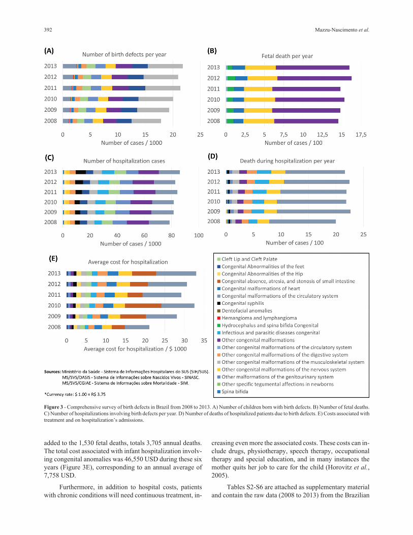
added to the 1,530 fetal deaths, totals 3,705 annual deaths.
The total cost associated with infant hospitalization involv-
ing congenital anomalies was 46,550 USD during these six
years (Figure 3E), corresponding to an annual average of
7,758 USD.
Furthermore, in addition to hospital costs, patients
with chronic conditions will need continuous treatment, in-
creasing even more the associated costs. These costs can in-
clude drugs, physiotherapy, speech therapy, occupational
therapy and special education, and in many instances the
mother quits her job to care for the child (Horovitz et al.,
2005).
Tables S2-S6 are attached as supplementary material
and contain the raw data (2008 to 2013) from the Brazilian
392 Mazzu-Nascimento et al.
Figure 3 - Comprehensive survey of birth defects in Brazil from 2008 to 2013. A) Number of children born with birth defects. B) Number of fetal deaths.
C) Number of hospitalizations involving birth defects per year. D) Number of deaths of hospitalized patients due to birth defects. E) Costs associated with
treatment and on hospitalization’s admissions.
Ministry of Health website. As can be seen, there is a lack
of specification in the records jeopardizing the identifica-
tion of the etiology of the possible teratogens. Better epide-
miological information would be very important for the
detection of new teratogens, as well as for their proper con-
trol. The microcephaly epidemics, as we will show below,
is an example of how the lack of proper registries in many
countries made it difficult to identify the real impact of the
Zika virus infection in pregnancy and the extent of the re-
sulting birth defects.
Brazilian cases of teratogenesis
Microcephaly outbreak in Brazil and Zika prenatalvirus infection
In 2015 there was a sudden appearance of Zika virus
(ZIKV) infections in Brazil, especially in the Northeast re-
gion (Campos et al., 2015). Climatic factors, lack of infra-
structure, and the population’s negligence offered
favorable conditions for the proliferation of Aedes sp. mos-
quitoes, the Zika virus vector (Costa et al., 2008). Further-
more, recent studies suggest a potential sexual transmission
of Zika virus (Musso et al., 2015; Atkinson et al., 2016;
D’Ortenzio et al., 2016), increasing the concern received
from public authorities. The principal reason for alarm is
the increased risk of the unborn babies to develop neurolog-
ical and brain abnormalities, characterized by microce-
phaly, when infected by Zika virus during pregnancy
(Mlakar et al., 2016; Rasmussen et al., 2016).
From 2010 to 2014 there was an average of 160 cases
of microcephaly per year in Brazil (Brazil Ministry of
Health, 2015). A drastic increase was reported from Octo-
ber 22, 2015 to June 18, 2016, with 8,039 suspected and
1,616 confirmed cases of microcephaly (Brazil Ministry of
Health, 2016). The cases occurred in 576 municipalities,
throughout all 26 Brazilian States and the Federal District.
In a study analyzing the first 1,501 cases of microcephaly
reported to the Brazilian MoH, 602 were included as possi-
bly related to ZIKV prenatal infection (depending on the
level of laboratorial, radiological or clinical evidence)
(França et al., 2016).
Microcephaly can be caused by genetic, environmen-
tal or multifactorial causes (Alcantara and O’Driscoll,
2014). The astonishing increase in the reported number of
cases of microcephaly in 2015 led the Brazilian MoH to de-
clare state of emergency in the country (Brazil MoH, 2015),
and on February, 2016, the World Health Organization
(WHO), in turn, declared the large number of cases of
microcephaly and neurological disorders and their possible
relationship with ZIKV a Public Health Emergency of In-
ternational Concern (PHEIC) (World Health Organization,
2016).
Soon after, the ZIKV genome was detected in the
amniotic fluid of pregnant women whose fetuses presented
microcephaly (Calvet et al., 2016), as well as in the brain of
aborted fetuses, in urine, and in cerebrospinal fluid (Brasil
et al., 2016; Mlakar et al., 2016).
Rasmussen et al. (2016), using Shepard’s criteria for
defining a human teratogen, concluded that there was
enough evidence to establish the causal relationship be-
tween ZIKV infection during pregnancy and microcephaly,
in addition to other brain anomalies. Studies in mice re-
vealed that the Brazilian Zika virus (ZIKVBR) is capable of
infecting the fetus, leading to intrauterine growth restric-
tion, including microcephaly (Cugola et al., 2016). Other
studies showed that ZIKV targets human brain cells
(Garcez et al., 2016), clearly revealing the association be-
tween ZIKV infection during the first trimester of preg-
nancy and microcephaly risk (Johansson et al., 2016).
Microcephaly can lead to several complications, such
as intellectual disability, growth retardation, strabism, epi-
lepsy, metabolic disorders, and cerebral palsy (Watemberg
et al., 2002; Ashwal et al., 2009), with an enormous eco-
nomic and social impact for the country in future years.
Therefore, effective actions should be implemented imme-
diately to contain the ZIKV spread. Those actions should
include better access to public sanitation and health poli-
cies, control of the Aedes vector, which also transmits other
tropical diseases such as dengue and yellow fever, along
with the search for effective vaccination and pharmacologi-
cal treatment.
Misoprostol
In developing countries there is an early sexual initia-
tion and lack of family planning. Women tend to be aware
of pregnancy weeks or even months later (De Moura and
Gomes, 2014), and there is a high rate of undesired preg-
nancies.
In countries were abortion is illegal, as in Brazil, in-
tentionally induced clandestine abortions contributes to
high numbers of maternal mortality. Around 5% of Brazil-
ian women end up opting for the illegal use of misoprostol
to terminate unwanted pregnancies (Vargas et al., 2000).
This medical drug is a synthetic prostaglandin E1 analog,
originally prescribed to treat gastric ulcers, which stimulate
uterine contractions, leading to miscarriage. In Brazil, it has
been banned from the market because of its use as an illegal
abortion method (Costa, 1998; Schuler et al., 1999; da Silva
Dal Pizzol et al., 2006; Allen and O’Brien, 2009). Miso-
prostol alone does not present great efficiency in inducing
abortions, and can induce fetal abnormalities, especially if
used in the first trimester of pregnancy (Gonzalez et al.,
1998; Costa, 1998; da Silva Dal Pizzol et al., 2006; Allen
and O’Brien, 2009). Furthermore, the increased use of
drugs or herbs with perceived abortive actions by the popu-
lation highlights the lack of control in selling and using pre-
scription medications.
A cohort study of pregnant women treated in prenatal
services in six Brazilian capitals revealed that 707 women
used products to induce menstruation, which includes her-
Teratogens: a public health issue 393
bal teas (34.4%), sex hormones (28.3%), and misoprostol
(17%) (Pizzol et al., 2008). The congenital anomaly risk
was 2.74 times greater for fetuses exposed to misoprostol
when compared to unexposed fetuses. Misoprostol is asso-
ciated mainly to congenital paralysis of the 6th and 7th cra-
nial nerves and to limb reduction defects due to fetal
vascular disruption (Gonzalez et al., 1998; Vargas et al.,
2000).
Thalidomide
Even after the immense international repercussion of
the teratogenic potential effects of thalidomide, many cases
of newborn malformation involving this medicine were
registered in Brazil in different years. Thalidomide is a drug
indicated for the treatment of erythema nodosum leprosum
(ENL), and more recently, to a number of different medical
conditions, due to its immunomodulatory properties (Vian-
na et al., 2011, 2015; Kim and Scialli, 2011;). The Latin-
American Collaborative Study of Congenital Malforma-
tions (ECLAMC) reported 34 thalidomide embryopathy
cases in South America from the 1960s to 1990s, of which
33 where in Brazil. Most of these cases involved mothers
treated for leprosy, whose babies presented birth defects,
such as phocomelia, hypoplastic glenoid, absence of
thumbs, absence or hypoplasia of radius, a third arm bone,
and polydactyly (Castilla et al., 1996).
Three cases were reported from 2005 to 2010. From
those, four involved mothers that underwent treatment for
ENL and were unaware of pregnancy, while the third case
involved self-medication of a pregnant woman who took
thalidomide prescribed for her mother, who was being
treated for multiple myeloma (Schuler-Faccini et al.,
2007). The majority of cases involves lack of medical infor-
mation, which is related to public health.
Despite safety concerns, the Brazilian population has
a high consumption of thalidomide. The state of São Paulo
leads the thalidomide drug distribution rates (5,889,210
thalidomide tablets), followed by Minas Gerais and Rio de
Janeiro, from 2005 to 2010. In this period, there were 2,802
reported cases of limb reduction defects, with 192 cases
compatible with a thalidomide embryopathy phenotype
(TEP) (Vianna et al., 2015). This demonstrates the serious-
ness of this public health issue.
Alcohol
In 2012, alcohol was responsible for almost 3.3 mil-
lion of deaths, corresponding to 5.9% of the total number of
deaths worldwide. Excessive alcohol consumption prevails
among adults aged 20-39 years, although alcohol use by
young people starts at early ages – even from 12.5 years old
(Vieira et al., 2007; World Health Organization, 2015b).
Alcohol abuse during pregnancy can lead to Neonatal Ab-
stinence Syndrome in babies. Alcohol is the teratogenic
agent responsible for the Fetal Alcohol Syndrome (FAS),
as well as Fetal Alcohol Spectrum Disorders (FASD), be-
ing a major non-genetic cause of intellectual disability and
behavioral problems (Abel and Sokol, 1987; Momino et al.,
2012; Chapman and Wu, 2013; O’Leary et al., 2013).
In 2015 a study was carried out in a Brazilian orphan-
age in Recife to investigate the frequency of FASD.
Children were evaluated by a multidisciplinary team, and
the following results were obtained: 50% of the childrens
mothers were reported as known alcohol abusers. Of these
children, 18% presented general developmental delay, 3%
had intellectual disabilities, 27% had cognitive impairment,
14% had attention deficit/hyperactivity, and 3% presented
autism. A total of 17% presented FASD, three children pre-
sented FAS, six presented partial FAS, and seven presented
neurological disorder related to alcohol. About 16% of
these children presented ocular changes, such as low vi-
sion, strabism and morphological changes of optic nerves,
which shows the devastating effects of drug abuse during
pregnancy. (Strömland et al., 2014)
Illicit drugs
Illicit drug consumption during pregnancy is another
public health problem involving a potential embryo-fetal
effect. The effects of these substances include low birth
weight, intrauterine growth restriction and placental
abruption, as well as premature birth or spontaneous abor-
tion (Holbrook and Rayburn, 2014). Cocaine and its deriva-
tives, such as crack and heroin, may have harmful effects
on pregnancy (Cherukuri et al., 1988; Rizk et al., 1996;
Costa et al., 2012). The extent of the problem can be esti-
mated by the increase in the demand of medical care and
costs with hospitalization and specialized treatment for
drug-addicted pregnant women, as well as the increase in
the number of premature births. Another issue is the in-
creased rate of sexually transmitted diseases or other mater-
nal infections related to illicit drug and alcohol use (Tüzün
et al., 1999; Hwang et al., 2000), which can also be terato-
genic, like syphilis.
Concluding remarks
This review presented fundamental aspects of terato-
genic agents, bringing an overview about the number of
cases of congenital anomalies, and their contribution to an
increase in mortality rates, hospitalizations and treatments
expenses for the Brazilian Unified Health System. We also
present cases of congenital malformations involving terato-
gens, as well as the definitive classification of Zika virus in-
fection as a teratogen and now a real public health problem.
Congenital anomalies caused by teratogenic agents
are essentially avoidable, with a great impact on public
health, on the economic and on social aspects. Public poli-
cies to prevent, care for, and treat these disabilities are ex-
tremely important to manage this public health issue. To
address these problems, a collaborative agreement among
the United Nations (UN), the WHO, the United Nations
Children’s Fund (UNICEF) and government leaders in
394 Mazzu-Nascimento et al.
2010 planned a series of low-cost and high-impact inter-
ventions to improve neonatal and infant health quality
(World Health Organization, 2015a). The lack of public
health structure in developing countries, however, still
causes problems, reducing the viability of the proposal.
Congenital abnormalities and neglected diseases are some-
what comparable, as both problems do not receive the nec-
essary attention and prevail in developing regions. In
Brazil, income transfers helped millions of people out of
extreme poverty, but are still far from solving public health
problems in the country. An intervention is needed, espe-
cially in the peripheral regions, with the application of pre-
ventive policies (Di Renzo et al., 2015). These policies may
differ according to the countries’ characteristics, but they
should by all means emphasize health education of profes-
sionals, and the public,investment in primary reproductive
healthcare, pregnancy planning, basic sanitation, and reli-
able registries with epidemiological data on congenital
anomalies.
Acknowledgments
The authors would like to thank the funding agencies
FAPESP (Grant Nr. 2011/13997-8), CNPq (Grants Nr.
311323/2011-1, Nr. 131306/2013-8 and 205453/2014-7)
and CAPES for scholarships and financial support.
References
Abel E and Sokol R (1987) Incidence of fetal alcohol sydrome and
economic impact of fas-related anamalies. Drug Alcohol
Depend 19:51-70.
Alcantara D and O’Driscoll M (2014) Congenital microcephaly.
Am J Med Genet Part C Semin Med Genet 166:124-139.
Allen R and O’Brien BM (2009) Uses of misoprostol in obstetrics
and gynecology. Rev Obstet Gynecol 2:159-168.
Alván G, Danielsson BR, Kihlström I, Lundborg P, Prame B,
Ridley E and Sannerstedt R (1995) Classification of drugs
for teratogenic risk. Eur J Clin Pharmacol 48:177-178.
Ashwal S, Michelson D, Plawner L and Dobyns WB (2009) Prac-
tice parameter: Evaluation of the child with microcephaly
(an evidence-based review): Report of the Quality Standards
Subcommittee of the American Academy of Neurology and
the Practice Committee of the Child Neurology Society.
Neurology 73:887-897.
Atkinson B, Hearn P, Afrough B, Lumley S, Carter D, Aarons EJ,
Simpson AJ, Brooks TJ and Hewson R (2016) Detection of
Zika Virus in semen. Emerg Infect Dis 22:160-107.
Barrow MV (1971) A brief history of teratology to the early 20th
century. Teratology 4:119-129.
Brasil P, Pereira Jr JP, Raja Gabaglia C, Damasceno L, Wakimoto
M, Ribeiro Nogueira RM, Carvalho de Sequeira P, Machado
Siqueira A, Abreu de Carvalho LM, Cotrim da Cunha D, et
al. (2016) Zika virus infection in pregnant women in Rio de
Janeiro - Preliminary report. N Engl J Med 375:2321-2334.
Brent RL (2001) The cause and prevention of human birth defects:
What have we learned in the past 50 years? Congenit Anom
(Kyoto) 41:3-21.
Bronberg R, Schuler-Faccini L, Ramallo V, Alfaro E and Dipierri
J (2014) Spatial and temporal analysis of infant mortality
from congenital malformations in Brazil (1996-2010). J
Community Genet 5:269-282.
Calvet G, Aguiarv RS, Melo AS, Sampaio SA, de Filippis I, Fabri
A, Araujo ES, de Sequeira PC, de Mendonça MCL, de
Oliveira L, et al. (2016) Detection and sequencing of Zika
virus from amniotic fluid of fetuses with microcephaly in
Brazil: A case study. Lancet Infect Dis 16:653-660.
Campos GS, Bandeira AC and Sardi SI (2015) Zika virus out-
break, Bahia, Brazil. Emerg Infect Dis 21:1885-1886.
Cassina M, Salviati L, Di Gianantonio E and Clementi M (2012)
Genetic susceptibility to teratogens: State of the art. Reprod
Toxicol 34:186-191.
Castilla E, Ashton-Prolla P, Barreda-Mejia E, Brunoni D, Caval-
canti D, Correa-Neto J, Delgadillo J, Dutra M, Felix T,
Giraldo A, et al. (1996) Thalidomide, a current teratogen in
South America. Teratology 54:273-277.
Chapman SLC and Wu Li-Tzy (2013) Substance abuse among in-
dividuals with intellectual disabilities. Res Dev Disabil
33:1147-1156.
Charlton-Menys V and Durrington PN (2008) Human cholesterol
metabolism and therapeutic molecules. Exp Physiol 93:27-
42.
Cherukuri R, Minkoff H, Feldman J, Parekh A and Glass L (1988)
A cohort study of alkaloidal cocaine (“ crack”) in prenancy.
Obstet Gynecol 72:147-151.
Cohlan SQ (1963) Teratogenic agents and congenital malforma-
tions. J Pediatr 63:650-659.
Conklin KA (2004) Chemotherapy-associated oxidative stress:
Impact on chemotherapeutic effectiveness. Integr Cancer
Ther 3:294-300.
Costa FS, Silva JJ da, Souza CM de and Mendes J (2008) Dinâ-
mica populacional de Aedes aegypti (L) em área urbana de
alta incidência de dengue. Rev Soc Bras Med Trop 41:309-
312.
Costa GM, Soibelman M, Zanchet DL, Costa PDM, Alberto C and
Salgado I (2012) Pregnant crack addicts in a psychiatric unit.
J Bras Psiquiatr 61:8-12.
Costa SH (1998) Commercial availability of misoprostol and in-
duced abortion in Brazil. Int J Gynaecol Obstet 63(Suppl
1):S131-S139.
Cugola FR, Fernandes IR, Russo FB, Freitas BC, Dias JLM,
Guimarães KP, Benazzato C, Almeida N, Pignatari GC,
Romero S, et al. (2016) The Brazilian Zika virus strain
causes birth defects in experimental models. Nature
534:267-271.
D’Ortenzio E, Matheron S, Lamballerie X de, Hubert B, Pior-
kowski G, Maquart M, Descamps D, Damond F, Yazdan-
panah Y and Leparc-Goffart I (2016) Evidence of sexual
transmission of Zika virus. N Engl J Med 374:2195-2198.
da Silva Dal Pizzol T, Knop FP and Mengue SS (2006) Prenatal
exposure to misoprostol and congenital anomalies: System-
atic review and meta-analysis. Reprod Toxicol 22:666-671.
De Moura LNB and Gomes KRO (2014) Planejamento familiar:
Uso dos serviços de saúde por jovens com experiência de
gravidez. Cienc Saude Colet 19:853-863.
Di Renzo GC, Conry JA, Blake J, DeFrancesco MS, DeNicola N,
Martin JN, McCue KA, Richmond D, Shah A, Sutton P, et
al. (2015) International Federation of Gynecology and Ob-
stetrics opinion on reproductive health impacts of exposure
Teratogens: a public health issue 395
to toxic environmental chemicals. Int J Gynecol Obstet.
131:219-225.
Edison RJ and Muenke M (2004) Central nervous system and
limb anomalies in case reports of first-trimester statin expo-
sure. N Engl J Med 350:1579-1582.
Egbe A, Uppu S, Lee S, Stroustrup A, Ho D and Srivastava S
(2015) Congenital malformations in the newborn popula-
tion: A population study and analysis of the effect of sex and
prematurity. Pediatr Neonatol 56:25-30.
Finnell RH (1999) Teratology: General considerations and princi-
ples. J Allergy Clin Immunol 103:337-342.
Food and Drug Administration (2014a) Content and format of la-
beling for human prescription drug and biological products
requirements for pregnancy and lactation labeling. Dep
Health Hum Serv Food Federal Regist 79:1-144.
França GV, Schuler-Faccini L, Oliveira WK, Henriques CMP,
Carmo EH, Pedi VD, Nunes ML, Castro MC, Serruya S,
Silveira MF, et al. (2016) Congenital Zika virus syndrome in
Brazil: A case series of the fi rst 1501 livebirths with com-
plete investigation. Lancet 388:891-897.
Garcez PP, Loiola EC, Costa RM da, Higa LM, Trindade P,
Delvecchio R, Nascimento JM, Brindeiro R, Tanuri A and
Rehen SK (2016) Zika virus impairs growth in human neu-
rospheres and brain organoids. Science 352:816-818.
Garcias GDL and Schüler-Faccini L (2004) The beliefs of moth-
ers in southern Brazil regarding risk-factors associated with
congenital abnormalities. Genet Mol Biol 27:147-153.
Giavini E and Menegola E (2012) Biomarkers of teratogenesis:
Suggestions from animal studies. Reprod Toxicol 34:180-
185.
Gilbert-Barness E (2010) Teratogenic causes of malformations.
Ann Clin Lab Sci 40:99-114.
Gonzalez CH, Marques-Dias MJ, Kim CA, Sugayama SM, Da
Paz JA, Huson SM and Holmes LB (1998) Congenital ab-
normalities in Brazilian children associated with misopros-
tol misuse in first trimester of pregnancy. Lancet 351:1624-
1627.
Hale F (1933) Pigs born without eye balls. J Hered 24:105-110.
Hansen JM (2006) Oxidative stress as a mechanism of terato-
genesis. Birth Defects Res C Embryo Today 78:293-307.
Holbrook BD and Rayburn WF (2014) Teratogenic risks from ex-
posure to illicit drugs. Obstet Gynecol Clin North Am
41:229-239.
Holmes LB (2002) Teratogen-induced limb defects. Am J Med
Genet 112:297-303.
Horovitz DDG, Llerena Jr JC and Mattos RA (2005) Atenção aos
defeitos congênitos no Brasil: Panorama atual. Cad Saúde
Pública 21:1055-1064.
Hwang LY, Ross MW, Zack C, Bull L, Rickman K and Holleman
M (2000) Prevalence of sexually transmitted infections and
associated risk factors among populations of drug abusers.
Clin Infect Dis 31:920-926.
Johansson MA, Mier-y-Teran-Romero L, Reefhuis J, Gilboa SM
and Hills SL (2016) Zika and the risk of microcephaly. N
Engl J Med 375:1-4.
Kalter H (2003) Teratology in the 20th century: Environmental
causes of congenital malformations in humans and how they
were established. Neurotoxicol Teratol 25:131-282.
Kim JH and Scialli AR (2011) Thalidomide: The tragedy of birth
defects and the effective treatment of disease. Toxicol Sci
122:1-6.
McMichael C, Waters E and Volmink J (2005) Evidence-based
public health: What does it offer developing countries? J
Public Health (Oxf) 27:215-221.
Menegola E, Di Renzo F, Broccia ML, Prudenziati M, Minucci S,
Massa V and Giavini E (2005) Inhibition of histone dea-
cetylase activity on specific embryonic tissues as a new
mechanism for teratogenicity. Birth Defects Res Part B -
Dev Reprod Toxicol 74:392-398.
Mlakar J, Korva M, Tul N, Popovic M, Poljak-Prijatelj M, Mraz J,
Kolenc M, Resman RK, Vesnaver VT, Voduek VF, et al.
(2016) Zika virus associated with microcephaly. N Engl J
Med 374:951-958.
Momino W, Félix TM, Abeche AM, Zandoná DI, Scheibler GG,
Chambers C, Jones KL, Flores RZ and Schüler-Faccini L
(2012) Maternal drinking behavior and Fetal Alcohol Spec-
trum disorders in adolescents with criminal behavior in
southern Brazil. Genet Mol Biol 35:960-965.
Musso D, Roche C, Robin E, Nhan T, Teissier A and Cao-
Lormeau VM (2015) Potential sexual transmission of zika
virus. Emerg Infect Dis 21:359-361.
Niakan KK, Han J, Pedersen RA, Simon C and Pera RAR (2012)
Human pre-implantation embryo development. Develop-
ment 139:829-841.
Nutbeam DON (2006) Health literacy as a public health goal: A
challenge for contemporary health education and communi-
cation strategies into the 21st century. Health Promot Int
15:259-268.
O’Leary C, Leonard H, Bourke J, D’Antoine H, Bartu A and
Bower C (2013) Intellectual disability: Population-based es-
timates of the proportion attributable to maternal alcohol use
disorder during pregnancy. Dev Med Child Neurol 55:271-
277.
Parker SE, Mai CT, Canfield MA, Rickard R, Wang Y, Meyer RE,
Anderson P, Mason CA, Collins JS, Kirby RS, et al. (2010)
Updated national birth prevalence estimates for selected
birth defects in the United States, 2004 - 2006. Birth Defects
Res (Part A) 88:1008-1016.
Passos-Bueno MR, Bertola D, Horovitz DDG, de Faria Ferraz VE
and Brito LA (2014) Genetics and genomics in Brazil: A
promising future. Mol Genet Genomic Med 2:280-291.
Petrini J, Damus K, Russell R, Poschman K, Davidoff MJ and
Mattison D (2002) Contribution of birth defects to infant
mortality in the United States. Teratology 66(Suppl
1):S3-S6.
Pizzol T da SD, Sanseverino MTV and Mengue SS (2008) Expo-
sure to misoprostol and hormones during pregnancy and risk
of congenital anomalies. Cad Saude Publica 24:1447-1453.
Raman-Wilms L, Tseng A, Wighardt S, Einarson T and Koren G
(1995) Fetal genital effects of first-trimester sex hormone
exposure: A meta-analysis. Obstet Gynecol 85:141-149.
Rasmussen SA, Jamieson DJ, Honein MA and Petersen LR
(2016) Zika virus and birth defects - Reviewing the evidence
for causality. N Engl J Med 374:1981-1987.
Reidpath DD and Allotey P (2003) Infant mortality rate as an indi-
cator of population health. J Epidemiol Community Health
57:344-346.
Rizk B, Atterbury JL and Groome LJ (1996) Reproductive risks of
cocaine. Hum Reprod Update 2:43-55.
Scholl TO and Johnson WG (2000) Folic acid: Influence on the
outcome of pregnancy. Am J Clin Nutr 71:1295S-303S.
396 Mazzu-Nascimento et al.
Schüler L, Pastuszak A, Sanseverino TV, Orioli IM, Brunoni D,
Ashton-Prolla P, Silva da Costa F, Giugliani R, Couto AM,
Brandao SB, et al. (1999) Pregnancy outcome after expo-
sure to misoprostol in Brazil: A prospective, controlled
study. Reprod Toxicol 13:147-151.
Schuler-Faccini L, Soares RCF, de Sousa ACM, Maximino C,
Luna E, Schwartz IVD, Waldman C and Castilla EE (2007)
New cases of thalidomide embryopathy in Brazil. Birth De-
fects Res A Clin Mol Teratol 79:671-672.
Shepard TH (1982) Detection of human teratogenic agents. J
Pediatr 101:810-815.
Shepard TH (1979) Teratogenicity of therapeutic agents. Curr
Probl Pediatr 10:1-42.
Shepard TH (1994) “Proof ” of human teratogenicity. Teratology
50:97-98.
Strömland K, Ventura LO, Mirzaei L, Fontes de Oliveira K,
Marcelino Bandim J, Parente Ivo A and Brandt C (2014) Fe-
tal alcohol spectrum disorders among children in a Brazilian
orphanage. Birth Defects Res A Clin Mol Teratol 103:178-
185.
Tüzün B, Tüzün Y and Wolf R (1999) Alcohol intake and sexually
transmitted diseases. Clin Dermatol 17:469-478.
van Gelder MMHJ, van Rooij IALM, Miller RK, Zielhuis GA, de
Jong-van den Berg LTW and Roeleveld N (2010) Terato-
genic mechanisms of medical drugs. Hum Reprod Update
16:378-94.
Vargas FR, Schuler-Faccini L, Brunoni D, Kim C, Meloni VF,
Sugayama SM, Albano L, Llerena Jr JC, Almeida JC, Duar-
te A, et al. (2000) Prenatal exposure to misoprostol and vas-
cular disruption defects: A case-control study. Am J Med
Genet 95:302-306.
Vianna FSL, Lopez-Camelo JS, Leite JCL, Sanseverino MTV,
Dutra MDG, Castilla EE and Schüler-Faccini L (2011) Epi-
demiological surveillance of birth defects compatible with
thalidomide embryopathy in Brazil. PLoS One 6:e21735.
Vianna FSL, Oliveira MZ, Sanseverino MTV, Morelo EF, Rabel-
lo Neto D de L, Lopez-Camelo J, Camey SA and Schuler-
Faccini L (2015) Pharmacoepidemiology and thalidomide
embryopathy surveillance in Brazil. Reprod Toxicol 53:63-
67.
Vieira DL, Ribeiro M, Romano M and Laranjeira RR (2007) Al-
cohol and adolescents: Study to implement municipal poli-
cies. Rev Saude Publica 41:1-8.
Watemberg N, Silver S, Harel S and Lerman-Sagie T (2002) Sig-
nificance of microcephaly among children with develop-
mental disabilities. J Child Neurol 17:117-122.
Waterham HR (2006) Defects of cholesterol biosynthesis. FEBS
Lett 580:5442-5449.
Wells PG, Kim PM, Laposa RR, Nicol CJ, Parman T and Winn
LM (1997) Oxidative damage in chemical teratogenesis.
Mutat Res 396:65-78.
Wilson JG (1977) Current status of teratology: General principles
and mechanisms derived from animal studies. In: JG Wilson
& FC Fraser (eds) Handbook of Teratology. Plenum Press,
New York, 488 pp.
Winn L and Wells P (1995) Free radical-mediated mechanisms of
anti-convulsant teratogenicity. Eur J Neurol 2:5-29.
Xu J, Murphy S, Kochanek K and Bastian B (2016) Deaths: Final
data for 2013. National vital statistics reports. Natl Cent
Heal Stat 64:1-117.
Internet resourcesBrazil Ministry of Health (2015) Microcephaly- epidemiological
bulletin. http://portalsaude.saude.gov.br/im-
ages/pdf/2015/novembro/30/Microcefalia-2-boletim.pdf,
accessed June 22, 2016.
Brazil Ministry of Health (2016) Microcephaly- epidemiological
bulletin. http://portalsaude.saude.gov.br/in-
dex.php/cidadao/princi-
pal/agencia-saude/24202-ministerio-da-saude-confirma-1-
616-casos-de-microcefalia-em-todo-o-pais, accessed June
22, 2016.
Food and Drug Administration (2014b) Code of Federal Regula-
tions Title 21. (21CFR201.57). Avalilable:
http://www.accessdata.fda.gov/scripts/cdrh/cfdocs/cfCFR/
CFRSearch.cfm?fr = 201.57, accessed June 2, 2014.
World Health Organization (2015a) Congenital anomalies. Fact
sheet No370.
http://www.who.int/mediacentre/factsheets/fs370/en/, ac-
cessed November 23, 2015.
World Health Organization (2015b) Alcohol. Fact sheet N°349.
http://www.who.int/mediacentre/factsheets/fs349/en/, ac-
cessed November 23, 2015.
World Health Organization (2016) WHO statement on the first
meeting of the International Health Regulations (2005)
(IHR 2005) Emergency Committee on Zika virus and ob-
served increase in neurological disorders and neonatal mal-
formations. http://www.who.int/mediacentre/news/state-
ments/2016/1st-emergency-committee-zika/en/, accessed
February 25, 2016.
Supplementary Material
The following online material is available for this article:
Table S1 - Main teratogen categories and respective em-
bryo-fetal effects during pregnancy.
Table S2 - Cases involving congenital anomalies from
2008 to 2013.
Table S3 - Fetal deaths involving congenital anomalies
from 2008 to 2013.
Table S4 - Infant hospitalizations involving congenital
anomalies from 2008 to 2013.
Table S5 - Number of deaths of infant hospitalized involv-
ing congenital anomalies from 2008 to 2013.
Table S6 - Average cost in US dollars of hospitalizations
involving congenital anomalies from 2008 to 2013
Associate Editor: Carlos F.M. Menck
License information: This is an open-access article distributed under the terms of theCreative Commons Attribution License (type CC-BY), which permits unrestricted use,distribution and reproduction in any medium, provided the original article is properly cited.
Teratogens: a public health issue 397