Embed Size (px)
Citation preview

1
Università degli Studi di Milano
GRADUATE SCHOOL OF VETERINARY SCIENCES FOR ANIMAL HEALTH AND FOOD SAFETY
Director: Prof. Vittorio Dell’Orto
Doctoral Program in Veterinary Clinical Sciences
XXIV CYCLE
Academic Year: 2011-2012
TENSION BAND HERNIORRHAPHY IN CANINE PERINEAL HERNIA: EVALUATION OF THE ROLE OF COLOPEXY ON SURGICAL AND
CLINICAL OUTCOME
Dott. Matteo Gobbetti RO8O45
Tutor: Coordinator: Prof. Stefano Romussi Prof. Angelo Belloli
2
Index
1. Introduction 3
2. Aim of the study 6
3. Clinical anatomy 8
4. Diagnosis and clinical classification 14
5. Surgical treatment of perineal hernia 19
5.1 Standard technique 5.2 Transpositional techniques 5.3 Prosthetic techniques 5.4 Pexy of abdominal and pelvic viscera 5.5 Orchiectomy 5.6 Surgical complications
6. Review of methodologies of collection of follow up in veterinary literature on perineal hernia 29
7. Matherials and method 32
7.1 Inclusion criteria 7.2 Diagnostic procedure 7.3 Treatment and experimental protocol 7.4 Follow up
8. Results 36 9. Discussion 42
10. Conclusion 48
11. References 50

3
Introduction
CHAPTER 1
4
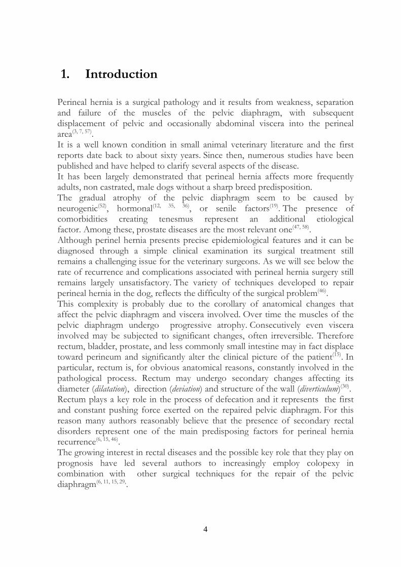
1. Introduction
Perineal hernia is a surgical pathology and it results from weakness, separation and failure of the muscles of the pelvic diaphragm, with subsequent displacement of pelvic and occasionally abdominal viscera into the perineal area(3, 7, 57). It is a well known condition in small animal veterinary literature and the first reports date back to about sixty years. Since then, numerous studies have been published and have helped to clarify several aspects of the disease. It has been largely demonstrated that perineal hernia affects more frequently adults, non castrated, male dogs without a sharp breed predisposition. The gradual atrophy of the pelvic diaphragm seem to be caused by neurogenic(52), hormonal(12, 35, 36), or senile factors(19). The presence of comorbidities creating tenesmus represent an additional etiological factor. Among these, prostate diseases are the most relevant one(47, 58). Although perinel hernia presents precise epidemiological features and it can be diagnosed through a simple clinical examination its surgical treatment still remains a challenging issue for the veterinary surgeons. As we will see below the rate of recurrence and complications associated with perineal hernia surgery still remains largely unsatisfactory. The variety of techniques developed to repair perineal hernia in the dog, reflects the difficulty of the surgical problem(46). This complexity is probably due to the corollary of anatomical changes that affect the pelvic diaphragm and viscera involved. Over time the muscles of the pelvic diaphragm undergo progressive atrophy. Consecutively even viscera involved may be subjected to significant changes, often irreversible. Therefore rectum, bladder, prostate, and less commonly small intestine may in fact displace toward perineum and significantly alter the clinical picture of the patient(15). In particular, rectum is, for obvious anatomical reasons, constantly involved in the pathological process. Rectum may undergo secondary changes affecting its diameter (dilatation), direction (deviation) and structure of the wall (diverticulum)(30). Rectum plays a key role in the process of defecation and it represents the first and constant pushing force exerted on the repaired pelvic diaphragm. For this reason many authors reasonably believe that the presence of secondary rectal disorders represent one of the main predisposing factors for perineal hernia recurrence(6, 15, 46). The growing interest in rectal diseases and the possible key role that they play on prognosis have led several authors to increasingly employ colopexy in combination with other surgical techniques for the repair of the pelvic diaphragm(6, 11, 15, 29,.
5
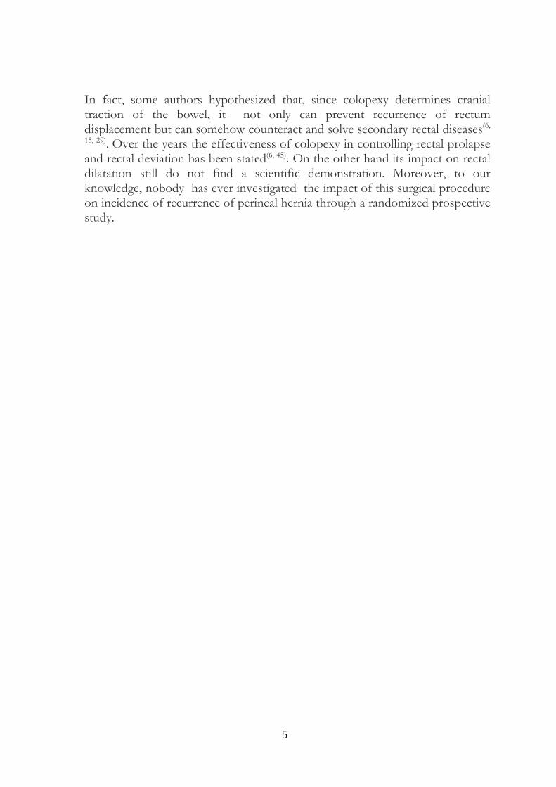
In fact, some authors hypothesized that, since colopexy determines cranial traction of the bowel, it not only can prevent recurrence of rectum displacement but can somehow counteract and solve secondary rectal diseases(6,
15, 29). Over the years the effectiveness of colopexy in controlling rectal prolapse and rectal deviation has been stated(6, 45). On the other hand its impact on rectal dilatation still do not find a scientific demonstration. Moreover, to our knowledge, nobody has ever investigated the impact of this surgical procedure on incidence of recurrence of perineal hernia through a randomized prospective study.
6
Aim of the study
CHAPTER 2
7
2. Aim of the study The objective of our study is to evaluate the influence of colopexy on both clinical and surgical outcome of dogs treated with perineal herniorrhapy and, if possible, to establish its impact on rectal dilatation. Moreover our second aim is to compare the results of our surgical approach to those almost published in veterinary literature. Our preliminary hypothesis is that colopexy might improve prognosis of dogs treated with herniorrhapy and could at least reduce the severity of secondary rectal dilatation.
8
Clinical anatomy
CHAPTER 3
9
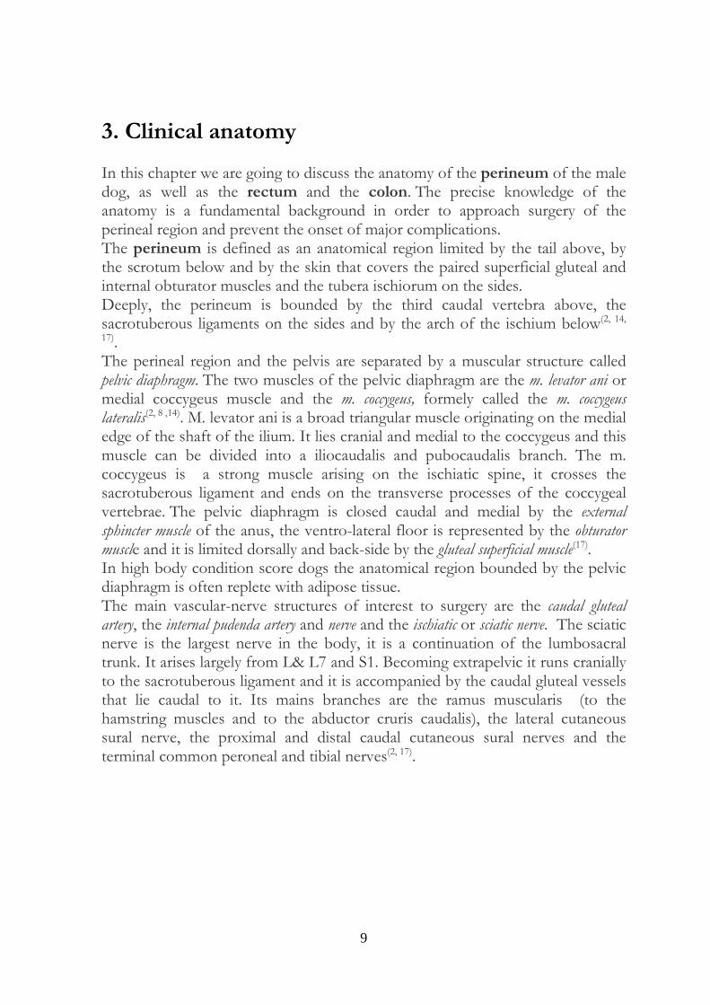
3. Clinical anatomy In this chapter we are going to discuss the anatomy of the perineum of the male dog, as well as the rectum and the colon. The precise knowledge of the anatomy is a fundamental background in order to approach surgery of the perineal region and prevent the onset of major complications. The perineum is defined as an anatomical region limited by the tail above, by the scrotum below and by the skin that covers the paired superficial gluteal and internal obturator muscles and the tubera ischiorum on the sides. Deeply, the perineum is bounded by the third caudal vertebra above, the sacrotuberous ligaments on the sides and by the arch of the ischium below(2, 14,
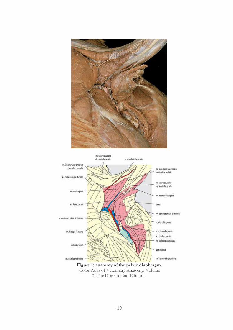
17). The perineal region and the pelvis are separated by a muscular structure called pelvic diaphragm. The two muscles of the pelvic diaphragm are the m. levator ani or medial coccygeus muscle and the m. coccygeus, formely called the m. coccygeus lateralis(2, 8 ,14). M. levator ani is a broad triangular muscle originating on the medial edge of the shaft of the ilium. It lies cranial and medial to the coccygeus and this muscle can be divided into a iliocaudalis and pubocaudalis branch. The m. coccygeus is a strong muscle arising on the ischiatic spine, it crosses the sacrotuberous ligament and ends on the transverse processes of the coccygeal vertebrae. The pelvic diaphragm is closed caudal and medial by the external sphincter muscle of the anus, the ventro-lateral floor is represented by the obturator muscle and it is limited dorsally and back-side by the gluteal superficial muscle(17). In high body condition score dogs the anatomical region bounded by the pelvic diaphragm is often replete with adipose tissue. The main vascular-nerve structures of interest to surgery are the caudal gluteal artery, the internal pudenda artery and nerve and the ischiatic or sciatic nerve. The sciatic nerve is the largest nerve in the body, it is a continuation of the lumbosacral trunk. It arises largely from L& L7 and S1. Becoming extrapelvic it runs cranially to the sacrotuberous ligament and it is accompanied by the caudal gluteal vessels that lie caudal to it. Its mains branches are the ramus muscularis (to the hamstring muscles and to the abductor cruris caudalis), the lateral cutaneous sural nerve, the proximal and distal caudal cutaneous sural nerves and the terminal common peroneal and tibial nerves(2, 17).
10
Figure 1: anatomy of the pelvic diaphragm.
Color Atlas of Veterinary Anatomy, Volume 3: The Dog Cat,2nd Edition.
11
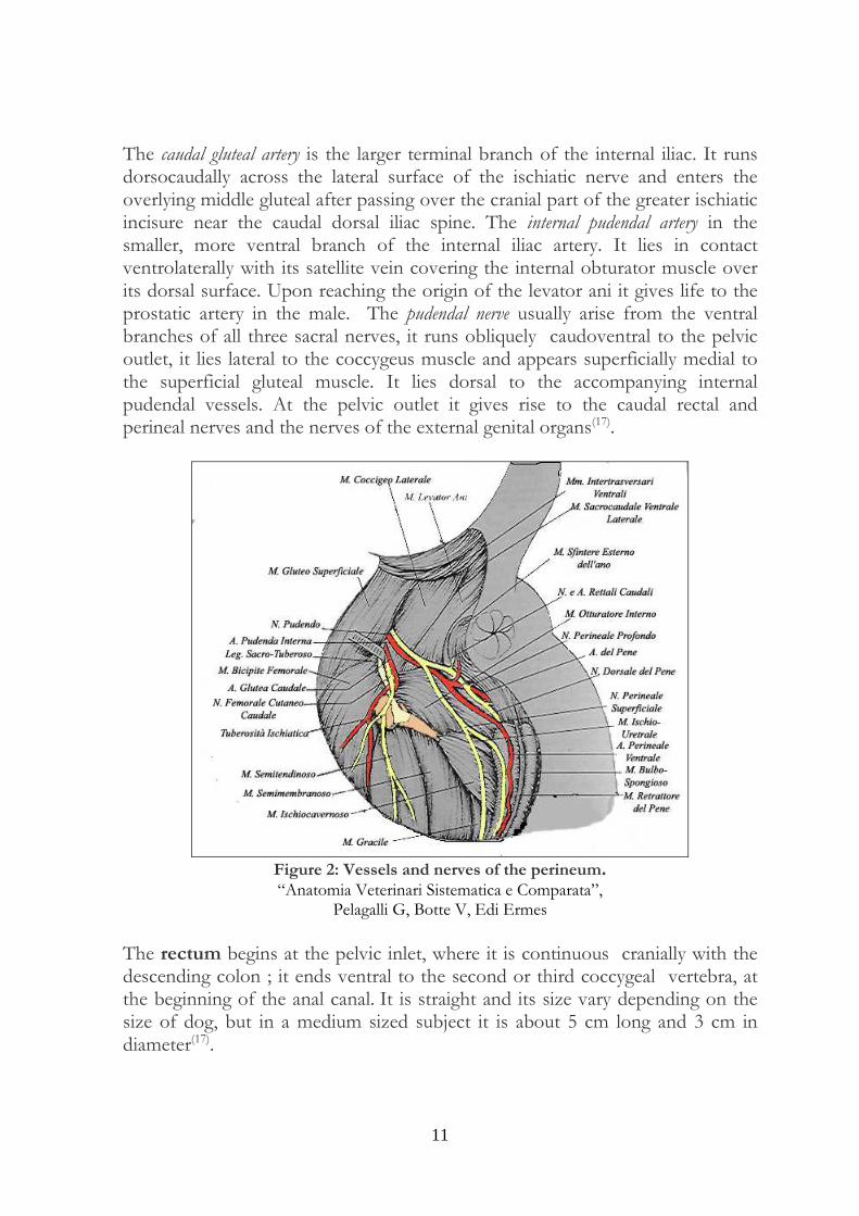
The caudal gluteal artery is the larger terminal branch of the internal iliac. It runs dorsocaudally across the lateral surface of the ischiatic nerve and enters the overlying middle gluteal after passing over the cranial part of the greater ischiatic incisure near the caudal dorsal iliac spine. The internal pudendal artery in the smaller, more ventral branch of the internal iliac artery. It lies in contact ventrolaterally with its satellite vein covering the internal obturator muscle over its dorsal surface. Upon reaching the origin of the levator ani it gives life to the prostatic artery in the male. The pudendal nerve usually arise from the ventral branches of all three sacral nerves, it runs obliquely caudoventral to the pelvic outlet, it lies lateral to the coccygeus muscle and appears superficially medial to the superficial gluteal muscle. It lies dorsal to the accompanying internal pudendal vessels. At the pelvic outlet it gives rise to the caudal rectal and perineal nerves and the nerves of the external genital organs(17).
Figure 2: Vessels and nerves of the perineum. “Anatomia Veterinari Sistematica e Comparata”,
Pelagalli G, Botte V, Edi Ermes
The rectum begins at the pelvic inlet, where it is continuous cranially with the descending colon ; it ends ventral to the second or third coccygeal vertebra, at the beginning of the anal canal. It is straight and its size vary depending on the size of dog, but in a medium sized subject it is about 5 cm long and 3 cm in diameter(17).
12
Dorsally, the rectum in attached to the ventral surface of the sacrum by the thin, 1 cm wide mesorectum. The peritoneal sheets of the mesorectum are continued on the sides of the pelvis as the parietal peritoneum. Caudally the visceral peritoneum from the rectum reflects forward at the acute angle in a frontal plane to become coextensive with the parietal peritoneum, which is derived from the lateral portion of the mesorectum. In this manner, a pararectal fossa (fossa pararectalis) is formed on each side. Ventrally, the peritoneum blends with that of the recto-vesical excavation (pouch of Douglas)(2, 17). The rectum is continued caudally into the anal canal, about 1 cm long, which interposed between the terminal part of the rectum and the anus itself. The internal anal sphincter is the caudal thickened part of the circular coat of the anal canal and it is composed of smooth and therefore involuntary muscle. Its thickness is significantly reduced compared to the external sphincter. On its lateral external surface in each side lies the anal sac, which is interposed largely between the two sphincter muscles. The duct from the anal sac crosses the caudal border of the internal sphincter. The post-ganglionic sympathetics reaches the muscle through the pelvic plexus and hypogastric nerves. The external anal sphincter is largely a circular band of striated, and therefore, voluntary muscle and is the chief guardian of the lumen of the anal canal(17). The mucosa of the anal canal and the sphincter muscles that surround it receive their blood mainly from the right and the left caudal rectal arteries that anastomose in the anal sphincters. The long cranial rectal artery may extend far enough caudally to furnish some blood to the anal canal. The middle rectal artery mainly supply blood to the prostate gland although it provides small branches to the wall of the caudal rectum and anal canal. The caudal and middle rectal artery arise from the internal pudenda artery while the cranial rectal arise directly from the aorta(17). The involuntary, smooth internal anal sphincter and retrococcygeus are supplied by autonomic fibers from the pelvic plexus. The parasympathetic portion comes to it through the pelvic nerves, branches from usually the first, second and third sacral nerves and the sympathetic portion is derived from the hypogastric nerves arise from the caudal mesenteric ganglion. The colon is divided into ascending, transverse and descending portions and their connecting flexures. The colon lies in the dorsal part of the abdominal cavity and is shaped like a shepherd’s crook or question mark. The hooked part of the colon lies cranial and to the right of the root of the mesentery. The cranial part of the crook is the transverse colon, the short right portion is the ascending colon. The descending colon is the longest segment of the colon. It extends from the left colic flexure to a transverse plane passing through the pelvic inlet, where it is continued by the rectum without demarcation. It follows
13
the curvature of the left lateral abdominal wall, usually covered by the greater omentum, from the dorsal part of the left costal arch to a point ventral to the promontory of the sacrum. The left ureter lies dorsal to its medial border initially but farther caudally this tube obliquely crosses the dorsal surface of the colon(17). The colon has four coats as found in the small intestine; these are the mucous, submucous, muscular and serous tunics. The mucous coat differs from that of the small intestine since there are no aggregated lymph nodules or intestinal villi. The submucous coat does not differ appreciably from that of the small intestine and it contains the submucous nerve plexuses and many vessels. The muscular coat is uniform in thickness. It has a stratum longitudinale and a stratum circulare(17).
14
Diagnosis and clinical classification
CHAPTER 4
15
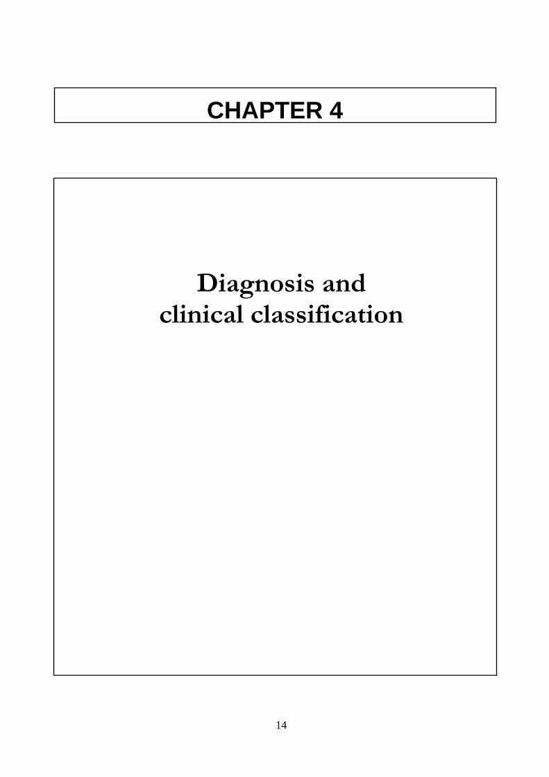
4. Diagnosis and clinical classification Diagnosis of perineal hernia is based on history, clinical signs and examination(19). In case of perineal hernia a simple digital rectal examination shows weakness and loss of integrity of pelvic diaphragm muscle and rotating the finger in a lateral or ventrolateral direction the tip can be easily identified in the subcutis of the perineal region. This clinical finding represent the gold standard for perineal hernia diagnosis(11, 16, 19).
Figure 3: rectal examination in a dog with
Perineal hernia.
Moreover most dogs are presented with a reducible perineal swelling. The swelling is usually located ventrolateral to the anus although ventral swelling with caudal projection of the anus becomes evident in some bilateral hernia (7, 28, 43, 46,
52). Once perineal hernia diagnosis is established, the role of the clinician is to assess the stage of the hernia and the secondary disease correlated. Always through the clinical examination it is possible to evaluate the following alterations: _ Site of the hernia: monolateral, bilateral, lateral, ventro-lateral. Detection of bilateral hernia seems to be a quite variable relief, it varies widely from 15% to 54% with a mean value of 36% (3,5,6,7,17,22,24,36,37,39,43,49,51,52). Some
16
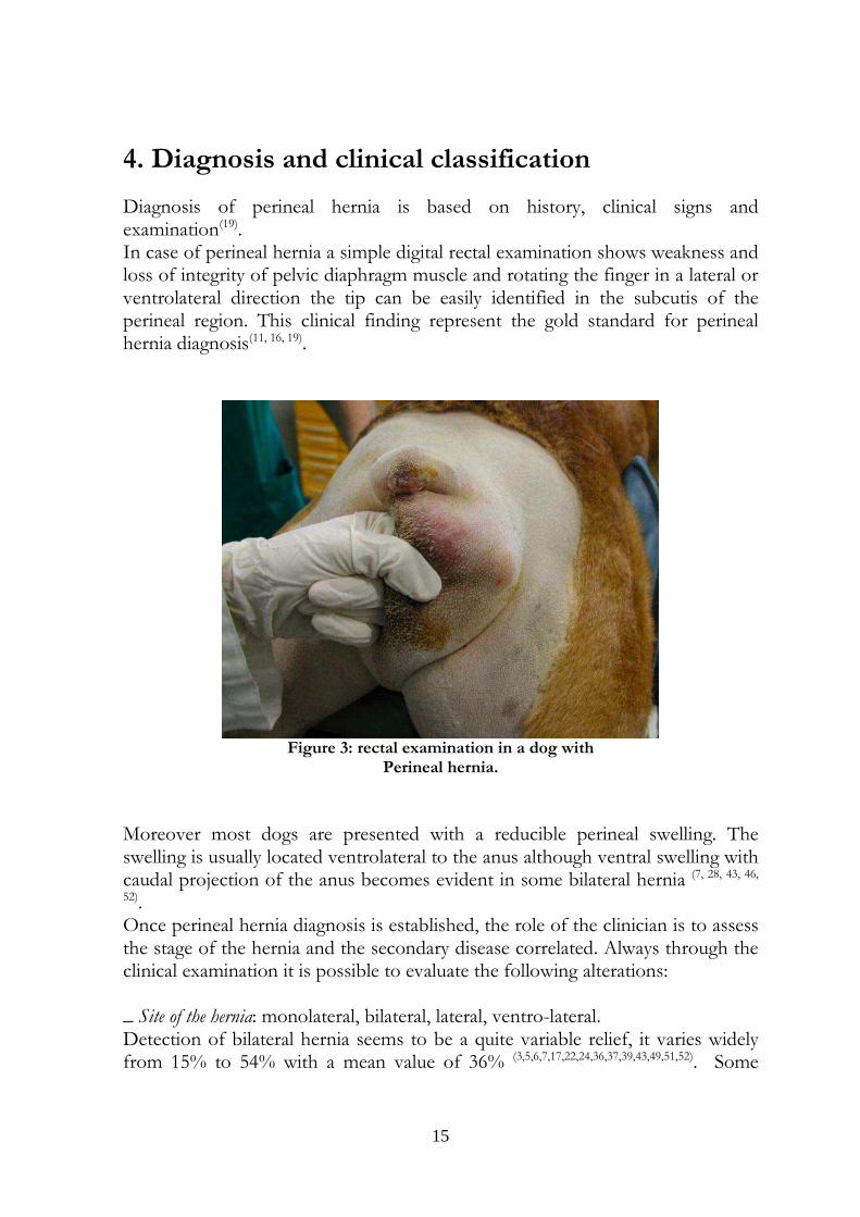
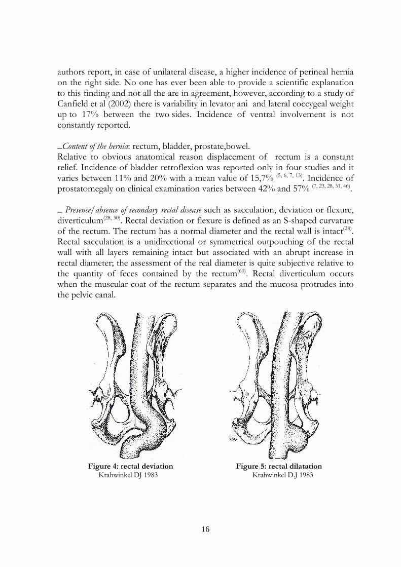
authors report, in case of unilateral disease, a higher incidence of perineal hernia on the right side. No one has ever been able to provide a scientific explanation to this finding and not all the are in agreement, however, according to a study of Canfield et al (2002) there is variability in levator ani and lateral coccygeal weight up to 17% between the two sides. Incidence of ventral involvement is not constantly reported. _Content of the hernia: rectum, bladder, prostate,bowel. Relative to obvious anatomical reason displacement of rectum is a constant relief. Incidence of bladder retroflexion was reported only in four studies and it varies between 11% and 20% with a mean value of 15,7% (5, 6, 7, 13). Incidence of prostatomegaly on clinical examination varies between 42% and 57% (7, 23, 28, 31, 46). _ Presence/absence of secondary rectal disease such as sacculation, deviation or flexure, diverticulum(28, 30). Rectal deviation or flexure is defined as an S-shaped curvature of the rectum. The rectum has a normal diameter and the rectal wall is intact(28). Rectal sacculation is a unidirectional or symmetrical outpouching of the rectal wall with all layers remaining intact but associated with an abrupt increase in rectal diameter; the assessment of the real diameter is quite subjective relative to the quantity of feces contained by the rectum(60). Rectal diverticulum occurs when the muscular coat of the rectum separates and the mucosa protrudes into the pelvic canal.
Figure 4: rectal deviation Figure 5: rectal dilatation Krahwinkel DJ 1983 Krahwinkel D.J 1983
17
Figure 6: rectal diverticulum
Krahwinkel DJ 1983
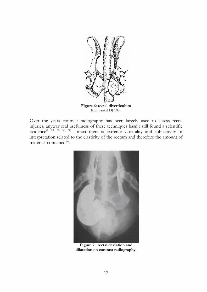
Over the years contrast radiography has been largely used to assess rectal injuries, anyway real usefulness of these techniques hasn’t still found a scientific evidence(1, 28, 30, 55, 60). Infact there is extreme variability and subjectivity of interpretation related to the elasticity of the rectum and therefore the amount of material contained(6).
Figure 7: rectal deviation and
dilatation on contrast radiography.
18
Since a universally accepted definition of rectal disease is a relatively recent acquisition in veterinary medicine it is difficult to perform a comparison of the incidence these diseases in the different studies. Thus the only author who has proposed a standardization of the clinical diagnosis of perineal hernia on the basis of the above criteria is Brissot HN; in a study published on Veterinary Surgery in 2004 the disease was staged as follows:
• Grade 0: no lesion.
• Grade 1: deviation without rectal dilatation.
• Grade 2: mild rectal dilatation (defined as asymmetric dilatation with fecal accumulation without visible perineal deformation; digital emptying of fecal material was easy).
• Grade 3: severe unilateral or mild to severe bilateral rectal dilatation (severe dilatation was defined as asymmetric dilatation with a visible bulge of the perineum with a large amount of fecal accumulation and impaction).
The use of imaging is an important step of the diagnostic work out. Primarily its role is to rule out any possible cause of chronic tenesmus as it represents one of the main suspected cause of the pathological process. Since this aspect is not related to the object of our study we are not going to deepen over this topic.
19
Surgical treatment of perineal hernia
CHAPTER 5
20
5. Surgical treatment of perineal hernia
Perineal hernia treatment may consist of either medical or elective surgical therapy. Medical therapy is indicated for preparing a patient for surgery but is unsuccessful at permanently controlling clinical signs(19). Therefore surgery represents the only effective approach. Since perineal hernia is a complex pathology and may involve different anatomical structures, surgical approach is often multistep and may include:
• Treatment of the damaged pelvic diaphragm
• Fixation of pelvic and abdominal structures
• Treatment of risk factors, such as prostatic disease (orchiectomy) Different surgical techniques have been proposed and they can be grossly divided in primary surgical repair (standard technique), transpositional techniques and surgical repair by mean of prosthetic structures.
5.1. Standard technique
This technique, also known as conventional or appositional technique, can simply be performed by suturing the damaged muscles of the pelvic diaphragm(7, 8, 13, 44, 60). Monofilament nylon sutures can be preplaced in order to appose the coccygeal muscles (if identifiable) to the external anal sphincter. Preplaced suture have to be tied and closure examined for any remaining defect in the pelvic diaphragm . Then a second row of suture must be placed between the subcutaneous fibrous connective tissue on the lateral side of the incision and the external anal sphincter on the medial side(13). The main limit of this approach is represented by the fact that perineal muscle are often atrophic and they are not safe structure for suture placement, this results in excessive high tension of the external sphincter and predispose suture dehiscence and hernia recurrence(6). In case of broad muscle atrophy some authors suggest to place sutures through the sacrotuberous ligament(3). Nevertheless the recurrence rate associated with this technique still remains between 19% and 46%(7, 18, 52).
21
5.2. Transpositional techniques Over the years different authors proposed novel surgical techniques in order to decrease the recurrence rate. All of these approaches share the same principle: use a vascularized muscle flap (a non-atrophied muscle, not involved in the pathological process) in order to provide additional reinforcement to the pelvic diaphragm . These techniques include superficial gluteal muscle transplantation(8, 55, 60), semitendinosus muscle flap(9), transposition of the internal obturator muscle(8, 19,
51, 53, 60). Transplantation of the superficial gluteal muscle was firstly described by Spreull et al in 1980. Superficial gluteal muscle must be identified immediately craniodorsal to the biceps where the latter arises from the sacrotuberous ligament. Its broad flat aponeurotic tendon is isolated and traced below the biceps muscle where it is cut at its insertion to the trochanter tertius. The muscle is then reflected taking care to preserve the anterior gluteal blood and nerve supply. The reflected belly of the muscle extends over the external anal sphincter muscle to which it is stitched. Main limits of this technique are the large wound required to expose the gluteal muscles, therefore surgery time may be relatively long. Moreover this approach does not provide a sufficient support in case of ventral hernia. Application of a semitendinosus muscle flap was described in two dogs by Chambers in 1991. The muscle must be dissected from the surrounding structures and the proximal vascular pedicle has to be identified. Then it must be transected at the midbelly and partially elevated by incising the lateral portion of its tendinous attachment to the ischium to allow further mobilization. The muscle can be rotated beneath the anus and the original medial border is sutured to the ventral aspect of the anal sphincter and the distal end of the flap is sutured to the contralateral internal obturator muscle. Although being relatively long and invasive this technique provide a strong support in case of ventral hernia. Transposition of the internal obturator muscle (IOMT) was described for the first time by Early TB in 1983. The muscle must be mobilised by severing its attachment to the caudal border of the ischium and the tendon is severed directly medial to the sacrotuberous ligament taking care to preserve pudendal nerve. Then the muscle must be reflected dorsally and sutured to the coccygeus and external sphincter muscle in order the close the ventral portion of the hernia. A conventional muscle apposition is then applied in order to the repair any eventual dorsal hernia. The technique can also be performed without severing the tendon of the internal obturator muscle(42) but this might greatly enhance tension at the suture line potentially increasing the recurrence rate(53).
22
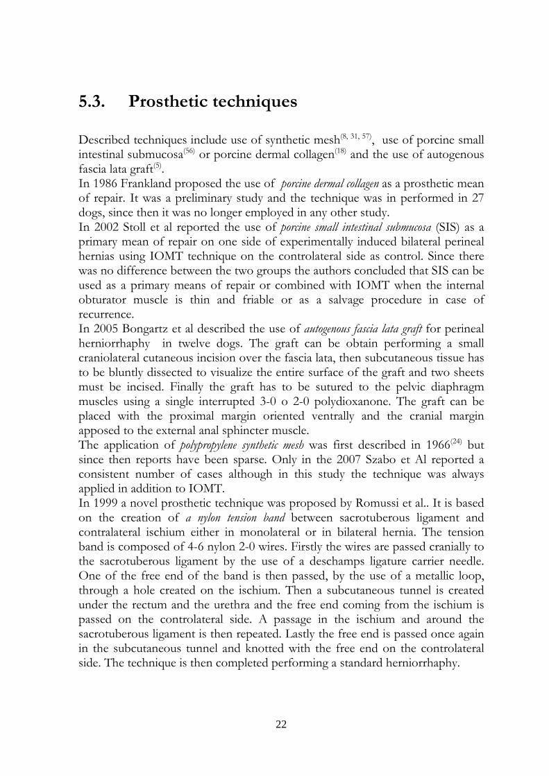
5.3. Prosthetic techniques Described techniques include use of synthetic mesh(8, 31, 57), use of porcine small intestinal submucosa(56) or porcine dermal collagen(18) and the use of autogenous fascia lata graft(5). In 1986 Frankland proposed the use of porcine dermal collagen as a prosthetic mean of repair. It was a preliminary study and the technique was in performed in 27 dogs, since then it was no longer employed in any other study. In 2002 Stoll et al reported the use of porcine small intestinal submucosa (SIS) as a primary mean of repair on one side of experimentally induced bilateral perineal hernias using IOMT technique on the controlateral side as control. Since there was no difference between the two groups the authors concluded that SIS can be used as a primary means of repair or combined with IOMT when the internal obturator muscle is thin and friable or as a salvage procedure in case of recurrence. In 2005 Bongartz et al described the use of autogenous fascia lata graft for perineal herniorrhaphy in twelve dogs. The graft can be obtain performing a small craniolateral cutaneous incision over the fascia lata, then subcutaneous tissue has to be bluntly dissected to visualize the entire surface of the graft and two sheets must be incised. Finally the graft has to be sutured to the pelvic diaphragm muscles using a single interrupted 3-0 o 2-0 polydioxanone. The graft can be placed with the proximal margin oriented ventrally and the cranial margin apposed to the external anal sphincter muscle. The application of polypropylene synthetic mesh was first described in 1966(24) but since then reports have been sparse. Only in the 2007 Szabo et Al reported a consistent number of cases although in this study the technique was always applied in addition to IOMT. In 1999 a novel prosthetic technique was proposed by Romussi et al.. It is based on the creation of a nylon tension band between sacrotuberous ligament and contralateral ischium either in monolateral or in bilateral hernia. The tension band is composed of 4-6 nylon 2-0 wires. Firstly the wires are passed cranially to the sacrotuberous ligament by the use of a deschamps ligature carrier needle. One of the free end of the band is then passed, by the use of a metallic loop, through a hole created on the ischium. Then a subcutaneous tunnel is created under the rectum and the urethra and the free end coming from the ischium is passed on the controlateral side. A passage in the ischium and around the sacrotuberous ligament is then repeated. Lastly the free end is passed once again in the subcutaneous tunnel and knotted with the free end on the controlateral side. The technique is then completed performing a standard herniorrhaphy.
23
Figure 8: a cadaveric scheme of the tension band technique. (Romussi et al 1999)
24
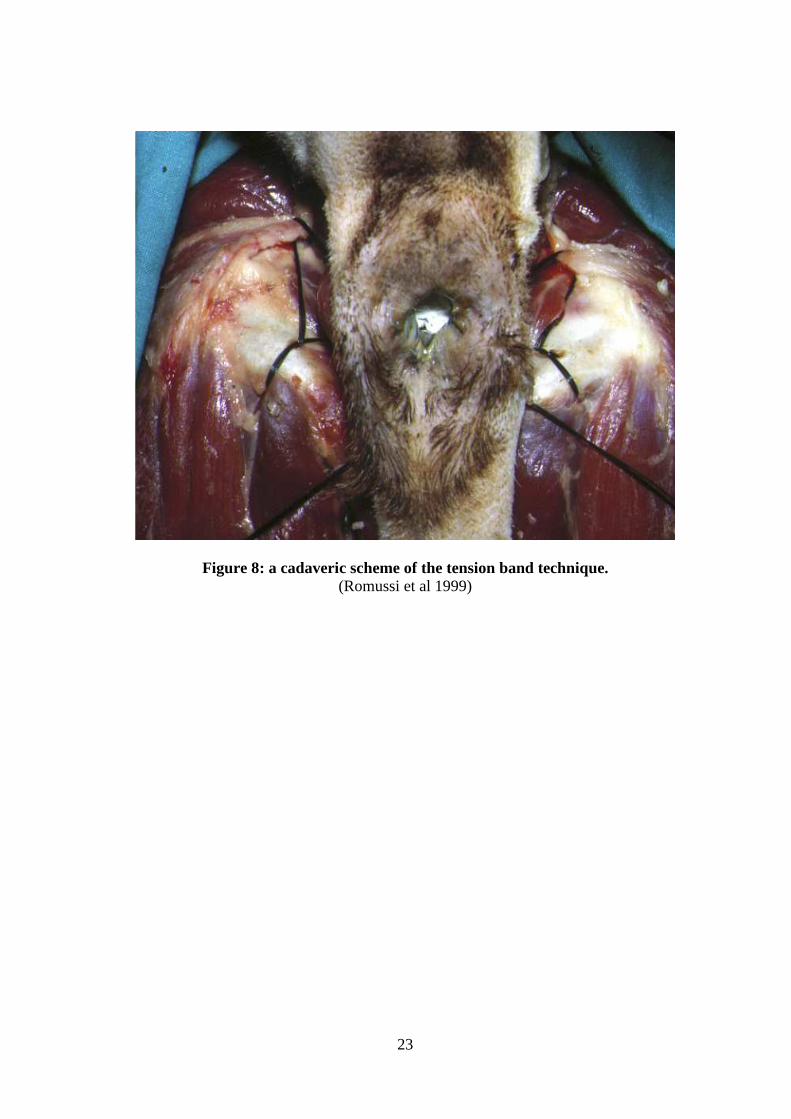
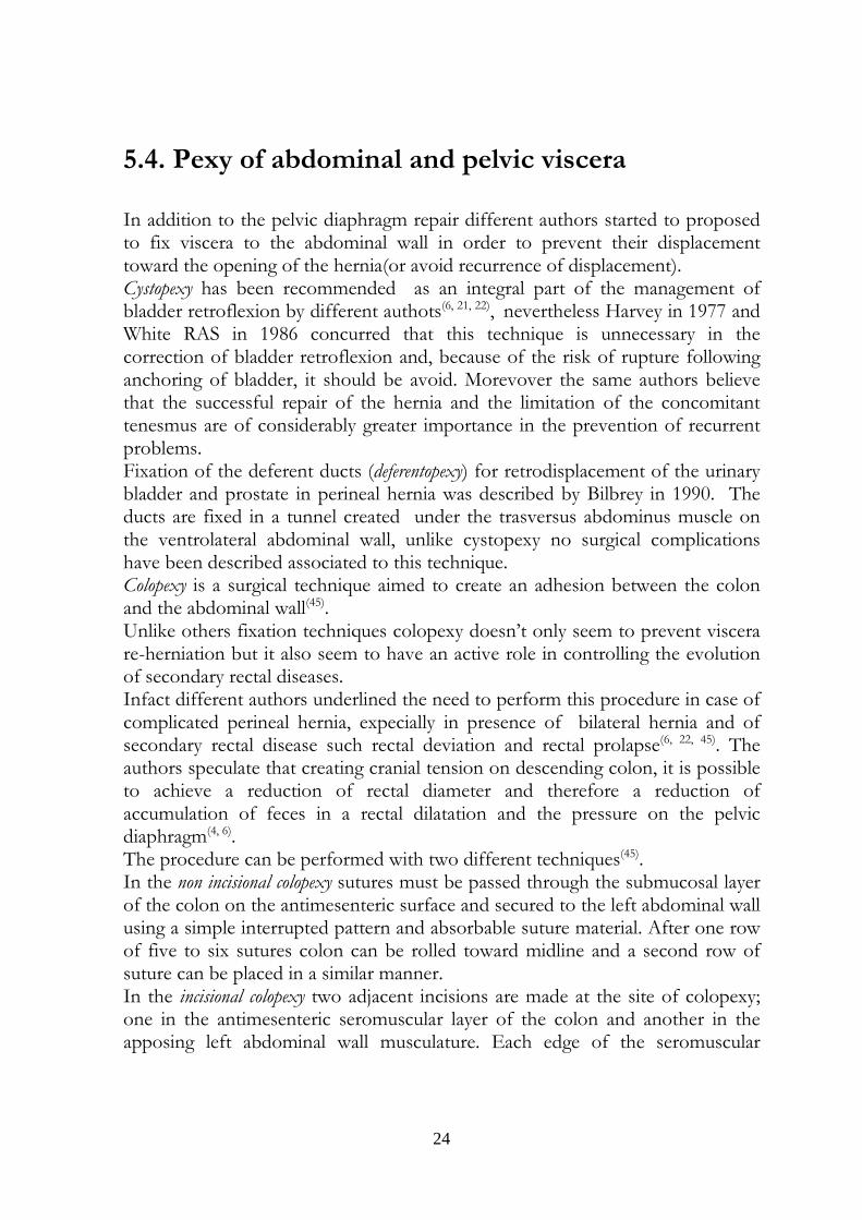
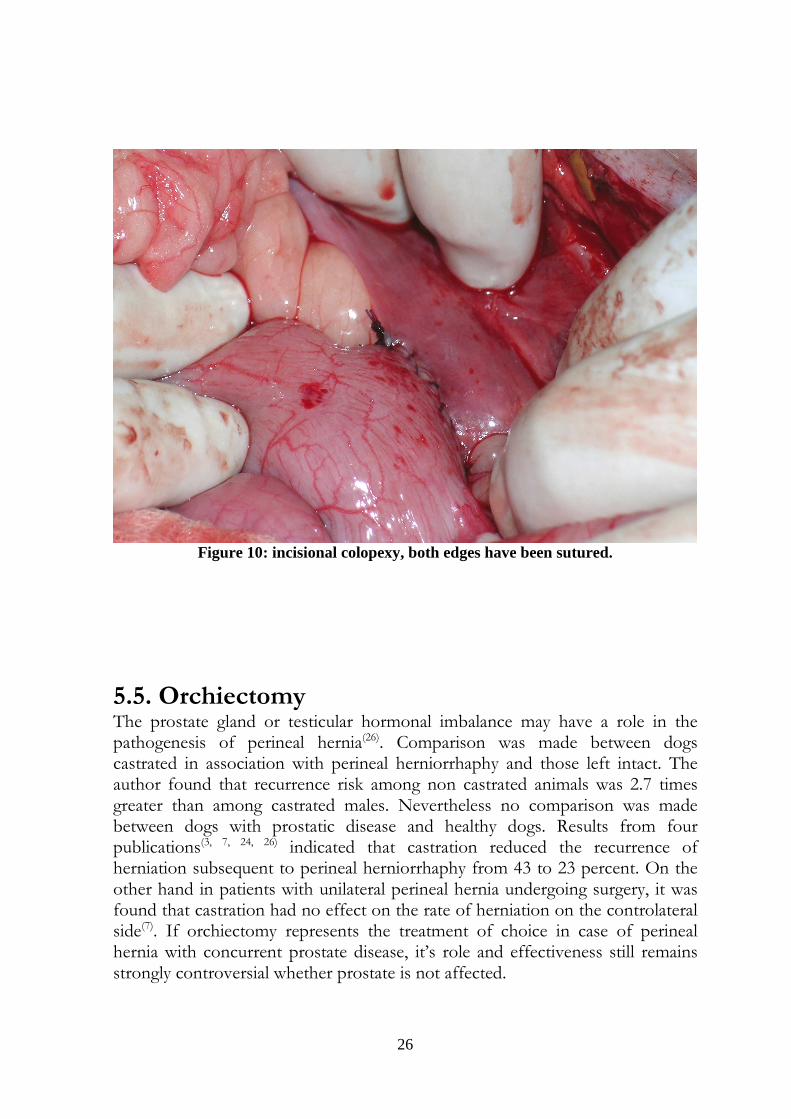
5.4. Pexy of abdominal and pelvic viscera In addition to the pelvic diaphragm repair different authors started to proposed to fix viscera to the abdominal wall in order to prevent their displacement toward the opening of the hernia(or avoid recurrence of displacement). Cystopexy has been recommended as an integral part of the management of bladder retroflexion by different authots(6, 21, 22), nevertheless Harvey in 1977 and White RAS in 1986 concurred that this technique is unnecessary in the correction of bladder retroflexion and, because of the risk of rupture following anchoring of bladder, it should be avoid. Morevover the same authors believe that the successful repair of the hernia and the limitation of the concomitant tenesmus are of considerably greater importance in the prevention of recurrent problems. Fixation of the deferent ducts (deferentopexy) for retrodisplacement of the urinary bladder and prostate in perineal hernia was described by Bilbrey in 1990. The ducts are fixed in a tunnel created under the trasversus abdominus muscle on the ventrolateral abdominal wall, unlike cystopexy no surgical complications have been described associated to this technique. Colopexy is a surgical technique aimed to create an adhesion between the colon and the abdominal wall(45). Unlike others fixation techniques colopexy doesn’t only seem to prevent viscera re-herniation but it also seem to have an active role in controlling the evolution of secondary rectal diseases. Infact different authors underlined the need to perform this procedure in case of complicated perineal hernia, expecially in presence of bilateral hernia and of secondary rectal disease such rectal deviation and rectal prolapse(6, 22, 45). The authors speculate that creating cranial tension on descending colon, it is possible to achieve a reduction of rectal diameter and therefore a reduction of accumulation of feces in a rectal dilatation and the pressure on the pelvic diaphragm(4, 6). The procedure can be performed with two different techniques(45). In the non incisional colopexy sutures must be passed through the submucosal layer of the colon on the antimesenteric surface and secured to the left abdominal wall using a simple interrupted pattern and absorbable suture material. After one row of five to six sutures colon can be rolled toward midline and a second row of suture can be placed in a similar manner. In the incisional colopexy two adjacent incisions are made at the site of colopexy; one in the antimesenteric seromuscular layer of the colon and another in the apposing left abdominal wall musculature. Each edge of the seromuscular
25
colonic incision is sutured separately to the corresponding edge of the incision in the abdominal wall. A simple interrupeted pattern can be used. Although similar surgical procedure (such as gastropexy) using as simple suturing technique has been reported to be unsuccessful in forming a permanent adhesion, Popovitch et al in 1994 did not find any difference in long term clinical outcome when comparing incisional and non incisional colopexy.
Figure 9: incisional colopexy, the first edge of the seromuscular incision
has been sutured.
26
Figure 10: incisional colopexy, both edges have been sutured.
5.5. Orchiectomy The prostate gland or testicular hormonal imbalance may have a role in the pathogenesis of perineal hernia(26). Comparison was made between dogs castrated in association with perineal herniorrhaphy and those left intact. The author found that recurrence risk among non castrated animals was 2.7 times greater than among castrated males. Nevertheless no comparison was made between dogs with prostatic disease and healthy dogs. Results from four publications(3, 7, 24, 26) indicated that castration reduced the recurrence of herniation subsequent to perineal herniorrhaphy from 43 to 23 percent. On the other hand in patients with unilateral perineal hernia undergoing surgery, it was found that castration had no effect on the rate of herniation on the controlateral side(7). If orchiectomy represents the treatment of choice in case of perineal hernia with concurrent prostate disease, it’s role and effectiveness still remains strongly controversial whether prostate is not affected.
27
5.6. Surgical complications Different surgical complications have been described over the years; reasonably every complication is mainly associated with a specific surgical technique. We are going to expose tipology and incidence of these complications as described in small animal veterinary literature. Intraoperative surgical complications can be roughly divided in three main categories: bleeding, injuries to the nerve structures and injury to the rectal wall. As previously described precise knowledge of the anatomy is a fundamental background in order to approach surgery of perineal region and prevent the onset of major complications. Significant bleeding occurs in case of main vessel injuries such as internal pudenda artery/vein and caudal gluteal artery. Due to obvious anatomical reasons IOMT is mainly associated to internal pudenda vessel bleeding. Bleeding from the caudal gluteal artery can be caused by passage of prosthetic material or suture around the sacrotuberous ligament. Similarly, injury to the sciatic nerve can occur with the same procedures and its incidence is reported in one study at 5%(7). After surgery animal may demonstrate nonweight bearing lameness secondary to sciatic pain. In dogs with sciatic nerve paralysis, the tarsus is extended and the toes knuckle if weight bearing is attempted. Cranial tibial and withdrawal reflexes are hyporeflexic or areflexic; sensation to the 3°-4° digitis and lateral aspect of the tarsus may be affected. This complication should be considered a surgical emergency(39). In rare cases sciatic nerve entrapment occurs several weeks after surgery, caused by a suture granuloma impinging on the sciatic nerve. Complete return of sciatic nerve function, after removal of a misplaced suture, may take several weeks to months(38). Sciatic nerve neuropraxia can result from positioning a dog in a perineal stand with the hind limb tied firmly forward; after surgery dogs exhibits knuckling on the paw but no sciatic pain is evident. However differentiating the two complications can be clinically difficult(38). Occasionally the anal sac or rectum can be penetrated by a suture during the repair of the hernia (potentially with any surgical technique) increasing the risk of postoperative infection. Urethral trauma has been also reported by Sereda et Al in 2002. Among postoperative complications wound infection is the most commonly reported. Wound site, near the anus, represents the main risk factor. The incidence of this complication varies widely between different studies and it has an average value of 26%(3, 6, 7, 43, 54). The appearance of fistulous tracts has been described almost exclusively after prosthetic techniques(48, 57).
28
Wound seroma varies from 6% to 33% (mean 16%) apparently without any correlation with a specific surgical procedure(22, 43, 46). Fecal incontinence has a mean incidence value of 8%(3, 7, 51), it is interesting to observe how this complication is more frequently described in oldest studies and its incidence decreases within the years. Urinary incontinence cannot be considered as a true surgical complication rather than a direct effect of bladder retroflexion resulting in nerves stretching. A study by White RAS in 1986 showed that 25% of dogs affected by bladder retroflextion developed permanent incontinence. Rarely, bladder necrosis and rupture has been described and they are due to retroflextion, moreover it has also been described as a surgical complication of cystopexy(24, 61). Some authors report the appeareance of rectal prolapse usually occurring during the immediate postoperative recovery phase of anesthesia because of rectal straining(38). The incidence of this complication seems to be reduced in those studies were colopexy is performed.
29
Review of methodologies of collection of follow up
in veterinary literature on perineal hernia
CHAPTER 6
30
6. Review of methodologies of collection of follow up in veterinary literature on perineal hernia In this chapter we are going to review methodologies of follow up collection employed in clinical studies on perineal hernia treatment. Specifically our interest will focus on follow up duration, method of follow up collection (clinical examination vs questionnaire), presence or absence of pre and post surgery clinical staging of rectal disease and evaluation of incidence rate of recurrence. Concerning to the duration of follow up period it is surprising to note that in five studies this data have not been specified(31,43, 44, 52, 59). A maximum follow up period of six months was available in two studies(5, 28). In studies published by Burrows FC, Harvey CE and Bellenger CR a minimum follow up period of six months was assessed but its maximum or medium value has not been indicated. In 1983 Hardie EM reassessed the patients one year after surgery. A mean follow up period of more than 24 months was available in five studies(6, 18, 46, 57, 60). The same heterogeneity is present if we analyze method of collection of follow up data. In six clinical studies the authors do not even specify if they have collected follow up through clinical examination (or data available on clinical record) or questionnaire(18, 21, 44, 52, 59, 60). A telephone questionnaire has been used as unique method of follow up data collection by five authors (4, 6, 23, 24, 57). Only two authors performed follow up through a clinical examination as a sole method(5, 43). In the remaining studies data has been collected either reviewing clinical record or through a questionnaire whether the quality and quantity of informations found on records were considered insufficient(3, 7, 28, 46). If we look at the method of collection and select only those studies with a clinical postoperative evaluation, follow up periods decrease greatly. Indeed in three studies we have found a maximum clinical follow up period of six months(5, 7, 28) while only one author performed a clinical re-examination 24 months after the day of surgery(46). Even less data can be found about clinical staging of rectal disease. In three studies the authors performed a pre operative classification of rectal anomalies, however classification criteria vary between the studies(3, 6, 28). Only one author(43) performed a pre and post operative assessment of rectal disease. The clinical studies provided inconstant data about presence of bilateral versus monolateral disease and the viscera herniated. Moreover only few studies stated the duration of symptoms before surgical treatment(6, 61). Also recurrence rate varies widely between studies. The use of standard technique showed a recurrence rate of 7%, 9.7%, 15%, 19%, 27%, 37,5% and 46%(7, 25, 31, 44, 59, 60). It’s interesting to note that studies with lowest recurrence rate

31
share the common lack of information about duration and method of follow up collection. Transposition of the superficial gluteal muscle showed a recurrence rate of 36% (60). In the eighties the use of internal obturator muscle transposition as unique surgical approach demonstrated a recurrence rate of 14%(43), 2,3%(23) and 5%(52). These publications varies greatly concerning follow up duration and method of collection, moreover Hardie EM and Sjollema BE performed the technique with a complete tenotomy while Orsher RJ did non performed tenotomy . Recently Vnuk D et al showed a recurrence rate of 11% with IOMT although nothing is specified about the execution of tenotomy and method of follow up. In 1986 Frankland AL et al performed perineal hernia repair with the use of porcine dermal collagen as a prosthetic technique and they demonstrated a recurrence rate of 41%. Bongartz A in 2005 did not have any recurrence treating fourteen perineal hernias by the use of autogenous fascia lata graft, on the other hand follow up period had a mean duration value of 5,6 months. Finally we are going to analyze clinical studies that combined two different surgical techniques. In 1983 Raffan PJ treated 52 perineal hernias both with IOMT and transposition of superficial gluteal muscle and they obtained a 2.6% recurrence rate. Brissot NH et al used a double stage approach to treat complicated bilateral hernia in 41 dogs, all the patients were treated both with colopexy (and cisto-deferentopexy whether necessary) and IOMT, the results showed a 9% recurrence rate. A 12.5% recurrence rate was obtained by Szabo S et al in 2007 treating their patient both with IOMT and polypropylene mesh. Incidence of recurrence in the study from Romussi S et al was 13%.
32
Matherials and methods
CHAPTER 7
33
7.1. Inclusion criteria Client owned dogs referred to the Sezione di Clinica Chirurgica of the Dipartimento di Scienze Cliniche Veterinarie of the Faculty of Veterinary Medicine of Milan from July 2007 to November 2010 were prospectively enrolled in the study when met the following inclusion criteria: confirmed clinical diagnosis of perineal hernia, presence of secondary rectal disease or complicated hernia(6), absence of any other concomitant disease of the perineum or any other comorbidities affecting the process of defecation.
7.2. Diagnostic procedures The evaluation of the patients included the compilation of a record aimed at thoroughly characterized the severity of the disease. The clinical record included data regarding signalment, history and any previous medical or surgical treatment. In particulary the presence and frequency of the following clinical signs were taken into account: tenesmus, dyschezia, ematochezia and dysuria. Moreover in each patient the extension of the hernia (monolateral versus bilateral, ventral extension), its content (prostate, bladder, bowel) presence/absence of prostatomegaly were described. We also planned to perform a clinical classification of the hernia based on rectal disease, we used a classification scheme proposed by Brissot NH et al in 2004: _ grade 0: no lesion. _ grade 1: deviation without rectal dilatation. _ grade 2: mild rectal dilatation (defined as asymmetric dilatation with fecal accumulation without visible perineal deformation, digital emptying of fecal material was easy). _ grade 3: severe rectal dilatation (asymmetric dilatation with a visible bulge of the perineum with a large amount of fecal accumulation and impaction). Abdomen x-rays were performed in order to assess the amount of fecal impaction in the colon. Ultrasound of the abdomen and cytology were performed if any prostate alteration was detect by clinical examination.
34
7.3. Treatment and experimental protocol A preoperative work out (blood analysis and echocardiography) was performed in each patient. Dogs were randomly allocated in two different groups. In group A patients were treated with nylon tension band technique(48). Dogs from group B were treated both with the nylon tension band technique and colopexy, in a one stage procedure. Orchiectomy was performed in both groups if any prostate disease was diagnosed. In group B colopexy end orchiectomy were performed first, the dogs were positioned in a sternal recumbency and perineal herniorrhaphy was achieved. The surgical technique employed for perineal hernia repair was described in a previous chapter. Colopexy was performed by the left flank with incisional musco-muscular technique(45). Perineal hernia repair was performed bilaterally in case of bilateral hernia or monolateral hernia with impairment of contralateral pelvic diaphragm. Perineal herniorrhaphy was performed in sternal recumbency with the pelvis slightly elevated above the head. The tail was partially clipped (the first 2/3 for large dogs with a long tail, the whole tail for dogs with a short tail or for small size dogs) and retracted and held over the back with care to avoid excessive tension and to keep the tail in the sagittal plane. Anal sacs were manually expressed and the caudal part of the rectum was evacuated digitally. An anal purse-string suture was placed before surgery. An urinary catheter was preplaced in all procedures. For laparotomy, dogs were positioned in a dorsal recumbency All surgical procedures were performed by the same surgeon. Duration of surgeries was recorded. Analgesia was administrated in the perioperative period as needed. Dogs were closely monitored in postoperative period and were generally discharged from the hospital 24-48 hours after surgery. Owners were instructed to monitor their dogs for any defecation problem, limit exercise to short ten minutes walks 3-4 times daily and keep clean the perineal surgical wound daily by use of a moistened gauze. The elisabetta collar was also considered mandatory for at least seven days. The diet was permanently modified adding vegetables in order to obtain softer stool.
7.4. Follow up Dogs were clinically revaluated 7 up to 15 days after surgery and the presence of any postoperative complication was reported. Then a clinical examination was scheduled at least 12 months after surgery in order to assess surgical outcome.
35
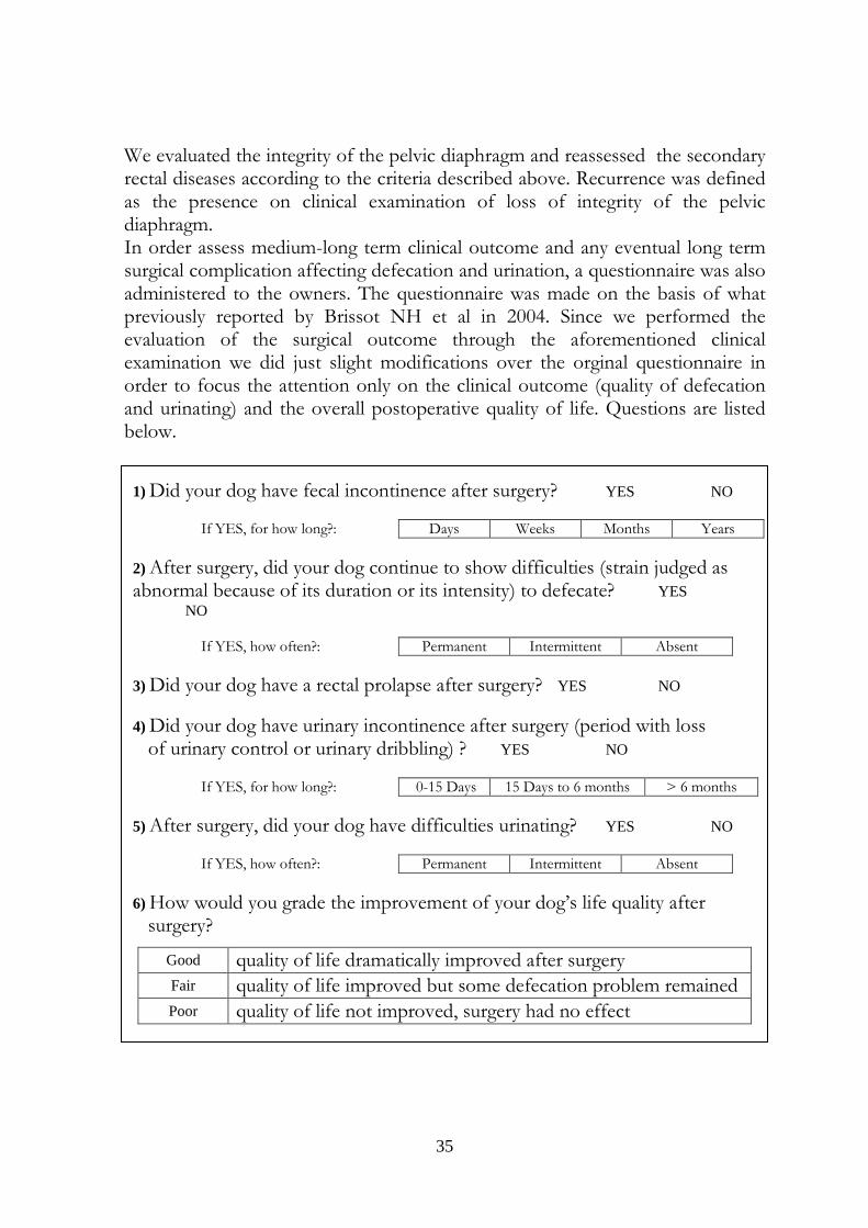
We evaluated the integrity of the pelvic diaphragm and reassessed the secondary rectal diseases according to the criteria described above. Recurrence was defined as the presence on clinical examination of loss of integrity of the pelvic diaphragm. In order assess medium-long term clinical outcome and any eventual long term surgical complication affecting defecation and urination, a questionnaire was also administered to the owners. The questionnaire was made on the basis of what previously reported by Brissot NH et al in 2004. Since we performed the evaluation of the surgical outcome through the aforementioned clinical examination we did just slight modifications over the orginal questionnaire in order to focus the attention only on the clinical outcome (quality of defecation and urinating) and the overall postoperative quality of life. Questions are listed below.
1) Did your dog have fecal incontinence after surgery? YES NO
If YES, for how long?: Days Weeks Months Years
2) After surgery, did your dog continue to show difficulties (strain judged as abnormal because of its duration or its intensity) to defecate? YES NO
If YES, how often?: Permanent Intermittent Absent
3) Did your dog have a rectal prolapse after surgery? YES NO 4) Did your dog have urinary incontinence after surgery (period with loss of urinary control or urinary dribbling) ? YES NO
If YES, for how long?: 0-15 Days 15 Days to 6 months > 6 months
5) After surgery, did your dog have difficulties urinating? YES NO
If YES, how often?: Permanent Intermittent Absent
6) How would you grade the improvement of your dog’s life quality after surgery?
Good quality of life dramatically improved after surgery
Fair quality of life improved but some defecation problem remained
Poor quality of life not improved, surgery had no effect

36
Results
CHAPTER 8
37
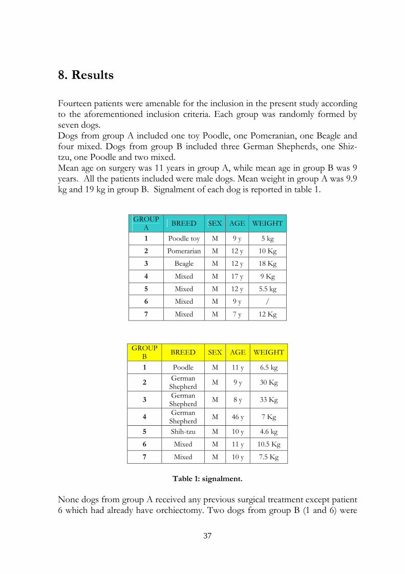
8. Results Fourteen patients were amenable for the inclusion in the present study according to the aforementioned inclusion criteria. Each group was randomly formed by seven dogs. Dogs from group A included one toy Poodle, one Pomeranian, one Beagle and four mixed. Dogs from group B included three German Shepherds, one Shiz-tzu, one Poodle and two mixed. Mean age on surgery was 11 years in group A, while mean age in group B was 9 years. All the patients included were male dogs. Mean weight in group A was 9.9 kg and 19 kg in group B. Signalment of each dog is reported in table 1.
Table 1: signalment.
None dogs from group A received any previous surgical treatment except patient 6 which had already have orchiectomy. Two dogs from group B (1 and 6) were
GROUP A
BREED SEX AGE WEIGHT
1 Poodle toy M 9 y 5 kg
2 Pomerarian M 12 y 10 Kg
3 Beagle M 12 y 18 Kg
4 Mixed M 17 y 9 Kg
5 Mixed M 12 y 5.5 kg
6 Mixed M 9 y /
7 Mixed M 7 y 12 Kg
GROUP B
BREED SEX AGE WEIGHT
1 Poodle M 11 y 6.5 kg
2 German Shepherd
M 9 y 30 Kg
3 German Shepherd
M 8 y 33 Kg
4 German Shepherd
M 46 y 7 Kg
5 Shih-tzu M 10 y 4.6 kg
6 Mixed M 11 y 10.5 Kg
7 Mixed M 10 y 7.5 Kg
38
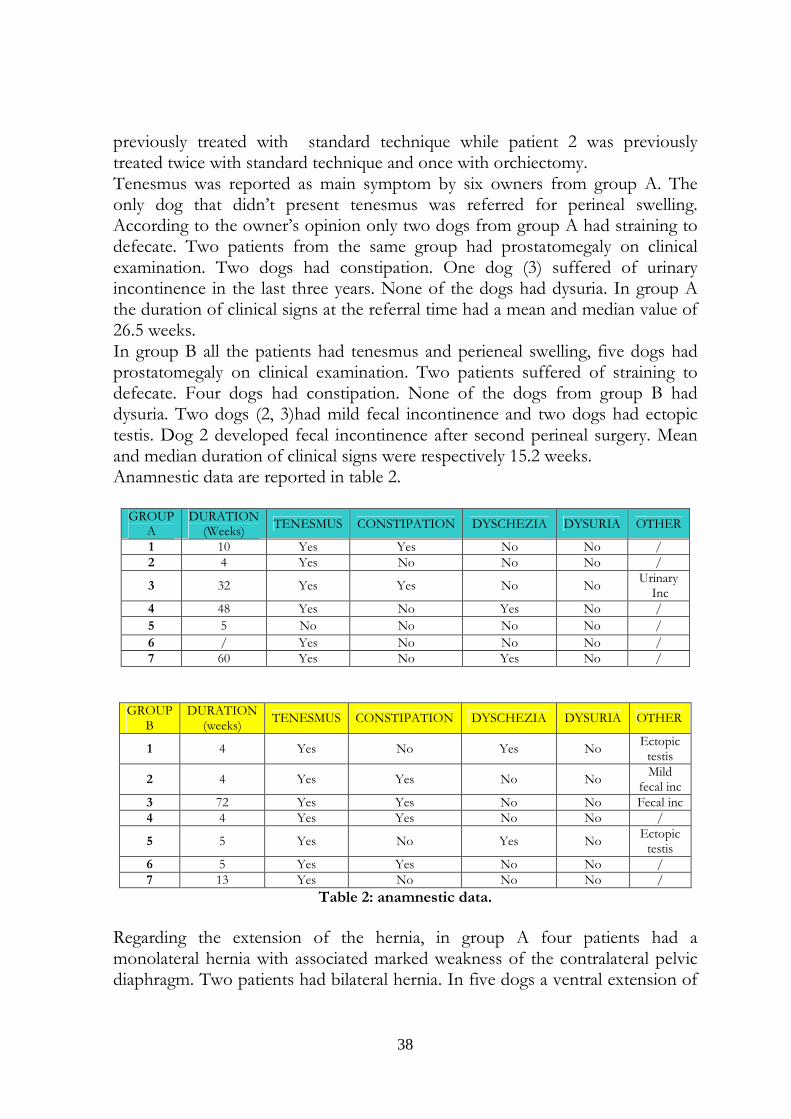
previously treated with standard technique while patient 2 was previously treated twice with standard technique and once with orchiectomy. Tenesmus was reported as main symptom by six owners from group A. The only dog that didn’t present tenesmus was referred for perineal swelling. According to the owner’s opinion only two dogs from group A had straining to defecate. Two patients from the same group had prostatomegaly on clinical examination. Two dogs had constipation. One dog (3) suffered of urinary incontinence in the last three years. None of the dogs had dysuria. In group A the duration of clinical signs at the referral time had a mean and median value of 26.5 weeks. In group B all the patients had tenesmus and perieneal swelling, five dogs had prostatomegaly on clinical examination. Two patients suffered of straining to defecate. Four dogs had constipation. None of the dogs from group B had dysuria. Two dogs (2, 3)had mild fecal incontinence and two dogs had ectopic testis. Dog 2 developed fecal incontinence after second perineal surgery. Mean and median duration of clinical signs were respectively 15.2 weeks. Anamnestic data are reported in table 2.
GROUP A
DURATION (Weeks)
TENESMUS CONSTIPATION DYSCHEZIA DYSURIA OTHER
1 10 Yes Yes No No / 2 4 Yes No No No /
3 32 Yes Yes No No Urinary
Inc 4 48 Yes No Yes No /
5 5 No No No No /
6 / Yes No No No / 7 60 Yes No Yes No /
GROUP
B DURATION
(weeks) TENESMUS CONSTIPATION DYSCHEZIA DYSURIA OTHER
1 4 Yes No Yes No Ectopic
testis
2 4 Yes Yes No No Mild
fecal inc 3 72 Yes Yes No No Fecal inc 4 4 Yes Yes No No /
5 5 Yes No Yes No Ectopic
testis 6 5 Yes Yes No No / 7 13 Yes No No No /
Table 2: anamnestic data.
Regarding the extension of the hernia, in group A four patients had a monolateral hernia with associated marked weakness of the contralateral pelvic diaphragm. Two patients had bilateral hernia. In five dogs a ventral extension of
39
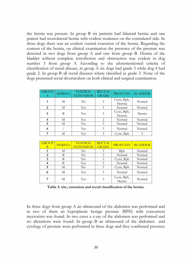
the hernia was present. In group B six patients had bilateral hernia and one patient had monolateral hernia with evident weakness on the contralaterl side. In three dogs there was an evident ventral extension of the hernia. Regarding the content of the hernia, on clinical examination the presence of the prostate was detected in two dogs from group A and one from group B. Hernia of the bladder without complete retroflexion and obstruction was evident in dog number 3 from group A According to the aforementioned criteria of classification of rectal disease, in group A six dogs had grade 3 while dog 4 had grade 2. In group B all rectal diseases where classified as grade 3. None of the dogs presented rectal diverticulum on both clinical and surgical examination.
Table 3: site, extension and rectal classification of the hernia.
In three dogs from group A an ultrasound of the abdomen was performed and in two of them an hyperplastic benign prostate (BPH) with concurrent mycrocists was found. In two cases a x-ray of the abdomen was performed and no alterations were found. In group B an ultrasound of the abdomen and cytology of prostate were performed in three dogs and they confirmed presence
GROUP A
HERNIA VENTRAL
EXTENSION RECTAL GRADE
PROSTATE BLADDER
1 M No 3 Cysts, Bph,
Hermia Normal
2 M Yes 3 Normal Normal
3 B Yes 3 Cysts, Bph,
Hermia Hernia
4 M Yes 2 Normal Normal 5 M Yes 3 Normal Normal
6 / Yes 3 Normal Normal
7 M Yes 3 Cysts, Bph 3
GROUP B
HERNIA VENTRAL
EXTENSION RECTAL GRADE
PROSTATE BLADDER
1 M No 3 Bph Normal 2 B No 3 Normal Normal 3 B Yes 3 Cysts, Bph Normal 4 B Yes 3 Normal Normal 5 M No 3 Cysts, Bph Normal
6 M Yes 3 Normal Normal
7 M Yes 3 Cysts, Bph,
Hernia Normal
40
of BPH. In three dogs a x-ray of the abdomen was performed and in two cases a huge fecal impaction was found. Concerning to the surgical plan, orchiectomy (1, 2, 7) was carried out in three dogs of the group A. In dogs 3 orchiectomy was not performed since the patient had a mild preoperative urinary incontinence. A mean surgical time was 87.5 min. In group B orchiectomy was performed in six patients and the mean duration of surgery was 129.3 min. All the patients included in the study have been treated with bilateral nylon tension band technique(48) since all the monolateral hernia had a concomitant weakness on the contralateral side. On postoperative clinical examination (7-15 days) in group A only one patient (dog 1) showed a delayed wound healing on the right side, the owner of dog 4 reported temporary appearance of straining to defecate for the first two days after surgery. In group B immediately after surgery two dogs (3, 6) had tenesmus and straining to defecate but their clinical condition normalized within ten days. One dog (4) developed a seroma of the perineal wound and it solved within ten days. The remaining dogs didn’t show any complication of the surgical wound both in perineal and abdominal area. In group A mean follow up duration was 525 days. Two patients (dogs 1 and 3) from group A developed recurrence of perineal hernia. Dog 1 developed recurrence 208 days after surgery and dog 3 developed recurrence 177 days after surgery. Obviously grading of rectal disease and questionnaire were not performed in these patients. According to the classification of the rectal diseases, to all the remaining five dogs a grade 2 was assigned, since a mild rectal dilatation was still present. Due to the repair of the hernia deviation and perineal bulging was no longer present. According to the questionnaire only dog 4 developed mild urinary incontinence and, to the owner’s opinion, it was more evident within the first six months and then it almost solved. Dog 6 developed mild fecal incontinence. Regarding urination and defecation no other dogs developed medium-long term complications. None of the patients developed rectal prolapse. The owners of the two dogs that had recurrence, graded as poor the improvement of quality of life while it was graded as good in the remaining five cases. In group B mean follow up duration was 545 days. None of the patients developed recurrence. According to the classification of rectal disease dog 1 had grade 0 while all the remaining patients had grade 2. According to the owner’s opinion five dogs (2, 4, 5, 6, 7) still had mild and intermittent tenesmus. One (2) of the two dogs that had fecal incontinence previous surgery still had it on long term follow up. None of the remaining dogs developed fecal incontinence.
41
Two dogs had mild urinary incontinence. In dog 1 incontinence had started soon after the first surgery but it didn’t change in frequency and severity after our surgery. In dog 4 mild urinary incontinence had started fifteen months after surgery. None of the patients had dysuria and rectal prolapse. All the owners graded as good the improvement of life quality.
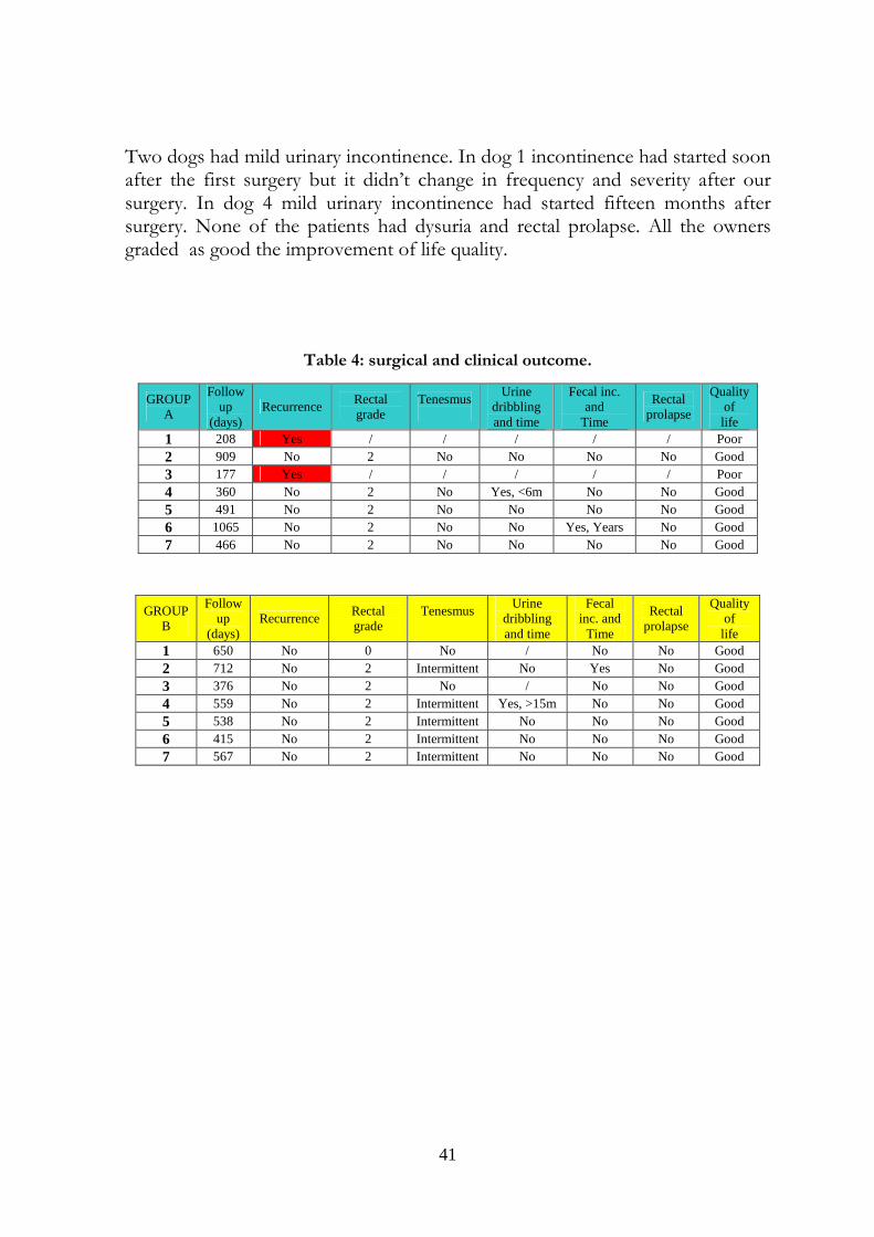
Table 4: surgical and clinical outcome.
GROUP A
Follow up
(days) Recurrence
Rectal grade
Tenesmus
Urine dribbling and time
Fecal inc. and
Time
Rectal prolapse
Quality of life
1 208 Yes / / / / / Poor 2 909 No 2 No No No No Good 3 177 Yes / / / / / Poor 4 360 No 2 No Yes, <6m No No Good 5 491 No 2 No No No No Good 6 1065 No 2 No No Yes, Years No Good 7 466 No 2 No No No No Good
GROUP B
Follow up
(days) Recurrence
Rectal grade
Tenesmus
Urine dribbling and time
Fecal inc. and
Time
Rectal prolapse
Quality of
life 1 650 No 0 No / No No Good 2 712 No 2 Intermittent No Yes No Good 3 376 No 2 No / No No Good 4 559 No 2 Intermittent Yes, >15m No No Good 5 538 No 2 Intermittent No No No Good 6 415 No 2 Intermittent No No No Good 7 567 No 2 Intermittent No No No Good
42
Discussion
CHAPTER 9

43
9. Discussion Surgical treatment of perineal hernia still represents a challenging issue for veterinary surgeons. More than sixty indexed clinical studies have been published over the years and at least ten different techniques have been proposed. These aspects reflect the complexity of the disease. The goal of surgical treatment of perineal hernia cannot only focus on the avoidance of the failure of pelvic diaphragm. As we mentioned in previous chapters, due to the chronicity of the process, lots of important alterations can affect pelvic and abdominal viscera, seriously increasing the severity of the disease. Krahwinkel DJ et al in 1983 firstly focused the attention on secondary rectal disease as potential negative prognostic factors. Moreover three years later Orsher RJ et al stated that clinical outcome cannot only rely on the effective repair of the pelvic diaphragm since many others aspects can influence the quality of life of a dog with perineal hernia. In fact a normal postoperative process of defecation is not only dependent to the absence of the hernia but also to the capability of rectum to eject its content and of the external sphincter to maintain continence. All these single processes act dependently and sinergically to affect the outcome. Even if the complexity of the assessment of results of perineal herniorrhaphy has been stated almost fourty years ago lots of successive studies still went on focusing only on the incidence of recurrence and complications related with the technique employed. In 1974 Amand WB firstly affirmed the need to perform “bowel surgery” in case of deviation and diverticulum. Anal resection turned out to be unsuccessful and correlated with a high risk of fecal contamination of the surgical site. However colopexy was firstly described almost ten years later(16). Over the years different authors demonstrated its role in controlling rectal prolapse and secondary rectal disease such as deviation(49). Some authors also affirmed that colopexy is effective in reducing rectal diameter(6, 15, 16, 29). However these statement did never find any scientific confirmation since none of the authors performed any clinical examination of rectal diseases in both pre and postoperative period. To our knowledge the effectiveness of colopexy in controlling rectal dilatation still remains confined to “author’s opinion” or “author’s experience”(6). Even less data are published about the real impact of colopexy on the incidence of recurrence. A recent retrospective study showed encouraging results(6). However in that study colopexy was performed in addition with other technique such as cystopexy or deferentopexy, the surgical outcome was assessed only through questionnaire and dogs treated with colopexy were not compared with a control group.
44
Starting from all these considerations we planned to perform a prospective randomized evaluation of both surgical and clinical impact of colopexy on perineal hernia treatment. Concerning the surgical outcome our results show a sharp distinction with regard on incidence of recurrence between the two groups. In fact we had a 28% of recurrence in group A and no recurrence in group B. On the other hand no evident differences can be found in the grading of rectal disease. All patients but one from group B still had a mild rectal dilatation (grade 2) on postoperative reassessment. If we can clearly affirm that colopexy has positively influenced the efficacy of the repair of perineal damage on the other hand our results do not seem to confirm some of the previously published hypothesis(6) according to which colopexy is effective in solving rectal dilatation. According to our results it is at least an uncommon event. If the annulment of rectal dilatation seem to be uncommon, nothing can be stated about the effectiveness of colopexy in at least reducing the diameter of the viscera. Since, with the exception of one patience, all dogs from both groups received the same score we are not able to verify our starting hypothesis. To our opinion this aspect may be dependent to the choice of grading system and to the lack of objective diagnostic tools. Actually there is no gold standard to differentiate and especially state the severity of rectal disease since both digital rectal examination and contrast radiography depend on the subjectivity of the clinician and the elasticity of the viscera. We decided to use a grading system proposed by Brissot et al in 2004. The criteria used by the authors are based on the only objectives clinical aspects of the disease: presence/absence of deviation and presence/absence of perineal bulge. Presence of deviation toward the opening of the pelvic diaphragm is the first stage of the rectal disease. To differentiate the severity of rectal disease the author used the presence/absence of perineal bulge, which is a simple and objective relief. Since any effective surgical repair inevitably involve the reduction of perineal bulge it almost obvious that all those hernia firstly classified as grade 3 received a lowest grade after surgery. The reduction of the grade is therefore due to the herniorrhaphy rather then the colopexy itself. In the absence of perineal bulge we were not able to rely on any other objective parameter in order to quantify the impact of colopexy on rectal dilatation. Moreover we performed a dietary change in the postoperative period and this may also has represented an important bias. Softer stool may have helped to reduce fecal impaction and therefore alter classification of rectal disease. Our results don not show any evident difference between group A and B with regard to surgical complications. With regard to short term complications, one patient had delayed surgical wound healing in group A and one dog had seroma in group B. Also the distribution of medium-long term complications affecting
45
defecation and urinating is similar between the two groups. Patience 2 from group B had faecal incontinence even previously surgical treatment while patient 6 from group A developed fecal incontinence years after surgery and since its surgical outcome was good it is not possible consider it certainly a direct surgical complication. Moreover some author hypothesize that this condition may be due to the evolution of the pathology (neurogenic damage or muscle atrophy) rather than a direct surgical complication(11). One patient per group developed urinary incontinence. In previous study this event has been mainly attributed to bladder retroflexion(61). However non of the dogs had bladder retroflexion in our study, one dog had a partial involvement of the bladder into the opening of hernia although there was no urinary obstruction and the bladder could be easily digitally replaced in the pelvic cavity. It is our opinion that it should be considered a direct complication of orchiectomy rather than herniorrhaphy itself. Surprisingly five owners from group B reported the persistence of mild and intermittent tenesmus, this clinical relief do not find any corresponding surgical data as the surgical outcome was equal in both groups. Since all the owners considered as good the overall postoperative quality of life, we speculated that frequency and severity of the symptom could be mild. Presence of tenesmus was observed in 50% of dogs in two previous studies using colopexy or cystopexy(15,
29) and a persistent recto-colitis has been suggested as main cause. This aspect has not been assessed in our study. We speculated that the persistent cranial traction exerted on the colon may be responsible for permanent mild tenesmus. More studies are needed to clarify this aspect. Obviously the two groups differ significantly with regard to surgical time, the double surgical approach in group B required a mean time 40 minutes longer than simple herniorrhaphy. Nevertheless colopexy allowed improved observation of important local anatomic structures with facilitated hernia repair, as previously reported(6). Main limits of our study are represented by the scarcity of the sample and the absence of strict inclusion criteria. These aspects may have at least partially influenced our results and we have to consider our study as preliminary. We believed that lack of strict inclusion criteria, especially with regard to all the possible and recognized prognostic factors, reduced the homogeneity of the sample between the groups, therefore representing possible bias. However if we carefully analyze distribution of prognostic factors it is interesting to note that they are mainly concentrated in group B. In fact two patients from group B had been previously treated respectively once and twice, moreover patients from group B had higher mean weight, hernias were more frequently bilateral and they all received grade 3. In light of what we have shown up the difference of incidence of recurrence between the two groups seem to be even more evident. The only negative prognostic factor that may have influenced the recurrence
46
incidence in group A is the higher percentage of ventral extension of the hernia. Infact the surgical repair of this portion of the hernia opening still remains one the most challenging issue in surgical treatment of perineal hernia. Some authors undermine the importance to perform other techniques, such as semitendinosus muscle flap in order to obviate to this problem(9, 10). Our second aim was to compare clinical and surgical outcome with data almost published in veterinary literature. As mentioned in a previous chapter collection of follow up data doesn’t seem to share any common criteria in veterinary literature. Studies differ widely in time of follow up and even more in methodologies. To our opinion one of the main limit is given by the fact that the surgical outcome (presence/absence of recurrence) has been frequently assessed only through questionnaire. Even in recent and exhaustive studies(6) no clinical examination is performed to revaluate the patient in medium-long term follow up. The onset of recurrence doesn’t not necessarily correlate with sudden and marked recurrence of clinical signs, at least in the initial stage. It is highly likely that the judgement of the owners could underestimate the problem. Wide range of recurrence rate between different studies reflects the complexity of the problem. Therefore we found difficulties in performing an evidence based comparison of our results. The highest success rate in surgical treatment of perineal hernia is 92%(6). In that study dogs were treated with a double staged approach: IOMT and colopexy, cystopexy and deferentopexy if needed. Our results from the group with a combined approach show 100% success rate, although we must admit that our sample is significantly more scarse (7 versus 42). Moreover this aspect can be explained by the choice of surgical technique employed for the repair of the hernia. As previously reported we decided to perform bilateral herniorrhaphy by the use of nylon tension band technique since all monolateral hernias had a significant weakness of contralateral pelvic diaphragm. This approach may have helped to prevent the occurrence of hernia on the contralteral side. In fact Brissot NH at al stated that surgical insuccess were mainly due to the occurrence of hernia on the controlateral side rather than real recurrence, also the author asserted that he would have become more likely to routinely use bilateral surgical repair approach. The incidence of recurrence with the only herniorrhapy (group A) is 28%, this result compares unfavourably with studies employing other studies. Nevertheless the scarcity of the sample has once again to be taken into account. Moreover we believe that the careful collection of follow up performed both through clinical and anamnestic data at least one year after surgery did not allowed any underestimation of recurrence. In both groups, occurrence of surgical complications is less frequent than in previous studies. No intraoperative complications have been reported. The
47
surgical technique involved the passage of a nylon band around sacrotuberous ligament, however in none of the case a damage of the caudal gluteal artery or the ischiatic nerve happened. Mean incidence of wound infection in veterinary literature is 26%, but with the exception of one seroma and one delayed healing all our postoperative periods were uneventful. Iatrogenic injuries of the urethra have been described(49). Due to the surgical technique employed for the herniorrhaphy, we had to perform in all patients a tunnel under the rectum. Nevetheless urethral damages were avoided thank to the preplacement of urinary catheter.
48
Conclusion
CHAPTER 10
49
10. Conclusion According to our results colopexy, whether combined with herniorrhapy, seems to affect positively the surgical outcome of dogs with complicated perineal hernias. Patients treated with both colopexy and herniorrhaphy had relevant lower incidence of recurrence compared with dogs treated with the only herniorrhaphy. On the other hand it was not possible to quantify the impact of colopexy on rectal dilatation. To our opinion it was due to the fact that previously reported classification criteria, based over clinical reliefs, did not demonstrate enough accuracy in the evaluation of grade of rectal dilatation and no other diagnostic tool was available. Nevertheless we can disprove previous hypothesis(6) according which colopexy is effective in solving rectal dilatation since it was uncommon event in our study. Surprisingly a slight worse clinical outcome was assessed in dogs treated with colopexy since a significant percentage of dogs still had mild tenusmus in the postoperative period. We did not find any correlation between this aspect and data regarding the surgical outcome. However all the owners declared as good the overall quality of life of their dogs. It was not possible to perform a satisfying compare between our studies and other data previously published due to the huge heterogeneity of duration and methodology of follow up collection. Therefore we want to undermine the importance, in the near future, to develop and employ standardize criteria in both the preoperative classification and postoperative follow up evaluation in veterinary literature on perineal hernia treatment. Since the scarcity of the sample and the lack of strict inclusion criteria we have to consider our results as preliminary. Therefore additional studies with larger population are needed to confirm our data.
50
References
CHAPTER 11
51
11. References
1. Amand WB: "Non neurogenic disorders of the anus and rectum". Vet Clin North Am. 1974; 4: 535-55
2. Ashdown RR: “Symposium in canine recto-anal disorders – clinical anatomy”. J Small Animal Pract. 1968; 9: 315-322.
3. Bellenger CR: "Perineal hernia in dogs". Aust Vet J. 1980; 56: 434-438.
4. Bilbrey SA, Smeak DD, DeHoff W:“Fixation of the deferent ducts for retrodisplacement of the urinary bladder and prostate in canine perinealc hernia”. Vet Surg. 1990; 19(1): 24-27.
5. Bongartz A. et al.: “Use of autogenous fascia lata graft for perineal herniorraphy in dogs”. Vet Surg. 2005; 34: 405413.
6. Brissot NH, Dupré PG, Bouvy MB: “Use of laparotomy in a staged approach for resolution of bilateral or complicated perineal hernia in 41 dogs”. Vet Surg. 2004; 33: 412-421.
7. Burrows FC, Harvey EC: “Perineal hernia in the dog”. J Small Anim. Pract. 1973; 14: 315-332.
8. Canfield BR, Bellenger RC: “Perineal hernia”. In Slatter: “Textbook of small animal surgery”, third edition, Saunders, 2002.
9. Chamber JN et Al: “Application of a semitendinosus muscle flap in two dogs”. Javma, 1991, 199(1): 84-86.
10. Buracco P: “Chirurgia dell’ernia perineale”. Proceedings 47° Congresso Nazionale SCIVAC “Chirurgia dei tessuti molli negli animali da compagnia”. Perugia, Italy, October 2003: 21-25.
11. Buracco P: “Ernia perineale: pessi, chiusura o entrambe?”. Proceedings 57° Congresso Nazionale SCIVAC, Perugia, Italy, October 2007:18-24.
12. Desai R: “An anatomical study of the canine male and female pelvic diaphragm and the effect of testosterone on the status of levator ani of male dogs”. J Am Animal Hosp Assoc. 1983; 18: 195-202.
13. Dieterich HF: "Symposium on surgical techniques in small animal practice. Perineal hernia repair in the canine". Vet Clin North Am. 1975; 5: 383-99.
14. Dorn AS: “A preliminary comparison of perineal hernia in the dog and man”. J Am Anim Hosp Assoc 1982; 18: 624-632.
52
15. Duprè G et al: ”The nature and treatment of perineal hernia-related lesions. A retrospective study of 60 cases and the definition of the protocol for treatment”. Prat Med Chir Anim Comp 1993, 23:333-344.
16. Early TB, Kolata RJ: “Perineal hernia in the dog: an alternative method of correction”. In Current Techniques in Small Animal Surgery, MJ Bojrab Editor, 1983:405-407.
17. Evans H. E.: “Miller’s anatomy of the dog”. 3 edt, Saunders, 1993. 445 451.
18. Frankland AL.: "Use of porcine dermal collagen in the repair of perineal hernia in dogs a preliminary report". Vet Rec. 1986; 119: 13-4.
19. Fossum et al.: “Chirurgia dell’apparato digerente”. In “Chirurgia dei piccoli animali”, second edt, Masson EV, 2004.
20. Galanty M.: “Perineal hernia in three cats”. Pol. J. Vet. Sci. 2005; 8(2): 1658.
21. Gambardella PC, Archibald J: ”Urinary system”. In Canine and feline surgery (eds Archibald and Catcott), 1st edn, Ch 8, American Veterinary Publications, 1983.
22. Gilley RS, Caywood DD, Lulich JP, Bowersox TS: "Treatment with a combined cystopexy-colopexy for dysuria and rectal prolapse after bilateral perineal herniorrhaphy in a dog". J Am Vet Med Assoc. 2003; 1717-21, 170.
23. Hardie EM : “Evaluation of internal obturator muscle transposition in treatment of pernieal hernia in dogs”. Vet Surg. 1983; 12: 69-72.
24. Harvey CE: “Treatment of perineal hernia in the dog – a reassessment”. J Small Animal Pract. 1977; 18: 505-511.
25. Harvey CE: “Anal and perineal diseases of the dog”. The Vet Annual. 1977; 150-155.
26. Hayes MH: “The epidemiologic features of perineal hernia in 771 dogs”. J Am Anim Hosp Assoc. 1978; 14: 703-707.
27. Head LL, Francis DA: “Mineralized paraprostatic cyst as a potential contributing factor in the development of perineal hernias in a dog”. Javma. 2002; 221(4): 533-535.
28. Hosgood G et al.: “Perineal herniorrhaphy: perioperative data from 100 dogs”. J Am Anim Hosp Assoc. 1995; 31: 331-342.
29. Huber DJ et al: ”Cystopexy and colopexy for the management of large or recurrent perineal hernia in the dog: 9 cases”. Vet Surg. 1997; 26: 253-254 (Abstract).
53
30. Krahwinkel DJ: “Rectal diseases and their role in perineal hernia”. Vet Surg. 1983; 12(3): 160-165.
31. Larsen JS: “Perineal herniorrhaphy in dogs”. Javma. 1966; 149(3): 277-280.
32. Lavely JA: “Priapism in dogs". Top Companion Anim Med. 2009; 24 :49-54.
33. Lewis DG: “Symposium in canine recto-anal disorders – clinical management”. J Small Anim Pract. 1968; 9: 329-336.
34. Lipowitz AJ et al.: “Testicular neoplasms and concomitant clinical changes in the dog”. Javma. 1973; 163(12): 1364-1368.
35. Mann FA: “Ernia perineale”. In: Bojrab MJ “Le basi patogenetiche delle malattie chirurgiche nei piccoli animali”. Prima edizione italiana, Giraldi editore 2001.
36. Mann FA et al.: “Serum testosterone and estradiol 17beta concentrations in 15 dogs with perineal hernia”. Javma. 1989; 194: 1578-1580.
37. Merchav R et al “Expression of relaxin receptor LRG7, canine relaxin and relaxinlike factor in the pelvic diaphragm muscolature of dogs with and without perineal hernia”. Vet Surg. 2005; 34: 476-481.
38. Marretta SM, Matthiesen DT: "Problems associated with the surgical treatment of diseases involving the perineal region". Probl Vet Med. 1989; 1: 215-42.
39. Matthiesen DT: “Diagnosis and managment of complications occurring after perineal herniorrhaphy in dogs”. Cont Ed Art 1989; 7: 797-822.
40. Niebauer GW et al.: “Relaxin of prostatic origin might be linked to perineal hernia formation in dogs”. An NY Acad Sci. 2005; 1041: 415-422.
41. Niles JD, Williams JM: “Perineal hernia with bladder retroflexion in a female cocker spaniel”. J Small Animal Pract.1999; 40: 92-94.
42. Orsher RJ: “ The surgical treatment of perineal hernia in dogs by transposition of the obturator muscle”. Cont Ed Art. 1985; 7: 233-238.
43. Orsher RJ: “Clinical and surgical parameters in dogs with perineal hernia, analysis of results of internal obturator transposition”. Vet Surg. 1986; 15: 253-258.
44. Petit GD. : “Perineal hernia in the dog”. Cornell Vet. 1962; 52: 261-279.
45. Popovitch CA, Holt D, Bright R: “Colopexy as a treatment for rectal prolapse in dogs and cats: a retrospective study of 14 cases”. Vet Surg. 1994; 23: 115-118.
46. Raffan PJ: “A new surgical technique for repair of perineal hernias in the dog”. J Small Anim Pract. 1983; 34: 13-19.
54
47. Robertson J: "Perineal hernia repair in dogs". Mod Vet Pract. 1984; 65: 365-8.
48. Romussi et al: “Perineal herniorrhaphy in the dog: preliminary results evaluation of a new operative technique”. Proceedings VI Congresso Nazionale SICV, Giardini di Naxos, June 1999: 366-374.
49. Sereda C, Fowler D, Shmon C.: “Iatrogenic proximal uretra obstruction after inadvertent prostatectomy during bilateral perineal herniorraphy in a dog”. Can. Vet. J. 2002; 43: 288-290.
50. Shahar R. et al.: “A possible association between acquired nontraumatic inguinal and perineal hernia in adult male dogs”. Can Vet J. 1996; 37: 614-616.
51. Sjollema BE, van Sluijs FJ: “Perineal hernia repair in the dog by transposition of the internal obturator muscle. II. Complications and results in 100 patients”. Vet Q. 1989; 11(1): 18-23.
52. Sjollema BE, Venker-van Haagen AJ, van Sluijs FJ, Hartman F, Goedegebuure SA: "Electromyography of the pelvic diaphragm and anal sphincter in dogs with perineal hernia". Am J Vet Res. 1993; 54: 185-90.
53. Van Sluijs FJ, Sjollema BE: “Perineal hernia repair in the dog by transposition of the internal obturator muslce. I. Surgical technique”. Vet Q. 1989; 11(1): 12-17.
54. Sontas BH et al.: “Perineal hernia because of retroflexion of the urinary bladder in a rottweiler bitch during pregnancy”. J Small Animal Pract. 2008; 49: 421425.
55. Spreull JSA., Frankland AL.: “Transplanting the superficial gluteal muscle in the treatment of perineal hernia and flexure of the rectum in the dog”. J Small Animal Pract. 1980; 21: 265-278.
56. Stoll MR et al.: “The use of porcine small intestinal submucosa as a biomaterial for perineal herniorrhaphy in the dog”. Vet Surg. 2002; 31: 379-390.
57. Szabo S, Wilkens B, Radasch RM: “Use of a polypropylene mesh in addition to internal obturator trans position: a review of 59 cases (2000-2004)”. J Am Anim Hosp Assoc. 2007; 43: 136-142.
58. Tobias KM: “Perineal Hernia”. In Manual of Soft Tissue Small Animal Surgery , first edition 2010, Wiley Blackwell Ed, chapter 47, pp 339-346.
59. Vnuk D et al.: “Application of a semitendinosus muscle flap in the treatment of perineal hernia in a cat”. Vet Rec. 2005; 156: 182-184.
60. Weaver AD, Omamegbe JO: “Surgical treatment of perineal hernia in the dog”. J Small Animal Pract. 1981; 22: 749-758.

55
61. White RAS: “Bladder retroflexion in the dog”. J small Anim Pract. 1986; 27: 735-736.