Embed Size (px)
Citation preview
Telomere end-binding proteins control the formationof G-quadruplex DNA structures in vivoKatrin Paeschke1,3, Tomas Simonsson2,3, Jan Postberg1, Daniela Rhodes2 & Hans Joachim Lipps1
Telomere end-binding proteins (TEBPs) bind to the guanine-rich overhang (G-overhang) of telomeres. Although the DNA bindingproperties of TEBPs have been investigated in vitro, little is known about their functions in vivo. Here we use RNA interference toexplore in vivo functions of two ciliate TEBPs, TEBPa and TEBPb. Silencing the expression of genes encoding both TEBPs showsthat they cooperate to control the formation of an antiparallel guanine quadruplex (G-quadruplex) DNA structure at telomeresin vivo. This function seems to depend on the role of TEBPa in attaching telomeres in the nucleus and in recruiting TEBPb tothese sites. In vitro DNA binding and footprinting studies confirm the in vivo observations and highlight the role of theC terminus of TEBPb in G-quadruplex formation. We have also found that G-quadruplex formation in vivo is regulated by thecell cycle–dependent phosphorylation of TEBPb.
Telomeres, the protein–DNA complexes at the termini of eukaryoticchromosomes, protect chromosomes from degradation. They preventthe telomeric ends from being recognized as sites of DNA damage,ensure complete replication of the genome and contribute to nucleararchitecture1–3. Telomeric DNA consists of tandem repeats of a shortsequence motif containing a guanine-rich strand which is oriented 5¢to 3¢ toward the chromosome terminus. The protrusion of theguanine-rich strand to form a single-stranded G-overhang is a featureconserved from simple eukaryotes to vertebrates4–6. Telomeres in themacronucleus of the ciliate protozoan Stylonychia lemnae end with aG-overhang consisting of 16 nucleotides (nt), T4G4T4G4 (Fig. 1a),which is bound by two TEBPs, TEBPa and TEBPb. These TEBPs arecharacterized by the presence of oligonucleotide/oligosaccharide–binding folds (OB folds) that are involved in single-stranded DNAbinding7,8. Recently it has been discovered that the fission yeast andhuman Pot1 proteins are structural homologs of TEBPa9, suggestingconservation in G-overhang binding and protection.
Biochemical and structural studies have shown that TEBPs canform two different complexes at telomeres and that these are likely tohave different functions. The 56-kDa TEBPa binds specifically to thetelomeric G-overhang10. This complex can further homodimerize, asseen in the crystal structure of a TEBPa–T4G4 complex11, suggesting apossible mechanism for telomere-telomere association. The 41-kDaTEBPb does not bind the telomeric G-overhang by itself, but hetero-dimerizes with TEBPa in a DNA-dependent manner12,13. The crystalstructure of the TEBPa-b heterodimer in complex with G4T4G4 showsthe DNA in an extended single-stranded conformation. The 3¢hydroxyl group of the last guanine is buried deep within the proteincomplex, suggesting it serves as a telomere cap14. Notably in relation
to the study presented here, the TEBPb present in the crystalstructure is truncated and lacks the C terminus. In vitro studieshave suggested that the C terminus of TEBPb can change the structureof telomeric G-overhangs15. Despite detailed knowledge of the way inwhich TEBPs bind to telomeric DNA in vitro, their in vivo functionshave remained unknown.
Besides having a role in recruiting TEBPs, telomeric G-overhangscan form G-quadruplex DNA structures16,17 at physiological saltconcentrations in vitro18. Four guanines can adopt a flat, cyclicHoogsteen hydrogen-bonding arrangement known as the guaninetetrad, in which each guanine serves as both hydrogen bondingacceptor and donor (Fig. 1b) and the stacking of guanine tetradsresults in G-quadruplex DNA structures (Fig. 1c). For instance, twotelomeric 3¢ T4G4T4G4 overhangs can form hairpins that dimerize togive rise to G-quadruplex DNA structures with antiparallel strands.
G-quadruplex DNA structures have attracted considerable attentionbecause they provide a simple mechanism for telomere-telomereinteractions19 and inhibit telomerase activity in vitro20. Althoughthere is accumulating in vitro evidence for G-quadruplex DNAstructures, demonstration of their in vivo formation has provenelusive. Recent analyses using specific antibodies have provided thefirst direct evidence that an antiparallel G-quadruplex DNA structureis present at telomeres in the macronucleus of stichotrichous ciliatesin vivo21.
Macronuclear DNA of stichotrichous ciliates consists of small DNAmolecules (nanochromosomes), each terminated by telomeres. Asingle macronucleus of Stylonychia contains about 1 � 108 nanochro-mosomes3. Several studies have suggested that the nanochromosomesare assembled into a higher-order structure in vivo via their
Received 31 May; accepted 3 August; published online 4 September 2005; doi:10.1038/nsmb982
1Institute of Cell Biology, University Witten/Herdecke, Stockumer Strasse 10, 58453 Witten, Germany. 2Medical Research Council, Laboratory of Molecular Biology,Hills Road, Cambridge CB2 2QH, UK. 3These authors contributed equally to this work. Correspondence should be addressed to H.J.L. ([email protected]) and D.R.([email protected]).
NATURE STRUCTURAL & MOLECULAR BIOLOGY VOLUME 12 NUMBER 10 OCTOBER 2005 8 4 7
ART IC L E SA R T IC L E S©
2005
Nat
ure
Pub
lishi
ng G
roup
ht
tp://
ww
w.n
atur
e.co
m/n
smb

telomeres22–24. Analysis of telomere organization has shown thattelomeres are attached to a subnuclear structure25, as has beenreported for other eukaryotic cells26–28. In a follow-up study, wediscovered that telomeres adopt a G-quadruplex DNA structurewhen attached to a subnuclear structure29. Both the attachment oftelomeres and the G-quadruplex DNA structure seem to have beenresolved during replication21,25. Nevertheless, the molecular natureof telomere attachment and the proteins involved in formation ofG-quadruplex DNA structures are still unknown.
Here we use RNA interference (RNAi) to silence expression of theTEBP genes, a strategy that for the first time permits analysis of theirin vivo functions. Our results show that TEBPa and TEBPb haveseparate but essential roles in telomere attachment and in the forma-tion and regulation of G-quadruplex DNA structures at telomeres.In vitro binding studies pinpoint the precise roles of the TEBPs in the
formation of G-quadruplex DNA structures. Furthermore, we findthat G-quadruplex formation in vivo is regulated by phosphorylationof TEBPb. These data allow us to propose a model for the way TEBPsmodulate telomere structure in vivo.
RESULTSTEBPa attaches telomeres in the nucleus and recruits TEBPbWe investigated whether the two TEBPs have a role in the attachmentof telomeres to a subnuclear structure by using nuclear electro-elution25,30, which is a powerful technique to test whether nuclearcomponents are bound to subnuclear structures in vivo. Cellswere embedded into low–melting point agarose and permeabilized,and then the DNA was restriction digested in the gel. An electric fieldwas then applied to elute unbound DNA and proteins. Fluorescencein situ hybridization (FISH) analysis using a telomeric DNA probeshowed that telomeres could not be electroeluted fromthe macronucleus (Fig. 2a,b). Similarly, immunolocalization ofTEBPa and TEBPb showed that both were bound to the subnuclearstructure in vivo (Fig. 2c–f). Replication of macronuclear DNAoccurs in a morphologically distinct region, the replication band(Fig. 2a–f, arrow), and this allowed us to observe the behaviorof telomeres and the two TEBPs by light microscopy during replica-tion. In macronuclei that had not been electroeluted, FISH withtelomeric probes (Fig. 2a) and immunolocalization of TEBPa(Fig. 2c) and TEBPb (Fig. 2e) showed a strong staining thatcolocalized with the replication band. After electroelution, neithertelomeres (Fig. 2b) nor TEBPs (Fig. 2d,f) could be detected in the
replication band, indicating that neitherremained attached to a subnuclear structure.
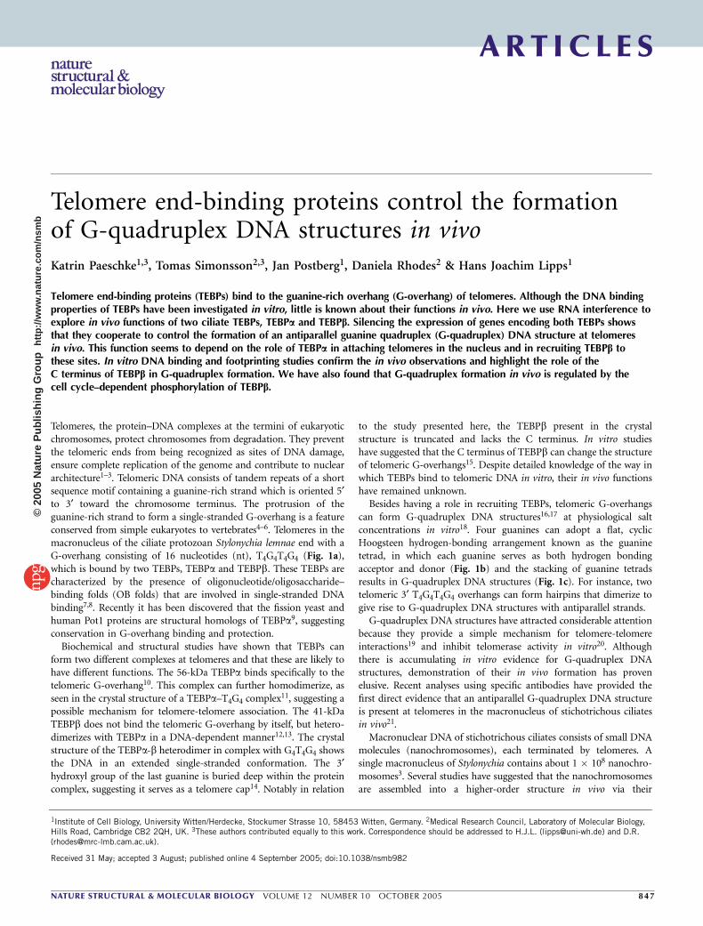
We next assessed the individual roles ofTEBPa and TEBPb in telomere attachmentin vivo by silencing their expression. This wasachieved by feeding cells with Escherichia coliexpressing double-stranded (ds) RNAdirected against the mRNAs encoding theproteins31. As shown by in situ antibodystaining and western analyses (Fig. 3a–f),expression of the TEBP genes was almostcompletely silenced 4 d after Stylonychiawere fed with dsRNA-expressing E. coli.This phenotype was seen in over 95% ofthe cells, consistent with an observed decreasein intensity of the respective TEBP bands inwestern analyses (Fig. 3). At this time thecells were fully viable, as shown by thepresence of replication bands and dividingcells. Silencing of TEBPa or TEBPbeventually led to cell death (B14–21 d).After this time telomeres were no longerdetectable by Southern analysis and theDNA was degraded and lost from the nucleus(data not shown). This effect was alsoobserved when different regions of theTEBP genes were targeted by RNAi.
3′-G4T4G4T4G4T4G4 . . .C4A4C4 . . .
. . . C4A4C4
. . . G4T4G4T4G4T4G4-3′
R
N N N
N
NN
N
NN
NN
N
N
N N N
N
N
N
N
H
H
H
HH
H
H
HHH
HR
R
T10
T9
G8
G7
G6
G5
T11
TEBPα TEBPβ
T12
G13
G14
G15
G16
3′ OH
R
H
OO
OO
a
b c
Figure 1 Schematic representation of a macronuclear nanochromosome.
(a) Each telomeric 3¢ overhang binds one TEBPa (blue) and one TEBPb(orange). (b) The hydrogen bonding pattern of guanine tetrads (yellow)
uniquely involves the exocyclic amino group N7 (bold). (c) Formation of an
antiparallel G-quadruplex DNA structure from two G-overhangs. Stabilizing
cations are shown as black spheres.
No RNAi
Beforeelectroelution
Afterelectroelution
Beforeelectroelution
Afterelectroelution
Beforeelectroelution
Afterelectroelution
TEBPα RNAi TEBPβ RNAi
a b
c d
e f
g h
i j
k l
m n
o p
q r
Figure 2 TEBPa attaches telomeres to a subnuclear structure and recruits TEBPb to the attachment
sites. The localization of telomeric DNA was examined by FISH analysis and TEBP localization was
examined by in situ antibody staining. (a–r) Stylonychia macronuclei after FISH analysis with telomeric
probe (yellow), or after in situ antibody staining of TEBPa (blue) or TEBPb (orange), before or after
electroeleution as indicated; arrows, replication band; scale bar represents 10 mm. Schematics show
constituents of complexes in the experiments: blue, TEBPa; orange, TEBPb; crosses, silencing of either
TEBPa (g–l) or TEBPb (m–r) expression by RNAi.
ART IC L E S
84 8 VOLUME 12 NUMBER 10 OCTOBER 2005 NATURE STRUCTURAL & MOLECULAR BIOLOGY
©20
05 N
atur
e P
ublis
hing
Gro
up
http
://w
ww
.nat
ure.
com
/nsm
b
In addition, we studied telomere attachment after silencing of eitherTEBPa or TEBPb expression using nuclear electroelution. Silencingof TEBPa expression by RNAi for 4 d resulted in the loss of telomereattachment, and telomeres could then be completely electroelutedfrom the nucleus (Fig. 2g,h). This was not the result of degradationof telomeric DNA, as shown by FISH analyses of nuclei beforeelectroelution (Fig. 2g) and by sequence analyses of telomeric DNA(Supplementary Tables 1–3 online). Silencing of TEBPa expression(Fig. 2i,j) followed by electroelution resulted in complete removalof the TEBPb from the macronucleus (Fig. 2k,l). In contrast, neitherthe telomeres (Fig. 2m,n) nor TEBPa (Fig. 2o,p) could be electro-eluted from the nucleus after silencing of TEBPb expression(Fig. 2q,r). These results show that telomere attachment is mediatedby TEBPa and that TEBPb localizes to telomeres through interactionswith TEBPa.
Both TEBPs are required for G-quadruplex formation in vivoWe asked whether tethering of telomeres in the nucleus is related tothe formation of G-quadruplex DNA structures in vivo. G-quadruplexDNA structures at macronuclear telomeres in vivo were detected usingantibodies generated by ribosome display from the Human Combi-natorial Antibody Library21,32 . These antibodies were highly specific,with affinities for telomeric G-quadruplex DNA structures betweenKd ¼ 0.125 nM and Kd ¼ 5 nM. Moreover, they could be used todistinguish between parallel and antiparallel conformations21. Nostaining was obtained with Sty3, an antibody against the parallelG-quadruplex conformation (Fig. 4a), providing an essential controlshowing that the observed G-quadruplex DNA structure was not anartifact that occurred upon antibody binding. Only Sty49, an antibodydirected against both G-quadruplex conformations, gave a positivesignal (Fig. 4b,c). Thus, only the antiparallel G-quadruplex DNAstructure is present in vivo21. Staining with Sty49 showed the samefocal organization as was observed in FISH analyses using a telomericprobe or after staining with antibodies against TEBPs. Therefore,it is likely that G-quadruplex staining overlaps with telomeres,as concluded earlier21. Moreover, the antibodies do not stain thereplication band (Fig. 4b). As antibodies against B-DNA reactstrongly with the replication band33, the absence of staining withantibodies against G-quadruplex DNA cannot be explained by loweraccessibility of the DNA in the replication band. From this weconclude that the telomeric G-quadruplex DNA structure is unfoldedduring replication.
To investigate whether the two TEBPs are involved in the formationof G-quadruplex DNA structures at telomeres, we again silenced theirexpression silenced by RNAi, but this time we analyzed the effect ontelomere structure with the Sty49 antibody. Silencing of either TEBPa(Fig. 4d) or TEBPb (Fig. 4e) led to the loss of antibody binding. Thus,both TEBPa and TEBPb are required for maintaining G-quadruplexDNA structures in vivo.
Both TEBPs are required for G-quadruplex formation in vitroTo elucidate how TEBPa and TEBPb are involved in the formation ofthe G-quadruplex DNA structure at telomeres, we analyzed theirin vitro DNA binding properties in detail. Stylonychia TEBPa andTEBPb were overexpressed in E. coli and purified to homogeneity. Amodel ciliate telomere consisting of 21 base pairs (bp) of double-stranded DNA and a single-stranded G-overhang (T4G4T4G4) at one3¢ end was constructed from synthetic DNA oligonucleotides. Previousstudies have shown that the G-overhang constitutes the binding sitefor the TEBPs10,34. Electrophoretic mobility shift assays (EMSAs) werecarried out under conditions aimed to mimic those in vivo. From thetypical size of a macronucleus (diameter B25–40 mm and lengthB80–120 mm), the approximate number of DNA molecules in amacronucleus (108) and Avogadro’s constant, we estimated themacronuclear telomere concentration in vivo to be 41 mM. Accord-ingly, the concentration of telomeric DNA in the in vitro bindingstudies presented here was 1 mM. We incubated the model telomereswith increasing concentrations of TEBPa and TEBPb and analyzedtheir binding properties by EMSAs in nondenaturing agarose gels(Fig. 5). TEBPa bound the G-overhang (Fig. 5a), whereas TEBPb onits own did not (Fig. 5b). TEBPb only bound the model telomere in
a
d e
b c
f
g
1 2
1 2
1 2
56 kDa
42 kDa
36 kDa
a Parallel Antiparallel
No RNAi TEBPβ RNAiTEBPα RNAi
b
c d e
Figure 4 Folding of telomeric G-overhangs into an antiparallel
G-quadruplex DNA structure requires both TEBPa and TEBPb in vivo.
(a,b) Staining of Stylonychia macronuclei with Sty3, an antibody specific
for parallel G-quadruplex DNA structures (a), or Sty49, an antibody that
detects both parallel and antiparallel G-quadruplex DNA structures (b).
The results suggest that telomeric G-overhangs adopt only the antiparallel
G-quadruplex DNA structure in vivo. G-quadruplex DNA structures are
absent from the replication band (arrow). Scale bar represents 10 mm.
(c–e) Staining of Stylonychia macronuclei with Sty49. Schematics are as inFigure 2. (c) Telomeric G-overhangs fold into antiparallel G-quadruplex DNA
structures before RNAi silencing. (d,e) Telomeric G-overhangs cannot fold
into antiparallel G-quadruplex DNA structures when either TEBPa (d) or
TEBPb (e) expression is silenced by RNAi.
Figure 3 Silencing of TEBPa and TEBPb gene expression by RNAi. (a–g)
In situ antibody staining of the two TEBPs (a,b,d,e; coloring as in Fig. 2;
scale bar represents 10 mm) and western analyses (c,f,g). (a,b) TEBPa in
control cells (a) and after silencing (b). (c) Western blots using antibodies
directed against TEBPa in control cells (lane 1) and after silencing TEBPa(lane 2). (d,e) TEBPb in control cells (d) and after silencing (e). (f) Western
blots using antibodies directed against TEBPb in control cells (lane 1) and
after silencing TEBPb (lane 2). (g) Control western blot using monoclonal
antibody 38F3 (EnCor Biotechnology) to fibrillarin/nop1p, after
silencing of TEBPa.
ART IC L E S
NATURE STRUCTURAL & MOLECULAR BIOLOGY VOLUME 12 NUMBER 10 OCTOBER 2005 8 4 9
©20
05 N
atur
e P
ublis
hing
Gro
up
http
://w
ww
.nat
ure.
com
/nsm
b

the presence of TEBPa (Fig. 5c), consistent with previous conclusionsfor the Oxytricha nova TEBPs34.
Next, we investigated the conformation of the DNA in the TEBPa-bheterodimer. This was done by using the Sty49 antibody to detect theantiparallel G-quadruplex DNA structure in vivo and by footprinting.If a G-quadruplex DNA structure was present in a complex, theexpectation was that the antibody would bind to it and changethe electrophoretic mobility of the complex. Owing to the limitingconcentration of the antibody, the telomere DNA concentration in thisexperiment was 0.1 mM. A band of unique electrophoretic mobilityindeed appeared, but only when the telomeric DNA had been boundto the TEBPa-b heterodimer before incubation with the antibody
(Fig. 6a, lanes 5–8). By contrast, the antibodydid not alter the electrophoretic mobility ofthe telomeric DNA on its own or whenincubated with only TEBPb (data notshown) or only TEBPa (Fig. 6a, lane 4).Although the intensity of the antibody-specific band was concentration dependent,it was not possible to reach saturation ofbinding owing to the limiting concentrationof the antibody. The electrophoretic mobilityof the band containing the antibody washigher than that of the band containing theTEBPa-TEBPb–DNA complex (Fig. 6a, lanes5–8), which suggests that a protein wasreleased from the complex upon antibodybinding. The most likely explanation is thatantibody binding caused displacement ofTEBPb. Whatever the composition of the
complex with the antibody is, it does not affect the interpretationthat formation of the G-quadruplex DNA structure requires bothTEBPa and TEBPb. These results strongly suggest that both TEBPaand TEBPb are required to fold the telomeric G-overhang into theG-quadruplex DNA structure that is present in vivo.
To further delineate the formation of the antiparallel G-quadruplexDNA structure, TEBPb lacking the C terminus (TEBPb1–230) wasproduced and tested in our binding studies. This experiment wasbased on two previous biochemical studies15,35, which had impliedthat the C terminus of TEBPb is required for the formation ofG-quadruplex DNA structures. The truncated TEBPb1–230 formed astable ternary complex with full-length TEBPa and telomeric DNA
(Fig. 6a, lane 10), but incubation with theantibody did not produce a band of uniqueelectrophoretic mobility (Fig. 6a, lane 11).This suggests that the complex with TEBPb1–230
contains single-stranded DNA, whereas thepresence of the full-length TEBPb results inthe formation of the antiparallel G-quadru-plex DNA structure. Thus, the C terminus ofTEBPb thus is responsible for formation ofthe G-quadruplex DNA structure.
To address directly the question of thefolding state of the G-overhang in the variousreconstituted TEBP–telomeric DNA com-plexes and to validate the interpretation ofour EMSAs, we probed the various com-plexes with dimethyl sulfate (DMS)(Fig. 1b). Whereas DMS specifically methy-lates non–hydrogen bonded N7 groups onguanines, it cannot react with N7 groupsinvolved in hydrogen bonding (Fig. 1b).Consequently, DMS protection provides areliable test for G-quadruplex DNA struc-tures. We incubated the complexes (Fig. 6a)with DMS and analyzed the methylationpatterns of the guanines by denaturing gelelectrophoresis. The eight guanines present inthe telomeric G-overhangs were readily mod-ified in naked DNA in the absence of cations,as expected, but also when the model telo-mere had been incubated with KCl overnightor with TEBPb alone (Fig. 6b, lanes 2, 3 and
ba c
DN
A
DN
A
DN
A
TEBPα-β–DNA
TEBPα TEBPβ
TEBPβ
TEBPα
TEBPα–DNA
DNA
Figure 5 TEBPb requires TEBPa in order to interact with telomeric DNA in vitro. (a,b) EMSA results
after incubation of the model telomere with increasing concentrations of TEBPa (a) or TEBPb (b).
(c) EMSA results after the model telomere–TEBPa complex was titrated with increasing concentrations
of TEBPb. The concentration of DNA was 1 mM and that of the proteins ranged from 0.25–4 mM.Analyses were carried out on a nondenaturing 0.4% (w/v) agarose gel. Blue, TEBPa; orange, TEBPb.
Cartoons, right, mark the migration of each complex and show its components.
a
Unm
odifi
ed D
NA
Unm
odifi
ed D
NA
DN
A in
TE
DN
A in
KC
I
DN
A +
α
DN
A +
β
DN
A +
α +
∆β
DN
A +
α +
β
1 2 3 4 5 6 7 8
G4 Ab
TEBPβ
1
TEBPα-β–DNA
TEBPα-∆β–DNA
TEBPα–DNA
G4 Ab–DNA
DNA
2 3 4 5 6 7 8 9 10 11
TEBPβ1–230
TEBPα
DN
A
b
Figure 6 Both TEBPs are required for G-quadruplex DNA formation and the basic C terminus of TEBPbis essential. (a) EMSA results. Proteins included in EMSAs are shown above the lanes. Addition of the
sty49 antibody to the G-quadruplex (G4 Ab) results in a complex of altered electrophoretic mobility
when the telomeric DNA is bound by the TEBPa-b heterodimer (lanes 6–8). In this experiment
the concentration of the telomeric DNA is 0.1 mM because of the limiting concentration of the
G-quadruplex antibody. By contrast, the complex with TEBPb1–230 (Db) does not show a band of
altered mobility when incubated with the G-quadruplex antibody (lane 11). Analysis was carried out
on a 0.4% (w/v) nondenaturing agarose gel. Blue, TEBPa; orange, TEBPb; gray, G4 Ab. Cartoons,
right, mark the migration of each complex and show its components. (b) DMS footprinting results.
Left, the sequence of the model telomere used in both gel mobility and footprinting experiments.
DNA concentration was 1 mM. Buffer conditions and proteins bound to the telomere before
DMS probing are shown above each lane (cartoons and coloring as in a). Analysis was carried out
on a 20% (w/v) denaturing polyacrylamide gel.
ART IC L E S
85 0 VOLUME 12 NUMBER 10 OCTOBER 2005 NATURE STRUCTURAL & MOLECULAR BIOLOGY
©20
05 N
atur
e P
ublis
hing
Gro
up
http
://w
ww
.nat
ure.
com
/nsm
b

5). Incubation with TEBPa alone or with TEBPa and TEBPb1–230
together led to some protection (Fig. 6b, lane 4). This might beexpected from the mode of binding observed in the crystal structure ofthe TEBPa-b heterodimer–DNA complex, in which the bases in thesingle-stranded G-overhang are buried in a protein cleft14. Only in thecomplex containing both full-length TEBPs were the two tracts ofguanines in the G-overhang protected from DMS modification(Fig. 6b, lane 7). This provides strong evidence that the G-overhangin this complex is folded into a G-quadruplex DNA structure. Incontrast, the G-overhang in the complex with TEBPa-b1–230 wasaccessible to modification, suggesting that it remains in a single-stranded form (Fig. 6b, lane 6). Overnight incubation of the modeltelomere (1 mM) in KCl did not result in the spontaneous formationof a G-quadruplex DNA structure, providing further evidence for theimportant roles of TEBPa and TEBPb in modulating the structure oftelomeric DNA. Thus, the DMS footprinting data (Fig. 6b) areentirely consistent with the results of antibody probing and EMSAs(Fig. 6a), and they provide further evidence that both full-lengthTEBPs are essential for the folding of single-stranded telomericG-overhangs into G-quadruplex DNA structures. The in vitro resultspinpoint the different functions of TEBPa and TEBPb. The role of
TEBPa is to bind the G-overhang and recruit TEBPb, which in turn isresponsible for the conversion of the single-stranded G-overhang intoa G-quadruplex DNA structure. Moreover, the formation of DNAstructure can be attributed to the very basic C terminus of TEBPb.
TEBPb phosphorylation regulates G-quadruplex formationStructural information on the TEBPa-TEBPb–DNA complex14,36 andthe observation that this complex blocks telomerase activity37 stronglysuggest that the TEBPa-b heterodimer bound to the telomeric DNAend needs to be disrupted for the end-replication machinery to gainaccess. The cyclin-dependent kinase (Cdk) family of proteins arepositive regulators of eukaryotic cell cycle progression. We consideredthe possibility38 that phosphorylation of TEBPa or TEBPb by a Cdkcould influence telomere accessibility. A search for potential phos-phorylation sites39 in the amino acid sequences of the Oxytricha andStylonychia TEBPs showed that TEBPa lacks a consensus Cdk2recognition sequence and cannot be phosphorylated in vitro byCdk2 (data not shown). In contrast, TEBPb contains two closelyspaced consensus Cdk2 recognition sequences38 (SupplementaryFig. 1 online). Both sites are located in the C terminus of TEBPband are conserved in Oxytricha and Stylonychia. To test the effect ofphosphorylation on the binding properties of TEBPb, it was phos-phorylated in vitro using Cdk2. EMSA experiments (Fig. 7) showedthat phosphorylated TEBPb was no longer able to bind to telomericDNA bound by TEBPa as in Figure 5c. Consequently, phosphoryla-tion of TEBPb prevents complex formation with TEBPa and thetelomeric G-overhang.
We next asked whether TEBPb is phosphorylated in vivo in a cellcycle–dependent manner. Stylonychia cells were synchronized40, pulse-labeled with [g-32P]ATP and electroeluted at different stages of the cellcycle. Although many proteins are phosphorylated during the S phase(Fig. 8a, lane 1), only one phosphorylated protein of molecular weight42 kDa was electroeluted during DNA replication (Fig. 8a, lane 3).This protein could not be electroeluted during other stages of the cellcycle (Fig. 8a, lane 2). Western analysis confirmed that the phos-phorylated 42-kDa protein was TEBPb (Fig. 8b). To investigate theeffect of in vivo phosphorylation of TEBPb on the formation of theG-quadruplex DNA structure, the activity of Cdk2 was inhibited withbutyrolactone41. As before (Fig. 4), the nuclei were stained in situ withthe Sty49 antibody. The G-quadruplex DNA structure was present inthe replication band (Fig. 8c), whereas no detectable amount ofG-quadruplex DNA in the replication band could be observed incontrol cells (Figs. 4 and 8). Together with the in vitro binding data(Fig. 7), these results suggest that phosphorylation of TEBPb duringS phase prevents binding of TEBPb to TEBPa complex or, more likely,that it allows TEBPb to dissociate from the TEBPa–G-overhangcomplex. These findings constitute strong evidence that phosphoryla-tion of TEBPb is required for the unfolding of the G-quadruplex DNAstructure during replication in vivo.
DISCUSSIONThe telomere end-binding proteins TEBPa and TEBPb bind telomericG-overhangs in the macronucleus of the ciliate Stylonychia. By usingRNAi to silence the expression of the two TEBP genes, we have now
DN
AphosphoTEBPβ
TEBPα
TEBPα-β–DNA
TEBPα–DNA
DNA
a b
42 kDa
c
d
1 2 3 1 2 3 4
Figure 8 TEBPb is phosphorylated during S phase, and inhibition of
phosphorylation prevents the G-quadruplex DNA structure from being
resolved during replication. (a) Visualization of nuclear proteins pulse-
labeled in vivo before and after electroelution. Lane 1, total phosphorylated
nuclear proteins during S phase; lane 2, electroeluted phosphorylated
nuclear proteins during G0 phase; lane 3, electroeluted phosphorylatednuclear proteins during S phase (lane 3). (b) Western blots of TEBPb.
Lanes 1 and 2, total nuclear proteins during S phase and G0 phase,
respectively; lanes 3 and 4, electroeluted nuclear proteins during G0
phase and S phase, respectively. (c,d) In situ antibody staining of the
G-quadruplex DNA structure in Cdk2-inhibited cells (c) and control (d).
Scale bar represents 10 mm.
Figure 7 Phosphorylated TEBPb does not interact with telomeric DNA
bound by TEBPa in vitro. Lane 1, free DNA; lane 2, control with
unphosphorylated TEBPb; lanes 3–8, titration of TEBPa-bound telomeric
DNA with phosphorylated TEBPb (phosphoTEBPb) in two-fold dilution steps.
The concentrations of DNA and TEBPa were 1 mM each, and the TEBPbconcentration ranged from 0.25–4 mM.
ART IC L E S
NATURE STRUCTURAL & MOLECULAR BIOLOGY VOLUME 12 NUMBER 10 OCTOBER 2005 8 5 1
©20
05 N
atur
e P
ublis
hing
Gro
up
http
://w
ww
.nat
ure.
com
/nsm
b

been able to characterize their in vivo functions in detail. We show thatTEBPa is important both for the attachment of telomeres to asubnuclear structure and for the recruitment of TEBPb to theattachment sites. We also show that antiparallel-type G-quadruplexDNA structure forms at telomeres when they are bound to asubnuclear structure in vivo. However, as telomere-telomere inter-actions remain intact when the TEBP complex is dissociated from thesubnuclear structure by high-salt treatment, attachment per se seemsto be independent from G-quadruplex formation18. Silencing of TEBPexpression by RNAi shows that formation of the G-quadruplex DNAstructure requires both TEBPa and TEBPb in vivo.In vitro DNA binding and footprinting studies using a model
telomere and purified TEBPs further reveal the separate roles ofTEBPa and TEBPb in formation of the G-quadruplex DNA structure.Under our experimental conditions, TEBPa is required first to bind tothe telomeric G-overhang and second to recruit TEBPb to thetelomere to form a stable DNA–protein complex. This discovery isonly in partial agreement with the previous report that TEBPb alonecan accelerate the formation of G-quadruplex DNA structuresin vitro15,35. TEBPb lacking the C-terminal domain (TEBPb1–230)still forms a ternary complex with TEBPa and the G-overhang, butantibody probing and footprinting by DMS did not detect theG-quadruplex DNA structure in the complex. These results showthat the C terminus of TEBPb, which is basic and has sequencehomology to the linker histone H1, is essential for promoting thefolding of the G-overhang into a G-quadruplex DNA structure. Thisobservation is in agreement with conclusions from previous in vitrobinding studies15,35 and also with the crystal structure of the TEBPa-bheterodimer in complex with G4T4G4, in which the DNA is in asingle-stranded form14. In this structure, TEBPb is truncated at residue260 and lacks the larger part of the C-terminal domain that we showis responsible for the formation of the G-quadruplex DNA structure.
Evidence for a possible regulation mechanism for G-quadruplexDNA formation comes from our analysis of the phosphorylation statesof the TEBPs. Our in vitro binding studies show that phosphorylationof TEBPb blocks interactions with the TEBPa-bound telomericG-overhang and hence prevents conversion of the single strand intothe G-quadruplex DNA structure. The importance of TEBPb phos-phorylation is further substantiated by in vivo studies, which showthat Stylonychia TEBPb becomes phosphorylated during S phase. Weshow a direct link between the phosphorylation state of TEBPb andG-quadruplex DNA formation. Inhibition of phosphorylation duringS phase results in the presence of G-quadruplex DNA structure inthe replication band, suggesting that phosphorylation of TEBPb isrequired for the unfolding of G-quadruplex DNA structure. Notably,the two putative Cdk2 phosphorylation sites are located in theC-terminal domain of TEBPb, which we have shown is responsible forG-quadruplex DNA formation.
Our results allow us to propose a model for the regulation ofG-quadruplex DNA structure formation in vivo. We show that in vivothe G-quadruplex DNA structure is antiparallel, rather than parallel.This is consistent with the observation that the nanochromosomes ofStylonychia macronuclei are joined end to end via their telomeres18,24,as the most likely structure resulting from the pairing of twoT4G4T4G4 overhangs is an antiparallel G-quadruplex42. The extensivethree-dimensional structural information on the Oxytricha TEBPahomodimer and TEBPa-b heterodimer in complex with telomericDNA11,14,36 provides insights into how G-quadruplex DNA formationat telomeres might occur. The crystal structure of TEBPa showed thatit forms a homodimer, which binds two telomeres and promotestelomere-telomere associations11. The DNA in this complex is single
stranded, which is consistent with our antibody probing andfootprinting data. In the second structure, that of the TEBPa-TEBPb–DNA complex14,36, the G-overhang is also bound in anextended and single-stranded form. However, this structure containeda truncated TEBPb that lacked the C terminus, which our in vitrobinding studies show is required for G-quadruplex DNA formation. Incomplex with TEBPb1–230, the DNA remains in a single-strandedconformation, whereas in complex with full-length TEBPb, the boundG-overhang adopts G-quadruplex DNA structure. As has been shownhere and also suggested elsewhere15, the highly basic C terminus ofTEBPb seems to act as a chaperone to promote G-quadruplex DNAformation. The discovery that TEBPb is phosphorylated in a cellcycle–dependent manner provides strong evidence that this protein isresponsible for regulating G-quadruplex DNA formation in vivo.TEBPb is phosphorylated during S phase, which is likely to result indissociation of the TEBPa-b heterodimer. Once this has occurred, theG-quadruplex DNA structure unfolds into a single strand, whichwould make telomeres accessible for the end-replication machinery.As G-quadruplex DNA structures are very stable under physiologicalconditions, it remains to be determined how they are resolved in vivoin a timely manner.
Could G-quadruplex DNA structures be a general telomere cappingstructure? Research on telomerase inhibitors has provided indirectsupport for the presence of G-quadruplex DNA structures at telo-meres in human cells. The use of ligands that stabilize G-quadruplexesresults in senescence and telomere shortening in telomerase-positivehuman cell lines43,44. G-quadruplex DNA structures have also beenimplicated in the function of the Rtel gene, which is required fortelomere elongation in mouse cells45. These observations are consis-tent with the view that G-quadruplex DNA structures may act as capsby making the 3¢ overhang inaccessible to telomerase20 and exonu-cleases. Our finding, that telomere end-binding proteins control theformation of G-quadruplexes in a cell cycle–dependent manner,indeed suggests that G-quadruplex DNA structures are a core com-ponent in telomere capping.
METHODSIsolation and electroelution of macronuclei. Stylonychia macronuclei were
isolated and electroeluted essentially according to established protocols25,30,46.
About 1 � 105 cells were embedded in 1.6% (w/v) low–melting point agarose.
The agarose blocks were melted at 65 1C and digested with agarase (Fermentas)
3 h at 42 1C. Nuclei were recovered by centrifugation at 1,000g for 5 min,
resuspended in 100 ml PBS containing 2% (v/v) formaldehyde and incubated
30 min at room temperature. Nuclei were transferred onto microscopic slides
and dried for in situ antibody staining and FISH analysis.
In situ antibody staining, FISH analysis and western analysis. These were
performed as described in refs. 21,25.
Inhibition of gene expression by RNA interference. To silence TEBPa and
TEBPb, fragments (500 bp) from various regions of the TEBPs were amplified
by PCR and cloned into the L4440 vector47. The vectors were transfected into
RNase III–deficient E. coli HT115 cells, and RNAi expression was induced by
adding IPTG to a final concentration of 0.4 mM when the cell culture reached
an OD600 of about 0.4. Specific inhibition of TEBPa or TEBPb expression was
achieved by feeding Stylonychia cells heat-inactivated E. coli containing the
appropriate L4440 vector mixed with Chlorogonium elongatum31. A strong
RNAi effect was observed 4 d after feeding. As a control, cells were fed with
E. coli containing the L4440 vector without an insert.
Oligonucleotides. Oligonucleotides were synthesized by Sigma-Genosys and
purified by denaturing polyacrylamide gel electrophoresis. The oligonucleotides
TelG (5¢-GCTACACTTGCCAT4G4T4G4T4G4-3¢) and TelC (5¢-C4A4TGGCAA
GTGTAGC-3¢) were annealed to form the model telomere with a 16-nt
ART IC L E S
85 2 VOLUME 12 NUMBER 10 OCTOBER 2005 NATURE STRUCTURAL & MOLECULAR BIOLOGY
©20
05 N
atur
e P
ublis
hing
Gro
up
http
://w
ww
.nat
ure.
com
/nsm
b
G-overhang. Oligonucleotide concentrations were estimated from OD260
measurements and sequence-specific extinction coefficients.
Protein expression and purification. The genes encoding the full-length
Stylonychia TEBPa (GenBank accession number AY751782) and TEBPb and
TEBPb1–230 were cloned into a modified Pet30a vector (Novagen) containing
an N-terminal 6� His-tag and S-tag followed by a tobacco etch virus (TEV)
protease cleavage site. Inserts were verified by sequencing and the plasmids
transfected into E. coli Rosetta cells (Novagen). Protein expression and
purification were carried out essentially as described for the dimerization
domains of the human TRF proteins48. The purification procedure involved
affinity purification using Ni-NTA resin (Qiagen) followed by TEV protease
cleavage to remove the 6� His-tag and S-tag. The TEV protease itself was 6�His-tagged, which allowed the cleaved proteins to be purified from the
TEV protease and the cleaved N-terminal 6� His-tag and S-tag by rebinding
to Ni-NTA resin. Finally, the proteins were purified to homogeneity by gel
filtration on a Superdex 200.
Electrophoretic mobility shift assays. DNA was radioactively labeled using
[g-32P]ATP (Amersham) and T4 polynucleotide kinase (New England Biolabs).
Unincorporated radiolabel was removed on Bio-Spin P30 columns (Bio-Rad).
Labeled DNA (1 mM) was incubated with protein in binding buffer (50 mM
Tris/HCl (pH 8.0), 125 mM KCl, 5 mM DTT and 10% (v/v) glycerol)
containing 100 mg ml–1 BSA (New England Biolabs) and 100 mg ml–1 sheared
E. coli DNA for 12 h at room temperature. Reaction mixtures were analyzed by
nondenaturing agarose gel electrophoresis (0.4% (w/v) agarose, 0.25� TB) at
7.5 V cm–1 for up to 120 min at 4 1C. Gels were dried onto DE81 paper
(Whatman) and scanned using a Typhoon 8600 imaging system (Amersham
Pharmacia Biotech).
Antibody mobility shift assays. Radiolabeled DNA (10 nM) was incubated
with protein (20 nM) in binding buffer containing 10 mg ml–1 BSA and 10 mg
ml–1 sheared E. coli DNA for 12 h at room temperature. The Sty49 antibody
was added and the reaction mixtures were incubated 2 h on ice before being
analyzed by nondenaturing agarose gel electrophoresis as above.
Dimethyl sulfate footprinting. Radiolabeled DNA was incubated with proteins
as described above before footprinting analysis. Methylation was carried out by
incubations with 0.2% (v/v) DMS (Sigma) for 10 min at 25 1C. Reactions were
terminated by adding 10 ml of a solution containing 1.5 M sodium acetate
(pH 9.0), 1.0 M b-mercaptoethanol, 100 mg ml–1 sonicated E. coli DNA and
10 mM EDTA. An aliquot of each reaction was taken out for immediate
analysis by nondenaturing agarose gel electrophoresis and the remainder was
purified by phenol-chloroform extraction. The DNA was cleaved with 1 M
piperidine, and the piperidine was removed by lyophilization. Samples were
analyzed on denaturing 20% (w/v) polyacrylamide (19:1 mono/bis) 8 M urea
gels. Gels were autoradiographed and scanned using a Typhoon 8600 imaging
system (Amersham Pharmacia Biotech).
In vitro and in vivo protein phosphorylation. Purified TEBPb (4 mM) was
incubated with 20 units Cdk2-cycline A (New England Biolabs) for 30 min at
30 1C in a buffer containing 50 mM Tris (pH 7.4), 10 mM b-mercaptoethanol,
10 mM MgCl2, 10 mM g-glycerol phosphate and 100 mM ATP. For analysis of
in vivo protein phosphorylation, cells were synchronized44 and pulse-labeled
(2 h) by the addition of 1 mCi ml–1 cell culture [g-32P]ATP (Amersham).
Nuclear proteins were separated on 15% (w/v) SDS polyacrylamide gels and
autoradiographed. In vivo inhibition of Cdk2 activity was achieved by the
addition of 10 mM butyrolactone (Acros); in situ antibody analysis was
performed as above.
Note: Supplementary information is available on the Nature Structural & MolecularBiology website.
ACKNOWLEDGMENTSThis work was supported by a grant from the Deutsche Forschungsgemeinschaftto H.J.L., a European Molecular Biology Organization short-term fellowshipto K.P. and a Wenner-Gren Foundations fellowship to T.S. We thank T. Cech(University of Colorado, Boulder, Colorado, USA) for providing antibodiesagainst the TEBP subunits and C. Schaffitzel (Swiss Federal Institute
of Technology, Zurich) for providing antibodies against G-quadruplexDNA structures.
COMPETING INTERESTS STATEMENTThe authors declare that they have no competing financial interests.
Published online at http://www.nature.com/nsmb/
Reprints and permissions information is available online at http://npg.nature.com/
reprintsandpermissions/
1. Zakian, V.A. Telomeres: beginning to understand the end. Science 270, 1601–1607(1995).
2. De Lange, T. Protection of mammalian telomeres. Oncogene 21, 532–540(2002).
3. Jonsson, F. & Lipps, H.J. The biology of telomeres in hypotrichous ciliates. inTelomerases, Telomeres and Cancer (eds. Krupp, G. & Parwaresch, R.) 205–222(Landes Bioscience, Kluwer Academic/Plenum Publishers, Georgetown, Texas, USA,2002).
4. Klobutcher, L.A., Swanton, M.T., Donini, P. & Prescott, D.M. All gene-sized DNAmolecules in four species of hypotrichs have the same terminal sequence and anunusual 3¢ terminus. Proc. Natl. Acad. Sci. USA 78, 3015–3019 (1981).
5. Wellinger, R.J., Ethier, K., Labrecque, P. & Zakian, V.A. Evidence for a new step intelomere maintenance. Cell 85, 423–433 (1996).
6. Makarov, V.L., Hirose, Y. & Langmore, J.P. Long G tails at both ends of humanchromosomes suggest a C strand degradation mechanism for telomere shortening.Cell 88, 657–666 (1997).
7. Murzin, A.G. OB(oligonucleotide/oligosaccharide binding)-fold: common structuraland functional solution for non-homologous sequences. EMBO J. 12, 861–867(1993).
8. Mitton-Fry, R.M., Anderson, E.M., Hughes, T.R., Lundblad, V. & Wuttke, D.S. Con-served structure for single-stranded telomeric DNA recognition. Science 296, 145–147(2002).
9. Theobald, D.L. & Wuttke, D.S. Prediction of multiple tandem OB-fold domains intelomere end-binding proteins Pot1 and Cdc13. Structure (Camb) 12, 1877–1879(2004).
10. Gottschling, D.E. & Zakian, V.A. Telomere proteins: specific recognition and protectionof the natural termini of Oxytricha macronuclear DNA. Cell 47, 195–205 (1986).
11. Peersen, O.B., Ruggles, J.A. & Schultz, S.C. Dimeric structure of the Oxytricha novatelomere end-binding protein alpha-subunit bound to ssDNA. Nat. Struct. Biol. 9,182–187 (2002).
12. Gray, J.T., Celander, D.W., Price, C.M. & Cech, T.R. Cloning and expression of genes forthe Oxytricha telomere-binding protein: specific subunit interactions in the telomericcomplex. Cell 67, 807–814 (1991).
13. Fang, G. & Cech, T.R. Oxytricha telomere-binding protein: DNA-dependent dimeriza-tion of the alpha and beta subunits. Proc. Natl. Acad. Sci. USA 90, 6056–6060(1993).
14. Horvath, M.P., Schweiker, V.L., Bevilacqua, J.M., Ruggles, J.A. & Schultz, S.C. Crystalstructure of the Oxytricha nova telomere end binding protein complexed with singlestrand DNA. Cell 95, 963–974 (1998).
15. Fang, G. & Cech, T.R. The beta subunit of Oxytricha telomere-binding protein promotesG-quartet formation by telomeric DNA. Cell 74, 875–885 (1993).
16. Sundquist, W.I. & Klug, A. Telomeric DNA dimerizes by formation of guanine tetradsbetween hairpin loops. Nature 342, 825–829 (1989).
17. Williamson, J.R., Raghuraman, M.K. & Cech, T.R. Monovalent cation-induced struc-ture of telomeric DNA: the G-quartet model. Cell 59, 871–880 (1989).
18. Lipps, H.J. In vitro aggregation of the gene-sized DNA molecules of the ciliateStylonychia mytilus. Proc. Natl. Acad. Sci. USA 77, 4104–4107 (1980).
19. Giraldo, R. & Rhodes, D. The yeast telomere-binding protein RAP1 binds toand promotes the formation of DNA quadruplexes in telomeric DNA. EMBO J. 13,2411–2420 (1994).
20. Zahler, A.M., Williamson, J.R., Cech, T.R. & Prescott, D.M. Inhibition of telomerase byG-quartet DNA structures. Nature 350, 718–720 (1991).
21. Schaffitzel, C. et al. In vitro generated antibodies specific for telomeric guanine-quadruplex DNA react with Stylonychia lemnae macronuclei. Proc. Natl. Acad. Sci.USA 98, 8572–8577 (2001).
22. Meyer, G.F. & Lipps, H.J. The formation of polytene chromosomes during macronucleardevelopment of the hypotrichous ciliate Stylonychia mytilus. Chromosoma 82,309–314 (1981).
23. Murti, K.G. & Prescott, D.M. Topological organization of DNA molecules in the macro-nucleus of hypotrichous ciliated protozoa. Chromosome Res. 10, 165–173 (2002).
24. Lipps, H.J., Gruissem, W. & Prescott, D.M. Higher order DNA structure in macro-nuclear chromatin of the hypotrichous ciliate Oxytricha nova. Proc. Natl. Acad. Sci.USA 79, 2495–2499 (1982).
25. Postberg, J. et al. Association of the telomere-telomere binding protein-complex ofhypotrichous ciliates with the nuclear matrix and dissociation during replication. J. CellSci. 114, 1861–1866 (2001).
26. de Lange, T. Human telomeres are attached to the nuclear matrix. EMBO J. 11,717–724 (1992).
27. de Lara, J., Wydner, K.L., Hyland, K.M. & Ward, W.S. Fluorescent in situ hybridizationof the telomere repeat sequence in hamster sperm nuclear structures. J. Cell.Biochem. 53, 213–221 (1993).
ART IC L E S
NATURE STRUCTURAL & MOLECULAR BIOLOGY VOLUME 12 NUMBER 10 OCTOBER 2005 8 5 3
©20
05 N
atur
e P
ublis
hing
Gro
up
http
://w
ww
.nat
ure.
com
/nsm
b

28. Laroche, T., Martin, S.G., Tsai-Pflugfelder, M. & Gasser, S.M. The dynamics of yeasttelomeres and silencing proteins through the cell cycle. J. Struct. Biol. 129, 159–174(2000).
29. Jonsson, F., Postberg, J., Schaffitzel, C. & Lipps, H.J. Organization of the macronucleargene-sized pieces of stichotrichous ciliates into a higher order structure via telomere-matrix interactions. Chromosome Res. 10, 445–453 (2002).
30. Jackson, D.A., Yuan, J. & Cook, P.R. A gentle method for preparing cyto- and nucleo-skeletons and associated chromatin. J. Cell Sci. 90, 365–378 (1988).
31. Paschka, A.G. et al. The use of RNAi to analyze gene function in spirotrichous ciliates.Eur. J. Protistol. 39, 449–454 (2003).
32. Schaffitzel, C., Hanes, J., Jermutus, L. & Pluckthun, A. Ribosome display: an in vitromethod for selection and evolution of antibodies from libraries. J. Immunol. Methods231, 119–135 (1999).
33. Lipps, H.J. et al. Antibodies against Z DNA react with the macronucleus but not themicronucleus of the hypotrichous ciliate Stylonychia mytilus. Cell 32, 435–441(1983).
34. Fang, G., Gray, J.T. & Cech, T.R. Oxytricha telomere-binding protein: separable DNA-binding and dimerization domains of the alpha-subunit. Genes Dev. 7, 870–882(1993).
35. Fang, G. & Cech, T.R. Characterization of a G-quartet formation reaction promotedby the beta- subunit of the Oxytricha telomere-binding protein. Biochemistry 32,11646–11657 (1993).
36. Horvath, M.P. & Schultz, S.C. DNA G-quartets in a 1.86 A resolution structure of anOxytricha nova telomeric protein-DNA complex. J. Mol. Biol. 310, 367–377 (2001).
37. Froelich-Ammon, S.J., Dickinson, B.A., Bevilacqua, J.M., Schultz, S.C. & Cech, T.R.Modulation of telomerase activity by telomere DNA-binding proteins in Oxytricha.Genes Dev. 12, 1504–1514 (1998).
38. Hicke, B. et al. Phosphorylation of the Oxytricha telomere protein: possible cell cycleregulation. Nucleic Acids Res. 23, 1887–1893 (1995).
39. Obenauer, J.C., Cantley, L.C. & Yaffe, M.B. Scansite 2.0: Proteome-wide predictionof cell signaling interactions using short sequence motifs. Nucleic Acids Res. 31,3635–3641 (2003).
40. Juranek, S.A., Jonsson, F., Maercker, C. & Lipps, H.J. The telomeres of replicatingmacronuclear DNA molecules of the ciliate Stylonychia lemnae. Protistology 1, 148–151 (2000).
41. Kitagawa, M. et al. Butyrolactone I, a selective inhibitor of cdk2 and cdc2 kinase.Oncogene 8, 2425–2432 (1993).
42. Rhodes, D. & Giraldo, R. Telomere structure and function. Curr. Opin. Struct. Biol. 5,311–322 (1995).
43. Riou, J.F. et al. Cell senescence and telomere shortening induced by a new series ofspecific G-quadruplex DNA ligands. Proc. Natl. Acad. Sci. USA 99, 2672–2677(2002).
44. Rezler, E.M., Bearss, D.J. & Hurley, L.H. Telomeres and telomerases as drug targets.Curr. Opin. Pharmacol. 2, 415–423 (2002).
45. Ding, H. et al. Regulation of murine telomere length by Rtel: an essential geneencoding a helicase-like protein. Cell 117, 873–886 (2004).
46. Ammermann, D., Steinbruck, G., von Berger, L. & Hennig, W. The development ofthe macronucleus in the ciliated protozoan Stylonychia mytilus. Chromosoma 45,401–429 (1974).
47. Timmons, L. & Fire, A. Specific interference by ingested dsRNA. Nature 395, 854(1998).
48. Fairall, L., Chapman, L., Moss, H., de Lange, T. & Rhodes, D. Structure of the TRFHdimerization domain of the human telomeric proteins TRF and TRF2. Mol. Cell 8,351–361 (2001).
ART IC L E S
85 4 VOLUME 12 NUMBER 10 OCTOBER 2005 NATURE STRUCTURAL & MOLECULAR BIOLOGY
©20
05 N
atur
e P
ublis
hing
Gro
up
http
://w
ww
.nat
ure.
com
/nsm
b