Embed Size (px)
Citation preview
1
Total deletion of in vivo telomere elongation capacity: anambitious but possibly ultimate cure for all age-related human
cancers
Aubrey D.N.J. de Grey, Department of Genetics, University of CambridgeF. Charles Campbell, Department of Surgery, Queen’s University of BelfastInderjeet Dokal, Department of Haematology, Imperial College, LondonLeslie J. Fairbairn, Paterson Institute for Cancer Research, ManchesterGerry J. Graham, Cancer Research UK Beatson Laboratories, GlasgowColin A.B. Jahoda, School of Biological and Biomedical Sciences, University of DurhamAndrew C.G. Porter, Faculty of Medicine, Imperial College, LondonCorrespondence to: Dr. Aubrey D.N.J. de Grey, Department of Genetics, University of Cambridge,Downing Street, Cambridge CB2 3EH, UK. Tel +44 1223 333963; Fax +44 1223 333992; [email protected] title: Truly curing cancer by ablating telomere elongation
AbstractDespite enormous effort, progress in reducing mortality from cancer remains modest. Can a true cancer"cure" ever be developed, given the vast versatility that tumours derive from their genomic instability?Here we consider the efficacy, feasibility and safety of a therapy that, unlike any available or indevelopment, could never be escaped by spontaneous changes of gene expression: the total eliminationfrom the body of all genetic potential for telomere elongation, combined with stem cell therapiesadministered about once a decade to maintain proliferative tissues despite this handicap. We term thistherapy WILT, for “Whole-body Interdiction of Lengthening of Telomeres”. We first argue that awhole-body gene-deletion approach, however bizarre it initially seems, is the only way truly toovercome the hypermutation that makes tumours so insidious. We then identify the key obstacles todeveloping such a therapy and conclude that, while some will probably be insurmountable for at least adecade, none is a clear-cut showstopper. Hence, given the absence of alternatives with comparable anti-cancer promise, we advocate working towards such a therapy.
IntroductionThe reason cancer is so hard to combat is depressingly simple; it comes down to just two facts. First,cancer cells are very similar in most respects to the non-cancer cells from which they arose, makingselective ablation of cancer cells intrinsically fraught. Second, unlike all other aspects of age-relateddegeneration cancer can acquire additional means of survival in response to whatever the body, or theclinician, may throw at it. Each cell in a tumour is a furnace of inventive potential, constantlyexperimenting with new combinations of gene expression as a result of its profound genomic instabilityand the availability of 6Gb of DNA to rearrange.What therapies might, in principle, overcome this versatility? An answer is evident when we considerthe mechanisms whereby tumours escape endogenous or medical attack, because those mechanismshave one very tangible thing in common: changes of gene expression. Immune stimulation is oftenescaped by loss of antigen presentation.1 Angiogenesis inhibition is escaped by up-regulating alternativeangiogenic pathways.2 Chemotherapy may be escaped by numerous mechanisms, including up-regulating transporters that keep the cell free of the toxin, or specific DNA repair capacities.3
2Telomerase inhibitors4 are too new for clinical data to be available, but can in theory be escaped byover-expression of telomerase or degradation or export of the inhibitor.We should therefore seek therapies that make such changes of gene expression impossible or at leasthighly improbable, even for the ever-inventive cancer cell. In principle, the deletion (not merelyinhibition) of a gene whose function is essential for cancers to progress would present a major challengeto cancer cells. Creation of a new gene out of nothing does of course occur on evolutionary timescales,but it is many orders of magnitude rarer than mutations causing changes of expression, so tumourswould never have enough cells or enough time to achieve it.Unfortunately, cancer cells have the same genome as non-cancer cells. Selective deletion of a gene fromcancer cells but not others faces the usual gene-expression problems: cancer cells can readily escape bydown-regulating the DNA maintenance machinery on which targeted deletion depends, and the need toavoid killing too many non-cancer cells intrinsically limits any such treatment’s therapeutic index.
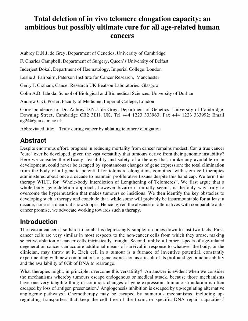
Figure 1. The WILT concept. An untreated cancer sufferer (a) dies rapidly once the cancer achievesmalignancy and metastasis. Conventional treatments (b) delay death somewhat, but the cancer typicallyevolves gene expression changes that outmauoeuvre the treatment. A hypothetical telomere-elongation-negative human (c) could not develop metastatic cancer, but would die relatively young from failure of
constantly-renewing tissues. A beneficiary of WILT (d) would maintain function of such tissues throughperiodic stem cell transplantation, so would not die of either cause.
A way around this is to eliminate the gene from all cells, and somehow to make it non-essential to thenon-cancer cell (or, at least, to the tissue of which that cell is a part) while preserving the cancer cell’s
3absolute requirement for it. This is the approach discussed here. The genes in question are thoseresponsible for maintaining telomere length through large numbers of cell divisions by telomereelongation. Tissue integrity would be preserved by, about every ten years, re-seeding the stem cellcompartments of all tissues reliant on continuous cell division with stem cells whose telomeres had beenlengthened ex vivo but whose telomere elongation machinery was deleted (Figure 1). Transformation ofa living organism—ultimately, a human—to a totally telomere elongation-incompetent state would beperformed gradually, using a variety of techniques discussed below. We suggest that, if (as we foresee)it could be implemented without serious side-effects, this therapy would almost completely eliminatecancer as an age-related cause of death, something which no other present or currently contemplatedtherapy would do. In fact, one’s risk of cancer would actually decline with age as telomere elongation-competent cells were progressively depleted.Here we discuss the plethora of obstacles to the development (even on a multi-decade timescale) of suchtherapy, which we term WILT, “Whole-body Interdiction of Lengthening of Telomeres”. We concludethat, while many of those obstacles are daunting, none is so insurmountable as to justify dismissing thisapproach. We foresee a considerable risk that, as sophisticated cell and gene therapy technologiesmature in the next decade or two, progress in reducing age-specific cancer death rates will lag behindthat for other major killers,5 and that the proportion of people dying from cancer will thus rise sharply. Itmay be that only by developing ambitious but extremely powerful anti-cancer therapies, such as WILT,can this be prevented.
Efficacy
Requirement of telomere elongation for human cancer progressionA typical clinically relevant cancer contains around 240 (1012) cells. Thus, at least 40 cell generationshave occurred in that cancer starting from the initiating non-cancer cell. In fact, however, this is a grossunderestimate: (a) there is abundant cell death in cancer, and (b) the development of cancer is multi-stage. Suppose, for simplicity, that a cancer develops as a result of two mutations—one to escapegrowth control and, later, one to induce angiogenesis. The former may allow it to grow to, say, 106 cells;the second, to 1012 cells. But that second mutation will have occurred in just one of the 106 cells whichharboured the first. Thus, starting from the cell in which the first mutation occurred, there will havebeen not 40 but 60 divisions—even ignoring the contribution of cell death.In practice, since cancer progression in humans requires many more than two mutations (except somerare childhood cancers), there are probably at least a few hundred cell generations between theoriginating non-mutant cell and the clinically relevant cancer.6 It is this that makes prevention oftelomere elongation a realistic way to prevent cancers from ever reaching an advanced stage. No humancell has ever been observed to divide more than 100 times without telomere elongation machinery.Moreover, even though the “end-replication problem”7,8 that originally inspired the telomere-basedexplanation of this “Hayflick limit”9 suggests that telomere loss per cell generation could be muchslower than is typically seen in vitro,10 only a modest extension of replicative capacity results fromgrowth in low oxygen.11 Direct evidence for the absolute requirement of telomere elongation forprogression of human cancers is widespread.12-14
TelomeraseAll mammals maintain telomere length in rapidly-dividing cells by reverse transcription of a 6-baseRNA template of which the telomere is a many-copy DNA tandem repeat. The RNA including thetemplate (hereafter “TERC”) and the reverse transcriptase (hereafter “TERT”) form a heterodimer calledtelomerase, which adds copies of this sequence to the end of one strand; the other strand is elongated bystandard DNA replication machinery.15 TERC is ubiquitous in human tissues, but TERT is expressedonly at trace levels in tissues that require telomere elongation and is undetectable in quiescent andpostmitotic cells.16,17 In about 90% of human cancers, however, TERT is highly expressed and telomere
4length thereby stabilised.18 Many non-cancer human cell types that normally senesce (have a finitereplicative capacity) in culture have been “immortalised” (given indefinite replicative capacity) byintroducing constitutively active TERT.19-21
Though telomerase may have cytoprotective properties not directly related to cell division-associatedtelomere elongation,22 it seems that neither telomerase subunit has any essential physiological functionexcept telomere elongation. For TERT in humans this is shown by its absence in nearly all cells andvery low levels in any cell type. In laboratory mice, which maintain their telomere length severalorganismal generations “ahead of the game” (allowing serial inbreeding of telomerase knockout mice, inwhich telomeres in the germ line progressively shorten), knockout of either TERC or TERT confers nodetectable phenotype for three generations.23,24 Thus, these genes are prime targets for WILT.
ALT (alternative lengthening of telomeres)However, about 10% of human cancers—predominantly mesenchyme-derived ones such as sarcomas, inwhich the proportion approaches 50%—do not express telomerase but nonetheless maintain telomerelength indefinitely both in vivo and in vitro.25 They do so by a mechanism termed ALT, for “AlternativeLengthening of Telomeres”. Moreover, cancers of epithelial tissues may express ALT rarely onlybecause they have the easier option to activate telomerase (which may be suppressed less thoroughly inepithelial than in mesenchymal tissues). Hence, epithelial-derived cancers might turn ALT on as easilyas sarcomas do if the telomerase route were denied them. A therapy that eliminated cancers’ ability toactivate telomerase but left ALT untouched might thus only modestly reduce age-specific cancermortality rates.The mechanism of ALT clearly involves a recombination-like process but the molecular details have notbeen determined. Short telomeres are the recipients of ALT events (but perhaps not exclusively) but it isunclear whether the telomere repeats added are chromosomal or extra-chromosomal in origin. Onemodel proposes that t-loop structures initiate intra-telomeric rolling circle replication in ALT cells,though there is currently no supporting evidence for this mechanism in human cells. A second model[which has some supporting evidence26] proposes strand invasion and copying of telomere repeats froma donor to a recipient telomere in a BIR (breakage-induced repair)–like process.27
Even with our limited current understanding of ALT, however, we can make one observation that givescause for optimism that, once its underlying genetic etiology is discovered, it will be amenable to thesame sort of manipulation as for telomerase. There are formally three types of possible explanation forwhy ALT is seen in certain cancers but not in normal tissues. One is that, like telomerase, ALT is anactivation of a gene or genes that are normally turned off in the cell type in which the cancer arises.(Candidate genes might be ones involved in meiosis or in a form of DNA repair that is only activatedunder certain circumstances, for example.) The second is that telomere elongation by recombination is aside-effect of a constitutive process (a ubiquitous DNA repair process, for example): that is, that it ishappening all the time in normal cells but is counterbalanced by a shortening process and so progressivetelomere lengthening is not seen in such cells. Indeed, there seems to be a system for actively shorteningtelomeres that have been lengthened by ALT.28 If this second mechanism were the basis of ALT itwould be a blow to the potential efficacy of WILT, because no gene would be available to be deletedwithout rapidly deleterious effects in normal cells. This seems unlikely, however, because a constitutivesystem in which telomere elongation by recombination is balanced by shortening of over-long telomereswould maintain telomere length by default in all tumours and there would be no pressure to activatetelomerase. Additionally, telomeric recombination has not been detected in normal cells.26,29 Finallythere is the possibility that the active players in ALT are indeed active in normal cells (performing non-telomeric DNA maintenance) but that, rather than being constantly lengthened and re-shortened, somesystem protects telomeres from being adventitiously lengthened by this machinery in the first place, andthis is lost in ALT cells. In this scenario, however, since the hypothetical constitutive function of theALT machinery is certainly not inter-chromosomal recombination, it seems plausible that (once it had
5been identified) judicious site-directed mutagenesis could delete its capacity for such recombinationwhile preserving its constitutive function.Hence, in summary, until the molecular basis of ALT is understood we cannot state how or whether itcan be ablated along with telomerase as part of WILT, but there is reason for optimism.
Chemotherapy and chemoresistanceThe characteristic that most centrally defines a cancer cell is its high division rate. Thus, the cell typesthat are hardest to distinguish from cancer cells, when designing a therapy, are those that themselvesdivide fast, such as in the bone marrow, skin and gut. The blood, in particular, is maintained by the rapiddivision of transit amplifying cells in bone marrow whose ablation as a result of anti-cancer therapy ishighly prejudicial to the welfare or even survival of the patient.30
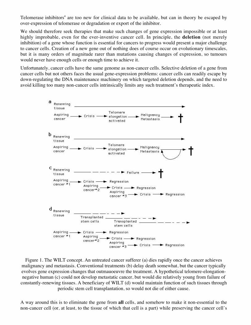
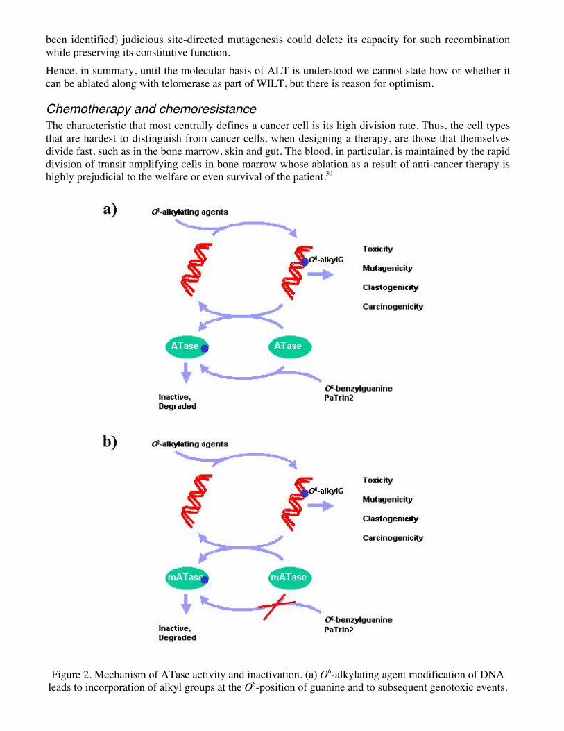
Figure 2. Mechanism of ATase activity and inactivation. (a) O6-alkylating agent modification of DNAleads to incorporation of alkyl groups at the O6-position of guanine and to subsequent genotoxic events.
6ATase repairs O6-alkylguanine lesions in a stoichometric and autoinactiavting manner. Small moleculepsueodosubstrates of ATase lead to inactivation and thus to sensitivity of cells to O6-alkylating agents.(b) Certain point-mutated versions of ATase are unable to react with pseudosubstrates, yet retain their
DNA-repair capacity.Conveniently, the tissues most at risk in this regard are just those that would also—again because oftheir rapid division—be eventually compromised by WILT. Such tissues must be maintained by theperiodic introduction of new cells that have been engineered ex vivo. This ex vivo manipulation canpotentially include manipulations to diminish sensitivity to anti-cancer agents.In fact, many groups have been exploring such an approach (independently of WILT, of course).31,32 Aprominent stratagem has been to exploit the cell’s spectacularly laborious mechanism for reversing aparticular type of DNA damage, alkylation of guanine at position 6; this is done by a protein thattransfers the alkyl group to itself and is then ubiquitinated and destroyed, rather than acting catalytically.This protein, O6-alkylguanine-DNA-alkyltransferase (ATase) is a first-class target for chemotherapeuticagents because of two additional features: first, there are small molecules that mimic O6-alkylation inDNA and act as pseudosubstrates of the protein leading to its inactivation, and second, there are single-amino-acid changes to the protein that render it almost completely resistant to such inactivation (Figure2). Hence, before chemotherapy with a combination of inactivator and O6-alkylating agent, the patientcan be transplanted with haemopoietic stem cells engineered to express inactivator-resistant ATase;then, a dose of the inhibitor/O6-alkylating agent combination that the cancer cell cannot survive willablate native bone marrow but leave the transplanted cells (and hence the patient) unscathed.33
This approach possesses an inherent shortcoming: engineered marrow—which must necessarily containhaemopoietic stem cells—could give rise to new cancers, which would be resistant to somechemotherapeutic challenges. Since haemopoietic stem cells can repopulate many tissues other than theblood,34,35 this problem might be severe if the patient lives many years after the treatment. However,when implemented in combination with WILT, no such concern exists: such cancers might arise, butthey could not reach a life-threatening stage.
Telomerase capture: a possible cancer escape route from WILT?The WILT concept assumes that creating a new gene is far harder than changing expression patterns.However, genomes can gain genes by lateral gene transfer. Could tumours escape WILT that way?A formal possibility is that the reverse transcriptase of a retrovirus might be recruited for telomereelongation; these work very differently from TERT, however,36 so this seems remote. Alternatively, acell engineered to lack telomerase but to be chemoresistant might fuse, in vivo, with one that waspresent natively and hence retained telomerase genes. Cell fusion (and phagocytosis of apoptotic bodies,which could be equivalent) has been demonstrated in vitro,37-39 but its in vivo relevance remainsunknown. Such fusion products would, anyway, still be susceptible to chemotherapeutic agents to whichresistance had not been transgenically conferred, so this would not seriously subvert WILT.
Feasibility
Stem cell technologyWe purposely say little here about the prospects for developing the stem cell technology needed forWILT. Clearly WILT could not be implemented until we can transform adult cells into stem cells of allrapidly-renewing tissues, expand them very substantially in vitro and introduce them into the personfrom whom they came;40 these are major advances. But numerous other advances necessary for WILTare also very considerable, so that we do not foresee any prospect of its being developed in under tenyears even in the best case. Progress in stem cell technology is presently so rapid that nothing canmeaningfully be predicted about where it will stand in ten years, let alone thereafter. Hence, we merely
7note that the necessary sophistication of stem cell therapy is likely to exist by then, and focus here onthe many other prerequisites for implementing WILT.
Ex vivo genetic manipulationThree genetic alterations are entailed in WILT (plus, perhaps, adding an inducible suicide gene forsafety):• Deletion of telomere-elongation genes;• Introduction of chemoresistance;• Elongation of telomeres to the length seen in a typical stem cell.Of these, the last is probably easiest, because it does not require genomic integration. Expression oftelomerase from an extrachromosomal transgene, in a cell that has already undergone the other requiredgenetic changes, would extend telomeres to somewhat more than the desired length. The cells wouldthen be grown for enough generations to allow confirmation of loss of the extrachromosomal transgene.Additionally, by incorporating a ‘suicide gene’ such as HSVTk into the extrachromosomal construct itshould be possible to selectively eliminate (using ganciclovir) any cells that retain the construct.41
Chemoresistance could be conferred by introducing a transgene (e.g. encoding a drug-resistant ATase)at a random position in the genome. This approach, however, in common with most gene additionstrategies for gene therapy, faces the severe problems of transgene silencing and instability aftertransplantation (not to mention potentially deleterious effects on genes close to the integration site),even if pre-transplant selection is used to enrich for transgene-expressing cells. Such problems can beavoided by modification of an endogenous gene; the fact that chemoresistance can sometimes beconferred by one or two amino-acid changes31 to an endogenous gene is particularly valuable in thiscontext. The best-established technique for endogenous gene modification is gene targeting byhomologous recombination between a target locus and a plasmid carrying several kbp of homologousdsDNA.42-45 Other, less developed methods involve triplex-forming oligonucleotides,46 single-strandedoligonucleotides47,48 or RNA/DNA oligonucleotides (RDOs).49,50 The choice of technique would bedriven mainly by the incidence of accompanying random mutations, rather than of the desired alteration,because selection for (e.g.) ATase inhibitor resistance can be used to enrich for rare events. Althoughthe incidence of accompanying random integrations is easier to assess (by simple Southern analysis) instandard gene targeting methods, oligonucleotides have the attraction that they are relatively easy toprepare and deliver. It is therefore important that oligonucleotide-based methods be thoroughlyvalidated, characterised and optimised.A problem with altering endogenous genes is that the target tissues are often sensitive tochemotherapeutic drugs because they naturally express low levels of drug-resistance mechanisms suchas ATase. Thus, small changes to endogenous genes might be more appropriate for ubiquitouslyexpressed enzymes such as thymidylate synthetase or dihydrofolate reductase, where specific mutationsconfer resistance to specific anti-metabolites.51,52 Other alternatives are to combine point mutations inthe coding regions with genomic changes to alter expression patterns, or to deliver the drug resistancetransgene on a mammalian artificial chromosome.53,54
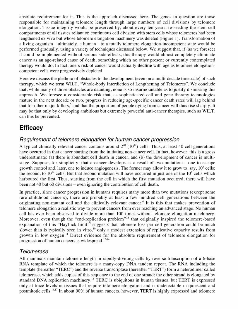
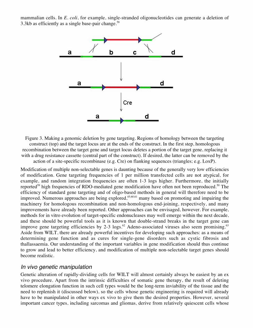
Deletion of telomere-elongation genes is more challenging. First, deletion of both copies of at least twoto four genes will be desirable. Second, these deletions will not confer an inherent selectable advantageon the cells (though with gene targeting this might be addressed by replacing the gene by a selectablemarker). Third, the alterations must delete large portions of the relevant genes, because less drasticchanges (such as single-base-pair changes to introduce a stop codon or frameshift) might be too easilyreverted in a cancer. This may constrain the choice of technique: targeted deletions (see Figure 3) ofseveral kbp (or Mbp if Cre/lox technologies are used) are a well established capability of standard genetargeting methods,45,55-57 while oligonucleotide-mediated approaches have so far been limited to basechanges or deletions of only one or two base pairs (although multiple modifications could be introducedsequentially). This situation may change, however, as oligonucleotide-based methods are developed in
8mammalian cells. In E. coli, for example, single-stranded oligonucleotides can generate a deletion of3.3kb as efficiently as a single base-pair change.58
Figure 3. Making a genomic deletion by gene targeting. Regions of homology between the targetingconstruct (top) and the target locus are at the ends of the construct. In the first step, homologous
recombination between the target gene and target locus deletes a portion of the target gene, replacing itwith a drug resistance cassette (central part of the construct). If desired, the latter can be removed by the
action of a site-specific recombinase (e.g. Cre) on flanking sequences (triangles; e.g. LoxP).Modification of multiple non-selectable genes is daunting because of the generally very low efficienciesof modification. Gene targeting frequencies of 1 per million transfected cells are not atypical, forexample, and random integration frequencies are often 1-3 logs higher. Furthermore, the initiallyreported59 high frequencies of RDO-mediated gene modification have often not been reproduced.50 Theefficiency of standard gene targeting and of oligo-based methods in general will therefore need to beimproved. Numerous approaches are being explored,45,60,61 many based on promoting and impairing themachinery for homologous recombination and non-homologous end-joining, respectively, and manyimprovements have already been reported. Other approaches can be envisaged, however. For example,methods for in vitro evolution of target-specific endonucleases may well emerge within the next decade,and these should be powerful tools as it is known that double-strand breaks in the target gene canimprove gene targeting efficiencies by 2-3 logs.62 Adeno-associated viruses also seem promising.63
Aside from WILT, there are already powerful incentives for developing such approaches: as a means ofdetermining gene function and as cures for single-gene disorders such as cystic fibrosis andthallassaemia. Our understanding of the important variables in gene modification should thus continueto grow and lead to better efficiency, and modification of multiple non-selectable target genes shouldbecome realistic.
In vivo genetic manipulationGenetic alteration of rapidly-dividing cells for WILT will almost certainly always be easiest by an exvivo procedure. Apart from the intrinsic difficulties of somatic gene therapy, the result of deletingtelomere elongation function in such cell types would be the long-term inviability of the tissue and theneed to replenish it (discussed below), so the cells whose genetic engineering is required will alreadyhave to be manipulated in other ways ex vivo to give them the desired properties. However, severalimportant cancer types, including sarcomas and gliomas, derive from relatively quiescent cells whose
9total requirement for cell division in a lifetime is low. Repopulating such tissues with engineered cellsappears very challenging.It may therefore be necessary to alter these cell types in situ. Unfortunately, this seems unlikely to beachievable soon by gene targeting, given the rate of random integration noted above. However, severalgroups are working to improve the reproducibility of high-efficiency oligonucleotide-mediated genetargeting, and other approaches are also on the horizon.61
Finally, we must remember that a therapy so advanced and technically difficult as WILT is not likely tobecome an attractive option until considerable progress has been made against all other major causes ofdeath and debilitation—particularly cardiovascular disease, neurodegeneration and diabetes—becauseonly then will the limitations of more conventional anti-cancer approaches become apparent as a steeprise in the proportion of people dying of cancer. Such advances will surely also require effective in vivogenetic manipulation.5 Thus, this aspect of WILT is not in fact a substantive obstacle to its developmentby the time it is needed.
Side-effectsCancer promotion?Cells with very short telomeres are genetically unstable. Indeed, in humans it is considered likely that aninitial phase of inadequate telomere maintenance early in tumorigenesis confers a “mutator” phenotype,accelerating the occurrence of further mutations—including activation of telomerase or ALT—thatallow the tumour to progress.64
This phenomenon should not affect the efficacy of WILT, however. When telomere elongation cannotbe activated, the mutator phenotype will if anything hasten the tumour’s demise by increasing theaccumulation of mutations that prevent cell division.
Telomerase knockout miceThe best laboratory models currently available to explore the WILT concept are mice engineered to lacktelomerase. Even though mouse ES cells lacking telomerase acquire an ALT-like character,65 this is notseen in vivo—perhaps because it is too rare, or perhaps because ALT is incompatible with the normaldifferentiated state of some mouse tissues. Thus, mice lacking telomerase can be inbred for successivegenerations to yield mice with telomeres too short to sustain highly proliferative tissues for the animal’snormal lifetime.66 The most prominent phenotypes observed are in the gonad, blood and skin.Interestingly, mice lacking p53 as well as telomerase show a delay in the major phenotype that can bemeasured at an early enough age not to be masked by the cancers that result from lack of p53, namelysterility: TERC-/- p53-/- mice can be bred for two generations longer than the simple TERC knockouts.67
It is important to understand why p53 ablation accelerates death from cancer even in late-generationtelomerase knockout mice.68 The number of cell divisions needed for a mouse cancer to grow bigenough to kill it is considerably fewer than in a human, for several reasons: (1) the cancer need not growas big; (2) it need not metastasise, whereas most human cancers only become life-threatening aftermetastasis; and (3) the telomere damage-detection system in mice is simpler—in particular, the Rbpathway does not exert a strong protective effect69—so fewer mutations are required. Also, as notedearlier, mouse cells can activate ALT quite easily in vitro; it has not been determined whether cancersdeveloped by TERC-/- p53-/- mice (or, indeed, by TERC-/- mice) are phenotypically ALT-like, thoughthey do become ALT-like after serial transplantation.70 The anti-cancer effect of short telomeres istherefore challenging to assay in mice. However, when these confounders are minimised, the effect isdramatic: in TERC-/- ApcMin mice, a mild reduction in telomere length increased the incidence of cancerat a given age, but severe telomere shortening reduced that risk to the point where no deaths at alloccurred by the age at which all TERC+/+ animals had died.71 Other models of cancer in TERC-/- micealso show resistance to cancer progression.72
10
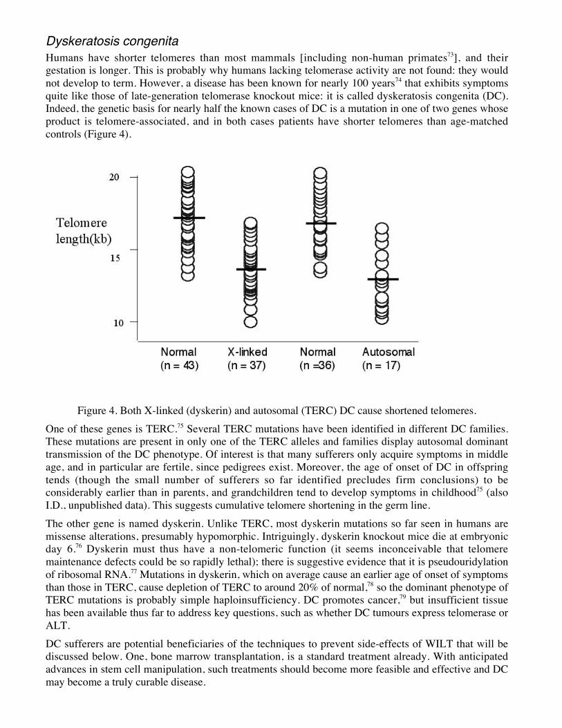
Dyskeratosis congenitaHumans have shorter telomeres than most mammals [including non-human primates73], and theirgestation is longer. This is probably why humans lacking telomerase activity are not found: they wouldnot develop to term. However, a disease has been known for nearly 100 years74 that exhibits symptomsquite like those of late-generation telomerase knockout mice: it is called dyskeratosis congenita (DC).Indeed, the genetic basis for nearly half the known cases of DC is a mutation in one of two genes whoseproduct is telomere-associated, and in both cases patients have shorter telomeres than age-matchedcontrols (Figure 4).
Figure 4. Both X-linked (dyskerin) and autosomal (TERC) DC cause shortened telomeres.One of these genes is TERC.75 Several TERC mutations have been identified in different DC families.These mutations are present in only one of the TERC alleles and families display autosomal dominanttransmission of the DC phenotype. Of interest is that many sufferers only acquire symptoms in middleage, and in particular are fertile, since pedigrees exist. Moreover, the age of onset of DC in offspringtends (though the small number of sufferers so far identified precludes firm conclusions) to beconsiderably earlier than in parents, and grandchildren tend to develop symptoms in childhood75 (alsoI.D., unpublished data). This suggests cumulative telomere shortening in the germ line.The other gene is named dyskerin. Unlike TERC, most dyskerin mutations so far seen in humans aremissense alterations, presumably hypomorphic. Intriguingly, dyskerin knockout mice die at embryonicday 6.76 Dyskerin must thus have a non-telomeric function (it seems inconceivable that telomeremaintenance defects could be so rapidly lethal); there is suggestive evidence that it is pseudouridylationof ribosomal RNA.77 Mutations in dyskerin, which on average cause an earlier age of onset of symptomsthan those in TERC, cause depletion of TERC to around 20% of normal,78 so the dominant phenotype ofTERC mutations is probably simple haploinsufficiency. DC promotes cancer,79 but insufficient tissuehas been available thus far to address key questions, such as whether DC tumours express telomerase orALT.DC sufferers are potential beneficiaries of the techniques to prevent side-effects of WILT that will bediscussed below. One, bone marrow transplantation, is a standard treatment already. With anticipatedadvances in stem cell manipulation, such treatments should become more feasible and effective and DCmay become a truly curable disease.
11
Avoidance of side-effectsThe problems that would certainly arise from ablating telomere elongation can, in principle, be avoidedby periodically reseeding all highly proliferative tissues with stem cells whose telomeres have beenlengthened ex vivo (but whose autonomous telomere elongation competence has not been restored, orhas been restored but then removed again). While this is conceptually straightforward, in practice itfaces major obstacles, which vary from tissue to tissue. Below we consider three key tissues needingthis restoration and the difficulties to be overcome.
Bone marrow reseedingThe haemopoietic system is a rapidly-renewing tissue for which replenishment techniques already exist:bone marrow transplantation (BMT) has long been routine. New problems would arise, however, inBMT for WILT.Firstly, for complete re-seeding of the haemopoietic system, substantial expansion of the transplantablestem cell population ex vivo would be required. Current efforts at expanding stem cell numbers utilisingcombinations of cytokines80 have been limited by several factors. Firstly, the most primitive stem cells,the so-called long term repopulating stem cells, are typically not expanded and are frequently lost bydifferentiation following prolonged culture in ex vivo expansion conditions. These are the key stem cellsfor long term maintenance of a transplant81 so this largely explains the failure of transplantability of exvivo expanded stem cell populations. Thus, there is much current interest in characterising factors whichwill allow self-renewal of these cells against a block in differentiation. Recently, many researchers havefocused on members of the Notch ligand family in this respect.82 However, although results arepromising, these analyses are at an early stage.A further complication is that following such culture, these cells will typically be proliferating. Undernormal circumstances, most stem cells are not proliferating and there is abundant evidence for arequirement for a G0/G1 state of haemopoietic stem cells for proper homing and engraftment followingtransplantation.83 Thus, pre-transplantation resetting of this ‘quiescent’ status will be essential. This maybe achieved using combinations of the well characterised inhibitors of stem cell proliferation appropriatefor long term repopulating stem cells.84 The same applies to molecules involved in the in vivo homingand engraftment of haemopoietic stem cells: expression of these molecules, such as the chemokinereceptor CXCR4, may be altered by culture conditions85 and need to be reset before transplantation.Thirdly, BMT works best when the stem cell niche has already been denuded of native cells.86 In certaindiseases of the haemopoietic system, however, this is not seen,87 indicating that functionallycompromised stem cells are less resistant to displacement by incoming, more robust ones. For secondand subsequent WILT reseedings, it may thus be possible to rely on the fact that many of the previousreseeding’s stem cells will be nearing exhaustion in terms of telomere length. The first treatment,however, might need chemotherapeutic or radiation-mediated depletion of the marrow to allow theengineered cells to engraft.
Gut reseedingThe gut lining consists of a juxtaposition of finger-shaped structured, termed villi, and invaginations,termed crypts. It is maintained by stem cells at or near the crypt base, which generate rapidly dividingcells that migrate up out of them and along the villi, eventually being shed at the tip.Several years ago, one of us (F.C.C.) and his colleagues developed a technique for surgicallyrepopulating the mouse colon with cells extracted from the small intestine. Despite considerable ex vivohandling, cell aggregates that had been plated onto freshly denuded colon developed in vivo intomorphologically normal crypts containing all four of the differentiated cell types normally observed.88
Clearly this augurs well for maintaining the gut in the context of WILT.However, daunting problems remain. One is fibrosis. The mouse work involved only a small area ofcolon; when similar surgery was done on newborn pigs, the resulting fibrosis precluded restoration of

12functional intestine (F.C.C., unpublished data). Further, for application to humans there would be arequirement to avoid surgery. The gut may, like the bone marrow, resist engraftment of new stem cellsin tissue already replete with them, so denudation may be required; this might be possible usingendoscopy technology, but that would risk short-term impairment of gut function. Finally, gut stem cellscultured in vitro lose proliferative capacity after only a few divisions. This sensitivity may or may not bealleviated by more sophisticated culturing technology (such as low oxygen).
Skin reseedingDC sufferers and late-generation TERC-/- mice both show severe epidermal dysfunction, and this willundoubtedly be a tissue needing replenishment in the context of WILT. However, epidermal functiondepends critically on the underlying tissue—the dermis.89 The dermis-epidermis interaction seems to becentral to epidermal maintenance and regeneration. Also, most of our skin grows hair, and it may be thehair follicles, from which several important types of skin cancer are believed to originate, that hold thekey to effective skin regeneration.90
As elsewhere, an important requirement for skin reseeding with a frequency of around a decade will beto control the differentiation process so that stem cells divide rarely enough to survive. Several factorshave been reported to exert appropriate influences. One is 14-3-3s, which inhibits differentiation.91
Reliable identification of stem cells is also important; high levels of b1 integrin expression,92 high a6integrin with low CD71 expression,93 Keratin 19,94 and p6395 are among markers championed as beingdiagnostic of the keratinocyte stem cell state. A definitive marker would permit refinement oftechniques already developed for isolating a pure population of these cells for tissue engineeringpurposes.96
Figure 5. Corneal epithelium is reprogrammed by the dermis as hair-forming epidermis, starting withthe hair follicle. Epidermis formation after grafting of corneal epithelium (A) proceeds through basal
layer formation (B), placode formation (C), epidermis formation at the follicle edge (D) and finally hairgrowth (E).
13A particularly useful tool for studying skin regeneration is the corneal epithelium. Its central region canbe removed without removing the stem cells from the surrounding germinal region (the limbus) andgrafted onto denuded dorsal dermis; the corneal cells are rapidly reprogrammed to a follicular state,including development of normal hair follicles.97 Importantly, hair follicle formation actually precedesformation of new epidermis, and epidermal differentiation spreads from the neck of the follicle (Figure5), reinforcing the idea that the follicle can be a repository of stem cells for the epidermis.90 Also, thistype of transdifferentiation work illustrates the possibility of using stem cells from an organ like the eyeto repopulate another like skin.Taken together, these and other observations give cause for optimism regarding skin regeneration,whether for WILT or for more traditional applications such as treatment of burns or DC. Since thedermis is a very slowly-renewing tissue, its capacity to orchestrate the behaviour of its epidermalcoating affords us great flexibility in the introduction of WILT cells. There is still some uncertainty as tothe numbers and exact location of stem cells in hair follicles but the most widely held view is that theyundergo bursts of activity corresponding with the periodic initiation of the follicle growth cycle.98
Certainly a follicle (not to mention its epidermal neighbourhood) contains more cells than a villus.Moreover, matrix epithelial cells at the base of actively growing follicles are among the fastest dividingcells in the body: in human scalp the matrix replaces itself every 23 hours, and corresponding mousecells are thought to divide every 13 hours.99,100 Since the active growing phase in scalp follicles can lastseveral years this represents many generations at the transit amplifying stage. Thus, the skin should notonly be relatively straightforward to repopulate: it should also not need reseeding very often.As a practical consideration and on the question of whether periodic reseeding of skin cells would be anacceptable practice and taken up if available, it is worth noting that many people currently pay for (oftenvery painful) chemical or laser “peels” of skin merely for cosmetic anti-ageing purposes.
Reseeding of other tissuesSimilar techniques would presumably have to be developed for many other epithelial tissues in order tomake WILT a viable therapy. These include the lung, on which stem cell therapy is already an activearea of research given its potential to treat diseases such as cystic fibrosis.101 Though several such tissuesmay present specific difficulties, we feel that the techniques developed for the three tissues discussedabove will probably be sufficiently versatile to be relatively easily adapted to these other tissues.
Frequency of reseedingThe utility of WILT depends on all rapidly-renewing tissues surviving for at least several years withstem cells that lack all telomere extension function. This implies stem cell generation times of at leasttwo months. That may be the natural rate in blood;102 what about other tissues?The tissue ostensibly of greatest concern here is the gut. In both DC sufferers and TERC-/- mice, gutphenotypes tend to arise contemporaneously with those of other tissues such as the blood. [In DC thereis an important exception—certain sufferers of Hoyeraal-Hreidarsson syndrome, a severe allelic variantof DC,103 show gut abnormalities before other problems.] Yet, mouse haemopoietic stem cells divideonly every few weeks,102 whereas gut stem cells have been calculated to divide once a day.104 [Thiscalculation depends on how many stem cells are present per crypt, which remains unclear: crypts aremonoclonal,105 but cells at an early stage of differentiation can return to the stem cell niche,106 so maybestem cells can also sometimes divide symmetrically to form two stem cells, as in the haemopoieticsystem.107] If blood and gut stem cell division frequencies differ by an order of magnitude, how can theage at which telomere maintenance deficiency affects those two tissues be comparable? In DC theremight be tissue-specific factors—for example, the gut might express more TERC—but no such loopholeseems available for TERC-/- mice. Resolution of this paradox is of high priority, as it may reveal aspectsof stem cell population dynamics that can be exploited for many therapeutic purposes. For WILT,however, the implication is inescapable that the gut should survive as long between transplants as theblood.
14
Senescent cell ablationTelomere elongation-incompetent stem cells, when compromised by over-short telomeres, may not fallobligingly upon their swords: they may need to be actively eliminated. Cultured cells with over-shorttelomeres remain alive in a distinctive “senescent” state long after losing the ability to divide; similarcells have been observed in vivo.108,109 They seem to be very rare except in cartilage,109 but might bedeleterious even so;110 WILT might raise their abundance considerably, with unknown consequences.Luckily, however, the possibility that even rare senescent cells may do harm in vivo has prompted workon their removal.111 One strategy being pursued is to incorporate into cells a “suicide” gene that inducesapoptosis if the cell adopts a senescent gene expression profile. Vaccination against cell surface markersof the senescent state is also a plausible approach. This work is at an early stage, but the fact that it isalready being aggressively attempted gives cause for optimism that it will be perfected within the ten-year minimum timeframe that we foresee for WILT.
Immune memoryUnlike the gut lining and epidermis, the blood comprises not only short-lived cells but also a smallminority of long-lived ones. The immune system relies on two types of long term memory, and thepossibility and consequences of loss of this memory as a result of WILT must be considered.One type is the memory of previous infections. While memory cells are undoubtedly good for us, theymay be of diminishing importance as we make progress against the many other aspects of aging thatmake the elderly more susceptible to infection in the first place. The immune system of a young personcontains relatively few memory cells, but one would not replace it with that of a centenarian.Additionally, to the extent that it may be desirable to preserve memory cell abundance and function, thesame technology proposed here for stem cell renewal may also be applicable to memory cells, which arerelatively oligoclonal and could be amplified along with telomere lengthening ex vivo.The other type of memory is self/non-self discrimination. Here we must bear in mind that lymphocytescannot participate in an immune response until maturation in either the thymus or the bone marrow. Onepart of this maturation process is the pre-emption of an autoimmune response, mainly by clonal deletionof cells that could react to self antigens.112 The memory of which cells to delete is thus not stored in thehaemopoietic system itself but in the stromal cells that vet immature lymphocytes. In the thymus, someof these cells are epithelial in origin and are maintained by stem cells,113 so retention of their self/nonselfmemory during ex vivo manipulation would be needed. Critically, however, these cells—and also thoseof other provenance, such as bone marrow-derived dentritic cells at the cortex/medulla boundary withinthe thymus—do not undergo the genomic reorganisation that occurs in the lymphocytes themselves.Thus, since the cells to be used for reseeding will be autologous, the selective pattern that theirdescendents in the thymus and bone marrow subsequently impose on maturing lymphocytes will be thesame as that imposed by native cells—that is, a faithful self/non-self divide. Autoimmune consequencesof WILT thus seem unlikely.
Putting it all together: development and implementation
Developing WILT in miceReseeding of rapidly-renewing tissues can be explored not only in normal mice, but also in telomerase-negative ones. Sixth-generation TERC-/- mice have a shortened lifespan as a result of failure of highlyproliferative tissues;66 successful reseeding of such tissues should, in principle, restore these animals'longevity. Similarly, mice with constitutive p53 activity exhibit shortened lifespan due to an accelerateddecline of function of various tissues, even though they are markedly protected from cancer;114 suchmice, when made TERC-/- in addition, would be ideal subjects for testing the benefit of reseedingrelevant tissues.
15
WILT in humans: when should it be initiated?Several issues of clinical judgement would arise when and if WILT became safely and affordablyavailable. The first is whether it would be advisable to use WILT pre-emptively, on individuals who didnot yet have cancer. This may not be generally advisable, as the main motivation would be to use high-dose chemotherapy to deplete “native” (telomere-elongation competent) stem cells, and the side-effectsof chemotherapy are considerable. However, factors such as a family history of early death from cancermay alter this judgement.A related issue concerns the frequency of administration of chemotherapy after a patient’s first WILTtreatment. If the reseeding of all relevant tissues is efficient, it should be possible to raise chemotherapydoses to a point that kills the large majority of “native” stem cells as well as the patient’s cancer itself,thus greatly reducing the risk of subsequent cancers. But this will ultimately be limited by the toxicity ofchemotherapy to cell types that are not replenished from stem cells, as well as by the degree ofchemoresistance of the engineered cells. Thus, especially at advanced ages when new tumours may beappearing more often, it may become appropriate to administer chemotherapy at milder doses but higherfrequency.The question of subsequent fertility must also be considered, since the proposed high doses ofchemotherapy might well cause permanent sterility, at least in males. This should be surmountable,however, by a variety of assisted reproduction technologies—not least by the option of introducingengineered germline stem cells in the same way as is proposed here for other constantly renewingtissues. Germ-line manipulations raise unique ethical and safety issues, of course, but these can only beevaluated when what the therapy might offer is clearly delineated.
ConclusionThe idea of eliminating from the body a function known to be essential for survival is a conceptual leapthat takes substantial justification even to contemplate, let alone implement. However, here we haveexamined its ramifications in detail and found that none is so clear-cut as to preclude the possibility thatWILT might be feasible. Given (a) the acknowledged inadequacy of present cancer therapies, (b) thepersuasive logic that no foreseeable therapy that could be escaped by changes of gene expression will domuch better, and (c) the likelihood that progress against other aspects of age-related decline will makecancer a progressively bigger menace,5 we conclude that serious consideration should be given, even atthis early stage, to development and refinement of the many techniques that would be necessary to makeWILT work.
AcknowledgementsWe are much indebted to Nicola Royle of the Department of Genetics, University of Leicester, whoparticipated (along with all of us) in the roundtable meeting at which the WILT concept was firstevaluated and also in the preparation of this article. She played as great a role as the other authors,contributing vital specialist expertise about ALT, but withdrew from authorship because of her concernsat the social and environmental consequences of the greatly increased human healthy life expectancythat successful development of WILT would help to bring about. We are equally indebted to StevenArtandi of Stanford University, who participated equall vitally in the aforementioned roundtablecontributing expertise about telomerase knockout mice; he was however unable to devote any time tothe preparation of this manuscript and therefore felt that he should not be an author.
References1. Muller L, Kiessling R, Rees RC, Pawelec G. 2002. Escape mechanisms in tumor immunity: an
update. J Environ Pathol Toxicol Oncol 21:277-330.2. Twombly R. 2002. First clinical trials of endostatin yield lukewarm results. J Natl Cancer Inst
94:1520-1521.
163. Sikic BI. 1999. Modulation of multidrug resistance: a paradigm for translational clinical research.
Oncology 13:183-187.4. White LK, Wright WE, Shay JW. 2001. Telomerase inhibitors. Trends Biotechnol 19:114-120.5. de Grey A, Ames BN, Andersen JK, Bartke A, Campisi J, Heward CB, McCarter RJM, Stock G.
2002. Time to talk SENS: critiquing the immutability of human aging. Ann NY Acad Sci 959:452-462.
6. Reddel RR. 2000. The role of senescence and immortalization in carcinogenesis. Carcinogenesis21:477-484.
7. Olovnikov AM. 1971. [Principle of marginotomy in template synthesis of polynucleotides.] DoklAkad Nauk SSSR 201:1496-1499.
8. Watson JD. 1972. Origin of concatemeric T7 DNA. Nat New Biol 239:197-201.9. Hayflick L, Moorhead PS. 1961. The limited in vitro lifetime of human diploid cell strains. Exp Cell
Res 25:585-621.10. von Zglinicki T. 2002. Oxidative stress shortens telomeres. Trends Biochem Sci 27:339-344.11. Packer L, Fuehr K. 1977. Low oxygen concentration extends the lifespan of cultured human diploid
cells. Nature 267:423-425.12. Hiyama E, Hiyama K, Yokoyama T, Matsuura Y, Piatyszek MA, Shay JW. 1995. Correlating
telomerase activity levels with human neuroblastoma outcomes. Nat Med 1:249-255.13. Hirano Y, Fujita K, Suzuki K, Ushiyama T, Ohtawara Y, Tsuda F. 1998. Telomerase activity as an
indicator of potentially malignant adrenal tumors. Cancer 83:772-776.14. Lin Y, Miyamoto H, Fujinami K, Uemura H, Hosaka M, Iwasaki Y, Kubota Y. 1996. Telomerase
activity in human bladder cancer. Clin Cancer Res 2:929-932.15. Shippen-Lentz D, Blackburn EH. 1990. Functional evidence for an RNA template in telomerase.
Science 247:546-552.16. Ramirez RD, Wright WE, Shay JW, Taylor RS. 1997. Telomerase activity concentrates in the
mitotically active segments of human hair follicles. J Invest Dermatol 108:113-117.17. Hsiao R, Sharma HW, Ramakrishnan S, Keith E, Narayanan R. 1997. Telomerase activity in normal
human endothelial cells. Anticancer Res 17:827-832.18. Shay JW, Bacchetti S. 1997. A survey of telomerase activity in human cancer. Eur J Cancer 33:787-
791.19. Bodnar AG, Ouellette M, Frolkis M, Holt SE, Chiu CP, Morin GB, Harley CB, Shay JW,
Lichtsteiner S, Wright WE. 1998. Extension of life-span by introduction of telomerase into normalhuman cells. Science 279:349-352.
20. Ramirez RD, Morales CP, Herbert BS, Rohde JM, Passons C, Shay JW, Wright WE. 2001. Putativetelomere-independent mechanisms of replicative aging reflect inadequate growth conditions. GenesDev 15:398-403.
21. Herbert BS, Wright WE, Shay JW. 2002. p16(INK4a) inactivation is not required to immortalizehuman mammary epithelial cells. Oncogene 21:7897-7900.
22. Cheong C, Hong KU, Lee HW. 2003. Mouse models for telomere and telomerase biology. Exp MolMed 35:141-153.
23. Rudolph KL, Chang S, Lee HW, Blasco M, Gottlieb GJ, Greider C, DePinho RA. 1999. Longevity,stress response, and cancer in aging telomerase-deficient mice. Cell 96:701-712.
24. Yuan X, Ishibashi S, Hatakeyama S, Saito M, Nakayama J, Nikaido R, Haruyama T, Watanabe Y,Iwata H, Iida M, Sugimura H, Yamada N, Ishikawa F. 1999. Presence of telomeric G-strand tails inthe telomerase catalytic subunit TERT knockout mice. Genes Cells 4:563-572.
25. Henson JD, Neumann AA, Yeager TR, Reddel RR. 2002. Alternative lengthening of telomeres inmammalian cells. Oncogene 21:598-601.
26. Varley H, Pickett HA, Foxon JL, Reddel RR, Royle NJ. 2002. Molecular characterization of inter-telomere and intra-telomere mutations in human ALT cells. Nat Genet 30:301-305.
27. Malkova A, Ivanov EL, Haber JE. 1996. Double-strand break repair in the absence of RAD51 inyeast: a possible role for break-induced DNA replication. Proc Natl Acad Sci USA 93:7131-7136.
1728. Perrem K, Bryan TM, Englezou A, Hackl T, Moy EL, Reddel RR. 1999. Repression of an
alternative mechanism for lengthening of telomeres in somatic cell hybrids. Oncogene 18:3383-3390.29. Baird DM, Coleman J, Rosser ZH, Royle NJ. 2000. High levels of sequence polymorphism and
linkage disequilibrium at the telomere of 12q: implications for telomere biology and humanevolution. Am J Hum Genet 66:235-250.
30. Mauch P, Constine L, Greenberger J, Knospe W, Sullivan J, Liesveld JL, Deeg HJ. 1995.Hematopoietic stem cell compartment: acute and late effects of radiation therapy and chemotherapy.Int J Radiat Oncol Biol Phys 31:1319-1339.
31. Rafferty JA, Hickson I, Chinnasamy N, Lashford LS, Margison GP, Dexter TM, Fairbairn LJ. 1996.Chemoprotection of normal tissues by transfer of drug resistance genes. Cancer Metastasis Rev15:365-383.
32. Maze R, Hanenberg H, Williams DA. 1997. Establishing chemoresistance in hematopoieticprogenitor cells. Mol Med Today 3:350-358.
33. Hobin DA, Fairbairn LJ. 2002. Genetic chemoprotection with mutant O6-alkylguanine-DNA-alkyltransferases. Curr Gene Ther 2:1-8.
34. Prockop DJ. 1997. Marrow stromal cells as stem cells for nonhaematopoietic tissues. Science276:71-74.
35. Krause DS. 2002. Plasticity of marrow-derived stem cells. Gene Ther 9:754-758.36. Miller MC, Liu JK, Colins K. 2000. Template definition by Tetrahymena telomerase reverse
transcriptase. EMBO J 19:4412-4422.37. Terada N, Hamazaki T, Oka M, Hoki M, Mastalerz DM, Nakano Y, Meyer EM, Morel L, Petersen
BE, Scott EW. 2002. Bone marrow cells adopt the phenotype of other cells by spontaneous cellfusion. Nature 416:542-545.
38. Ying QL, Nichols J, Evans EP, Smith AG. 2002. Changing potency by spontaneous fusion. Nature416:545-548.
39. Bergsmedh A, Szeles A, Henriksson M, Bratt A, Folkman MJ, Spetz AL, Holmgren L. 2001.Horizontal transfer of oncogenes by uptake of apoptotic bodies. Proc Natl Acad Sci 98:6407-6411.
40. Gurdon JB, Colman A. 1999. The future of cloning. Nature 402:743-746.41. Spencer DM. 2000. Developments in suicide genes for preclinical and clinical applications. Curr
Opin Mol Ther 2:433-440.42. Capecchi MR. 1989. Altering the genome by homologous recombination. Science 244:1288-1292.43. Koller BH, Smithies O. 1992. Altering genes in animals by gene targeting. Annu Rev Immunol
10:705-730.44. Porter AC, Dallman MJ. 1997. Gene targeting: techniques and applications to transplantation.
Transplantation 64:1227-1235.45. Yanez RJ, Porter AC. 1998. Therapeutic gene targeting. Gene Ther 5:149-159.46. Knauert MP, Glazer PM. 2001. Triplex forming oligonucleotides: sequence-specific tools for gene
targeting. Hum Mol Genet 10:2243-2245.47. Igoucheva O, Alexeev V, Yoon K. 2001. Targeted gene correction by small single-stranded
oligonucleotides in mammalian cells. Gene Ther 8:391-399.48. Kenner O, Kneisel A, Klingler J, Bartelt B, Speit G, Vogel W, Kaufmann D. 2002. Targeted gene
correction of hprt mutations by 45 base single-stranded oligonucleotides. Biochem Biophys ResCommun 299:787-792.
49. Brachman EE, Kmiec EB. 2002. The 'biased' evolution of targeted gene repair. Curr Opin Mol Ther4:171-176.
50. Taubes G. 2002. Gene therapy. The strange case of chimeraplasty. Science 298:2116-2120.51. Landis DM, Heindel CC, Loeb LA. 2001. Creation and characterization of 5-fluorodeoxyuridine-
resistant Arg50 loop mutants of human thymidylate synthase. Cancer Res 61:666-672.52. Allay JA, Persons DA, Galipeau J, Riberdy JM, Ashmun RA, Blakley RL, Sorrentino BP. 1998. In
vivo selection of retrovirally transduced hematopoietic stem cells. Nat Med 10:1136-1143.53. Larin Z, Mejia JE. 2002. Advances in human artificial chromosome technology. Trends Genet
18:313-319.
1854. Cooke H. 2001. Mammalian artificial chromosomes as vectors: progress and prospects. Cloning
Stem Cells 3:243-249.55. Mombaerts P, Clarke AR, Hooper ML, Tonegawa S. 1991. Creation of a large genomic deletion at
the T-cell antigen receptor beta-subunit locus in mouse embryonic stem cells by gene targeting. ProcNatl Acad Sci USA 88:3084-3087.
56. Muller U. 1999. Ten years of gene targeting: targeted mouse mutants, from vector design tophenotype analysis. Mech Dev 82:3-21.
57. Mills AA, Bradley A. 2001. From mouse to man: generating megabase chromosomerearrangements. Trends Genet 17:331-339.
58. Ellis HM, Yu D, DiTizio T, Court DL. 2001. High efficiency mutagenesis, repair, and engineeringof chromosomal DNA using single-stranded oligonucleotides. Proc Natl Acad Sci USA 98:6742-6746.
59. Kren BT, Bandyopadhyay P, Steer CJ. 1998. In vivo site-directed mutagenesis of the factor IX geneby chimeric RNA/DNA oligonucleotides. Nat Med 4:285-290.
60. Vasquez KM, Marburger K, Intody Z, Wilson JH. 2001. Manipulating the mammalian genome byhomologous recombination. Proc Natl Acad Sci USA 98:8403-8410.
61. Westphal SP. 2002. Designer animals made easy. New Scientist 173:6.62. Jasin M. 1996. Genetic manipulation of genomes with rare-cutting endonucleases. Trends Genet
12:224-228.63. Hirata R, Chamberlain J, Dong R, Russell DW. 2002. Targeted transgene insertion into human
chromosomes by adeno-associated virus vectors. Nat Biotechnol 20:735-738.64. Loeb LA. 2001. A mutator phenotype in cancer. Cancer Res 61:3230-3239.65. Niida H, Shinkai Y, Hande MP, Matsumoto T, Takehara S, Tachibana M, Oshimura M, Pansdorp
PM, Furuichi Y. 2000. Telomere maintenance in telomerase-deficient mouse embryonic stem cells:characterization of an amplified telomeric DNA. Mol Cell Biol 20:4115-4127.
66. Lee HW, Blasco MA, Gottlieb GJ, Horner JW, Greider CW, DePinho RA. 1998. Essential role ofmouse telomerase in highly proliferative organs. Nature 392:569-574.
67. Chin L, Artandi SE, Shen Q, Tam A, Lee SL, Gottlieb GJ, Greider CW, DePinho RA. 1999. p53deficiency rescues the adverse effects of telomere loss and cooperates with telomere dysfunction toaccelerate carcinogenesis. Cell 97:527-538.
68. Artandi SE, DePinho RA. 2000. A critical role for telomeres in suppressing and facilitatingcarcinogenesis. Curr Opin Genet Dev 10:39-46.
69. Smogorzewska A, de Lange T. 2002. Different telomere damage signaling pathways in human andmouse cells. EMBO J 21:4338-4348.
70. Chang S, Khoo CM, Naylor ML, Maser RS, DePinho RA. 2003. Telomere-based crisis: functionaldifferences between telomerase activation and ALT in tumor progression. Genes Dev 17:88-100.
71. Rudolph KL, Millard M, Bosenberg MW, DePinho RA. 2001. Telomere dysfunction and evolutionof intestinal carcinoma in mice and humans. Nat Genet 28:155-159.
72. Gonzalez-Suarez E, Samper E, Flores JM, Blasco MA. 2000. Telomerase-deficient mice with shorttelomeres are resistant to skin tumorigenesis. Nat Genet 26:114-117.
73. Kakuo S, Asaoka K, Ide T. 1999. Human is a unique species among primates in terms of telomerelength. Biochem Biophys Res Commun 263:308-314.
74. Zinsser F. 1906. Atropha cutis reticularis cum pigmentatione, dystropia ungium et leukoplakia oris.Ikonogr Dermatol 5:219-223.
75. Vulliamy T, Marrone A, Goldman F, Dearlove A, Bessler M, Mason PJ, Dokal I. 2001. The RNAcomponent of telomerase is mutated in autosomal dominant dyskeratosis congenita. Nature 413:432-435.
76. He J, Navarrete S, Jasinski M, Vulliamy T, Dokal I, Bessler M, Mason PJ. 2002. Targeted disruptionof Dkc1, the gene mutated in X-linked dyskeratosis congenita, causes embryonic lethality in mice.Oncogene 21:7740-7744.
1977. Ruggero D, Grisendi S, Piazza F, Rego E, Mari F, Rao PH, Cordon-Cardo C, Pandolfi PP. 2003.
Dyskeratosis congenita and cancer in mice deficient in ribosomal RNA modification. Science299:259-262.
78. Mitchell JR, Wood E, Collins K. 1999. A telomerase component is defective in the human diseasedyskeratosis congenita. Nature 402:551-555.
79. Dokal I. 2000. Dyskeratosis congenita in all its forms. Br J Haematol 110:768-779.80. McNiece I, Briddell R. 2001. Ex vivo expansion of hematopoietic progenitor cells and mature cells.
Exp Hematol 29:3-11.81. Graham GJ, Wright EG. 1997. Haemopoietic stem cells: their heterogeneity and regulation. Int J
Exp Path 78:197-218.82. Ohshi K, Varnum-Finney B, Bernstein ID. 2002. Delta-1 enhances marrow and thymus repopulating
ability of human CD34+CD38- cord blood cells. J Clin Invest 110:1165-1174.83. Yong KL, Fahey A, Pizzey A, Linch DC. 2002. Influence of cell cycling and cell division on
transendothelial migration of CD34(+) cells. Br J Haematol 119:500-509.84. Graham GJ. Growth inhibitors in haemoiesis and leukaemogenesis. 1997. Baillieres Clin Haematol
10:467-483.85. Whetton AD, Graham GJ. 1999. Homing and mobilization in the stem cell niche. Trends Cell Biol
9:233-238.86. Stewart FM, Zhong S, Wuu J, Hsieh CC, Nilsson SK, Quesenberry PJ. 1998. Lymphohematopoietic
engraftment in minimally myeloablated hosts. Blood 91:3681-3687.87. Georges GE, Storb R. 2002. Stem cell transplantation for aplastic anemia. Int J Hematol 75:141-146.88. Tait IS, Flint N, Campbell FC, Evans GS. 1994. Generation of neomucosa in vivo by transplantation
of dissociated rat postnatal small intestinal epithelium. Differentiation 56:91-100.89. Jahoda CA, Reynolds AJ. 2001. Hair follicle dermal sheath cells: unsung participants in wound
healing. Lancet 358:1445-1448.90. Taylor G, Lehrer MS, Jensen PJ, Sun TT, Lavker RM. 2000. Involvement of follicular stem cells in
forming not only the follicle but also the epidermis. Cell 102:451-461.91. Dellambra E, Golisano O, Bondanza S, Siviero E, Lacal P, Molinari M, D’Atri S, De Luca M. 2000.
Downregulation of 14-3-3sigma prevents clonal evolution and leads to immortalization of primaryhuman keratinocytes. J Cell Biol 149:1117-1130.
92. Watt FM. 1998. Epidermal stem cells: markers, patterning and the control of stem cell fate. PhilTrans R Soc Lond B 353:831-837.
93. Tani H, Morris RJ, Kaur P. 2000. Enrichment for murine keratinocyte stem cells based on cellsurface phenotype. Proc Natl Acad Sci USA 97:10960-10965.
94. Michel M, Torok N, Godbout MJ, Lussier M, Gaudreau P, Royal A, Germain L. 1996. Keratin 19 asa biochemical marker of skin stem cells in vivo and in vitro: keratin 19 expressing cells aredifferentially localized in function of anatomic sites, and their number varies with donor age andculture stage. J Cell Sci 109:1017-1028.
95. Pellegrini G, Dellambra E, Golisano O, Martinelli E, Fantozzi I, Bondanza S, Ponzin D, McKeon F,De Luca M. 2001. p63 identifies keratinocyte stem cells. Proc Natl Acad Sci USA 98:3156-3161.
96. Dunnwald M, Tomanek-Chalkley A, Alexandrunas D, Fishbaugh J, Bickenbach JR. 2001. Isolatinga pure population of epidermal stem cells for use in tissue engineering. Exp Dermatol 10:45-54.
97. Ferraris C, Chevalier G, Favier B, Jahoda CA, Dhouailly D. 2000. Adult corneal epithelium basalcells possess the capacity to activate epidermal, pilosebaceous and sweat gland genetic programs inresponse to embryonic dermal stimuli. Development 127:5487-5495.
98. Cotsarelis G, Sun TT, Lavker RM. 1990. Label-retaining cells reside in the bulge area ofpilosebaceous unit: implications for follicular stem cells, hair cycle, and skin carcinogenesis. Cell61:1329-1337.
99. Bullough WS, Laurence EB. 1958. The mitotic activity of the follicle. In: Montagna W, Ellis RA,editors. The biology of hair growth. New York: Academic Press, Inc. p. 171-187.
100. Van Scott EJ, Ekel TM, Auerbach R. 1963. Determinants of rate and kinetics of cell division inscalp hair. J Invest Dermatol 4:269-273.
20101. Mason RJ, Williams MC, Moses HL, Mohla S, Berberich MA. 1997. Stem cells in lung
development, disease and therapy. Am J Respir Cell Mol Biol 16:355-363.102. Abkowitz JL, Golinelli D, Harrison DE, Guttdorp P. 2000. In vivo kinetics of murine hemopoietic
stem cells. Blood 96:3399-3405.103. Knight SW, Heiss NS, Vulliamy TJ, Aalfs CM, McMahon C, Richmond P, Jones A, Hennekam
RC, Poustka A, Mason PJ, Dokal I. 1999. Unexplained aplastic anaemia, immunodeficiency, andcerebellar hypoplasia (Hoyeraal-Hreidarsson syndrome) due to mutations in the dyskeratosiscongenita gene, DKC1. Br J Haematol 107:335-339.
104. Marshman E, Booth C, Potten CS. 2002. The intestinal epithelial stem cell. BioEssays 24:91-98.105. Schmidt GH, Garbutt DJ, Wilkinson MM, Ponder BA. 1985. Clonal analysis of intestinal crypt
populations in mouse aggregation chimaeras. J Embryol Exp Morphol 85:121-130.106. Kim KM, Shibata D. 2002. Methylation reveals a niche: stem cell succession in human colon
crypts. Oncogene 21:5441-5449.107. de Haan G, Van Zant G. 1999. Dynamic changes in mouse hematopoietic stem cell numbers during
aging. Blood 93:3294-3301.108. Dimri GP, Lee X, Basile G, Acosta M, Scott G, Roskelley C, Medrano EE, Linskens M, Rubelj I,
Pereira-Smith O, Peacocke M, Campisi J. 1995. A biomarker that identifies senescent human cells inculture and in aging skin in vivo. Proc Natl Acad Sci USA 92:9363-9367.
109. Martin JA, Buckwalter JA. 2002. Aging, articular cartilage chondrocyte senescence andosteoarthritis. Biogerontology 3:257-264.
110. Campisi J. 1997. Aging and cancer: the double-edged sword of replicative senescence. J AmGeriatr Soc 45:482-488.
111. Campisi J. 2003. Consequences of cellular senescence and prospects for reversal. Biogerontology 4(Suppl. 1):13.
112. Lo D, Reilly CR, Burkly LC, DeKoning J, Laufer TM, Glimcher LH. 1997. Thymic stromal cellspecialization and the T-cell receptor repertoire. Immunol Res 16:3-14.
113. Blackburn CC, Manley NR, Palmer DB, Boyd RL, Anderson G, Ritter MA. 2002. One for all andall for one: thymic epithelial stem cells and regeneration Trends Immunol 23: 391-395.
114. Tyner SD, Venkatachalam S, Choi J, Jones S, Ghebranious N, Igelmann H, Lu X, Soron G, CooperB, Brayton C, Hee Park S, Thompson T, Karsenty G, Bradley A, Donehower LA. 2002. p53 mutantmice that display early ageing-associated phenotypes. Nature 415:45-53.


![Research Paper MiR-185 targets POT1 to induce telomere ... · and induce telomere fragility, replication fork stalling, and telomere elongation [5, 6]. POT1 is a key protein linking](https://img.pdfslide.us/doc/110x75/603d50e8cb3cfc37ff77b2c6/research-paper-mir-185-targets-pot1-to-induce-telomere-and-induce-telomere-fragility.jpg)


![Intrarenal arteriosclerosis and telomere attrition ...€¦ · Telomere length is a well-established marker of biological age [4]. Although telomere length is partly heritable, there](https://img.pdfslide.us/doc/110x75/5f2629fb310cc83259516f06/intrarenal-arteriosclerosis-and-telomere-attrition-telomere-length-is-a-well-established.jpg)